Embed Size (px)
Citation preview

Methionine Ligand Interaction in a Blue Copper ProteinCharacterized by Site-Selective Infrared SpectroscopyAmanda L. Le Sueur, Richard N. Schaugaard, Mu-Hyun Baik,† and Megan C. Thielges*
Department of Chemistry, Indiana University, 800 East Kirkwood Avenue, Bloomington, Indiana 47405, United States
*S Supporting Information
ABSTRACT: The reactivity of metal sites in proteins is tuned byprotein-based ligands. For example, in blue copper proteins suchas plastocyanin (Pc), the structure imparts a highly elongatedbond between the Cu and a methionine (Met) axial ligand tomodulate its redox properties. Despite extensive study, a completeunderstanding of the contribution of the protein to redox activityis challenged by experimentally accessing both redox states ofmetalloproteins. Using infrared (IR) spectroscopy in combinationwith site-selective labeling with carbon−deuterium (C−D)vibrational probes, we characterized the localized changes at theCu ligand Met97 in the oxidized and reduced states, as well as theZn(II) or Co(II)-substituted, the pH-induced low-coordinate, the apoprotein, and the unfolded states. The IR absorptions of (d3-methyl)Met97 are highly sensitive to interaction of the sulfur-based orbitals with the metal center and are demonstrated to beuseful reporters of its modulation in the different states. Unrestricted Kohn−Sham density functional theory calculationsperformed on a model of the Cu site of Pc confirm the observed dependence. IR spectroscopy was then applied to characterizethe impact of binding to the physiological redox partner cytochrome (cyt) f. The spectral changes suggest a slightly stronger Cu−S(Met97) interaction in the complex with cyt f that has potential to modulate the electron transfer properties. Besides providingdirect, molecular-level comparison of the oxidized and reduced states of Pc from the perspective of the axial Met ligand andevidence for perturbation of the Cu site properties by redox partner binding, this study demonstrates the localized spatialinformation afforded by IR spectroscopy of selectively incorporated C−D probes.
■ INTRODUCTION
Metal sites in proteins serve essential roles in biologicalfunction. To suit their particular role, metalloproteins can tailortheir activity through control of protein-based metal ligands.For example, the Cu site in type I blue Cu proteins, named fortheir unusual spectral features, show heightened midpointpotentials compared to small molecule Cu systems attributed tothe distinct coordination environment created by the protein.1
One intensively studied blue Cu protein is plastocyanin (Pc),found in the thylakoid lumen of plants or the cytoplasm ofcyanobacteria, where it shuttles electrons between cytochrome(cyt) f and photosystem I. In Pc, the Cu is ligated by fourprotein-based ligands: cysteine (Cys), methionine (Met), andtwo histidines (His) ligands (Figure 1). A hallmark of Cucenters in blue Cu proteins such as Pc is a short Cu−S(Cys)bond of ∼2.1 Å that combined with an unusually long Cu−S(Met) bond of ∼2.9 Å creates a distorted tetrahedralcoordination geometry. This geometry is thought to resultfrom a weak interaction between Cu and the Met axial ligand,which in turn induces Cu−S(Cys) orbital mixing and a shorterand stronger Cu−Cys bond, to result in the characteristicstrong visible absorption spectrum2 and possibly optimizedmidpoint potential, reorganization energy, and/or electrontransfer (ET) pathway through the protein.3,4 In addition to theimpact of a metalloprotein itself in dictating the metal
environment, the binding by redox partners can alter the ETproperties.5 Thus, fully understanding the Pc function and theprotein’s role in modulating metal center reactivity requires a
Received: April 15, 2016Published: May 10, 2016
Figure 1. Structural model of the Pc−cyt f complex (PDB 1TU2).6
Inset: expanded view of the Cu site of Pc with introduced deuteriumsat Met97 highlighted in purple.7
Article
pubs.acs.org/JACS
© 2016 American Chemical Society 7187 DOI: 10.1021/jacs.6b03916J. Am. Chem. Soc. 2016, 138, 7187−7193

complete assessment of Cu−S(Met) and other metal−ligandbonds and how they change in the biological redox reaction.Pc and other blue copper proteins have been extensively
studied and their electronic structures in the oxidized stateilluminated via visible, electron paramagnetic resonance (EPR),and magnetic circular dichroism spectroscopy.4 However, theliterature is nearly devoid of experimental studies of thereduced state because its filled d orbitals preclude character-ization by most spectroscopic methods. The few examplesinclude the application of X-ray absorption spectroscopy(XAS), but unfortunately, the contribution from the Cu−S(Met) bond in the free protein was undetectable orambiguous.5,8 Photoelectron spectroscopy is applicable forcharacterization of reduced Cu sites, but is only experimentallyfeasible with small molecule biomimetic complexes.9 Crystalstructures of oxidized and reduced proteins discern lengtheningof Cu−ligand bonds in the reduced state for most Pchomologues, although the differences are very small (∼0.1A).10,11 Thus, characterization of the reduced state has beenchallenging, and methods to address this limitation aredesirable.Toward generating a more complete understanding of the
Cu site of Pc, we applied infrared (IR) spectroscopy incombination with selective labeling with carbon−deuterium(C−D) bonds to achieve localized characterization of Pc fromthe perspective of the Met axial ligand. Because IR spectroscopyprobes vibrational rather than electronic transitions, it isapplicable to both redox states. Additionally, the use ofapproximately local mode vibrations such as C−D bonds asIR probes enables characterization of one distinct location in Pcwith high spatial precision, and the inherent fast time scale ofIR spectroscopy ensures resolution of all heterogeneity in theenvironment of the C−D probe (via inhomogeneous line widthbroadening) as the interconversion of all protein conforma-tional states is expected to be slow on the IR time scale. Whilethe complexity and congestion of the vibrational spectrachallenge the application of IR spectroscopy for the study ofproteins, site-selective incorporation of groups such as C−Dbonds that absorb in a “transparent” spectral region (∼1900−2300 cm−1) provides frequency-resolved vibrational bands thatcan be accurately analyzed to characterize local sites inproteins.12 Moreover, incorporation of C−D bonds as IRprobes in proteins is completely nonperturbative, which is anespecially important consideration for the study of metalcenters and their ligands as small perturbations can have largeeffects on their properties. Previously, the C−D vibrations of(d3-methyl)methionine have been demonstrated as usefulprobes of local environmental changes in the proteinscytochrome c and dihydrofolate reductase.12−18 The absorptionfrequencies of C−D bonds are sensitive to electronicperturbation at adjacent heteroatoms,12 suggesting that theCD3 group should be a sensitive probe of the interactionsbetween Met97 and the Cu center of Pc.We site-specifically introduced (d3-methyl)methionine at the
axial ligand position 97 in Nostoc cyanobacterial Pc (d3-Met) asa local IR reporter of the Cu site. Unique incorporationrequired removal of initiator Met1 and the only othermethionine residue Met66. Met1 was removed via substitutionof the two residues after the initiator methionine with alanine incombination with coexpression of methionine aminopepti-dase,19 and Met66 was removed via mutagenesis to leucine.Expression of the modified Pc in minimal media containing (d3-methyl)methionine enabled unique labeling at Met97. We then
utilized the C−D vibrations to probe the interaction of theMet97 ligand with the Cu center of Pc. Despite the long andpresumably weak Cu−S(Met) bond, the IR spectra show thatthe ligand is highly sensitive to the nature of the metal center,help elucidate the underlying interactions, and furthermoresuggest that they are sensitive to the binding of the protein’sphysiological redox partner, cyt f.
■ METHODS AND MATERIALSSample Preparation. The leader sequence (first 34 amino acids)
of the petE gene encoding Pc in Nostoc PCC7119 was removed toachieve cytoplasmic expression according to literature procedures.20
The truncated gene was subcloned as an NdeI−XhoI restrictionfragment into vector pET28a. To ensure expression of Pc containing aunique methionine at position 97 (d3-Met97 Pc), two alanine codonswere introduced after the initiator methionine codon and Pc wascoexpressed with methionine aminopeptidase (using plasmid pMetAP-GTG-21a19) to promote removal of the initiator methionine residue.In addition, the only other methionine of Pc, Met66, was substitutedwith leucine using site-directed mutagenesis. The integrities of the finalplasmid construct and the expressed Pc were verified by DNAsequencing and mass spectrometry, respectively. The same amino acidnumbering for the modified Pc [hereafter referred to as Pc] was usedas derived from PDB entry 1TU2 for Nostoc Pc and excludes the initialmethionine and the non-native precursor sequence “MAA-” for claritywhen comparison is made to literature numbering. The expressions ofPc and cyt f21 were performed as described in the literature with minormodifications. Pc was expressed in minimal media containingsupplemented M9 minimal salts and a 400 mg/L concentration ofeach amino acid excluding methionine. The media also containedeither 400 mg/L methionine or 50 mg/L (d3-methyl)methionine(Cambridge Isotopes) for expression of unlabeled or d3-Met97 Pc,respectively. Preparation of Pc with Cu substituted by Zn(II) orCo(II) and in the unfolded, apoprotein, and low-pH states proceededwith established protocols, which are described in detail in theSupporting Information. FT IR data of all proteins and Pc−cyt fprotein complexes with the exception of the unfolded and apoproteinstates were taken in low ionic strength (1 mM) sodium phosphatebuffer. For comparison, FT IR data were also taken of the oxidized andreduced Cu protein, as well as the Zn(II)- and Co(II)-substituted Pc,under buffer conditions identical to those used for the unfolded andapoprotein states, and can be found in the Supporting Information. Allprotein variants were characterized with circular dichroism and UV−vis spectroscopy (Figures S2−S4).
FT IR Measurements. FT IR spectroscopy was performed with anAgilent Cary 670 FT IR spectrometer using a liquid nitrogen cooledmercury−cadmium−telluride detector at 4 cm−1 resolution at 2000cm−1. IR absorption spectra of d3-Met97 Pc were generated usingtransmission spectra of unlabeled and labeled Pc acquired underidentical conditions. For both the reference and sample, 10000 scanswere averaged after the chamber was purged with dry nitrogen for 30min. Experiments were performed using a band-pass filter (ThorlabsFB4500-500) with a center frequency of 4500 nm and full width athalf-maximum (fwhm) of 500 nm. The spectra were generated using aBlackman−Harris function for apodization, a zero filling factor of 8,and the Mertz phase correction algorithm. All experiments wereperformed in at least triplicate. A residual slowly varying baseline in theabsorption spectra was removed by fitting a polynomial to a spectralregion of ∼200 cm−1 excluding the d3-Met absorption bands (Matlab7.8.0). The CD3 symmetric stretch absorptions of the baseline-corrected spectra were then fit to a Gaussian or sum of Gaussianfunctions (Supporting Information).
Calculations. Unrestricted Kohn−Sham density functional theory(DFT) calculations were performed with the quantum chemicalprogram package ORCA 3.0.322 using the hybrid exchange-correlationfunctional B3LYP and PBE0. Ahlrich’s def2-SVP basis set was utilizedfor geometry optimizations and vibrational mode calculations.23 H−Disotope shifts for vibration calculations were obtained by changing the
Journal of the American Chemical Society Article
DOI: 10.1021/jacs.6b03916J. Am. Chem. Soc. 2016, 138, 7187−7193
7188
mass of the hydrogens of interest to 2.0141 au and performing anormal-mode analysis using the existing Hessian.For computational expediency a small model was obtained by
truncating a crystal structure of Nostoc Pc (PDB 2GIM) to theimmediate coordination environment of Cu. The two metal-bound Hissites were replaced by imidazole ligands, Met was represented by ethylmethyl sulfide, and Cys was modeled by methanethiolate (Figure S8).Free geometry optimization of the small model of the Cu site failed
to produce a structure where ethyl methyl sulfide remained attached tothe Cu center, indicating that the coordination geometry is enforcedby the protein. To simulate this effect, all the bond angles of the metal-bound ligand atoms where Cu was the central atom were constrainedto the values obtained in the crystal structure. Imaginary modesresulting from this procedure were very small, suggesting that this is areasonable protocol from a computational perspective. Any imaginarymodes present in the resulting structures were eliminated by manuallydisplacing the structure along the offending mode(s).A larger structure was also formed by truncating the same crystal
structure in a way that preserved much of the H-bonding interactionswith the ligands in the secondary coordination sphere (Figure S9).These structures did not require constraints to converge to minimaand did not return imaginary modes.
■ RESULTS AND DISCUSSION
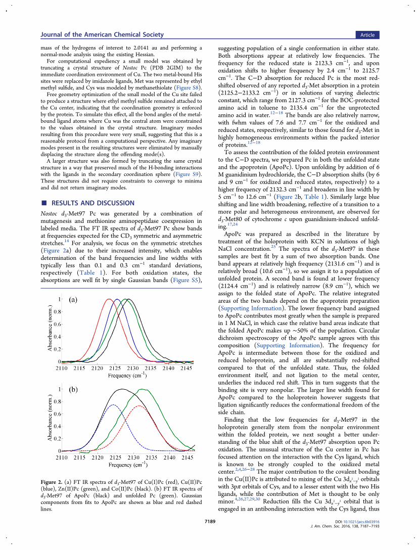
Nostoc d3-Met97 Pc was generated by a combination ofmutagenesis and methionine aminopeptidase coexpression inlabeled media. The FT IR spectra of d3-Met97 Pc show bandsat frequencies expected for the CD3 symmetric and asymmetricstretches.14 For analysis, we focus on the symmetric stretches(Figure 2a) due to their increased intensity, which enablesdetermination of the band frequencies and line widths withtypically less than 0.1 and 0.3 cm−1 standard deviations,respectively (Table 1). For both oxidation states, theabsorptions are well fit by single Gaussian bands (Figure S5),
suggesting population of a single conformation in either state.Both absorptions appear at relatively low frequencies. Thefrequency for the reduced state is 2123.3 cm−1, and uponoxidation shifts to higher frequency by 2.4 cm−1 to 2125.7cm−1. The C−D absorption for reduced Pc is the most red-shifted observed of any reported d3-Met absorption in a protein(2125.2−2133.2 cm−1) or in solutions of varying dielectricconstant, which range from 2127.3 cm−1 for the BOC-protectedamino acid in toluene to 2135.4 cm−1 for the unprotectedamino acid in water.12−18 The bands are also relatively narrow,with fwhm values of 7.6 and 7.7 cm−1 for the oxidized andreduced states, respectively, similar to those found for d3-Met inhighly homogeneous environments within the packed interiorof proteins.12−18
To assess the contribution of the folded protein environmentto the C−D spectra, we prepared Pc in both the unfolded stateand the apoprotein (ApoPc). Upon unfolding by addition of 6M guanidinium hydrochloride, the C−D absorption shifts (by 6and 9 cm−1 for oxidized and reduced states, respectively) to ahigher frequency of 2132.3 cm−1 and broadens in line width by5 cm−1 to 12.6 cm−1 (Figure 2b, Table 1). Similarly large blueshifting and line width broadening, reflective of a transition to amore polar and heterogeneous environment, are observed ford3-Met80 of cytochrome c upon guanidinium-induced unfold-ing.17,24
ApoPc was prepared as described in the literature bytreatment of the holoprotein with KCN in solutions of highNaCl concentration.25 The spectra of the d3-Met97 in thesesamples are best fit by a sum of two absorption bands. Oneband appears at relatively high frequency (2131.6 cm−1) and isrelatively broad (10.6 cm−1), so we assign it to a population ofunfolded protein. A second band is found at lower frequency(2124.4 cm−1) and is relatively narrow (8.9 cm−1), which weassign to the folded state of ApoPc. The relative integratedareas of the two bands depend on the apoprotein preparation(Supporting Information). The lower frequency band assignedto ApoPc contributes most greatly when the sample is preparedin 1 M NaCl, in which case the relative band areas indicate thatthe folded ApoPc makes up ∼50% of the population. Circulardichroism spectroscopy of the ApoPc sample agrees with thiscomposition (Supporting Information). The frequency forApoPc is intermediate between those for the oxidized andreduced holoprotein, and all are substantially red-shiftedcompared to that of the unfolded state. Thus, the foldedenvironment itself, and not ligation to the metal center,underlies the induced red shift. This in turn suggests that thebinding site is very nonpolar. The larger line width found forApoPc compared to the holoprotein however suggests thatligation significantly reduces the conformational freedom of theside chain.Finding that the low frequencies for d3-Met97 in the
holoprotein generally stem from the nonpolar environmentwithin the folded protein, we next sought a better under-standing of the blue shift of the d3-Met97 absorption upon Pcoxidation. The unusual structure of the Cu center in Pc hasfocused attention on the interaction with the Cys ligand, whichis known to be strongly coupled to the oxidized metalcenter.2,4,26−28 The major contribution to the covalent bondingin the Cu(II)Pc is attributed to mixing of the Cu 3dx2−y2 orbitalswith 3pπ orbitals of Cys, and to a lesser extent with the two Hisligands, while the contribution of Met is thought to be onlyminor.4,26,27,29,30 Reduction fills the Cu 3dx2−y2 orbital that isengaged in an antibonding interaction with the Cys ligand, thus
Figure 2. (a) FT IR spectra of d3-Met97 of Cu(I)Pc (red), Cu(II)Pc(blue), Zn(II)Pc (green), and Co(II)Pc (black). (b) FT IR spectra ofd3-Met97 of ApoPc (black) and unfolded Pc (green). Gaussiancomponents from fits to ApoPc are shown as blue and red dashedlines.
Journal of the American Chemical Society Article
DOI: 10.1021/jacs.6b03916J. Am. Chem. Soc. 2016, 138, 7187−7193
7189

decreasing the Cu−S bond order. Self-consistent field Xα-scattered wave calculations on models of the oxidized andreduced states find the potential for interaction of Met sulfur-based orbitals with the 3dz2 orbital of Cu in a pseudo-σ bond,but the orbital mixing is very minor in either state.26,29 Thus,the Cu−S(Met97) bond likely differs in Cu(II)Pc and Cu(I)Pcthrough primarily ionic interactions, which consequently shoulddominate the spectral changes of d3-Met97. However, thestrength of the Cu−S(Met97) interaction in turn can beaffected by the Cu−S(Cys89) bonding, as a compensatoryrelationship is observed between the two.Because oxidation of Cu(I)Pc to Cu(II)Pc leads to changes
in both metal charge and electronic configuration, unravelingthe contributions to the differences in the C−D absorptions isdifficult. To do so, we prepared and characterized Pc with theCu site substituted by either Zn(II) or Co(II) (Zn(II)Pc andCo(II)Pc), as well as in the low-pH state of Cu(I)Pc. Both theCu(I)Pc and Zn(II)Pc contain metal centers with d10
configurations, and differences in the bonding in these speciesshould be primarily ionic in nature. Thus, comparison of thesesamples enables isolation of the spectral changes due toeffectively doubling the metal charge in the absence of effectsdue to d orbital mixing. Additionally, at low pH the His92ligand dissociates from the metal center of Cu(I)Pc, a changethought to serve as a regulatory response to conditions ofpotentially damaging light levels during photosynthesis,31
leaving a three-coordinate site with a contracted Cu−S(Met97)bond.11 In this state the Cu(I) maintains the same charge andelectronic configuration, but the decreased distance fromMet97 should strengthen their ionic interaction. In contrast,the Co(II)Pc, Cu(II)Pc, and Zn(II)Pc have the same formal 2+charge, although the effective nuclear charge, Zeff, increasesslightly across this series. These metal ions more dramaticallydiffer in electronic structure. Unlike the d9 configuration ofCu(II)Pc, Zn(II)Pc is a d10 system, whereas Co(II)Pc is a d7
system. We attempted to prepare Ni(II)-substituted Pc, butwere ultimately not successful, consistent with previousreports.32 Comparison of this series of analogous specieshelps to delineate the spectral effects associated with covalentinteractions involving the protein-based ligands and the metalcenter.To understand the differences in the C−D frequencies of the
Pc variants, we first consider Cu(I)Pc and Zn(II)Pc, the d10
systems intended to isolate the influence of metal charge. TheZn(II)Pc shows a C−D absorption at a relatively highfrequency of 2127.7 cm−1. The additional positive charge at
the metal increases the C−D frequency by 4.4 cm−1. To furtherassess the relationship between the C−D frequency and ionicinteraction of Met97 with the Cu center, we characterized thelow-pH state of Cu(I)Pc, which contains a three-coordinatemetal center with a shorter Cu(I)−S(Met97) bond. The IRspectrum of this sample is best fit by a sum of two bands(Figure 3). A dominant band (66% relative area) appears at
2123.2 cm−1, within the error of the frequency observed forreduced Cu(I)Pc at pH 7. A second band (34% relative area) isshifted to higher frequency by 5.2 cm−1. We assign this higherfrequency band to the His92-dissociated state. The 34%population of the dissociated state indicated by the relativeintegrated band area differs from what a simple calculationbased on the apparent pKa of 5.09
33 of His92 would predict,namely, a population of the dissociated state of ∼90%. Thismismatch must be evaluated with caution, however, given howsensitive population differentials are to slight energy changesand considering some of the innate uncertainties in themeasurements: the potential variation in the C−D crosssections is relatively large, accurate peak integration ischallenging, and the steepness of the pH titration curve neara side chain’s pKa introduces significant errors. Interestingly, nodifferences in the IR spectrum were observed at pH 4 forCu(II)Pc, which does not undergo the acid-induced transition(Supporting Information). Thus, formation of three-coordinateCu(I) at low pH also blue shifts the CD3 symmetric absorption(by 5.2 cm−1). This is in agreement with the Cu(I)Pc/Zn(II)Pcdata that indicate formation of a stronger ionic interaction
Table 1. Parameters from Gaussian Fits to Experimental Spectra and Computational Analysis for the d3-Met97 SymmetricStretcha
experimental crystal structure data, Pc/ pseudoazurincomputational
(PBE0)
species ν (cm−1) fwhmb (cm−1) bond length (Å) ν (cm−1)
Cu(I)Pc 2123.3 ± 0.1 7.7 ± 0.1 2.911/2.835 2195.4Cu(II)Pc 2125.7 ± 0.1 7.6 ± 0.1 2.810/2.735 2200.1ZnPc 2127.7 ± 0.04 9.2 ± 0.4 NA/2.636 2204.4CoPc 2128.8 ± 0.1 8.4 ± 0.6 NA/2.537 2201.8ApoPc 2124.4 ± 0.4 (44%), 2131.6 ± 0.3 (56%) 8.9 ± 0.01, 10.6 ± 1.8unfolded Pc 2132.3 ± 0.1 12.6 ± 1.2Cu(I)Pc, pH 4 2123.2 ± 0.4 (66%), 2128.4 ± 1.5 (34%) 7.2 ± 1.0, 7.9 ± 0.3Cu(I)Pc−cyt f-bound 2123.6 ± 0.1 7.5 ± 0.2Cu(II)Pc−cyt f-bound 2126.1 ± 0.03 6.6 ± 0.2aPercentages denote relative areas for Gaussian components. bFull width at maximum peak height.
Figure 3. FT IR spectra of d3-Met97 of Cu(I)Pc at pH 4 (red).Gaussian components from fits are shown as blue and green dashedlines.
Journal of the American Chemical Society Article
DOI: 10.1021/jacs.6b03916J. Am. Chem. Soc. 2016, 138, 7187−7193
7190

between the metal and Met97 engenders a blue shift of the C−D frequency, whether due to increased metal charge or adecreased interaction distance.These results likewise imply that the more positively charged
metal centers of Cu(II)Pc and Co(II)Pc compared to Cu(I)Pcshould contribute to blue shifting of the C−D frequencies.Indeed, the spectrum of the Co(II)Pc shows a C−D absorptionat a high frequency of 2128.8 cm−1. However, the effect of thecharge of the metal alone does not fully explain the differences.Consideration of the expected Zeff of the metals predicts thatthe frequencies for Co(II)Pc, Cu(II)Pc, and Zn(II)Pc shouldslightly increase across the series but vary only marginallyrelative to their difference from Cu(I)Pc, which is contrary toexperimental observation. Instead, the span in the C−Dfrequencies among these species (3 cm−1) is substantialcompared to the 4.4 cm−1 difference between Zn(II)Pc andCu(I)Pc. In addition, Co(II) is expected to have the smallestZeff of the metal series, but the C−D absorption for Co(II)Pcshows the highest frequency.Since consideration of metal charge alone cannot fully
explain the spectral differences among the Pc variants, weexamined the potential contribution from bond covalency. BothCo(II) and Cu(II) have partially unoccupied 3d orbitals thatmay mix with protein side chains that act as ligands. Co(II)Pccontains a metal center with partially unoccupied dx2−y2, dxy, anddz2 orbitals. Unlike Cu(II)Pc, the interaction of Cys89 with thedx2−y2 orbital of Co(II) is expected to be weak.32,34 Instead, thedominant covalent interaction of Cys89 with Co(II) involves aσ interaction with the dxy orbital; however, the relativeintensities of the corresponding visible absorptions indicatethe σ interaction with Co(II) is weaker than the π interaction ofCu(II) with Cys89.32 The mixing of the occupied Cys89 orbitalwith an unoccupied Cu(II) orbital would decrease themagnitude of positive charge at the metal, and so is predictedto be associated with a decrease in the C−D frequency. This iscontrary to our observation that the C−D frequency forCo(II)Pc is higher than that for Zn(II)Pc. Importantly, incontrast to the Cu(I/II) and Zn(II) proteins, in Co(II)Pc acovalent interaction between the Co(II) and Met97 itselfpotentially contributes to the spectrum. Distinctly, in Co(II)Pc,a partially occupied dz2 orbital is available and aligned tointeract with the sulfur-based orbitals of Met97. Because theinteraction of the orbitals of Met97 with the unoccupied metalorbitals is likely to have a stabilizing effect similar to that of theionic interaction with the metal charge, it is expected to resultin similar blue shifting of the C−D frequency. Therefore, weattribute the high frequency of the C−D band for the Co(II)Pcto the combined effects of its sensitivity to the positive metalcharge and increased orbital mixing of Met97 uniquely withCo(II).With a better understanding of the contributions to the
frequency changes of the C−D absorption for the Pc variants,we reconsider the spectral differences associated with oxidation.As already discussed, metal−S(Met97) covalent interactions arenot predicted to substantially contribute to the differences inmetal binding with Cu(I)Pc, Cu(II)Pc, or Zn(II)Pc, so in theseproteins the C−D absorption should primarily report onchanges in the strength of the ionic interaction of Met97 withthe positively charged metal center. In agreement with this, theIR data do not suggest mixing of the singly occupied dx2−y2orbital of Cu(II) with those of Met97, as the C−D frequency islower for Cu(II)Pc than Zn(II)Pc, rather than higher as foundfor Co(II)Pc, which has the partially occupied dz2 orbital
oriented for interaction with Met97. The dominant covalentinteraction in Cu(II)Pc involves mixing of an occupied Cys89orbital with a partially unoccupied orbital of Cu(II), whichresults in significant charge transfer from the Cys89 ligand tothe metal. Consequently, the Cu−S(Cys89) interaction isexpected to reduce the effective positive metal charge felt by d3-Met97 to a value intermediate between those for Cu(I)Pc andZn(II)Pc. Given the positive correlation between themagnitude of positive metal charge and frequency indicatedby the data for Cu(I)Pc and Zn(II)Pc, the intermediatefrequency observed for the Cu(II) protein is in line with theintermediate effective metal charge felt by Met97 when theeffect of Cu(II)−Cys89 covalent bonding is considered. Themagnitude of the frequency difference associated with thiseffect is notably substantial. In comparison to the 4.4 cm−1
lower C−D frequency associated with an approximate decreaseof one unit of positive metal charge from Zn(II)Pc to Cu(I)Pc,a 2 cm−1 lower C−D frequency is found for Cu(II)Pc than forZn(II)Pc. Thus, the magnitude of the frequency shift attributedto Cu(II)−S(Cys89) covalent mixing is nearly half as large asresults from a decrease in one unit of charge at the metal.Interestingly, this is consistent with combined theoretical andexperimental data which suggest that the highest occupiedmolecular orbital (HOMO) of Cu(II)Pc possesses ∼40% Cys3pπ character.34 Thus, the IR data indicate a very strongCu(II)−Cys89 covalent interaction.In addition to the frequency differences among the metal
series of Pc, the absorption line widths for Zn(II)Pc andCo(II)Pc are broader by 2 cm−1 than found for the native Cuproteins. Assuming that the line width changes are dominatedby inhomogeneous broadening, as is typically the case forabsorptions in proteins,24,38 the broader line widths suggestgreater heterogeneity in the metal-substituted Pc. This could bedue to smaller force constants intrinsic to the metal−ligandbonds of Co(II) and Zn(II), as found in previous calculationsof model complexes with these metals.34
To confirm that the observed spectral changes reflectsensitivity to the positively charged metal, DFT were performedon a model of the metal site in Pc using the PBE0 and B3LYPfunctionals. A minimal model consisted of a metal ion carryingmethyl sulfide, methanethiolate, and two imidazoles, as mimicsof the Met, Cys, and His ligands, respectively. The bond angleswere constrained to those observed in the crystal structures ofPc, and the Zn(II)Pc and Co(II)Pc models used the samestarting geometry as the native Cu protein. Optimizedgeometries for the oxidized, reduced, Zn(II)Pc, and Co(II)Pcstates are shown in Figure S8, and results from harmonicfrequency calculations for the CD3 group are reported in Tables1 and S2. To assess secondary ligand sphere effects, largermodels of the metal sites in all variants of Pc were generated bytruncation of the crystal structures to include only the primarycoordination environment and several hydrogen-bondinginteractions found within the surrounding protein that weredeemed likely to enforce the primary coordination environment(Figure S9). Whereas geometry optimization of the minimalmodels of the metal center in the absence of structuralconstraints did not converge to structures in which all theligands remained attached, the larger models converged tostructures that yielded C−D frequencies that replicated thetrend found for the constrained minimal models (Table S2),suggesting that the secondary ligand sphere is necessary tomaintain the structural integrity of the metal sites, but does not
Journal of the American Chemical Society Article
DOI: 10.1021/jacs.6b03916J. Am. Chem. Soc. 2016, 138, 7187−7193
7191

otherwise contribute significantly to the observed differences inthe C−D absorptions.Compared to Cu(I)Pc, the calculated frequencies for all the
other states (Cu(II)Pc, Zn(II)Pc, Co(II)Pc, and Cu(I) in thethree-coordinate complex) are shifted to higher frequency, andthus generally reproduce the experimentally observed trend. Inagreement with the empirical interpretation, the calculationssuggest that the variation in C−D frequencies reflects theirsensitivity to the positive charge of the metal. At the B3LYP-def2-SVP level of theory, the HOMO of ethyl methyl sulfide isalmost entirely sulfur p in character, but also has contributionsfrom the C−D σ and σ* orbitals. Interaction of the Met97 witha more positively charged metal lowers the sulfur p orbitalenergy, allowing a better interaction with the C−D σ orbitalcompared to the σ* orbital, which in turn increases thecontribution of the C−D σ orbitals to the HOMO and leads toa higher C−D frequency (Figure S11). Consistent with thisidea, modeling the interaction of the ethyl methyl sulfide with apoint charge of varying magnitude recapitulates the trend inC−D frequencies (Table S3). As there were no basis functionscentered on the point charge for these latter calculations, thecalculated frequency trend exclusively reflects the influence ofan ionic interaction.Although the calculations generally capture the influence of
the positive charge of the metal on the C−D frequency, theyunderestimate the frequency for Co(II)Pc. As this is the onlycomplex in which the orbitals of Met97 have the potential tosignificantly mix with orbitals of the metal, it is possible that thecalculations do not sufficiently capture the covalency of theCo(II)−Met97 interaction, which is likely to be highly sensitiveto the metal site geometry used in the calculations. Theexperimentally determined frequencies do show an inversecorrelation with the lengths of the metal−S(Met) bonds fromcrystal structures of Pc, which are available for all the speciesbut the Zn(II)Pc and Co(II)Pc, or from structures ofpseudoazurin, which contains the same ligand set as Pc,39
which further supports that the IR spectra of d3-Met97 reporton sensitivity to its interaction with the metal center. Thespectral, computational, and structural data for the differentspecies of Pc thus are compatible with the presence of a weakionic interaction of Met97 with Cu(I), a stronger but stillrelatively weak ionic interaction with Cu(II) due to the strongcovalent bonding of Cu−S(Cys89), a relatively strong ionicinteraction with Zn(II), and a strong partially covalentinteraction with Co(II).Finally, to further test whether d3-Met97 is sensitive to
changes that might contribute to biological function, wecharacterized the effects of binding the physiological redoxpartner, cyt f (Figure 4, Table 1). Whereas under physiologicalconditions reduced cyt f transfers an electron to oxidized Pc, welooked at the binding-induced changes with both proteins inthe oxidized or reduced state to eliminate complications due tothe redox equilibrium. For the oxidized and reduced states weobserve small, but reproducible and significant, 0.4 and 0.3cm−1 (± <0.1 cm−1) shifts in the d3-Met97 absorptions tohigher frequency upon Pc−cyt f binding. This indicates that theeffects of cyt f binding are transduced to Met97 at the metalsite. The spectral changes are not likely dominated by the effectof surface water displacement by cyt f binding, which wouldcause the C−D probe to experience a more nonpolarenvironment and result in a red shift, contrary to observation.Alternately, our study of the metal series of Pc suggests that theblue shift upon cyt f binding reflects a stronger interaction
between the Cu and Met97. A crude but illustrative comparisonof the magnitude of the frequency change associated with cyt fbinding finds it is ∼10% that due to a change of one unit ofmetal charge. Consistent with this interpretation, XAS studiesof oxidized and reduced Pc find spectral contributions fromCu−S(Met) in the complex with cyt f but not the free protein,which suggests formation of a shorter Cu−S(Met) bond in thecomplex.5
In addition, the C−D absorptions show a significant decrease(by 1 cm−1) in line width upon binding cyt f in the oxidizedstate. This suggests that binding of Pc to cyt f leads to a morehomogeneous environment at Met97, consistent with greaterconformational restriction in the protein−protein complex. Theabsorption narrowing is found only for the oxidized proteincomplex, so the IR data report lower conformationalheterogeneity in that state. XAS of the Pc−cyt f complexsimilarly finds different conformational restrictions for theoxidized and reduced states, but, in contrast to our study,reports greater restriction in the reduced state.5,8 Nevertheless,the IR data provide evidence that complexation with cyt f likelyimpacts the ET properties of Pc. Indeed, there are otherexamples of complexation with electron transfer partnersinfluencing ET reactions.40,41
Previous studies of a range of blue Cu proteins and their axialligand mutants have identified an inverse correlation betweenthe strength of an axial ligand interaction and the midpointpotential of the metal center.1,3 On the basis of this observation,an increase in the Cu(II)−Met97 interaction upon binding withcyt f suggested by the spectral data would predict a reduction inthe midpoint potential. Interestingly, this aligns with previousmeasurement of a 30 mV decrease in the reduction potentialfor Pc in complex with cyt f.42 Additionally, the ET ratesthrough possible pathways in proteins strongly depend on thecoupling between the ligands connecting the pathway and themetal. One of two competing ET pathways specifically evokedin Pc is mediated through the Cu−S(Cys89) covalent bond.3
Given the compensatory relationship between the Met97 andCys89 bonding with Cu(II), the stronger Cu−S(Met97)interaction potentially reflects a corresponding weaker Cu-(II)−S(Cys89) bond, which could have a great impact on thecontribution of the ET pathway mediated through it.
■ CONCLUSIONSThe data clearly reveal that the absorptions of the C−D bondssite-selectively incorporated at the Met axial ligand of Pc aresensitive to the nature of the Cu center. This sensitivity likely
Figure 4. FT IR spectra of d3-Met97 of unbound Cu(I)Pc (red),unbound Cu(II)Pc (blue), cyt f-bound Cu(I)Pc (black), and cyt f-bound Cu(II)Pc (green).
Journal of the American Chemical Society Article
DOI: 10.1021/jacs.6b03916J. Am. Chem. Soc. 2016, 138, 7187−7193
7192

results from variation in the interaction between sulfur-centeredorbitals of Met97 and the positively charged metal center,which itself is influenced by interaction with the other ligands,presumably most significantly Cys89. The approach enablesdirect, nonperturbative, molecular-level comparison of theoxidized state to the reduced state of Pc, as well as to severalother variants, with the same experimental method. Ourspectral data support the presence of the strong covalentinteraction between Cu(II) and Cys89, and that Met97 acts tostabilize the metal center via ionic interactions. The spectralchanges observed upon binding cyt f suggest increased Cu−Met97 interaction in the complex, which illustrates amechanism for the modulation of the ET properties fromcomplexation with a redox partner. Further characterization ofPc, including the site-selective deuteration of other ligands,promises to more fully elucidate the nature of the Cu site andhow it is modulated for function. Similarly, the IR character-ization of ligands in other blue Cu proteins with varied metalsites and other proteins with presumably weak metal−ligandbonds should help illuminate how they contribute to tuning thereactivity for metalloproteins’ specific function in biology.
■ ASSOCIATED CONTENT*S Supporting InformationThe Supporting Information is available free of charge on theACS Publications website at DOI: 10.1021/jacs.6b03916.
Experimental details of protein expression, spectroscopicmeasurement and fitting parameters, and computationaldetails (PDF)
■ AUTHOR INFORMATIONCorresponding Author*[email protected] Address†M.-H.B.: Department of Chemistry, Korea Advanced Instituteof Science and Technology, 291 Daehak-ro, Yuseong-gu,Daejeon 305-701, Republic of Korea.NotesThe authors declare no competing financial interest.
■ ACKNOWLEDGMENTSWe thank Marcellus Ubbink (Leiden University) for providingexpression plasmids for Pc and cyt f. A.L.L. and M.C.T .thankIndiana University and the Department of Energy (Grant DE-FOA-0000751) for funding.
■ REFERENCES(1) Gray, H. B.; Malmstrom, B. G.; Williams, R. J. P. JBIC, J. Biol.Inorg. Chem. 2000, 5, 551.(2) Solomon, E. I.; Hare, J. W.; Gray, H. B. Proc. Natl. Acad. Sci. U. S.A. 1976, 73, 1389.(3) Randall, D. W.; Gamelin, D. R.; LaCroix, L. B.; Solomon, E. I. J.Biol. Inorg. Chem. 2000, 5, 16.(4) Solomon, E. I.; Szilagyi, R. K.; DeBeer George, S.; Basumallick, L.Chem. Rev. 2004, 104, 419.(5) Diaz-Moreno, I.; Diaz-Quintana, A.; Diaz-Moreno, S.; Subias, G.;De la Rosa, M. A. FEBS Lett. 2006, 580, 6187.(6) Diaz-Moreno, I.; Diaz-Quintana, A.; De la Rosa, M. A.; Ubbink,M. J. Biol. Chem. 2005, 280, 18908.(7) Pettersen, E. F.; Goddard, T. D.; Huang, C. C.; Couch, G. S.;Greenblatt, D. M.; Meng, E. C.; Ferrin, T. E. J. Comput. Chem. 2004,25, 1605.
(8) Cruz-Gallardo, I.; Diaz-Moreno, I.; Diaz-Quintana, A.; De laRosa, M. A. FEBS Lett. 2012, 586, 646.(9) Guckert, J. A.; Lowery, M. D.; Solomon, E. I. J. Am. Chem. Soc.1995, 117, 2817.(10) Guss, J. M.; Bartunik, H. D.; Freeman, H. C. Acta Crystallogr.,Sect. B: Struct. Sci. 1992, 48, 790.(11) Guss, J. M.; Harrowell, P. R.; Murata, M.; Norris, V. A.;Freeman, H. C. J. Mol. Biol. 1986, 192, 361.(12) Chin, J. K.; Jimenez, R.; Romesberg, F. E. J. Am. Chem. Soc.2001, 123, 2426.(13) Cremeens, M. E.; Fujisaki, H.; Zhang, Y.; Zimmermann, J.;Sagle, L. B.; Matsuda, S.; Dawson, P. E.; Straub, J. E.; Romesberg, F. E.J. Am. Chem. Soc. 2006, 128, 6028.(14) Thielges, M. C.; Case, D. A.; Romesberg, F. E. J. Am. Chem. Soc.2008, 130, 6597.(15) Sagle, L. B.; Zimmermann, J.; Dawson, P. E.; Romesberg, F. E. J.Am. Chem. Soc. 2004, 126, 3384.(16) Zimmermann, J.; Thielges, M. C.; Yu, W.; Dawson, P. E.;Romesberg, F. E. J. Phys. Chem. Lett. 2011, 2, 412.(17) Sagle, L. B.; Zimmermann, J.; Matsuda, S.; Dawson, P. E.;Romesberg, F. E. J. Am. Chem. Soc. 2006, 128, 7909.(18) Chin, J. K.; Jimenez, R.; Romesberg, F. E. J. Am. Chem. Soc.2002, 124, 1846.(19) Liao, Y.-D.; Jeng, J.-C.; Wang, C.-F.; Wang, S.-C.; Chang, S.-T.Protein Sci. 2004, 13, 1802.(20) Scanu, S.; Forster, J.; Finiguerra, M. G.; Shabestari, M. H.;Huber, M.; Ubbink, M. ChemBioChem 2012, 13, 1312.(21) Albarran, C.; Navarro, J. A.; Molina-Heredia, F. P.; Murdoch, P.d. S.; De la Rosa, M. A.; Hervas, M. Biochemistry 2005, 44, 11601.(22) Neese, F. WIREs Comput. Mol. Sci. 2012, 2, 73.(23) Schaf̈er, A.; Horn, H.; Ahlrichs, R. J. Chem. Phys. 1992, 97, 2571.(24) Thielges, M. C.; Zimmermann, J.; Dawson, P. E.; Romesberg, F.E. J. Mol. Biol. 2009, 388, 159.(25) Koide, S.; Dyson, H. J.; Wright, P. E. Biochemistry 1993, 32,12299.(26) Gewirth, A. A.; Solomon, E. I. J. Am. Chem. Soc. 1988, 110,3811.(27) Penfield, K. W.; Gay, R. R.; Himmelwright, R. S.; Eickman, N.C.; Norris, V. A.; Freeman, H. C.; Solomon, E. I. J. Am. Chem. Soc.1981, 103, 4382.(28) Solomon, E. I.; Clendening, P. J.; Gray, H. B.; Grunthaner, F. J.J. Am. Chem. Soc. 1975, 97, 3878.(29) Penfield, K. W.; Gewirth, A. A.; Solomon, E. I. J. Am. Chem. Soc.1985, 107, 4519.(30) Scott, R. A.; Hahn, J. E.; Doniach, S.; Freeman, H. C.; Hodgson,K. O. J. Am. Chem. Soc. 1982, 104, 5364.(31) Rochaix, J.-D. Biochim. Biophys. Acta, Bioenerg. 2011, 1807, 375.(32) McMillin, D. R.; Rosenberg, R. C.; Gray, H. B. Proc. Natl. Acad.Sci. U. S. A. 1974, 71, 4760.(33) Hass, M. A. S.; Thuesen, M. H.; Christensen, H. E. M.; Led, J. J.J. Am. Chem. Soc. 2004, 126, 753.(34) Gorelsky, S. I.; Basumallick, L.; Vura-Weis, J.; Sarangi, R.;Hodgson, K. O.; Hedman, B.; Fujisawa, K.; Solomon, E. I. Inorg. Chem.2005, 44, 4947.(35) Libeu, C. A. P.; Kukimoto, M.; Nishiyama, M.; Horinouchi, S.;Adman, E. Biochemistry 1997, 36, 13160.(36) Gessmann, R.; Papadovasilaki, M.; Drougkas, E.; Petratos, K.Acta Crystallogr., Sect. F: Struct. Biol. Commun. 2015, 71, 19.(37) Gessmann, R.; Kyvelidou, C.; Papadovasilaki, M.; Petratos, K.Biopolymers 2011, 95, 202.(38) Chung, J. K.; Thielges, M. C.; Lynch, S. R.; Fayer, M. D. J. Phys.Chem. B 2012, 116, 11024.(39) Dennison, C. Dalton Trans 2005, 21, 3436.(40) Drepper, F.; Hippler, M.; Nitschke, W.; Haehnel, W.Biochemistry 1996, 35, 1282.(41) Roncel, M.; Boussac, A.; Zurita, J. L.; Bottin, H.; Sugiura, M.;Kirilovsky, D.; Ortega, J. M. JBIC, J. Biol. Inorg. Chem. 2003, 8, 206.(42) Malkin, R.; Knaff, D. B.; Bearden, A. J. Biochim. Biophys. Acta,Bioenerg. 1973, 305, 675.
Journal of the American Chemical Society Article
DOI: 10.1021/jacs.6b03916J. Am. Chem. Soc. 2016, 138, 7187−7193
7193