Embed Size (px)
Citation preview

www.wjpr.net Vol 7, Issue 19, 2018.
244
Abinokhauno et al. World Journal of Pharmaceutical Research
METHICILIN RESISTANT STAPHYLOCOCCUS AUREUS IN
CLINICAL SAMPLES AND FOMITES
Abinokhauno Solomon*1, Iyevhobu Kenneth O.
1, Oghena Marcus
1, Obodo Basil
Nnaemeka1 and Ebadan Maxwel
2
1Department of Medical Laboratory Science, Faculty of Basic Medical Science, Ambrose
Alli University, Ekpoma, Edo State, Nigeria.
2Quality Assurance Clina-Lancet Laboratories Limited, Victoria Island, Lagos State, Nigeria.
ABSTRACT
A total of 200 swabs from clinical samples and fomites which
comprised of 105 swabs from the skin, 56 swabs from nasal and 38
swabs from fomites i.e laboratory coats, each were collected and
examined. From this study, 30(15.0%) were identified as
Staphylococcus aureus, and 170(85.5%) showed no growth or other
species of bacteria and analysis show that it was statistically significant
P<0.05, the prevalence of Methicillin/ Oxacillin Resistant
Staphylococcus aureus (MRSA/ORSA) was 13(6.5%) with skin having
the highest occurrences of 9(4.5%) and nasal passage and fomites
having 4(2.0%) and 0(0.0%) respectively and the Methicillin/ Oxacillin
Sensitive Staphylococcus aureus (MSSA/OSSA) having prevalence of
15(7.5%), 2(1.0%) and 0(0.0%) for isolates from the skin, nasal passage and formites
respectively and when the number of MRSA isolated was compare with MSSA statistically it
was found not to be statistically significant p-value=0.193, p>0.05. When mean diameter of
Oxacillin was used to compared the mean diameter of Vacomycin it was 5.3±2.83 vs
17.4±18.47; tcal=2.072,p-val=0.068, and df=9, respectively, it was not statistically significant
i.e P>=0.05, at α=95% or 0.05, the mean, standard deviation, F-value calculated, degree of
freedom, p-values of the diameters of zones of inhibitions of Oxacillin, Vacomycin, and
sulbatam, when mean diameter of Oxacillin was used to compared the mean diameter of
Vacomycin and Sulbatam is was 5.3±2.83 vs 17.4±18.47 and 1.11±0.281; Fcal Between
groups=6.1.47, p-val=0.06, and df=29 respectively, it was not statistically significant i.e
P>=0.05, at α=95% or 0.05. However, there seems to be a steady rise of MRSA isolates
World Journal of Pharmaceutical Research SJIF Impact Factor 8.074
Volume 7, Issue 19, 244-255. Research Article ISSN 2277– 7105
Article Received on
15 Oct. 2018,
Revised on 05 Nov. 2018,
Accepted on 26 Nov. 2018
DOI: 10.20959/wjpr201819-13819
*Corresponding Author
Abinokhauno Solomon
Department of Medical
Laboratory Science, Faculty
of Basic Medical Science,
Ambrose Alli University,
Ekpoma, Edo State, Nigeria.

www.wjpr.net Vol 7, Issue 19, 2018.
245
Abinokhauno et al. World Journal of Pharmaceutical Research
resistance to commonly used antibiotics like Sulbatam and Vacomycin but the present rate is
still low in comparison to values in some other studies.
KEYWORDS: Staphylococcus, Vacomycin, Methicillin, Fomites, Resistant.
INTRODUCTION
Methicillin-resistant Staphylococcus aureus (MRSA) are strains of Staphylococcus aureus
which are resistant to methicillin and related penicillins and are particularly difficult to treat
because they are also resistant to most other common antibiotics (Cheesbrough, 2000).
Although Staphylococcus aureus infections were historically treatable with common
antibiotics, emergence of drug-resistant organisms is now a major concern. MRSA was
endemic in hospitals by the late 1960s, but it appeared rapidly and unexpectedly in
communities in the 1990s and is now prevalent worldwide (Deleo, 2009; Liebowitz, 2009).
Staphylococci are gram positive cocci of uniform size, occurring characteristically in groups
but also singly and in pairs. They are non-motile and non-capsulated (Cheesbrough, 2000).
Staphylococcus aureus is the most medically important member in terms of pathogenicity of
the group (Ochei and Kolhatkar, 2000).
Staphylococcus is present in the nose of 30% of healthy people and may be found on the skin.
It causes infection most commonly at sites of lowered host resistance, such as damaged skin
or mucous membrane (Humphrey, 2007). Although 50 – 60% of patients with MRSA are
merely colonised (i.e. they carry the bacteria but do not have symptoms or an illness), serious
infections such as those involving the blood stream, respiratory tract and bones or joints do
occur (Humphrey, 2007). S. aureus causes boils, pustules, styes, impetigo, infections of
wounds (cross-infections), ulcers and burns, osteomyelitis, mastitis, septicaemia, meningitis,
pneumonia and pleural empyema. Also, toxic food poisoning (rapid onset, no fever), toxic
shock syndrome and toxic skin exfoliation (Chessbrough, 2000).
Mannitol salt agar is a useful selective medium for recovering S. aureus from faecal
specimens when investigating staphylococcal food poisoning. It can also be used to screen for
nasal carriers. S. aureus ferments mannitol and is able to grow on agar containing 70 – 100g/l
sodium chloride. Mannitol salt agar containing 75g/l sodium is recommended particularly for
isolating MRSA strains (Cheessbrough, 2000).

www.wjpr.net Vol 7, Issue 19, 2018.
246
Abinokhauno et al. World Journal of Pharmaceutical Research
On mannitol salt agar, S. aureus produces yellow colonies (Ochei and Kolhatkar, 2000). The
MRSAs are usually sensitive to vancomycin (Ochei and Kolhatkar, 2000). Flucloxacillin and
chloxacillin are used to treat -lactamase (penicilinase) producing staphylococci. Vacomycin
is often needed to treat MRSA infections. Antibacterial resistance to penicillin may occur due
to the -lactamase production, cell membrane alterations reducing antibiotic uptake (gram
negative bacteria), or changes in the penicillin-binding protein as occurs with MRSA
(Cheesbrough, 2000).
There is no effective immunisation with toxoids or bacterial vaccines for preventing the
spread of S. aureus (Levinson and Jawetz, 2002). The control and prevention of MRSA
involves early and reliable detection in the laboratory through surveillance, patient isolation
when admitted to hospital, good professional practice by all healthcare workers (including
compliance with hand hygiene guidelines), effective hospital hygiene programmes and
sensible use of antibiotics (Humphrey, 2007).
MRSA are potential source for the spread of nosocomial infections in patients and healthcare
works, cause patients that are hospitalized to over stay in the hospital and spend financial
resources up to 400% that is, four times up to what they would have spent without Hospital
acquired nosocomial infections (Ahlstrom, 2011), CDC reported that MRSA or ORSA are
primary source of nosocomial infections, which could be transferred from patients to patients,
patients to health workers or health workers to health workers or health workers to patients.
Then, this project determines the prevalence of MRSA in clinical samples and fomites to
validated the claims of CDC and other researchers that had worked on such work else were.
To the best of our ability this research work has been done in the area of study.
MATERIALS AND MEHODS
This project work was carried out in both Ekpoma, Esan West Local Government Area, Edo
State, Nigeria with longitude 6.13oE and latitude 6.73
oN having a population of about 61,870
people (Population of Cities, 2007) and in Irrua Specialist Teaching Hospital (formerly
Otibhor Okhae Teaching Hospital), Irrua was established by decree 92 of 1993 to provide
tertiary health care delivery services to the people of Edo State. The hospital is located in
Irrua, Edo central senatorial district, along the Benin-Abuja Highway at about 87 kilometers
north of Benin City, the Edo State capital.

www.wjpr.net Vol 7, Issue 19, 2018.
247
Abinokhauno et al. World Journal of Pharmaceutical Research
Subject Sector
A target of 200 clinical samples and fomites served as the test subject for this project work.
Research Design
This project work was carried out within a period of two months. 200 swab samples from
clinical samples and fomites (laboratory coat) were collected randomly and used for this
project work.
Materials Used
The following materials and apparatus were used for the bacteriological analysis: mannitol
salt agar, Nutrient agar, swab sticks, Petri dishes, conical flask, distilled water, autoclave,
Bunsen burner, inoculating wire loop, binocular microscope, weighing balance, measuring
cylinder, glass slides, human plasma, hydrogen peroxide, crystal violet Lugol’s iodine,
acetone, neutral red, sensitivity discs (Oxacillin, Vacomycin and Sulbatam) and other
reagents.
Media Used
The media used were mannitol salt agar and Nutrient agar.
Sample Collection
Two hundred (200) swab specimen used were collected randomly from apparently clinical
samples of skin, nasal and fomites i.e laboratory coat from irrua specialist teaching hospital,
Irrua, Edo State. Nasal swabs were collected in good light vision from subjects bending their
heads backward to collect the specimens deep down the anterior passages using a sterile swab
stick, Both right and left nostrils were used, swab were also taken from skin and fomites
(laboratory coat), were swabbed bearing labels as swabs, code number and date of collection.
The swab sticks were carefully returned to their sterile containers, sealed with adhesive tape
and labelled accordingly. Collected specimens were taken to the laboratory where
bacteriological analysis was carried out immediately.
Procedure for Culture
(i) The swab sticks were used to make wells of inoculum on each nutrient agar surface.
(ii) Spreading was done by streaking from the primary inoculum using inoculation wire loop
to obtain discrete colonies.
(iii)The plates were then incubated at 37oC for 24 hours.

www.wjpr.net Vol 7, Issue 19, 2018.
248
Abinokhauno et al. World Journal of Pharmaceutical Research
(iv) Growths were observed after incubation.
(v) Suspected colonies were confirmed by other methods which include gram staining.
Gram Staining Procedure
(i) A smear was made and allowed to air dry and then fixed with gentle heat by passing the
slide three times over a Bunsen flame.
(ii) It was stained with crystal violet for one minute.
(iii)It was washed with tap water.
(iv) Lugol’s iodine was applied and left for one minute.
(v) It was washed with tap water.
(vi) Decolourisation was done with acetone until no more colour appeared to ooze out of the
smear for 2 seconds.
(vii) It was washed immediately with tap water.
(viii) It was counterstained with neutral red for 2 minutes.
(ix) It was washed with tap water.
(x) It was blotted with blotting paper and dried.
(xi) It was examined microscopically using 100X objective with immersion.
Biochemical Test
The biochemical tests that were used for this work include:
(i) Catalase test
(ii) Coagulase test
Catalase Test: This test helps to differentiate staphylococci from streptococci.
Method
(i) 3ml of H2O2 was placed in a test tube
(ii) With sterile plastic rod, some colonies of the organism were picked and immersed in the
H2O2 solution.
(iii)Immediate gas bubbling was observed.

www.wjpr.net Vol 7, Issue 19, 2018.
249
Abinokhauno et al. World Journal of Pharmaceutical Research
Coagulase Test
Procedure
(i) Two separate drops of saline were placed on a slide
(ii) Colonies of the organism were emulsified in each of the drops to make thick suspensions.
(iii)The tip of straight wire loop was dipped into the undiluted plasma and the adhering traces
of plasma was mixed into one of the bacterial suspensions.
(iv) Immediate coarse clumping of the mixture was looked for within 10 seconds.
— Tube test was performed to confirm all slide test coagulase negative staphylococcus, were
truly negative for coagulase.
Antibiotic Sensitive Test
2g/disc Oxacilin discs and other antibiotic discs such as 8g/disc Vacomycin and 8g/disc
Sulbatam (manufactured by Abtek Biologicals Ltd) were used to test the susceptibility of
staphylococcal isolates obtained. The test isolates were inoculated into peptone water broth
and the inoculum was used to seed Nutrient agar agar plate. The antibiotic discs were placed
aseptically on the seeded plate. This was incubated at 37oC for 24 hours and examined for
zones of inhibition. The zones of inhibition were measured in millimetres and recorded.
Statistical Analysis
Chi-square was used to determine the difference between MRSA and MSSA, t- student test
and Anova statistical analysis was used to determine the differences in the level of sensitive
to Oxacilin and Vacomycin, and Oxacilin, Vacomycin and Sulbatam by Methicillin Sensitive
Staphylococcus aureus.
RESULTS
A total of 200 swabs from clinical samples and fomites which comprised of 106(53%) swabs
from the skin, 56(28%) swabs from nasal and 38(19%) swabs from fomites i.e laboratory
coats, each were collected and examined. With the help of biochemical characterisation,
30(15.0%) were identified as Staphylococcus aureus, other species of bacteria 58(29%) and
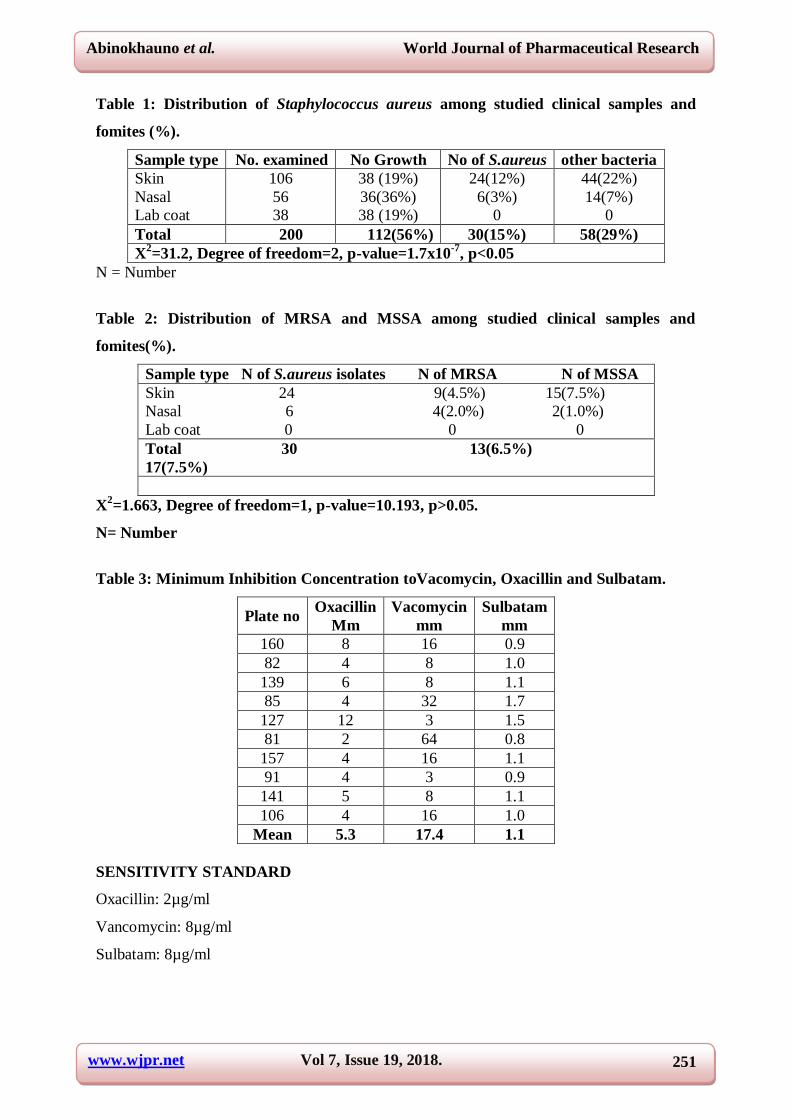
112(56%) showed no growth (Table 1). Staphylococcus aureus was more isolated from
swabs from the skin 24(12.0%), followed by nasal swab with 6(3.0%) and swab from fomites
i.e laboratory coat with 0(0.0%) when the number of isolates was compared within the
samples statistically it was found to be statistically significant, p<0.05.

www.wjpr.net Vol 7, Issue 19, 2018.
250
Abinokhauno et al. World Journal of Pharmaceutical Research
Among the various Staphylococcus aureus isolated from the various samples, the prevalence
of Methicillin/ Oxacillin Resistant Staphylococcus aureus (MRSA/ORSA) was 13(6.5%)
with skin having the highest occurrences of 9(4.5%) and nasal passage and fomites having
4(2.0%) and 0(0.0%) respectively and the Methicillin/ Oxacillin Sensitive Staphylococcus
aureus (MSSA/OSSA) having a prevalences of 15(7.5%), 2(1.0%) and 0(0.0%) for isolates
from the skin, nasal passage and formites respectively and when the number of MRSA
isolated was compare with MSSA statistically it was found not to be statistically significant ,
p>0.05.(Table 2).
The Minimum inhibition concentration to Oxacillin, Vacomycin and Sulbatam is shown on
table 3.
The mean, standard deviation, t student statistic calculated, and p values of the diameters of
zones of inhibitions of Oxacillin and Vacomycin, when mean diameter of Oxacillin was used
to compared the mean diameter of Vacomycin it was 5.3±2.83 vs 17.4±18.47; tcal=2.072, p-
val=0.068, and df=9 respectivey, it was not statistically significant i.e P>=0.05, at α=95% or
0.05 as shown on table 4.
The mean, standard deviation, F-value calculated, degree of freedom, p-values of the
diameters of zones of inhibitions of Oxacillin, Vacomycin, and sulbatam, when mean
diameter of Oxacillin was used to compared the mean diameter of Vacomycin and Sulbatam
is was 5.3±2.83 vs 17.4±18.47 and 1.11±0.281; Fcal Between groups=6.1.47, p-val=0.006,
and df=29 respectivey, it was not statistically significant i.e P>=0.05, at α=95% or 0.05 and
using LSD Post Hoc Test to do multiple comparisons between Oxacillin against Vacomycin
and Sulbatam; Vacomycin against Oxacillin and Sulbatam and Sulbatam against Oxacillin
and Vacomycin with p-values of 0.018 and 0.393; 0.018 and 0.02; and 0.393 and 0.02
showing that mean diameter of Oxacillin was significantly different from Vacomycin while
the mean diameter of Oxacillin is not differ from that of Sulbatam, while the mean diameter
of Sulbatam differs from that of Vacomycin as shown on table 5.
www.wjpr.net Vol 7, Issue 19, 2018.
251
Abinokhauno et al. World Journal of Pharmaceutical Research
Table 1: Distribution of Staphylococcus aureus among studied clinical samples and
fomites (%).
Sample type No. examined No Growth No of S.aureus other bacteria
Skin
Nasal
Lab coat
106
56
38
38 (19%)
36(36%)
38 (19%)
24(12%)
6(3%)
0
44(22%)
14(7%)
0
Total 200 112(56%) 30(15%) 58(29%)
X2=31.2, Degree of freedom=2, p-value=1.7x10
-7, p<0.05
N = Number
Table 2: Distribution of MRSA and MSSA among studied clinical samples and
fomites(%).
Sample type N of S.aureus isolates N of MRSA N of MSSA
Skin 24 9(4.5%) 15(7.5%)
Nasal 6 4(2.0%) 2(1.0%)
Lab coat 0 0 0
Total 30 13(6.5%)
17(7.5%)
X2=1.663, Degree of freedom=1, p-value=10.193, p>0.05.
N= Number
Table 3: Minimum Inhibition Concentration toVacomycin, Oxacillin and Sulbatam.
Plate no Oxacillin
Mm
Vacomycin
mm
Sulbatam
mm
160 8 16 0.9
82 4 8 1.0
139 6 8 1.1
85 4 32 1.7
127 12 3 1.5
81 2 64 0.8
157 4 16 1.1
91 4 3 0.9
141 5 8 1.1
106 4 16 1.0
Mean 5.3 17.4 1.1
SENSITIVITY STANDARD
Oxacillin: 2µg/ml
Vancomycin: 8µg/ml
Sulbatam: 8µg/ml

www.wjpr.net Vol 7, Issue 19, 2018.
252
Abinokhauno et al. World Journal of Pharmaceutical Research
Table 4: Mean, standard deviation, t-student statistic calculated, p-values of the
diameters of zones of inhibition of Oxacillin and Vacomycin on MSSA.
Test value
Oxacillin Vacomycin Comparison (test value=5.3) t value p-value Significant
MeanZone diameter Standard deviation
5.30 17.40 2.83 18.34
2.072 0.068 p>0.05
Table 5a: Anova of multiple comparison of diameter of Oxacillin, Vacomycin and
Sulbatam.
(a) ANOVA Sum of Squares df Mean Square F Non Sig.
Between Groups 1431.101 2 715.550 6.147 .006
Within Groups 3143.209 27 116.415
Total 4574.310 29
Table 5b: Post hoc test multiple comparison of diameter of Oxacillin, Vacomycin and
Sulbatam.
(TEST VALUE) GROUP Sig.
OXACILIN Vacomycin .018
Sulbatam .393
VACOMYCIN Oxacilin .018
Sulbatam .002
SULBATAM Oxacilin .393
Vacomycin .002
*. The mean difference is significant at the 0.05
level.
DISCUSSION
Despite recognising Staphylococcus species as regional flora of the skin and mucus
membrane, certain species have been found frequently as aetiological agent of a variety of
human infections. The most common among these infections are the superficial supportive
infection caused by Staphylococcus aureus. Under minding the introduction of chemotherapy
and recent improvement in medical services, methicillin resistant Staphylococcus aureus
(MRSA) strain emerge and has posed a major threat to public health in treatment and
management of Staphylococcus aureus infection. The increasing prevalence of MRSA among
Staphylococcus aureus strains resulted in a significant increase in the utilization of
vancomycin. Methicillin resistant Staphylococcus aureus (MRSA) have been recognized as a
major strain of Staphylococcus aureus prevalent as cause of nosocomial infection which often
results to increase in morbidity and mortality (Schaumacher-Perdreau, 1999).

www.wjpr.net Vol 7, Issue 19, 2018.
253
Abinokhauno et al. World Journal of Pharmaceutical Research
A total of 200 swabs from clinical samples and fomites which comprised of 106(53%) swabs
from the skin, 56(28%) swabs from nasal and 38(19%) swabs from fomites i.e laboratory
coats, each were collected and examined. With the help of biochemical characterisation,
30(15.0%) were identified as Staphylococcus aureus, and 112(56%) showed no growth or
other species of bacteria and analysis show that it was statistically significant P<0.05. the
overall prevalence of MRSA was 13(6.5%) it was noticed that the highest number isolates
incident of MRSA on the skin 9(4.5%) while Nasal passage had 4(2.0%) and lab coat had
0(0.0%), the difference noticed between MRSA on the skin and Nasal passage could be the
difference in the number of samples used from skin and nasal passage and also due to
geographical distribution of micro-organism, and this work in line with work of Ayliffe et al.,
(1988); Majumdar et al (2009); who reported 9.0%, and 7.5% but was in constraint to
Akpaka et al (2006) who reported 23.0%. and Okodua et al., (2013) of 29.7% although
samples differences, samples size and Techniques has effect on the overall prevalence of
MRSA.
Methicillin/Resistance Staphylococcus aureus (MRSA) and Methicillin/Sensitive
Staphylococcus aureus (MSSA)] among studied population and fomites had 43.3%
prevalence which is in variation with previous work done in different region of Nigeria
where Taiwo et al., (2004) that, reported a prevalence of 34.7% in Illorin, Kwara State.
Comparatively, study area, sample size, sample type, implementation of standard operating
procedures in work place might account for the observed differences. The highest occurrence
of MRSA was seen in skin 30% and 13.3% from nasal nares while 56.6%of the
Staphylococcus aureus isolated in our study population were methicillin sensitive
Staphylococcus aureus (MSSA).
It was noticed that the diameter of zone of inhibition of Vacomycin was greater than those of
Oxacillin and Sulbatam, with the mean diameter and standard deviation of 5.30±2.83,
17.4±18.47 and 1.11±0.28 respectively, Vacomycin was the most active antibiotic which
shows a greater mean and standard compare to those of Oxacillin and Sulbatam. This is in
agreement with report of CDC, (2010), and with all available literature, but the Sulbatam was
resistant to all the MRSA as shown from its mean diameter and standard deviation of
1.11±0.28, but to the MRSA, Oxacillin was in the boarderline between sensitive and
intermediate with mean diameter and standard deviation of 5.30±2.83.

www.wjpr.net Vol 7, Issue 19, 2018.
254
Abinokhauno et al. World Journal of Pharmaceutical Research
There was no significant difference between mean diameter and standard deviation of the
susceptible MRSA to Vacomycin and Oxacillin with a tcal value of 2.072 and Pvalue of 0.068
and this also agreed on available literature.
CONCLUSION
Evidence from the results obtained has shown that the 13(6.5%) of the studied samples were
Methicillin Resistant Staphylococcus aureus and that the level of resistance shown by MRSA
isolates to other antibiotics when compared with that of methicillin-sensitive Staphylococcus
aureus (MSSA) isolates, is by far higher.
However, there seems to be a steady rise of MRSA isolates resistance to commonly used
antibiotics like Sulbatam and Vacomycin but the present rate is still low in comparison to
values in some other studies.
From this study, haven established the prevalence of MRSA in skin and nasal nares, it is
necessary for medical Personnel, especially those involved in routine care, monitoring and
prescription of commonly used antibiotics to pay attention to the prevalence of methicillin/
resistance Staphylococcus aureus (MRSA), methicillin/ sensitive Staphylococcus aureus
(MSSA) in treatment of Staphylococcus aureus infections. These outbreaks were associated
with certain higher risk groups, including individuals who used IV drugs, participants in close
contact sports, and residents living together in crowded conditions, such as inmates, military
recruits, and disabled individuals in group homes. A regular surveillance of both nosocomial,
Hospital Acquired methicillin/ resistance Staphylococcus aureus (HA-MRSA) and
Community Acquired methicillin resistant Staphylococcus aureus (CA-MRSA) infection is
necessary to succumb it prevalence in the hospital settings and community at large. Regular
monitoring of antibiotic sensitivity pattern of Staphylococcus aureus must be made
mandatory, to control further spread of its infections. Proper hygiene should be maintained at
all times by all health workers to reduce its spread and infection by contact. Hand –washing,
particularly is very important by all health workers after each procedure with a patient before
touching the next patient. This will stop the transmission of MRSA. The use of disposable
aprons and gloves by hospital staff should be practice thereby reducing the risk of
transmission in the hospital.

www.wjpr.net Vol 7, Issue 19, 2018.
255
Abinokhauno et al. World Journal of Pharmaceutical Research
From this study, MRSA are on the continual rising in comparison with other studies done.
With this increase prevalence rate of MRSA, within the next two decades there might not be
any reliable treatment left for most Staph. aureus infections.
It is hereby recommended that combination therapy with good therapeutic effect should be
considered for the treatment of Staph. aureus
REFERENCES
1. Ayliffe GAJ, Babb JR, Davies, JG, Lilly HA. Hand disinfection: a comparison of various
agents in laboratory and ward studies. J Hosp Infect., 1988; 11: 226–243.
2. Ahlstrom, D. (2011). "New strain of MRSA superbug discovered in Dublin hospitals".
The Irish Times. http://www.irishtimes.com, Retrieved May 3, 2011.
3. CDC (2006): CDC Guideline "Management of Multidrug-Resistant Organisms in
Healthcare Settings, 2006".
4. CDC, "Guidelines for Infection Control in Health Care Personnel,". Centers for Disease
Control and Prevention. http://www.cdc.gov, Retrieved, December, 2010; 18: 2013.
5. Cheesbrough M. Staphylococcus aureus In: District Laboratory Practice in Tropical
Countries, Part 2. Cambridge University Press, UK, 2000; 133: 155-158.
6. Deleo FR, Chambers HF. Re-emergence of antibiotic-resistant Staphylococcus aureus in
the genomics era. J.Clin Invest. 2009; 2464-2474.
7. Humphreys H. Staphylococcus In: Medical Microbiology (17th edition). Churchill
Livingstone. 2007; 172: 174-175.
8. Levinson W, Jawetz E. Staphylococcus In: Medical Microbiology and Immunology (7th
edition). Lange Medical Books/ McGraw-Hill. 2002; 95.
9. Liebowitz LD. MRSA burden and interventions. Int. Antimicrobial Agents, 2009; 34:
11–13.
10. Ochei J, Kolhatkar A. Methicillin-Resistant Staphylococcus aureus In: Medical
Laboratory Science Theory and Practice. Tata McGraw-Hill Publishing Company
Limited, New Delhi, 2000; 813, 665, 805-806.
11. Schumacher-Perdreau F. Clinical significance and laboratory diagnosis of coagulase –
negative staphylococci. Clinical Microbiology Newsletter, 1999; 13: 97-101.
12. Taiwo DA, Ikeh EI, Olayemi A. Prevalence of Methicillin-resistance Staphylococcus
aureus in Illorin Nigeria. African Journal of Clinical and Experimental Microbiology,
2004; 5: 342-14.