Embed Size (px)
Citation preview

Metarhizium anisopliae Pathogenesis of MosquitoLarvae: A Verdict of Accidental DeathTariq M. Butt1*, Bethany P. J. Greenfield1, Carolyn Greig1, Thierry G. G. Maffeis2, James W. D. Taylor1,
Justyna Piasecka1, Ed Dudley3, Ahmed Abdulla1, Ivan M. Dubovskiy4, Inmaculada Garrido-Jurado5,
Enrique Quesada-Moraga5, Mark W. Penny2, Daniel C. Eastwood1
1College of Science, Swansea University, Swansea, United Kingdom, 2College of Engineering, Swansea University, Swansea, United Kingdom, 3College of Medicine,
Swansea University, Swansea, United Kingdom, 4 Institute of Systematics and Ecology of Animals, Siberian Branch of Russian Academy of Sciences, Novosibirsk, Russia,
5Department of Agricultural and Forestry Sciences, University of Cordoba, Cordoba, Spain
Abstract
Metarhizium anisopliae, a fungal pathogen of terrestrial arthropods, kills the aquatic larvae of Aedes aegypti, the vector ofdengue and yellow fever. The fungus kills without adhering to the host cuticle. Ingested conidia also fail to germinate andare expelled in fecal pellets. This study investigates the mechanism by which this fungus adapted to terrestrial hosts killsaquatic mosquito larvae. Genes associated with the M. anisopliae early pathogenic response (proteinases Pr1 and Pr2, andadhesins, Mad1 and Mad2) are upregulated in the presence of larvae, but the established infection process observed interrestrial hosts does not progress and insecticidal destruxins were not detected. Protease inhibitors reduce larval mortalityindicating the importance of proteases in the host interaction. The Ae. aegypti immune response to M. anisopliae appearslimited, whilst the oxidative stress response gene encoding for thiol peroxidase is upregulated. Cecropin and Hsp70 genesare downregulated as larval death occurs, and insect mortality appears to be linked to autolysis through caspase activityregulated by Hsp70 and inhibited, in infected larvae, by protease inhibitors. Evidence is presented that a traditional host-pathogen response does not occur as the species have not evolved to interact. M. anisopliae retains pre-formed pathogenicdeterminants which mediate host mortality, but unlike true aquatic fungal pathogens, does not recognise and colonise thelarval host.
Citation: Butt TM, Greenfield BPJ, Greig C, Maffeis TGG, Taylor JWD, et al. (2013) Metarhizium anisopliae Pathogenesis of Mosquito Larvae: A Verdict of AccidentalDeath. PLoS ONE 8(12): e81686. doi:10.1371/journal.pone.0081686
Editor: Jorge Luis Folch-Mallol, Universidad Autonoma del estado de Morelos, Mexico
Received August 21, 2013; Accepted October 24, 2013; Published December 13, 2013
Copyright: � 2013 Butt et al. This is an open-access article distributed under the terms of the Creative Commons Attribution License, which permits unrestricteduse, distribution, and reproduction in any medium, provided the original author and source are credited.
Funding: INBIOSOIL is an EU funded project. INTERREG IVA (http://wefo.wales.gov.uk/programmes/territorial/irelandwales/?lang = en). KESS (http://www.higherskillswales.co.uk/kess/). RFBR (http://www.rfbr.ru/rffi/eng/about). Spanish Ministry of Science (http://www.mineco.gob.es/portal/site/mineco/idi). EU FP7(http://inbiosoil.uni-goettingen.de/). The funders had no role in study design, data collection and analysis, decision to publish, or preparation of the manuscript.
Competing Interests: The authors have declared that no competing interests.
* E-mail: [email protected]
Introduction
Mosquitoes vector a wide range of diseases (e.g. dengue, yellow
fever and malaria) which can have a devastating impact on
human health. Almost half the world’s population is at risk to
mosquito-transmitted diseases and the range has expanded due
to climate change and increased trade [1]. Many chemical
pesticides have been withdrawn due to the risks they pose to
humans and the environment, and development of resistance in
pest populations. Recent studies show that Metarhizium anisopliae, a
soil borne fungal pathogen of terrestrial insects, offers an
environmentally friendly alternative to chemicals for the control
of mosquitoes. M. anisopliae will kill adult and larval stages of
Aedes, Anopheles and Culex mosquitoes [2,3] yet the mechanism of
how this terrestrial pathogen kills the aquatic larval stage is
unclear. Strains of M. anisopliae have been developed to control a
wide range of terrestrial arthropods including pests of agro-forests
crops and vectors of human and animal diseases [4,5]. Infection
of terrestrial arthropod hosts by M. anisopliae, like that of most
other entomopathogenic fungi, follows a consistent pattern.
Firstly, spores adhere to the surface of the host cuticle followed
by germination and differentiation of an appressorium from
which a narrow penetration peg is produced which penetrates
the cuticle using a combination of enzymes and mechanical force
[6,7]. Following colonization of the hemocoel, the fungus erupts
through the intersegmental membranes and differentiates conid-
iophores and conidia. The key pathogenicity determinants of M.
anisopliae include cuticle degrading enzymes like Pr1 (subtilisin
protease) and toxic cyclic peptides like destruxins [8]. Fungal
virulence appears to be correlated with Pr1 and destruxin
production; with hyper producers being more virulent [8].
Equally important are the adhesins, Mad1 and Mad2, which play
an important role in adhesion of M. anisopliae to the insect cuticle
[9]. Disruption of the Mad1 gene reduces virulence by reducing
adhesion of conidia to the host surface [10]. It is assumed that
the pattern of infection outlined above occurs in aquatic
mosquito larvae [2].This paper demonstrates for the first time
that M. anisopliae kills the mosquito larvae via a mechanism which
does not entail the traditional infection processes.
Based on the limited number of observational studies
conducted on M. anisopliae infection of mosquito larvae, possible
routes of invasion have been reported including entry via
penetration of the cuticle, the respiratory siphon or alimentary
canal, however, the precise mechanism remains elusive. Lacey
PLOS ONE | www.plosone.org 1 December 2013 | Volume 8 | Issue 12 | e81686

et al., [11] noted that when larvae of Culex quinquefasciatus broke
the water tension with their perispiracular valves for air intake,
floating conidia of M. anisopliae adhered to the inside surface of
the valves, germinated and invaded the siphon tip tissue, then
extended into and blocked the trachea resulting in suffocation
and death. Lacey et al., [11] also noted that conidia suspended in
the water were ingested and occluded the larval gut, initiating
mortality within 6 to 24 hr after ingestion. In contrast, Riba
et al., [12] reported that M. anisopliae conidia killed Ae. aegypti
within 1.1 days before intra-hemocoelic invasion. Some workers
suggest that death is due to colonization of the hemocoel by the
fungus [2,12], others suggest it is due to toxins released by
ingested conidia without colonisation of the hemocoel [11,13].
Materials and Methods
Fungal strains and productionMetarhizium anisopliae isolate ARSEF 4556, identified as highly
pathogenic to mosquitoes and midges [4], was maintained on
Sabouraud dextrose agar (SDA) or broken Basmati rice [14].
Conidia used in assays had over 95% viability. A green
fluorescence protein (GFP) transformed strain of Metarhizium
brunneum EAMa 01/58 Su was maintained on SDA.
Mosquito source and maintenanceAedes aegypti (strain AeAe) eggs, obtained from the London
School of Hygiene and Tropical Medicine, were hatched in
distilled water and the larvae fed on TetraminH fish food, at room
temperature (22uC62uC).
Inoculation of Aedes larvae with Metarhizium conidiaAssays were performed using 24 well plates (Nunc, Roskilde,
Denmark) with one larva per well. M. anisopliae ARSEF 4556 was
assayed at 107 conidia ml21 against L324 larvae. Additional
assays were done using heat killed conidia to determine the role
of extracellular enzymes in pathogenesis. Extracellular enzymes
were denatured by wrapping the conidia in aluminium foil and
autoclaving for 15 min at 121uC. Conidial viability was assessed
using the plate count technique [15]. Control larvae were
exposed to either 1ml 0.03% Aq Tween 80 or distilled water.
Larval mortality was recorded daily up to 7 days. All assays were
performed at room temperature with a 16L:8D photoperiod.
There were 24 larvae per assay which was repeated three times.
This format was used in subsequent assays to study host-
pathogen interactions, in particular, insect defense responses and
regulation of M. anisopliae pathogenicity determinants.
Larvae (n = 20) were inoculated with conidia of ARSEF 4556 as
outlined above and examined at 0, 24, 48 and 72 hr post
inoculation (pi). Healthy and infected larvae were examined by
light microscopy (LM) to determine if there were preferential sites
for spore adhesion and to monitor passage of the fungal conidia
through the gut. Larvae (n = 20) were also examined by cryo
scanning electron microscopy (SEM) using a Hitachi S4800 field
emission microscope equipped with a Quorum PPT2000 cryo-
genic stage and preparation chamber. Full details on the cryo-
SEM are provided in Text S1 in File S1. Additional studies were
done using a GFP-transformed strain of M. brunneum. The surface
and gut contents of infected Ae. aegypti larvae (n = 10)as well as fecal
pellets were examined by fluorescence microscopy (FM) using a
Zeiss fluorescence microscope.
Mass spectrometry analysis of destruxinsAssays were performed using 24-multi-well plates with ten Ae.
aegypti larvae (L3–4) per well containing 1 ml aqueous suspension
of 16107 conidia ARSEF 4556 or 1 ml of distilled water
(control). After 24 hr incubation, the larvae were removed and
prepared for destruxin extraction and analysis by mass spectros-
copy as described by Butt et al., [16]. The effect of destruxins on
Ae. aegypti larvae was also tested by introducing larvae to 1 ml of
distilled water spiked with 1nmole of destruxin A (Sigma-Aldrich)
and determining larval survival. Briefly, destruxin extracts were
analyzed by nano-reverse phase liquid chromatography (Ultimate
Pump, LC-Packing, Dionex, The Netherlands) using an electro-
spray ion trap MS (LCQ Deca XP, ThermoElectron, Hemel
Hempstead, UK). Matrix Assisted Laser Deionisation Mass
Spectroscopy (MALDI MS) was used to confirm the levels of
detection below which any destruxins might be exhibited within
the larvae themselves. For this purpose, larval extract was re-
dissolved in a matrix solution and 1 ml of the resulting mixture
was spotted onto the MALDI plate and allowed to dry at room
temperature. A 10 mg/mL alpha cyanocinnaminic acid (CHCA)
in 50:50 0.1% trifluoroacetic acid (TFA): acetonitrile (ACN)
matrix solution was used and a Voyager DE-STR instrument
(Applied Biosystems, UK) was utilized in reflectron mode. An
acceleration voltage of 20,000V and a grid voltage of 70% was
utilized in order to study any peptides present and a 1pmole
standard of destruxin A applied to the plate as a control and as a
method of determining the level below which any destruxins are
Figure 1. Heat killed treatment increased the survival of Ae.aegypti larvae. Late 3rd24th instar Ae. aegypti larvae (n = 72) wereinoculated with live and heat killed conidia of M. anisopliae. Kaplan-Meier method was used to plot cumulative survival curves of larvaeafter inoculation, log-rank test was used to assess difference in survivalbetween treatments. The curves of non-exposed and heat killedtreatment show no statistical difference. Live conidia significantlydecreased survival compared to heat killed conidia (p,0.001). Larvaewith no fungal treatment were used as a negative control.The aim ofthis study was to establish the cause of death as this could haveprofound implications on the formulation and deployment ofMetarhizium and other terrestrial entomopathogenic fungi to controlmosquito larvae. This study, utilising a combination of microscopy,molecular and biochemical methods, shows that Metarhizium invasivestrategies, evolved for terrestrial hosts including adult mosquitoes, havenot evolved for killing aquatic insect hosts with resultant mortality inmosquito larvae being multi-factorial.doi:10.1371/journal.pone.0081686.g001
Fungal Pathogenesis of Mosquito Larvae
PLOS ONE | www.plosone.org 2 December 2013 | Volume 8 | Issue 12 | e81686

present if signal intensity were below the standard. Full details
are provided in Text S2 in File S1.
Enzymology(i) Protease inhibition assays. To determine if the
extracellular proteases were responsible for larval mortality.
Larvae (n = 24) were exposed to M. anisopliae ARSEF 4556
conidia containing either chicken egg white (0.1 mg/ml), EDTA
(1 mM) or a2-macroglobulin (1 mg/ml) which were inhibitors
specific for Pr1, metalloprotease and global (serine, cysteine,
metallo-) proteases, respectively. All the inhibitors were pur-
chased from Sigma-Aldrich. Controls consisted of buffer and
buffered inhibitor. Mortality was recorded at 0, 12, 24, 36, 48
and 72 hr pi. Assays were also done using heat killed conidia at
107 conidia ml21.
(ii) Caspase assays. Activity of caspases 2, 3, 7 and 8 was
assayed using luminometric kits in accordance with the manufac-
turer’s guidelines (Promega). Six larvae were examined per
treatment with the endpoint luminescence being measured after
1 hr, in four replicate wells for each larvae. Full details are given in
Text S3 in File S1.
Transcript quantification of insect and fungus-derivedgenes
(i) Samples, RNA extraction and cDNA synthesis. Full
details of the transcript quantification are given in Text S4 in
Figure 2. M. anisopliae do not attach to or invade mosquito hemocoel. Larvae inoculated with 16107 conidia ml21 M. anisopliae, 48 hr post-inoculation, were subjected to Cryo-SEM to establish areas of attachment and penetration of the fungus. No conidia were observed attached to thesurface of the head (A), abdomen and siphon (B). Cross section of control larva (C) and infected larva (D) showing the gut lumen (GL), midgutepithelium (MG), and peritrophic matrix (PM). Conidia of M. anisopliae were restricted to and appeared to occlude the gut lumen. (D2E). Both swollenand collapsed conidia were observed in the gut lumen with no evidence of conidia penetrating the gut wall (F) and invading the hemocoel (H).Light,fluorescence and scanning electron microscopydoi:10.1371/journal.pone.0081686.g002
Fungal Pathogenesis of Mosquito Larvae
PLOS ONE | www.plosone.org 3 December 2013 | Volume 8 | Issue 12 | e81686

File S1. Briefly, Ae. aegypti larvae (L3–4) (n = 3, 10 larvae per
replicate) were exposed to M. anisopliae ARSEF 4556, controls
included larvae not exposed to fungus and a terrestrial insect,
Tenebrio molitor. Samples were frozen under liquid nitrogen and
stored at 280uC until required. All samples were ground with a
micropestle and total RNA extractions carried out using the
RNeasy Micro kit (Qiagen) following the manufacturer’s
instructions. RNA concentration and purity was assessed at 260
and 280 nm absorbance using a Nanophotometer (Implen).
Total RNA (1 mg) was either RQ1 RNase-free DNAse (Promega)
treated and reverse transcribed using the qScript cDNA synthesis
kit (Quanta Biosciences), or reverse transcribed using the
QuantiTect Reverse Transcription kit (Qiagen) with gDNA
elimination reaction, for the experiment to quantify insect-
derived transcripts and fungus-derived transcripts, respectively.
Relative cDNA quantity was analyzed by PCR using two
reference genes for insect or fungal cDNA samples to ensure
consistency between values: Ae. aegypti ribosomal S7 (accession
number: AAEL009496) and ribosomal protein 49/L32
(AAEL003396) and M. anisopliae 18S rRNA and elongation
factor tEF (Table S1 in File S1).
(ii) Quantitative PCR (qPCR). Transcript levels were
determined using the Rotor-Gene 6000 (Corbett Research) or
CFX96TM Real-Time PCR detection system (Biorad) for Ae.
aegypti and M. anisopliae gene targets respectively. Primers were
designed to amplify key Ae. aegypti response genes and M.
anisopliae pathology-related genes (Table S1 in File S1).
The accompanying software for each qPCR instrument was
used to analyze the raw data and carry out quality control for
each sample. The cycle threshold (Ct) value was determined for
each reaction and normalized to the geometric mean of the
appropriate endogenous reference genes. Relative gene expres-
sion was calculated using the comparative Ct method (2-DDCt)
following established methodology [17].
ROS production, lipid peroxidation and antioxidantsystem activityThree insects per time point were sample were homogenized in
100 ml of ice-cold phosphate buffered saline (10 mM phosphate
buffer, 150 mM NaCl, pH 7.2) containing phenylthiourea (1 mg/
ml). The homogenate was centrifuged for 5 min, 10,000 g at 4uCand activities determined as described by Dubovskiy et al.[18], in
12 replicates for ROS production and Lipid peroxidation and 9
replicates for Super Oxide Dismutase (SOD), Glutathione-S-
transferase (GST) and Catalase activity. Full details are provided
in Text S5 in File S1.
Statistical AnalysisDifferences in mosquito larvae survival between live and heat
killed conidia and protease inhibited conidia were analysed using
Kaplan-Meier survival analysis to plot cumulative survival
functions by treatment with pairwise comparison over log-rank
test [4]. Biochemical and molecular data sets were analyzed
using two-way Analysis of Variance (ANOVA) with Bonferroni’s
post-test. Prior to analysis gene expression data was logarithm
(base 10) transformed, conforming to ANOVA assumption of
homogeneity of variance [19]. All statistical analyses were carried
out using SPSS v21.0 [20] and GraphPad Prism v5.0 (GraphPad
Software, USA).
Figure 3. Metarhizium conidia expressing GFP in the gut and fecal pellets demonstrating activity and viability. Larvae inoculated withconidia of a GFP transformed strain of Metarhizium brunneum were examined 48 hr pi to assess viability and activity of the conidia. (A) Conidiaocclude most of the gut lumen, some autofluorescence is seen in the head and thorax region. (B) Numerous conidia are active and expressing theGFP within the gut lumen. (C) High magnification of conidia expressing GFP, the non-fluorescing conidia may be inactive or dead, (D) Fecal pelletbeing expelled from an infected larva showing many active conidia.doi:10.1371/journal.pone.0081686.g003
Fungal Pathogenesis of Mosquito Larvae
PLOS ONE | www.plosone.org 4 December 2013 | Volume 8 | Issue 12 | e81686

Results
Virulence of Metarhizium anisopliae ARSEF 4556 for Aedesaegypti larvaeAe. aegypti larvae were highly susceptible to M. anisopliae 4556
with the earliest mortalities being observed 24 hr pi and
percentage mortality reaching 60%290% between 72 and
96 hr pi (Fig. 1). Heat killed conidia were used to determine
whether conidia caused blockage within the larvae or were
required to actively bring about mortality. Mortality was
significantly higher in larvae exposed to live conidia than those
exposed to heat killed conidia (p,0.001) and there was no
significant difference in mortality between the heat killed conidia
(p,0.153) and the untreated control (Fig. 1).
Metarhizium conidia fail to infect Aedes larvaeMicroscopy studies clearly showed little or no attachment of
conidia to the surface of the mosquito body (Figs. 2A and B) with
conidia being concentrated in the gut lumen (Figs. 2D-F). In some
larvae, the conidia occluded the gut lumen while in others gaps
were observed between the conidial clumps. None of the conidia
in the gut had produced a germ tube and whereas some conidia
were hydrated and swollen others appeared collapsed (Fig. 2F).
Conidia had a prominent hydrophobin rodlet layer with little
evidence of mucilage secretion. FM showed that conidia in the gut
and those present in fecal pellets expressed the GFP and were
clearly viable and active (Fig. 3). Most non-fluorescing GFP
conidia were probably quiescent.
SEM examination of cross sections of abdomen showed no
obvious differences in the appearance of the gut epithelial cells,
peritrophic matrix and other structures of treated and untreated
(control) larvae (Figs. 2C and D–F). There was no visible
evidence of damage to the internal organs. Compact fecal pellets
were produced by control and infected larvae (Fig. 3) suggesting
the peritrophic matrix, peristalsis and other gut functions were
intact at least until the time of death. The fungus never crossed
the gut; it did not colonize the hemocoel as it would terrestrial
arthropod hosts.
Destruxins are not the cause of larval deathDestruxins, common virulence determinants of M. anisopliae,
were not detected by mass spectroscopy in Ae. aegypti larvae that
had ingested conidia of M. anisopliae. The LCMS analysis was
utilized to profile for any destruxin signals present using constant
neutral loss signals established in our laboratories that highlight
low levels of destruxins [16]. No signals were detected which
represent destruxins in any of the five replicate larval extractions
(See supplementary figure S2) and re-analysing the data for the
m/z of specific destruxins also confirmed their absence (data not
shown). The LCMS system was tested using a 10fmole
cytochrome C digest and 1 pmole destruxin A, however system
test data was not routinely recorded and therefore the larval
Figure 4. Destruxins not detected in larval extracts. Larval extracts were analysed by MALDI ToF alongside a destruxin A standard. Nodestruxin signals were detected with both larval extract and control larvae containing only those ions formed due to the MALDI matrix itself.doi:10.1371/journal.pone.0081686.g004
Fungal Pathogenesis of Mosquito Larvae
PLOS ONE | www.plosone.org 5 December 2013 | Volume 8 | Issue 12 | e81686

extracts were also analysed by MALDI ToF analysis alongside a
destruxin A standard (Fig. 4). As can be seen the standard
destruxin provides an excellent signal at the 1pmole level whilst
the control experiment and larval extracts contain only those ions
formed due to the MALDI matrix itself and no destruxin signals
can be determined. This allows us to confirm the absence of
destruxins, at least up to the 1pmole level per larval experiment.
Metarhizium pathogenicity genes expressed in mosquitogut and faecesProteases (Pr1, Pr2) and adhesins (Mad1, Mad2) play a key role
in fungal pathogenicity, expression of the genes for these
components was analyzed and was shown to be generally much
higher in the gut lumen and remained high in the fecal pellets
(Fig. 5). Expression of Pr1 was significantly lower in conidia in
the presence of Ae. aegypti larvae compared with all the other
treatments (F(5,47) = 96.09, p,0.01) but was high in infected
living, infected dead and fecal pellet samples (F(5,47) = 96.09,
p,0.05) even when compared with the T. molitor treatment
(F(5,47) = 96.09, p,0.001). Pr1 expression was greatest in fecal
pellets (F(5,47) = 96.09, p,0.001) then infected larvae (F(5,47)
= 96.09, p,0.05) followed by infected dead larvae (Fig. 5). All
treatments had a significantly higher expression of Pr2 than the
spore pellets not exposed to larvae with expression being highest
in infected living larvae (F(5,47) = 96.09, p,0.001) then the fecal
pellet (F(5,47) = 96.09, p,0.01; Fig. 5). Spore pellets exposed to
larvae and infected dead larvae had lower and significantly
similar expression levels. Conidia exposed to T. molitor had
similar Pr2 expression to the spore pellet exposed to the larvae,
infected dead larvae and the fecal pellets (Fig. 5).
The pattern of expression of Mad1 was comparable with the
expression of Pr2 with the exception of a slightly lower relative
expression in the fecal pellet (Fig. 5). Infected living larvae
showed significantly greater expression than any other treatment
(F(5,47) = 96.09, p,0.01; Fig. 5). Greatest expression levels of
Mad2 were detected in fecal pellets (F(5,47) = 96.09, p,0.001),
T. molitor (F(5,47) = 96.09, p,0.001) and infected dead (F(5,47)
= 96.09, p,0.01) when compared with the spores not exposed to
larvae (Fig. 5).
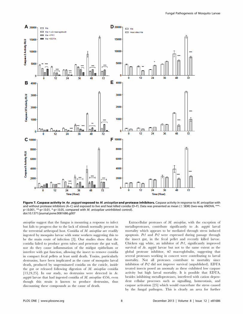
Mortality linked to fungal protease-induced apoptosisOne possible mechanism that may eventually lead to mortality
of the larvae is the activation of apoptotic pathways in the larvae
(involving caspase enzymes) by active agents released by the
conidia. As the active agents identified were proteases, the effect
of inhibiting these enzymes on larval mortality was investigated
and the larval caspase activity also monitored.
Mortality of larvae incubated with the fungus was significantly
lower in the presence of protease inhibitors with the exception of
EDTA which was not significantly different from the M. anisopliae
treated larvae (Fig. 6) The inhibition with chicken egg white
increased percentage survival after treatment with the fungus from
approximately 10% to 30%, whilst inhibition by a2-macroglobulin
improved this value to 50% of the untreated larvae. As well as the
effect of the proteases produced by the fungus on larval survival,
the study of the effect of the inhibition of such bioactive entities on
apoptosis was studied. Activity of caspases 2, 3/7 and 8 was
significantly higher in Ae. aegypti larvae inoculated with live conidia
of M. anisopliae without protease inhibitors than with inhibitors
(Fig. 7). Interestingly, in the M. anisopliae treated larvae, activity
increased dramatically, concomitant with larval mortality, be-
tween 36 and 72 hr pi (Figs. 6, 7). Caspase activity was
significantly lower in larvae in the presence of protease inhibitors
for the whole period of the assay (F(5,72) = 661.39, F(5,72) = 90.4,
F(5,72) = 75.42 (caspase 3/7, 2 and 8 respectively) p,0.001;
Fig. 7A, B and C). Caspase 2, 3/7 and 8 activity was generally
Figure 5. Metarhizium pathogenicity genes expressed in mosquito gut and faeces. Expression of protease (Pr1A, Pr2) and adhesin (Mad1,Mad2) pathogenicity related genes by conidia of M. anisopliae, 48 hr pi, analysed by quantitative PCR. SP2: Spore pellet in the absence of Ae. aegyptilarvae, SP+: Spore pellet in presence of larvae, IL: infected live larvae, ID: infected dead larvae, FP: mosquito fecal pellet, TEN: Tenebrio molitor(terrestrial host) positive control. Data was presented as mean (6 SEM) means with different letters denoting statistical differences (two-way ANOVA).Data normalized to average dCt of SP2.doi:10.1371/journal.pone.0081686.g005
Fungal Pathogenesis of Mosquito Larvae
PLOS ONE | www.plosone.org 6 December 2013 | Volume 8 | Issue 12 | e81686

lower 24–72 hr pi in the presence of EDTA than the other
inhibitors (F(5,72) = 1359.03, F(5,72) = 486.01, F(5,72) = 271.46
(caspase 3/7, 2 and 8 respectively) p,0.001; Fig. 7A, B and C).
Oxidative stress is not an obvious mediator of apoptosisOxidative stress within organisms can also be a trigger for the
initiation of apoptosis and therefore various indicators of such
stress were studied in larvae with and without conidia.
Examination of reactive oxygen species generation, lipid perox-
idation, catalase, superoxide dismutase and glutathione-S-trans-
ferase activity during two days of pathogenesis revealed no major
differences between uninfected and M. anisopliae infected mos-
quito larvae except glutathione-S-transferase activity which was
higher (F(2,48) = 12.10, p,0.01) 48 hr pi in infected (Figs. S1A–
E). At 72 hr pi when most larvae were dead or dying we found
the decrease in ROS generation (F(2,60) = 5.05, p,0.01); lipid
peroxidation (F(2,59) = 1.66, p,0.05); catalase (F(2,48) = 6.77,
p,0.01); and glutathione-S-transferase (F(2,48) = 12.10, p,0.01)
activity in infected insects (Figs. S1A2E).
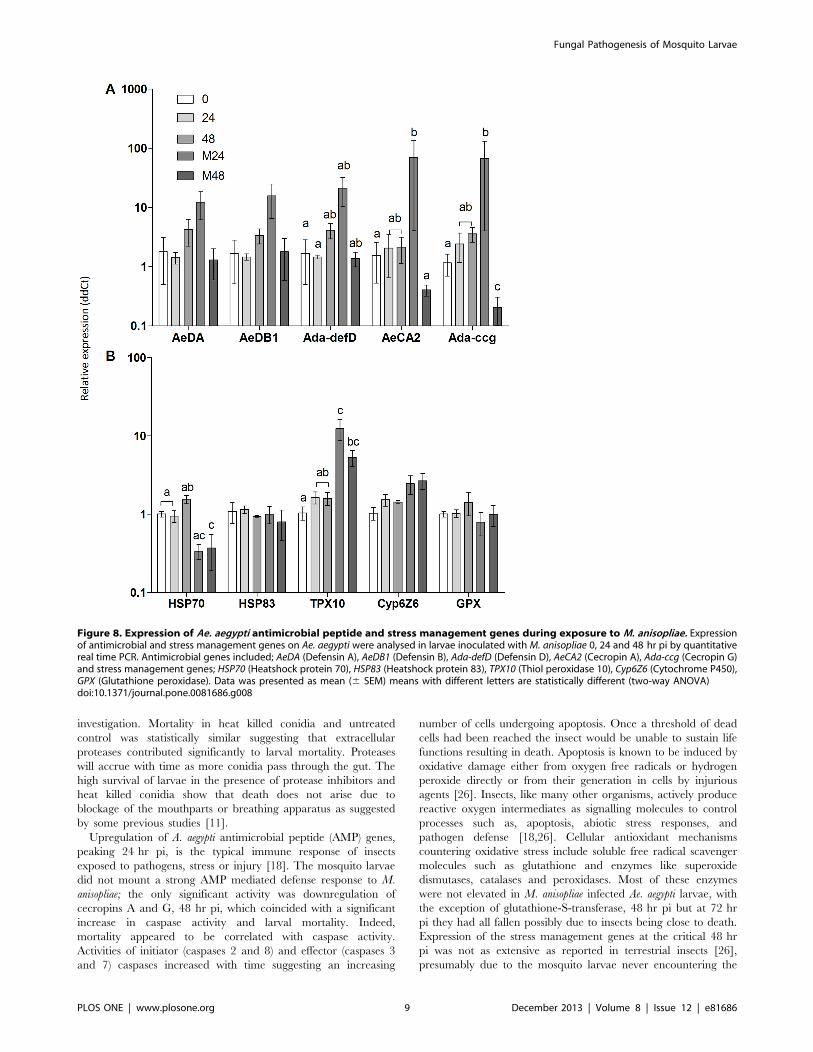
Immune and stress management systems fail to protectmosquito larvaeGiven the release of active proteases by the fungus and
elicitation of a pathogenic response when exposed to the larvae,
the study of the larval response to the fungus was also
undertaken. The analysis examined the larval defense mecha-
nisms (predominantly antimicrobial peptides) as well as the stress
response of the larvae. Expression of the antimicrobial peptide
(AMP) genes AeDA and AeDB was not significantly up regulated
following Ae. aegypti larval ingestion of live M. anisopliae conidia
24 hr pi (Fig. 8A). While expression levels of Ada-DefD and Ada-
CcG was significantly greater in samples exposed to M. anisopliae
after 24 hr compared with time zero (F(4,50) = 16.12, p,0.05) it
was not significantly different from the 24 hr untreated control
(Fig. 8A). However, the gene was significantly down regulated in
larvae exposed to M. anisopliae 48 hr pi compared with the other
treatments (F(4,50) = 16.12, p,0.05; Fig. 8A). AeCA2 was
significantly down regulated in larvae at 48 hr pi compared
with 24 hr pi (F(4,50) = 16.12, p,0.01) but these did not differ
from unexposed larvae at 48 hr and 24 hr, respectively (Fig. 8A).
Of the five stress management genes examined, a significant
increase in expression of the TPX10 was observed in larvae
exposed to M. anisopliae 24 hr compared with the time zero
(F(4,40) = 7.71, p,0.001) and 24 hr untreated control (F(4,40)
= 7.71, p,0.01; Fig. 8B). While there was a significant increase
in expression of TPX10 at 48 hr in exposed larvae compared
with time zero (F(4,40) = 7.71, p,0.05) this was not significant
compared with the unexposed larvae at 48 hr (Fig. 8B). In
contrast, the Hsp70 gene was down-regulated in larvae exposed
to conidia 48 hr pi compared with the untreated control (F(4,40)
= 7.71, p,0.01; Fig. 8B). No significant changes were observed
for both AMPs and stress genes in the untreated controls at 0, 24
and 48 hr (Fig. 8A and B).
Discussion
This study shows that conidia of M. anisopliae do not firmly
adhere to the surface of the cuticle of Ae. aegypti larvae and do
not gain entry by penetrating the host cuticle. Conidia have been
reported adhering to the cuticle, particularly the siphon and
mouthparts of the fungus [21], thereby infecting the larvae in a
similar manner with which it infects terrestrial hosts [2].
Conidia attach to terrestrial hosts initially via passive hydro-
phobic forces followed by secretion of enzymes and adhesion
compounds to anchor the spore to the cuticle surface [6,7]. The
failure of conidia to adhere to the cuticle of terrestrial hosts has
been attributed to the cuticle chemistry, with some compounds
altering hydrophobicity or being fungistatic [6,22]. It is feasible
that the mosquito larval cuticle is not conducive for adhesion,
with any mucilage produced by the fungus being diluted in the
water. In contrast, aquatic pathogens of mosquitoes such as
Lagenidium giganteum (Oomycetes) and Coelomomyces punctatus
(Chytridiomycetes) produce zoospores that can attach to and
penetrate the larval cuticle before colonizing the hemocoel.
Culicinomyces clavisporus, an aquatic Sordariomycete related to M.
anisopliae, produces conidia which, following ingestion by the
larvae, adhere to and penetrate through the chitinous wall in the
fore- and hindgut [23].
In conidia of M. anisopliae, Mad1 expression in the presence of
mosquito larvae suggest the fungus had responded to cuticular
cues in a similar manner to a terrestrial host despite its failure to
adhere through passive hydrophobic forces. Mad1 was up
regulated particularly inside the gut of live insects suggesting
that the fungus had perceived additional cues. Mad2 was not
upregulated in the same manner, however, expression of both
these genes was significantly higher in the gut of dead insects and
fecal pellets possibly due to nutritional stress which would also
explain why no germ tubes were produced. Nutrient starvation is
known to up regulate Mad2 [9]. The concomitant upregulation
of Mad1, Mad2, Pr1 and Pr2 by the ungerminated conidia of M.
Figure 6. Survival of Ae. aegypti larvae in presence of proteaseinhibitor. Ae. aegypti larvae (n = 72) were inoculated by M. anisopliaewith and without protease inhibitors. CEW: Chicken Egg White a Pr1specific inhibitor, a2 mac: a2 macroglobulin a global protease inhibitorand EDTA a metalloprotease inhibitor. Kaplan-Meier method was usedto plot cumulative survival curves of larvae after inoculation, log-ranktest was used to assess differences in survival between treatments.Uninhibited conidia caused greater mortality than conidia treated withinhibitors with the exception of EDTA (p,0.001). Controls consist ofeither 0.05% Aqueous Tween only, or 0.05% Aqueous Tween withprotease inhibitor.Caspase activity, particularly caspases 3/7 and 8, wasconsistently higher in Ae. aegypti larvae exposed to live conidiacompared to the heat killed conidia 48–72 hr pi (F(5,54) = 203.60,F(5,54) = 71.15 (caspase 3/7 and 8 respectively) p,0.001; Fig. 7B–D).Caspase activity elicited by heat killed conidia increased over time up to72 hr pi.doi:10.1371/journal.pone.0081686.g006
Fungal Pathogenesis of Mosquito Larvae
PLOS ONE | www.plosone.org 7 December 2013 | Volume 8 | Issue 12 | e81686
anisopliae suggest that the fungus is mounting a response to infect
but fails to progress due to the lack of stimuli normally present in
the terrestrial arthropod host. Conidia of M. anisopliae are readily
ingested by mosquito larvae with some workers suggesting this to
be the main route of infection [3]. Our studies show that the
conidia failed to produce germ tubes and penetrate the gut wall,
nor do they cause inflammation of the midgut epithelium or
interfere with gut function, allowing the insect to remove conidia
in compact fecal pellets at least until death. Toxins, particularly
destruxins, have been implicated as the cause of mosquito larval
death, produced by ungerminated conidia on the cuticle, inside
the gut or released following digestion of M. anisopliae conidia
[13,24,25]. In our study, no destruxins were detected in Ae.
aegypti larvae that had ingested conidia of M. anisopliae 4556, even
though this strain is known to produce destruxins, thus
discounting these compounds as the cause of death.
Extracellular proteases of M. anisopliae, with the exception of
metalloproteases, contribute significantly to Ae. aegypti larval
mortality which appears to be mediated through stress induced
apoptosis. Pr1 and Pr2 were expressed during passage through
the insect gut, in the fecal pellet and recently killed larvae.
Chicken egg white, an inhibitor of Pr1, significantly improved
survival of Ae. aegypti larvae but not to the same extent as the
global protease inhibitor, a2 macroglobulin, suggesting that
several proteases working in concert were contributing to larval
mortality. Not all proteases contribute to mortality since
inhibition of Pr2 did not improve survival (unpublished). EDTA
treated insects posed an anomaly as these exhibited low caspase
activity but high larval mortality. It is possible that EDTA,
besides inhibiting metalloproteases, interfered with cation depen-
dent cellular processes such as signalling, homeostasis, and
caspase activation [25] which would exacerbate the stress caused
by the fungal pathogen. This is clearly an area for further
Figure 7. Caspase activity in Ae. aegypti exposed toM. anisopliae and protease inhibitors. Caspase activity in response toM. anisopliae withand without protease inhibitors (A–C) and exposed to live and heat killed conidia (D–F). Data was presented as mean (6 SEM) (two-way ANOVA, ***-p,0.001, **-p,0.01, *-p,0.05, compared with M. anisopliae uninhibited control).doi:10.1371/journal.pone.0081686.g007
Fungal Pathogenesis of Mosquito Larvae
PLOS ONE | www.plosone.org 8 December 2013 | Volume 8 | Issue 12 | e81686
investigation. Mortality in heat killed conidia and untreated
control was statistically similar suggesting that extracellular
proteases contributed significantly to larval mortality. Proteases
will accrue with time as more conidia pass through the gut. The
high survival of larvae in the presence of protease inhibitors and
heat killed conidia show that death does not arise due to
blockage of the mouthparts or breathing apparatus as suggested
by some previous studies [11].
Upregulation of A. aegypti antimicrobial peptide (AMP) genes,
peaking 24 hr pi, is the typical immune response of insects
exposed to pathogens, stress or injury [18]. The mosquito larvae
did not mount a strong AMP mediated defense response to M.
anisopliae; the only significant activity was downregulation of
cecropins A and G, 48 hr pi, which coincided with a significant
increase in caspase activity and larval mortality. Indeed,
mortality appeared to be correlated with caspase activity.
Activities of initiator (caspases 2 and 8) and effector (caspases 3
and 7) caspases increased with time suggesting an increasing
number of cells undergoing apoptosis. Once a threshold of dead
cells had been reached the insect would be unable to sustain life
functions resulting in death. Apoptosis is known to be induced by
oxidative damage either from oxygen free radicals or hydrogen
peroxide directly or from their generation in cells by injurious
agents [26]. Insects, like many other organisms, actively produce
reactive oxygen intermediates as signalling molecules to control
processes such as, apoptosis, abiotic stress responses, and
pathogen defense [18,26]. Cellular antioxidant mechanisms
countering oxidative stress include soluble free radical scavenger
molecules such as glutathione and enzymes like superoxide
dismutases, catalases and peroxidases. Most of these enzymes
were not elevated in M. anisopliae infected Ae. aegypti larvae, with
the exception of glutathione-S-transferase, 48 hr pi but at 72 hr
pi they had all fallen possibly due to insects being close to death.
Expression of the stress management genes at the critical 48 hr
pi was not as extensive as reported in terrestrial insects [26],
presumably due to the mosquito larvae never encountering the
Figure 8. Expression of Ae. aegypti antimicrobial peptide and stress management genes during exposure to M. anisopliae. Expressionof antimicrobial and stress management genes on Ae. aegypti were analysed in larvae inoculated with M. anisopliae 0, 24 and 48 hr pi by quantitativereal time PCR. Antimicrobial genes included; AeDA (Defensin A), AeDB1 (Defensin B), Ada-defD (Defensin D), AeCA2 (Cecropin A), Ada-ccg (Cecropin G)and stress management genes; HSP70 (Heatshock protein 70), HSP83 (Heatshock protein 83), TPX10 (Thiol peroxidase 10), Cyp6Z6 (Cytochrome P450),GPX (Glutathione peroxidase). Data was presented as mean (6 SEM) means with different letters are statistically different (two-way ANOVA)doi:10.1371/journal.pone.0081686.g008
Fungal Pathogenesis of Mosquito Larvae
PLOS ONE | www.plosone.org 9 December 2013 | Volume 8 | Issue 12 | e81686

fungus and evolving an appropriate response. Most notable was
the downregulation of Hsp70 and upregulation of TPX10. Hsp70
has vital housekeeping functions, maintaining homeostasis and
protecting cells against thermal and oxidative stress [27]. It can
directly inhibit apoptosis upstream of caspase 3 activation
[27,28]. Hsp70 is activated by a wide range of factors including
cytokines, energy (ATP) depletion and reactive oxygen species
[27]. The downregulation of Hsp70 would predispose the
mosquito larvae to apoptosis. Thiol peroxidases (TPx) play an
important antioxidant role in a wide range of organisms
including insects. They utilize thioredoxin as a substrate to carry
out detoxification of reactive oxygen species [29]. Thiol
peroxidases can inhibit apoptosis [29], therefore, upregulation
of TPX10 may be an attempt by the M. anisopliae infected larvae
to contain apoptosis.
This study shows for the first time that mortality of mosquito
larvae exposed to M. anisopliae is multifactorial. It is not due to
invasion and colonisation of the host, as proposed by other
workers, but entails M. anisopliae proteases triggering stress
induced apoptosis which ultimately leads to host death, hence
the verdict of accidental death. The fungus has the machinery to
infect terrestrial insect hosts and although some of this apparatus
is expressed in the mosquito larvae it is ineffective in the aquatic
environment. Likewise, the mosquito larvae did not mount a
strong defenseresponse as for C. clavisporus [23]. Presumably,
mosquito larvae have either not evolved appropriate pathogen
recognition receptors to identify M. anisopliae derived pathoge-
nicity associated molecular patterns, as is the case for terrestrial
hosts [30] or alternatively, the lack of success with regard to the
fungal colonization limits the insects ability to recognise the
attempted infection. Failure of M. anisopliae to colonize and
sporulate on the mosquito host would result in no horizontal
transfer of inoculum and for biocontrol management strategies
would require regular application unlike the aquatic pathogens
which can cause epizootics because of their ability to reproduce
in mosquitoes and other aquatic invertebrates [3]. Genetic or
physiological manipulation of M. anisopliae to over produce
proteases could accelerate larval mortality and pose little
environmental risks because of the inability of the fungus to
infect or reproduce in mosquito larvae.
Supporting Information
File S1 Figure S1-S2, Table S1, Text S1-S5. Figure S1.Limited antioxidant activity in mosquito larvae exposed to M.
anisopliae. Activity of mosquito larvae exposed and not exposed to
conidia of M. anisopliae. (A) Reactive oxygen species (ROS)
generation, and activity of (B) MDA (lipid peroxidation), (C)
catalase, (D) Superoxide dismutase (SOD),and (E) glutathione-S-
transferase (GST). Data presented as mean 6 (SEM) (Two-way
ANOVA, **-p,0.01, *-p,0.05 compared with uninfected for the
same time point). Figure S2. LCMS chromatogram showing no
detectable Metarhizium anisopliae destruxin in Aedes aegypti larval
extracts. Table S1. Metarhizium anisopliae and Aedes aegypti loci used
for expression analysis. Text S1. Cryo-SEM. Text S2. Analysis ofdestruxins. Text S3. Enzyme and enzyme inhibitor assays. TextS4. Transcript quantification of insect and fungus-derived genes.
Text S5. ROS production, lipid peroxidation and antioxidant
system activity.
(DOCX)
Author Contributions
Conceived and designed the experiments: TMB BPJG DCE. Performed
the experiments: TMB BPJG CG TGGM JWDT JP ED AA IMD IGJ
MWP. Analyzed the data: TMB BPJG ED IMD DCE. Contributed
reagents/materials/analysis tools: TMB TGGM ED EQM. Wrote the
paper: TMB BPJG ED DCE.
References
1. Medlock JM, Hansford KM, Schaffner F, Versteirt V, Hendrickx G, et al. (2012)
A review of the invasive mosquitoes in Europe: ecology, public health risks, and
control options. Vector borne and zoonotic diseases (Larchmont, NY) 12: 435–
447.
2. Bukhari T, Middelman A, Koenraadt CJM, Takken W, Knols BGJ (2010)
Factors affecting fungus-induced larval mortality in Anopheles gambiae and
Anopheles stephensi. Malaria journal 9: 22.
3. Scholte E-J, Knols BGJ, Samson R A, Takken W (2004) Entomopathogenic
fungi for mosquito control: a review. Journal of insect science (Online) 4: 19.
4. Ansari MA, Pope EC, Carpenter S, Scholte E-J, Butt TM (2011) Entomopatho-
genic fungus as a biological control for an important vector of livestock disease:
the Culicoides biting midge. PloS one 6: e16108.
5. Faria M, Wraight S (2007) Mycoinsecticides and Mycoacaricides: A compre-
hensive list with worldwide coverage and international classification of
formulation types. BioControl 43: 237–256.
6. Butt TM, Ibrahim L, Clark SJ, Beckett A (1995) The germination behaviour of
Metarhizium anisopliae on the surface of aphid and flea beetle cuticles.
Mycological Research 99: 945–950.
7. Vestergaard S, Butt TM, Bresciani J, Gillespie A, Eilenberg J (1999) Light and
electron microscopy studies of the infection of the western flower thrips
Frankliniella occidentalis (Thysanoptera: Thripidae) by the entomopathogenic
fungus Metarhizium anisopliae. Journal of invertebrate pathology 73: 25–33.
8. Schrank A, Vainstein MH (2010) Metarhizium anisopliae enzymes and toxins.
Toxicon?: official journal of the International Society on Toxinology 56: 1267–
1274.
9. Wang C, St Leger RJ, Leger RJS (2007) The MAD1 adhesin of Metarhizium
anisopliae links adhesion with blastospore production and virulence to insects,
and the MAD2 adhesin enables attachment to plants. Eukaryotic cell 6: 808–
816.
10. Barelli L, Padilla-Guerrero IE, Bidochka MJ (2011) Differential expression of
insect and plant specific adhesin genes, Mad1 and Mad2, in Metarhizium
robertsii. Fungal biology 115: 1174–1185.
11. Lacey L, Lacey L, Roberts DW (1988) Route of invasion and histopathology of
Metarhizium anisopliae in Culex quinquefasciatus. Journal of invertebrate
pathology 52: 108–118.
12. Riba G, Keita A, Soares GG, Ferron P (1986) Comparative studies of
Metarhizium anisopliae and Tolypocladium cylindrosporum as pathogens of
mosquito larvae. Journal of American Mosquito Control Association 2: 469–473.
13. Crisan E (1971) Mechanism responsible for release of toxin by Metarhizium
spores in mosquito larvae. Journal of invertebrate pathology 17: 260–264.
14. Ansari MA, Butt TM (2011) Effects of successive subculturing on stability,
virulence, conidial yield, germination and shelf-life of entomopathogenic fungi.
Journal of applied microbiology 110: 1460–1469.
15. Goettel C, Inglis D (1997) Fungi: Hyphomycetes. In: Lacey L, editor. Manual of
Techniques in insect Pathology. San Diego: Academic press. 213–249.
16. Butt T, Hadj N, Skrobek A (2009) Mass spectrometry as a tool for the selective
profiling of destruxins; their first identification in Lecanicillium longisporum.
Mass Spectrometry: 1426–1434.
17. Livak KJ, Schmittgen TD (2001) Analysis of relative gene expression data using
real-time quantitative PCR and the 2(-Delta Delta C(T)) Method. Methods San
Diego Calif 25: 402–408.
18. Dubovskiy IM, Whitten MMA, Yaroslavtseva ON, Greig C, Kryukov VY, et al.
(2013) Can insects develop resistance to insect pathogenic fungi? PloS one 8:
e60248.
19. Eastwood DC, Mead A, Sergeant MJ, Burton KS (2008) Statistical modelling of
transcript profiles of differentially regulated genes. BMC molecular biology 9:
66.
20. IMB corp (2012) IBM SPSS Statistics for Windows.
21. Miranpuri G, Khachatourians G (1991) Infection sites of the entomopathogenic
fungus Beauveria bassiana in the larvae of the mosquito Aedes aegypti.
Entomologia Experimentalis et Applicata 18: 955–964.
22. Lord J, Howard R (2004) proposed role for the cuticular fatty amides of
Liposcelis bostrychophila (Psocoptera: Liposcelidae) in preventing adhesion of
entomopathogenic fungi with dry-conidia. Mycopathologia: 10.
23. Sweeney A (1983) The time-mortality response of mosquito larvae infected with
the fungus Culicinomyces. Journal of Invertebrate Pathology 42: 162–166.
24. Al-Aidroos K, Roberts DW (1978) Mutants of Metarhizium anisopliae with
increase virulence toward mosquito larvae. Canadian Journal of Genetics and
Cytology 20: 211–219.
Fungal Pathogenesis of Mosquito Larvae
PLOS ONE | www.plosone.org 10 December 2013 | Volume 8 | Issue 12 | e81686

25. Tantral L, Malathi K, Kohyama S, Silane M, Berenstein A, et al. (2004)
Intracellular calcium release is required for caspase-3 and -9 activation. Cell
biochemistry and function 22: 35–40.
26. Cooper DM, Granville DJ, Lowenberger C (2009) The insect caspases.
Apoptosis?: an international journal on programmed cell death 14: 247–256.
27. Mayer MP, Bukau B (2005) Hsp70 chaperones: cellular functions and molecular
mechanism. Cellular and molecular life sciences?: CMLS 62: 670–684.
28. Li CY, Lee JS, Ko YG, Kim JI, Seo JS (2000) Heat shock protein 70 inhibits
apoptosis downstream of cytochrome c release and upstream of caspase-3activation. The Journal of biological chemistry 275: 25665–25671.
29. Hambarde S, Singh V, Chandna S (2013) Evidence for involvement of cytosolic
thioredoxin peroxidase in the excessive resistance of Sf9 Lepidopteran insectcells against radiation-induced apoptosis. PloS one 8: e58261.
30. Gottar M, Gobert V, Matskevich AA, Reichhart J-M, Wang C, et al. (2006)Dual detection of fungal infections in Drosophila via recognition of glucans and
sensing of virulence factors. Cell 127: 1425–1437.
Fungal Pathogenesis of Mosquito Larvae
PLOS ONE | www.plosone.org 11 December 2013 | Volume 8 | Issue 12 | e81686