Embed Size (px)
Citation preview

I
METABOLIC MODULATION BY MEDIUM CHAIN
TRIGLYCERIDES PREVENTS CARDIAC REMODELING
IN SPONTANEOUSLY HYPERTENSIVE RAT
SAIFUDEEN ISMAEL
Ph.D. THESIS
2015
SREE CHITRA TIRUNAL INSTITUTE FOR MEDICAL
SCIENCES AND TECHNOLOGY, TRIVANDRUM
THIRUVANANTHAPURAM
II
METABOLIC MODULATION BY MEDIUM CHAIN
TRIGLYCERIDES PREVENTS CARDIAC REMODELING
IN SPONTANEOUSLY HYPERTENSIVE RAT
A THESIS PRESENTED BY
SAIFUDEEN ISMAEL
TO
SREE CHITRA TIRUNAL INSTITUTE FOR MEDICAL
SCIENCES AND TECHNOLOGY, TRIVANDRUM
THIRUVANANTHAPURAM
IN PARTIAL FULFILMENT OF THE REQUIREMENT
FOR THE AWARD OF
DOCTOR OF PHILOSAPHY
2015
I
CERTIFICATE
I, Saifudeen Ismael, hereby certify that I had personally carried out the work depicted in
the thesis entitled, “Metabolic modulation by medium chain triglycerides prevents
cardiac remodeling in Spontaneously Hypertensive Rat” under the supervision of Dr.
R Renuka Nair, except where external help was sought and acknowledged. No part of
the thesis has been submitted for the award of any other degree or diploma prior to this
date.
Date Saifudeen Ismael

II
Declaration by Guide
Dr R Renuka Nair
Division of Cellular and Molecular Cardiology
Sree Chitra Tirunal Institute for Medical Sciences and Technology
Thiruvananthapuram-695 011, India.
This is to certify that Saifudeen Ismael, in the Division of Cellular & Molecular
Cardiology of this Institute, has fulfilled the requirements of the regulations relating to
the nature and prescribed period of research for the PhD degree of the Sree Chitra
Tirunal Institute for Medical Sciences and Technology, Trivandrum. The study entitled
“Metabolic modulation by medium chain triglycerides prevents cardiac remodeling
in Spontaneously Hypertensive Rat” was carried out under my direct supervision. No
part of the thesis has been submitted for the award of any other degree or diploma prior
to this date. Clearance was obtained from the Institutional Animal Ethics Committee for
carrying out the study.
Date Dr R.Renuka Nair

III
The thesis entitled
METABOLIC MODULATION BY MEDIUM CHAIN
TRIGLYCERIDES PREVENTS CARDIAC REMODELING IN
SPONTANEOUSLY HYPERTENSIVE RAT
Submitted by
SAIFUDEEN ISMAEL
For the degree of
Doctor of Philosophy
of
SREE CHITRA TIRUNAL INSTITUTE FOR
MEDICAL SCIENCES AND TECHNOLOGY, TRIVANDRUM.
Thiruvananthapuram
Is evaluated and approved
Name of the Guide Name of the thesis examiner

IV
CONTENTS
LIST OF TABLES VII
ABBREVIATIONS XII
SYNOPSIS XV
I. INTRODUCTION 1
II. REVIEW OF LITERATURE 7
II.1. CARDIAC HYPERTROPHY 8
II.2. CARDIAC METABOLISM 20
II.3. METABOLISM IN CARDIAC HYPERTROPHY 35
II.4. MODULATION OF ENERGY METABOLISM IN CARDIAC HYPERTROPHY 46
III. METHODOLOGY 56
III.1. DESIGN OF THE STUDY 57
III.2. MATERIALS 62
III.3. COMPOSITION OF REAGENTS AND BUFFERS 65
III.4. EXPERIMENTAL STUDIES 70
III.5. METHODOLOGY 77
III.6. STATISTICAL ANALYSIS 86
IV. RESULTS 87
V. DISCUSSION 123
VI. SUMMARY AND CONCLUSION 138
VII. BIBLIOGRAPHY 144
VIII. PUBLICATION 174
V
ACKNOWLEDGEMENT
I consider myself privileged to have had the opportunity to carry out my doctoral studies
in the Division of Cellular and Molecular Cardiology, Sree Chitra Tirunal Institute for
Medical Sciences and Technology, Thiruvananthapuram. I thank Dr. K Radhakrishnan,
Dr. Jaganmohan Tharakan and Dr. Asha Kishore, the former and present Directors, for
extending support and excellent facilities required for research programs in this institute.
I am particularly indebted to my mentor, Dr. R. Renuka Nair for her continued
encouragement and constant support in my research program, which has helped me
greatly in the successful completion of my thesis. I consider it as a great opportunity to
do my doctoral programme under her guidance and to learn from her research expertise.
I express sincere gratitude to the members of my Doctoral Advisory Committee, Dr. N
Jayakumari, Dr S Harikrishnan, Dr. Annie John and Dr. T.R Santhosh Kumar for their
support and suggestions. A special thanks to Dr. S Lakshmi and her team, Department of
Molecular Medicine, Regional Cancer Centre, Thiruvananthapuram for the help
extended to me for Real Time PCR analysis.
I wish to express my thanks to Dr. VS Harikrishnan, Division of Laboratory Animal
Science, for the maintance and supply of experimental animals

VI
My heartfelt thanks to Mrs. K Remani and Mr. James.T for their technical support and
assistance. I also appreciate the help rendered by Mrs. Sulochana, Mr. Mithun, Mr.
Anoop, Mr. Manoj, Mr Najeeb and Mr. Sunil.
I acknowledge the financial support received from the Indian Council of Medical
Research and Life Science Research Board, DRDO, India.
I convey my gratefulness to the Department of Pathology and Department of
Biochemistry for their help in the research program. I am thankful to Dr. T.V Anil
Kumar and Dr. Shabareshwaran for helping me in histological analysis.
I appreciate the goodwill extended to me by all my colleagues which has been crucial in
the progress of my studies. Last, but not the least I would like to thank my loving family
and friends for their strength and support towards me at all occasions.
Above all, I owe it to the Lord Almighty for granting me this opportunity and enabling
me to achieve this goal.
Saifudeen Ismael
VII
LIST OF TABLES
Table 1: List of genes and its primers used for real time PCR analysis 81
Table 2: Blood pressure of SHR following 4 months of supplementation with MCT 109
Table 3. Lipid profile of SHR following supplemention with MCT 121
LIST OF FIGURES
Figure 1: Distinct features of pathological cardiac hypertrophy 11
Figure 2: Classification of cardiac hypertrophy 13
Figure 3: Linkages between cardiac power, ATP hydrolysis, oxidative phosphorylation,
and NADH generation by dehydrogenases in cardiac metabolism. 23
Figure 4: Schematic representation of normal cardiac metabolism. 25
Figure 5: The transcriptional control of cardiac fatty acid oxidation in hypertrophic
heart. 40
Figure 6: Proposed mechanism(s) for enhanced glycolysis in cardiac hypertrophy. 43
Figure 7: Structure of even (Tricaprylin) and odd (Triheptanoin) Medium chain
triglyceride 60
Figure 8: Set up for blood pressure measurement 78
Figure 9: Systolic and diastolic blood pressure of 6 months old male SHR and
normotensive wistar (W) rat. 88
Figure 10: CD36 expression of 6 months old SHR and normotensive Wistar (W) rat. 89
Figure 11: Immunohistochemical analysis of CD36 expression of 6 months old SHR
and normotensive wistar (W) rat. 90
VIII
Figure 12: Hypertrophy index of the 6 months old SHR and normotensive Wistar (W)
rat. 91
Figure 13: Myocyte cross sectional area of 6 months old SHR and normotensive Wistar
(W) rat . 92
Figure 14: Myocardial interstitial fibrosis of 6 months old SHR and normotensive Wistar
(W) rat . 93
Figure 15: Myocardial perivascular fibrosis of 6 months old SHR and normotensive
Wistar (W) rat. 94
Figure 16: mRNA expression of BNP in 6 months old SHR and normotensive Wistar
(W) rat. 95
Figure 17: Protein expression of Calcineurin A in 6 months old SHR and normotensive
Wistar (W) rat determined by Western blotting. 96
Figure 18: mRNA expression of PPAR-α and MCAD of 6 months old SHR and
normotensive wistar (W) rat. 97
Figure 19: mRNA expression of PFK-1in 6 months old SHR and normotensive Wistar
(W) rat. 97
Figure 20: Cardiac malondialdehyde (MDA) and protein carbonyl levels of 6 months
old SHR and normotensive Wistar (W) rat. 98
Figure 21: Myocardial 3-nitrotyrosine levels of 6 months old SHR and normotensive
wistar (W) rat . 99
IX
Figure 22: Hypertrophy index of 4 month old SHR following 60 days of
supplementation with Even chain (Tricaprylin) or Odd chain (Triheptanoate) MCT.
101
Figure 23: Myocyte cross sectional area of 4 month old SHR following 60 days of Even
chain or Odd chain MCT supplementation (5% of the feed) compared with age and
sex matched untreated SHR. 102
Figure 24: Myocardial interstitial fibrosis of 4 month old SHR following 60 days of
Even chain or Odd chain MCT supplementation (5% of the feed) compared with
age and sex matched untreated SHR. 103
Figure 25: Myocardial Malone dialdehyde levels of 4 month old SHR following 60 days
of supplementation with Even chain or Odd chain MCT (5% of the feed)
compared with age and sex matched untreated SHR. 104
Figure 26: Body weight of 4 month old SHR following 60 days of supplementation
with Even chain or Odd chain MCT (5% of the feed) compared with age and sex
matched untreated SHR. 104
Figure 27: Serum lipid profile of 4 month old SHR following 60 days of
supplementation with Even chain or Odd chain MCT (5% of the feed) compared
with age and sex matched untreated SHR. 105
Figure 28: Fold change in the mRNA expression of PPAR- in 6 and 10 months SHR
following 4 months of supplementation with MCT (5% of the feed) (SHR M)
compared with age and sex matched untreated control. 107
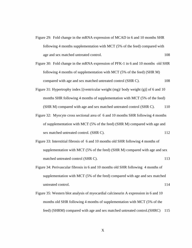
X
Figure 29: Fold change in the mRNA expression of MCAD in 6 and 10 months SHR
following 4 months supplementation with MCT (5% of the feed) compared with
age and sex matched untreated control. 108
Figure 30: Fold change in the mRNA expression of PFK-1 in 6 and 10 months old SHR
following 4 months of supplementation with MCT (5% of the feed) (SHR M)
compared with age and sex matched untreated control (SHR C). 108
Figure 31: Hypertrophy index [(ventricular weight (mg)/ body weight (g)] of 6 and 10
months SHR following 4 months of supplementation with MCT (5% of the feed)
(SHR M) compared with age and sex matched untreated control (SHR C). 110
Figure 32: Myocyte cross sectional area of 6 and 10 months SHR following 4 months
of supplementation with MCT (5% of the feed) (SHR M) compared with age and
sex matched untreated control. (SHR C). 112
Figure 33: Interstitial fibrosis of 6 and 10 months old SHR following 4 months of
supplementation with MCT (5% of the feed) (SHR M) compared with age and sex
matched untreated control (SHR C). 113
Figure 34: Perivascular fibrosis in 6 and 10 months old SHR following 4 months of
supplementation with MCT (5% of the feed) compared with age and sex matched
untreated control. 114
Figure 35: Western blot analysis of myocardial calcineurin A expression in 6 and 10
months old SHR following 4 months of supplementation with MCT (5% of the
feed) (SHRM) compared with age and sex matched untreated control.(SHRC) 115
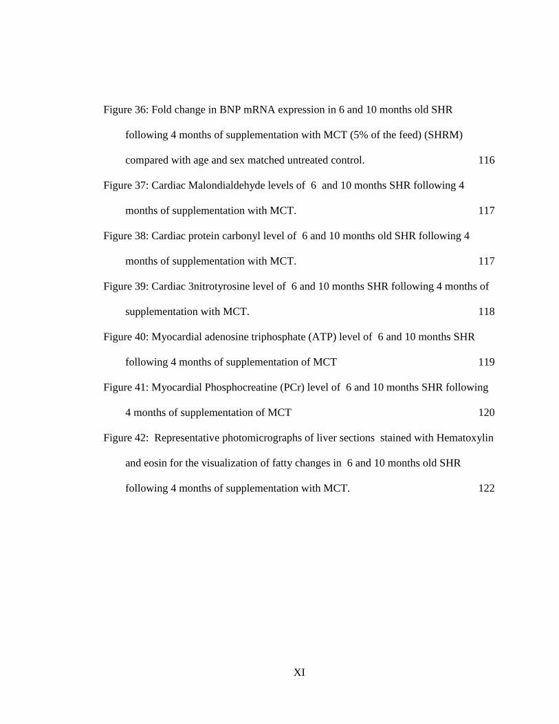
XI
Figure 36: Fold change in BNP mRNA expression in 6 and 10 months old SHR
following 4 months of supplementation with MCT (5% of the feed) (SHRM)
compared with age and sex matched untreated control. 116
Figure 37: Cardiac Malondialdehyde levels of 6 and 10 months SHR following 4
months of supplementation with MCT. 117
Figure 38: Cardiac protein carbonyl level of 6 and 10 months old SHR following 4
months of supplementation with MCT. 117
Figure 39: Cardiac 3nitrotyrosine level of 6 and 10 months SHR following 4 months of
supplementation with MCT. 118
Figure 40: Myocardial adenosine triphosphate (ATP) level of 6 and 10 months SHR
following 4 months of supplementation of MCT 119
Figure 41: Myocardial Phosphocreatine (PCr) level of 6 and 10 months SHR following
4 months of supplementation of MCT 120
Figure 42: Representative photomicrographs of liver sections stained with Hematoxylin
and eosin for the visualization of fatty changes in 6 and 10 months old SHR
following 4 months of supplementation with MCT. 122
XII
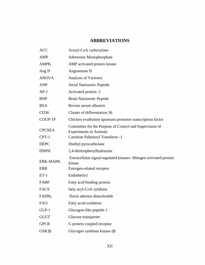
ABBREVIATIONS
ACC Acetyl-CoA carboxylase
AMP Adenosine Monophosphate
AMPK AMP activated protein kinase
Ang II Angiotensin II
ANOVA Analysis of Varience
ANP Atrial Natriuretic Peptide
AP-1 Activated protein -1
BNP Brain Natriuretic Peptide
BSA Bovine serum albumin
CD36 Cluster of differentation 36
COUP-TF Chicken ovalbumin upstream promoter transcription factor
CPCSEA Committee for the Purpose of Control and Supervision of
Experiments in Animals
CPT-1 Carnitine Palmitoyl Transferse -1
DEPC Diethyl pyrocarbonate
DNPH 2,4-dinitrophenylhydrazine
ERK-MAPK Extracellular signal-regulated kinases- Mitogen activated protein
kinase
ERR Estrogen-related receptor
ET-1 Endothelin1
FABP Fatty acid binding protein
FACS fatty acyl-CoA synthase
FADH2 flavin adenine dinucleotide
FAO Fatty acod oxidation
GLP-1 Glucagon-like peptide-1
GLUT Glucose transporter
GPCR G protein coupled receptor
GSK3 Glycogen synthase kinase-3
XIII
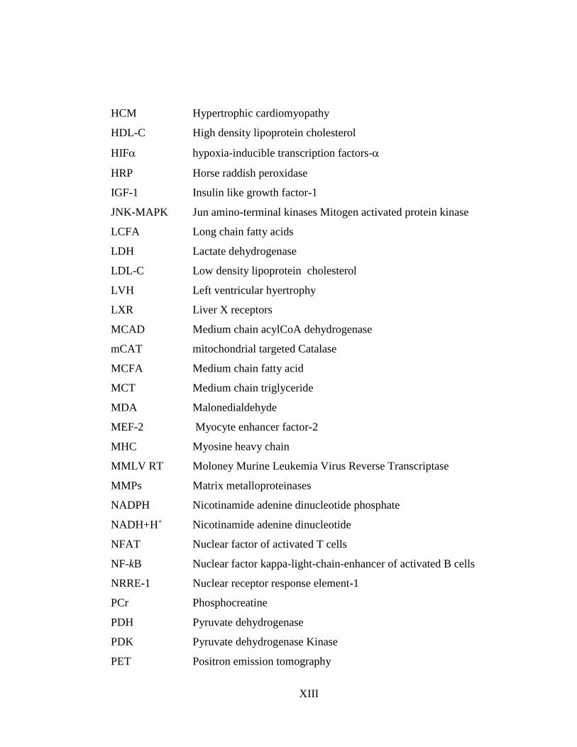
HCM Hypertrophic cardiomyopathy
HDL-C High density lipoprotein cholesterol
HIF hypoxia-inducible transcription factors-
HRP Horse raddish peroxidase
IGF-1 Insulin like growth factor-1
JNK-MAPK Jun amino-terminal kinases Mitogen activated protein kinase
LCFA Long chain fatty acids
LDH Lactate dehydrogenase
LDL-C Low density lipoprotein cholesterol
LVH Left ventricular hyertrophy
LXR Liver X receptors
MCAD Medium chain acylCoA dehydrogenase
mCAT mitochondrial targeted Catalase
MCFA Medium chain fatty acid
MCT Medium chain triglyceride
MDA Malonedialdehyde
MEF-2 Myocyte enhancer factor-2
MHC Myosine heavy chain
MMLV RT Moloney Murine Leukemia Virus Reverse Transcriptase
MMPs Matrix metalloproteinases
NADPH Nicotinamide adenine dinucleotide phosphate
NADH+H+ Nicotinamide adenine dinucleotide
NFAT Nuclear factor of activated T cells
NF-kB Nuclear factor kappa-light-chain-enhancer of activated B cells
NRRE-1 Nuclear receptor response element-1
PCr Phosphocreatine
PDH Pyruvate dehydrogenase
PDK Pyruvate dehydrogenase Kinase
PET Positron emission tomography

XIV
PFK-1 Phosphofructokinase-1
PGC-1 Peroxisome proliferator-activated receptor gamma coactivator 1-alpha
PI3K Phosphoinositide 3-kinase
PKA Protein Kinase A
PKC Protein Kinase C
PKD Protein Kinase D
PPAR Peroxisome proliferator-activated receptor α
ROS Reactive oxygen species
RXR Retinoid X receptor
SDS Sodium dodecyl sulphate
SHR Spontaneously hypertensive rat
SRF Diethyl pyrocarbonate
TBARS Thiobarbituric acid reactive substances
TCA Tricloro acetic acid
TEMED Tetramethylethylenediamine
TIMPs Tissue inhibitors of metalloproteinases
VLCAD Very long chain acyl CoA dehydrogenase

XV
SYNOPSIS

XVI
Left ventricular hypertrophy (LVH) is an adaptive mechanism triggered in the heart in
response to pathological stimuli such as hypertension or aortic stenosis and is regarded
as the major risk factor for adverse cardiovascular events and sudden death. Attenuation
of LVH is associated with better maintenance of cardiac function and prevention of
adverse cardiac remodeling. Two factors intimately linked with pathological
hypertrophy are oxidative stress and change in cardiac metabolic profile. Oxidation of
long-chain fatty acids are the major metabolic fuel for the healthy heart. In pathological
cardiac hypertrophy, there is a decrease in the oxidation of long chain fatty acids and
increased relative contribution of glucose for energy production, co-ordinated by down
regulation of fatty acid oxidizing enzymes, consequent to diminished activity of
Peroxisome proliferator-activated receptor–alpha (PPAR-α). PPAR-α is a ligand
activated transcription factor that regulates the expression of genes involved in the fatty
acid oxidation. Long chain fatty acids are the natural ligands of PPAR-α. The
availability of the long chain fatty acids is limited when the sarcolemmal fatty acid
transporter CD36 is defective. In patients with hypertrophic cardiomyopathy, defective
CD36 results in the decreased PPAR-α activity. The transporter is also defective in
Spontaneously hypertensive rat (SHR). Several studies have reported the association
between reduced rate of fatty acid oxidation and development of cardiac hypertrophy.
Hence it is anticipated that, restoration of fatty acid metabolism will help to maintain
the metabolic status and prevent progressive cardiac remodeling. Medium chain
triglycerides (MCT) have the capacity to bypass CD36 and serve as substrate for fatty
acid oxidation.
XVII
Medium chain triglycerides are triglycerides of saturated fatty acids of chain length 8-12
carbon atoms, and their oxidation is independent of cytoplasmic and mitochondrial fatty
acid transport systems such as CD36 and Carnitine palmitoyl transferase-1 (CPT-1).
Hence, they are regarded as direct fuel for β-oxidation. Several studies have reported the
beneficial effect of MCT on heart. Inclusion of MCT is reported to improve contractile
function of the hypertrophic heart and its consumption is not associated with the
development of coronary artery diseases. Hence, the study was based on the hypothesis
that stimulation of fatty acid metabolism by supplementation of medium chain
triglyceride can prevent and reverse adverse cardiac remodeling in Spontaneously
hypertensive rat.
The study was designed with following objectives:
Validate the suitability of the experimental model by screening for markers of
cardiac hypertrophy, metabolic shift and oxidative stress in spontaneously
hypertensive rat (SHR) in comparison with normotensive wistar rat (W)
Compare the cardiac response to stimulation of fatty acid oxidation by
supplementation with odd and even chain MCT
Evaluate the effect of supplementation with MCT on cardiac remodeling and
oxidative stress at the initial and established stages of cardiac hypertrophy
XVIII
Spontaneously hypertensive rat (SHR), was used as the experimental model to
investigate cardiovascular response to metabolic stimulation by MCT. The SHR
replicates the clinical progression of hypertension in humans; wherein early
development of hypertension is followed by a long stable period of compensated cardiac
hypertrophy that slowly progresses to heart failure. Before initiation of experimental
studies, it was confirmed that the stock of SHR available in the laboratory carries the
essential features of hypertrophy and metabolic alteration. Adult SHR (6 months old)
were compared with age and sex matched Wistar rat (W). Hypertrophy was quantified
from hypertrophy index [Heart weight / body weight ratio (mg/g)], myocyte cross
sectional area, myocardial fibrosis, mRNA expression of Brain natriureic peptide (BNP)
and expression of Calcineurin A. Myocardial malonedialadehyde, protein carbonyl and
3-nitro tyrosine were used as the markers for myocardial oxidative stress. Metabolic
remodeling was assessed from mRNA expression of PPARα, medium chain acylCoA
dehydrogenase (MCAD) and phophofructokinase-1 (PFK-1).
MCT is available in two different forms, odd and even chain MCT based on the number
of carbons. Odd chain MCT is reported to be anaplerotic in nature. Anaplerosis is the re-
filling of the catalytic intermediates of the Citric acid cycle. Therefore, there is
possibility of difference in their metabolic properties. A pilot study was therefore carried
out to identify the type of triglyceride that induces better cardiac response. To select the
ideal MCT 2-month-old SHR were supplemented with 5% (v/w) Ticaprylin or

XIX
Triheptanoate of the total feed for 2 months. Morphological and histological markers of
cardiac hypertrophy and markers of oxidative stress were analyzed.
Experimental studies were carried out in SHR to investigate the cardiovascular response
to metabolic stimulation by MCT supplementation. Animals at initial (2months old) and
established (6 months old) stages of cardiac hypertrophy were treated with 5% MCT
along with standard feed for 4 months and its cardiovascular response was analyzed
from morphological histological and molecular markers. Effect of MCT on metabolic
modulation was assessed from the expression of Peroxisome proliferator activated
receptor (PPAR)-α, medium chain acylCoA dehydrogenase (MCAD) and
phosphofructokinase-1 (PFK-1) mRNA by Real Time PCR. Hypertrophy was quantified
from hypertrophy index [Heart weight / body weight ratio (mg/g)], myocyte cross
sectional area, myocardial fibrosis, mRNA expression of Brain natriureic peptide (BNP)
and expression of Calcineurin A. Myocardial malonedialadehyde, protein carbonyl and
3-nitrotyrosine were used as the markers for myocardial oxidative stress. Cardiac energy
level was assessed by biochemical estimation of adenosine triphosphate (ATP) and
phosphocreatine. Blood pressure was measured noninvasively. Commercially available
kits were used for determination of lipid profile.

XX
Comparison of the markers of cardiac hypertrophy, metabolic shift and oxidative
stress in spontaneously hypertensive rat (SHR) with normotensive Wistar rat (W):
The study has confirmed that Blood pressure of SHR was significantly higher than that
of Wistar. Morphological, histological and molecular markers confirmed the presence of
LVH in SHR. A shift in substrate preference away from fatty acid oxidation towards
glucose was seen as assessed from the expression of PPARα, MCAD and PFK-1. The
decrease in fatty acid oxidation is possibly caused by non-availability of substrate due to
down regulation of the fatty acid transporter CD-36. Hypertrophic response in SHR is
associated with increased oxidative stress.
Cardiac response to supplementation with Odd chain and Even chain triglycerides:
Even chain and odd chain triglycerides have shown variable cardiac response. Although
both sources of MCT decreased myocardial oxidative stress, prevention of hypertrophic
response was relativel better with even chain MCT. Hence tricaprylin was used as the
source of MCT for further experimental studies. Both treatments did not affect body
weight and lipid profile.
Stimulation fatty acid oxidation in initial and established stages of cardiac
hypertrophy:
Supplementation of MCT stimulated fatty acid metabolism in the initial and established
stages of cardiac hypertrophy in SHR. Stimulation of fatty acid metabolism prevented

XXI
progressive cardiac remodeling independent of blood pressure. MCT modulated cardiac
oxidative stress. The reduction of cardiac hypertrophy is possibly associated with
decrease in oxidative stress. The study shows that stimulation of fatty acid metabolism
by supplementation of MCT under hypertrophic condition is beneficial to the heart. The
treatment did not induce dislipidemia or obesity.
The above observations lead to the conclusion that, contrary to the belief that stimulation
of Fatty acid oxidation can be detrimental in the presence of hypertrophy, this study has
shown that, restoration of the metabolic profile is beneficial to the heart.
Supplementation of medium chain triglycerides prevented progressive cardiac
remodeling in hypertrophic heart, possibly mediated by reduction of oxidative stress. As
hypertension is associated with consequences other than hypertrophy, MCT can be used
only as a supplement along with antihypertensives. Preclinical studies will help to
determine the therapeutic implications of supplementation with MCT in patients with
hypertensive heart disease.

1
I. INTRODUCTION

2
Left ventricular hypertrophy is an adaptive mechanism triggered in the heart in response
to pathological stimuli such as hypertension or aortic stenosis and is regarded as the
major risk factor for adverse cardiovascular events and sudden death. Attenuation of
LVH is associated with maintenance of cardiac function and reduction in cardiac injury.
Oxidation of long-chain fatty acids forms the major metabolic fuel for the healthy heart
and supplies 60-70% of myocardial ATP. The remaining energy comes from glucose and
lactate oxidation. In pathological cardiac hypertrophy, there is a decrease in the
oxidation of long chain fatty acids and increased relative contribution of glucose for the
energy maintenance. The decrease in the metabolic substrate preference for fatty acid is
possibly caused by reduction in the expression of Cluster of Differentation-36 (CD36), a
major sarcolemmal fatty acid transporter, that limits the availability of long chain fatty
acids. A number of reports testify the role of CD36 deficiency in the metabolic alteration
and the consequent cardiac remodeling. Tanaka et al reported the direct association
between CD36 deficiency and development of hypertrophic cardiomyopathy in the
Japanese population(Tanaka et al., 1997). Pharmacological inhibition of CD36 was
associated with development of cardiac hypertrophy independent of blood pressure in
rodents (Kusaka et al., 1995). Defective CD36 in spontaneously hypertensive rat (SHR)
is identified as the primary determinant of altered myocardial metabolism and
myocardial hypertrophy (Hajri et al., 2001). Deficiency of CD36 has been reported
recently by Magida et al in mouse models with familial hypertrophic cardiomyopathy
(Magida and Leinwand, 2014). These studies highlight the concept that decreased fatty
acid oxidation is secondary to reduced CD36 expression in cardiomyocytes. As chronic
3
reduction in fatty acid availability can lead to pathological remodeling, it is presumed
that restoration of fatty acid oxidation will help to maintain the metabolic status and
prevent progressive cardiac remodeling.
Although enhanced glucose metabolism is more economical in terms of oxygen cost,
chronic reliance on glucose metabolism is inefficient for maintaining contractile function
under work overload. (Ingwall, 2009) Nevertheless, substantially enhanced insulin
independent glucose utilization by GLUT-1 overexpression prevented the progression of
cardiac dysfunction during hypertrophy (Liao et al., 2002a). However such enhanced
glucose utilization by the approach of genetic modification is of less clinical relevance.
Kolwicz et al reported that sustenance of myocardial fatty acid metabolism preserved
myocardial energetics and prevented the development of cardiac hypertrophy under
pressure overload, showing that that maintenance of inherent cardiac metabolic profile
is beneficial under pathological conditions (Kolwicz et al., 2012). Similarly, studies have
reported that high dietary fat could activate mitochondrial oxidative metabolism and
alleviate adverse cardiac remodeling associated with development of obesity (Chess et
al., 2008, 2009; Duda et al., 2008; Okere et al., 2005). The therapeutic strategies aimed
at reduction of hemodynamic overload does not essentially modulate myocardial
metabolism or regress cardiac remodeling. Hence, the study was designed to evaluate
cardiac response to metabolic modulation by supplementation of Medium chain
triglyceride (MCT) at the initial and established stages of cardiac hypertrophy

4
Medium chain triglycerides (MCT) have the capacity to bypass CD36 and serve as
substrate for fatty acid oxidation. Medium chain triglycerides are triglycerides of
saturated fatty acids of chain length 8-12 carbon atoms, and their oxidation is
independent of cytoplasmic and mitochondrial fatty acid transport systems such as CD36
and Carnitine palmitoyl transferase-1 (CPT-1). Hence, they are regarded as direct fuel
for β-oxidation. Several studies have reported the beneficial effect of MCT on heart.
Inclusion of MCT is reported to improve contractile function of the hypertrophic heart
and its consumption is not associated with the development of coronary artery diseases.
Therefore, this study was based on the hypothesis that, “Reactivation of fatty acid
metabolism by supplementation of medium chain triglyceride can prevent and reverse
adverse cardiac remodeling.” The study was carried out in spontaneously hypertensive
rat, an animal model of chronic pressure overload induced left ventricular hypertrophy.
The cardiac response to stimulation of fatty acid metabolism was examined in animals of
two different age groups, as metabolic modulation using pharmacological ligands had
yielded positive results in young SHR but not in older animals. (Purushothaman et al.,
2011b). Further, the studies that reported positive response to MCT was carried out
either in young SHR or surgical models.
MCT is available in two different forms, odd and even chain MCT based on number of
carbons. Odd chain MCT is reported to be anaplerotic in nature. Anaplerosis is the re-
filling of the catalytic intermediates of the Citric acid cycle. Therefore, there is the
possibility of difference in their metabolic properties. A pilot study was therefore carried
5
out to identify the type of triglyceride that can induce a comparatively better cardiac
response, and then use that for further experiments. This study was carried out with the
following objectives
i. Validate the suitability of the experimental model by screening for markers of
cardiac hypertrophy, metabolic shift and oxidative stress in spontaneously
hypertensive rat (SHR) in comparison with normotensive Wistar rat (W)
ii. Compare the cardiac response to stimulation of fatty acid oxidation by
supplementation with odd and even chain MCT
iii. Assess cardiac response to stimulation of fatty acid oxidation by supplementation
of medium chain triglycerides at the initial and established stages of cardiac
hypertrophy
SHR in the initial and established stages of cardiac hypertrophy were supplemented
with 5% MCT (v/w) along with standard feed for 4 months and its cardiovascular
response was analyzed for morphological, histological and molecular markers. Effect of
MCT on metabolic modulation was assessed from the expression of Peroxisome
proliferator-activated receptor (PPAR)-α, medium chain acylCoA dehydrogenase
(MCAD) and phosphofructokinase-1 (PFK-1) mRNA by Real Time PCR. Evaluation of
cardiac remodeling was based on morphological, histological and molecular markers of
cardiac hypertrophy. Myocardial malonedialadehyde, protein carbonyl and 3-
6
nitrotyrosine were used as the markers for myocardial oxidative stress. Cardiac energy
level was assessed by biochemical estimation of adenosine triphosphate (ATP) and
phosphocreatine. Blood pressure was measured noninvasively. Commercially available
kits were used for determination of lipid profile.
A brief description of the morphological and molecular changes associated with
cardiac hypertrophy and the energy metabolism of the normal and hypertrophied heart
are given in the next chapter. Literature on the role of CD36 in the development of
cardiac hypertrophy and strategies adopted for modulation of energy metabolism in
cardiac hypertrophy and the consequence are also reviewed.
The design of study and experimental methodology are given in the third chapter,
In the fourth chapter, the results are presented and the findings discussed in the
light of available information.
Salient observations of the study are listed in the fifth chapter. The conclusion
and scope for further studies are also given.
The references cited in the text are listed in ‘Bibliography’.

7
II. REVIEW OF LITERATURE

8
II.1. CARDIAC HYPERTROPHY
The heart is a dynamic organ capable of self regulation and adaption in response to
alterations in workload associated with developmental, physiological and pathological
stimuli. As the postnatal growth of the heart is closely matched to its functional load,
heart muscle enlarges its size and mass to counterbalance the chronic increase in wall
stress resulting in cardiac hypertrophy. Cardiac hypertrophy is defined as the
enlargement or overgrowth of cardiac mass due to an increase in size of its terminally
differentiated myocytes and can broadly be classified as either pathological or
physiological depending on the nature of the stimulus and the phenotype.
II.1.1. Pathological cardiac hypertrophy
Pathological cardiac hypertrophy is considered as an intermediate stage in the
progression to heart failure. The heart undergoes remodeling in response to multitude of
stimuli such as hemodynamic overload, neurohormonal activation and sarcomeric gene
mutation. In response to hormonal, genetic and mechanical stimuli, ventricular myocytes
hypertrophy by increasing the number of sarcomere units per cell without increase in
cell number. Although the stimuli are different, the molecular and cellular events
converge into a process called cardiac hypertrophy which is regarded as a compensatory
phase (Kehat and Molkentin, 2010). According to Law of Laplace, under chronic
hemodynamic overload, the heart tends to increase its wall thickness to diminish wall
stress; and maintenance of cardiac output forms the basis for the adaptive nature of the
cardiac growth. (Vidt and Prisant, 2005). Sustained hypertrophy is maladaptive and is an
9
independent risk factor for myocardial infarction, arrhythmia and sudden death
(Nagendran et al., 2013). Regression of cardiac hypertrophy reduces the risk of heart
failure and ventricular dysfunction independent of the treatment adopted (Esposito et
al., 2002; Frey et al., 2004; Yusuf et al., 2000). The defining features of cardiac
hypertrophy are an increase in cardiomyocyte size, enhanced protein synthesis, and
reinduction of fetal cardiac gene programme. It is also associated with overproduction of
reactive oxygen species (ROS) and reactivation of fetal cardiac metabolism: a shift in
substrate preference from fatty acid to glucose (Barger et al., 2000; Takimoto and Kass,
2007).
Hypertension is the common cause for the development of left ventricular hypertrophy
(LVH), where the heart must work harder to counterbalance the chronic increase in wall
stress. As a result, the muscle cells within the heart enlarge leading to an increase in size
and mass. However, prolonged wall stress results in ventricular dilation, contractile
dysfunction and eventually leads to heart failure. LVH is also induced by various
neurohormones such as angiotensin II, aldosterone, norepinephrine and insulin,
independent of pressure overload (Db and Lj, 1996). In the 1960s, Meerson and
colleagues classified development of pathological hypertrophy into different stages. The
developing phase of hypertrophy, with ventricular hyperfunction where work load
exceeds the work output is followed by a compensatory phase, where increased
workload is compensated by the cardiac growth with maintenance of resting cardiac
output. Finally, the decompensation phase with deterioration of cardiac output and filling
10
progressively leads to heart failure (Frey et al., 2004).The duration and progression of
these stages depend on the magnitude and type of overload.
Pathological cardiac hypertrophy is accompanied with molecular changes that are
observed during fetal cardiac development, such as reactivation of fetal gene program.
Fetal recapitulation includes stimulation of atrial natriuretic peptide (ANP), B-type
natriuretic peptide (BNP) and genes for fetal isoforms of contractile proteins, such as
skeletal -actin and -myosin heavy chain (MHC). This fetal recapitulation is
accompanied by downregulation of adult genes, such as -MHC and the calcium-
handling protein sarco(endo)plasmic reticulum Ca2+-ATPase (Zhao et al., 2004). Heart
parallely shifts its substrate preference from fatty acid towards glucose under cardiac
hypertrophy, the hallmark of fetal cardiac metabolism (Huss and Kelly, 2005). The
morphological, histological and biochemical changes associated with hypertrophy are
illustrated in figure.1. The reactivation of fetal gene program is mediated by various
transcription factors (TFs) such as NFAT, NF-kB, MEF2, GATA4, and SRF which play
a prominent role in embryonic development. (Akazawa and Komuro, 2003; Oka et al.,
2007). Aberrant expression of genes involved in fetal contractile proteins and cardiac
metabolism leads to progressive myocardial dysfunction and irreversible pathogenesis.
Decreased expression of fetal genes is accompanied by improvement of ventricular
function. Hence, they can be used as the markers of adverse cardiac remodeling. The
reactivation of fetal gene expression in the adult myocardium is mediated by the

11
combined action of transcription factors, chromatin remodeling and post transcriptional
regulation at different levels of gene expression (Dirkx et al., 2013).
Figure 1: Charecteristic features of pathological cardiac hypertrophy
During cardiac hypertrophy, the myocardium undergoes structural remodeling to
accommodate the hypertrophied myocytes by modulating extracellular matrix and
myocardial capillary network. The extracellular matrix is principally composed of
collagen with smaller amounts of elastin, laminin, and fibronectin. Although collagen
types I, III, and V coexist in the myocardium, type I forms the major component (85% of

12
the total collagen). The extracellular fibrillar collagen provides structural integrity to
adjoining myocytes, and aids myocyte contraction that translates into efficient cardiac
contraction (Barsotti et al., 1993; Weber et al., 1994). Pathological LVH is associated
with increased interstitial and perivascular fibrosis. Compared to the normal
myocardium, the extent of fibrosis is inversely proportional to the number of surviving
myocytes and is directly related to the degree of hypertrophy. Excessive collagen
deposition replaces the myocyte loss due to apoptosis and necrosis. Hypertensive cardiac
remodeling involves initial deposition of type-III collagen, followed by type-I collagen.
Disproportionate accumulation of collagen increases ventricular stiffnesss, contractile
uncoupling, altered microcirculation and electrical uncoupling resulting in impaired
cardiac function and adverse cardiovascular events. The augmented cardiac fibrosis and
reduced capillary density increases the oxygen diffusion distance resulting in myocardial
ischemia, and is likely to contribute to the progressive transition to heart failure
(Gradman and Alfayoumi, 2006).
Based on the phenotype of the heart and the initiating stimuli, cardiac hypertrophy has
classically been subdivided as concentric or eccentric (Kehat and Molkentin, 2010)
(Fig.2). Concentric hypertrophy refers to an increase in relative wall thickness and
cardiac mass, with a small reduction in chamber volume. Pathological stimuli such as
hypertension and aortic stenosis produces systolic wall stress and results in concentric
cardiac hypertrophy. In contrast, in eccentric hypertrophy the increase in cardiac mass is
due to increased chamber volume. The stimuli such as aortic regurgitation and
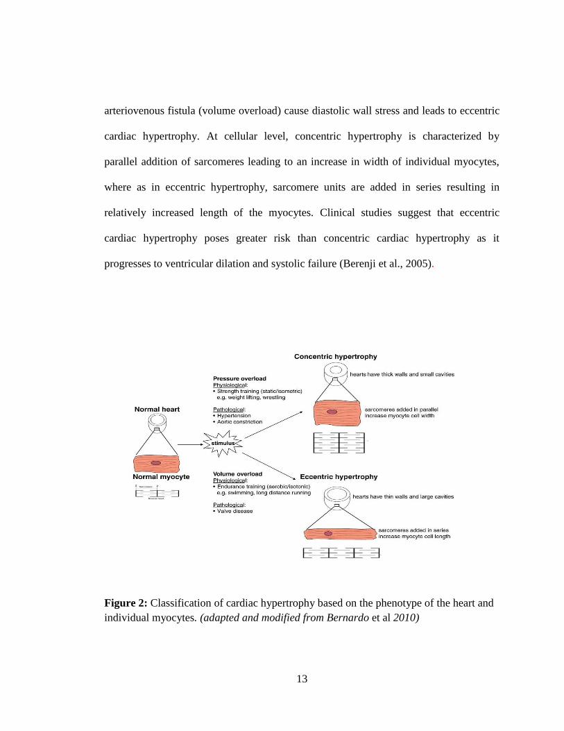
13
arteriovenous fistula (volume overload) cause diastolic wall stress and leads to eccentric
cardiac hypertrophy. At cellular level, concentric hypertrophy is characterized by
parallel addition of sarcomeres leading to an increase in width of individual myocytes,
where as in eccentric hypertrophy, sarcomere units are added in series resulting in
relatively increased length of the myocytes. Clinical studies suggest that eccentric
cardiac hypertrophy poses greater risk than concentric cardiac hypertrophy as it
progresses to ventricular dilation and systolic failure (Berenji et al., 2005).
Figure 2: Classification of cardiac hypertrophy based on the phenotype of the heart and
individual myocytes. (adapted and modified from Bernardo et al 2010)
14
II.1.2. Physiological Cardiac hypertrophy
Physiological cardiac hypertrophy in contrast to pathological hypertrophy is
advantageous and is distinct from the latter hypertrophy at the molecular, cellular and
functional levels. Physiological hypertrophy develops in response to chronic exercise
training with preserved or enhanced cardiac function, and does not lead to failure
(Fagard, 1997). It is characterized by normal or enhanced cardiac function, improved
cardiac metabolism, and absence of pathological features such as fibrosis and
reactivation of fetal genes with enhanced fatty acid and glucose oxidation (McMullen
and Jennings, 2007). Physiological cardiac hypertrophy is also observed during post
natal growth, endurance exercise training and pregnancy and is reversible once the
stimuli have been relieved. The mechanism by which the heart adapts differently to
physiological and pathological stimuli remains mysterious. One school of thought is that
most pathological stimuli are chronic while physiological stimuli are intermittent in
nature, presuming that the duration of the stimulus determines the phenotype of the
heart. Perrino et al applied pathological stimuli for intermittent periods to evaluate the
role of duration of stimuli on this divergent cardiac phenotype, simulating the duration
of exercise training (Perrino et al., 2006). Intermittent pathological stress induced
cardiac abnormalities with diastolic dysfunction and vascular rarefaction. They
demonstrated that the duration of stress determines the degree of cardiac hypertrophy
whereas nature of the stress determines the cardiac phenotype whether it is physiological
or pathological. The development of athletic heart is a beneficial adaptive response with
decreased resting and submaximal heart rates and increased filling time and venous

15
return. These adaptations can facilitate the myocardium to satisfy the increased demands
of exercise while maintaining or enhancing normal function (Iemitsu et al., 2001; Richey
and Brown, 1998; Shapiro and Smith, 1983).
II.1.2.1. Athletes heart and sudden cardiac death
The athlete's heart has generally been defined as a benign increase in heart mass,
representing a physiological adaptation to chronic exercise training. There are several
media reports on increased incidence of sudden death in elite athletes due to
cardiovascular abnormalities. United states based 12 year survey in high-school athletes
reported that the frequency of sudden death is considerably low (1:200,000 per year )
(Maron et al., 2009). Large autopsy-based studies revealed that, hypertrophic
cardiomyopathy accounts for one-third of sudden death in US followed by congenital
coronary artery anomalies and congenital malformations (Maron, 2003). To address the
clinical consequence of cardiac remodeling in trained athletes, a study conducted in
young Olympic athletes reported that extreme and uninterrupted endurance training over
long periods of time (up to 17 years) does not induce cardiac structural and functional
abnormalities or occurrence of cardiovascular events (Pelliccia et al., 2010). Thus, it is
generally considered that cardiac hypertrophy in response to exercise training itself is
not the cause for sudden death in elite athletes. However, a complete understanding of
cellular check points that distinguishes physiological from pathological cardiac
remodeling is critical for pharmacological targeting for prevention of the transition from
compensated cardiac hypertrophy to failure.

16
II.1.3. Signaling cascades mediating cardiac hypertrophy
The signaling pathway mediating physiological cardiac hypertrophy is largely attributed
to insulin-like growth factor-1(IGF-1) and growth hormones and is transduced by
phosphoinositide 3-kinase (PI3K)/Akt signaling (Dorn and Force, 2005). The
involvement of the IGF-1/PI3K/Akt signaling in mediating physiological hypertrophy
was evaluated in mice with cardiac specific transgenic expression of constitutively active
or dominant-negative mutant forms of signaling intermediates. Cardiac-specific deletion
of the insulin-like growth factor-1 receptor prevented the development of exercise-
induced cardiac hypertrophy (Kim et al., 2008). Mice with cardiac-specific expression of
constitutively active PI3K resulted in larger hearts with the features of physiological
cardiac hypertrophy (Shioi et al., 2000), whereas dominant-negative PI3K expression
attenuated the development of exercise-induced physiological but not the pathological
hypertrophy induced by pressure overload, demonstrating the role of PI3K pathway in
the development of adaptive hypertrophy. Phosphoinositide 3-kinase (p110alpha) plays
a critical role in the induction of physiological, but not pathological, cardiac
hypertrophy. Similarly, Akt1-/- mice were resistant to exercise-induced cardiac
hypertrophy, however, it developed greater cardiac hypertrophy in response to
pathological stimuli such as aortic constriction demonstrating its critical role in
mediating physiological cardiac hypertrophy (DeBosch et al., 2006).
The best characterized signaling cascades for mediating pathological cardiac
hypertrophy are the G protein-coupled receptor (GPCR) mediated Gαq signaling
17
activated by Ang II, ET-1 and catecholamines with down stream activation of mitogen
activated protein kinases (MAPKs), protein kinase C (PKC) and D (PKD) and
calcineurin. In response to hypertrophic stimuli such as pressure overload, various
paracrine and autocrine factors such as Ang II, ET-1 and noradrenaline (norepinephrine,
NE) are released and plays a critical role in the development of pathological cardiac
hypertrophy (Arai et al., 1995; Rapacciuolo et al., 2001; Schunkert et al., 1990;
Yamazaki et al., 1999). These factors activate GPCR, causing dissociation and activation
of Gαq/11 and downstream signaling proteins, including phospholipase C (PLC),
MAPKs, PKC and protein kinase A (PKA). The role of Gαq/11 in mediating pathological
cardiac hypertrophy was evaluated in transgenic mouse models with cardiac specific
over-expression of Gαq and inhibitory peptide specific for Gq-coupled receptor
signaling (D’Angelo et al., 1997; Wettschureck et al., 2001). Downstream of Gq,
mitogen-activated protein kinases (MAPKs) and some protein kinase (PK) C isoforms
have been implicated in mediating pathological cardiac hypertrophy. MAPKs such as the
extracellular signal-regulated kinases (ERKs), the c-Jun amino-terminal kinase (JNKs),
and the p38-MAPKs are activated in cardiac myocytes in response to GPCR agonists
(AT1 receptors, endothelin receptors and α1-ARs) and in pressure overload (Sadoshima
et al., 1995; Sugden, 2001; Takeishi et al., 2001; Yamazaki et al., 1993). Pressure
overload also stimulates GPCR mediated PKC and PKD activation to trigger
hypertrophic responses (Dorn and Force, 2005; Harrison et al., 2006).

18
Another target of Gq is the calcium-dependent phosphatase, calcineurin that contributes
to pathological cardiac hypertrophic gene transcription by desphosphorylating
transcription factors known as nuclear factor of activated T cells (NFAT) (Wilkins and
Molkentin, 2004). Calcineurin is activated by a sustained Ca2+ plateau and is insensitive
to transient Ca2+ fluxes such as that which occur in response to cardiomyocyte
contraction. Activated NFAT translocates to the nucleus, where it associates with other
transcription factors such as GATA4 and myocyte enhancer factor 2 (MEF2) to initiate
the transcription of hypertrophic gene program (Frey and Olson, 2003; Wilkins et al.,
2002). Clinical studies revealed that calcineurin activity is increased in patients with LV
hypertrophy and failure (Haq et al., 2001). Transgenic expression of activated
calcineurin in mice developed profound cardiac hypertrophy which rapidly progressed to
congestive heart failure and it was prevented by pharmacological inhibition of
calcineurin (Molkentin et al., 1998). It is reported that calcineurin activity was elevated
only in pathological models but not in the physiological models testifying its role in
pathological cardiac remodeling (Wilkins et al., 2004).
II.1.4. Role of Oxidative stress in mediating Cardiac hypertrophy
Growing evidence highlights that oxidative stress is the contributing factor for the
development and progression of cardiac hypertrophy (Li et al., 2002). Oxidative stress
occurs when free radical synthesis outweighs the intrinsic antioxidant capabilities of the
cell, and has been implicated in the genesis of pathological cardiac hypertrophy and
heart failure (Takimoto and Kass, 2007). It is established that inhibition of endogenous
19
antioxidant enzymes can stimulate myocyte hypertrophy and was inhibited by the
addition of antioxidants (Siwik et al., 1999). Extracellular stimuli such as Angiotensin II,
Endothelin-I, tumor necrosis factor-, 1-adrenergic agonists and mechanical stretch
induced hypertrophic response mediated by stimulating ROS production in cardiac
myocytes, which could be abolished by antioxidants. (Amin et al., 2001; Cheng et al.,
1999; Nakamura et al.;1998; Pimentel et al., 2001). Angiotensin II induces the activation
of NADPH oxidase, an enzyme primarily devoted for ROS production. Several
extracellular stimuli are capable of inducing cardiac hypertrophy through various
downstream signaling molecules such as PKC; the MAPKs p38, JNK, apoptosis-
signaling kinase 1 (ASK-1), and ERK1/2; PI3K; Akt; several tyrosine kinases, NF-κB;
and calcineurin; and these factors can be activated directly or indirectly by ROS
(Giordano, 2005). ROS also induces activation of redox-sensitive protein kinases such
as the mitogen activated protein kinase (MAPK) superfamily and activity of
transcription factors like NF-kB, AP-1 (activated protein –I) (Li et al., 2002).
Increased ROS generation induces cardiac contractile dysfunction by modifying the
activity of proteins involved in excitation-contraction coupling, such as sarcolemmal ion
channels and exchangers and sarcoplasmic reticulum calcium release channels (Seddon
et al., 2007). Superoxides produced by hypertrophic stimuli interact with nitric oxide
(NO) to form peroxynitrite and decreases its bioavailability, leading to coronary vascular
endothelial dysfunction (Shah and MacCarthy, 2000). ROS are involved in several
processes such as an increase in fibroblast proliferation, transformation into matrix-
20
generating myofibroblasts, the expression of pro-fibrotic genes and alterations in the
balance between the activities of MMPs and TIMPs. ROS also have a potent role in
extracellular matrix remodeling by modulating the activity of matrixmetalloproteinases
(Preeta and Nair, 2000; Siwik et al., 2001).
Although several antioxidants have shown a positive role in modulating oxidative stress
and cardiac hypertrophy, their potential in maintaining mitochondrial redox status is
poor. Mitochondria and nicotinamide adenine dinucleotide phosphate (NADPH)
oxidase a major site of intracellular ROS production, and NOX4 isoform of NADPH
oxidase is located in mitochondria (Dai et al., 2011). Mitochondria targeted antioxidants
such as Mito-Q and Szeto-Schiller (SS)-31 peptide have ameliorated cardiac
hypertrophy and dysfunction (Dai et al., 2011; Graham et al., 2009). Furthermore, it is
demonstrated that overexpression of mitochondrial targeted Catalase (mCAT) reduced
age-dependent left ventricular hypertrophy and diastolic dysfunction (Dai et al., 2009).
II.2. CARDIAC METABOLISM
The heart has a tremendous capacity for ATP generation, allowing it to function as an
efficient pump throughout the life of the organism. Cardiac function depends on the
delicate equilibrium between work performance to meet requirements of the body and
energy metabolism to maintain its contractile function. In the healthy heart the processes
of ATP synthesis and breakdown are exquisitely matched such that there is never a

21
significant fall in ATP concentration, even with large increases in cardiac output
(Stanley et al., 2005). Heart utilizes 60–70% of the ATP generated to fuel contractile
function, and the remaining 30–40% is principally used by the sarcoplasmic reticulum
Ca2+-ATPase and other ion pumps (Gibbs, 1978).
The heart is considered as a metabolic omnivore with the capacity to oxidize wide range
of substrates such as fatty acids, carbohydrates, ketone bodies, lactate and even amino
acids. The contribution of each substrate to the overall production of ATP is tightly
regulated, with each pathway possessing a considerable degree of plasticity and
interdependence. The metabolic flexibility confers the advantage to the myocardium to
adapt to a variety of physiological and pathological conditions by maintaining sufficient
ATP level for sustaining cardiac function. Under normal aerobic conditions, the heart
relies primarily on fatty acids as substrates for oxidative metabolism. Fatty acid -
oxidation normally contributes 60–70% of total ATP production in the healthy adult
heart; the remainder is provided mainly by the oxidation of glucose and lactate
oxidation, and a lesser extent by the oxidation of ketone bodies. Despite relatively
limited lipid storage capacity, fatty acid oxidation serves as the primary myocardial ATP
generating pathway.
Heart is a highly oxidative organ and derives 90% of ATP from oxidative
phosphorylation in the mitochondria and the remainder is from glycolysis and to a lesser
extent from the citric acid cycle (Krebs cycle). In the mitochondria, the high-energy

22
phosphate bond in ATP can be transferred to creatine by mitochondrial creatine kinase to
form phosphocreatine (PCr). Due to its smaller molecular weight than ATP, PCr can
diffuse through the mitochondrial membrane into the cytosol where it acts as a buffer to
maintain constant ATP level by reactions catalyzed by the cytosolic creatine kinase.
Because of its continuous mechanical work, the heart has a high rate of ATP hydrolysis
(≈0.5 μmol/g wet weight per second). Nevertheless, the high-energy phosphate pool in
the heart is relatively small and can be exhausted within a few seconds. Therefore,
cardiac work depends essentially on ATP generation, and impairments in this process
can rapidly induce contractile dysfunction.
Mitochondria occupy ~30% of the cardiomyocyte volume and are organized in rows
between myofilaments, with constant diffusion distance within the core of the
myofilaments, making them the cell type with the highest mitochondria content (Schaper
et al., 1985). The NADH+H+ and FADH2 produced during fatty acid -oxidation
pathway, the citric acid cycle (TCA cycle), and to a lesser extent from the pyruvate
dehydrogenase (glucose oxidation) reaction and glycolysis fuel the mitochondrial
electron transport chain and synthesize ATP by oxidative phosphorylation. There is a
coordinated link between the rate of oxidation of energy substrate and the contractile
performance of the heart. Thus, an increase in contractile function results in a
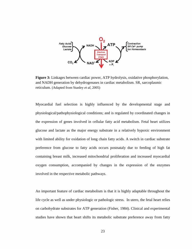
concomitant increase in the utilization of metabolic substrate for ATP production.
(Fig.3)
23
Figure 3: Linkages between cardiac power, ATP hydrolysis, oxidative phosphorylation,
and NADH generation by dehydrogenases in cardiac metabolism. SR, sarcoplasmic
reticulum. (Adapted from Stanley et al, 2005)
Myocardial fuel selection is highly influenced by the developmental stage and
physiological/pathophysiological conditions; and is regulated by coordinated changes in
the expression of genes involved in cellular fatty acid metabolism. Fetal heart utilizes
glucose and lactate as the major energy substrate in a relatively hypoxic environment
with limited ability for oxidation of long chain fatty acids. A switch in cardiac substrate
preference from glucose to fatty acids occurs postnataly due to feeding of high fat
containing breast milk, increased mitochondrial proliferation and increased myocardial
oxygen consumption, accompanied by changes in the expression of the enzymes
involved in the respective metabolic pathways.
An important feature of cardiac metabolism is that it is highly adaptable throughout the
life cycle as well as under physiologic or pathologic stress. In utero, the fetal heart relies
on carbohydrate substrates for ATP generation (Fisher, 1984). Clinical and experimental
studies have shown that heart shifts its metabolic substrate preference away from fatty

24
acid towards glucose under various pathological conditions such as pressure- or volume
overload–induced hypertrophy and also in heart failure, resembling the fetal metabolic
program. Similarly, in the uncontrolled diabetic state, myocardium uses fatty acids
almost exclusively to maintain energy level due to insulin resistance and high circulating
free fatty acids (Wall and Lopaschuk, 1989). Conversely with aging, there is a decline
in fatty acid metabolism, and the proportion of glucose metabolism to overall substrate
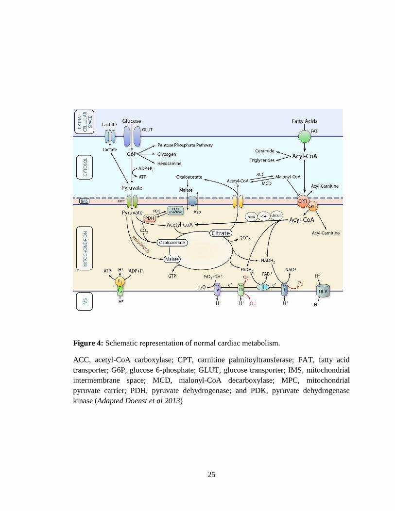
metabolism increases (Kates et al., 2003; McMillin et al., 1993). A schematic
representation of normal cardiac metabolism is given in fig.4.
25
Figure 4: Schematic representation of normal cardiac metabolism.
ACC, acetyl-CoA carboxylase; CPT, carnitine palmitoyltransferase; FAT, fatty acid
transporter; G6P, glucose 6-phosphate; GLUT, glucose transporter; IMS, mitochondrial
intermembrane space; MCD, malonyl-CoA decarboxylase; MPC, mitochondrial
pyruvate carrier; PDH, pyruvate dehydrogenase; and PDK, pyruvate dehydrogenase
kinase (Adapted Doenst et al 2013)

26
II.2.1. Myocardial fatty acid metabolism
Myocardial fatty acid uptake is primarily determined by plasma concentration of
nonesterified fatty acids. Free fatty acids are highly hydrophobic and associated with
proteins or covalently bound with coenzyme A. Major sources of plasma fatty acid are
triglycerides which are released from adipocytes, chylomicrons and very low density
lipoproteins that are hydrolyzed by lipoprotein lipase bound to the capillary endothelial
cells and cardiomyocytes. The major site of fatty acid oxidation is the mitochondrial
matrix and is highly dependent on the intracellular delivery of fatty acids. Uptake of
fatty acids into the cardiomyocyte is mediated either by passive diffusion or by protein-
mediated fatty acid uptake from microvascular compartments; and is mediated by fatty
acid translocase (FAT), or a plasma membrane fatty acid binding protein (FABPpm). A
specific 88-kDa FAT protein called CD36 is abundantly expressed in skeletal and
cardiac muscle and appears to be the predominant form of FAT in the heart.
Once fatty acids are transported into the sarcoplasm, the free fatty acids bind to FABP,
the primary intracellular carrier of non esterified fatty acids and are then activated by
fatty acyl-CoA synthase (FACS) to fatty acyl-CoA. FABP and FACS protein along with
CD36 are assembled on the cytosolic side of the sarcolemmal membrane to ensure the
immediate esterification of fatty acids entering into the sarcoplasm to fatty acyl-CoA,
making them water soluble (Lopaschuk et al., 1994). Oxidation of fatty acids primarily
occur in the mitochondria and to a small extent in peroxisomes. Hence fatty acids have
to be transported into the mitochondrial matrix from cytosol. As inner mitochondrial

27
membrane is impermeable to fatty acyl-CoA, the transport of long chain acyl-CoA is
mediated by carnitine dependent transport system, consisting of palmitoyl transferases
(CPT) I and II and carnitine acyl translocase (CAT). CPT-I is present on the outer
mitochondrial membrane. It binds to fatty acyl CoAs and catalyzes the formation of fatty
acyl carnitines which are transported to the mitochondrial inter-membrane space. There,
CAT translocates fatty acyl carnitines into the matrix (in exchange for carnitine), where
CPT-II re-esterifies acyl carnitines into acyl CoAs. Within the matrix, acyl CoAs can
then be progressively metabolized by fatty acid oxidation. CPT-1 is a regulatory enzyme
that controls the mitochondrial uptake of long chain fatty acids with two different
isoforms CPT-1α and β. CPT-I the liver isoform is found throughout the body except
cardiac and skeletal muscle and brown adipose tissue. CPT-1 is the main isoform in the
heart.
Once taken up by the mitochondria, fatty acyl-CoA undergoes -oxidation, a process
that repeatedly cleaves off two carbon acetyl-CoA units, generates energy in the form of
reduced NADH+H+ and FADH2, that are subsequently utilized for the formation of ATP
by electron transport chain and oxidative phosphorylation. Four main enzyme classes are
involved in the mitochondrial -oxidation. The first step is catalyzed by acyl-CoA
dehydrogenase, followed by 2-enoyl-CoA hydratase, and then 3-hydroxyacyl-CoA
dehydrogenase. The final step is 3-ketoacyl-CoA thiolase (3-KAT), which regenerates
acyl-CoA for another round of -oxidation. Acyl-CoA dehydrogenase and 3-
hydroxyacyl-CoA dehydrogenase generate FADH2 and NADH+H+, respectively, and the

28
acetyl-CoA formed from -oxidation is fed into citric acid cycle (CAC) and generates
NADH+H+, GTP and FADH2.
II.2.2. Glucose Metabolism
Glucose that is used by the heart is derived from exogenous glucose and glycogen stores.
Glucose transport into cardiomyocytes is mediated by the transmembrane glucose
gradient and the glucose transporters (GLUTs) in the sarcolemma. Although GLUT1 is
the major glucose transporter in the fetal heart and contributes to constitutive glucose
uptake, in the adult heart, GLUT4 is the predominant isoform and mediates the bulk of
basal myocardial glucose uptake and is sensitive to insulin stimulation. After uptake,
free glucose is rapidly phosphorylated to glucose-6-phosphate (G6P), which
subsequently enters many metabolic pathways. There is a translocation of glucose
transporters from intracellular vesicles to the sarcolemmal membrane in response to
insulin stimulation, increased work demand, or ischemia (Stanley et al., 1997; Young et
al., 2000, 1997).
During glycolysis, glucose is converted into pyruvate with the net production of two
molecules of ATP and two molecules of NADH+H+. Under normoxic condition,
pyruvate is oxidized by the pyruvate dehydrogenase (PDH) complex to form acetyl
coenzyme A (CoA), which then feeds into the TCA cycle. Alternatively, under hypoxic
condition, pyruvate can be converted to lactate by the enzyme lactate dehydrogenase
(LDH), to regenerate the NAD+ required to maintain glycolysis. The PDH complex is
29
rate-limiting for glucose oxidation, and is highly sensitive to product inhibition by
Acetyl-CoA. When high rates of fatty acid oxidation is present, there is an increase in
the concentration of Acetyl-CoA, which in turn can inhibit glucose oxidation.
The myocardium produces more amount of lactate under the conditions of ischemia and
poorly controlled diabetes, when there is accelerated glycolysis in the face of impaired
oxidation of pyruvate (Stanley et al., 1997). Lactate transport across the cardiac
sarcolemma is facilitated by the monocarboxylic acid transporter-1. Lactate is extracted
from the blood, converted to pyruvate in the cytosol, and further oxidized to acetyl-CoA
in the mitochondrial matrix. In the normal healthy human heart, pyruvate is derived in
approximately equal proportions from glycolysis and lactate uptake
Glycolytic enzymes are clustered together and arranged as complexes and are bound to
sarcomere, and sarcoplasmic reticulam where they synthesize ATP which is readily
available for ion pumps and other membrane structures (Pierce and Philipson, 1985).
Glycolytically generated ATP is preferentially used by the sarcoplasmic reticulum to
fuel Ca2+ uptake and by the sarcolemma to maintain ion homeostasis. (Entman et al.,
1977; Weiss and Lamp, 1989)
Acetyl-CoA, the common end product of glucose and fatty acid oxidation, is further
metabolized by citric acid cycle (tricarboxylic acid cycle or Krebs cycle) to generate
GTP (or ATP), CO2, and reducing equivalents such as NADH+H+ and FADH2. As the

30
intermediates of citric acid cycle are used for many biosynthetic pathways such as
aminoacids and nucleic acids, the constant availability of the metabolic intermediates are
critical. The reducing equivalents generated (NADH+H+ and FADH2) by glycolysis and
citric acid cycle enter the electron transport chain (ETC) for oxidative phosphorylation.
II.2.2.1. Accessory Pathways of Glucose Metabolism
Glucose oxidation by glycolysis not only produces pyruvate for further oxidation but
also yields metabolic intermediates which can enter into additional accessory pathways
of biological significance that do not lend to ATP generation (Kolwicz et al., 2013).
Glucose 6-phosphate (G-6-P) produced by the hexokinase reaction may also be
channeled into glycogen synthesis or the pentose phosphate pathway (PPP). The PPP is
an important source of reduced nicotinamide adenine dinucleotide phosphate (NADPH)
and 5-carbon sugars. NADPH plays a critical role in the cellular antioxidant defense
system by maintaining the level of reduced glutathione (Wu et al., 2004). The 5-carbon
sugars formed such as ribose 5-phosphate becomes a substrate for nucleotide synthesis
(Zimmer, 1992), and xylulose 5-phosphate has been suggested as a transcriptional
signaling molecule (Doiron et al., 1996). In addition to the PPP a small amount of G-6-P
enters into the hexosamine biosynthetic pathway yielding uridine diphosphate-N-
acetylglucosamine, a monosaccharide donor for O-GlcNAcylation of proteins (Wells et
al., 2001). Recent evidence suggests a role for these pathways in the pathophysiology
of heart disease despite small fluxes (Doenst et al., 2013).

31
II.2.3. Ketone Body Metabolism
The heart extracts and oxidizes ketone bodies (-hydroxybutyrate and acetoacetate) in a
concentration dependent manner. Plasma ketone bodies are formed from fatty acids in
the liver. The contribution of ketone bodies for myocardial energy production is
considered to be minor as they are the minor substrate for the myocardium under normal
physiological condition and its uptake is mediated by Monocarboxylate transporter
(Halestrap and Price, 1999). During prolonged fasting, on high fat diet, poorly controlled
diabetes and heart failure, blood level of ketone bodies increases and results in enhanced
use by the heart (McNulty et al., 2000; Wentz et al., 2010). Elevated cardiac ketone body
oxidation is said to inhibit the utilization of glucose and fatty acids in the heart
presumably mediated through product inhibition on PDH by Acetyl-CoA (Kodde et al.,
2007).
II.2.4. Regulation of Cardiac Metabolism
The cardiac metabolic machinery is designed to generate high amounts of ATP to meet
the elevated energy demand during elevated workload. The control of these energy
producing pathways is complex, but the different pathways normally work in perfect
harmony to ensure that the energy requirement of the myocardium are met (Lopaschuk
and Kelly, 2008). The utilization of fatty acids and glucose is tightly linked and
coregulated. Use of one substrate may directly restrict the use of the other, and this
reciprocal inter-regulatory relationship between glucose oxidation and fatty acid
oxidation was originally described by Philip Randle, and is known as glucose/fatty acid
32
cycle or Randle cycle. The myocardial substrate selection is influenced by arterial
carbon substrate concentration, hormone concentrations such as insulin, glucagon-like
peptides (GLPs), and catecholamines, coronary flow, inotropic state, and the nutritional
status of the tissue; and is tightly regulated at multiple levels. The heart employs
different mechanisms to adapt to acute and chronic changes in energy demand. Acute
changes in energy demand are met by coordinated activation or inactivation of enzymes
and transporters and are achieved by modulating the level of protein phosphorylation,
co-factors or allosteric compounds. These mechanisms allow for the rapid adaptation to
acute metabolic stresses such as exercise, ischemia or fasting. Similarly, chronic changes
in energy demand is maintained by adjustments in the rate of ‘‘metabolic’’ gene
expression of key enzymes. Chronic changes in cardiac substrate preference occur
during various physiological as well as pathophysiological conditions, such as postnatal
development, cardiac hypertrophy, diabetes and ischemia; and are mediated by
regulation of metabolic enzymes at a transcriptional and/or post-translational level
(Taegtmeyer, 2000a).
Cardiac metabolic activity is regulated at transcriptional levels of metabolic enzymes by
several nuclear transcription factors. Peroxisome proliferator–activated receptor
(PPAR)-α nuclear receptor transcription factor regulates the expression of genes
involved in fatty acid uptake and oxidation. Although PPARα is the predominant
isoform in the heart, PPARδ and PPARγ isoforms may also modulate FA metabolism

33
( Leone et al., 1999). The increased fatty acid utilization by the activation of PPARα
leads reciprocally to a decrease in glucose utilization. HIF1α (hypoxia-inducible
transcription factors) is another nuclear receptor transcription factor which has several
target genes that increase O2 delivery or survival during hypoxia. These include genes
involved in the upregulation of glucose metabolism (e.g transporters, dehydrogenases
and kinases) (Semenza, 2000). HIF1α is one of the few transcription factors that
regulates the myocardial adaptation to ischemia and hypoxia (Semenza, 2011).
The estrogen-related receptor (ERR) family is comprised of ERRα, ERRβ and ERRγ.
Among them ERRα and ERRγ seem to be important mediators of myocardial
metabolism. ERRα in association with PPAR-α activates over 90 distinct genes involved
in multiple key energy producing pathways, including FA and glucose metabolism
suggesting an intense interaction between both nuclear receptors (Huss et al., 2004).
Myocardial ERRα expression increases considerably during the post-natal period, in
parallel with the global upregulation of enzymes involved in cellular fatty acid uptake
and mitochondrial oxidation. The transcriptional activities of PPARs and ERRs are
potently induced by interacting with members of the PPAR-γ coactivator-1 (PGC-1)
family. PGC-1α coactivates transcription factors which regulates mitochondrial
biogenesis (Vega et al., 2000). It serves as the master modulator of oxidative energy
metabolism.
34
AMP activated protein kinase (AMPK) is a highly conserved heterotrimeric enzyme that
act as the energy sensor of the myocardium, activated by cellular stresses that deplete
ATP and acts as an indicator of intracellular ATP/AMP (Heidrich et al., 2010). AMPK
promotes energy-producing catabolic pathways and inhibits energy-consuming anabolic
metabolism (Arad et al., 2007). Activated AMPK enhances uptake and oxidative
metabolism of fatty acids and also glucose transport and glycolysis. AMPK stimulates
the fatty acid metabolism by inhibiting acetyl-CoA carboxylase (ACC), thereby
decreasing the myocardial level of malonoyl-CoA, an inhibitor of Carnitine palmitoyl
transferase-I (CPT-I) (Dyck and Lopaschuk, 2006). Additionally, AMPK increases
sarcolemmal expression of fatty acid transporter (FAT/CD36) and membrane-associated
fatty acid binding protein (FABPpm) (Chabowski et al., 2006). AMPK also stimulates
glucose metabolism by increased translocation of glucose transporters, GLUT1 and
GLUT4, from intracellular reservoirs to the sarcolemma to mediate glucose uptake
(Fryer et al., 2002). AMPK stimulates glycolysis directly by activating
phosphofructokinases (PFK1), the primary regulatory enzyme in the glycolytic pathway
by increasing the level of fructose-2,6-biphosphate, an allosteric stimulator of PFK1
(Marsin et al., 2000). AMPK is activated by various physiological and pathological
conditions like exercise, hypoxia, ischemia, and neurohumoral factors.

35
II.3. METABOLISM IN CARDIAC HYPERTROPHY
Intricately associated with the defining features of cardiac hypertrophy such as increase
in cardiomyocyte size, shift in myosin isoform, and reinduction of fetal cardiac gene
program, altered expression of extra cellular protein and enhanced oxidative stress,
myocardial metabolism undergoes a reprogramming in response to left ventricular
hypertrophy. During hypertrophic response, the relative contribution of fatty acids to the
overall energy production decreases and glucose becomes the favored fuel. (Barger and
Kelly,1999). Increased glucose utilization, characterized by accelerated rates of basal
glucose uptake and glycolysis, is not accompanied by correspondingly enhanced rates of
glucose oxidation, suggesting an ‘uncoupling’ of glycolysis and glucose oxidation in
cardiac hypertrophy. (Allard et al.,1994; Sambandam et al. 2002). A prominent decrease
in fatty acid oxidation with enhanced glucose utilization suggests that, the pattern of
energy substrate utilization in cardiac hypertrophy resembles that in fetal hearts called
fetal recapitulation (Lopaschuk et al., 1991). Kagaya et al. demonstrated that the
metabolic remodeling is not the consequence of work overload per se, but hypertrophic
response itself is the ultimate cause (Kagaya et al., 1990). As the long-chain fatty acid
oxidation is more efficient in terms of ATP synthesis per substrate molecule utilized, a
substantial increase in glucose oxidation is required to balance the myocardial energy
status in response to metabolic remodeling (Abdurrachim et al., 2015).
36
The cardiac phenotype due to defective fatty acid oxidation underscores the association
between reduced rate of fatty acid oxidation and development of cardiac hypertrophy.
Cardiac hypertrophy was provoked in rats by feeding fat-free diet (Panos and Finerty
1953). Humans with inborn errors in mitochondrial fatty acid oxidation enzymes often
develop ventricular hypertrophy in the absence of stimuli such as hypertension (Kelly et
al., 1992). Inhibition of mitochondrial fatty acid oxidation in animal models results in
the development of cardiac hypertrophy (Bressler et al., 1989; Rupp and Jacob, 1992).
Patients with idiopathic dilated cardiomyopathy have decreased myocardial fatty acid
oxidation as assessed by PET (Dávila-Román et al., 2002). In Spontaneously
hypertensive rat, a widely used experimental model for left ventricular hypertrophy
(LVH), myocardial metabolism is characterized by increased glucose utilization and
reduced palmitate oxidation (Christe and Rodgers, 1994a). Hypertensive left ventricular
hypertrophy is associated with abnormal myocardial fatty acid metabolism (de las
Fuentes et al., 2006). Cardiac specific deletion of Carnitine palmitoyl transferase-I
(CPT-I) is associated with development of cardiac hypertrophy and increased mortality
in mice (Haynie et al., 2014). Deficiency of long chain and very long chain acyl CoA
dehydrogenase is associated with development of cardiac hypertrophy in mice (Cox et
al., 2009). Though initially adaptive, prolonged low rate of fatty acid oxidation results in
various cardiovascular consequences including increased oxidative damage. Modulating
cardiac metabolism is therefore a therapeutic target for the treatment of heart disease.
37
Although alteration in metabolic phenotype may be adaptive under non stressful
conditions with improvement in myocardial oxygen efficiency, this metabolic profile is
inefficient in maintaining the myocardial energy level under increased work load,
leading to depletion of myocardial energy status and reduced capacity to maintain
myocyte lipid balance leading to cardiac dysfunction (Lehman and Kelly, 2002a;
Neubauer, 2007) . The changes in substrate utilization may influence the heart’s ability
to withstand a subsequent metabolic stress, such as ischemia– reperfusion (Taegtmeyer,
2000b). PPARα null mouse heart (PPARα-/-) with decreased fatty acid oxidation could
not maintain myocardial ATP concentration under stressful condition, and showed
impaired contractile performance when subjected to high workload challenge (Luptak et
al., 2005; Watanabe et al., 2000). In an experimental study, a substantial increase in
glucose metabolism by GLUT1 overexpression modulated the contractile performance
of PPAR-/- mice under severe work overload, showing that inactivation of PPARα could
be corrected by enhancing insulin-independent glucose utilization by overexpression of
transporter GLUT1 in PPARα-/- hearts and prevented the development of heart failure
under chronic pressure overload. (Liao et al., 2002b; Luptak et al., 2005). However, it is
accepted that the ability to increase glucose utilization is limited in adult hearts, and is
insufficient to sustain the contractile function in the face of severely decreased capacity
to oxidize fatty acids. These findings imply that switching the substrate preference from
fatty acid to glucose hampers the heart’s ability to cope with subsequent metabolic
stresses and myocardial energetics, and is therefore considered maladaptive
(Taegtmeyer, 2000b).

38
II.3.1. Mechanism underlying decreased fatty acid oxidation in cardiac
hypertrophy
Experimental studies in the last decade have revealed a number of molecular
mechanisms that induces metabolic remodeling to adapt to hypertrophic condition, such
as peroxisome proliferator-activated receptors (PPARs), adenosine monophosphate-
activated protein kinase, and peroxisome proliferator-activated receptor-γ coactivator 1
(PGC1). The decline in fatty acid oxidation in the hypertrophic heart is the direct
consequence of a transcriptional downregulation in the expression of the fatty acid
oxidation genes (Sack et al., 1996; van Bilsen et al., 1998). Experimental studies in
animal models have shown that the expression of genes involved in fatty acid uptake and
metabolism was diminished under hypertrophic condition (Akki et al., 2008; de las
Fuentes et al., 2003; Remondino et al., 2000). The decreased expression of genes
involved in fatty acid metabolism is consequent to a decline in the activity and
expression of the nuclear receptor PPAR-α, the transcription factor that plays a crucial
role in the transcriptional regulation of genes involved in fatty acid metabolism (Huss
and Kelly, 2004; van Bilsen et al., 2004). The PPAR-/- mice has shown reduced cardiac
expression of genes involved in mitochondrial fatty acid oxidation ( Leone et al., 1999;
Watanabe et al., 2000). The cardiac mRNA expression of PPAR- and PPAR gamma
coactivator 1 (PGC-1) were coordinately down regulated in the mouse model of
pressure overload induced cardiac hypertrophy in parallel with the reduced expression of
fatty acid oxidation enzyme genes (Barger et al., 2000). PGC-1 is a co-activator of
39
PPAR-, and is involved in the transcriptional control of genes involved in fatty acid
oxidation. These results demonstrate that one prominent mechanism involved in the
downregulation of FAO enzyme expression in the hypertrophied heart involves
repression of PPAR- and PGC-1 gene expression (Lehman and Kelly, 2002a).
During cardiac hypertrophic growth, the DNA binding activity of PPAR was rapidly
inhibited at the post-transcriptional level by a phosphorylation at the NH2-terminal AB
domain by an extracellular-regulated kinase (Fig.5). The mitogen-activated protein
kinase (ERK– MAPK), is an obvious candidate implicated in the hypertrophic growth;
and pharmacological inhibition of ERK–MAPK activation with PD98059 prevented the
decrease in fatty acid oxidation in cellular model of cardiomyocyte hypertrophy.
Conversely, pharmacological inhibition of p38 and c-Jun N-terminal kinase (JNK)–
MAPK did not prevent the decrease in fatty acid oxidation under hypertrophic
condition (Barger et al., 2000).
Cardiac expression of mitochondrial fatty acid oxidation enzyme gene expression is
antagonized in the hypertrophied heart by the transcriptional repressors COUP-TF, Sp1,
and Sp3, with a concurrent decline in the nuclear protein levels of PPAR. (Fig. 5) The
mRNA expression and activity of fetal nuclear receptor transcription factor chicken
ovalbumin upstream promoter transcription factor I (COUP-TF) and Sp (Sp1 and 3)
factors were found to be increased in the hypertrophied heart, accompanied with
reciprocal expression pattern of PPAR. The enhanced DNA binding activities of Sp1,
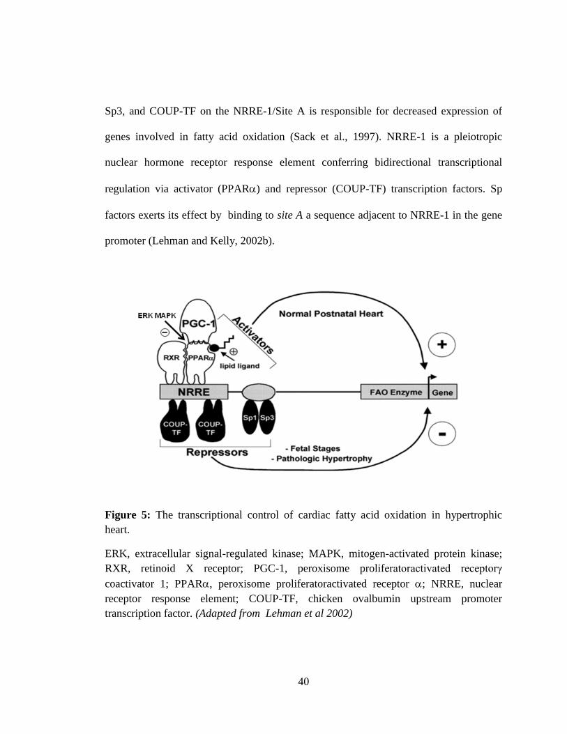
40
Sp3, and COUP-TF on the NRRE-1/Site A is responsible for decreased expression of
genes involved in fatty acid oxidation (Sack et al., 1997). NRRE-1 is a pleiotropic
nuclear hormone receptor response element conferring bidirectional transcriptional
regulation via activator (PPAR) and repressor (COUP-TF) transcription factors. Sp
factors exerts its effect by binding to site A a sequence adjacent to NRRE-1 in the gene
promoter (Lehman and Kelly, 2002b).
Figure 5: The transcriptional control of cardiac fatty acid oxidation in hypertrophic
heart.
ERK, extracellular signal-regulated kinase; MAPK, mitogen-activated protein kinase;
RXR, retinoid X receptor; PGC-1, peroxisome proliferatoractivated receptorγ
coactivator 1; PPAR, peroxisome proliferatoractivated receptor ; NRRE, nuclear
receptor response element; COUP-TF, chicken ovalbumin upstream promoter
transcription factor. (Adapted from Lehman et al 2002)
41
II.3.2. Mechanism of increased glucose utilization in cardiac hypertrophy
Cardiac glucose uptake is mediated by the glucose transporters GLUT-1 and GLUT-4.
The increased utilization in cardiac hypertrophy is mediated through insulin independent
glucose transporter GLUT-1, whereas insulin dependent glucose uptake through GLUT-
4 is diminished (Ritchie and Delbridge, 2006). The enhanced myocardial basal glucose
uptake associated with an elevation of the GLUT-1/GLUT-4 transporter ratio was
observed in the hypertophic heart of SHR and was accompanied with diminished insulin
stimulated glucose uptake (Paternostro et al., 1995). GLUT4-/- mice developed cardiac
hypertrophy with reduced fatty acid oxidation (Abel et al., 1999; Stenbit et al., 2000).
It is proposed that the increased glycolytic flux during cardiac hypertrophy is mediated
by the activation of an intracellular energy sensor Adenosine Mononucleotide Phosphate
-activated protein kinase (AMPK) (Nascimben et al., 2004). AMPK, a metabolic sensor
acts as a metabolic master switch under pathophysiological conditions. As the oxidation
of long chain fatty acids is efficient in terms of ATP produced per molecule oxidized
compared to glucose, prolonged low rate of fatty acid oxidation results in chronic
depletion of energy reserve. AMPK acts as an energy sensor and, when the cytosolic
AMP/ATP concentration increases, AMPK stimulates metabolic pathways that conserve
ATP and inhibits pathways that consume ATP (Hardie et al., 1999; Tian et al., 2001).
However, Allard et al demonstrated that the activation of AMPK during pressure
overload is independent of the energy status of the heart. AMPK was activated in
42
hypertophic heart even in the absence of measurable change in energy level. (Allard et
al., 2007).
AMPK was found to activate cardiac phosphofructokinase-2 (PFK-2) by
phosphorylation, leading to augmented synthesis of fructose-2,6-bisphosphate (F-2,6-
P2) from fructose-6-phosphte, an allosteric activator of the phosphofructokinase (PFK),
the rate-limiting glycolytic enzyme (Marsin et al., 2000). Similarly, enhanced AMPK
activity stimulates the translocation of the glucose transporters (GLUT) onto the plasma
membrane and enhances glucose uptake (fig.6). Increased AMPK activity has been
reported in rat model of pressure overload left ventricular hypertrophy (LVH) in
response to chronic change in myocardial energetics manifested by increased insulin
independent glucose uptake (Tian et al., 2001). In addition to regulation of myocardial
glucose uptake, AMPK also regulates fatty acid metabolism. Pharmacological activation
of AMPK increased fatty acid oxidation in rat skeletal muscle, mediated by inhibition of
acetylCoA carboxylase (ACC), subsequent to malonyl-CoA mediated inhibition of
CPT-I. (Merrill et al., 1997). In contrast, decreased fatty acid oxidation has been shown
in parallel with increased AMPK activity in hypertrophied hearts under chronic pressure
overload (Allard et al., 1994).
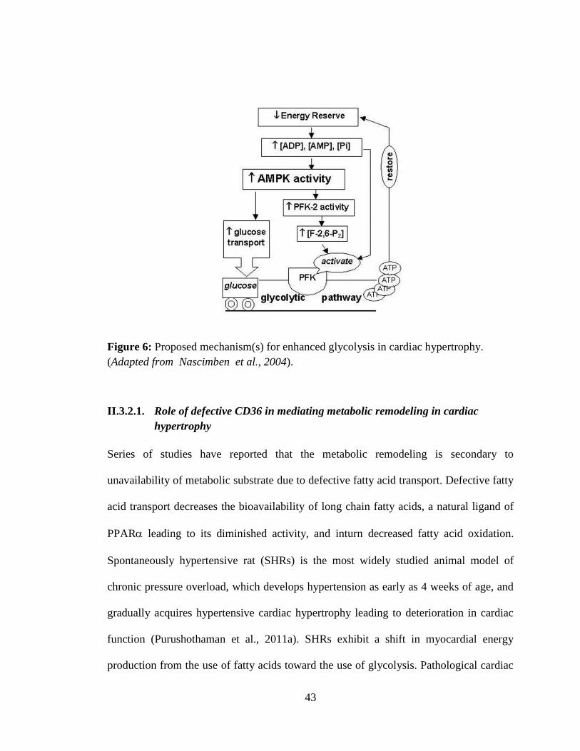
43
Figure 6: Proposed mechanism(s) for enhanced glycolysis in cardiac hypertrophy.
(Adapted from Nascimben et al., 2004).
II.3.2.1. Role of defective CD36 in mediating metabolic remodeling in cardiac
hypertrophy
Series of studies have reported that the metabolic remodeling is secondary to
unavailability of metabolic substrate due to defective fatty acid transport. Defective fatty
acid transport decreases the bioavailability of long chain fatty acids, a natural ligand of
PPAR leading to its diminished activity, and inturn decreased fatty acid oxidation.
Spontaneously hypertensive rat (SHRs) is the most widely studied animal model of
chronic pressure overload, which develops hypertension as early as 4 weeks of age, and
gradually acquires hypertensive cardiac hypertrophy leading to deterioration in cardiac
function (Purushothaman et al., 2011a). SHRs exhibit a shift in myocardial energy
production from the use of fatty acids toward the use of glycolysis. Pathological cardiac

44
hypertrophy in SHRs also results in an abnormal regulation of proteins and transcription
of the key enzymes and transporters involved in myocardial energy metabolism (Coburn
et al., 2000). Congenic mapping and microarray screening in SHR identified CD36 on
rat chromosome 4 as a defective gene, resulting from recombination between wild type
gene and Pseudo gene affecting post translational modification and membrane targeting
of the protein ( Aitman et al., 1999; Lauzier et al., 2011). Reduced expression of CD36
in SHR is associated with reduced long chain fatty acid uptake. Expression of wild type
CD36 in SHR improved defective fatty acid oxidation without affecting hypertension
(Pravenec et al., 2001). CD36 is a highly glycosylated integral membrane protein, that
plays an important role as a LCFA-binding/transport protein that shuttles 40-80% of
fatty acids from the extracellular compartment into the cytoplasm (Brinkmann et al.,
2002). SHRs are characterized by a restricted utilization of exogenous LCFAs for energy
production, which has been attributed to a genetic defect in CD36. In addition, the
cytoplasmic content of fatty acid binding protein (FABP) is found to be decreased in
SHR making SHR an ideal model of defective transporter mediated metabolic
remodeling (Vork et al., 1992).
Clinical studies have reported the effect of defective fatty acid transport system on
cardiac structure and function. Tanaka et al demonstrated the prevalence of association
between CD36 deficiency and defective fatty acid uptake in patients with hypertrophic
cardiomyopathy (HCM) (Tanaka et al., 1997). High prevalence of FAT/CD36
(478T→478C) point mutation has been reported in Japanese population resulting in

45
FAT/CD36 deficiency. The point mutation results in the replacement of Proline (90Pro)
with Serine (Ser) in CD36 resulting in post translational failure, with consequent
shortening of the half life of the protein (Kashiwagi et al., 1995). Patients, homozygous
for the 478T→478C substitution developed hypertrophic cardiomyopathy with reduced
uptake of long chain fatty acid ( Tanaka et al., 1997).This finding was the first clinical
evidence of a possible causal association between defective fatty acid transporter and
development of cardiomyopathy. Another study conducted in a larger patient population
showed that 40% of the hypertrophic cardiomyopathic patients had CD36 mutation
(Okamoto et al., 1998). These observations were challenged by Nakamura et al, who
could not show any significant difference in cardiac phenotype between HCM patients
with normal CD36 expression and those showing CD36 deficiency (Nakamura et al.,
1999). Variable etiology can account for the discrepancy in the observations. Deficiency
of CD36 has been reported recently by Magida et al in mouse models with familial
hypertrophic cardiomyopathy (Magida and Leinwand, 2014). Defective CD36 did not
affect the normal survival of the animals under basal conditions but showed reduced
ischemic tolerance, increased susceptibility to arrhythmia and impaired response to
adrenergic stimulation (Irie et al., 2003; Labarthe, 2004).
In addition to FAT/CD36 deficiency, direct interventions in cardiac fatty acid handling
eventually entails myocardial hypertrophy. Cardiac hypertrophy in animal models
showed decline in myocardial carnitine content (El Alaoui-Talibi et al., 1997).
Pharmcological inhibition of carnitine acyl transferase-I (CPT-I) and Fatty acid

46
translocase (FAT/CD36) was associated with increased heart weight in rat compared to
untreated control (Koundakjian et al., 1984; Kusaka et al., 1995). Similarly, cardiac
specific fatty acid binding protein (H-FABP) knock-out mice developed exercise
intolerance and cardiac hypertrophy at older age (Binas et al., 1999). The development
of cardiac hypertrophy in juvenile visceral steatosis mice was alleviated by
administration of carnitine (Horiuchi et al., 1993; Jalil et al., 2006). These observations
justify that reduced substrate availability due to defective fatty acid transporter CD36
can be a leading cause for the development of cardiac hypertrophy independent of
hypertension.
II.4. MODULATION OF ENERGY METABOLISM IN CARDIAC
HYPERTROPHY
Pathologic cardiac hypertrophy is associated with the reappearance of the fetal metabolic
profile characterized by decreased fatty acid oxidation and increased relative
contribution of glucose as energy source (Barger et al., 2000). As cardiac hypertrophy is
an important predictor for heart failure, the prolonged substrate preference for glucose is
closely associated with impairment of cardiac energy balance and loss of contractile
reserve (Neubauer, 2007). Cardiac interventions enhancing oxidation of carbohydrate
over fatty acid were shown to enhance the function of the ischemic reperfused heart or
the failing heart. The interventional studies in animals were carried out using drugs,
which enhances glucose oxidation and inhibits fatty acid oxidation such as ranolazine,
47
oxfenicine, trimetazidine. However, it remains unanswered, how long this reduced fatty
acid oxidation would be tolerated by the diseased heart, as it is observed that humans
with genetic defects in fatty acid oxidation develop cardiomyopathies. Nevertheless,
chronic decrease in fatty acid oxidation is associated with detrimental cardiac
consequences such as intracellular lipid accumulation and energy deficit. Energy
production from diminished fatty acid oxidation is not likely to be fully compensated by
enhanced glucose use, under chronic workload. The diminished fatty acid oxidation
leads to progressive deterioration of cardiac function during hemodynamic overload.
Cardiac specific PPAR null mouse (PPAR-/-) hearts with permissive increases in
glucose oxidation was not able to maintain the myocardial energetics during high
workload (Luptak et al., 2005). Cardiac specific overexpression of the insulin-
independent glucose transporter (glucose transporter 1 [GLUT1]) to recapitulate
metabolic profile of hypertrophic heart, has shown increased glucose oxidation, with
reciprocal decreases in cardiac fatty acid oxidation (Liao et al., 2002). These mice were
protected against contractile dysfunction and left ventricular dilation when subjected to
pressure overload (ascending aortic constriction) with maintenance of myocardial
energetics, demonstrating that the increased dependence on glucose per se is not
detrimental to the heart under pressure overload. Similarly, transgenic mice with
overexpression of liver X receptors (LXRs) was protected against cardiac remodeling
induced by chronic pressure overload by stimulation of glucose oxidation (Cannon et
al., 2015). These genetic modification approaches of metabolic modulations are of less
clinical relevance because of its limited clinical applicability.
48
Partial inhibition of fatty acid oxidation by pharmacological inhibition of Carnitine
palmitoyl transferase-I (CPT-I), a mitochondrial long chain fatty acid oxidation enzyme
prevented adverse cardiac remodeling and progression of heart failure in rat model of
pressure overload and in canine model of pacing induced heart failure (Lionetti et al.,
2005; Rupp and Vetter, 2000). CPT-I inhibition prevented transcriptional
downregulation of cardiac energy metablism enzymes and sarcoplasmic reticulum
calcium uptake. However, prolonged CPT-I inhibition resulted in the aggravation of
cardiac hypertrophy and development of cardiac lipotoxicity (Haynie et al., 2014; He et
al., 2012). As the cardiac glucose oxidation is highly insulin dependent, impaired insulin
signaling in combination with diminished fatty acid oxidation can result in severe
limitations of substrate oxidation. These reports demonstate that the inherent cardiac
capacity to increase glucose utilization is insufficient to sustain normal energy supply
under stress when fatty acid oxidation is severely impaired (Allard et al., 1994). It has
been reported that hypertrophic and failing heart are less responsive to insulin and
insulin resistant, limit the utilization of glucose as the sole energy source. (Ashrafian et
al., 2007; Paternostro et al., 1999, 1995; Witteles et al., 2004). However, administration
of Glucagon like peptide-1(GLP-I) improved left ventricular performance in pacing
induced dilated cardiomyopathy by increase in glucose uptake and oxidation,
accompanied by stimulation of insulin secretion and insulin sensitivity (Nikolaidis et al.,
2004).

49
Taken together, it is clear that enhancing glucose oxidation in cardiac hypertrophy
improves cardiac function and energetics in short term experiments and are less
promising for chronic use. (Kolwicz et al., 2013). Supporting this perception, several
animal studies reported that sustenance of fatty acid metabolism in hypertrophic heart
preserves cardiac function and energetics (Chess et al., 2009; Duda et al., 2008; Kolwicz
et al., 2012).
Reactivation of fatty acid oxidation in hypertrophied hearts is expected to restore a more
'normal' metabolic physiology, thereby maintaining cardiac function. Modulating cardiac
metabolism is therefore presumed as a therapeutic avenue for the treatment of heart
disease. Modulation of fatty acid metabolism by pharmacologic intervention in cardiac
hypertrophy has presented variable effects on the heart consequent to pharmacological
stimulation of fatty acid metabolism. Fenofibrate attenuated left ventricular
hypertrophy, fibrosis and improved diastolic dysfunction in young SHR and surgical
models of cardiac hypertrophy (Chen et al., 2007; Li et al., 2009; Ogata et al., 2002a;
Zou et al., 2013). Chronic activation of PPAR with fenofibrate did not influence Left
ventricular dysfunction and dilation despite upregulation of fatty acid oxidation and
markers of LV hypertrophy in infarct model of heart failure. (Morgan et al., 2006)
However, Briqadeau et al. reported that fenofibrate reduced cardiac hypertrophy and
slowed down the progression to heart failure on stimulation of fatty acid oxidation
enzymes in pacing induced porcine model of heart failure (Brigadeau et al., 2007).
Fenofibrate not only attenuated the development of hypertrophy and ventricular
50
dysfunction, but also prevented myocardial inflammation (Ichihara et al., 2006). In
aldosterone induced hypertension, fenofibrate improved LV hypertrophy and fibrosis
independent of blood pressure (Lebrasseur et al., 2007). Fenofibrate also improved
cardiac remodeling and function in rat model of severe volume overload (Dhahri et al.,
2013). Pharmacological reactivation of PPAR-α with fibrates have shown regression of
cardiac hypertrophy and fibrosis along with increased life expectancy in SHR (Chen et
al., 2007; Linz et al., 2009). Concurrently, severe depression of cardiac power and
efficiency in the hypertrophied heart has also been reported (Young et al., 2001). Age
associated variation in cardiac response on short term treatment with fenofibrate, with
positive effect in young animals and negative response in older animals was also
reported (Purushothaman et al., 2011b). Recently, we have observed ligand specific
cardiac response to stimulation of fatty acid metabolism in spontaneously hypertensive
rat on administration of fenofibrate and medium chain triglyceride (Ismael et al., 2015).
A series of studies in the past decade have suggested that high fat diet attenuates adverse
cardiac remodeling. Dietary guidelines for cardiovascular diseases recommend high
carbohydrate/low fat diet. However, Chess et al have reported that high dietary fat can
activate PPAR-α and mitochondrial oxidative metabolism without inducing adverse
cardiac remodeling in mice with transverse aortic constriction (Chess et al., 2008, 2009).
High fat diet in 10 week old Dahl salt sensitive rat resulted in the attenuation of cardiac
hypertrophy and improved contractile performance of the heart, inspite of increased
myocardial triglyceride content (Okere et al., 2005). Similarly, consumption of a low
51
carbohydrate/high fat diet attenuated pressure overload induced LVH, remodeling and
contractile dysfunction compared to a high carbohydrate/low fat diet in rats that
underwent aortic banding (Duda et al., 2008). The altered gene expression such as
increased atrial natriuretic factor mRNA, myosin heavy chain isoform switch, and
decreased activity of citrate synthase and medium chain acyl-coenzyme A
dehydrogenase associated with hypertrophy were attenuated by high-fat feeding in Dahl
salt sensitive rat (Okere et al., 2006). In 5-6 week old SHR fed a low carbohydrate/ high
fat diet, Bosse et al observed that the degree of cardiac hypertrophy was not affected
despite reduction in blood pressure (Bosse et al., 2013). Adverse effects of high fat diet
are also reported. Consumption of fat based diet is associated with increased
myocardial triglyceride content, plasma fatty acids and development of obesity in
rodents (Chess et al., 2009; Okere et al., 2005). In mice that underwent transverse aortic
constriction, high fat diet worsened cardiac hypertrophy and dysfunction (Raher et al.,
2008). Similarly, chronic high fat diet induced myocardial hypertrophy and fibrosis in 7-
week old CD1 mice by inhibition of Glycogen synthase kinase-3 (Wang et al., 2015).
Concomitantly, in 9-11 week old Dahl salt sensitive rat, diet based on simple sugars such
as fructose and sucrose is shown to be associated with increased left ventricular
dysfunction and mortality (Sharma et al., 2008, 2007). Conventionally, saturated fats are
not recommend as its intake is presumed to increase the risk of cardiovascular diseases.
The contradictory observations from different approaches for stimulation of fatty acid
metabolism raises the question whether reactivation of fatty acid metabolism is

52
detrimental during pathological cardiac hypertrophy. Kolwicz et al carried out an
interesting experiment in mice with cardiac specific deletion of Acetyl-CoA
carboxylase-2, as the latter is known to inhibit entry of long chain fatty acids into
mitochondria by inhibition of malonyl coA production. In transgenic mice exposed to 8
weeks of pressure overload by transverse aortic constriction, hearts with elevated rate of
fatty acid oxidation prevented myocardial hypertrophy and fibrosis (Kolwicz et al.,
2012), suggesting that maintenance of inherent cardiac metabolic profile is beneficial
under pathological condition.
II.4.1. Medium chain triglycerides, a metabolic substrate to bypass defective CD36
Medium chain triglycerides are esters formed from glycerol and saturated
medium-chain fatty acids (MCFAs) with a chain length between 8-12 carbons. Coconut
and palm oils are the dietary sources with the highest concentration of MCTs. Unlike
long chain fatty acids, their structural and physiochemical properties confer unique
metabolic characteristics (Odle, 1997). Their uptake is independent of cellular and
subcellular fatty acid transport systems such as CD36 and CPT-1 making them direct
fuel for -oxidation (Labarthe et al., 2008). Naturally occurring medium chain fatty
acids in oils and foods are caproic acid (hexanoic acid, C6:0), caprylic acid (octanoic
acid, C8:0) and capric acid (decanoic acid, C10:0). The proportion of MCFAs is about
15%, 7.9%, 6.8%, 6.9%, 6.6% and 7.3% of total fatty acid in coconut oil, palm kernel
oil, butter, milk, yogurt and cheese, respectively (Bach and Babayan, 1982; Nagao and
Yanagita, 2010). They are isolated by lipid fractionation of edible fat and are
53
commercial formulations are mainly comprised of C8:0 and C10:0. They form a minor
constituent of classical diet. The use of MCTs in food products is recognized as safe
(GRAS status) by US Food and Drug Administration since 1994 (Traul et al., 2000).
In contrast to long chain triglycerides (LCT), MCTs are hydrosoluble and are rapidly
hydrolysed to MCFAs that are absorbed into the portal vein and are rapidly metabolized
by the liver. In the liver, MCFAs are directed towards oxidation rather than
incorporation into lipids, showing its high propensity for oxidation. Excess acetyl-CoA
formed by β-oxidation leads to ketone body synthesis. Acetoacetate and β-
hydroxybutyrate, are the quantitatively most important MCFA-derived ketone bodies.
Odd-carbon MCFAs also lead to the formation of the C-5 ketone body analogs β-
ketopentanoate and β-hydroxypentanoate from propionyl-CoA metabolism
MCT were first introduced in the 1950s for the treatment of lipid absorption disorders
(Bach and Babayan, 1982). Over the past decades, MCTs have also been used for the
nutritional management of patients with inherited long chain fatty acid β-oxidation
disorders. MCFAs, which bypass the enzyme defect and thereby restore energy
production, is shown to improve clinical symptoms, particularly cardiac hypertrophy and
dysfunction, in most patients associated with very long chain acyl-CoA dehydrogenase
(VLCAD) deficiency (Brown-Harrison et al., 1996; Cox et al., 1998; Duran et al., 1991;
Touma et al., 2001).
54
MCT is available in two different forms, odd and even chain MCT based on number of
carbons atom (Fig.7). Odd chain MCT is reported to be more anaplerotic than even
chain MCT. Anaplerosis is the re-filling of the catalytic intermediates of the Citric acid
cycle. MCTs with an odd number of carbons were reported to be superior to even-carbon
MCTs in improving the cardiomyopathy of one patient with genetic LCFA oxidation
deficiency (Roe et al., 2002). The -oxidation of one molecule of heptanoate (7 Carbon
MCT) yields two acetyl-CoA and one propionyl-CoA molecules, while that of octanoate
yields four acetyl-CoA units. The Propionyl-CoA, three carbon compound formed from
heptanoate is further metabolized to succinyl-CoA, which is an anaplerotic reaction that
feeds the pool of Citric acid cycle intermediates. Dietary regimen substituting
tricaprylin (even chain MCT) with triheptanoin (odd chain MCT) improved
cardiomyopathy in few patients with very long-chain acyl-CoA dehydrogenase
deficiency. Triheptanoin intake enhanced ketone body synthesis without inducing
propionyl-CoA overload (Roe et al., 2002). Recently, Nguyen et al demonstrated that
odd chain MCT triheptanoin feeding improved diastolic function and cardiac
hypertrophy in rodent model of pressure overload (Nguyen et al., 2015).
Several studies reported the beneficial effect of MCT on heart. 14 years of prospective
study revealed that intake of short and medium chain fatty acids are not associated with
the development of coronary heart disease (Hu et al., 1999). The impaired capacity of
SHR hearts to withstand an acute adrenergic stress could be improved by increasing the
contribution of energy production by supplementation with 0.2 mM octanoate (a eight
55
carbon triglyceride) (Labarthe et al., 2005). Octanoate reduced cardiac tissue damage in
response to acute adrenergic stimulation represented by decreased LDH release and
improved cardiac function. Inclusion of octonate in the perfusate normalized accelerated
glycolysis, improved energy status and the contractile function in the hypertrophic
working heart induced by aortic contriction (Allard et al., 2007). Similarly, addition of
caprylic acid (8C) to the perfusate improved ischemic recovery of CD36 knock out
working hearts, in comparison with long chain fatty acid palmitate (Irie et al., 2003).
Additionally, supplementation of octanate enhanced contractile function of volume
overload heart (Alaoui-Talibi et al., 1992). Supplementation of MCT prior to exercise
training improved oxidative metabolism and decreased cardiac workload in patients
with long-chain fatty acid oxidation defect (Behrend et al., 2012). Dietary administration
of MCT improved LV dysfunction in mice with diabetic cardiomyopathy (Finck et al.,
2003).
There are few studies reporting beneficial effects of MCT-enriched diet in SHR.
Reactivation of fatty acid metabolism by supplementation of medium chain triglycerides
(MCT) was found to prevent cardiac hypertrophy and improve cardiac function in
young SHR with persistent hypertension (Hajri et al., 2001; Iemitsu et al., 2008; Rupp et
al., 1995; Shimojo et al., 2004). However, data concerning the potential of MCT to
promote reverse remodeling in older animals and the effect of supplementation of MCT
on cardiac oxidative stress and energy status are not available.

56
III. METHODOLOGY
57
III.1. DESIGN OF THE STUDY
Oxidation of long-chain fatty acids is the major energy source for the healthy
heart. Pathological cardiac hypertrophy is accompanied by reactivation of fetal cardiac
metabolism, decreased oxidation of long chain fatty acids and increased relative
contribution of glucose for the energy production. Prolonged low rate of fatty acid
oxidation is associated with adverse cardiovascular consequences including increased
oxidative stress. This metabolic preference is mediated by down regulation of the
expression of genes involved in fatty acid metabolism. Studies have shown that the
reduced fatty acid oxidation in patients with hypertrophic cardiomyopathy, and also in
Spontaneously hypertensive rat (SHR) are due to reduced expression of the fatty acid
transporter, CD36 (Cluster of differentiation-36) . Hence, it is hypothesized that
supplementation of Medium chain triglyceride that by passes the defective fatty acid
transport can maintain myocardial metabolism to prevent adverse cardiac remodeling.
Medium chain triglycerides are triglycerides of saturated fatty acids of chain length 8-12
carbon atoms, and their oxidation is independent of cytoplasmic and mitochondrial fatty
acid transport systems such as CD36 and Carnitine palmitoyl transferase-1 (CPT-1).
Hence, they are regarded as direct fuel for β-oxidation. Several studies have reported the
beneficial effect of MCT on heart. MCT is known to improve contractile function of the
hypertrophic heart. Consumption of MCT is not associated with the development of
coronary artery diseases. MCT is available in two different forms, odd and even chain
58
triglycerides, based on number of carbons. Odd chain MCT is reported to be anaplerotic
in nature. Anaplerosis is the re-filling of the catalytic intermediates of the Citric acid
cycle. Therefore, there is possibility of difference in their metabolic properties.
The study is aimed at regression of cardiac hypertrophy for prevention of progressive
cardiac remodeling. Experimental studies have shown that older animals are resistant to
treatment. There are no reports on the use of MCT for regression of cardiac hypertrophy
in chronic pressure overload associated with CD36 deficiency. Spontaneously
hypertensive rat, an animal model of pressure overload was used as the experimental
model. Cardiac remodeling in SHR follows the same pattern as the clinical course of
hypertension induced cardiac hypertrophy and failure.
The study was designed with following objectives
i. Validate the suitability of the experimental model by screening for CD36
deficiency and markers of cardiac hypertrophy, metabolic shift and oxidative
stress, by comparison of Spontaneously hypertensive rat (SHR) with
normotensive Wistar rat (W)
ii. Compare the cardiac response to stimulation of fatty acid oxidation by
supplementation with odd and even chain MCT
59
iii. Asses cardiac response to stimulation of fatty acid oxidation by supplementation
of medium chain triglycerides at initial and established stages of cardiac
hypertrophy.
(i) Prior to experimental studies, suitability of SHR as the experimental model for
pathological cardiac hypertrophy was validated. Morphological, histological and
molecular markers of cardiac hypertrophy, metabolic shift and oxidative stress of 6
month old SHR were compared with age and sex matched normotensive Wistar rat (W).
Wistar-Kyoto rats were avoided as they have a tendency to develop cardiac hypertrophy.
Blood pressure of the animals was monitored by tail cuff spygmomanometer. The
expression of CD36 in Wistar and SHR was assessed by Western blotting. Cardiac index
[heart weight/ body weight(mg/g)], myocyte cross sectional area, mRNA expression of
Brain natriuretic peptide (BNP), calcineurin A expression were used as the markers of
cardiac hypertrophy.
Cardiac metabolism in SHR was evaluated from mRNA expression of Peroxisome
prolifrator-activated receptor- (PPAR ) and one of its target genes Medium chain
acylCoA dehydrogenase (MCAD), and Phosphofructokinase-1(PFK-1), a glycolytic
enzyme by Real time PCR analysis. Myocardial fibrosis was evaluated from histological
analysis of myocardial sections with Picrosirius red staining; and interstitial and
perivascular fibrosis was quantified.
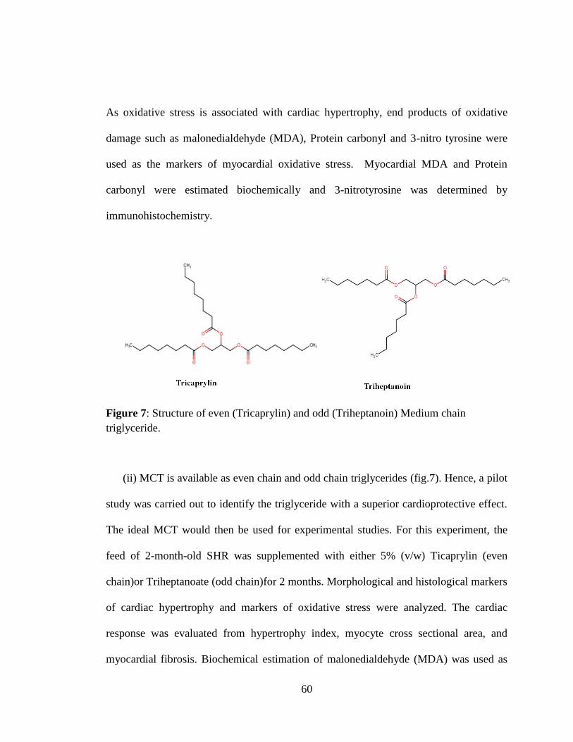
60
As oxidative stress is associated with cardiac hypertrophy, end products of oxidative
damage such as malonedialdehyde (MDA), Protein carbonyl and 3-nitro tyrosine were
used as the markers of myocardial oxidative stress. Myocardial MDA and Protein
carbonyl were estimated biochemically and 3-nitrotyrosine was determined by
immunohistochemistry.
Figure 7: Structure of even (Tricaprylin) and odd (Triheptanoin) Medium chain
triglyceride.
(ii) MCT is available as even chain and odd chain triglycerides (fig.7). Hence, a pilot
study was carried out to identify the triglyceride with a superior cardioprotective effect.
The ideal MCT would then be used for experimental studies. For this experiment, the
feed of 2-month-old SHR was supplemented with either 5% (v/w) Ticaprylin (even
chain)or Triheptanoate (odd chain)for 2 months. Morphological and histological markers
of cardiac hypertrophy and markers of oxidative stress were analyzed. The cardiac
response was evaluated from hypertrophy index, myocyte cross sectional area, and
myocardial fibrosis. Biochemical estimation of malonedialdehyde (MDA) was used as
61
the marker of oxidative stress. Since fat based diet was used for experimental purpose,
serum lipid profile was determined by commercially available kit (Agappe Diagnostics,
India).
(iii) Cardiac response to stimulation of fatty acid metabolism by supplementation of
medium chain triglycerides was assessed at initial and established stages of cardiac
hypertrophy. Young, 2-months-old and older 6-months-old SHR were supplemented
with Medium chain triglycerides (MCT) for 4 months, with the objective of preventing
cardiac remodeling in the former, and promoting reverse remodeling in the latter group.
Stimulation of cardiac fatty acid metabolism was substantiated from the expression of
peroxisome proliferator-activated receptor alpha (PPAR-α) and medium chain acyl CoA
dehydrogenase (MCAD -a key enzyme in β-oxidation of fatty acids) by real time RT-
PCR analysis. Consequence of reactivation of fatty acid metabolism on glucose
metabolism was evaluated from the mRNA expression of phosphofructokinase-1 (PFK-
1), marker enzyme for glycolysis. Cardiac response to metabolic stimulation was
evaluated based on hypertrophy index, cell size, morphometric analysis of ventricular
collagen (interstitial and perivascular), expression of brain natriuretic peptide (BNP) and
calcineurin A. Effect of MCT on oxidative stress was assessed, as the latter is associated
with pathological hypertrophy. Myocardial malondialdehyde (MDA- a stable terminal
metabolite of lipid peroxidation), 3-nitrotyrosine and protein carbonyl (end product of
protein oxidation) were used as markers of oxidative stress. The energy level was

62
evaluated from biochemical estimation of ATP and phosphocreatine. Blood pressure and
serum lipid profile were also determined.
The study was approved by The Institutional Animal Ethics Committee. The housing
care and the management of these animals were in accordance with Committee for the
Purpose of Control and Supervision of Experiments in Animals (CPCSEA) Guidelines.
III.2. MATERIALS
III.2.1. Fine chemicals- The source is given in italics
Bovine Serum Albumin (BSA), Disodium hydrogen phosphate, Potassium chloride,
Potassium dihydrogen phosphate, EDTA, glucose, Diethyl pyrocarbonate (DEPC),
Protease inhibitor cocktail, Acrylamide, bisacrylamide, -mercaptoethanol, TEMED,
Ammonium persulphate, Color burst electrophoresis marker, Direct red, , Sodium
dodecyl sulphate (SDS), Trizma base, Agarose, Glycine, Thiobarbituric acid, Tricloro
acetic acid (TCA), Tris–HCl, Perchloric acid, Pottassium bicarbonate, Methyl orange,
Triethanolamine, NADP, Ketamine and Xylazine, Glucose-6-phosphate dehydrogenase,
Hexokinase, Creatine kinase, 2,4 dinitrophenyl hydrazine (DNPH), digitonin, ethyl
acetate and streptomycin sulphate. (Sigma-Aldrich), Nitrocellulose membrane (Millipore
USA). Odd and Even Chain MCT for initial experiments were gifted from Jayant agro-
organics limited, Mumbai, India. MCT (Tricaprylin) for major experiments were
purchased from Sigma Aldrich.
63
Antibodies: Monoclonal anti-calcineurin antibody (Thermo), Anti CD36 antibody
(Abcam), monoclonal anti-beta actin antibody (Sigma), Trizol reagent, anti-mouse IgM
antibody and monoclonal anti rabbit antibody (Sigma-Aldrich), Polyclonal anti
dystrophin antibody (Santa cruz Biotechnology),
Kits: Coomassie protein assay reagent, DNase –I kit (amplication grade- Sigma), Super
signal West Femto Substrate kit (Thermo scientific USA). The fine chemicals for cDNA
synthesis including RT buffer, RNase inhibitor, oligo dT primers, dNTPs and M-MLV
Reverse Transcriptase were purchased from Promega. Power SYBR Green for real time
PCR was purchased from Applied Biosystems,
Total cholesterol, Triglycerides, HDL cholesterol and LDL Cholesterol estimation kits
were purchased from Agappe Diagnostics India.
III.2.2. Routine Chemicals
Sodium chloride, Phenol red, chloroform, Propanol, ethanol, Methanol Hydrochloric
acid, glycerol, formalin, Isopropanol, Xylene, Formalin,. Routine chemicals were
purchased from Sisco Research Laboratories, Nice chemicals, Merck, India.
III.2.3. Instruments used
Eppendorf centrifuge 5415 R, Incubators (Beston India; Kemi, India), Weighing balance
(Sartorius, USA and Ohaus), Homogenizer (IKA, Labortechnik, Germany), Deepfreezer

64
-20oC (Vestfrost), Deepfreezer -80oC (Sanyo), Steam distillation unit (Beston),
Microwave oven (IFB) Water bath (LKB, Sweden), Ice machine (Hoshizaki, Japan), pH
meter (Labindia), Low speed magnetic stirrer (Remi, India), Hot air oven (Tempo,
India), EASY pure UV/UF compact reagent grade water system (Barnstead, USA),
ELISA reader (Bio-Tek instruments, USA), UV- visible Spectrophotometer (Shimadzu),
Electrophoresis unit (Biorad laboratories, USA), Mini Blot (Biorad laboratories, USA),
Programmable Thermal Cycler (MJ Research Inc, USA), Submarine electrophoresis unit
(Bangalore Genei, India), UV-Transilluminator (Bangalore Genei, India). Small Animal
Noninvasive Blood Pressure System (NIBP200A) (Biopac Systems, USA), BIOPAC
Data Acquisition Unit (MP35/MP30), Applied Biosystem 7500 real time PCR system
(Applied Biosystem).
III.2.4. Software used
Image-Pro Plus 5.1 for image analysis (Media Cybernetics)
BSL PRO for NIBP (Biopac)
ImageJ (NIH)
65
III.3. COMPOSITION OF REAGENTS AND BUFFERS
III.3.1. Phosphate buffered saline (PBS) –pH 7.4
NaCl 137mM,
KCl 2.7mM,
KH2PO4 1.76mM,
Na2HPO4 10.14mM.
III.3.2. DEPC-treated deionized water
1 ml of DEPC in one litre of deionized water, stirred for 30 minutes at room temperature
and autoclaved
III.3.3. 10% Buffered formalin
NaH2PO4 (unhydrous) – 3.5g
Na2HPO4 (unhydrous) – 6.5g
Formalin - 100ml
Distilled Water - 900ml
III.3.4. Harris Hematoxylin
Alum -100g
Hematoxylin - 5g
Ethanol -50ml
Mercuric oxide - 2.5g
Distilled water - 1000ml
66
III.3.5. Eosin Solution
Eosin -1gm
Propanol -100ml
III.3.6. Sirius Red stain
Sirius red (direct red) - 0.1g
Saturated picric acid -100ml
III.3.7. RIPA buffer
Tris HCl 234 mg
NaCl 261 mg
EDTA 11.6 mg
1% Triton X100 3 ml
1% sodium deoxy cholate 3 ml
Made up to 30 ml with distilled water
III.3.8. Electrode buffer (pH 8.3) for SDS–polyacrylamide gel electrophoresis
(SDS- PAGE)
Tris base 25 mM
Glycine 192 mM
SDS 0.1%
Deionized water 100ml
67
III.3.9. SDS gel-loading buffer (6X)
SDS (9% w/v)
Bromophenol blue (0.03%)
β-mercaptoethanol (9%),
Glycerol 50% v/v)
1M Tris HCl- pH 6.8 18.75 ml
III.3.10. Resolving gel buffer (pH 8.8)
Tris base 18.165 g
in 80 ml deionized water. pH adjusted to 8.8 using HCl, made upto 100 ml and stored at
room temperature.
III.3.11. Stacking gel buffer (pH 6.8)
30 ml from resolving buffer was measured, pH adjusted to 6.8 using HCl and made up to
45ml. Stored at room temperature.
III.3.12. 30% Acrylamide solution
29.2 g of acrylamide and 0.8g bis acrylamide dissolved in distilled water and made up
to 100ml. Solution stored in dark at room temperature.
III.3.13. 10% Ammonium per sulphate solution
0.1 g Ammonium Per sulphate (APS) dissolved in 1ml distilled water.
III.3.14. 10% SDS solution
1g of SDS dissolved in 10 ml distilled water
68
III.3.15. Blocking buffer
Skimmed Milk 0.5g
1x TBS- Tween 20 10ml
III.3.16. Towbin’s buffer (Transfer buffer)
Tris base 3.027 g
Glycine 14.4 g
Methanol 200 ml
Deionized water 800 ml
III.3.17. Tris borate EDTA buffer (TBE) (5X, pH 8.3)
Tris base 54 g
boric acid 27.5 g
0.5 M EDTA ( pH 8.0) 20 ml
Deionized water 1000ml
III.3.18. DNA/RNA loading dye
Bromophenol blue 0.25%
Xylene cynol 0.25%
EDTA 1mM
Glycerol 50% in Nuclease free water
III.3.19. Ethidium bromide (Stock solution)
1mg in 1ml water, 5ul of this stock solution added to 20ml of 1% agarose gel for
electrophoresis.
69
III.3.20. Tris-buffered saline (TBS) (10X, pH 7.6)
Tris base 24.2 g
Sodium chloride 80 g
Distilled water 1000ml
III.3.21. Triethanolamine buffer (TEA) (pH 7.6)
Triethanolamine HCl 4.65g
Water 500 ml
pH was adjusted with NaOH
70
III.4. EXPERIMENTAL STUDIES
Spontaneously hypertensive rat (SHR) was used as the in vivo experimental
model of left ventricular hypertrophy (LVH). It simulates the clinical progression of
hypertension induced left ventricular hypertrophy. SHR develops hypertension at 4
weeks of age, left ventricular hypertrophy at 8 weeks followed by metabolic shift.
Spontaneously hypertensive rats were purchased from Animal Resource Centre,
Australia and the Wistar rats were maintained in the Division of Laboratory Animal
Science of the Institute (SCTIMST). The animals were housed at 22±2°C in 55±10%
relative himidity in individually ventilated cages. Light levels measured at 1 meter
height were less than 300 Lux and a 12:12 hour dark: light pattern was maintained. The
rats were fed with standard pelletted feed and drinking water ad libitum.
III.4.1. Comparison of markers of cardiac hypertrophy, metabolic shift and
oxidative stress in Spontaneously hypertensive rat (SHR) with
normotensive wistar rat (W).
Prior to the experimental studies it was decided to confirm that the strain of SHR
available at the Institute was deficient for CD36. The animals were characterized for
markers of cardiac hypertrophy, metabolic shift and oxidative stress in SHR.
Normotensive Wistar rat were used as the control for comparison. Morphological,
histological and molecular features were analyzed in SHR and compared with Wistar rat.
6 months old male SHR were compared with age and sex matched Wistar rat.
71
III.4.1.1. Assessment of markers of cardiac hypertrophy, oxidative stress and
metabolic shift in SHR
The expression of CD36 and indicators of cardiac hypertrophy, oxidative stress
and metabolic shift in SHRs was assessed using morphological, histological,
biochemical and molecular indicators compared with age and sex matched Wistar rat.
Hemodynamic measurement of six month old animals 4 from each strain was recorded.
After measurement of body weight, the animals were euthanized under deep surgical
plane of anesthesia using 5mg/kg Xylazine and 70mg/kg Ketamine followed by 10mg/kg
body weight of 1% Thiopentone sodium as i/p injections. The heart was immediately
dissected out, ventricles separated from the atrium and weighed. Hypertrophy index was
used as the morphological indicator of cardiac hypertrophy and it was compared with
age and sex matched normotensive Wistar rat. Hypertrophy index was determined as the
ratio of total ventricular weight to body weight [Ventricular weight in milligram
(mg)/Body weight in gram (g)]. Protein expression of CD36 was carried out to confirm
CD36 deficiency. Calcineurin A expression and mRNA expression of Brain natriuretic
peptide (BNP) were used as the molecular markers of cardiac hypertrophy. Histological
markers such as myocyte cross sectional area and myocardial fibrosis (interstitial and
perivascular fibrosis) were determined and compared with Wistar rat. To determine
myocyte cross sectional area heart sections were immunostained for dystrophin to obtain
a clear outline of the cell. Dystrophin is a member of complex proteins which anchor
muscular cytoskeleton to the extracellular matrix through plasma membrane. Cross
sectional area of a minimum of 100 muscle fibers were measured from each ventricular
72
section and quantified by ImageJ 1.45s software (National Institute of Health). To
maintain consistency, mid-ventricular region of the heart was excised and fixed in
formalin for histological analysis.
As oxidative stress is an essential factor associated with cardiac hypertrophy, markers of
end products of oxidative damage such as Malonedialdehyde (MDA), protein carbonyl
and 3nitrotyrosine level were determined in SHR and compared with Wistar rat.
Malonedialdehyde and protein carbonyls were biochemically measured from myocardial
tissue and myocardial 3-nitrotyrosine were determined by immunohistochemistry.
Another factor intimately associated with cardiac hypertrophy is a shift in substrate
preference from fatty acids towards glucose. The metabolic status in SHR was evaluated
from the mRNA expression of Peroxisome priliferator-activated receptor (PPAR)-,
Medium chain acyl CoA dehydrogenase, (MCAD) and phosphofructokinase (PFK)-1 by
Real time polymerase chain reaction. mRNA expression was quantified using ΔΔCT
method with - actin as the loading control gene (Livak and Schmittgen, 2001).
73
III.4.2. Comparison of cardiac response to odd and even chain mct
supplementation in SHR
III.4.2.1. Evaluation of the cardiac response to Supplementation of even and odd
chain MCT in SHR
MCT is available in two forms, Even chain MCT and Odd chain MCT.
Therefore, a pilot study was carried out to select the ideal MCT to reactivate fatty acid
metabolism in SHR. Tricaprylin and triheptonoate were used as the sources of even
chain and odd chain MCT respectively. As MCT is available in the liquid form, 50 ml of
MCT was blended with 1000g of standard rat chow (5% MCT with standard feed). To
evaluate the cardiac response, 2 months old male SHR were supplemented with 5%
MCT. Age and sex matched untreated SHR served as the experimental control. SHR
were parallely fed and each group contained 4 animals (n=4). Morphological and
histological markers of cardiac hypertrophy were evaluated following 2 months of
treatment. Hypertrophy index, myocyte cross sectional area and myocardial interstitial
fibrosis were used as the markers for the evaluation of the cardiac hypertrophy.
Myocardial malonedialdehyde , a marker of oxidative stress was also evaluated. To
examine the consequence of fat based diet on lipid profile, serum triglyceride, total
cholesterol and high density lipoprotein (HDL) cholesterol were evaluated. LDL was
expressed as the calculated value.

74
III.4.3. Assessment of cardiac response to stimulation of fatty acid metabolism by
supplementation of medium chain triglycerides at the initial stage and established
stage of cardiac hypertrophy
Two months old SHR served as the model for initial stages of cardiac
hypertrophy. This represents the stage prior to the occurrence of metabolic shift. Six
months old SHR served as the model for the established stage of cardiac hypertrophy.
This phase is the stable phase of cardiac hypertrophy. Age and sex matched untreated
SHR served as the experimental control. Each group contained six animals. The feed of
the SHR were supplemented with 5% MCT (v/w) (tricaprylin) for 4 months with the
assumption that maintenance of fatty acid metabolism can prevent adverse cardiac
remodeling. The animals were fed parallely. On completion of treatment, after
measurement of body weight and hemodynamic evaluation, the animals were euthanized
under deep surgical plane of anesthesia using 5mg/kg Xylazine and 70mg/kg Ketamine
followed by 10mg/kg body weight of 1% Thiopentone sodium as i/p injections. The
heart was immediately dissected out, ventricles separated from the atrium and weighed.
Mid-ventricular region was excised and fixed in formalin for histological analysis. The
ventricles were then rinsed in ice-cold saline, frozen in liquid nitrogen and stored at -
80°C. Morphological, histological and molecular markers of cardiac hypertrophy were
evaluated following 4 months of MCT supplementation. The markers of oxidative stress
and myocardial metabolism were determined.
75
Evaluation of cardiovascular response to MCT supplementation.
III.4.3.1. Metabolic changes on supplementation of MCT in the initial and
established stages of cardiac hypertrophy
Cardiac hypertrophy in SHR is associated with a shift in energy substrate preference
from fatty acid towards glucose. MCT was supplemented based on the assumption that it
can maintain the fatty acid metabolism in SHR. Effect of MCT on cardiac metabolism
was evaluated from the mRNA expression of PPAR-, MCAD and PFK-1 as
determined by Real time polymerase chain reaction. PPAR- and MCAD are markers of
fatty acid metabolism where as PFK-1 is marker for glucose metabolism.
III.4.3.2. Assessment of the effect of MCT supplementation on markers of cardiac
hypertrophy
Hypertrophy is characterized by increased myocardial mass with increased myocyte
cross sectional area and interstitial fibrosis. Effect of metabolic stimulation on markers
of cardiac hypertrophy was evaluated from the morphological, histological and
molecular markers of cardiac hypertrophy. Hypertrophy index was calculated as
previously described (Section III.4.1.1) and used as the morphological marker of cardiac
hypertrophy. Myocyte cross sectional area was determined by immunostaining of
myocardial sections with anti dystrophin antibodies to get the clear outline of the
individual cell membrane. Myocardial fibrosis was assessed by picrosirius red staining
of the myocardial sections and interstitial and perivascular fibrosis was determined.
Protein expression of Calcineurin A and mRNA expression of BNP were used as the

76
molecular markers of adverse cardiac remodeling and the procedure described in III.5.2
and III.5.6 was followed.
III.4.3.3. Consequence of MCT supplementation on markers of oxidative stress
The effect of metabolic stimulation with MCT on oxidative stress was evaluated by
biochemical estimation of cellular end products of oxidative damage such as
molonedialdehyde and protein carbonyl. Myocardial level of 3-nitrotyrosine was also
determined in tissue sections.
III.4.3.4. Effect of MCT supplementation on blood pressure
Effect of metabolic modulation on blood pressure was monitored by noninvasive tail
cuff sphygmomanometer after warming and restraining the animals. Systolic and
diastolic pressure was measured as described in the Section III.5.1
III.4.3.5. Myocardial energy level in response to MCT supplementation
Myocardial energy level was determined by biochemical evaluation of Adenosine
Triphosphate (ATP) and Phosphocreatine(PCr).
III.4.3.6. Effect of MCT supplementation on serum lipid profile
Serum triglyceride, Total cholesterol, HDL were estimated by commercially available
estimation kits (Agappe Diagnonotics. India).

77
III.4.3.7. Hepatic lipotoxicty
As fat based diets can cause hepatic lipid accumulation, liver tissue was histologically
analyzed for any indication of lipotoxicity by hemotoxyline–eosin staining .
III.5. METHODOLOGY
III.5.1. MEASUREMENT OF BLOOD PRESSURE
On completion of the experiments, resting systolic and diastolic arterial pressure of the
animals were measured. Blood pressure was measured using Noninvasive Blood
Pressure Monitoring System for Small Animals (BIOPAC Systems.Inc) (Fig.7). The
measurements were taken with a tail-cuff sphygmomanometer after restraining the
animals.
The tail cuff was placed proximally on the tail to occlude the blood flow. Upon deflation
of the cuff, blood pressure was determined which coincided with the restoration of
caudal artery pulse. This was achieved by using a piezo electric pulse transducer placed
distal to the cuff, which was coupled to the Data Acquisition System. For accurate
noninvasive blood pressure measurement, the tail of the animal was pre-warmed to
32OC.
At least 6 determinations were made in every session of blood pressure measurement
and the average was obtained. The systolic blood pressure value coincided with the
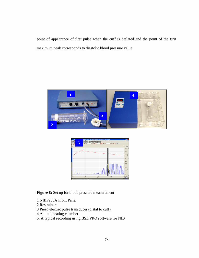
78
point of appearance of first pulse when the cuff is deflated and the point of the first
maximum peak corresponds to diastolic blood pressure value.
Figure 8: Set up for blood pressure measurement
1 NIBP200A Front Panel
2 Restrainer
3 Piezo electric pulse transducer (distal to cuff)
4 Animal heating chamber
5. A typical recording using BSL PRO software for NIB
1
2
3
4
5
79
III.5.2. GENE EXPRESSION ANALYSIS
III.5.2.1. RNA Isolation
The myocardial tissue for RNA isolation was snap frozen in liquid nitrogen and stored at
-800C. Frozen myocardial tissue (100 mg) was thoroughly homogenized in 1ml Trizol
reagent (Sigma) with a homogenizer (Ultra-Turrax). The homogenate was incubated at
room temperature for 10 minutes for the dissociation of nuclear proteins. 0.2 ml of
chloroform/ml of Trizol was added to the homogenate, mixed by vigorous shaking and
centrifuged at 12000 rpm for 10 minutes. The aqueous layer was collected and 0.5 ml of
Isopropanol was added to precipitate the RNA, followed by centrifugation at 12000 rpm
for 15 minutes. The pellet formed was washed with 75% ethanol at 7500 rpm for 8
minutes and air dried. The final pellet was resuspended in 20l nuclease free water. The
RNA was quantified at A260nm using Epoch spectrophometer (Biotek, USA). The purity
of RNA was determined from the A260/280 ratio spectroscopically. The integrity of the
RNA was evaluated by 1% agarose gel electrophoresis. Genomic DNA contamination
of RNA sample was removed by RNase free DNase-1 treatment according to
manufacture’s protocol.
III.5.2.2. cDNA synthesis
Rat-specific primers for the genes were designed by Oscimum Biosolutions (India).
Real Time PCR was performed in a total volume of 20 l, containing 60 ng of the cDNA
derived from reverse transcription using Power SYBR Green technology. The threshold
80
cycle (CT) for fluorescence development was used to calculate the fold changes applying
the ‘‘2-∆∆CT method’’ (Livak and Schmittgen, 2001).
Construction of cDNA- 2 g of each RNA sample was reverse transcribed to cDNA
with MMLV reverse transcriptase using oligo dT primers. Briefly, to the 2 µg RNA ,
2µl oligo dT primer was added and was incubated for 5’ at 700C for removing the
unwanted secondary RNA structure and cooled immediately by placing it in ice for 5-10’
and centrifuged for 10 seconds. The sample was added to the reaction mixture
containing dNTP, RNasin, MMLV-RT, RT buffer and water, and was incubated at 370C
for an hour. The sample was kept at 900C for 5’ to inactivate reverse transcriptase
enzyme.
III.5.2.3. Real Time PCR analysis
Real Time RT PCR was performed using Power SYBR Green technology on Applied
Biosystem 7500 real time PCR system. Concentration of the primers were optimized by
conventional polymerase chain reaction. PCR amplification was performed in a total
volume of 20 µl, containing 60 ng of the cDNA derived from reverse transcription, 200-
500nM of primer, and 10 µl of 2X Power SYBR Green mastermix. Each reaction was
incubated for 2 min at 50°C and 10 min at 95°C and then subjected to 40 cycles, each
involving denaturation at 95°C for 15s and annealing/extension at 60°C for 1 min. In this
way, the threshold cycle (CT) for fluorescence development was obtained. All samples
were run in triplicate. The “2-∆∆CT method” for comparing the relative expression results
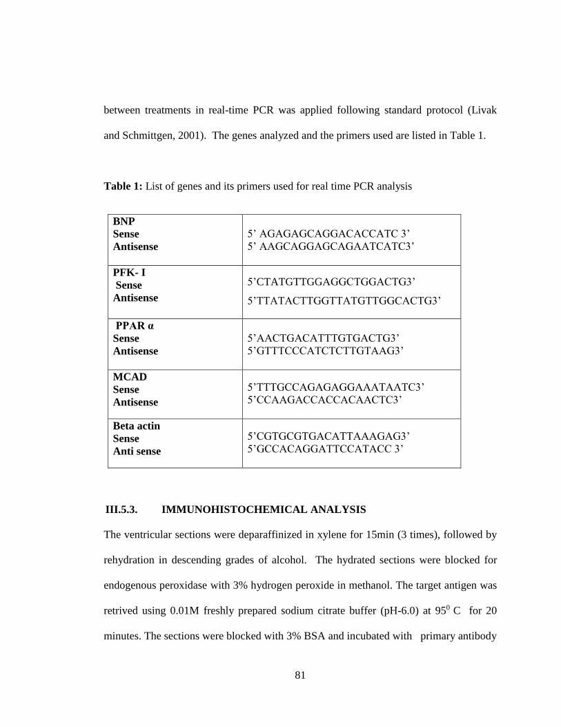
81
between treatments in real-time PCR was applied following standard protocol (Livak
and Schmittgen, 2001). The genes analyzed and the primers used are listed in Table 1.
Table 1: List of genes and its primers used for real time PCR analysis
BNP
Sense
Antisense
5’ AGAGAGCAGGACACCATC 3’
5’ AAGCAGGAGCAGAATCATC3’
PFK- I
Sense
Antisense
5’CTATGTTGGAGGCTGGACTG3’
5’TTATACTTGGTTATGTTGGCACTG3’
PPAR α
Sense
Antisense
5’AACTGACATTTGTGACTG3’
5’GTTTCCCATCTCTTGTAAG3’
MCAD
Sense
Antisense
5’TTTGCCAGAGAGGAAATAATC3’
5’CCAAGACCACCACAACTC3’
Beta actin
Sense
Anti sense
5’CGTGCGTGACATTAAAGAG3’
5’GCCACAGGATTCCATACC 3’
III.5.3. IMMUNOHISTOCHEMICAL ANALYSIS
The ventricular sections were deparaffinized in xylene for 15min (3 times), followed by
rehydration in descending grades of alcohol. The hydrated sections were blocked for
endogenous peroxidase with 3% hydrogen peroxide in methanol. The target antigen was
retrived using 0.01M freshly prepared sodium citrate buffer (pH-6.0) at 950 C for 20
minutes. The sections were blocked with 3% BSA and incubated with primary antibody
82
at 4oC overnight. The dilutions of the primary antibodies were - Dystrophin- 1:50 and
CD36 and 3-nitro tyrosine-1:100. After washing with PBS, the sections were treated
with HRP conjugated secondary antibody (dilution 1:200) for an hour at room
temperature. After washing off unbound secondary antibody, the sections were treated
with the chromogen, diaminobenzidine (DAB) (Sigma)
III.5.4. HAEMATOXYLIN AND EOSIN STAINING
Tissue sections are rehydrated in descending grades of alcohol and brought to the water.
Hydrated sections were stained with hematoxylin for 3 minutes followed by
differentiation in acid alcohol for 30 seconds. Bluing was done in running tap water.
Then the sections were stained with Eosin for one minute. The dehydrated sections were
mounted with DPX.
III.5.5. SIRIUS RED STAINING
The extent of myocardial fibrosis was determined by Sirius red staining of cardiac cross
sections. Briefly, deparafinized and rehydrated cardiac sections were stained with 1%
sirius red in saturated aqueous solution of picric acid for 90 minutes followed by wash
with two changes of acidified water. Fibrosis was expressed as the percentage of stained
area in a particular microscopic field. Perivascular and gap areas were excluded from the
measurement of interstitial fibrosis. For the determination of Perivascular fibrosis
percentage of stained area were normalized by vessel area and quantified by ImageJ.
83
III.5.6. WESTERN BLOTTING
Western blot analysis was carried out following the procedure described by Maniatis
et al (1982). 100mg of tissue samples were homogenized in 1ml of RIPA buffer
containing protease inhibitor cocktail (Sigma). The extracts were kept in ice with
intermittent vortexing for 30min and centrifuged at 12,000rpm for 20 minutes at 40C.
The supernatant was collected and protein concentration was determined using
Coomassie protein assay reagent (Sigma Aldrich).
40 µg of total protein was fractionated on 10% SDS-polyacrylamide gels at 100
V and electroblotted to nitrocellulose membranes. The electrophoresis and transfer
system used was the mini gel system from Biorad. At the end of transfer, membrane was
taken out from the assembly and washed with deionized water and stained reversibly
with ponceau S to ensure that the transfer was successful. The membrane was washed
twice with Tris-buffered saline containing 0.1% Tween-20 (TBS-T) for 5’ to remove the
stain. The nonspecific binding sites were blocked with 5% nonfat milk in TBS-T at room
temperature. After blocking, the membrane was incubated with primary antibody
solution (Santa Cruz, dilution 1:500) overnight at 40C in a shaker.
The membranes were then washed three times in TBS-T and incubated with horseradish
peroxidase-conjugated anti-rabbit secondary antibody at room temperature for 1 hour.
Immunoreactive bands were visualized using chemiluminescence detection kit
(Thermo). The membrane was re-probed with anti-β actin antibody. The images were

84
captured on Syngene gel documentation system. Protein expression levels were
normalized to β-actin.
III.5.7. MEASUREMENT OF MALONDIALDEHYDE
MDA levels were measured using the thiobarbituric acid reactive substances (TBARS)
method of Buege and Aust (Buege and Aust, 1978). MDA level was assessed in cardiac
tissue as well as in serum samples. The malondialdehyde concentration was determined
colorimetrically from the intensity of the chromogen (color complex) formed when lipid
peroxides resulting from oxidative stress reacted with thiobarbituric acid (TBA). Heart
tissue was homogenized in 0.1M Tris HCl buffer (pH 7.5). 1ml of the homogenate was
mixed with 2 ml of a TCA-TBA reagent (5% w/V) [TCA 0.375% (w/v) TBA, 0.25 mol/l
HCl]. The complete mixture was heated for 15 minutes in boiling water bath and
centrifuged at 3000 rpm for 10 minutes. The absorbance of the supernatant was
determined at 535nm against a blank that contained all the reagents except the sample
(tissue extract).
III.5.8. ASSAY OF PHOSPHOCREATINE (PCr) AND ADENOSINE
TRIPHOSPHATE (ATP)
PCr and ATP levels were determined by the enzymatic method described by Heinz and
Weiber (Heinz and Weiber, 1985). 100mg of fresh myocardial tissue was ground in
900l of 6% ice cold Perchloric acid and centrifuged. The supernatant was neutralized
with potassium bicarbonate using methyl orange as indicator and used for analysis. For
85
the measurement of ATP, the solutions were added to the sample in the following order.
0.4 ml 50mmol/l trietahnolamine buffer (pH 7.5), 0.01 ml 12.7 mmol/L NADP, 0.05 ml
0.01mol/l MgCl2, 0.005ml 0.5 mol/l glucose and 0.005 ml 87.5 kU/l Glucose -6-
phosphate dehydrogenase. The solution was mixed and read at 339 nm until a constant
absorbance (A1) was obtained. 0.005 ml 70kU/l Hexokinase was added and absorbance
(A2) was monitored. The change in absorbance (A2- A1) is proportional to the amount
of ATP present in the sample. For the determination of Creatine phosphate, Absorbance
(A1) was monitored after the addition of Hexokinase. Then 0.01 ml of 1900kU/l
Creatine kinase was added to the reaction mixture and read (A2) at 339nm. The change
in absorbance is directly proportional to the amount of PCr.
III.5.9. ASSAY OF PROTEIN CARBONYL
The protein carbonyl was assessed from end product of reaction between 2,4-
dinitrophenylhydrazine (DNPH) with carbonyl to form a Schiff base which was analyzed
spectroscopically (Levine et al., 1990). 100mg of heart tissue was minced and
homogenized in 1 ml 50mM phosphate buffer (pH 7.4) containing 0.1% digitonin and
incubated for 15minutes at room temperature. The contents were centrifuged at 6000g
for 10 minutes at room temperature to remove debris. Inorder to remove nucleic acid, the
supernatant was treated with 10% streptomycin sulphate at room temperature for 15
minutes. The sample was centrifuged at 6000g for 10minutes and supernatant was
collected. Samples were taken in two vials and each was mixed with 0.4 ml 20%
trichloro acetic acid (TCA), incubated at room temperature for 15 minutes and

86
centrifuged at 2800g for 5 minutes. To one vial 0.5ml Dinitrophenylhydrazine (0.2% in
2M HCl) was added, while to the other tube 0.5ml 2M HCl was added. The vials were
kept at room temperature in dark for 1 hour. 0.5 ml 20% TCA was added and
centrifuged at 3400g for 10 minutes and the supernatant was discarded. The pellet was
washed three times with 1.5 ml of ethanol/ethylacetate (1:1) (v/v) to remove free DNPH
and lipid contaminants. Finally the pellet was dissolved in 1.25ml 6M guanidine
hydrochloride solution. The samples were read at 370nm using HCl treated sample as
blank.
III.6. STATISTICAL ANALYSIS
All values are expressed as mean + Standard Deviation (SD). ANOVA was carried out
when there were more than 2 groups. The difference between groups was assessed by the
two-tailed Student t-test. p< 0.05 was considered statistically significant.

87
IV. RESULTS
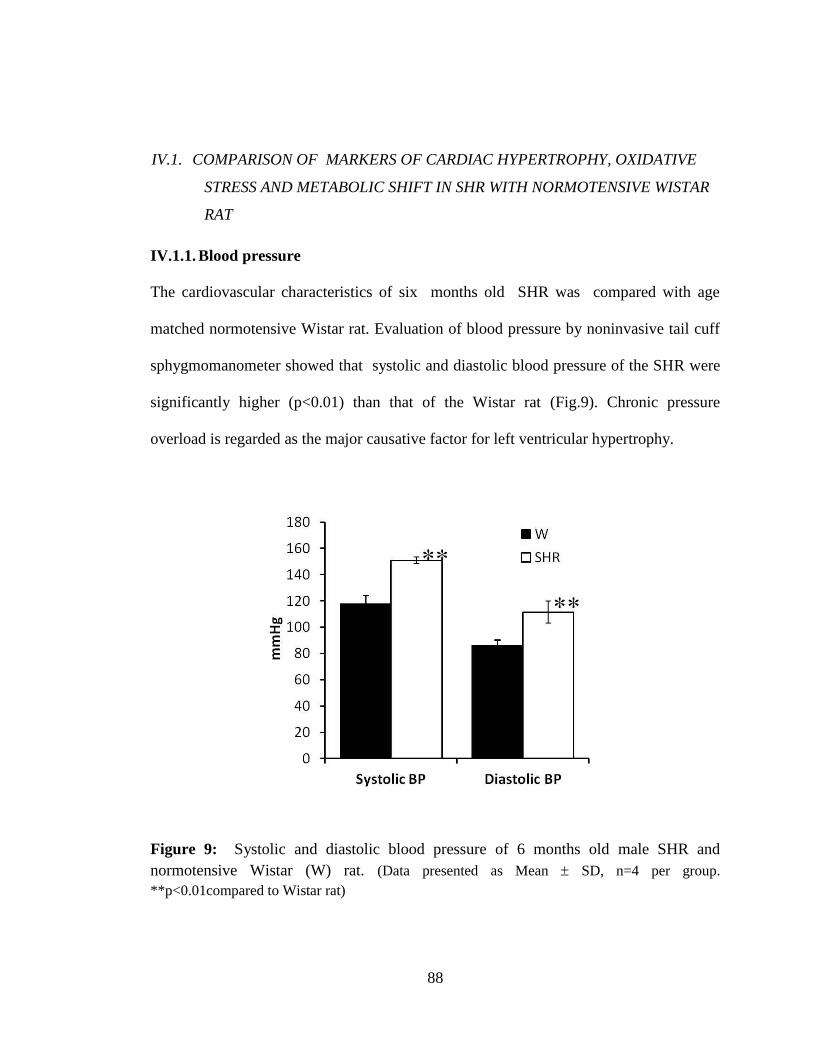
88
IV.1. COMPARISON OF MARKERS OF CARDIAC HYPERTROPHY, OXIDATIVE
STRESS AND METABOLIC SHIFT IN SHR WITH NORMOTENSIVE WISTAR
RAT
IV.1.1. Blood pressure
The cardiovascular characteristics of six months old SHR was compared with age
matched normotensive Wistar rat. Evaluation of blood pressure by noninvasive tail cuff
sphygmomanometer showed that systolic and diastolic blood pressure of the SHR were
significantly higher (p<0.01) than that of the Wistar rat (Fig.9). Chronic pressure
overload is regarded as the major causative factor for left ventricular hypertrophy.
Figure 9: Systolic and diastolic blood pressure of 6 months old male SHR and
normotensive Wistar (W) rat. (Data presented as Mean SD, n=4 per group.
**p<0.01compared to Wistar rat)

89
IV.1.2. Expression of CD36
Metabolic changes in SHR is possibly the consequence of reduced expression of
FAT/CD36. Inorder to evaluate the expression of CD36 in the stock of SHR used for the
experimental studies, the protein expression was analyzed by Western blot analysis.
SHR showed significantly reduced (p<0.05) expression of CD36 (fig.10).
Immunohistochemical analysis also confirmed reduced expression of CD36 in SHR.
(Fig. 11)
Figure 10: CD36 expression of the 6 months old SHR and normotensive Wistar (W) rat
(Data presented as Mean SD, n=4 per group **p<0.01 compared to Wistar rat)

90
Figure 11: Immunohistochemical analysis of CD36 expression of the 6 months old
SHR and normotensive wistar (W) rat – Representative photomicrographs
IV.1.3. Hypertrophy index
The ratio of heart weight (mg) to body weight (g) was used as the morphological marker
of cardiac hypertrophy, and was found to be significantly higher (p<0.05) in SHR
compared to Wistar rat (Fig:12)

91
Figure 12: Hypertrophy index of the 6 months old SHR and normotensive Wistar (W)
rat. (Data presented as Mean SD, n=4 per group. *p<0.05 compared to Wistar rat)
IV.1.4. Histological assessment for cardiac hypertrophy
Histological markers of cardiac remodeling such as myocyte cross sectional area and
cardiac fibrosis were determined by Image J software. Myocyte cross sectional area was
significantly higher (p<0.01) in SHR than Wistar rat (Fig.13). Myocardial fibrosis was
measured as interstitial fibrosis and perivascular fibrosis. Extent of fibrosis for both the
variables was significantly higher in SHR than that of Wistar rat (Fig. 14, 15).

92
Figure 13: Myocyte cross sectional area of 6 months old SHR and normotensive Wistar
(W) rat . Myocardial sections were immunostained with anti dystrophin antibody to get a
clear outline of the cell membrane. (A) Representative photomicrographs. (B) Graphical
representation of data. (Data presented as Mean SD, n=4 per group. ** p<0.01 compared
to Wistar)
A
B

93
Figure 14: Myocardial interstitial fibrosis of 6 months old SHR and normotensive
Wistar (W) rat . Myocardial sections were stained with Picrosirius red to visualize the
interstitial fibrosis. Collagen appears red. (A) Representative photomicrographs (B)
Graphical representation of data. (Data presented as Mean SD, n=4 per group. ** p<0.01
compared to Wistar)
B
A
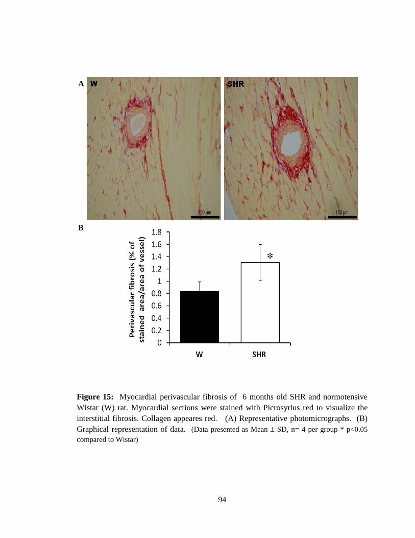
94
Figure 15: Myocardial perivascular fibrosis of 6 months old SHR and normotensive
Wistar (W) rat. Myocardial sections were stained with Picrosyrius red to visualize the
interstitial fibrosis. Collagen appeares red. (A) Representative photomicrographs. (B)
Graphical representation of data. (Data presented as Mean SD, n= 4 per group * p<0.05
compared to Wistar)
B
A
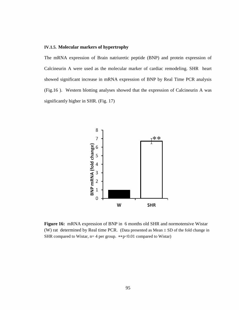
95
IV.1.5. Molecular markers of hypertrophy
The mRNA expression of Brain natriuretic peptide (BNP) and protein expression of
Calcineurin A were used as the molecular marker of cardiac remodeling. SHR heart
showed significant increase in mRNA expression of BNP by Real Time PCR analysis
(Fig.16 ). Western blotting analyses showed that the expression of Calcineurin A was
significantly higher in SHR. (Fig. 17)
Figure 16: mRNA expression of BNP in 6 months old SHR and normotensive Wistar
(W) rat determined by Real time PCR. (Data presented as Mean SD of the fold change in
SHR compared to Wistar, n= 4 per group. p<0.01 compared to Wistar)

96
Figure 17: Protein expression of Calcineurin A in 6 months old SHR and normotensive
Wistar (W) rat determined by Western blotting (A) Graphical presentation of data. (B)
Representative blots showing expression of Calcineurin A and the loading control actin
(Data presented as Mean SD of the fold change in SHR compared to Wistar n= 4 per group,
p<0.05 compared to Wistar)
IV.1.6. Cardiac energy metabolism
The mRNA expression of PPAR-, MCAD and PFK-1 were used as the molecular
markers of cardiac metabolism. mRNA expression was evaluated by Real time PCR
analysis. SHR showed significantly decreased expression of genes involved in fatty acid
metabolism such as PPAR- and MCAD (p<0.01) (Fig.18) and increased expression of
the glycolytic enzyme PFK-1 (p<0.05) (Fig.19). The observations confirm the presence
of shift in substrate preference from fatty acid to glucose in SHR.
A B
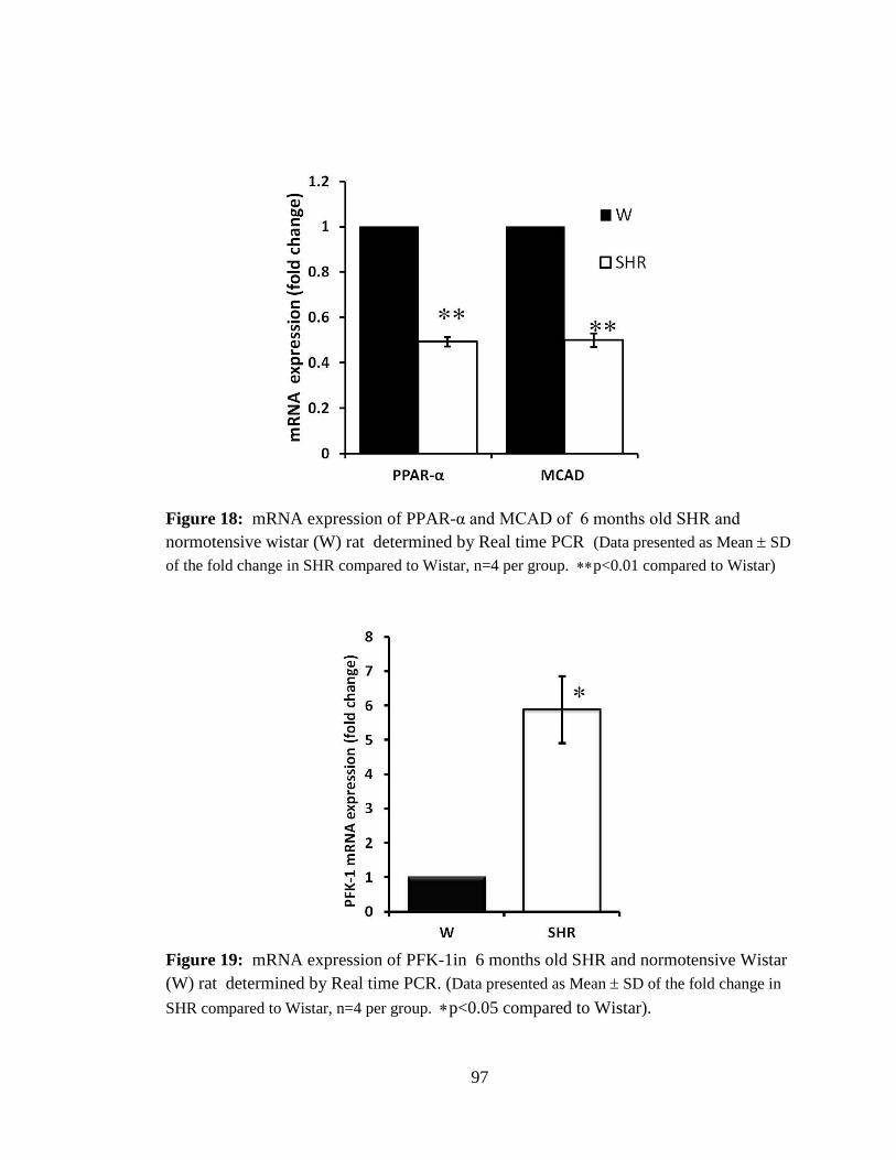
97
Figure 18: mRNA expression of PPAR-α and MCAD of 6 months old SHR and
normotensive wistar (W) rat determined by Real time PCR (Data presented as Mean SD
of the fold change in SHR compared to Wistar, n=4 per group. p<0.01 compared to Wistar)
Figure 19: mRNA expression of PFK-1in 6 months old SHR and normotensive Wistar
(W) rat determined by Real time PCR. (Data presented as Mean SD of the fold change in
SHR compared to Wistar, n=4 per group. p<0.05 compared to Wistar).
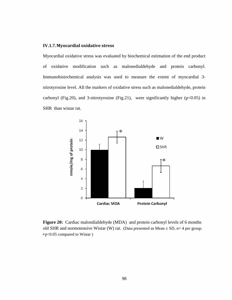
98
IV.1.7. Myocardial oxidative stress
Myocardial oxidative stress was evaluated by biochemical estimation of the end product
of oxidative modification such as malonedialdehyde and protein carbonyl.
Immunohistochemical analysis was used to measure the extent of myocardial 3-
nitrotyrosine level. All the markers of oxidative stress such as malonedialdehyde, protein
carbonyl (Fig.20), and 3-nitrotyrosine (Fig.21), were significantly higher (p<0.05) in
SHR than wistar rat.
Figure 20: Cardiac malondialdehyde (MDA) and protein carbonyl levels of 6 months
old SHR and normotensive Wistar (W) rat. (Data presented as Mean SD, n= 4 per group.
p<0.05 compared to Wistar )
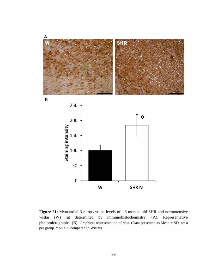
99
Figure 21: Myocardial 3-nitrotyrosine levels of 6 months old SHR and normotensive
wistar (W) rat determined by immunohistochemistry. (A). Representative
photomicrographs (B). Graphical representation of data. (Data presented as Mean SD, n= 4
per group. * p<0.05 compared to Wistar)
A
B

100
IV.2. CARDIAC RESPONSE TO EVEN CHAIN AND ODD CHAIN MCT
SUPPLEMENTATION IN YOUNG SHR
MCT is available in two different forms, Even chain MCT and Odd chain MCT with
difference in their metabolic properties. A pilot study was therefore carried out to select
the type of triglyceride that would be relatively more effective. For this experiment, 2-
month-old male SHR were supplemented with either 5% Tricaprylin or Triheptanoate
(v/w) of the total feed for 2 months. Morphological and histological markers of cardiac
hypertrophy and markers of myocardial oxidative stress were analyzed. SHR showed
variable cardiac response to Even and Odd chain MCT. Hypertrophy index was
significantly decreased (p<0.05) by Even chain MCT, but it was unaffected by Odd
chain MCT (Fig.22). Myocyte cross sectional area and interstitial fibrosis were used as
the histological markers of cardiac remodeling. Although cardiomyocyte cross sectional
area was significantly decreased (p<0.01) by both types of MCT (Fig.23), interstitial
fibrosis was significantly decreased (p<0.01) only with even chain MCT (Fig.24),
Interstitial fibrosis was unaffected by odd chain MCT. There was no significant
difference in the myocyte cross sectional area between Even chain and Odd chain MCT
supplemented SHR. Myocardial malonedialdehyde was used as the marker of oxidative
stress, and was found to be decreased by both types of MCT (p<0.01), with the extent of
decrease being greater for even chain MCT (Fig.25). MCT did not induce obesity as
assessed from the body weight in the treated and untreated groups (Fig.26).
Supplementation of both type of MCT did not affect serum lipid profile of the SHR,

101
assessedby biochemical estimation of total cholesterol, triglyceride, HDL cholesterol and
LDL cholesterol (Fig.27).
Figure 22: Hypertrophy index of 4 month old SHR following 60 days of
supplementation with Even chain (Tricaprylin) or Odd chain (Triheptanoate) MCT (5%
of the feed) compared with age and sex matched untreated SHR. (Data presented as Mean
SD, n= 4 per group, ANOVA p< 0.05 p<0.05 Even chain vs. Control, ##
p<0.01 Even chain
vs. Odd chain).
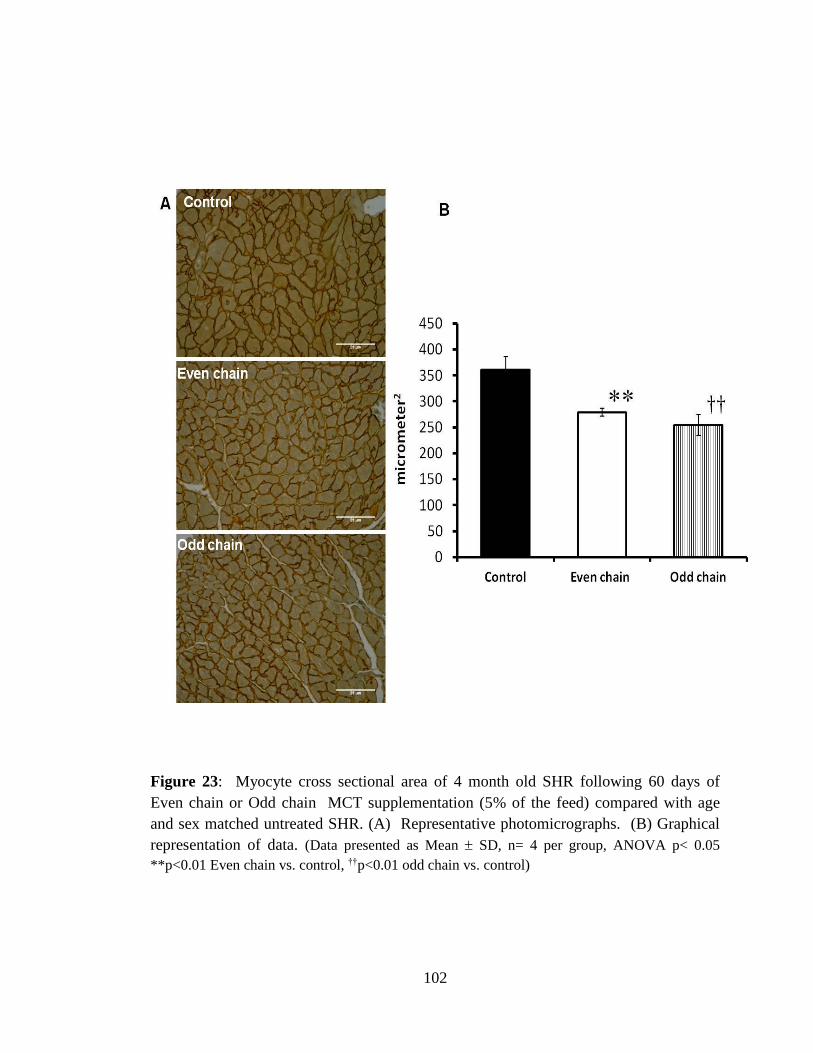
102
Figure 23: Myocyte cross sectional area of 4 month old SHR following 60 days of
Even chain or Odd chain MCT supplementation (5% of the feed) compared with age
and sex matched untreated SHR. (A) Representative photomicrographs. (B) Graphical
representation of data. (Data presented as Mean SD, n= 4 per group, ANOVA p< 0.05
**p<0.01 Even chain vs. control, ††p<0.01 odd chain vs. control)

103
Figure 24: Myocardial interstitial fibrosis of 4 month old SHR following 60 days of
Even chain or Odd chain MCT supplementation (5% of the feed) compared with age
and sex matched untreated SHR. Myocardial sections were stained with Picro Siriusred,
to visualize the collagen. Collagen appears red. (A) Representative photomicrographs.
(B) Graphical representation of data. (Data presented as Mean SD, n= 4 per group,
ANOVA p< 0.05, **p<0.01 Even chain vs. control).

104
Figure 25: Myocardial Malone dialdehyde levels of 4 month old SHR following 60 days
of supplementation with Even chain or Odd chain MCT (5% of the feed) compared
with age and sex matched untreated SHR. (Data presented as Mean SD, n= 4 per group,
ANOVA p< 0.05 **p<0.01 Even chain vs. control, ††p<0.01 odd chain vs. control, #p<0.05
Even chain vs. odd chain)
Figure 26: Body weight of 4 month old SHR following 60 days of supplementation
with Even chain or Odd chain MCT (5% of the feed) compared with age and sex
matched untreated SHR. (Data presented as Mean SD, n= 4 per group, ANOVA p=NS)

105
Figure 27: Serum lipid profile of 4 month old SHR following 60 days of
supplementation with Even chain or Odd chain MCT (5% of the feed) compared with
age and sex matched untreated SHR. (Data presented as Mean SD, n= 4 per group,
ANOVA p= NS)
IV.3. CARDIAC RESPONSE TO STIMULATION OF FATTY ACID OXIDATION BY
SUPPLEMENTATION OF MEDIUM CHAIN TRIGLYCERIDES AT INITIAL
AND ESTABLISHED STAGES OF CARDIAC HYPERTROPHY
Male Spontaneously hypertensive rats (SHR) of two different age groups representing
the initial and established stages of hypertrophy were used as the experimental model to
stimulate fatty acid metabolism. The even chain fatty acid Tricaprylin was used for the
experiments as the cardiac response was found to be relatively better than that of the odd
chain fatty acid Triheptanoate. 2-months-old (initial stages of hypertrophy) and 6-
months-old (established hypertrophy) SHR were supplemented with Medium chain
106
triglycerides (MCT) for 4 months to prevent hypertrophy in the former and regress
cardiac remodeling in the latter group. The animals were sacrificed respectively at 6
months and 10 months of age for evaluation of the cardiovascular response to treatment.
Cardiac response to MCT supplementation was evaluated based on cardiac morphology
and the expression of genes associated with fatty acid oxidation (FAO). Hypertrophy
index, cell size, morphometric analysis of ventricular collagen, expression of brain
natriuretic peptide (BNP) and calcineurin A were used as markers of adverse cardiac
remodeling. Myocardial malondialdehyde (MDA- a stable terminal metabolite of lipid
peroxidation), 3nitrotyrosine and protein carbonyl (end product of protein oxidation)
were used as markers of oxidative stress. Cardiac fatty acid oxidation was evaluated
based on the expression of PPAR-α and medium chain acyl CoA dehydrogenase
(MCAD -a key enzyme in β-oxidation of fatty acids) by Real time RT-PCR analysis.
The energy level was evaluated from biochemical estimation of ATP and
phosphocreatine.
IV.3.1. Effect of MCT on myocardial metabolism
To modulate the myocardial metabolism in SHR with early and established stages of
cardiac hypertrophy 2 months and 6 months old SHR were supplemented with 5% MCT
along with standard feed for 4 months. The effect of MCT on myocardial metabolism
was determined from the mRNA expression of PPAR-, MCAD and PFK-1 by Real
time PCR analysis. PPAR- and MCAD served as the molecular indicators for the
stimulation of fatty acid oxidation in SHR. PPAR- (*p<0.01) and MCAD (*p<0.05)

107
were significantly increased in initial and established stages of cardiac hypertrophy by
supplementation of MCT (Fig.28,29). Increased PPAR- expression along with
increased MCAD mRNA indicates stimulation of fatty acid oxidation. The consequence
of MCT supplementation on glucose oxidation was determined by mRNA expression
of PFK-1. Expression of PFK-1 was not significantly affected by the treatment (Fig.30).
Figure 28: Fold change in the mRNA expression of PPAR- in 6 and 10 months old
SHR following 4 months of supplementation with MCT (5% of the feed) (SHR M)
compared with age and sex matched untreated control (SHR C). (Data expressed as Mean
SD of the fold change in treated compared to untreated SHR, n = 6/group; **p<0.01 compared
to untreated control)

108
Figure 29: Fold change in the mRNA expression of MCAD in 6 and 10 months old
SHR following 4 months supplementation with MCT (5% of the feed) compared with
age and sex matched untreated control. (Data expressed as Mean SD of the fold change
in treated (SHR M) compared to untreated SHR (SHR C), n = 6/group; *p<0.05 compared to
untreated control)
Figure 30: Fold change in the mRNA expression of PFK-1 in 6 and 10 months old SHR
following 4 months of supplementation with MCT (5% of the feed) (SHR M) compared
with age and sex matched untreated control (SHR C). (Data expressed as Mean SD of
the fold change in treated compared to untreated SHR, n = 6/group)

109
IV.3.2. Cardiovascular response to stimulation of fatty acid oxidation
A. Blood pressure:
Blood pressure was monitored following 4 months of treatment. Irrespective of the age
of the animal, supplementation of MCT did not cause any change in systolic or diastolic
blood pressure in both the age groups. (Table 2)
Table 2: Mean ( SD) blood pressure of 6 and 10 months old SHR following 4 months
of supplementation with MCT (5% of the feed) (SHR M) compared with age and sex
matched untreated control (SHR C) (n=6/group)
B. Hypertrophy index:
Hypertrophy index was used as the morphological indicator of cardiac hypertrophy,
represented as the ratio of heart weight to body weight. Reactivation of fatty acid
metabolism by MCT significantly decreased hypertrophy index (p<0.05) in younger rat
and was maintained in older rats in comparison with age-matched control SHR (Fig.31).
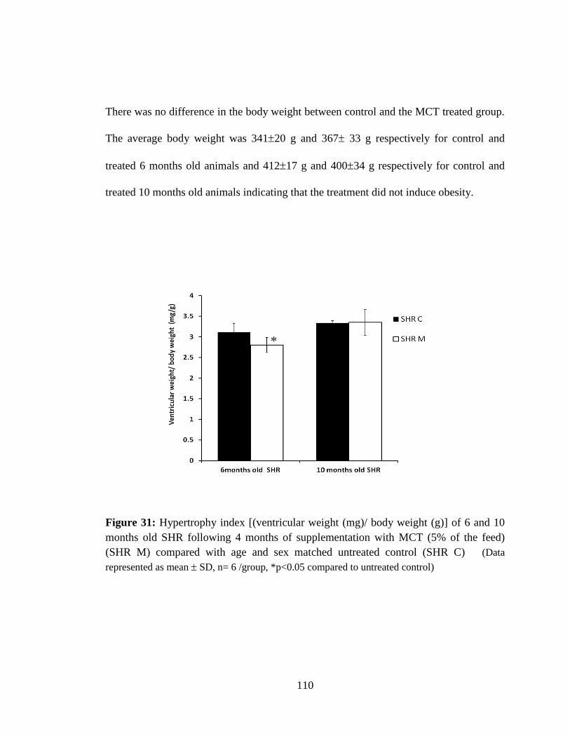
110
There was no difference in the body weight between control and the MCT treated group.
The average body weight was 34120 g and 367 33 g respectively for control and
treated 6 months old animals and 41217 g and 40034 g respectively for control and
treated 10 months old animals indicating that the treatment did not induce obesity.
Figure 31: Hypertrophy index [(ventricular weight (mg)/ body weight (g)] of 6 and 10
months old SHR following 4 months of supplementation with MCT (5% of the feed)
(SHR M) compared with age and sex matched untreated control (SHR C) (Data
represented as mean SD, n= 6 /group, *p<0.05 compared to untreated control)

111
C. Myocyte cross sectional area and myocardial fibrosis:
Increased cardiomyocyte cross sectional area and interstitial fibrosis are the histological
features of myocardial hypertrophy. The sarcolemma of the individual myocytes was
visualized by immunohistochemical staining for dystrophin and picro sirius red staining
was used for assessment of fibrosis. The mean cross sectional area of cardiomyocytes
and interstitial fibrosis decreased significantly with treatment in both the age groups
(Fig.32,33). Perivascular fibrosis was unaffected in either of the age groups in response
to MCT supplementation (Fig. 34).

112
Figure 32: Myocyte cross sectional area of 6 and 10 months old SHR following 4
months of supplementation with MCT (5% of the feed) (SHR M) compared with age and
sex matched untreated control. (SHR C)Mid ventricular cross sections were
immunostained with dystrophin to get a clear outline of the cells. (A) Representative
photomicrographs. (B) Graphical representation of data. (Data presented as Mean SD,
n=6/group, ** p<0.01 and * p<0.05 compared to untreated control).

113
Figure 33: Interstitial fibrosis of 6 and 10 months old SHR following 4 months of
supplementation with MCT (5% of the feed) (SHR M) compared with age and sex
matched untreated control (SHR C) For the visualization of the fibrosis myocardial
sections were stained with Picrosirius red. Collagen appears red. (A) Representative
photomicrographs. (B) Graphical representation of data. (Data presented as Mean SD, n=
6/group. * p<0.05 compared to untreated control)

114
Figure 34: Perivascular fibrosis in 6 and 10 months old SHR following 4 months of
supplementation with MCT (5% of the feed) compared with age and sex matched
untreated control. For the visualization of the fibrosis myocardial sections were stained
with Picrosirius red. Collagen appears red. (A) Representative photomicrographs. (B)
Graphical representation of data. (Data presented as Mean SD, n= 6/group.)
A
B
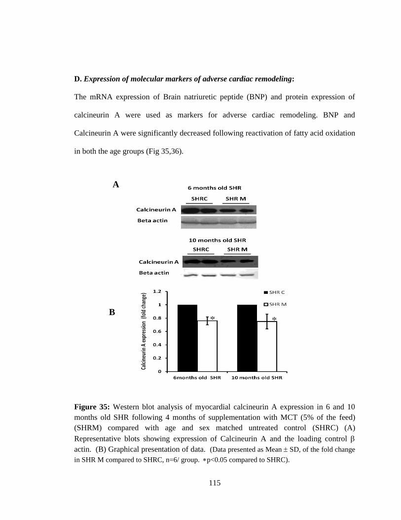
115
D. Expression of molecular markers of adverse cardiac remodeling:
The mRNA expression of Brain natriuretic peptide (BNP) and protein expression of
calcineurin A were used as markers for adverse cardiac remodeling. BNP and
Calcineurin A were significantly decreased following reactivation of fatty acid oxidation
in both the age groups (Fig 35,36).
Figure 35: Western blot analysis of myocardial calcineurin A expression in 6 and 10
months old SHR following 4 months of supplementation with MCT (5% of the feed)
(SHRM) compared with age and sex matched untreated control (SHRC) (A)
Representative blots showing expression of Calcineurin A and the loading control
actin. (B) Graphical presentation of data. (Data presented as Mean SD, of the fold change
in SHR M compared to SHRC, n=6/ group. p<0.05 compared to SHRC).
A
B

116
Figure 36: Fold change in BNP mRNA expression in 6 and 10 months old SHR
following 4 months of supplementation with MCT (5% of the feed) (SHRM) compared
with age and sex matched untreated control. (Data expressed as mean SD of fold change in
expression of SHRM compared to SHRC, n=6/ group.**p<0.01 compared to SHRC)
E. Effect of MCT supplementation on markers of oxidative stress:
Malonedialdehyde (MDA), a stable end product of lipid peroxidation and protein
carbonyl and 3- nitrotyrosine, end product of protein oxidation were used as the markers
of oxidative stress. Increased lipid peroxidation is the expected consequence of high fat
diet. However, MCT supplementation decreased myocardial lipid peroxidation,
significantly in younger SHR (Fig.37). Myocardial protein carbonyl and 3-nitrotyrosine
level were significantly reduced by metabolic stimulation in both the age groups (Figs
38,39).

117
Figure 37: Cardiac Malondialdehyde levels of 6 and 10 months SHR following 4
months of supplementation with MCT (5% of the feed) (SHR M) compared with age and
sex matched untreated control.(SHR C). (Data expressed as mean SD, n=6/group,
**p<0.01 compared to untreated control)
Figure 38: Cardiac protein carbonyl level of 6 and 10 months old SHR following 4
months of supplementation with MCT (5% of the feed) (SHR M)compared with age and
sex matched untreated control (SHR C). (Data expressed as mean SD, n=6/group,
**p<0.01compared to SHR C)

118
Figure 39: Cardiac 3nitrotyrosine level of 6 and 10 months old SHR following 4 months
of supplementation with MCT (5% of the feed) (SHR M) compared with age and sex
matched untreated control (SHR C) detected by immunohistochemistry of mid
ventricular sections. (A) Representative photomicrographs. (B) Graphical
representation of data. (Data presented as Mean SD, n= 6 * p<0.05 compared to untreated
control).
A
B
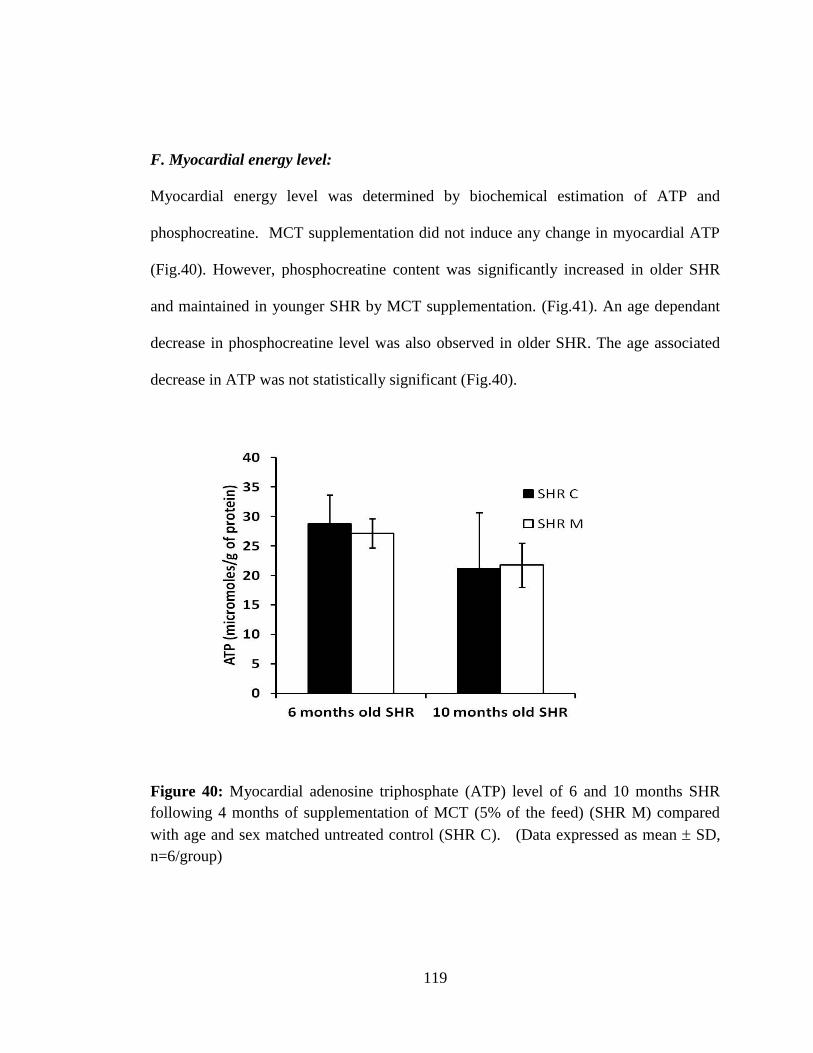
119
F. Myocardial energy level:
Myocardial energy level was determined by biochemical estimation of ATP and
phosphocreatine. MCT supplementation did not induce any change in myocardial ATP
(Fig.40). However, phosphocreatine content was significantly increased in older SHR
and maintained in younger SHR by MCT supplementation. (Fig.41). An age dependant
decrease in phosphocreatine level was also observed in older SHR. The age associated
decrease in ATP was not statistically significant (Fig.40).
Figure 40: Myocardial adenosine triphosphate (ATP) level of 6 and 10 months SHR
following 4 months of supplementation of MCT (5% of the feed) (SHR M) compared
with age and sex matched untreated control (SHR C). (Data expressed as mean SD,
n=6/group)

120
Figure 41: Myocardial Phosphocreatine (PCr) level of 6 and 10 months SHR following
4 months of supplementation of MCT (5% of the feed) (SHR M) compared with age and
sex matched untreated control (SHR C). (Data expressed as mean SD, n=6/group,
*p<0.05compared to SHR C, #p<0.05 compared to 6 months old SHR C)
G. Lipid profile and Hepatic Lipid accumulation:
MCT supplementation did not induce any change in serum lipid profile of the SHR.
(Table.3). MCT maintained serum triglycerides, total cholesterol, HDL-C, and LDL-C in
SHR. Consumption of MCT did not induce any hepatic lipid accumulation in
comparison with control SHR (Fig.42).
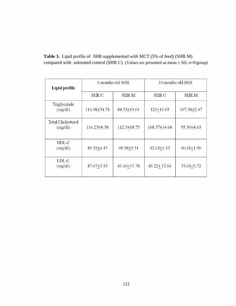
121
Table 3. Lipid profile of SHR supplemented with MCT (5% of feed) (SHR M)
compared with untreated control (SHR C) (Values are presented as mean SD, n=6/group)
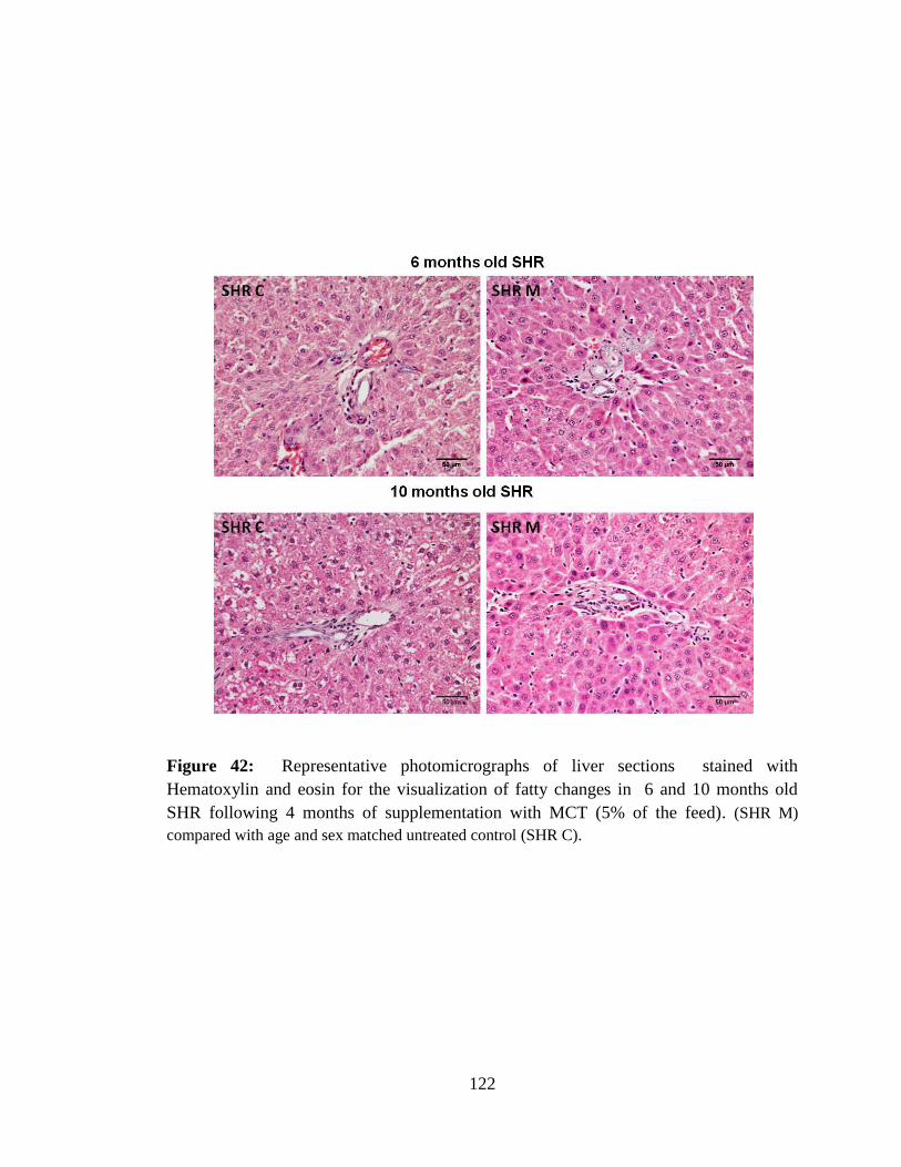
122
Figure 42: Representative photomicrographs of liver sections stained with
Hematoxylin and eosin for the visualization of fatty changes in 6 and 10 months old
SHR following 4 months of supplementation with MCT (5% of the feed). (SHR M)
compared with age and sex matched untreated control (SHR C).

123
V. DISCUSSION
124
Left ventricular hypertrophy (LVH) is an adaptive mechanism triggered in the heart in
response to pathological stimuli such as hypertension or aortic stenosis. However it is a
major risk factor for adverse cardiovascular events and sudden death. Attenuation of
LVH reduces the risk for cardiac failure and myocardial injury. During pathological
hypertrophy, heart shifts its metabolic substrate preference from fatty acids to glucose
(Allard, 2004; Allard et al., 2007). Chronic reliance on glucose metabolism is inefficient
for maintaining contractile function especially under work overload (Ingwall, 2009). The
decreased fatty acid utilization is possibly caused by reduction in the expression of
Cluster of Differentation-36 (CD36), a major sarcolemmal fatty acid transporter that
limits the availability of long chain fatty acids. A number of reports testify the role of
CD36 deficiency in the metabolic alteration and the consequent cardiac remodeling. As
chronic reduction in fatty acid oxidation can lead to pathological cardiac remodeling, it
is presumed that restoration of fatty acid metabolism by bypassing the defective CD36
will help to maintain the metabolic status and prevent progressive cardiac remodeling.
Hence, the study was carried out based on the hypothesis that reactivation fatty acid
oxidation by supplementation of medium chain triglyceride (MCT) can prevent adverse
cardiac remodeling in chronic pressure overload. The study was carried out in
Spontaneously hypertensive rat, an animal model of chronic pressure overload induced
left ventricular hypertrophy. Wistar (W) rats were used as the normotensive control.

125
V.1. COMPARISON OF MARKERS OF CARDIAC HYPERTROPHY, METABOLIC
SHIFT AND OXIDATIVE STRESS IN SPONTANEOUSLY HYPERTENSIVE RAT
(SHR) WITH NORMOTENSIVE WISTAR RAT (W)
The Spontaneously hypertensive rat (SHR) is a genetic model of hypertension induced
left ventricular hypertrophy (LVH). SHR simulates the clinical progression of
hypertension with a long term stable phase of left ventricular hypertrophy followed by
progressive heart failure (Boluyt et al., 1995). Elucidation of mechanisms that restrict the
development of cardiac hypertrophy in SHR may aid in the development of therapeutic
strategies that prevent progressive cardiac remodeling.
Before initiating the experimental studies the presence of cardiac hypertrophy, metabolic
shift and oxidative stress was validated in adult six-months old SHR (the established
stage of cardiac hypertrophy) by comparison with age and sex matched normotensive
Wistar rat. Wistar-Kyoto rats were avoided as they have a tendency to develop cardiac
hypertrophy (Aiello et al., 2004). Morphological and histological analysis testified
that, SHR is characterized by left ventricular hypertrophy. Hypertrophy index [heart
weight (mg)/ body weight (g)] used as morphological marker of cardiac hypertrophy,
was found to be significantly higher in 6 months old SHR compared to Wistar rat (W)
(Fig.12).
Systolic and diastolic blood pressure of SHR, as determined by tail cuff
sphygmomanometer was significantly higher in SHR (Fig.9). Histological assessment
126
showed that myocyte cross sectional area, interstitial and perivascular fibrosis (Fig.13-
15) were significantly higher in SHR confirming the presence of cardiac hypertrophy.
Longitudinal studies in SHR had shown that SHR develop hypertension as early as one
month of age, followed by hypertrophy in the 2nd month (Purushothaman et al., 2011a).
Molecular markers of cardiac hypertrophy such as Brain natriuretic peptide (BNP)
mRNA and Calcineurin A protein expression were significantly higher in SHR than
normotensive Wistar rat (W) (Fig.16,17). Calcineurin is a Ca2+ dependant serine-
threonine phosphatase that acts as a signaling molecule and mediates the development
of pathological cardiac hypertrophy; and progressively increases with age in SHR (Zou
et al., 2002).
Development of cardiac hypertrophy in SHR is associated with a shift in substrate
preference from fatty acid to glucose mediated by downregulation of the expression of
genes involved in fatty acid metabolism (Christe and Rodgers, 1994b). Previous studies
have reported that the reduction in fatty acid oxidation in SHR is due to defective CD36,
a major plasmalemmal fatty acid transporter (Hajri et al., 2001). The stock of SHR used
for experimental studies also showed reduced expression of CD36 in heart, which was
confirmed by Western blotting and immunohistochemistry (Fig.10,11). The reduced
CD36 expression in SHR limits the availability of the long chain fatty acids, the natural
ligand of PPARα. This is possibly the cause for the reduction in the fatty acid oxidation

127
as apparent from the reduced mRNA expression of PPARα and one of its target gene
MCAD (Fig.18).
CD 36 is a highly glycolsylated integral membrane protein that shuttles 40-80% of fatty
acid into the cardiomyocytes (Brinkmann et al., 2002). SHR carry a defective CD36 on
rat chromosome 4, formed by recombination between wild type gene and Pseudo gene
affecting post translational modification and intracellular localization changes the
CD36's capacity to transport long chain fatty acids (Lauzier et al., 2011). Expression of
wild type CD36 in SHR background improved defective fatty acid oxidation without
affecting hypertension (Pravenec et al., 2001, 2003). SHRs are characterized by a
restricted utilization of exogenous LCFAs for energy production, which has been
attributed to a genetic defect in CD36. Age dependant downregulation of CD36 mRNA
was also reported in SHR (Purushothaman et al., 2011b). The decreased fatty acid
oxidation in SHR was accompanied by increased mRNA expression of PFK-1 (Fig.19),
indicating accelerated glycolysis as a compensatory mechanism for sustenance of energy
metabolism.
Apart from the shift in substrate preference from fatty acid to glucose, cardiac
remodeling in hypertensive heart disease is associated with enhanced oxidative stress.
Invitro and invivo studies carried out earlier have linked oxidative stress with cardiac
hypertrophy (Adiga and Nair, 2008; Preeta and Nair, 2000; Purushothaman et al.,
2011a). Oxidative stress in SHR was assessed by biochemical estimation of

128
malonedialdehyde (MDA) and protein carbonyl. Myocardial 3-nitrotyrosine was
determined by immunohistochemistry. Malonedialdehyde, protein carbonyl and 3-
nitrotyrosine levels were significantly higher in SHR (Fig.20,21), showing the
association of oxidative stress with cardiac hypertrophy. Studies reported that use of
antioxidants improved endothelial function and attenuated cardiac hypertrophy in young
Stroke prone Spontaneously hypertensive rat (Graham et al., 2009). The observations
ensure that the animals used for the experimental studies had cardiac hypertrophy,
oxidative stress and shift in energy substrate preference.
V.2. COMPARISON OF CARDIAC RESPONSE TO STIMULATION OF FATTY ACID
OXIDATION BY SUPPLEMENTATION WITH ODD AND EVEN CHAIN
MEDIUM CHAIN TRIGLYCERIDES
MCT is available in two different forms, odd and even chain MCT based on number of
carbons. Odd chain MCT is reported to be anaplerotic in nature. Anaplerosis is the re-
filling of the catalytic intermediates of the Citric acid cycle. MCTs with an odd number
of carbons were reported to be superior to even-carbon MCTs in improving the
cardiomyopathy of one patient with genetic LCFA oxidation deficiency (Roe et al.,
2002). Dietary regimen substituting tricaprylin (even chain MCT) with triheptanoin
(odd chain MCT) improved cardiomyopathy in few patients with very long-chain acyl-
CoA dehydrogenase deficiency. Triheptanoin intake enhanced ketone body synthesis
without inducing propionyl-CoA overload (Roe et al., 2002). Recently, Nguyen et al.
demonstrated that odd chain MCT triheptanoin feeding improved diastolic function and

129
cardiac hypertrophy in rodent model of pressure overload (Nguyen et al., 2015). Positive
cardiac response to even chain triglycerides has also been reported (Labarthe et al.,
2008). Hence, there exists the possibility of difference in their metabolic properties. A
pilot study was therefore carried out to examine whether there would be a differential
response depending on the type of triglyceride. For the experimental studies, 2-month-
old SHR were supplemented with either Tricaprylin or Triheptanoate (5% of the total
feed) for 2 months. Morphological and histological markers of cardiac hypertrophy and
markers of oxidative stress were analyzed.
Even chain and odd chain triglycerides have shown variable cardiac response. Although
both the sources of MCT decreased myocardial malonedialdehyde level, the decrease
was more significant with even chain MCT (Tricaprylin) (Fig.25). Morphologial and
histological markers of cardiac hypertrophy also showed divergent response. Although
myocardial cross sectional area was significantly decreased by both the types of MCT
(Fig.23), the hypertrophy index decreased only with even chain MCT (Fig.22). Similarly,
myocardial fibrosis was decreased only with even chain MCT and not with odd chain
MCT (Fig.24). Positive cardiac remodeling was significantly better with even chain
MCT. Hence, tricaprylin was used as the source of MCT for further experimental
studies. Both treatments did not affect body weight and lipid profile (Fig.26,27).

130
V.3. CARDIAC RESPONSE TO STIMULATION OF FATTY ACID OXIDATION BY
SUPPLEMENTATION OF MEDIUM CHAIN TRIGLYCERIDES AT INITIAL AND
ESTABLISHED STAGES OF CARDIAC HYPERTROPHY
Hypertension induced LVH is a convergent point for many risk factors leading to heart
failure. During this hypertrophic response, the heart switches its substrate preference
from fatty acid towards glucose. Several studies have reported the association between
reduced rate of fatty acid oxidation and development of cardiac hypertrophy. Cardiac
hypertrophy was provoked in rats on fat-free diet (Panos and Finerty, 1954). Humans
with inborn errors in mitochondrial fatty acid oxidation enzymes often develop
ventricular hypertrophy even in the absence of hypertension (Kelly et al., 1992).
Inhibition of mitochondrial FAO in animals resulted in the development of cardiac
hypertrophy (Rupp and Jacob, 1992). Patients with idiopathic dilated cardiomyopathy
showed downregulation of FAO enzymes and/or decreased myocardial fatty acid
oxidation as assessed by Positron emission tomography (Dávila-Román et al., 2002).
Though initially adaptive, prolonged low rate of fatty acid oxidation is associated with
cardiovascular consequences including increased oxidative damage and decreased
energy reserve. Hence, restoration of fatty acid metabolism is expected to prevent
adverse cardiac remodeling. Peroxisome proliferator-activated receptor-α (PPARα) is a
critical regulator of myocardial fatty acid uptake and utilization. Studies reporting
positive cardiac changes consequent to pharmacological stimulation of fatty acid
metabolism were carried out in young rat or surgical models of cardiac hypertrophy
(Chen et al., 2007; Li et al., 2009; Ogata et al., 2002b; Zou et al., 2013). Age associated

131
variation in cardiac response to ligand-mediated activation of PPARα was observed
(Purushothaman et al, 2011b). Fenofibrate stimulated fatty acid oxidation and prevented
development of hypertrophy in young SHR, but aggravated hypertrophy in older
animals. An age associated downregulation of CD36 expression was observed in SHR
(Purushothaman et al., 2011b). Variation in CD36 expression can account for this
paradoxical cardiac response on pharmacological stimulation of fatty acid metabolism,
as defective CD36 in SHR limits entry of long chain fatty acid into the cardiomyocytes.
In the absence of metabolic substrate, on stimulation of fatty acid metabolism, this
substrate deficiency is fulfilled by mobilization of the endogenous triglycerides that in
turn results in the imbalance of the cellular redox status, leading to adverse cardiac
consequences (Ismael et al., 2015). The lack of metabolic substrate availability
consequent to defective CD36 is the primary determinant of altered metabolism and
cardiac hypertrophy in SHR (Hajri et al., 2001). Defective CD36 did not affect the
normal survival of the animals under basal conditions but showed reduced ischemic
tolerance, increased susceptibility to arrhythmia under pressure overload and impaired
response to adrenergic stimulation. (Irie et al., 2003; Labarthe, 2004).
A number of reports testify the role of CD36 deficiency in the metabolic alteration and
the consequent cardiac remodeling. Tanaka et al. reported the direct association between
CD36 deficiency and development of hypertrophic cardiomyopathy in the Japanese
population (Tanaka et al., 1997). Pharmacological inhibition of CD 36 was associated
with development of cardiac hypertrophy independent of blood pressure in rodents

132
(Kusaka et al., 1995). Altered myocardial metabolism and myocardial hypertrophy in
SHR is attributed to defective CD36 (Hajri et al., 2001). Deficiency of CD36 has been
reported recently by Magida et al in mouse models with familial hypertrophic
cardiomyopathy (Magida and Leinwand, 2014). These studies highlight that the
decreased fatty acid oxidation is secondary to reduced CD36 expression in
cardiomyocytes. Therefore, it was assumed that, supplementation with medium chain
triglyceride can overcome the maladaptive changes due to deficit in fatty acid oxidation
as the substrate availability is independent of the fatty acid transporter CD36.
Spontaneously hypertensive rats in the initial and established stages of cardiac
hypertrophy were supplemented with 5% MCT, tricaprylin a triglyceride of caprylic acid
along with standard feed for 4 months.
The study has shown that substrate mediated stimulation of fatty acid metabolism
prevents progressive cardiac remodeling in SHR. Supplementation of MCT in SHR
stimulated fatty acid metabolism as indicated by increased expression of PPAR-α and
one of its target genes MCAD, on comparison with age matched untreated control (Fig.
28,29). Increased PPAR-α mRNA expression along with increased MCAD mRNA
indicate stimulation of fatty acid metabolism by the activation PPAR-α. PFK-1 level
was not affected by MCT supplementation (Fig.30).
Metabolic stimulation by MCT prevented the development and progression of adverse
cardiac remolding in SHR, as indicated by decreased cardiomyocyte cross sectional area
133
and interstitial fibrosis compared with untreated SHR (Fig.32,33). Significant decrease
in hypertrophy index was apparent in the younger age group (Fig.31). Interstitial
fibrosis was also significantly reduced by the treatment. Studies have reported that,
supplementation of short and medium chain triglycerides prevented the development of
hypertrophy in young SHR (Hajri et al., 2001; Iemitsu et al., 2008; Shimojo et al., 2004).
Stimulation of fatty acid oxidation was associated with a significant decrease in the
expression of BNP mRNA and the protein Calcineurin A both of which serve as
molecular markers for adverse cardiac remodeling (Fig.35,36). Pharmacological
inhibition of calcineurin A is reported to attenuate cardiac hypertrophy and prevent
transition to heart failure (Sakata et al., 2000). Morphological, histological as well as
molecular indicators of hypertrophy were significantly reduced in SHR of both age
groups, confirming that supplementation with MCT in the diet prevents cardiac
hypertrophy.
It is now accepted that ROS are involved in the pathophysiology of myocardial
hypertrophy and failure. Invitro and invivo studies carried out earlier have linked
oxidative stress with cardiac hypertrophy (Adiga and Nair, 2008; Preeta and Nair, 2000;
Purushothaman et al., 2011a). Superoxide at low levels stimulate hypertrophy, but
chronic or high levels of superoxide result in cumulative cellular damage leading to
secondary mitochondrial damage and eventual progression to heart failure(Sawyer et al.,
2002). The direct correlation of oxidative stress with cardiac hypertrophy implies that
reduction of oxidative stress is an essential mediator of positive cardiac remodeling.
134
Oxidative stress was assessed from cardiac malonedialdehyde (MDA), protein carbonyl
and 3-nitrotyrosine levels. Protein carbonyl and cardiac 3-nitrotyrosine levels decreased
significantly with the treatment, and cardiac MDA level was found to be significantly
lower in young SHR, signifying that the metabolic stimulation by supplementation with
MCT decreased oxidative stress (Fig.37-39). Reduction of oxidative stress is supported
by the observation that intake of medium chain fatty acids prevents the accumulation of
oxidatively damaged proteins and lipid in skeletal muscle by inhibition of reactive
oxygen species (Montgomery et al., 2013).
There was no significant difference in diastolic or systolic blood pressure on MCT
supplementation (Table 2), showing blood pressure independent attenuation of LVH.
Perivascular fibrosis was unaffected by metabolic stimulation despite decrease in
oxidative stress (Fig.34) possibly, due to the fact that blood pressure was unaffected by
the treatment. This observation is supported by previous report by Iemitsu et al. (2008)
It is popularly known that cardiac decompensation consequent to shift in energy
substrate preference from fatty acid to glucose is due to energy deficit. Pathological
cardiac hypertrophy is manifested by depletion of phosphocreatine, the high energy
reserve; with preserved ATP levels (Kolwicz et al., 2013; Liao et al., 1996). In the
present study, an age dependant decrease in myocardial phosphocreatine content was
observed in older SHR compared to younger animals, which was normalized by MCT
supplementation (Fig.41). However MCT treatment maintained the cardiac ATP levels

135
in both age group (Fig.40). Studies have reported that, SHRs have similar cardiac high
energy phosphate content at younger age (15 week old) compared to normotensive
Wistar rat (Dodd et al., 2012), but diminishes gradually during sustained phase of
cardiac hypertrophy (Perings et al., 2000; Shimamoto et al.,1982) These results imply
that the positive cardiac response to the treatment seen in SHR is possibly due to
maintenance of energy level as well as decrease in oxidative stress.
Apart from its effect on cardiac hypertrophy MCT are known to have other beneficial
effects on the heart. Intake of short and medium-chain fatty acids was not associated
with coronary artery disease; ( Hu et al., 1999) and improvement in contractile function
and ATP level of the hypertrophied heart and capacity to withstand acute adrenergic
stress has been reported (Allard et al., 2007; Labarthe, 2004). Improved ischemic
recovery of working hearts has also been reported on perfusion with caprylic acid (Irie
et al., 2003). Additionally, supplementation of octanate enhanced contractile function of
volume overloaded heart (Alaoui-Talibi et al., 1992). Supplementation of MCT prior to
exercise training improved oxidative metabolism and decreased cardiac workload in
patients with long-chain fatty acid oxidation defect (Behrend et al., 2012). Dietary
administration of MCT improved LV dysfunction in mice with diabetic cardiomyopathy
(Finck et al., 2003).
Dietary guidelines for cardiovascular diseases recommend high carbohydrate/low fat
diet. High fat diet is reported to worsen cardiac hypertrophy and dysfunction. (Raher et
136
al.,2008). However, several studies have reported that high dietary fat could activate
mitochondrial oxidative metabolism and alleviate adverse cardiac remodeling (Chess et
al., 2009, 2008; Duda et al., 2008; Okere et al., 2006, 2005). It is also reported that high
fat diet did not affect the degree of cardiac hypertrophy in SHR even with reduction in
blood pressure (Bosse et al., 2013). Conventionally, saturated fats are not recommended
as its intake is associated with elevated risk of cardiovascular diseases. Onge et al
reported that MCT consumption at moderate level of 10-20% of energy intake does not
increase the risk of cardiovascular disease (St-Onge et al., 2008). Consumption of MCT
is not associated with the development of coronary artery disease (Hu et al., 1999). The
current study has shown that supplementation of 5% of the diet with MCT does not
produce increase in body weight or affect the lipid profile. On the other hand, it can be
cardioprotective. There was no sign of hepatic lipid accumulation. Clinical studies that
have reported dislipidemia on MCT consumption have used diet with more than 30% of
calorie intake from MCT. (Hill et al., 1990; Swift et al., 1992)
The observations leads to the inference that, the positive cardiac changes in response to
MCT are independent of blood pressure. Cardiac hypertrophy is regarded as an
adaptation to pressure overload. The observations of the study indicate that decrease in
blood pressure does not essentially lead to regression of hypertrophy, and that adverse
cardiac remodeling can be prevented regardless of the maintenance of hypertension. The
absence of a direct correlation of blood pressure with hypertrophy is supported by the

137
observation of blood pressure independent cardiac hypertrophy in Wistar-Kyoto rats
(Aiello et al., 2004).
V.4. LIMITATION AND FUTURE DIRECTION
The beneficial effect of MCT supplementation would be appreciated with functional
assessment, but this could not be done due to lack of facility
Future direction should be aimed at clinical validation of the experimental observation.
It would be interesting to examine whether reduction of oxidative stress can restore the
original metabolic profile.
138
VI. SUMMARY AND CONCLUSION

139
VI.1. SUMMARY
The development of left ventricular hypertrophy (LVH) is an adaptive response to
chronic pressure overload. Sustained hypertrophy is maladaptive and is a clinical
predictor for heart failure. Oxidation of long-chain fatty acids serves as the major
metabolic fuel for the healthy heart. In pathological cardiac hypertrophy, there is a
decrease in the oxidation of long chain fatty acids and increased relative contribution of
glucose for the maintenance of cardiac energy requirements. Prolonged lower rate of
fatty acid oxidation is associated with adverse cardiovascular consequences.
Pharmacological activation of Peroxisome proliferator-activated receptor α (PPARα)
revealed an age dependent differential cardiac response in SHR possibly linked to the
stage of hypertrophy; despite stimulation of fatty acid oxidation. Studies in
Spontaneously hypertensive rat (SHR) and patients with hypertrophic cardiomyopathy
leads to the postulation that, the reduced fatty acid oxidation is possibly caused by
reduced expression of CD36, a major sarcolemmal fatty acid transporter. A temporal
variation was observed in the expression of CD36, with decreased expression in older
animals. As Medium chain triglycerides have the capacity to bypass CD36 and serve as
substrate for fatty acid oxidation, it is presumed that restoration of fatty acid oxidation
by supplementation of MCT will help to maintain the metabolic status and prevent
progressive cardiac remodeling. Male Spontaneously hypertensive rat were used as the
experimental model.
140
SHR in the initial and established stage of hypertrophy were supplemented with 5%
Medium chain triglycerides (MCT) - Tricaprylin, along with standard feed for 4 months.
Stimulation of fatty acid oxidation was assessed from the expression of PPARα and
Medium chain acylCoA dehydrogenase (MCAD) mRNA by Real time polymerase chain
reaction. Hypertrophy was assessed by hypertrophy index, myocyte cross sectional area,
extent of cardiac fibrosis, mRNA expression of Brain natriuretic peptide (BNP) and
expression of the protein calcineurin A. Malonedialdehyde and protein carbonyl assay,
and histological analysis of 3-nitrotyrosine were used as the markers of oxidative stress.
Myocardial energy level was determined by biochemical estimation of ATP and
phosphocreatine.
The significant findings of the present study are
Comparison of morphological, histological and molecular markers of
hypertrophy with age and sex matched Wistar rats confirmed that SHR
used for the experimental studies developed Left Ventricular Hypertrophy
Fatty acid oxidation was decreased as indicated by decreased mRNA
expression of MCAD and PPAR and increased PFK-1 mRNA expression
indicates increase in glucose oxidation in SHR in comparison with Wistar
rat.

141
SHR was characterized by reduced expression of the fatty acid transporter
CD36, a possible cause for diminished Fatty acid oxidation.
Hypertrophy in SHR was associated with increased oxidative stress as
indicated by increased levels of cardiac malonedialdehyde, protein
carbonoyl and 3-nitrotyrosine compared to Wistar rats
The cardiac response to supplementation with MCT can depend on the type
of fatty acid. Supplementation of Odd chain and Even chain triglycerides in
2-month-old SHR (5% of feed v/w) for 2 months indicated that decrease in
hypertrophy index and interstitial fibrosis was significantly better with
Even chain MCT compared to Odd chain MCT.
Reduction of oxidative stress was apparent with both the treatments but
was significantly greater with Even chain MCT.
Supplementation with Even chain MCT in SHR in the initial and established
stages of cardiac hypertrophy stimulated fatty acid oxidation as indicated by
increased mRNA expression of MCAD and PPAR, with maintenance of
PFK-1 mRNA expression.
Stimulation of fatty acid oxidation by MCT attenuated progressive cardiac
remodeling independent of blood pressure.
142
Modulation of fatty acid oxidation increased myocardial energy level
significantly in older SHR.
Stimulation of Fatty acid oxidation by MCT was accompanied by decrease
in cardiac oxidative stress.
Reduction of cardiac hypertrophy is possibly the consequence of decrease in
oxidative stress and maintenance of myocardial energy level.
Consolidation of the observations lead to the inference that restoration of
Fatty acid oxidation by MCT supplementation is beneficial to the
hypertrophic heart
Supplementation of MCT was not associated with the development of
obesity and dislipidemia.

143
VI.2. CONCLUSION
Contrary to the belief that stimulation of Fatty acid oxidation can be detrimental in the
presence of hypertrophy, this study has shown that, restoration of FAO can be
cardioprotective, irrespective of stage of the hypertrophy in SHR. The findings of this
study is supported by the observations of Kolwicz et al, where they have shown that
maintenance of inherent metabolic profile prevents adverse cardiac remodeling under
work overload (Kolwicz et al., 2012). This is the first study to show that stimulation of
fatty acid oxidation in older rat with chronic pressure overload confers beneficial effects
on the hypertrophic heart. The observations can have therapeutic implications and
provides insight for further studies; to obtain clinical correlates by supplementation of
MCT in the diet of individuals with CD36 deficiency. As hypertension is associated with
consequences other than hypertrophy, MCT can be used only as a supplement along with
antihypertensives. Preclinical studies will help to determine the therapeutic implications
for supplementation with MCT in patients with hypertensive heart disease.

144
VII. BIBLIOGRAPHY

145
Abdurrachim, D., Luiken, J.J.F.P., Nicolay, K., Glatz, J.F.C., Prompers, J.J., Nabben,
M., 2015. Good and bad consequences of altered fatty acid metabolism in heart
failure: evidence from mouse models. Cardiovasc. Res. 106, 194–205.
doi:10.1093/cvr/cvv105
Abel, E.D., Kaulbach, H.C., Tian, R., Hopkins, J.C., Duffy, J., Doetschman, T.,
Minnemann, T., Boers, M.E., Hadro, E., Oberste-Berghaus, C., Quist, W.,
Lowell, B.B., Ingwall, J.S., Kahn, B.B., 1999. Cardiac hypertrophy with
preserved contractile function after selective deletion of GLUT4 from the heart.
J. Clin. Invest. 104, 1703–1714. doi:10.1172/JCI7605
Adiga, I.K., Nair, R.R., 2008. Multiple signaling pathways coordinately mediate reactive
oxygen species dependent cardiomyocyte hypertrophy. Cell Biochem. Funct. 26,
346–351. doi:10.1002/cbf.1449
Aiello, E.A., Villa-Abrille, M.C., Escudero, E.M., Portiansky, E.L., Pérez, N.G., de
Hurtado, M.C.C., Cingolani, H.E., 2004. Myocardial hypertrophy of
normotensive Wistar-Kyoto rats. Am. J. Physiol. Heart Circ. Physiol. 286,
H1229–1235. doi:10.1152/ajpheart.00779.2003
Aitman, T.J., Glazier, A.M., Wallace, C.A., Cooper, L.D., Norsworthy, P.J., Wahid,
F.N., Al-Majali, K.M., Trembling, P.M., Mann, C.J., Shoulders, C.C., Graf, D.,
St Lezin, E., Kurtz, T.W., Kren, V., Pravenec, M., Ibrahimi, A., Abumrad, N.A.,
Stanton, L.W., Scott, J., 1999. Identification of Cd36 (Fat) as an insulin-
resistance gene causing defective fatty acid and glucose metabolism in
hypertensive rats. Nat. Genet. 21, 76–83. doi:10.1038/5013
Akazawa, H., Komuro, I., 2003. Roles of cardiac transcription factors in cardiac
hypertrophy. Circ. Res. 92, 1079–1088.
doi:10.1161/01.RES.0000072977.86706.23
Akki, A., Smith, K., Seymour, A.-M.L., 2008. Compensated cardiac hypertrophy is
characterised by a decline in palmitate oxidation. Mol. Cell. Biochem. 311, 215–
224. doi:10.1007/s11010-008-9711-y

146
Alaoui-Talibi, Z. el, Landormy, S., Loireau, A., Moravec, J., 1992. Fatty acid oxidation
and mechanical performance of volume-overloaded rat hearts. Am. J. Physiol. -
Heart Circ. Physiol. 262, H1068–H1074.
Allard, M.F., 2004. Energy substrate metabolism in cardiac hypertrophy. Curr.
Hypertens. Rep. 6, 430–435.
Allard, M.F., Parsons, H.L., Saeedi, R., Wambolt, R.B., Brownsey, R., 2007. AMPK and
metabolic adaptation by the heart to pressure overload. Am. J. Physiol. Heart
Circ. Physiol. 292, H140–148. doi:10.1152/ajpheart.00424.2006
Allard, M.F., Schönekess, B.O., Henning, S.L., English, D.R., Lopaschuk, G.D., 1994.
Contribution of oxidative metabolism and glycolysis to ATP production in
hypertrophied hearts. Am. J. Physiol. 267, H742–750.
Amin, J.K., Xiao, L., Pimental, D.R., Pagano, P.J., Singh, K., Sawyer, D.B., Colucci,
W.S., 2001. Reactive oxygen species mediate alpha-adrenergic receptor-
stimulated hypertrophy in adult rat ventricular myocytes. J. Mol. Cell. Cardiol.
33, 131–139. doi:10.1006/jmcc.2000.1285
Arad, M., Seidman, C.E., Seidman, J.G., 2007. AMP-activated protein kinase in the
heart: role during health and disease. Circ. Res. 100, 474–488.
doi:10.1161/01.RES.0000258446.23525.37
Arai, M., Yoguchi, A., Iso, T., Takahashi, T., Imai, S., Murata, K., Suzuki, T., 1995.
Endothelin-1 and its binding sites are upregulated in pressure overload cardiac
hypertrophy. Am. J. Physiol. 268, H2084–2091.
Ashrafian, H., Frenneaux, M.P., Opie, L.H., 2007. Metabolic mechanisms in heart
failure. Circulation 116,434–448. doi:10.1161/CIRCULATIONAHA.107.702795
Bach, A.C., Babayan, V.K., 1982. Medium-chain triglycerides: an update. Am. J. Clin.
Nutr. 36, 950–962.
Barger, P.M., Brandt, J.M., Leone, T.C., Weinheimer, C.J., Kelly, D.P., 2000.
Deactivation of peroxisome proliferator-activated receptor-alpha during cardiac
hypertrophic growth. J. Clin. Invest. 105, 1723–1730. doi:10.1172/JCI9056

147
Barger, P.M., Kelly, D.P., 1999. Fatty Acid Utilization in the Hypertrophied and Failing
Heart: Molecular Regulatory Mechanisms. J. Med. Sci. July 1999 318.
Barsotti, A., Dini, F.L., Nardini, V., Di Muzio, M., Gallina, S., Di Napoli, P., Calafiore,
A.M., Trevi, G., 1993. [From myocardial hypertrophy to heart failure: role of the
interstitium]. Cardiol. Rome Italy 38, 67–77.
Behrend, A.M., Harding, C.O., Shoemaker, J.D., Matern, D., Sahn, D.J., Elliot, D.L.,
Gillingham, M.B., 2012. Substrate oxidation and cardiac performance during
exercise in disorders of long chain fatty acid oxidation. Mol. Genet. Metab. 105,
110–115. doi:10.1016/j.ymgme.2011.09.030
Berenji, K., Drazner, M.H., Rothermel, B.A., Hill, J.A., 2005. Does load-induced
ventricular hypertrophy progress to systolic heart failure? Am. J. Physiol. Heart
Circ. Physiol. 289, H8–H16. doi:10.1152/ajpheart.01303.2004
Binas, B., Danneberg, H., McWhir, J., Mullins, L., Clark, A.J., 1999. Requirement for
the heart-type fatty acid binding protein in cardiac fatty acid utilization. FASEB
J. Off. Publ. Fed. Am. Soc. Exp. Biol. 13, 805–812.
Boluyt, M.O., Bing, O.H., Lakatta, E.G., 1995. The ageing spontaneously hypertensive
rat as a model of the transition from stable compensated hypertrophy to heart
failure. Eur. Heart J. 16 Suppl N, 19–30.
Bosse, J.D., Lin, H.Y., Sloan, C., Zhang, Q.-J., Abel, E.D., Pereira, T.J., Dolinsky,
V.W., Symons, J.D., Jalili, T., 2013. A low-carbohydrate/high-fat diet reduces
blood pressure in spontaneously hypertensive rats without deleterious changes in
insulin resistance. Am. J. Physiol. - Heart Circ. Physiol. 304, H1733–H1742.
doi:10.1152/ajpheart.00631.2012
Bressler, R., Gay, R., Copeland, J.G., Bahl, J.J., Bedotto, J., Goldman, S., 1989. Chronic
inhibition of fatty acid oxidation: new model of diastolic dysfunction. Life Sci.
44, 1897–1906.
Brigadeau, F., Gelé, P., Wibaux, M., Marquié, C., Martin-Nizard, F., Torpier, G.,
Fruchart, J.-C., Staels, B., Duriez, P., Lacroix, D., 2007. The PPARalpha
activator fenofibrate slows down the progression of the left ventricular

148
dysfunction in porcine tachycardia-induced cardiomyopathy. J. Cardiovasc.
Pharmacol. 49, 408–415. doi:10.1097/FJC.0b013e3180544540
Brinkmann, J.F.F., Abumrad, N.A., Ibrahimi, A., van der Vusse, G.J., Glatz, J.F.C.,
2002. New insights into long-chain fatty acid uptake by heart muscle: a crucial
role for fatty acid translocase/CD36. Biochem. J. 367, 561–570.
doi:10.1042/BJ20020747
Brown-Harrison, M.C., Nada, M.A., Sprecher, H., Vianey-Saban, C., Farquhar, J.,
Gilladoga, A.C., Roe, C.R., 1996. Very long chain acyl-CoA dehydrogenase
deficiency: successful treatment of acute cardiomyopathy. Biochem. Mol. Med.
58, 59–65.
Buege, J.A., Aust, S.D., 1978. Microsomal lipid peroxidation. Methods Enzymol. 52,
302–310.
Cannon, M.V., Silljé, H.H., Sijbesma, J.W., Vreeswijk-Baudoin, I., Ciapaite, J., van der
Sluis, B., van Deursen, J., Silva, G.J., de Windt, L.J., Gustafsson, J.-Å., van der
Harst, P., van Gilst, W.H., de Boer, R.A., 2015. Cardiac LXRα protects against
pathological cardiac hypertrophy and dysfunction by enhancing glucose uptake
and utilization. EMBO Mol. Med. 7, 1229–1243.
doi:10.15252/emmm.201404669
Chabowski, A., Momken, I., Coort, S.L.M., Calles-Escandon, J., Tandon, N.N., Glatz,
J.F.C., Luiken, J.J.F.P., Bonen, A., 2006. Prolonged AMPK activation increases
the expression of fatty acid transporters in cardiac myocytes and perfused hearts.
Mol. Cell. Biochem. 288, 201–212. doi:10.1007/s11010-006-9140-8
Chen, H., Chen, J., Wang, X., Yu, M., 2007. [PPAR alpha activator fenofibrate
regressed left ventricular hypertrophy and increased myocardium PPAR alpha
expression in spontaneously hypertensive rats]. Zhejiang Xue Xue Bao Yi Xue
Ban J. Zhejiang Univ. Med. Sci. 36, 470–476.
Cheng, T.H., Shih, N.L., Chen, S.Y., Wang, D.L., Chen, J.J., 1999. Reactive oxygen
species modulate endothelin-I-induced c-fos gene expression in cardiomyocytes.
Cardiovasc. Res. 41, 654–662.

149
Chess, D.J., Khairallah, R.J., O’Shea, K.M., Xu, W., Stanley, W.C., 2009. A high-fat
diet increases adiposity but maintains mitochondrial oxidative enzymes without
affecting development of heart failure with pressure overload. Am. J. Physiol.
Heart Circ. Physiol. 297, H1585–1593. doi:10.1152/ajpheart.00599.2009
Chess, D.J., Lei, B., Hoit, B.D., Azimzadeh, A.M., Stanley, W.C., 2008. Effects of a
high saturated fat diet on cardiac hypertrophy and dysfunction in response to
pressure overload. J. Card. Fail. 14, 82–88. doi:10.1016/j.cardfail.2007.09.004
Christe, M.E., Rodgers, R.L., 1994a. Altered glucose and fatty acid oxidation in hearts
of the spontaneously hypertensive rat. J. Mol. Cell. Cardiol. 26, 1371–1375.
doi:10.1006/jmcc.1994.1155
Christe, M.E., Rodgers, R.L., 1994b. Altered glucose and fatty acid oxidation in hearts
of the spontaneously hypertensive rat. J. Mol. Cell. Cardiol. 26, 1371–1375.
doi:10.1006/jmcc.1994.1155
Coburn, C.T., Knapp, F.F., Febbraio, M., Beets, A.L., Silverstein, R.L., Abumrad, N.A.,
2000. Defective uptake and utilization of long chain fatty acids in muscle and
adipose tissues of CD36 knockout mice. J. Biol. Chem. 275, 32523–32529.
doi:10.1074/jbc.M003826200
Cox, G.F., Souri, M., Aoyama, T., Rockenmacher, S., Varvogli, L., Rohr, F., Hashimoto,
T., Korson, M.S., 1998. Reversal of severe hypertrophic cardiomyopathy and
excellent neuropsychologic outcome in very-long-chain acyl-coenzyme A
dehydrogenase deficiency. J. Pediatr. 133, 247–253.
Cox, K.B., Liu, J., Tian, L., Barnes, S., Yang, Q., Wood, P.A., 2009. Cardiac
hypertrophy in mice with long-chain acyl-CoA dehydrogenase or very long-chain
acyl-CoA dehydrogenase deficiency. Lab. Investig. J. Tech. Methods Pathol. 89,
1348–1354. doi:10.1038/labinvest.2009.86
D’Angelo, D.D., Sakata, Y., Lorenz, J.N., Boivin, G.P., Walsh, R.A., Liggett, S.B.,
Dorn, G.W., 1997. Transgenic Galphaq overexpression induces cardiac
contractile failure in mice. Proc. Natl. Acad. Sci. U. S. A. 94, 8121–8126.

150
Dai, D.-F., Chen, T., Szeto, H., Nieves-Cintrón, M., Kutyavin, V., Santana, L.F.,
Rabinovitch, P.S., 2011. Mitochondrial targeted antioxidant Peptide ameliorates
hypertensive cardiomyopathy. J. Am. Coll. Cardiol. 58, 73–82.
doi:10.1016/j.jacc.2010.12.044
Dai, D.-F., Santana, L.F., Vermulst, M., Tomazela, D.M., Emond, M.J., MacCoss, M.J.,
Gollahon, K., Martin, G.M., Loeb, L.A., Ladiges, W.C., Rabinovitch, P.S., 2009.
Overexpression of catalase targeted to mitochondria attenuates murine cardiac
aging.Circulation119,2789–2797.
doi:10.1161/CIRCULATIONAHA.108.822403
Dávila-Román, V.G., Vedala, G., Herrero, P., de las Fuentes, L., Rogers, J.G., Kelly,
D.P., Gropler, R.J., 2002. Altered myocardial fatty acid and glucose metabolism
in idiopathic dilated cardiomyopathy. J. Am. Coll. Cardiol. 40, 271–277.
Db, J., Lj, D., 1996. Cardiac hypertrophy and failure in hypertension. Curr. Opin.
Nephrol. Hypertens. 5, 186–191.
De las Fuentes, L., Herrero, P., Peterson, L.R., Kelly, D.P., Gropler, R.J., Dávila-Román,
V.G., 2003. Myocardial fatty acid metabolism: independent predictor of left
ventricular mass in hypertensive heart disease. Hypertension 41, 83–87.
DeBosch, B., Treskov, I., Lupu, T.S., Weinheimer, C., Kovacs, A., Courtois, M.,
Muslin, A.J., 2006. Akt1 is required for physiological cardiac growth.
Circulation 113, 2097–2104. doi:10.1161/CIRCULATIONAHA.105.595231
Dhahri, W., Couet, J., Roussel, É., Drolet, M.-C., Arsenault, M., 2013. Fenofibrate
reduces cardiac remodeling and improves cardiac function in a rat model of
severe left ventricle volume overload. Life Sci. 92, 26–34.
doi:10.1016/j.lfs.2012.10.022
Dirkx, E., da Costa Martins, P.A., De Windt, L.J., 2013. Regulation of fetal gene
expression in heart failure. Biochim. Biophys. Acta 1832, 2414–2424.
doi:10.1016/j.bbadis.2013.07.023
Dodd, M.S., Ball, D.R., Schroeder, M.A., Le Page, L.M., Atherton, H.J., Heather, L.C.,
Seymour, A.-M., Ashrafian, H., Watkins, H., Clarke, K., Tyler, D.J., 2012. In

151
vivo alterations in cardiac metabolism and function in the spontaneously
hypertensive rat heart. Cardiovasc. Res. 95, 69–76. doi:10.1093/cvr/cvs164
Doenst, T., Nguyen, T.D., Abel, E.D., 2013. Cardiac Metabolism in Heart Failure
Implications Beyond ATP Production. Circ. Res. 113, 709–724.
doi:10.1161/CIRCRESAHA.113.300376
Doiron, B., Cuif, M.H., Chen, R., Kahn, A., 1996. Transcriptional glucose signaling
through the glucose response element is mediated by the pentose phosphate
pathway. J. Biol. Chem. 271, 5321–5324.
Dorn, G.W., Force, T., 2005. Protein kinase cascades in the regulation of cardiac
hypertrophy. J. Clin. Invest. 115, 527–537. doi:10.1172/JCI200524178
Duda, M.K., O’Shea, K.M., Lei, B., Barrows, B.R., Azimzadeh, A.M., McElfresh, T.E.,
Hoit, B.D., Kop, W.J., Stanley, W.C., 2008. Low-carbohydrate/high-fat diet
attenuates pressure overload-induced ventricular remodeling and dysfunction. J.
Card. Fail. 14, 327–335. doi:10.1016/j.cardfail.2007.11.003
Duran, M., Wanders, R.J., de Jager, J.P., Dorland, L., Bruinvis, L., Ketting, D., Ijlst, L.,
van Sprang, F.J., 1991. 3-Hydroxydicarboxylic aciduria due to long-chain 3-
hydroxyacyl-coenzyme A dehydrogenase deficiency associated with sudden
neonatal death: protective effect of medium-chain triglyceride treatment. Eur. J.
Pediatr. 150, 190–195.
El Alaoui-Talibi, Z., Guendouz, A., Moravec, M., Moravec, J., 1997. Control of
oxidative metabolism in volume-overloaded rat hearts: effect of propionyl-L-
carnitine. Am. J. Physiol. 272, H1615–1624.
Entman, M.L., Bornet, E.P., Van Winkle, W.B., Goldstein, M.A., Schwartz, A., 1977.
Association of glycogenolysis with cardiac sarcoplasmic reticulum: II. Effect of
glycogen depletion, deoxycholate solubilization and cardiac ischemia: evidence
for a phorphorylase kinase membrane complex. J. Mol. Cell. Cardiol. 9, 515–
528.
Esposito, G., Rapacciuolo, A., Prasad, S.V.N., Takaoka, H., Thomas, S.A., Koch, W.J.,
Rockman, H.A., 2002. Genetic Alterations That Inhibit In Vivo Pressure-

152
Overload Hypertrophy Prevent Cardiac Dysfunction Despite Increased Wall
Stress. Circulation 105, 85–92. doi:10.1161/hc0102.101365
Fagard, R.H., 1997. Impact of different sports and training on cardiac structure and
function. Cardiol. Clin. 15, 397–412.
Finck, B.N., Han, X., Courtois, M., Aimond, F., Nerbonne, J.M., Kovacs, A., Gross,
R.W., Kelly, D.P., 2003. A critical role for PPARalpha-mediated lipotoxicity in
the pathogenesis of diabetic cardiomyopathy: modulation by dietary fat content.
Proc. Natl. Acad. Sci. U. S. A. 100, 1226–1231. doi:10.1073/pnas.0336724100
Fisher, D.J., 1984. Oxygenation and metabolism in the developing heart. Semin.
Perinatol. 8, 217–225.
Frey, N., Katus, H.A., Olson, E.N., Hill, J.A., 2004. Hypertrophy of the Heart A New
Therapeutic Target? Circulation 109, 1580–1589.
doi:10.1161/01.CIR.0000120390.68287.BB
Frey, N., Olson, E.N., 2003. Cardiac hypertrophy: the good, the bad, and the ugly. Annu.
Rev. Physiol. 65, 45–79. doi:10.1146/annurev.physiol.65.092101.142243
Fryer, L.G.D., Foufelle, F., Barnes, K., Baldwin, S.A., Woods, A., Carling, D., 2002.
Characterization of the role of the AMP-activated protein kinase in the
stimulation of glucose transport in skeletal muscle cells. Biochem. J. 363, 167–
174.
Gibbs, C.L., 1978. Cardiac energetics. Physiol. Rev. 58, 174–254.
Giordano, F.J., 2005. Oxygen, oxidative stress, hypoxia, and heart failure. J. Clin. Invest.
115, 500–508. doi:10.1172/JCI24408
Gradman, A.H., Alfayoumi, F., 2006. From left ventricular hypertrophy to congestive
heart failure: management of hypertensive heart disease. Prog. Cardiovasc. Dis.
48, 326–341. doi:10.1016/j.pcad.2006.02.001
Graham, D., Huynh, N.N., Hamilton, C.A., Beattie, E., Smith, R.A.J., Cochemé, H.M.,
Murphy, M.P., Dominiczak, A.F., 2009. Mitochondria-Targeted Antioxidant
MitoQ10 Improves Endothelial Function and Attenuates Cardiac Hypertrophy.
Hypertension 54, 322–328. doi:10.1161/HYPERTENSIONAHA.109.130351

153
Hajri, T., Ibrahimi, A., Coburn, C.T., Knapp, F.F., Kurtz, T., Pravenec, M., Abumrad,
N.A., 2001. Defective Fatty Acid Uptake in the Spontaneously Hypertensive Rat
Is a Primary Determinant of Altered Glucose Metabolism, Hyperinsulinemia, and
Myocardial Hypertrophy. J. Biol. Chem. 276, 23661–23666.
doi:10.1074/jbc.M100942200
Halestrap, A.P., Price, N.T., 1999. The proton-linked monocarboxylate transporter
(MCT) family: structure, function and regulation. Biochem. J. 343 Pt 2, 281–299.
Haq, S., Choukroun, G., Lim, H., Tymitz, K.M., del Monte, F., Gwathmey, J., Grazette,
L., Michael, A., Hajjar, R., Force, T., Molkentin, J.D., 2001. Differential
activation of signal transduction pathways in human hearts with hypertrophy
versus advanced heart failure. Circulation 103, 670–677.
Hardie, D.G., Salt, I.P., Hawley, S.A., Davies, S.P., 1999. AMP-activated protein kinase:
an ultrasensitive system for monitoring cellular energy charge. Biochem. J. 338 (
Pt 3), 717–722.
Harrison, B.C., Kim, M.-S., van Rooij, E., Plato, C.F., Papst, P.J., Vega, R.B.,
McAnally, J.A., Richardson, J.A., Bassel-Duby, R., Olson, E.N., McKinsey,
T.A., 2006. Regulation of cardiac stress signaling by protein kinase d1. Mol.
Cell. Biol. 26, 3875–3888. doi:10.1128/MCB.26.10.3875-3888.2006
Haynie, K.R., Vandanmagsar, B., Wicks, S.E., Zhang, J., Mynatt, R.L., 2014. Inhibition
of carnitine palymitoyltransferase1b induces cardiac hypertrophy and mortality
in mice. Diabetes Obes. Metab. 16, 757–760. doi:10.1111/dom.12248
He, L., Kim, T., Long, Q., Liu, J., Wang, P., Zhou, Y., Ding, Y., Prasain, J., Wood, P.A.,
Yang, Q., 2012. Carnitine Palmitoyltransferase-1b (CPT1b) Deficiency
Aggravates Pressure-Overload-Induced Cardiac Hypertrophy due to Lipotoxicity.
Circulation 126, 1705–1716. doi:10.1161/CIRCULATIONAHA.111.075978
Heidrich, F., Schotola, H., Popov, A.F., Sohns, C., Schuenemann, J., Friedrich, M.,
Coskun, K.O., von Lewinski, D., Hinz, J., Bauer, M., Mokashi, S.A., Sossalla, S.,
Schmitto, J.D., 2010. AMPK - Activated Protein Kinase and its Role in Energy

154
Metabolism of the Heart. Curr. Cardiol. Rev. 6, 337–342.
doi:10.2174/157340310793566073
Heinz, F., Weiber, H., 1985. Metabolites: lipids, amino acids and related compounds. In
Methods of enzymatic analysis., 3rd edition. ed. Edited by H.U. Bergmeyer.
Weinheim and Academic Press, New York.
Hill, J.O., Peters, J.C., Swift, L.L., Yang, D., Sharp, T., Abumrad, N., Greene, H.L.,
1990. Changes in blood lipids during six days of overfeeding with medium or
long chain triglycerides. J. Lipid Res. 31, 407–416.
Horiuchi, M., Yoshida, H., Kobayashi, K., Kuriwaki, K., Yoshimine, K., Tomomura, M.,
Koizumi, T., Nikaido, H., Hayakawa, J., Kuwajima, M., 1993. Cardiac
hypertrophy in juvenile visceral steatosis (jvs) mice with systemic carnitine
deficiency. FEBS Lett. 326, 267–271.
Hu, F.B., Stampfer, M.J., Manson, J.E., Ascherio, A., Colditz, G.A., Speizer, F.E.,
Hennekens, C.H., Willett, W.C., 1999. Dietary saturated fats and their food
sources in relation to the risk of coronary heart disease in women. Am. J. Clin.
Nutr. 70, 1001–1008.
Hu, F.B., Stampfer, M.J., Manson, J.E., Ascherio, A., Colditz, G.A., Speizer, F.E.,
Hennekens, C.H., Willett, W.C., 1999. Dietary saturated fats and their food
sources in relation to the risk of coronary heart disease in women. Am. J. Clin.
Nutr. 70, 1001–1008.
Huss, J.M., Kelly, D.P., 2005. Mitochondrial energy metabolism in heart failure: a
question of balance. J. Clin. Invest. 115, 547–555. doi:10.1172/JCI24405
Huss, J.M., Kelly, D.P., 2004. Nuclear receptor signaling and cardiac energetics. Circ.
Res. 95, 568–578. doi:10.1161/01.RES.0000141774.29937.e3
Huss, J.M., Torra, I.P., Staels, B., Giguère, V., Kelly, D.P., 2004. Estrogen-related
receptor alpha directs peroxisome proliferator-activated receptor alpha signaling
in the transcriptional control of energy metabolism in cardiac and skeletal
muscle. Mol. Cell. Biol. 24, 9079–9091. doi:10.1128/MCB.24.20.9079-
9091.2004

155
Ichihara, S., Obata, K., Yamada, Y., Nagata, K., Noda, A., Ichihara, G., Yamada, A.,
Kato, T., Izawa, H., Murohara, T., Yokota, M., 2006. Attenuation of cardiac
dysfunction by a PPAR-alpha agonist is associated with down-regulation of
redox-regulated transcription factors. J. Mol. Cell. Cardiol. 41, 318–329.
doi:10.1016/j.yjmcc.2006.05.013
Iemitsu, M., Miyauchi, T., Maeda, S., Sakai, S., Kobayashi, T., Fujii, N., Miyazaki, H.,
Matsuda, M., Yamaguchi, I., 2001. Physiological and pathological cardiac
hypertrophy induce different molecular phenotypes in the rat. Am. J. Physiol.
Regul. Integr. Comp. Physiol. 281, R2029–2036.
Iemitsu, M., Shimojo, N., Maeda, S., Irukayama-Tomobe, Y., Sakai, S., Ohkubo, T.,
Tanaka, Y., Miyauchi, T., 2008. The benefit of medium-chain triglyceride
therapy on the cardiac function of SHRs is associated with a reversal of
metabolic and signaling alterations. Am. J. Physiol. - Heart Circ. Physiol. 295,
H136–H144. doi:10.1152/ajpheart.01417.2006
Ingwall, J.S., 2009. Energy metabolism in heart failure and remodelling. Cardiovasc.
Res. 81, 412–419. doi:10.1093/cvr/cvn301
Irie, H., Krukenkamp, I.B., Brinkmann, J.F.F., Gaudette, G.R., Saltman, A.E., Jou, W.,
Glatz, J.F.C., Abumrad, N.A., Ibrahimi, A., 2003. Myocardial recovery from
ischemia is impaired in CD36-null mice and restored by myocyte CD36
expression or medium-chain fatty acids. Proc. Natl. Acad. Sci. U. S. A. 100,
6819–6824. doi:10.1073/pnas.1132094100
Ismael, S., Purushothaman, S., Harikrishnan, V.S., Nair, R.R., 2015. Ligand specific
variation in cardiac response to stimulation of peroxisome proliferator-activated
receptor-alpha in spontaneously hypertensive rat. Mol. Cell. Biochem.
doi:10.1007/s11010-015-2435-x
Jalil, M.A., Horiuchi, M., Wakamatsu, M., Li, M.X., Begum, L., Suzuki, K., Kobayashi,
K., Oka, T., Saheki, T., 2006. Attenuation of cardiac hypertrophy in carnitine-
deficient juvenile visceral steatosis (JVS) mice achieved by lowering dietary
lipid. J. Biochem. (Tokyo) 139, 263–270. doi:10.1093/jb/mvj032
156
Kagaya, Y., Kanno, Y., Takeyama, D., Ishide, N., Maruyama, Y., Takahashi, T., Ido, T.,
Takishima, T., 1990. Effects of long-term pressure overload on regional
myocardial glucose and free fatty acid uptake in rats. A quantitative
autoradiographic study. Circulation 81, 1353–1361.
doi:10.1161/01.CIR.81.4.1353
Kashiwagi, H., Tomiyama, Y., Honda, S., Kosugi, S., Shiraga, M., Nagao, N.,
Sekiguchi, S., Kanayama, Y., Kurata, Y., Matsuzawa, Y., 1995. Molecular basis
of CD36 deficiency. Evidence that a 478C-->T substitution (proline90-->serine)
in CD36 cDNA accounts for CD36 deficiency. J. Clin. Invest. 95, 1040–1046.
doi:10.1172/JCI117749
Kates, A.M., Herrero, P., Dence, C., Soto, P., Srinivasan, M., Delano, D.G., Ehsani, A.,
Gropler, R.J., 2003. Impact of aging on substrate metabolism by the human heart.
J. Am. Coll. Cardiol. 41, 293–299.
Kehat, I., Molkentin, J.D., 2010. Molecular Pathways Underlying Cardiac Remodeling
During Pathophysiological Stimulation. Circulation 122, 2727–2735.
doi:10.1161/CIRCULATIONAHA.110.942268
Kelly, D.P., Hale, D.E., Rutledge, S.L., Ogden, M.L., Whelan, A.J., Zhang, Z., Strauss,
A.W., 1992. Molecular basis of inherited medium-chain acyl-CoA
dehydrogenase deficiency causing sudden child death. J. Inherit. Metab. Dis. 15,
171–180.
Kim, J., Wende, A.R., Sena, S., Theobald, H.A., Soto, J., Sloan, C., Wayment, B.E.,
Litwin, S.E., Holzenberger, M., LeRoith, D., Abel, E.D., 2008. Insulin-like
growth factor I receptor signaling is required for exercise-induced cardiac
hypertrophy. Mol. Endocrinol. Baltim. Md 22, 2531–2543.
doi:10.1210/me.2008-0265
Kodde, I.F., van der Stok, J., Smolenski, R.T., de Jong, J.W., 2007. Metabolic and
genetic regulation of cardiac energy substrate preference. Comp. Biochem.
Physiol. A. Mol. Integr. Physiol. 146, 26–39. doi:10.1016/j.cbpa.2006.09.014

157
Kolwicz, S.C., Olson, D.P., Marney, L.C., Garcia-Menendez, L., Synovec, R.E., Tian,
R., 2012. Cardiac-specific deletion of Acety-CoA carboxylase 2 prevents
metabolic remodeling during pressure-overload hypertrophy. Circ. Res. 111,
728–738. doi:10.1161/CIRCRESAHA.112.268128
Kolwicz, S.C., Purohit, S., Tian, R., 2013. Cardiac metabolism and its interactions with
contraction, growth, and survival of cardiomyocytes. Circ. Res. 113, 603–616.
doi:10.1161/CIRCRESAHA.113.302095
Koundakjian, P.P., Turnbull, D.M., Bone, A.J., Rogers, M.P., Younan, S.I., Sherratt,
H.S., 1984. Metabolic changes in fed rats caused by chronic administration of
ethyl 2[5(4-chlorophenyl)pentyl]oxirane-2-carboxylate, a new hypoglycaemic
compound. Biochem. Pharmacol. 33, 465–473.
Kusaka, Y., Tanaka, T., Okamoto, F., Terasaki, F., Matsunaga, Y., Miyazaki, H.,
Kawamura, K., 1995. Effect of sulfo-N-succinimidyl palmitate on the rat heart:
myocardial long-chain fatty acid uptake and cardiac hypertrophy. J. Mol. Cell.
Cardiol. 27, 1605–1612.
Labarthe, F., 2004. Fatty acid oxidation and its impact on response of spontaneously
hypertensive rat hearts to an adrenergic stress: benefits of a medium-chain fatty
acid. AJP Heart Circ. Physiol. 288, H1425–H1436.
doi:10.1152/ajpheart.00722.2004
Labarthe, F., Gélinas, R., Des Rosiers, C., 2008. Medium-chain fatty acids as metabolic
therapy in cardiac disease. Cardiovasc. Drugs Ther. Spons. Int. Soc. Cardiovasc.
Pharmacother. 22, 97–106. doi:10.1007/s10557-008-6084-0
Labarthe, F., Khairallah, M., Bouchard, B., Stanley, W.C., Des Rosiers, C., 2005. Fatty
acid oxidation and its impact on response of spontaneously hypertensive rat
hearts to an adrenergic stress: benefits of a medium-chain fatty acid. Am. J.
Physiol. Heart Circ. Physiol.288, H1425–1436. doi:10.1152/ajpheart.00722.2004
Lauzier, B., Merlen, C., Vaillant, F., McDuff, J., Bouchard, B., Beguin, P.C., Dolinsky,
V.W., Foisy, S., Villeneuve, L.R., Labarthe, F., Dyck, J.R.B., Allen, B.G.,
Charron, G., Des Rosiers, C., 2011. Post-translational modifications, a key

158
process in CD36 function: Lessons from the spontaneously hypertensive rat
heart. J. Mol. Cell. Cardiol. 51, 99–108. doi:10.1016/j.yjmcc.2011.04.001
Lebrasseur, N.K., Duhaney, T.-A.S., De Silva, D.S., Cui, L., Ip, P.C., Joseph, L., Sam,
F., 2007. Effects of fenofibrate on cardiac remodeling in aldosterone-induced
hypertension.uHypertensionh50,489–496.
doi:10.1161/HYPERTENSIONAHA.107.092403
Lehman, J.J., Kelly, D.P., 2002a. Transcriptional activation of energy metabolic
switches in the developing and hypertrophied heart. Clin. Exp. Pharmacol.
Physiol. 29, 339–345.
Lehman, J.J., Kelly, D.P., 2002b. Gene regulatory mechanisms governing energy
metabolism during cardiac hypertrophic growth. Heart Fail. Rev. 7, 175–185.
Leone, T.C., Weinheimer, C.J., Kelly, D.P., 1999. A critical role for the peroxisome
proliferator-activated receptor α (PPARα) in the cellular fasting response: The
PPARα-null mouse as a model of fatty acid oxidation disorders. Proc. Natl.
Acad. Sci. 96, 7473–7478. doi:10.1073/pnas.96.13.7473
Leone, T.C., Weinheimer, C.J., Kelly, D.P., 1999. A critical role for the peroxisome
proliferator-activated receptor alpha (PPARalpha) in the cellular fasting
response: the PPARalpha-null mouse as a model of fatty acid oxidation
disorders. Proc. Natl. Acad. Sci. U. S. A. 96, 7473–7478.
Levine, R.L., Garland, D., Oliver, C.N., Amici, A., Climent, I., Lenz, A.G., Ahn, B.W.,
Shaltiel, S., Stadtman, E.R., 1990. Determination of carbonyl content in
oxidatively modified proteins. Methods Enzymol. 186, 464–478.
Li, C.-B., Li, X.-X., Chen, Y.-G., Zhang, C., Zhang, M.-X., Zhao, X.-Q., Hao, M.-X.,
Hou, X.-Y., Gong, M.-L., Zhao, Y.-X., Bu, P.-L., Zhang, Y., 2009. Effects and
mechanisms of PPARalpha activator fenofibrate on myocardial remodelling in
hypertension. J. Cell. Mol. Med. 13, 4444–4452. doi:10.1111/j.1582-
4934.2008.00484.x
159
Li, J.-M., Gall, N.P., Grieve, D.J., Chen, M., Shah, A.M., 2002. Activation of NADPH
oxidase during progression of cardiac hypertrophy to failure. Hypertension 40,
477–484.
Liao, R., Jain, M., Cui, L., D’Agostino, J., Aiello, F., Luptak, I., Ngoy, S., Mortensen,
R.M., Tian, R., 2002a. Cardiac-Specific Overexpression of GLUT1 Prevents the
Development of Heart Failure Attributable to Pressure Overload in Mice.
Circulation 106, 2125–2131. doi:10.1161/01.CIR.0000034049.61181.F3
Liao, R., Jain, M., Cui, L., D’Agostino, J., Aiello, F., Luptak, I., Ngoy, S., Mortensen,
R.M., Tian, R., 2002b. Cardiac-Specific Overexpression of GLUT1 Prevents the
Development of Heart Failure Attributable to Pressure Overload in Mice.
Circulation 106, 2125–2131. doi:10.1161/01.CIR.0000034049.61181.F3
Liao, R., Nascimben, L., Friedrich, J., Gwathmey, J.K., Ingwall, J.S., 1996. Decreased
energy reserve in an animal model of dilated cardiomyopathy. Relationship to
contractile performance. Circ. Res. 78, 893–902.
Linz, W., Wohlfart, P., Baader, M., Breitschopf, K., Falk, E., Schäfer, H.-L., Gerl, M.,
Kramer, W., Rütten, H., 2009. The peroxisome proliferator-activated receptor-
alpha (PPAR-alpha) agonist, AVE8134, attenuates the progression of heart
failure and increases survival in rats. Acta Pharmacol. Sin. 30, 935–946.
doi:10.1038/aps.2009.58
Lionetti, V., Linke, A., Chandler, M.P., Young, M.E., Penn, M.S., Gupte, S., d’
Agostino, C., Hintze, T.H., Stanley, W.C., Recchia, F.A., 2005. Carnitine
palmitoyl transferase-I inhibition prevents ventricular remodeling and delays
decompensation in pacing-induced heart failure. Cardiovasc. Res. 66, 454–461.
doi:10.1016/j.cardiores.2005.02.004
Livak, K.J., Schmittgen, T.D., 2001. Analysis of relative gene expression data using
real-time quantitative PCR and the 2(-Delta Delta C(T)) Method. Methods San
Diego Calif 25, 402–408. doi:10.1006/meth.2001.1262

160
Lopaschuk, G.D., Belke, D.D., Gamble, J., Itoi, T., Schönekess, B.O., 1994. Regulation
of fatty acid oxidation in the mammalian heart in health and disease. Biochim.
Biophys. Acta 1213, 263–276.
Lopaschuk, G.D., Kelly, D.P., 2008. Signalling in cardiac metabolism. Cardiovasc. Res.
79, 205–207. doi:10.1093/cvr/cvn134
Lopaschuk, G.D., Spafford, M.A., Marsh, D.R., 1991. Glycolysis is predominant source
of myocardial ATP production immediately after birth. Am. J. Physiol. 261,
H1698–1705.
Luptak, I., Balschi, J.A., Xing, Y., Leone, T.C., Kelly, D.P., Tian, R., 2005. Decreased
Contractile and Metabolic Reserve in Peroxisome Proliferator–Activated
Receptor-α–Null Hearts Can Be Rescued by Increasing Glucose Transport and
Utilization.DCirculationA112,2339–2346.
doi:10.1161/CIRCULATIONAHA.105.534594
Magida, J.A., Leinwand, L.A., 2014. Metabolic crosstalk between the heart and liver
impacts familial hypertrophic cardiomyopathy. EMBO Mol. Med. 6, 482–495.
doi:10.1002/emmm.201302852
Maron, B.J., 2003. Sudden Death in Young Athletes. N. Engl. J. Med. 349, 1064–1075.
doi:10.1056/NEJMra022783
Maron, B.J., Doerer, J.J., Haas, T.S., Tierney, D.M., Mueller, F.O., 2009. Sudden deaths
in young competitive athletes: analysis of 1866 deaths in the United States, 1980-
2006.SCirculationa119,1085–1092.
doi:10.1161/CIRCULATIONAHA.108.804617
Marsin, A.S., Bertrand, L., Rider, M.H., Deprez, J., Beauloye, C., Vincent, M.F., Van
den Berghe, G., Carling, D., Hue, L., 2000. Phosphorylation and activation of
heart PFK-2 by AMPK has a role in the stimulation of glycolysis during
ischaemia. Curr. Biol. CB 10, 1247–1255.
McMillin, J.B., Taffet, G.E., Taegtmeyer, H., Hudson, E.K., Tate, C.A., 1993.
Mitochondrial metabolism and substrate competition in the aging Fischer rat
heart. Cardiovasc. Res. 27, 2222–2228.
161
McMullen, J.R., Jennings, G.L., 2007. Differences between pathological and
physiological cardiac hypertrophy: novel therapeutic strategies to treat heart
failure. Clin. Exp. Pharmacol. Physiol. 34, 255–262. doi:10.1111/j.1440-
1681.2007.04585.x
McNulty, P.H., Jacob, R., Deckelbaum, L.I., Young, L.H., 2000. Effect of
hyperinsulinemia on myocardial amino acid uptake in patients with coronary
artery disease. Metabolism. 49, 1365–1369. doi:10.1053/meta.2000.9510
Merrill, G.F., Kurth, E.J., Hardie, D.G., Winder, W.W., 1997. AICA riboside increases
AMP-activated protein kinase, fatty acid oxidation, and glucose uptake in rat
muscle. Am. J. Physiol. 273, E1107–1112.
Molkentin, J.D., Lu, J.R., Antos, C.L., Markham, B., Richardson, J., Robbins, J., Grant,
S.R., Olson, E.N., 1998. A calcineurin-dependent transcriptional pathway for
cardiac hypertrophy. Cell 93, 215–228.
Montgomery, M.K., Osborne, B., Brown, S.H.J., Small, L., Mitchell, T.W., Cooney,
G.J., Turner, N., 2013. Contrasting metabolic effects of medium- vs. long-chain
fatty acids in skeletal muscle. J.Lipid Res. jlr.M040451.
doi:10.1194/jlr.M040451
Morgan, E.E., Rennison, J.H., Young, M.E., McElfresh, T.A., Kung, T.A., Tserng, K.-
Y., Hoit, B.D., Stanley, W.C., Chandler, M.P., 2006. Effects of chronic
activation of peroxisome proliferator-activated receptor-α or high-fat feeding in a
rat infarct model of heart failure. Am. J. Physiol. - Heart Circ. Physiol. 290,
H1899–H1904. doi:10.1152/ajpheart.01014.2005
Nagao, K., Yanagita, T., 2010. Medium-chain fatty acids: functional lipids for the
prevention and treatment of the metabolic syndrome. Pharmacol. Res. 61, 208–
212. doi:10.1016/j.phrs.2009.11.007
Nagendran, J., Waller, T.J., Dyck, J.R.B., 2013. AMPK signalling and the control of
substrate use in the heart. Mol. Cell. Endocrinol. 366, 180–193.
doi:10.1016/j.mce.2012.06.015
162
Nakamura, K., Fushimi, K., Kouchi, H., Mihara, K., Miyazaki, M., Ohe, T., Namba, M.,
1998. Inhibitory effects of antioxidants on neonatal rat cardiac myocyte
hypertrophy induced by tumor necrosis factor-alpha and angiotensin II.
Circulation 98, 794–799.
Nakamura, T., Sugihara, H., Inaba, T., Kinoshita, N., Adachi, Y., Hirasaki, S., Matsuo,
A., Azuma, A., Nakagawa, M., 1999. CD36 deficiency has little influence on the
pathophysiology of hypertrophic cardiomyopathy. J. Mol. Cell. Cardiol. 31,
1253–1259. doi:10.1006/jmcc.1999.0957
Nascimben, L., Ingwall, J.S., Lorell, B.H., Pinz, I., Schultz, V., Tornheim, K., Tian, R.,
2004. Mechanisms for Increased Glycolysis in the Hypertrophied Rat Heart.
Hypertension 44, 662–667. doi:10.1161/01.HYP.0000144292.69599.0c
Neubauer, S., 2007. The Failing Heart — An Engine Out of Fuel. N. Engl. J. Med. 356,
1140–1151. doi:10.1056/NEJMra063052
Nguyen, T.D., Shingu, Y., Amorim, P.A., Schwarzer, M., Doenst, T., 2015. Triheptanoin
Alleviates Ventricular Hypertrophy and Improves Myocardial Glucose Oxidation
in Rats With Pressure Overload. J. Card. Fail. doi:10.1016/j.cardfail.2015.07.009
Nikolaidis, L.A., Elahi, D., Hentosz, T., Doverspike, A., Huerbin, R., Zourelias, L.,
Stolarski, C., Shen, Y., Shannon, R.P., 2004. Recombinant Glucagon-Like
Peptide-1 Increases Myocardial Glucose Uptake and Improves Left Ventricular
Performance in Conscious Dogs With Pacing-Induced Dilated Cardiomyopathy.
Circulation 110, 955–961. doi:10.1161/01.CIR.0000139339.85840.DD
Odle, J., 1997. New insights into the utilization of medium-chain triglycerides by the
neonate: observations from a piglet model. J. Nutr. 127, 1061–1067.
Ogata, T., Miyauchi, T., Sakai, S., Irukayama-Tomobe, Y., Goto, K., Yamaguchi, I.,
2002a. Stimulation of peroxisome-proliferator-activated receptor alpha (PPAR
alpha) attenuates cardiac fibrosis and endothelin-1 production in pressure-
overloaded rat hearts. Clin. Sci. Lond. Engl. 1979 103 Suppl 48, 284S–288S.
doi:10.1042/CS103S284S

163
Ogata, T., Miyauchi, T., Sakai, S., Irukayama-Tomobe, Y., Goto, K., Yamaguchi, I.,
2002b. Stimulation of peroxisome-proliferator-activated receptor alpha (PPAR
alpha) attenuates cardiac fibrosis and endothelin-1 production in pressure-
overloaded rat hearts. Clin. Sci. Lond. Engl. 1979 103 Suppl 48, 284S–288S.
doi:10.1042/CS103S284S
Oka, T., Xu, J., Molkentin, J.D., 2007. Re-employment of developmental transcription
factors in adult heart disease. Semin. Cell Dev. Biol. 18, 117–131.
doi:10.1016/j.semcdb.2006.11.012
Okamoto, F., Tanaka, T., Sohmiya, K., Kawamura, K., 1998. CD36 abnormality and
impaired myocardial long-chain fatty acid uptake in patients with hypertrophic
cardiomyopathy. Jpn. Circ. J. 62, 499–504.
Okere, I.C., Chess, D.J., McElfresh, T.A., Johnson, J., Rennison, J., Ernsberger, P., Hoit,
B.D., Chandler, M.P., Stanley, W.C., 2005. High-fat diet prevents cardiac
hypertrophy and improves contractile function in the hypertensive dahl salt-
sensitive rat. Clin. Exp. Pharmacol. Physiol. 32, 825–831. doi:10.1111/j.1440-
1681.2005.04272.x
Okere, I.C., Young, M.E., McElfresh, T.A., Chess, D.J., Sharov, V.G., Sabbah, H.N.,
Hoit, B.D., Ernsberger, P., Chandler, M.P., Stanley, W.C., 2006. Low
carbohydrate/high-fat diet attenuates cardiac hypertrophy, remodeling, and
altered gene expression in hypertension. Hypertension 48, 1116–1123.
doi:10.1161/01.HYP.0000248430.26229.0f
Panos, T.C., Finerty, J.C., 1954. Effects of a Fat-Free Diet on Growing Male Rats with
Special Reference to the Endocrine System Five Figures. J. Nutr. 54, 315–331.
Paternostro, G., Clarke, K., Heath, J., Seymour, A.M., Radda, G.K., 1995. Decreased
GLUT-4 mRNA content and insulin-sensitive deoxyglucose uptake show insulin
resistance in the hypertensive rat heart. Cardiovasc. Res. 30, 205–211.
Paternostro, G., Pagano, D., Gnecchi-Ruscone, T., Bonser, R.S., Camici, P.G., 1999.
Insulin resistance in patients with cardiac hypertrophy. Cardiovasc. Res. 42, 246–
253. doi:10.1016/S0008-6363(98)00233-8

164
Pelliccia, A., Kinoshita, N., Pisicchio, C., Quattrini, F., Dipaolo, F.M., Ciardo, R., Di
Giacinto, B., Guerra, E., De Blasiis, E., Casasco, M., Culasso, F., Maron, B.J.,
2010. Long-term clinical consequences of intense, uninterrupted endurance
training in olympic athletes. J. Am. Coll. Cardiol. 55, 1619–1625.
doi:10.1016/j.jacc.2009.10.068
Perings, S.M., Schulze, K., Decking, U., Kelm, M., Strauer, B.E., 2000. Age-related
decline of PCr/ATP-ratio in progressively hypertrophied hearts of spontaneously
hypertensive rats. Heart Vessels 15, 197–202.
Perrino, C., Naga Prasad, S.V., Mao, L., Noma, T., Yan, Z., Kim, H.-S., Smithies, O.,
Rockman, H.A., 2006. Intermittent pressure overload triggers hypertrophy-
independent cardiac dysfunction and vascular rarefaction. J. Clin. Invest. 116,
1547–1560. doi:10.1172/JCI25397
Pierce, G.N., Philipson, K.D., 1985. Binding of glycolytic enzymes to cardiac
sarcolemmal and sarcoplasmic reticular membranes. J. Biol. Chem. 260, 6862–
6870.
Pimentel, D.R., Amin, J.K., Xiao, L., Miller, T., Viereck, J., Oliver-Krasinski, J., Baliga,
R., Wang, J., Siwik, D.A., Singh, K., Pagano, P., Colucci, W.S., Sawyer, D.B.,
2001. Reactive oxygen species mediate amplitude-dependent hypertrophic and
apoptotic responses to mechanical stretch in cardiac myocytes. Circ. Res. 89,
453–460.
Pravenec, M., Landa, V., Zidek, V., Musilova, A., Kren, V., Kazdova, L., Aitman, T.J.,
Glazier, A.M., Ibrahimi, A., Abumrad, N.A., Qi, N., Wang, J.-M., Lezin, E.M.S.,
Kurtz, T.W., 2001. Transgenic rescue of defective Cd36 ameliorates insulin
resistance in spontaneously hypertensive rats. Nat. Genet. 27, 156–158.
doi:10.1038/84777
Preeta, R., Nair, R.R., 2000. Superoxide anions mediate proliferative response in cardiac
fibroblasts. Indian J. Med. Res. 111, 127–132.
Purushothaman, S., Renuka Nair, R., Harikrishnan, V.S., Fernandez, A.C., 2011a.
Temporal relation of cardiac hypertrophy, oxidative stress, and fatty acid
165
metabolism in spontaneously hypertensive rat. Mol. Cell. Biochem. 351, 59–64.
doi:10.1007/s11010-011-0711-y
Purushothaman, S., Sathik, M.M., Nair, R.R., 2011b. Reactivation of Peroxisome
Proliferator-activated Receptor Alpha in Spontaneously Hypertensive Rat: Age-
associated Paradoxical Effect on the Heart. J. Cardiovasc. Pharmacol. 58, 254–
262. doi:10.1097/FJC.0b013e31822368d7
Raher, M.J., Thibault, H.B., Buys, E.S., Kuruppu, D., Shimizu, N., Brownell, A.-L.,
Blake, S.L., Rieusset, J., Kaneki, M., Derumeaux, G., Picard, M.H., Bloch, K.D.,
Scherrer-Crosbie, M., 2008. A short duration of high-fat diet induces insulin
resistance and predisposes to adverse left ventricular remodeling after pressure
overload. Am. J. Physiol. Heart Circ. Physiol. 295, H2495–2502.
doi:10.1152/ajpheart.00139.2008
Rapacciuolo, A., Esposito, G., Caron, K., Mao, L., Thomas, S.A., Rockman, H.A., 2001.
Important role of endogenous norepinephrine and epinephrine in the
development of in vivo pressure-overload cardiac hypertrophy. J. Am. Coll.
Cardiol. 38, 876–882.
Remondino, A., Rosenblatt-Velin, N., Montessuit, C., Tardy, I., Papageorgiou, I.,
Dorsaz, P.A., Jorge-Costa, M., Lerch, R., 2000. Altered expression of proteins of
metabolic regulation during remodeling of the left ventricle after myocardial
infarction. J. Mol. Cell. Cardiol. 32, 2025–2034. doi:10.1006/jmcc.2000.1234
Richey, P.A., Brown, S.P., 1998. Pathological versus physiological left ventricular
hypertrophy: a review. J.Sports Sci. 16, 129–141. doi:10.1080/026404198366849
Ritchie, R.H., Delbridge, L.M.D., 2006. Cardiac hypertrophy, substrate utilization and
metabolic remodelling: cause or effect? Clin. Exp. Pharmacol. Physiol. 33, 159–
166. doi:10.1111/j.1440-1681.2006.04342.x
Roe, C.R., Sweetman, L., Roe, D.S., David, F., Brunengraber, H., 2002. Treatment of
cardiomyopathy and rhabdomyolysis in long-chain fat oxidation disorders using
an anaplerotic odd-chain triglyceride. J. Clin. Invest. 110, 259–269.
doi:10.1172/JCI15311

166
Rupp, H., Jacob, R., 1992. Metabolically-modulated growth and phenotype of the rat
heart. Eur. Heart J. 13 Suppl D, 56–61.
Rupp, H., Schulze, W., Vetter, R., 1995. Dietary medium-chain triglycerides can prevent
changes in myosin and SR due to CPT-1 inhibition by etomoxir. Am. J. Physiol.
- Regul. Integr. Comp. Physiol. 269, R630–R640.
Rupp, H., Vetter, R., 2000. Sarcoplasmic reticulum function and carnitine
palmitoyltransferase-1 inhibition during progression of heart failure. Br. J.
Pharmacol. 131, 1748–1756. doi:10.1038/sj.bjp.0703741
Sack, M.N., Disch, D.L., Rockman, H.A., Kelly, D.P., 1997. A role for Sp and nuclear
receptor transcription factors in a cardiac hypertrophic growth program. Proc.
Natl. Acad. Sci. U. S. A. 94, 6438–6443.
Sadoshima, J., Qiu, Z., Morgan, J.P., Izumo, S., 1995. Angiotensin II and other
hypertrophic stimuli mediated by G protein-coupled receptors activate tyrosine
kinase, mitogen-activated protein kinase, and 90-kD S6 kinase in cardiac
myocytes. The critical role of Ca(2+)-dependent signaling. Circ. Res. 76, 1–15.
Sakata, Y., Masuyama, T., Yamamoto, K., Nishikawa, N., Yamamoto, H., Kondo, H.,
Ono, K., Otsu, K., Kuzuya, T., Miwa, T., Takeda, H., Miyamoto, E., Hori, M.,
2000. Calcineurin Inhibitor Attenuates Left Ventricular Hypertrophy, Leading to
Prevention of Heart Failure in Hypertensive Rats. Circulation 102, 2269–2275.
doi:10.1161/01.CIR.102.18.2269
Sambandam, N., Lopaschuk, G.D., Brownsey, R.W., Allard, M.F., 2002. Energy
metabolism in the hypertrophied heart. Heart Fail. Rev. 7, 161–173.
Sawyer, D.B., Siwik, D.A., Xiao, L., Pimentel, D.R., Singh, K., Colucci, W.S., 2002.
Role of Oxidative Stress in Myocardial Hypertrophy and Failure. J. Mol. Cell.
Cardiol. 34, 379–388. doi:10.1006/jmcc.2002.1526
Schaper, J., Meiser, E., Stämmler, G., 1985. Ultrastructural morphometric analysis of
myocardium from dogs, rats, hamsters, mice, and from human hearts. Circ. Res.
56, 377–391.
167
Schunkert, H., Dzau, V.J., Tang, S.S., Hirsch, A.T., Apstein, C.S., Lorell, B.H., 1990.
Increased rat cardiac angiotensin converting enzyme activity and mRNA
expression in pressure overload left ventricular hypertrophy. Effects on coronary
resistance, contractility, and relaxation. J. Clin. Invest. 86, 1913–1920.
doi:10.1172/JCI114924
Seddon, M., Looi, Y.H., Shah, A.M., 2007. Oxidative stress and redox signalling in
cardiac hypertrophy and heart failure. Heart Br. Card. Soc. 93, 903–907.
doi:10.1136/hrt.2005.068270
Semenza, G.L., 2011. Hypoxia-inducible factor 1: regulator of mitochondrial
metabolism and mediator of ischemic preconditioning. Biochim. Biophys. Acta
1813, 1263–1268. doi:10.1016/j.bbamcr.2010.08.006
Semenza, G.L., 2000. HIF-1: mediator of physiological and pathophysiological
responses to hypoxia. J. Appl. Physiol. Bethesda Md 1985 88, 1474–1480.
Shah, A.M., MacCarthy, P.A., 2000. Paracrine and autocrine effects of nitric oxide on
myocardial function. Pharmacol. Ther. 86, 49–86.
Shapiro, L.M., Smith, R.G., 1983. Effect of training on left ventricular structure and
function. An echocardiographic study. Br. Heart J. 50, 534–539.
Sharma, N., Okere, I.C., Barrows, B.R., Lei, B., Duda, M.K., Yuan, C.L., Previs, S.F.,
Sharov, V.G., Azimzadeh, A.M., Ernsberger, P., Hoit, B.D., Sabbah, H., Stanley,
W.C., 2008. High-sugar diets increase cardiac dysfunction and mortality in
hypertension compared to low-carbohydrate or high-starch diets. J. Hypertens.
26, 1402–1410. doi:10.1097/HJH.0b013e3283007dda
Sharma, N., Okere, I.C., Duda, M.K., Johnson, J., Yuan, C.L., Chandler, M.P.,
Ernsberger, P., Hoit, B.D., Stanley, W.C., 2007. High Fructose Diet Increases
Mortality in Hypertensive Rats Compared to a Complex Carbohydrate or High
Fat Diet. Am. J. Hypertens. 20, 403–409. doi:10.1016/j.amjhyper.2006.09.022
Shimamoto, N., Goto, N., Tanabe, M., Imamoto, T., Fujiwara, S., Hirata, M., 1982.
Myocardial energy metabolism in the hypertrophied hearts of spontaneously
hypertensive rats. Basic Res. Cardiol. 77, 359–357.
168
Shimojo, N., Miyauchi, T., Iemitsu, M., Irukayama-Tomobe, Y., Maeda, S., Ohkubo, T.,
Tanaka, Y., Goto, K., Yamaguchi, I., 2004. Effects of medium-chain triglyceride
(MCT) application to SHR on cardiac function, hypertrophy and expression of
endothelin-1 mRNA and other genes. J. Cardiovasc. Pharmacol. 44 Suppl 1,
S181–185.
Shioi, T., Kang, P.M., Douglas, P.S., Hampe, J., Yballe, C.M., Lawitts, J., Cantley, L.C.,
Izumo, S., 2000. The conserved phosphoinositide 3-kinase pathway determines
heart size in mice. EMBO J. 19, 2537–2548. doi:10.1093/emboj/19.11.2537
Siwik, D.A., Pagano, P.J., Colucci, W.S., 2001. Oxidative stress regulates collagen
synthesis and matrix metalloproteinase activity in cardiac fibroblasts. Am. J.
Physiol. Cell Physiol. 280, C53–60.
Siwik, D.A., Tzortzis, J.D., Pimental, D.R., Chang, D.L., Pagano, P.J., Singh, K.,
Sawyer, D.B., Colucci, W.S., 1999. Inhibition of copper-zinc superoxide
dismutase induces cell growth, hypertrophic phenotype, and apoptosis in
neonatal rat cardiac myocytes in vitro. Circ. Res. 85, 147–153.
Stanley, W.C., Lopaschuk, G.D., Hall, J.L., McCormack, J.G., 1997. Regulation of
myocardial carbohydrate metabolism under normal and ischaemic conditions.
Potential for pharmacological interventions. Cardiovasc. Res. 33, 243–257.
Stanley, W.C., Lopaschuk, G.D., McCormack, J.G., 1997. Regulation of energy
substrate metabolism in the diabetic heart. Cardiovasc. Res. 34, 25–33.
doi:10.1016/S0008-6363(97)00047-3
Stanley, W.C., Recchia, F.A., Lopaschuk, G.D., 2005. Myocardial substrate metabolism
in the normal and failing heart. Physiol. Rev. 85, 1093–1129.
doi:10.1152/physrev.00006.2004
Stenbit, A.E., Katz, E.B., Chatham, J.C., Geenen, D.L., Factor, S.M., Weiss, R.G., Tsao,
T.-S., Malhotra, A., Chacko, V.P., Ocampo, C., Jelicks, L.A., Charron, M.J.,
2000. Preservation of glucose metabolism in hypertrophic GLUT4-null hearts.
Am. J. Physiol. - Heart Circ. Physiol. 279, H313–H318.
St-Onge, M.-P., Bosarge, A., Goree, L.L.T., Darnell, B., 2008. Medium chain
triglyceride oil consumption as part of a weight loss diet does not lead to an
adverse metabolic profile when compared to olive oil. J. Am. Coll. Nutr. 27,
547–552.
Sugden, P.H., 2001. Signalling pathways in cardiac myocyte hypertrophy. Ann. Med.
33, 611–622.
169
Swift, L.L., Hill, J.O., Peters, J.C., Greene, H.L., 1992. Plasma lipids and lipoproteins
during 6 d of maintenance feeding with long-chain, medium-chain, and mixed-
chain triglycerides. Am. J. Clin. Nutr. 56, 881–886.
Taegtmeyer, H., 2000a. Genetics of energetics: transcriptional responses in cardiac
metabolism. Ann. Biomed. Eng. 28, 871–876.
Taegtmeyer, H., 2000b. Metabolism--the lost child of cardiology. J. Am. Coll. Cardiol.
36, 1386–1388.
Takeishi, Y., Huang, Q., Abe, J., Glassman, M., Che, W., Lee, J.D., Kawakatsu, H.,
Lawrence, E.G., Hoit, B.D., Berk, B.C., Walsh, R.A., 2001. Src and multiple
MAP kinase activation in cardiac hypertrophy and congestive heart failure under
chronic pressure-overload: comparison with acute mechanical stretch. J. Mol.
Cell. Cardiol. 33, 1637–1648. doi:10.1006/jmcc.2001.1427
Takimoto, E., Kass, D.A., 2007. Role of Oxidative Stress in Cardiac Hypertrophy and
Remodeling. Hypertension 49, 241–248.
doi:10.1161/01.HYP.0000254415.31362.a7
Tanaka, T., Okamoto, F., Sohmiya, K., Kawamura, K., 1997. Lack of myocardial iodine-
123 15-(p-iodiphenyl)-3-R,S-methylpentadecanoic acid (BMIPP) uptake and
CD36 abnormality--CD36 deficiency and hypertrophic cardiomyopathy. Jpn.
Circ. J. 61, 724–725.
Tanaka, T., Sohmiya, K., Kawamura, K., 1997. Is CD36 deficiency an etiology of
hereditary hypertrophic cardiomyopathy? J. Mol. Cell. Cardiol. 29, 121–127.
doi:10.1006/jmcc.1996.0257
Tian, R., Musi, N., D’Agostino, J., Hirshman, M.F., Goodyear, L.J., 2001. Increased
adenosine monophosphate-activated protein kinase activity in rat hearts with
pressure-overload hypertrophy. Circulation 104, 1664–1669.
Touma, E.H., Rashed, M.S., Vianey-Saban, C., Sakr, A., Divry, P., Gregersen, N.,
Andresen, B.S., 2001. A severe genotype with favourable outcome in very long
chain acyl-CoA dehydrogenase deficiency. Arch. Dis. Child. 84, 58–60.

170
Traul, K.A., Driedger, A., Ingle, D.L., Nakhasi, D., 2000. Review of the toxicologic
properties of medium-chain triglycerides. Food Chem. Toxicol. Int. J. Publ. Br.
Ind. Biol. Res. Assoc. 38, 79–98.
Van Bilsen, M., Smeets, P.J.H., Gilde, A.J., van der Vusse, G.J., 2004. Metabolic
remodelling of the failing heart: the cardiac burn-out syndrome? Cardiovasc.
Res. 61, 218–226.
Vega, R.B., Huss, J.M., Kelly, D.P., 2000. The coactivator PGC-1 cooperates with
peroxisome proliferator-activated receptor alpha in transcriptional control of
nuclear genes encoding mitochondrial fatty acid oxidation enzymes. Mol. Cell.
Biol. 20, 1868–1876.
Vidt, D.G., Prisant, L.M., 2005. Hypertensive Heart Disease. J. Clin. Hypertens. 7, 231–
238. doi:10.1111/j.1524-6175.2005.04119.x
Vork, M.M., Trigault, N., Snoeckx, L.H., Glatz, J.F., van der Vusse, G.J., 1992.
Heterogeneous distribution of fatty acid-binding protein in the hearts of Wistar
Kyoto and spontaneously hypertensive rats. J. Mol. Cell. Cardiol. 24, 317–321.
Wall, S.R., Lopaschuk, G.D., 1989. Glucose oxidation rates in fatty acid-perfused
isolated working hearts from diabetic rats. Biochim. Biophys. Acta 1006, 97–
103.
Wang, Z., Li, L., Zhao, H., Peng, S., Zuo, Z., 2015. Chronic high fat diet induces cardiac
hypertrophy and fibrosis in mice. Metabolism. 64, 917–925.
doi:10.1016/j.metabol.2015.04.010
Watanabe, K., Fujii, H., Takahashi, T., Kodama, M., Aizawa, Y., Ohta, Y., Ono, T.,
Hasegawa, G., Naito, M., Nakajima, T., Kamijo, Y., Gonzalez, F.J., Aoyama, T.,
2000. Constitutive regulation of cardiac fatty acid metabolism through
peroxisome proliferator-activated receptor alpha associated with age-dependent
cardiac toxicity. J. Biol. Chem. 275, 22293–22299. doi:10.1074/jbc.M000248200
Weber, K.T., Sun, Y., Tyagi, S.C., Cleutjens, J.P., 1994. Collagen network of the
myocardium: function, structural remodeling and regulatory mechanisms. J. Mol.
Cell. Cardiol. 26, 279–292. doi:10.1006/jmcc.1994.1036

171
Weiss, J.N., Lamp, S.T., 1989. Cardiac ATP-sensitive K+ channels. Evidence for
preferential regulation by glycolysis. J. Gen. Physiol. 94, 911–935.
Wells, L., Vosseller, K., Hart, G.W., 2001. Glycosylation of nucleocytoplasmic proteins:
signal transduction and O-GlcNAc. Science 291, 2376–2378.
Wentz, A.E., d’ Avignon, D.A., Weber, M.L., Cotter, D.G., Doherty, J.M., Kerns, R.,
Nagarajan, R., Reddy, N., Sambandam, N., Crawford, P.A., 2010. Adaptation of
myocardial substrate metabolism to a ketogenic nutrient environment. J. Biol.
Chem. 285, 24447–24456. doi:10.1074/jbc.M110.100651
Wettschureck, N., Rütten, H., Zywietz, A., Gehring, D., Wilkie, T.M., Chen, J., Chien,
K.R., Offermanns, S., 2001. Absence of pressure overload induced myocardial
hypertrophy after conditional inactivation of Galphaq/Galpha11 in
cardiomyocytes. Nat. Med. 7, 1236–1240. doi:10.1038/nm1101-1236
Wilkins, B.J., Dai, Y.-S., Bueno, O.F., Parsons, S.A., Xu, J., Plank, D.M., Jones, F.,
Kimball, T.R., Molkentin, J.D., 2004. Calcineurin/NFAT coupling participates in
pathological, but not physiological, cardiac hypertrophy. Circ. Res. 94, 110–118.
doi:10.1161/01.RES.0000109415.17511.18
Wilkins, B.J., De Windt, L.J., Bueno, O.F., Braz, J.C., Glascock, B.J., Kimball, T.F.,
Molkentin, J.D., 2002. Targeted disruption of NFATc3, but not NFATc4, reveals
an intrinsic defect in calcineurin-mediated cardiac hypertrophic growth. Mol.
Cell. Biol. 22, 7603–7613.
Wilkins, B.J., Molkentin, J.D., 2004. Calcium-calcineurin signaling in the regulation of
cardiac hypertrophy. Biochem. Biophys. Res. Commun. 322, 1178–1191.
doi:10.1016/j.bbrc.2004.07.121
Witteles, R.M., Tang, W.H.W., Jamali, A.H., Chu, J.W., Reaven, G.M., Fowler, M.B.,
2004. Insulin resistance in idiopathic dilated cardiomyopathy: a possible
etiologic link. J. Am. Coll. Cardiol. 44, 78–81. doi:10.1016/j.jacc.2004.03.037
Wu, G., Fang, Y.-Z., Yang, S., Lupton, J.R., Turner, N.D., 2004. Glutathione
metabolism and its implications for health. J. Nutr. 134, 489–492.

172
Yamazaki, T., Komuro, I., Yazaki, Y., 1999. Role of the renin-angiotensin system in
cardiac hypertrophy. Am. J. Cardiol. 83, 53–57. doi:10.1016/S0002-
9149(99)00259-3
Yamazaki, T., Tobe, K., Hoh, E., Maemura, K., Kaida, T., Komuro, I., Tamemoto, H.,
Kadowaki, T., Nagai, R., Yazaki, Y., 1993. Mechanical loading activates
mitogen-activated protein kinase and S6 peptide kinase in cultured rat cardiac
myocytes. J. Biol. Chem. 268, 12069–12076.
Young, L.H., Coven, D.L., Russell, R.R., 2000. Cellular and molecular regulation of
cardiac glucose transport. J. Nucl. Cardiol. 7, 267–276. doi:10.1016/S1071-
3581(00)70016-X
Young, L.H., Renfu, Y., Russell, R., Hu, X., Caplan, M., Ren, J., Shulman, G.I., Sinusas,
A.J., 1997. Low-Flow Ischemia Leads to Translocation of Canine Heart GLUT-4
and GLUT-1 Glucose Transporters to the Sarcolemma In Vivo. Circulation 95,
415–422. doi:10.1161/01.CIR.95.2.415
Young, M.E., Laws, F.A., Goodwin, G.W., Taegtmeyer, H., 2001. Reactivation of
Peroxisome Proliferator-activated Receptor α Is Associated with Contractile
Dysfunction in Hypertrophied Rat Heart. J. Biol. Chem. 276, 44390–44395.
doi:10.1074/jbc.M103826200
Yusuf, S., Sleight, P., Pogue, J., Bosch, J., Davies, R., Dagenais, G., 2000. Effects of an
angiotensin-converting-enzyme inhibitor, ramipril, on cardiovascular events in
high-risk patients. The Heart Outcomes Prevention Evaluation Study
Investigators. N. Engl. J. Med. 342, 145–153.
doi:10.1056/NEJM200001203420301
Zhao, M., Chow, A., Powers, J., Fajardo, G., Bernstein, D., 2004. Microarray analysis of
gene expression after transverse aortic constriction in mice. Physiol. Genomics
19, 93–105. doi:10.1152/physiolgenomics.00040.2004
Zimmer, H.G., 1992. The oxidative pentose phosphate pathway in the heart: regulation,
physiological significance, and clinical implications. Basic Res. Cardiol. 87,
303–316.
173
Zou, J., Le, K., Xu, S., Chen, J., Liu, Z., Chao, X., Geng, B., Luo, J., Zeng, S., Ye, J.,
Liu, P., 2013. Fenofibrate ameliorates cardiac hypertrophy by activation of
peroxisome proliferator-activated receptor-α partly via preventing p65-NFκB
binding to NFATc4. Mol. Cell. Endocrinol. 370, 103–112.
doi:10.1016/j.mce.2013.03.006
Zou, Y., Yamazaki, T., Nakagawa, K., Yamada, H., Iriguchi, N., Toko, H., Takano, H.,
Akazawa, H., Nagai, R., Komuro, I., 2002. Continuous blockade of L-type Ca2+
channels suppresses activation of calcineurin and development of cardiac
hypertrophy in spontaneously hypertensive rats. Hypertens. Res. Off. J. Jpn. Soc.
Hypertens. 25, 117–124.

174
VIII. PUBLICATIONS
175
PUBLICATIONS
1. S. Ismael, S Lakshmi, K Remani, R.R. Nair, Metabolic Modulation by Medium Chain
Triglycerides Reduces Oxidative Stress and Ameliorates CD36 Mediated Cardiac
Remodeling in Spontaneously Hypertensive Rat in Initial and Established Stages of
Hypertrophy, J. Card. Fail. (2016). doi:10.1016/j.cardfail.2016.08.001.
2. S. Ismael, S. Purushothaman, V.S. Harikrishnan, R.R. Nair, Ligand specific variation in
cardiac response to stimulation of peroxisome proliferator-activated receptor-alpha in
spontaneously hypertensive rat, Mol. Cell. Biochem. 406 (2015) 173–182.
doi:10.1007/s11010-015-2435-x.
3. Mukund A Prabhu, S Ismael, Remani K, R.R Nair, S Harikrishnan. Association study of
Transforming growth factor ß superfamily genes with non-regression of pulmonary
hypertension following balloon mitral valvotomy: a pilot study. J Heart Valve Disease.
(In press)
CONFERENCE PROCEEDINGS
1. Saifudeen Ismael, V S Harikrishnan, R Renuka Nair. Age dependant paradoxical effect
of medium chain triglycerides on cardiac oxidative stress in spontaneously hypertensive
rat. Abstract page number: 77-78. International seminar on “Recent biochemical
approaches in therapeutics”, Department of Biochemistry, University of Kerala,
Thiruvananthapuram on January 2013.
2. Saifudeen Ismael, V S Harikrishnan, R Renuka Nair . Reactivation of fatty acid
metabolism by medium chain triglycerides modulates oxidative stress and promotes
cardiac antiremodeling in spontaneously hypertensive rat. Abstract page 34-35. 6th
International Conference on Recent Advances in Cardiovascular Sciences organized by
International Academy of Cardiovascular Sciences held at Delhi Institute of
Pharmaceutical Sciences and Research (DIPSAR), New Delhi on January 2014 and
received Naranjan Dhalla Best Poster Award.
3. Saifudeen Ismael, R Renuka Nair. Prevention of adverse cardiac remodeling in
Spontaneously Hypertensive Rat: Ligand specific response to stimulation of
Peroxisome proliferator-activated receptor-alpha. Abstract page number 59. Indo-
Canadian Symposium on Heart Failure, at Rajiv Gandhi Centre for Biotechnology,
Thiruvananthapuram on March 2015.