Embed Size (px)
Citation preview

McKinley/O’Loughlin/Bidle
Anatomy and Physiology: An Integrative Approach, 3/e
Instructor Answer Key to In-chapter and End-of-chapter Questions
Chapter 1
Answers to “What Did You Learn?”
1. A health care worker’s knowledge of surface anatomy will allow the person to determine if the patient has a
pulse (by knowing the locations of various pulse points) and be able to palpate the appropriate part of the chest to
start CPR.
2. Anatomy is the study of structure and form. Physiology is the study of how the structures function.
3. Cardiovascular.
4. Anatomists focus on the form and structure of the small intestine. They examine the cells and tissues that form the small intestine, and describe the layers of the small intestinal wall. Physiologists focus on the function of the small intestine. They examine how the muscle of the smooth intestine propels food through the digestive tract and describe the process by which nutrients are broken down and absorbed. Both anatomists and physiologists know that form and function of the small intestine are interrelated.
5. When you study with a partner, you each can help the other identify gaps in your knowledge, keep study
sessions focused and on track, and serve as a sounding board when explaining a concept.
6. The ability of organisms to respond to stimuli such as changes in either their external or internal environment
provides them with a mechanism for maintaining a constant internal environment, even as the environment around
them changes.
7. A higher level of organization does contain all of the levels beneath it. Each level of organization is a function
of the arrangement of its subsequent subunits, which in turn are a function of the organization of their subunits.
Therefore, each level of organization is dependent on the organization of all of the levels below.
8. The urinary system is responsible for filtering and removing waste products from the blood.
9. A transverse plane, also called a horizontal or cross-sectional plane, would divide the mouth into superior and
inferior sections.
10. Proximal.
11. The term antebrachial refers to the forearm, the portion of the upper limb between the elbow and wrist.
12. The lungs are located within the thoracic cavity. The serous membranes surrounding them consist of the
parietal pleura, lining the inside of the body wall, and the visceral pleura, lining the individual lungs.
13. Epigastric
14. A homeostatic system consists of a receptor, such as a sensory neuron in the skin or a stretch receptor within a
muscle, that detects either an internal or external stimulus; a control system that integrates the input from the
receptor, such as the brain or an endocrine gland; and an effector, such as a muscle or a gland, that causes changes
in response to the stimulus.
15. The body may respond to a drop in temperature by decreasing the diameter of blood vessels carrying blood
to the surface of the skin, thereby decreasing the amount of heat lost to external environment. Another response

involves stimulation of skeletal muscles, causing “shivering” and thereby generating heat internally.
16. Negative feedback systems involve responses that are in opposition to the stimulus, thereby maintaining the
environment near the set point or normal level. Conversely, positive feedback systems entail a series of responses,
each increasing in intensity, until a climax event is reached, at which point the system will return to homeostasis.
17. Diabetes, an inability of the body to maintain blood sugar levels, may result in damage to anatomical structures
throughout the body due to high levels of glucose.
Answers to “Do You Know the Basics?”
1. B
Feedback: Surface anatomy correlates superficial markings on the surface of the body and skin to deeper anatomical
features.
2. C
Feedback: Organs are often composed of several tissue types working in concert to perform a common function.
3. A
Feedback: An organism’s metabolism is the sum of all of its biochemical reactions.
4. C
Feedback: A midsagittal or median plane separates the body into equal right and left halves as compared to simply a
sagittal section, which separates the body into unequal right and left portions. There can be numerous sagittal planes
but only one possible midsagittal section along the midline of the body.
5. D Feedback: The term proximal is used to describe the position of a structure on an appendage closest to the point
of attachment to the trunk. Although in standard anatomical position a structure that is proximal is often also
superior, proximal is the correct term for describing the position along an appendage. The term superior may be
used to describe positions along the axis of the body, closer to the head.
6. A
Feedback: The patellar region is the anterior portion of the knee. The popliteal region is the posterior portion of the
knee.
7. A
Feedback: The diaphragm comprises the barrier between the superior thoracic cavity and the inferior abdominal
cavity. The pelvic cavity is located inferior to the superior edges of the pelvic bones.
8. D
Feedback: The pleural cavity surrounding the lungs consists of the parietal pleura, lining the internal walls of the
thoracic cavity, and the visceral pleura, lining the surface of the lungs.
9. B
Feedback: Homeostasis is an automated process for maintaining a constant internal environment.
10. D
Feedback: The effector increasing the stimulus is an example of positive feedback. In a negative feedback
system, the response moves the system in opposition to the stimulus, back toward the set point.
11. Anatomy is the study of structure and form, whereas physiology is the study of how the structures function.
It is important to understand the anatomy of a structure in order to understand how it performs its function.
Conversely, understanding the function of an anatomical feature helps to put into perspective the significance of
its arrangement.

12. The simplest level of organization within an organism is found at the chemical level and is composed of atoms
and molecules. At the cellular level of organization, molecules are organized into cells and subcellular components,
forming the basic units of life. Groupings of similar cells performing similar functions are referred to as tissues,
and groups of tissues may be found working in concert, forming organs at the organ level of organization. Related
groups of organs working together in order to coordinate activities within the organism are called organ systems.
13. A hierarchical organization, metabolism, growth and development, responsiveness, regulation, and
reproduction are characteristics common to all living organisms. All living things are arranged in a hierarchical
manner with increasing levels of complexity from molecules to cells. They are capable of metabolism, growth and
development, and responsiveness to stimuli. They are also able to regulate their internal environment in order to
maintain homeostasis, ultimately surviving long enough to reproduce.
14. The human body consists of eleven organ systems. They are the integumentary, skeletal, muscular,
nervous, endocrine, cardiovascular, lymphatic, respiratory, urinary, digestive, and reproductive systems.
15. A body in anatomical position is standing upright with the feet flat on the floor. The upper limbs are at the
side of the body with palms facing anteriorly. The head is level and the eyes are looking forward. The anatomic
position is the point of common reference used by anatomists and physiologists for accuracy and clarity. It
provides an initial point of reference, from which all anatomic parts are described.
16. The forearm is the antebrachial region, the wrist is the carpal region, the chest is the thoracic region, the
armpit is the axillary region, the thigh is the femoral region, and the entire foot is the pes.
17. The cranial cavity and vertebral canal are located within the posterior aspect of the body. The cranial cavity
houses the brain and the vertebral canal contains the spinal cord.
18. The serous membranes are found lining the compartments of the ventral cavity of the body. They consist of a
parietal layer lining the inside of the body wall and a visceral layer covering internal organs. In between the two
membranes is a potential space, the serous cavity, which contains serous fluid.
19. A homeostatic system consists of a receptor that detects an internal or external stimulus, a control system that
integrates the input from the receptor, and an effector, such as a muscle or a gland, that causes changes in response
to the stimulus.
20. Negative feedback systems involve responses that are in opposition to the stimulus, thereby maintaining the
environment near the set point or normal level. Conversely, positive feedback systems entail a series of responses,
each increasing in intensity until a climax event is reached, at which point the system will return to homeostasis.
Answers to “Can You Apply What You’ve Learned?”
1. B
Feedback: The pain is coming from a region below the umbilicus, hence it is in the lower portion of the abdomen
and it is located on the right side. It is therefore in the right lower quadrant.
2. D
Feedback: The right iliac region is located just medial to the pelvic bones.
3. B
Feedback: X-rays are not absorbed by soft tissue such as the appendix. They are usually used to visualize dense
structures.
4. B
Feedback: Sweat glands release sweat at the surface of the skin.

5. B
Feedback: Serotonin is a neurotransmitter responsible for regulating both pathways associated with depression in
the brain and gastric motility in the stomach. Drugs such as SSRIs are used to treat depression in individuals with
low levels of serotonin in the brain by inhibiting its reuptake by neurons. Because the SSRI drugs cannot
specifically target the brain, they also have an effect within the digestive system, causing nausea and diarrhea.
Answers to “Can You Synthesize What You’ve Learned?”
1. Lynn has broken the bones within her forearm, the radius and ulna. She has an abrasion on her chin as
well as bruising on her buttocks and thigh.
2. The epinephrine counteracted the effect of the bee sting, acting in opposition to the stimulus; it was
therefore an example of negative feedback.
3. X-rays and CT scans are optimal for visualizing dense tissues, such as tumors. An MRI or ultrasound
would be better suited for examining soft tissues.

Interactive Case Studies and the Human Body (11-20)
The Male Body
Case Study 11
Hematology
Polycythemia
Answers:
1. The disorder of this individual is polycythemia. 2. The arterial O2 saturation and erythropoietin levels are important in confirming
that the increased hematocrit is not due to hypoxemia or an abnormally elevated erythropoietin level. The O2 saturation level would indicate if there is a physiologic stimulus for the increased erythrocyte production.
3. Phlebotomy is the letting of blood for transfusion pheresis, diagnostic testing, or experimental procedures.
4. Phlebotomy (removal of the whole blood) removes both blood cells and plasma. The plasma volume is replaced within days, whereas the erythrocytes take several weeks to be replaced.
5. Myelosuppressive therapy is therapy for the suppression of the bone marrow's production of blood cells and platelets.
6. Myelosuppressive therapy may be needed to suppress the erythrocyte production in the myeloid tissue if the hematocrit continues to rise after the phlebotomies.

Interactive Case Studies and the Human Body (1-10)
The Female Body
Case Study 1
Hematology
AIDS
Answers:
1. This individual has Acquired Immunodeficiency Syndrome (AIDS) caused by the Human Immunodeficiency Virus (HIV).
2. The hematocrit abnormality is caused by the dehydration. 3. Some current treatments include: AZT (Zidovudine) and ddI (Didanosine), both
antiretroviral agents which slow the replication of the virus, prevent occurrence or recurrence of opportunistic infections, and boost the immune system.
4. The individual is experiencing hypokalemia prior to treatment. 5. This abnormal potassium level could cause cardiac arrhythmias due to the
hyperpolarization of the resting membrane potential.

1
Table of Contents
1. LearnSmart Labs: Blood ....................................................................................................................... 2
2. LearnSmart Labs: Diffusion ............................................................................................................... 11
3. LearnSmart Labs: Digestive System ................................................................................................... 18
4. LearnSmart Labs: DNA ..................................................................................................................... 29
5. LearnSmart Labs: EMG ..................................................................................................................... 39
6. LearnSmart Labs: Endocrine Structure and Function .......................................................................... 44
7. LearnSmart Labs: Eye and Vision 1 .................................................................................................... 62
8. LearnSmart Labs: Eye and Vision 2 .................................................................................................... 71
9. LearnSmart Labs: Heart and ECG ...................................................................................................... 79
10. LearnSmart Labs: How Enzymes Function ....................................................................................... 90
11. LearnSmart Labs: Human Genetics ............................................................................................... 102
12. LearnSmart Labs: Lab Safety ......................................................................................................... 110
13. LearnSmart Labs: Mendalian Genetics .......................................................................................... 115
14. LearnSmart Labs: Microscopy ....................................................................................................... 130
15. LearnSmart Labs: Mitosis and Meiosis ......................................................................................... 142
16. LearnSmart Labs: Osmosis .......................................................................................................... 148
17. LearnSmart Labs: Pulse Rate and Blood Pressure ......................................................................... 154
18. LearnSmart Labs: Reflex Arc and Reflexes .................................................................................... 159
19. LearnSmart Labs: Respiratory System ........................................................................................... 163
20. LearnSmart Labs: Scientific Method .............................................................................................. 174
21. LearnSmart Labs: Skeletal Muscle Structure and Function ............................................................ 181

2
LearnSmart Labs: Blood
General Lab Outline
Total Time: 2 hr, 15 min
I. Core Concepts: Blood (15 min) II. Blood Smear and Differential White Cell Count (40 min) III. Hematocrit (20 min) IV. Hemoglobin Content (20 min) V. Blood Typing Test (20 min) VI. Final Summary Questions (10 min) VII. Reports
Assessed Learning Outcomes
1. Core Concepts: Blood a. Recall that blood is composed of plasma and the formed elements b. Structure and function of the formed elements
i. Recall the structure and function of red blood cells ii. Recall the structure and function of white blood cells
iii. Recall the structure and function of platelets iv. Compare the structure and function of the formed elements
c. Understand the basis of blood typing i. Recall the red blood cells are covered in antigens, and plasma contain antibodies
for foreign antigens ii. Match blood types and antibodies
iii. Explain when transfusion reactions occur d. Recall how to safely handle human blood
2. Blood Smear and Differential White Cell Count a. Pre-lab Briefing
i. Recall the steps to perform a blood smear ii. Recall how to perform a differential white blood cell count
b. Identify different white blood cells i. Identify platelets in a blood smear slide
ii. Identify erythrocytes in a blood smear slide iii. Identify neutrophils in a blood smear slide iv. Identify lymphocytes in a blood smear slide v. Identify monocytes in a blood smear slide
vi. Identify eosinophils in a blood smear slide vii. Identify basophils in a blood smear slide
3
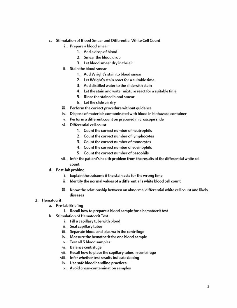
c. Stimulation of Blood Smear and Differential White Cell Count i. Prepare a blood smear
1. Add a drop of blood 2. Smear the blood drop 3. Let blood smear dry in the air
ii. Stain the blood smear 1. Add Wright’s stain to blood smear 2. Let Wright’s stain react for a suitable time 3. Add distilled water to the slide with stain 4. Let the stain and water mixture react for a suitable time 5. Rinse the stained blood smear 6. Let the slide air dry
iii. Perform the correct procedure without guidance iv. Dispose of materials contaminated with blood in biohazard container v. Perform a different count on prepared microscope slide
vi. Differential cell count 1. Count the correct number of neutrophils 2. Count the correct number of lymphocytes 3. Count the correct number of monocytes 4. Count the correct number of eosinophils 5. Count the correct number of basophils
vii. Infer the patient’s health problem from the results of the differential white cell count
d. Post-lab probing i. Explain the outcome if the stain acts for the wrong time
ii. Identify the normal values of a differential’s white blood cell count
iii. Know the relationship between an abnormal differential white cell count and likely diseases
3. Hematocrit a. Pre-lab Briefing
i. Recall how to prepare a blood sample for a hematocrit test b. Stimulation of Hematocrit Test
i. Fill a capillary tube with blood ii. Seal capillary tubes
iii. Separate blood and plasma in the centrifuge iv. Measure the hematocrit for one blood sample v. Test all 5 blood samples
vi. Balance centrifuge vii. Recall how to place the capillary tubes in centrifuge
viii. Infer whether test results indicate doping ix. Use safe blood handling practices x. Avoid cross-contamination samples

4
c. Post-lab Probing i. Explain the purpose of a hematocrit test
ii. Recall the normal hematocrit levels 4. Hemoglobin Content
a. Pre-lab Briefing i. Recall how to prepare a blood sample for a hemoglobin test
b. Simulation of Hemoglobin Test i. Test the three blood samples and positive and negative controls
ii. Stir until all hemoglobin is out of the red blood cells iii. Measure the hemoglobin content iv. Use safe blood handling practices v. Avoid cross-contaminating samples
vi. Recall why hemolysis applicators are used vii. Infer whether test results indicate doping
c. Post-lab Probing i. Explain the purpose of hemoglobin test
ii. Recall the normal hemoglobin content 5. Blood Typing Test
a. Pre-lab Briefing i. Recall how to determine the blood type
ii. Recall which transfusions lead to transfusion reactions b. Simulation of Blood Typing Test
i. Test all blood samples ii. Label the test slides
iii. Recall how the slides should be labeled iv. Add blood from only one patient to each slide v. Add the test serum to the labeled spot on the slide
vi. Determine the blood type vii. Use safe blood handling practices
viii. Avoid cross-contaminating blood samples ix. Recall why toothpicks are used in this experiment x. Use your results to determine who can donate blood to whom
c. Post-lab Probing i. Realize the need for type O packed cell transfusion when donor and recipient do
not exactly match 6. Final Summary Questions
a. Differentiate between the purpose of the various blood tests
INSTRUCTOR NOTE
: Safety requirements for blood handling may vary slightly from those used in this lab. Students may become frustrated if they begin to miss questions. Remind them that when missing a question they should remediate using the provided learning resource, most often a Slide, or the Library for that topic.

5
Student Instructions for Lab Experiments
In the following exercises you will perform tests that allow you to examine the nature of blood and also let you evaluate different samples of blood.
Overview for All Experiments:
These tests are useful diagnostic tools for physicians because blood composition reflects the status of many body functions and malfunctions.
Before getting started on the actual lab, I would like to go over some core concepts related to blood testing. Then you will proceed with the experiments.
In this experiment, you will prepare a microscope slide with a blood smear and perform a differential white blood cell count.
Differential WBC Count:
Before you start, I want to make sure that you have the necessary knowledge to execute the experiments.
Let’s make sure you know how to prepare a blood smear microscope slide and how to perform a differential white blood cell count.
Important to Know About Blood Samples:
• What is a blood smear and how to make one • How to stain a blood sample • How to identify the different white blood cells • What is a differential white blood cell count
Drag the labels from the right hand side to the correct locations on the slide. Select “Submit” when you are done.

6
INSTRUCTOR NOTE
Drag the labels from the right hand side to the correct locations on the slide. Select “Submit” when you are done.
: Often the Coach will appear at the top right. Sometimes students think she is in the way of completing the exercise. However, if they are patient, she will disappear when she completes talking. Students can reactivate her and make her repeat instructions by clicking on her refresh icon.

7
Simulator:
Click the Instructions button and follow the steps to make a blood smear.
Move the slide to the microscope to view it.
First, correctly focus the microscope slide. Move to the x40 objective to complete the count.
Hematocrit:
In this experiment, you will measure the hematocrit of blood samples.
Before we begin, I want to make sure you have the knowledge you need to execute the experiment and interpret your results.
Let’s learn more about the hematocrit of a blood sample
Important to Know About Hematocrit Testing:
• What is the hematocrit value • How is a hematocrit test performed
Drag the labels from the right hand side to the correct locations on the slide. Select “Submit” when you are done.

8
Simulator:
Click the Instructions button and follow the steps to determine the hematocrit.
Compare the hematocrit to blood doping samples.
INSTRUCTOR NOTE: Students will see a number of possible combinations of doping results and hematocrit levels. If they repeat the experiment, they should expect different results. Each student should have different results.
Hemoglobin Content:
In this experiment, you will measure the hemoglobin content of blood samples.
Before we begin, I want to make sure you have the knowledge you need to execute the experiment and interpret your results.
Let’s learn more about the hemoglobin content of blood and how to determine it.
Important to Know About Hemoglobin
• Hemoglobin in red blood cells • How to measure the hemoglobin content of blood
Student labeling activity before entering lab simulation

9
Simulator:
Click the Instructions button and follow the steps to determine the hemoglobin content.
Compare the hemoglobin content to blood doping samples.
INSTRUCTOR NOTE: The two halves in the hemoglobinometer will not have a line between them when the correct reading is available. Students will see a number of possible combinations of doping results and hemoglobin concentrations. If they repeat the experiment, they should expect different results. Each student should have different results.
Blood Typing:
In this experiment, you will determine the blood type of some blood samples.
Before we begin, I want to make sure you have the knowledge you need to execute the experiment and interpret your results.
Let’s learn more about the blood typing test.
Important to Know About Blood Typing
• How to determine the blood type • Transfusion reactions
Student labeling activity before entering lab simulation

10
Simulator:
Click the Instructions button and follow the steps to determine the blood types.
Remember to label your slides with the sample and test types.
INSTRUCTOR NOTE: Students will see a number of possible combinations of blood types. If they repeat the experiment, they should expect different results. Each student should have different results.
Final Summary Questions a. Differentiate between the purpose of the various blood tests
INSTRUCTOR NOTE: These final summary questions are designed to assess students that have completed all components of the lab. If you only assign some of the exercises and have not instructed students on the other techniques in class, your students may struggle with some of these questions.
Type of Student Report
Students are provided the following types of reports at the conclusion of these lab experiments.
I. Blood Smear and Differential White Cell Count – Debriefing II. Hematocrit – Debriefing III. Hemoglobin Content – Debriefing IV. Blood Typing Test – Debriefing