Embed Size (px)
Citation preview

Materials Science and Engineering C 68 (2016) 383–396
Contents lists available at ScienceDirect
Materials Science and Engineering C
j ourna l homepage: www.e lsev ie r .com/ locate /msec
Application of chemical mechanical polishing process on titaniumbased implants
Z. Ozdemir a, A. Ozdemir b, G.B. Basim a,⁎a Ozyegin University, Faculty of Engineering, Mechanical Engineering Department, Nisantepe Mevki, Orman Sokak No: 26, Cekmekoy, Istanbul 34794, Turkeyb TUBITAK Gebze Campus, Genetic Engineering and Biotechnology Institute, P.O Box 21, Gebze, Kocaeli 41470, Turkey
⁎ Corresponding author.E-mail address: [email protected] (G.B. Bas
http://dx.doi.org/10.1016/j.msec.2016.06.0020928-4931/© 2016 Elsevier B.V. All rights reserved.
a b s t r a c t
a r t i c l e i n f oArticle history:Received 14 January 2016Received in revised form 26 April 2016Accepted 1 June 2016Available online 06 June 2016
Modification of the implantable biomaterial surfaces is known to improve the biocompatibility of metallic im-plants. Particularly, treatments such as etching, sand-blasting or laser treatment are commonly studied to under-stand the impact of nano/micro roughness on cell attachment. Although, the currently utilized surfacemodification techniques are known to improve the amount of cell attachment, it is critical to control the levelof attachment due to the fact that promotion of bioactivity is needed for prosthetic implants while the cardiacvalves, which are also made of titanium, need demotion of cells attachment to be able to function. In thisstudy, a new alternative is proposed to treat the implantable titanium surfaces by chemical mechanical polishing(CMP) technique. It is demonstrated that the application of CMP on the titanium surface helps in modifying thesurface roughness of the implant in a controlled manner (inducing nano-scale smoothness or controlled nano/micro roughness). Simultaneously, it is observed that the application of CMP limits the bacteria growth byforming a protective thin surface oxide layer on titanium implants. It is further shown that there is an optimallevel of surface roughness where the cell attachment reaches a maximum and the level of roughness is control-lable through CMP.
© 2016 Elsevier B.V. All rights reserved.
Keywords:Implantable biomaterialsSurface structuringChemical mechanical polishingBiocompatibility
1. Introduction
Biomaterials are commonly used to make implantable structuressuch as dental prostheses, orthopedic devices, cardiac pacemakers,stents and catheters [1]. The choice of an adequate implant materialfor a selected application is based on the bio-stability and bio-compati-bility of the material once the required mechanical strength and dura-bility is achieved. Commercially, pure titanium and its alloys (mostcommonly Ti-6Al-4V) are widely used as structural biomaterials dueto their extraordinary properties such as high mechanical strength perunit volume in addition to their high corrosion resistance due to theirstable passive oxide layer [1–4]. The native oxide layer of titaniumthat spontaneously forms in air is in nanometer scale (3–10 nm) andit is typically amorphous and stoichiometrically defective [5–6]. To bea protective oxide film, the formed TiO2 layer has to be continuous,pore free and adhesive. Hence, although the native oxide film of titani-um is known to be protective, additional oxidation treatments such aschemical etching [6], thermal treatment [7] and electrochemical anodi-zation [8] of the titanium surface are practiced to grow an oxide layer bycontrolling the thickness, porosity, crystal structure of the oxide toenhance the wear characteristics as well as the bio-compatibility.
im).
The nano-scale oxide layer of titaniumhasmultiple functionalities inbiomaterial implant Spontaneously, when the titanium oxide film iscontinuous and pore free, it can also prevent the titanium ion dissolu-tion once the implant is implanted applications. First of all, it is knownto promote the biocompatibility of the titanium by enhancing cell at-tachment [9]. It also serves as an adhesion layer between the implantand the bone tissue (commonly simulated with hydroxyapatite-HA)particularly when an anatase crystalline structure is formed [10],which justifies the deposition of the TiO2 layers on the bare titanium im-plants [7]. In addition, the formation of a protective oxide film of titani-um helps shield the implant surface against corrosion by stopping theoxygen diffusion as shown by electrochemical analyses in detail [4]. Asthe implants are exposed to aggressive environments such as in bodyfluids (particularly in the acidic mouth environment when the dentalimplants are considered) the surface properties of this film becomesmore important [4]. Consequently, it is favorable to promote the forma-tion of the protective oxide layer of titanium during its processing forthe implant applications.
In addition to the characteristics of the titanium oxide film, thesurface topography of the titanium implants has also been studied in-tensively to analyze the effect of surface roughness on the biocompati-bility. There are many processing techniques adopted to modify theimplantmaterial surface roughness and concurrently the chemical com-position [11]. Gupta and coworkers classified the available surface
384 Z. Ozdemir et al. / Materials Science and Engineering C 68 (2016) 383–396
modification techniques into four main groups including (i) themethods to increase surface roughness, (ii) chemical etching, (iii) vari-ous methods of coating and (iv) the surface chemical and chemical to-pography modifications [8]. Blasting is one of the most commonlypracticed techniques to increase surface roughness in which particlesof various diameters of mainly alumina (Al2O3) and titania (TiO2) withparticle size ranging from small, medium to large grit are used to blastthe surfaces of the implant [12,13]. The obtained roughness dependson the particle size, time of blasting, pressure and distance from thesource of particle to the implant surface. The createdmicro-scale rough-ness allows adhesion, proliferation and differentiation of bone cells(osteoblasts) yet the soft tissue cells (fibroblasts) adhere to the surfacewith difficulty and hence this method can limit soft tissue proliferationand increase bone formation. However, some particles are commonlyleft on the surface after blasting which may impair bone formation bya possible competitive action on calcium ions [14]. Chemical etching isanother method in which the implant is dipped into an acidic environ-ment and the surface structure changes as a function of acid type, con-centration and exposure time. This method has also been observed topromote osteointegration and implemented following the sand blastingon the implant surfaces as well [15]. However, a yellowish, blurrylooking film was observed to form on the implant surface after theetch operation [16]. Blasting method is also practiced by using only hy-droxyapatite (HA) particles or biphasic calcium phosphate (BCP) parti-cles. BCP is a mixture of the hydroxyapatite and beta-tricalciumphosphate. Implementation of an etch procedure is also common fol-lowing the BCP treatment. The advantage of BCP is that the surfacecan be structured at micro-scale due to higher hardness of the BCP par-ticles relative to pure HA. Moreover, even if the HA or BCP particlesmaybe left on the implant post blasting operation, they do not adverselyimpact biocompatibility since both of these minerals are known to havesimilar compositions to the bone tissue and they are commonly utilizedto mimic the osteoblast attachment for implant studies [17]. Other thanblasting and etching, application of coating on the implant surfaceswithHA by using plasma spraying, laser ablation, pulsed laser deposition,sputtering or simply dip coating are also practiced [8]. While thesemethods help improve the cell attachment, they are also prone todelamination at the implant/coating interface, which may fail thehealthy integration of the implant with the surrounding tissue [8,18].Hydroxyapatite composites with zirconia and alumina were alsoshown to be good coating materials particularly for dental implantspromoting the mechanical properties of the implants in addition to en-hanced osteointegration [18]. Finally, the surface chemical and chemicaltopography modifications involve the treatment of the implant surfaceby the control of surface chemistry and attachment of proteins andcell adhesionmolecules on the surfaces of the implant through electricaldouble layer interactions [8,19].
All the surface modification methods mentioned so far tend toform random surface structures on the implant surface rangingfrom nano to micro scale roughness. The isotropic nature of the sur-face structures result in equal probability of the cell attachment onthe implant surface. To induce anisotropic surface structuring,more recently introduced alternative of implant surface structuringis the utilization of lasers [20–23]. The use of lasers allows themanufacturing of anisotropic surfaces which can help grow thecells in a specific direction. Furthermore, laser structuring can helpin controlling cell attachment between the osteoblast which prefermicro-scale roughness and the fibroblast which is preferably betterattached on the nano-rough surfaces [22,23]. It is also known thatalthough micro-scale roughness has been studied intensively to pro-mote cell attachment, the cellular events takes place on the nano-scale for cell-substrate interactions as the nano-metric cues havebeen shown to influence the cell activities [6,22,23]. Therefore, thescale of surface roughness from nano towardsmicro is critical to con-trol bioactivity by selective promotion or demotion of the cellattachment.
In the present study we introduce chemical mechanical polishing(CMP) technique as an alternative to surface structuring of the biomed-ical implants [24]. CMP has initially been introduced for glass polishingand extended into the planarization of the interlayer metal connectorsand dielectrics in microelectronics manufacturing [25]. In CMP process,the top film surface of the material is exposed to the chemicals inthe polishing slurry which is made of submicron size particles andcorrosives. This interaction forms a chemically altered topfilm that is re-moved by the mechanical action of the slurry abrasive particles. There-fore, it is a different method as compared to the mechanical polishingtechniques used for implant surface finishing [26]. The chemically al-tered top films have to be a protective oxide to enable planarizationby stopping chemical corrosion on the recessed metal surfaces whilethe elevated structures are polished [27]. Titanium CMP is performedin microelectronics to planarize Ti/TiN layers used as barriers to alumi-num interconnect diffusion to the dielectric layers [28]. Furthermore, ithas been shown by an earlier study that the application of CMP on Tifilms has been very successful in terms of creating a titanium oxidefilm on the surface that might also help promote biocompatibility inaddition to helping removal of the reacted and contaminated surfacelayers [16,29]. The comparison of the electrochemical etch to CMPapplication on titanium has shown that the titanium surfaces treatedby CMP using colloidal silica slurries and an oxidizer concentration of3 wt% were much clear and compositionally continuous than the yel-lowish and blurry titanium oxide layers formed by electrochemicaletching. Furthermore, in this study CMP is synergistically utilized to in-duce nano-scale smoothness or nano/micro scale roughness on thebioimplant surface. Particularly, we focus on the dental implants tochange the surface roughness in a control manner. Implementation ofthe CMP process on titanium bio-implants results in a synergy by(i) cleaning the implant surface from potentially contaminated surfacelayers by removing a nano-scale top layer during the process, (ii) simul-taneously creating a non-porous and continuous nano-scale oxide filmon the surface to limit any further contamination to minimize risk ofinfection and prevent corrosion and (iii) inducing controlled surfacesmoothness/roughness by designing the CMP process variables suchas slurry particle size, solids loading as well as the oxidizer type andconcentration.
In order to demonstrate the use of CMP on dental implants, bothtitanium plates and dental implants were processed. The CMP processwas carried out by using alumina based slurries and H2O2 as an oxidizeron commercially pure (cp) Ti samples with different polishing padsto modify surface topography. CMP characterization was performedby material removal rate, surface roughness by using atomic forcemicroscopy (AFM) and wettability analysis (through contact anglemeasurements) in addition to the surface topography and elementalcomposition analyses on the treated surface by X-ray diffraction(XRD), energy dispersive X-ray spectroscopy (EDX) and X-ray photo-electron spectroscopy (XPS). Biological evaluations were performedby cytotoxicity evaluations in addition to bacterial and cell attachmenttests and hydroxyapatite adhesion through wet deposition.
2. Materials and methods
2.1. Materials
The original titanium foil sample surface, which is considered asbaseline in this experimental study, was annealed. Fig. 1a illustratesthe optical micrograph of the anodized titanium plate surface (200X)as well as the SEM cross sectional image illustrating the thick and po-rous oxide layer with 30–40 μm thickness. Fig. 1b shows the titaniumbased dental implant used to implement the optimized CMP conditionsthrough hand polishing on a 3-D sample. The dental implants wereprovided by MODE Medical Company and they were only shaped bymachining prior to their exposure to the CMP testing.

Fig. 1. Surface structures of (a) optical image of baseline anodized titanium plate sample (200× magnification) and SEM x-section illustrating thick surface oxide, (b) baseline machineshaped dental implant sample with no additional surface treatment (4× magnification).
385Z. Ozdemir et al. / Materials Science and Engineering C 68 (2016) 383–396
2.2. Methods
2.2.1. Chemical mechanical polishing experimentsCMP experimentswere conducted on a tabletop Tegrapol-31 polish-
er. Fig. 2a shows the 2-dimensional standard CMP set up as it is devel-oped for the polishing and planarization of the 2-D structures. CMPslurries were prepared by using 5% weight alumina (Al2O3) abrasiveswith 50 nmparticle size at pH4 using nitric acid. In order to provide sta-bility, the suspensionswere ultrasonicated long enough by repeated pHadjustment until the slurry was fully stabilized. CMP tests were con-ducted at 70 N downforce which is equivalent to a 7.88 psi pressureon the used sample size (14 × 14 mm). The titanium plates werepolished by using a SubaIV subpad stacked under a polytex buff pad tosmoothen the surface while protecting the macro-scale shape of theplates. This soft-pad configuration enables a gentle interaction betweenthe pad and the implant surface providing local smoothening whilemaintaining the physical shape of the surface that is required for thescrew pitch of the dental implants. In addition, two sizes of sand paper(silicon carbide 150C and P320) were used in place of the polishingpad to create the micro structures through CMP. Fig. 2b and c illustratethe CMP pads and the sample holder, respectively. In Fig. 2b, the picturewith dark colored square surface structure belongs to the Polytex padused as the prime pad and the lighter colored pad with the circularholes belongs to the Suba IV sub-pad. Hydrogen peroxide (H2O2−Sigma Aldrich, %34.5–36.5 purity) was utilized as an oxidizer in theCMP experiments except for the baseline sample which received noadditional treatment. Furthermore, one of the samples was polishedwithout an oxidizer to expose the underlying titanium metal in orderto understand the effect of formation of an oxide layer during CMP onbio-compatibility. Samples ran with the polymeric CMP pads and
Fig. 2. CMP configuration for 2-D titanium plates (a) tabletop CMP to
abrasive papers were polished for 2 min with 3 wt% oxidizer addition.Material removal rates were calculated through weighing the samplespre and post polish by Swiss Made ES125SM model precise scientificbalance (five digits after the decimal point, 0.01 mg accuracy). All sam-pleswere cleaned in ultrasonic bathwith pH 4-adjustedwater for 5minand dried with nitrogen gas before they were characterized. Same ex-perimental procedure was implemented on the 3-D dental implantsamples obtained from Mode Medical Limited by using a polymericbrush and flowing slurry on the samples as they were hand polished.
2.2.2. Surface characterization experiments
2.2.2.1. Wettability characterization. All the 2-D and 3-D samples werecharacterized for wettability through contact angle measurementswith simulated body fluid (SBF) by using a KSV ATTENSION Theta LiteOptic Contact Angle Goniometer using the sessile drop method. Fivedrops were measured on each sample. The drop images were storedby a camera and an image analysis system calculated the contact angle(Θ) from the shape of the drops.
2.2.2.2. Surface topography and roughness characterization. The surfacetopographies of the 2-D specimens were examined by NanomagneticsAtomic ForceMicroscope (AFM) using contactmode. Surface roughnessvalues were recorded on 10 × 10 μm scan area and reported as an aver-age of minimum three measurements taken on the samples. CMP gen-erated metal oxide thin films are verified to be in nanometer scales(1–10 nm) and high energy beams of the Scanning ElectronMicroscopy(SEM) results in damage on these ultra-thinfilms, or require a coating tobe applied (which changes the nature of the thin oxide layer). There-fore, AFM technique was preferred for characterization of the CMP
ol (b) polytex and Suba IV polishing pads and (c) sample holder.

386 Z. Ozdemir et al. / Materials Science and Engineering C 68 (2016) 383–396
induced titanium surfaces (AFM does not require a coating applicationon the surface) [27,30]. SEM analyses were conducted to obtain thecross sectional analyses on the original titanium plate by JEOL JIB-4501SEM to analyze the thickness of the anodized titanium oxidelayer. Furthermore, profilometry analyses were performed to measurethe roughness values of the titanium plates at a larger scale by using aMitutoyo SJ-400 profilometer. 4 mm lengths were scanned on the sam-ples on three different locations and averaged for the average roughness(Ra) and average roughness depth (Rz) values.
2.2.2.3. Surface crystallographic structure analyses. In order to analyze thechanges in the nature of the very top thin film that is modified by thechemical action of the CMP process, grazing angle GIXRD analyseswere also conducted by using PANalytical, X'PERT Pro MPD modelXRD analyzer. The XRD profiles were collected between 20–80° of 2Θangles with a step interval of 0.02° using grazing angles for themeasurements.
2.2.2.4. Surface chemical composition analyses. The chemical nature of thetitanium surfaces was also studied through X-ray photoelectron spec-troscopy (XPS) and Energy Dispersive X-ray (EDX) analyses. PHOIBOSHAS 3500 150 R5e XPS [HW Type 30:14] tool was used to comparethe electronic states of the Ti2p and O1s of the titanium plates pre andpost CMP. Furthermore, EDX analyses were performed on a JEOL JIB-4501 MultiBeam Scanning Electron Microscope (SEM). The titaniumpeaks were investigated at 0.4 and 4.5 eV and oxygen peak was ana-lyzed at the 0.525 eV as reference values [13].
2.2.3. Biostability and biocompatibility analyses
2.2.3.1. Cytotoxicity analyses. ISO 10993-5 cytotoxicity test procedurewas adapted to evaluate the cell viability on the samples treated withand without CMP. L929 mice fibroblast cells were used to representthe mammalian system. Cells were counted and seeded onto the wellplate at a concentration of 104 cells/plate. The titanium samples werekept in the solutions prepared as per the ISO 10993-5 procedures for72 h and the solution extracts were added to the cell plates at 37 °Cand retained for 24 h in a %5-CO2 media. Cell viability was evaluatedvia WST-1 agent by colorimetric testing.
2.2.3.2. Bacteria attachment evaluations. Titanium plates were sterilizedin an autoclave at 120 °C temperature for 20 min before the microbio-logical analysis. Cronobacter sakazakii (Gram-) bacteria was used asthe species to evaluate the bacteria attachment. 100 μl of microorgan-isms from the nutrient broth microbial stock were spread on nutrientagar plates under sterile conditions. After the cultivation of bacteria,sterilized Ti samples were placed into each plate and incubated at37 °C. The bacteria growth zone was observed over 1, 3 and 7 daysand quantified by measuring the thickness of the colonies grown atthe periphery of the plates through photographs taken on the sam-ples (Fig. 3a).
Fig. 3. Biological evaluation set-up for (a) bacterial growth analyses, (b) cell attachment testcounting fibroblast cells.
2.2.3.3. Cell growth analyses. Titanium plates were cut into circular disksfor the cell-growth evaluation to fit into well plate-18 and sterilizedwith UV radiation. L929 fibroplast cells were amplified in the laboratoryfor the proliferation on the samples. The cells were seeded directly ontop of the Ti plates which were placed at the bottom of the wells. Thenutrient medium was changed every 3 days to help keep the fibroblastcells alive. After 1, 3, and 5 days of incubation periods, the grown cellswere washed off the titanium plates.
In order to test the cell attachment on the titanium plates, initially7.8 g DMEM-F12 (Dulbecco's Modified Eagle Medium, D8900 Sigma)and 0.6 g NHCO3 (dissolved in 450 ml ultra-pure water) was mixed toprepare a total 500 ml solution. The pH of the solution was adjusted to7.3 with the addition of NaOH. 50 ml FBS (fetal bovine serum) wasadded afterwards to make a 10 wt% FBS concentrated solution. 5 ml ofantibiotic Penicillin Streptomysin (100X) was added as the last compo-nent and the solution was mixed thoroughly and filtered as a final step.L929 fibroblast cells were grown in the prepared nutrient media insidethewell plates in the presence of the prepared Ti samples. The excessivesolution was removed from the well plates with vacuum ant the plateswere washed with phosphate buffer solution (PBS). The grown cellswere separated from the plates by using Tripsin into the well plates.The cellswere placed into the falcon tubes and centrifuged at room tem-perature at 1300 rpm. The settled cells were diluted to a 104 cells/cm2
concentration and transferred on to the Thoma lamel for countingunder the microscope as shown in Fig. 3b and c.
2.2.3.4. Hydroxyapatite attachment analyses. Hydroxyapatite (HA) isknown to be mimicking the bone tissue as it is well studied in the liter-ature and consequently it helps promote the osteoblast cell attachmentas a coating on the implants [31–35]. In order tomimic the bone cell re-sponse, HA attachment was evaluated on the titanium implants by pre-paring a solution by using Ca and P routes namely the calcium nitrate(Ca(NO3)2) and diammonium hydrogen sulphate ((NH4)2HPO4) ac-cording to the reaction given in the following chemical Eq. (1) [31].
10Ca NO3ð Þ2 þ 6 NH4ð Þ2HPO4 þ 8NH4OH→Ca10 PO4ð Þ6 OHð Þ2þ 20NH4NO3 þ 6H2O ð1Þ
Before deposition, titanium samples were washed in distilled waterin an ultrasonic bath. Deposition was carried out by dipping thetitanium plates into the HA solutions for 72 h [32,33]. The HA growthwas evaluated through weight differences pre and post the coatingprocedure. This evaluation is an indication of how well the osteoblastcells will attach to the CMP processed surface in addition to the plausi-bility of coating the implant surfaces with HA to further promotebiocompatibility.
3. Results and discussion
The baseline titanium plates were anodized and hence had a porousoxide layer on the surface as it is seen in Fig. 1a. In order to understandthe impact of the presence of oxide as well as the nature of the oxide
incubation well plates and (c) microscopic image of L929 cells on Thoma lamel used for

387Z. Ozdemir et al. / Materials Science and Engineering C 68 (2016) 383–396
film on the titanium surface, a baseline sample was subjected to surfacecharacterization and biological evaluations. In addition, another samplewas prepared by implementing CMP treatment by using 3 wt% aluminananoparticle containing slurry at pH 4 without addition of the H2O2 ox-idizer. This approach tunes the CMP process to function only mechani-cally and helps remove the surface oxide of the plates and expose thebare titaniumwithout planarization due to the lack of chemical compo-nent. In addition, CMP was performed in the presence of oxidizer addi-tion to the alumina slurries (3 wt% H2O2) in order to compare thetitanium oxide films that forms through the CMP process to the baselineanodized oxide film in terms of bioactivity performance. In these pre-liminary CMP evaluations, a relatively soft polytex buff-pad was usedon top of a SUBA IV subpad to provide smooth surface finish whileprotecting the topography of the surface such as the screw crests androots of the dental implants. Following these treatments, two types ofabrasive papers were used to induce micro-scale roughness to the tita-nium plates to evaluate the effect of surface nano-structuring on thematerial and biological responses by using the slurries in the presenceof 3 wt% oxidizer. Consequently, five types of sample surfaces were pre-pared as (i) baseline, (ii) CMP without H2O2 which exposes bare titani-um surface, (iii) CMP treated in the presence of H2O2, (iv) CMP treatedin the presence of oxidizer by using 45-μm grid abrasive paper and (v)CMP treated in the presence of oxidizer by using 90-μm grid abrasivepaper. These five samples were evaluated for their CMP responses, sur-face characterization as well as the biological performances. Moreover,the 3-D dental implants were also exposed to the same set of treat-ments, as detailed in the materials and the methods section, and evalu-ated for their CMP responses and project corresponding biologicalperformances.
3.1. CMP performance and wettability evaluations
Titanium plates treated with the five conditions as described abovewere initially characterized for the CMPmaterial removal rate, wettabil-ity, and surface topography responses evaluated through surface rough-ness measurements. Table 1 summarizes the post CMP evaluations ofthe experiments conducted on the baseline and CMP treated titaniumplates. It can be seen that the material removal rates of the samplespolished on the polymeric pads were negligible. Particularly, the CMPtest conducted without the addition of oxidizer resulted in only0.007 μm/min material removal rate. This result is expected since thematerial removal is driven by the continuous chemical attack on thesurface by the oxidizer during the CMP operation. Consequently, whenthe oxidizer is added into the system, material removal rate of 0.5–0.9 μm/min was obtained. On the other hand, polishing with the abra-sive papers resulted inmuchhigher removal rates due to the highly pro-nounced mechanical action provided by the fixed abrasive particlesembedded into the polishing papers. Although, this very high level ofmaterial removal rates are typically not desired for the CMP applica-tions, it must be noted here once again that the abrasive papers helpmodulate the surface roughness significantly to understand the effectof roughness on the bioimplant performance. Fig. 4a also illustratesthe trend in the material removal rates of the samples treated with
Table 1Material removal rate, wettability and surface roughness responses of the titanium plate samp
Sample CMP conditions Material removal rate (nm/min) We
Time (min) Pad type
As is – – – 84.CMP without H2O2 2 Polytex pad 7 ± 2 45.CMP with 3 wt% H2O2 2 Polytex pad 505 ± 395 34.CMP with 3 wt% H2O2 2 Ab.P.(45 μm) 30,113 ± 3039 54.CMP with 3 wt% H2O2 2 Ab.P.(90 μm) 37,260 ± 3882 65.
various CMP conditions highlighting the much higher removal rateswith the abrasive papers.
Fig. 4b summarizes the contact angle measurements taken with thesimulated body fluid on the titaniumplates representing thewettabilityof implant surface in the body environment. The high contact anglevalue obtained on the untreated baseline titanium sample can be attrib-uted to the surface oxide formed by anodization which has a porousstructure [34,35]. The trapped air in the porous titanium oxide is be-lieved to increase the hydrophobicity of the surface. The CMP processperformed without the oxidizer addition resulted in removal of thetop oxide layer and exposed the titanium surface with a ~ 45° contactangle measured, which is approximately half of the value measuredon the baseline sample (85°). This observation confirms that the thickanodized oxide film was removed from the titanium surface duringthe polishing with water since the wettability response of the samplechanged significantly. The decrease in the contact angle of the bare tita-nium surface can be attributed to the higher surface energy of the fresh-ly exposed titanium atoms resulting in higher interaction with thewatermolecules that leads to higher surfacewettability and hence a de-crease in the contact angle value. For the following treatments whereCMP was conducted in the presence of an oxidizer, the effect of surfaceroughness on the contact angle response started to dominate in parallelto the observations in the literature [21,22]. Fig. 5 shows theAFMmicro-graphs and corresponding cross sectional analyses of the titaniumplatestreated by using five different experimental conditions. Here, it can beseen that the surfaces with a smoother surface finish, such as in thecase of CMP application in the presence of the oxidizer (Fig. 5c), resultedin more wettability and hence a lower contact angle, and the surfaceswith the induced micro-roughness (such as the samples polished withabrasive papers) resulted in a higher contact angle that can be attribut-ed to the loweredwettability through the trapped air pocketswithin thegrooves on the surface. Onemore important factor that can be observedby comparing the micrographs in Fig. 5 is that, the CMP of the surfacesby using the soft polytex pad helps smoothen the surfaces locallywhile protecting the macro scale topography. This effect can be seenwhen the cross-sectional profiles of the baseline sample (Fig. 5a) andthe CMP treated sample (Fig. 5c) are compared.
Table 1 summarizes the surface roughness evaluations of thesamples treated with the five experimental conditions by taking10 × 10 μm surface scans and averaging the measurements on threesamples. The original sample had a high root mean square (RMS) sur-face roughness (486 ± 17 nm) that can be attributed to the porousoxide layer formed by anodization. When the surface was buffed withCMPwithout the addition of the H2O2, the porous surface oxide was re-moved and the surface topography reduced to 205 ± 32 nm. CMP pro-cess in the presence of the oxidizer at 3 wt% and using the polytex buffpad further reduced the surface roughness to 128± 41 nm, achieving asmoother surface finish. When the abrasive papers were used, on theother hand, surface roughness increased as the grid size increasedreaching up to 517 ± 88 nm when the 90 mm grit size paper was usein place of the pad material.
The larger scale roughness measurements conducted with theprofilometer were consistent with the AFM roughness values on thehigh surface roughness samples including the baseline sample and the
les treated with five different experimental conditions.
ttability contact angle (θ) RMS surface roughness (nm) Profilometer surfaceroughness (nm)
Ra Rz
4 ± 0.7 486 ± 17 425 ± 40 2660 ± 506 ± 1.2 205 ± 32 410 ± 20 2570 ± 603 ± 2.6 128 ± 41 350 ± 30 2470 ± 1202 ± 0.2 423 ± 56 350 ± 50 2770 ± 601 ± 0.7 517 ± 88 540 ± 220 4170 ± 1800

Fig. 4. Results of the (a) material removal rate and (b) surface wettability of the baseline and CMP treated Ti plates.
388 Z. Ozdemir et al. / Materials Science and Engineering C 68 (2016) 383–396
samples processed with CMP by using the abrasive papers. As it canbe seen in Table 1, the baseline sample was measured as 486 ± 17 nmby AFM and 425 ± 40 nm (Ra) by the profilometer as an example.These values are statistically the same. However, for the samplesprocessed by using the soft polishing pad in the absence and thepresence of the oxidizer, AFM roughness values were reported as205 ± 32 nm and 128 ± 41 nm, whereas profilometer roughnessvalues were 410 ± 20 nm and 350 ± 30 nm, respectively. For thesesmoother samples, the local roughness measurements by AFM aregiving smaller values as compared to the larger scale measurementsby the profilometer and the results are statistically different. Thisfinding is supporting the fact that the CMP treatment by the smoothpad usage is decreasing the surface roughness locally while stillprotecting the global curvature of the sample as desired. This resultis also confirmed from the Rz measurement taken by theprofilometer, in that, the average roughness depths are comparablefor the relatively smoother samples (~2400–2800 nm) yet the sam-ple treated by using the largest grid abrasive paper has a Rz valueof 4170 nm. These results further support the utilization of CMP asa method to control the surface roughness to enable the tuning forthe needed biocompatibility.
Fig. 5. Post CMP treatment AFMmicrographs and pre and post CMP cross sectional images of tCMP with 3% H2O2 oxidizer (d) post CMP with 3% H2O2 oxidizer with 45 μm grit abrasive pap
3.2. Characterization of the surface oxide layer
The nature of the surface oxide forming on the titanium plates char-acterized for elemental composition as well as for the crystal structure.In order to determine the elemental composition, XPS analyses wereperformed. Furthermore, the changes in the crystallographic nature ofthe titanium surface in the absence and presence of the oxidizer in theCMP slurries were analyzed by XRD analyses to understand the protec-tive nature of the surface oxide.
Fig. 6 shows the XPS spectrum of CMP treated titanium plates in theabsence (only by using water in the CMP slurry) and presence of 3 wt%H2O2 at the 2p orbital region of the titanium (Ti2p-region), and the 1sorbital region of the oxygen (O1s-region). This analysis was conductedto determine the changes in the intensities of the typical titanium andthe oxygen peaks of the titanium plates as they are known to confirmthe protective oxide formation on the surface [16]. A prominent Ti2p3/2 peak was observed at 459 eV region (Fig. 6a), which correspondsto the binding energy of Ti 2p3/2peak conformed to that of Ti in TiO2.Similarly, O 1s peak is positioned at the 530–535 eV region (Fig. 6b),which can be assigned to the three chemical components of oxygennamely (i) the lattice oxygen O2− (530.3 eV), (ii) the bridging and
he titanium plates (a) as received baseline sample (b) post CMP without oxidizer (c) poster and (e) post CMP with 3% H2O2 oxidizer with 90 μm grit abrasive paper.

389Z. Ozdemir et al. / Materials Science and Engineering C 68 (2016) 383–396
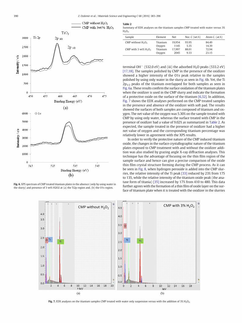
Fig. 6.XPS spectrum of CMP treated titanium plates in the absence (only by usingwater inthe slurry) and presence of 3 wt% H2O2 at (a) the Ti2p region and, (b) the O1s region.
Fig. 7. EDX analyses on the titanium samples CMP treated with
Table 2Summary of EDX analyses on the titanium samples CMP treated with water versus 3%H2O2.
Sample Element Net Nor. C (wt.%) Atom C. (at.%)
CMP without H2O2 Titanium 19,954 93.95 84.49Oxygen 1145 5.35 14.39
CMP with 3 wt% H2O2 Titanium 17,997 88.01 72.94Oxygen 2045 9.33 23.15
390 Z. Ozdemir et al. / Materials Science and Engineering C 68 (2016) 383–396
terminal OH− (532.0 eV) and (iii) the adsorbed H2O peaks (533.2 eV)[17,18]. The samples polished by CMP in the presence of the oxidizershowed a higher intensity of the O1s peak relative to the samplespolished by using only water in the slurry as seen in Fig. 6b. Yet, the Ti2p3/2 peaks of the titanium overlapped for both samples as seen inFig. 6a. These results confirm the surface oxidation of the titaniumplateswhen the oxidizer is used in the CMP slurry and indicate the formationof a protective oxide on the surface of the titanium [6,32]. In addition,Fig. 7 shows the EDX analyses performed on the CMP treated samplesin the presence and absence of the oxidizer with soft pad. The resultsshowed the surfaces of both samples are composed of titanium and ox-ygen. The net value of the oxygenwas 5.30% on the sample treatedwithCMP by using only water, whereas the surface treated with CMP in thepresence of oxidizer had a value of 9.02% as summarized in Table 2. Asexpected, the sample treated in the presence of oxidizer had a highernet value of oxygen and the corresponding titanium percentage wasrelatively lower in agreement with the XPS results.
In order to verify the protective nature of the CMP induced titaniumoxide, the changes in the surface crystallographic nature of the titaniumplates exposed to CMP treatment with and without the oxidizer addi-tion was also studied by grazing angle X-ray diffraction analyses. Thistechnique has the advantage of focusing on the thin film region of thesample surface and hence can give a precise comparison of the oxidethin film crystal structure forming during the CMP process. As it canbe seen in Fig. 8, when hydrogen peroxide is added into the CMP slur-ries, the relative intensity of the Ti peak [33] reduced by 23% from 175to 135, while the relative intensity of the titanium oxide peak (the ana-tase form of titania) [35] increased by 17% from 410 to 480. This datafurther agreeswith the formation of a thin filmof oxide layer on the sur-face of titanium plate when it is treated with the oxidizer in the slurries
water only suspension versus with the addition of 3% H2O2.

Fig. 8. XRD analyses on the titanium samples CMP treated with water only suspension versus with the addition of 3% H2O2.
391Z. Ozdemir et al. / Materials Science and Engineering C 68 (2016) 383–396
during the CMP process. It can be concluded that a denser film of titaniais formedwhen oxidizer is used as compared to the sample CMP treatedby using only water. This is due to the faster conversion of the titaniumatoms into titaniumdioxide in the presence of theH2O2 by oxidation re-action enhancing the protective nature of the oxide film on the implantmaterial surface [32,37,38].
3.3. Bio-stability and bio-compatibility evaluations
3.3.1. Cell viability analysesIn order to understand if there are any adverse effects of CMP treat-
ment on the titanium implant material, the preliminary biological anal-yses were conducted to test the cell viability after the CMP applicationand the results were compared to the untreated sample. Fig. 9 showsthe cytotoxicity test results conducted to evaluate the percent cell activ-ity on the polished surfaces as compared to the baseline and the knownpositive and negative samples [39]. The results confirmed that the cellviabilitywas not affected by the CMPprocesswithin the 72 h of the test-ing period. Furthermore, it is expected that the formation of the protec-tive oxide films of titaniumwill further limit the titanium dissolution inlonger term and hence improve the cell viability, which needs to bestudied through in vivo evaluations.
Fig. 9. Cell viability on the titanium samples treated with CMP as compared to the baselinebiocompatibility [35].
3.3.2. Bacteria growth analysesPost CMP treatment biological evaluations were also performed
through the bacteria growth analyses. Fig. 10 illustrates the growthzone thickness of the bacteria when the treated titanium plates wereplated upside down in the petri dishes containing the nutrient fluidafter 1, 3 and 7 days [40]. The baseline sample with a thick and porousoxide layer has shown an increase in the bacteria growth after thefirst day as the layer thickness increased from ~1.4 mm to ~1.8 mm.The same observation with a more pronounced effect has been notedon the titanium plate on which the oxide layer was removed throughCMP application without using an oxidizer. The bacteria zone thicknessincreased to ~1.9mm from thefirst day value of 0.9mm. This is believedto be due to the oxidation of the bare titaniumsurface in the nutrient so-lution. As the bacteria are known to grow on the oxide surfaces, the in-crease in the bacteria growth zone is potentially promoted when theoxide is formed [3,6]. This trend can also be explained by the increasedhydrophobicity of the surface by the formation of the oxide, which pro-motes the biocompatibility [41]. When CMP is performed with 3 wt%H2O2 addition, however, the results indicate that the increasing surfaceroughness promoted the bacteria zone thickness which can be attribut-ed to the better adhesion of the bacteria on the rougher surface. Yet, allthe sampleswere observed to retain an almost constant bacteria growthzone as a function of time after the CMP application. The consistency of
and control samples to observation of the CMP process and chemical applied samples
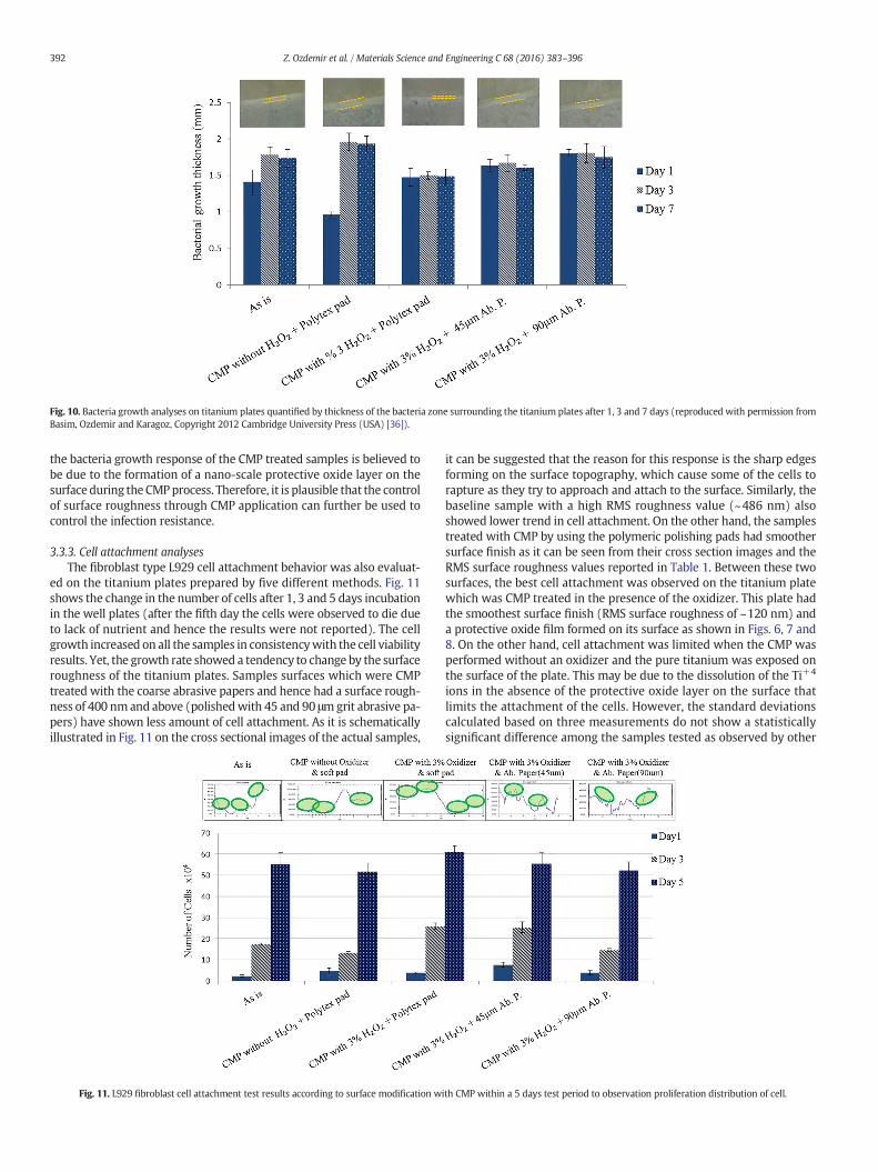
Fig. 10. Bacteria growth analyses on titanium plates quantified by thickness of the bacteria zone surrounding the titanium plates after 1, 3 and 7 days (reproduced with permission fromBasim, Ozdemir and Karagoz, Copyright 2012 Cambridge University Press (USA) [36]).
392 Z. Ozdemir et al. / Materials Science and Engineering C 68 (2016) 383–396
the bacteria growth response of the CMP treated samples is believed tobe due to the formation of a nano-scale protective oxide layer on thesurface during the CMPprocess. Therefore, it is plausible that the controlof surface roughness through CMP application can further be used tocontrol the infection resistance.
3.3.3. Cell attachment analysesThe fibroblast type L929 cell attachment behavior was also evaluat-
ed on the titanium plates prepared by five different methods. Fig. 11shows the change in the number of cells after 1, 3 and 5 days incubationin the well plates (after the fifth day the cells were observed to die dueto lack of nutrient and hence the results were not reported). The cellgrowth increased on all the samples in consistencywith the cell viabilityresults. Yet, the growth rate showed a tendency to change by the surfaceroughness of the titanium plates. Samples surfaces which were CMPtreated with the coarse abrasive papers and hence had a surface rough-ness of 400 nmand above (polishedwith 45 and 90 μmgrit abrasive pa-pers) have shown less amount of cell attachment. As it is schematicallyillustrated in Fig. 11 on the cross sectional images of the actual samples,
Fig. 11. L929 fibroblast cell attachment test results according to surface modification w
it can be suggested that the reason for this response is the sharp edgesforming on the surface topography, which cause some of the cells torapture as they try to approach and attach to the surface. Similarly, thebaseline sample with a high RMS roughness value (~486 nm) alsoshowed lower trend in cell attachment. On the other hand, the samplestreated with CMP by using the polymeric polishing pads had smoothersurface finish as it can be seen from their cross section images and theRMS surface roughness values reported in Table 1. Between these twosurfaces, the best cell attachment was observed on the titanium platewhich was CMP treated in the presence of the oxidizer. This plate hadthe smoothest surface finish (RMS surface roughness of ~120 nm) anda protective oxide film formed on its surface as shown in Figs. 6, 7 and8. On the other hand, cell attachment was limited when the CMP wasperformed without an oxidizer and the pure titanium was exposed onthe surface of the plate. This may be due to the dissolution of the Ti+4
ions in the absence of the protective oxide layer on the surface thatlimits the attachment of the cells. However, the standard deviationscalculated based on three measurements do not show a statisticallysignificant difference among the samples tested as observed by other
ith CMP within a 5 days test period to observation proliferation distribution of cell.

393Z. Ozdemir et al. / Materials Science and Engineering C 68 (2016) 383–396
researcher earlier [42]. Hence it can be stated that the CMP implemen-tation increases the tendency of cell attachment when it is applied inthe presence of oxidizers and using a soft pad promoting the smooth-ness. The sensitivity to the surface structuring is alignedwith the earlierliterature findings where the nano-scale structuring was observed toenhance the cell attachment [22,23].
3.3.4. Hydroxyapatite attachment analysesHydroxyapatite (HA) has been widely used as a coating material for
dental implants due to its chemical composition similar to natural bonemineral and its capability to promote bone regeneration [19,42–44]. Inthis study,we have evaluated theHAattachment on the samples treatedwith CMP and once again compared the attachment performance to thebaseline sample. Fig. 12a and b illustrate the HA attachment and thechange in the RMS surface roughness values as a function of the HAcoating, respectively. It can be seen that the attachment of HA increasedwith the increasing surface roughness. The smoothest surface obtainedby the CMP in the presence of the oxidizer and the polytex pad resultedin the minimum amount of HA attachment which was 1.4 mg/72 h,while the surface with the highest roughness (polished with 90-μmgrid paper) resulted in 2.1 mg attachment/72 h. In addition, the postHA coating surface roughness valueswere higherwhen the original sur-face roughness was higher. The AFMmicrographs given in Fig. 12b alsoclearly show the change in surfacemorphologywith theHA coating. It is
Fig. 12. HA attachment evaluation of the titanium samples (a) amount of HA attachment as acoating.
interesting to note that although the pre-HA coating surface roughnessof the baseline sample was similar to the roughness values obtainedwhen the abrasive papers were used for CMP applications (45 and 90-μm size sandpapers), the HA attachment was not similar on these sam-ples. Fig. 13 demonstrates this difference much better when the crosssectional AFM micrographs of all the samples are compared pre andpost HA deposition. Obviously, CMP induced surface roughness helpedpromote the HA attachment with a thicker layer deposited on the sur-face. This observation also supports the enhanced biocompatibility ofthe surfaces when CMP is applied since the HA attachment is knownto promote the cell activity with increasing roughness on the HA coatedsurface reported to increase the osteoblast cell attachment in the earlierstudies [42,43].
3.4. CMP application on 3-D dental implants
Fig. 14 summarizes the CMP performance of the 3-D dental implantsampleswhichwere obtained fromMODEMedikal Inc. The same exper-imental procedure was followed on the 3-D dental implant samples byreplacing the polymeric pad with a polymeric brush and hand polishingthe samples all over their exposed surfaces with the alumina basedpolishing slurry. CMP performances were evaluated based on themate-rial removal rates measured by the change in unit volume as a functionof time and also measuring the wettability responses of the implant
function of surface treatment and (b) measured RMS roughness values pre and post HA

Fig. 13. Post HA coating AFMmicrographs and pre and post cross sectional analyses of the titaniumplates (a) as received baseline sample (b) post CMPwithout oxidizer (c) post CMPwith3% H2O2 oxidizer (d) post CMP with 3% H2O2 oxidizer with 45 μm grit abrasive paper and (e) post CMP with 3% H2O2 oxidizer with 90 μm grit abrasive paper.
394 Z. Ozdemir et al. / Materials Science and Engineering C 68 (2016) 383–396
surfaces on a pre-selected region where the screw pitch is the same. Itcan be observed that both the material removal rate responses(Fig. 14a) and the wettability results maintained the same trend as
observed on the titanium plates. These results are encouraging in thatthe biological responses of the 3-D implants are expected to be similarto the 2-D equivalents.
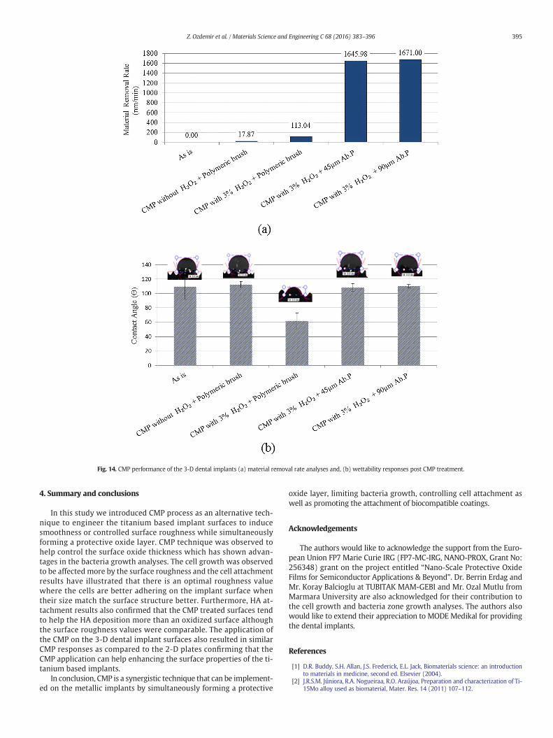
Fig. 14. CMP performance of the 3-D dental implants (a) material removal rate analyses and, (b) wettability responses post CMP treatment.
395Z. Ozdemir et al. / Materials Science and Engineering C 68 (2016) 383–396
4. Summary and conclusions
In this study we introduced CMP process as an alternative tech-nique to engineer the titanium based implant surfaces to inducesmoothness or controlled surface roughness while simultaneouslyforming a protective oxide layer. CMP technique was observed tohelp control the surface oxide thickness which has shown advan-tages in the bacteria growth analyses. The cell growth was observedto be affected more by the surface roughness and the cell attachmentresults have illustrated that there is an optimal roughness valuewhere the cells are better adhering on the implant surface whentheir size match the surface structure better. Furthermore, HA at-tachment results also confirmed that the CMP treated surfaces tendto help the HA deposition more than an oxidized surface althoughthe surface roughness values were comparable. The application ofthe CMP on the 3-D dental implant surfaces also resulted in similarCMP responses as compared to the 2-D plates confirming that theCMP application can help enhancing the surface properties of the ti-tanium based implants.
In conclusion, CMP is a synergistic technique that can be implement-ed on the metallic implants by simultaneously forming a protective
oxide layer, limiting bacteria growth, controlling cell attachment aswell as promoting the attachment of biocompatible coatings.
Acknowledgements
The authors would like to acknowledge the support from the Euro-pean Union FP7 Marie Curie IRG (FP7-MC-IRG, NANO-PROX, Grant No:256348) grant on the project entitled “Nano-Scale Protective OxideFilms for Semiconductor Applications & Beyond”. Dr. Berrin Erdag andMr. Koray Balcioglu at TUBITAK MAM-GEBI and Mr. Ozal Mutlu fromMarmara University are also acknowledged for their contribution tothe cell growth and bacteria zone growth analyses. The authors alsowould like to extend their appreciation to MODE Medikal for providingthe dental implants.
References
[1] D.R. Buddy, S.H. Allan, J.S. Frederick, E.L. Jack, Biomaterials science: an introductionto materials in medicine, second ed. Elsevier (2004).
[2] J.R.S.M. Júniora, R.A. Nogueiraa, R.O. Araújoa, Preparation and characterization of Ti-15Mo alloy used as biomaterial, Mater. Res. 14 (2011) 107–112.
396 Z. Ozdemir et al. / Materials Science and Engineering C 68 (2016) 383–396
[3] C.N. Elias, J.H.C. Lima, R. Valiev, M.A. Meyers, Biomedical applications of titaniumand its alloys, JOM Biol. Mater. Sci. 60 (2008) 46–49.
[4] P. Schmutz, N.-C. Quach-Vu, I. Gerber, Metallic medical implants: electrochemicalcharacterization of corrosion processes, Electrochem. Soc. Interface 1 (2008) 35–40.
[5] E. Gemelli, N.H.A. Camargo, Oxidation kinetics of commercially pure titanium,Matéria (Rio J.) 12 (2007) 525–531.
[6] F. Variola, Y.-H. Yi, L. Richert, J.D. Wuest, F. Rosei, A. Nanci, Tailoring the surface prop-erties of Ti6Al4V by controlled chemical oxidation, Biomaterials 29 (2008) 1285–1298.
[7] H. Guleryuz, H. Cimenoglu, Effect of thermal oxidation on corrosion and corrosion-wear behavior of a Ti-6Al-4V alloy, Biomaterials 25 (2004) 3325–3333.
[8] Y.J. Cho, S.J. Heo, J.Y. Koak, S.K. Kim, S.J. Lee, J.H. Lee, Promotion of osseointegration ofanodized titanium implants with a 1α,25-dihydroxyvitamin D3 submicron particlecoating, Int. J. Oral Maxillofac. Implants 26 (2011) 1225–1232.
[9] K. Shibata, A. Kamegai, Titanium in dentistry: biocompatibility of titanium, Quintes-sence (1988) 35–41.
[10] I. Jouanny, S. Labdi, P. Aubert, C. Buscema, O. Maciejak, M.-H. Berger, V. Guipont, M.Jeandin, Structural and mechanical properties of titanium oxide thin films for bio-medical application, Thin Solid Films 518 (2010) 3212–3217.
[11] A. Gupta, M. Dhanraj, G. Sivagami, Implant surface modification: review of litera-ture, Internet J. Dent. Sci. 7 (2009) 10.5580.
[12] D.-H. Li, B.-L. Liu, J.-C. Zou, K.-W. Xu, Improvement of osseointegration of titaniumdental implants by a modified sandblasting surface treatment: an in vivo interfacialbiomechanics study, Implant. Dent. 8 (1999) 289–294.
[13] H. Kim, S.-H. Choi, J.-J. Ryu, S.-Y. Koh, J.-H. Park, I-S.Lee, The biocompatibility of SLA-treated titanium implants, Biomed. Mater. 3 (2008), http://dx.doi.org/10.1088/1748-6041/3/2/025011.
[14] A. Wennerberg, T. Albrektsson, B. Andersson, Bone tissue response to commerciallypure titanium implants blasted with fine and course particles of aluminium oxide,Int. J. Oral Maxillofac. Implants 11 (1996) 38–45.
[15] R.G. Singh, A comparative analysis of sandblasted and acid etched and polished tita-nium surface on enhancement of osteogenic potential: an in vitro study, J. Dent. Im-plant. 2 (2012) 15–18.
[16] S. Okawa, K. Watanabe, Chemical mechanical polishing of titanium with colloidalsilica containing hydrogen peroxide-mirror polishing and surface properties, Dent.Mater. J. 28 (2009) 68–74.
[17] M.-J. Lee, B.-O. Kim, S-J.Yu, Clinical evaluation of biphasic calcium phosphate graftingmaterial in the treatment of human periodontal intrabony defects, J. Periodontal.Implant. Sci. 42 (2012) 127–135.
[18] I. Mobasherpour, M.S. Hashjin, S.S.T. Razavi, R.D. Kamachali, Effect of the additionZrO2Al2O3 on nanocrystalline hydroxyapatite bending strength and fracture tough-ness, J. Ceram. Int. 35 (2009) 1569–1574.
[19] N. Lumbikanonda, R. Sammons, Bone cell attachment to dental implants of differentsurface characteristics, Int. J. Oral Maxillofac. Implants 16 (2001) 627–636.
[20] A. Kurella, N.B. Dahotre, Review paper: surface modification for bioimplants: therole of laser surface engineering, J. Biomater. Appl. 20 (2005) 4–50.
[21] N. Mirhosseini, P.L. Crouse, M.J.J. Schmidth, D. Garrod, Laser surface micro-texturingof Ti-6Al-4V substrates for improved cell integration, Appl. Surf. Sci. 253 (2007)7738–7743.
[22] H. Kenar, E. Akman, E. Kacar, A. Demir, H. Park, H. Abdul-Khalid, C. Aktas, E. Karaoz,Femtosecond laser treatment of 316L improves its surface nanoroughness and car-bon content and promotes osseointegration: an in vitro evaluation, Colloids Surf. B:Biointerfaces 108 (2013) 305–312.
[23] M. Oberringer, E. Akman, J. Lee,W.Metzgez, C.K. Akkan, E. Kacar, A. Demir, H. Abdul-Khalid, N. Pütz, G. Wennemuth, T. Pohlemann, M. Veith, C. Aktas, Reducedmyofibroblast differentiation on femtosecond laser treated 316LS stainless steel,Mater. Sci. Eng. C 33 (2013) 901–908.
[24] G.B. Basim, O. Bebek, S.O. Orhan, Z. Ozdemir, The Method of Processing Multidimen-sional Objects and Large and Curved Surfaces Using Chemical and Mechanical NanoStructure Method and Configuration of Robotic Arm Employed in Realizing ThisMethod”, PCT Patent Application, 2014 PCT/TR2014/000530.
[25] G.B. Basim, Engineered Particulate Systems for Chemical Mechanical Planarization,Lambert Academic Publishing, 2011 ISBN 978-3-8433-6346-4.
[26] C. Sitting, M. Textor, N.D. Spencer, Surface characterization of implant materials c.p.Ti, Ti-6Al-7Nb and Ti-6Al-4V with different pretreatments, J. Mater. Sci. Mater. Med.10 (1999) 35–46.
[27] F.B. Kaufman., D.B. Thomson, R.E. Broadie, M.A. Jaso,W.L. Guthrie, M.B. Pearson, M.B.Small, J. Electrochem. Soc. 138 (1991) 3460.
[28] V.S. Chathapuram, T. Du, K.B. Sundaram, V. Desai, Role of oxidizer in the chemicalmechanical planarization of the Ti/TiN barrier layer, Microelectron. Eng. 65 (2003)478–488.
[29] Y. Tanaka, E. Kobayashi, S. Hirotomo, K. Asami, H. İmai, T. Hanawa, Calcium phos-phate formation on titanium by low-voltage electrolytic treatments, J. Mater. Sci.Mater. Med. 18 (2007) 797–806.
[30] A. Karagoz, V. Craciun, G.B. Basim, Characterization of nano-scale protective oxidefilms: application on metal chemical mechanical planarization, ECS J. Solid StateSci. Technol. 4 (2) (2015) 1–8.
[31] M. dat-Shojai, M.-T. Khorasani, E. Dinpanah-Khoshdargi, A. Jamshidi, Synthesismethods for nanosized hydroxyapatite with diverse structures, Acta Biomater. 9(2013) 7591–7621.
[32] E. SaMcCafferty, J.P. Wightman, An X-ray photoelectron spectroscopy sputter profilestudy of the native air-formed oxide film on titanium, Appl. Surf. Sci. 143 (1999)92–100.
[33] Diffraction Data, JCPDS 44–1294.[34] J. Bico, U. Thiele, D. Quere, Wetting of textured surfaces, Colloids Surf. A
Physicochem. Eng. Asp. 206 (2002) 41–46.[35] S.-H. Hsu, W.M. Sigmund, Artificial hairy surfaces with a nearly perfect hydrophobic
response, Langmuir 26 (3) (2010) 1504–1506.[36] Diffraction Data, JCPDS 89–4921.[37] S. Ilango, G. Raghavan, M. Kamruddin, S. Bera, A.K. Tyagi, Surface morphology of
annealed titanium/silicon bilayer in the presence of oxygen, Appl. Phys. Lett. 87(2005) 101911 http://dx.doi.org/10.1063/1.2042537.
[38] A.J. Nathananel, D. Mangalaraj, N. Ponpandian, Controlled growth and investigationon the morphology and mechanical properties of hydroxyapatite/titania nanocom-posite thin films, Compos. Sci. Technol. 70 (2010) 1645–1651.
[39] G.B. Basim, A. Karagoz, Z. Ozdemir, Characterization of chemically modified thinfilms for optimization of metal CMP applications, MRS Proceedings, 1560, MRSSpring Meeting, San Francisco, CA, 2013 http://dx.doi.org/10.1557/opl.2013.876.
[40] G.B. Basim, Z. Ozdemir, A. Karagoz, Evaluation of infection resistance of biologicalimplants through CMP based micro-patterning, MRS Proceedings, 1464, 2012http://dx.doi.org/10.1557/opl.2012.1469.
[41] A.S. Zuruzi, Y.H. Yeo, A.J. Monkowski, C.S. Ding, N.C. MacDonald, Superhydrophilicityon microstructured titanium surfaces via a superficial titania layer with intercon-nected nanoscale pores, Nanotechnology 24 (2013) 245304.
[42] K. Li, K. Crosby, M. Sawicki, L.L. Shaw, Y. Wang, Effects of surface roughness of hy-droxyapatite on cell attachment and proliferation, J. Biotechnol. Biomaterials 2(2012).
[43] S. Xu, Y. Xiaoyu, S. Yuan, T. Minhua, L. Jian, N. Aidi, L. Xing, Morphology improve-ment of sandblasted and acid-etched titanium surface and osteoblast attachmentpromotion by hydroxyapatite coating, Rare Metal Mater. Eng. 44 (2015) 67–72.
[44] A.K. Nayak, Hydroxyapatite synthesis methodologies: an overview, Int. J. ChemTechRes. 2 (2010) 903–907.