Embed Size (px)
Citation preview

Materials in Nanotechnology
Nanoparticles for Medical Applications
Terence Kuzma
Introduction• Nanomanufacturing supplies many unique
materials and processes for biological applications. These may range from nanoparticles for drug delivery to prosthetic devices.
• So it is necessary to understand how biological entities like cells interact with products crafted on the nanoscale.
• This packet will review basic biology with emphasis on scale and interface with materials. Then we will look at common nanomanufacturedbiomaterials with associated applications.

Introduction• So what we need to review first is the
boundaries of the application. So we will overview the basic cell structure.
• Next we will look at cell function, particularly the function of DNA and the importance of proteins.
• We also need to look at the cell, and how it views other structures in vivo. So basically we need to appreciate the cell interfacing with nanomaterials, or material interface on the nanoscale.
• The final overview will be on cancer, specifically how we can use nanomaterials to defeat cancer.

Outline • Biocompatibility• Quick overview of cellular interaction
– Scale, size, generic animal cell• Nanoscale materials for biological
interaction– Liposomes– Metal Nanoparticles– Nanoshells– Examples of bionano applications

Biocompatibility• Biocompatibility is the ability of a material to
perform with an appropriate host response in a specific application.
• To engineer biocompatibility, the nanotechnologist must amalgamate an understanding of materials and biological response.
• The first part of this packet focuses on cellular function. This allows us to appreciate biological scale, and cellular activity.
• The second part of this packet examines the biological response to engineered materials at the nanoscale.
Biocompatibility• The biological response to engineered
material should consider both the short term response, and the long term response.
• Acute response is the near term reaction of the body to the biomaterial.
• Long term response can be chemical release, chemical degradation, shedding particles, etc.
Outline • Biocompatibility• Quick overview of cellular interaction
– Scale, size, generic animal cell• Nanoscale materials for biological
interaction– Liposomes– Metal Nanoparticles– Nanoshells– Examples of bionano applications
Cell Size• The logistics of carrying out metabolism sets
limits on the size range of cells• As an object of a particular shape increases in
size, its volume grows proportionally more rapidly than its surface area
• Eukaryotic cells– Have a nucleus– ~5 to 100 m in diameter, depending upon function
Why Are Cells Microscopic?
• For objects of the same shape, the smaller the object, the greater its surface area to volume ratio. (Also the nanoparticle definition)
• If cells were larger, rates of chemical exchange with the extracellular environment might be inadequate to maintain the cell due to the great distance between the cell membrane and the nucleus
Why Are Cells Microscopic?
• By dividing a large cell into smaller cells, the surface-area to-volume ratio is maximized– This serves the cell’s need for acquiring
nutrients and expelling waste products• This relationship explains why larger
organisms do not have larger cells, but more of them
Cell Size• How does this “standardization” of cell size
impact nanotechnology?• Universal size for cells means universal
scaling and this dictates the manufacturing tool set.
• Same design algorithms and tools for mice cells as elephant cells.
• Techniques and applications can be shared across the nano-biomaterial market.
Cell Size vs. Surface Area
1
5
5
1(A) (B) (C)
A B C
Surface Area 6 150 750
Volume 1 125 125
Surface Area to
Volume Ratio
6 1.2 6
Public Domain: Generated by CNEU Staff for free use
American Air filter Company
Relative Particle Sizes
40 µm – Barely Visible to the Naked Eye
Gas Molecules
Virus
Tobacco Smoke
Bacteria
Fog
Adult Red Blood Cell
Flour dust, pollens
Human Hair
Beach Sand
0.1 - 1 nm
2 – 100 nm
10 – 300 nm
0.2 – 10 µm
1 – 50 µm
7.5 µm
5 – 50 µm
50 – 120 µm
100 µm and up
Relative Particle Sizes• Interestingly, the cell generally accepts particles
that are 100nm or less. Bigger particles, and non-spherical particles are not readily accepted.
• The nanoparticles must also elicit familiarity with the cell, so coating such as proteins and sugars greatly aid acceptance.
• So will be looking at the cell and ligands to develop engineered medicines.
• Later we will introduce the liposome, a made made biological nanoparticle that is widely used in medicine today. The liposome incorporates many of the discussion topics in this presentation.
The Animal Cell
Source: http://www.nsf.gov/news/overviews/biology/interactive.jsp
Form Fits Function• The previous slide of the animal cell is a generic
representation of a cell, no one cell really looks like that.
• Cells are extremely diverse in appearance.– modified for specific purposes, express functionality.
• Different cells contain different amounts of organelles, depending on the role they play in the body.
• Form Fits Function, and we select and modify materials to interact with cell function
• Again, we will reference cell function, then relate this information with materials.

Cell Diversity
Epithelial cells- Line body cavities
Connective Tissue cell Erythrocytes-Red and white blood cells
http://www.nida.nih.gov/pubs/Teaching/Teaching2/largegifs/slide5.gif
Neuron
http://www.nih.gov/news/research_matters/june2009/06082009immune.htmhttp://www.ncbi.nlm.nih.gov/bookshelf/
br.fcgi?book=eurekah&part=A36863
Skeletal Muscle cellsOsteocytes- bone cells
http://wheat.pw.usda.gov/~lazo/methods/minn/chap-shoot2.fm.html
http://2002annualreport.nichd.nih.gov/deb/images/bondy_fig4.jpg
http://www.nlm.nih.gov/medlineplus/ency/images/ency/fullsize/19495.jpg
Examining Cell Structure and Related Function
• Key points are the role of proteins, the role of DNA, and cell communication and interaction with materials.
• We will first take a look at the cell wall.• The wall itself is made of a self assembling phospholipid.• Phospholipids are also used as a base to create liposomes
that are used for drug delivery systems that we will discuss later.
• The cell wall is made of a phospholipid sheet that has ports that regulate nutrients, waste material, communication, and interaction with other cells. So this is how cells, “see”, “communicate”, and “carry out life functions”.
• Proteins are the “key” that opens and closes these ports.• Man made materials must interact or communicate with the
cell.

Membrane Structure
Lipid bilayer
Hydrophilic heads
Hydrophobictails
Source: https://www.llnl.gov/str/JanFeb06/Schwegler.html

So what is a Phospholipid?• Natural self assembly unit• A phospholipid is a molecule related to fat• The molecule is comprised of:
– A hydrophilic (water loving) phosphate head – Two hydrophobic (water fearing) fatty acid
(hydrocarbon) tails• When phospholipids are added to water, they
self-assemble.– The phosphate head points towards the water,
keeping the fatty acid tails away from the water, forming a bilayer
– These bilayers act as cell walls.
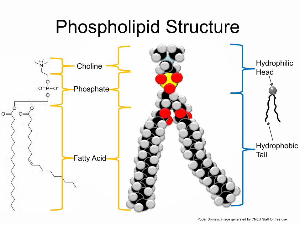
Phospholipid Structure
Fatty Acid
Phosphate
Choline Hydrophilic Head
Hydrophobic Tail
Public Domain: image generated by CNEU Staff for free use

The Nucleus• Nuclear envelope is also formed from self
assembled bilayer of phospholipid like the external cell wall.
• The most prominent organelle in an animal cell.• The nucleus houses deoxyribonucleic acid
(DNA), which is responsible for protein synthesis in the cell.
• The nuclear envelope protects the DNA.• The DNA is like a “hard drive”, and it contains all
the “programs” the body needs to carry out life functions.

The Animal Cell
Source: http://www.nsf.gov/news/overviews/biology/interactive.jsp

The Nuclear Envelope• The entire nucleus is separated from the cytoplasm
(the rest of the cell) by a nuclear envelope– A double membrane, lipid bilayers, separated by a
space of 20 to 30 nm– The envelope is perforated by pores that are about
100nm in diameter, these regulate the transport of macromolecules and particles.
– Naturally for the nanotechnologist these size parameters determine material interaction, material process tool set, and characterization tools.
– The nucleus houses DNA, that is packaged as chromosomes during cell division.

DNA and Proteins• Why do we care about DNA and proteins?• DNA is an excellent example of controlled self
assembly.– DNA is transcribed into RNA, which is
translated into proteins– DNA is the “software/template”, proteins carry
out life functions• We will review DNA first, then see how DNA is
used to create proteins later in the presentation.

Deoxyribonucleic Acid (DNA)• Chemically, DNA consists of a series of nucleotides
– The building block of nucleic acids, made up of a five carbon sugar, deoxyribose, covalently bonded to a nitrogenous base, base pairs, and a phosphate group.
– The phosphate of one nucleotide is bonded to the sugar of the next nucleotide in line, resulting in a sugar-phosphate “backbone” from which the bases project
– Co·don, A set of three consecutive nucleotides in a strand of DNA or RNA that provides the genetic information to code for a specific amino acid that will be incorporated into a protein chain or serve as a termination signal.
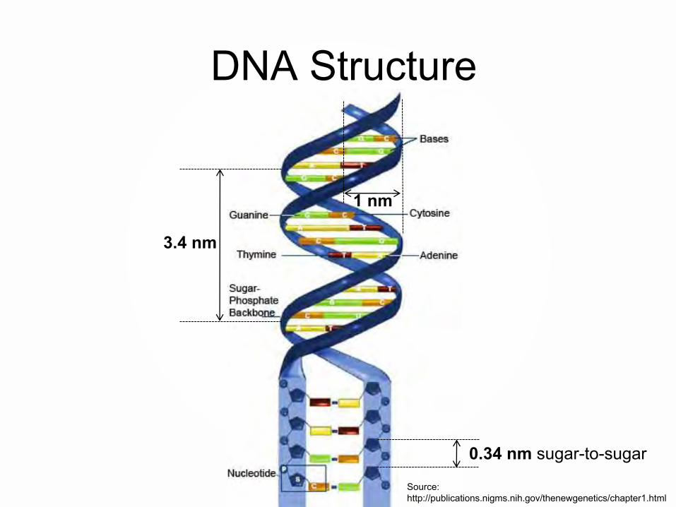
DNA Structure
Source: http://publications.nigms.nih.gov/thenewgenetics/chapter1.html
1 nm
3.4 nm
0.34 nm sugar-to-sugar

Sugar-phosphate backbone
Deoxyribonucleic Acid (DNA), Single Strand
Public Domain: image generated by CNEU Staff for free use, 2009

Assembly Rules of DNA• Purines bond to pyrimidines and vice versa via
hydrogen bonding– never to each other
• Bonding always occurs in the following convention– A-T– C-G– This allows ½ of the DNA chain to be replicated
• Human DNA contains about 6 billion base pairs.• Nature uses three base pairs to define a specific
amino acid, and these amino acids are coupled together to form functional proteins.
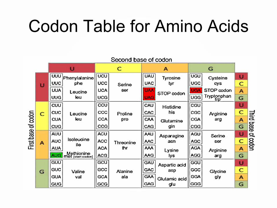
Codon Table for Amino Acids
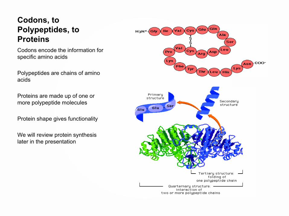
Codons, to Polypeptides, to ProteinsCodons encode the information for specific amino acids
Polypeptides are chains of amino acids
Proteins are made up of one or more polypeptide molecules
Protein shape gives functionality
We will review protein synthesis later in the presentation
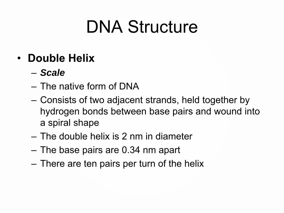
DNA Structure• Double Helix
– Scale
– The native form of DNA– Consists of two adjacent strands, held together by
hydrogen bonds between base pairs and wound into a spiral shape
– The double helix is 2 nm in diameter– The base pairs are 0.34 nm apart – There are ten pairs per turn of the helix

DNA Structure
Source: http://publications.nigms.nih.gov/thenewgenetics/chapter1.html
1 nm
3.4 nm
0.34 nm sugar-to-sugar
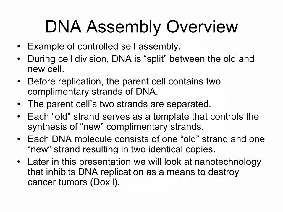
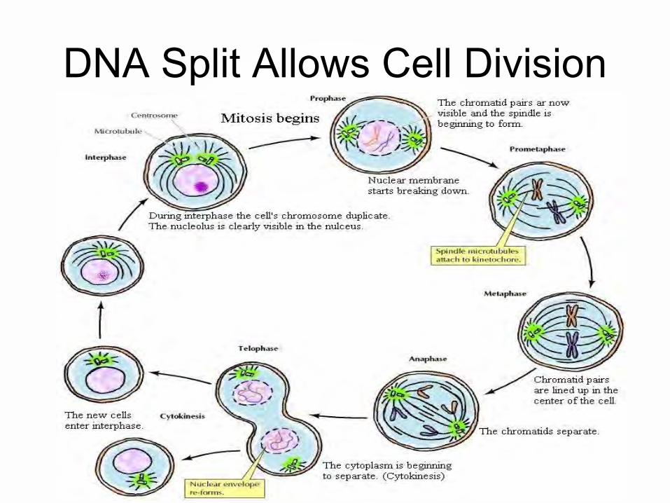
DNA Assembly Overview• Example of controlled self assembly.• During cell division, DNA is “split” between the old and
new cell. • Before replication, the parent cell contains two
complimentary strands of DNA.• The parent cell’s two strands are separated.• Each “old” strand serves as a template that controls the
synthesis of “new” complimentary strands.• Each DNA molecule consists of one “old” strand and one
“new” strand resulting in two identical copies. • Later in this presentation we will look at nanotechnology
that inhibits DNA replication as a means to destroy cancer tumors (Doxil).
© 2013 The Pennsylvania State University
DNA Split Allows Cell Division

DNA ReplicationG C
TA
G C
G C
TA
TA
TA
C G
AT
AT
GA
G
G
A
A
A
C
T
T
CT
C
C
T
T
T
G
A
A
CT
C
C
T
T
T
G
A
A
GA
G
G
A
A
A
C
T
T
CT
C
C
T
T
T
G
A
A
GA
G
G
A
A
A
C
T
T
1) Before DNA is replicated it two completestrands
2) The enzyme helicase splits the two strands of DNA apart
3) The two original strands of DNA serve as templates for the self-assembly of two new complement strands of DNA starting with the nucleotides
4) After the nucleotides are aligned they connect to one another to form the sugar phosphate backbone, and are now two complete DNA chains
GA
G
G
A
A
A
C
T
T
CT
C
C
T
T
T
G
A
A
GA
G
G
A
A
A
C
T
T
CT
C
C
T
T
T
G
A
A
Public Domain: image generated by CNEU Staff for free use, 2009

© 2013 The Pennsylvania State University
DNA Split Allows Cell Division
http://unit4biology.wikispaces.com/Cell+Division+and+Reproduction
© 2013 The Pennsylvania State University
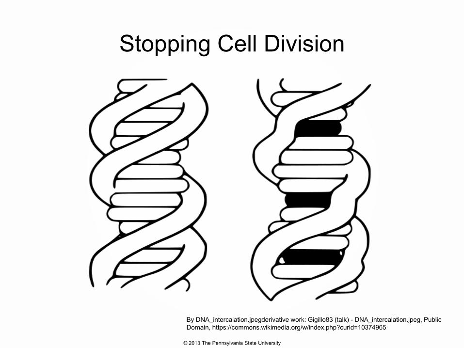
Stopping Cell Division• Cancer is known to have uncontrolled rapid cell
division. One way to stop cancer is preventing cell division.
• The man made nanoparticle that we are headed to discussing is the liposome.
• The particular liposome we will look at contains a drug that prevents DNA replication, and therefore stops cell division.
• This method is called intercalation
© 2013 The Pennsylvania State University
Stopping Cell Division• DNA intercalators are used in chemotherapeutic
treatment to inhibit DNA replication in rapidly growing cancer cells.
• Examples include doxorubicin (adriamycin) and daunorubicin (both of which are used in treatment of Hodgkin's lymphoma), and dactinomycin (used in Wilm's tumour, Ewing's Sarcoma, rhabdomyosarcoma).
© 2013 The Pennsylvania State University
Stopping Cell Division
By DNA_intercalation.jpegderivative work: Gigillo83 (talk) - DNA_intercalation.jpeg, Public Domain, https://commons.wikimedia.org/w/index.php?curid=10374965
Materials in Nanotechnology
Nanoparticles for Medical Applications
Part 2
Terence Kuzma

Outline • Biocompatibility• Quick overview of cellular interaction
– Scale, size, generic animal cell• Nanoscale materials for biological
interaction– Liposomes– Metal Nanoparticles– Nanoshells– Examples of bionano applications
© 2013 The Pennsylvania State University
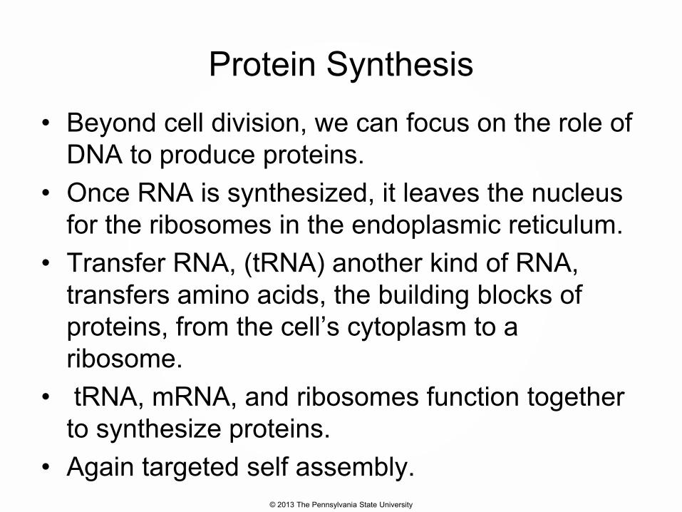
Protein Synthesis• Beyond cell division, we can focus on the role of
DNA to produce proteins.• Once RNA is synthesized, it leaves the nucleus
for the ribosomes in the endoplasmic reticulum.• Transfer RNA, (tRNA) another kind of RNA,
transfers amino acids, the building blocks of proteins, from the cell’s cytoplasm to a ribosome.
• tRNA, mRNA, and ribosomes function together to synthesize proteins.
• Again targeted self assembly.
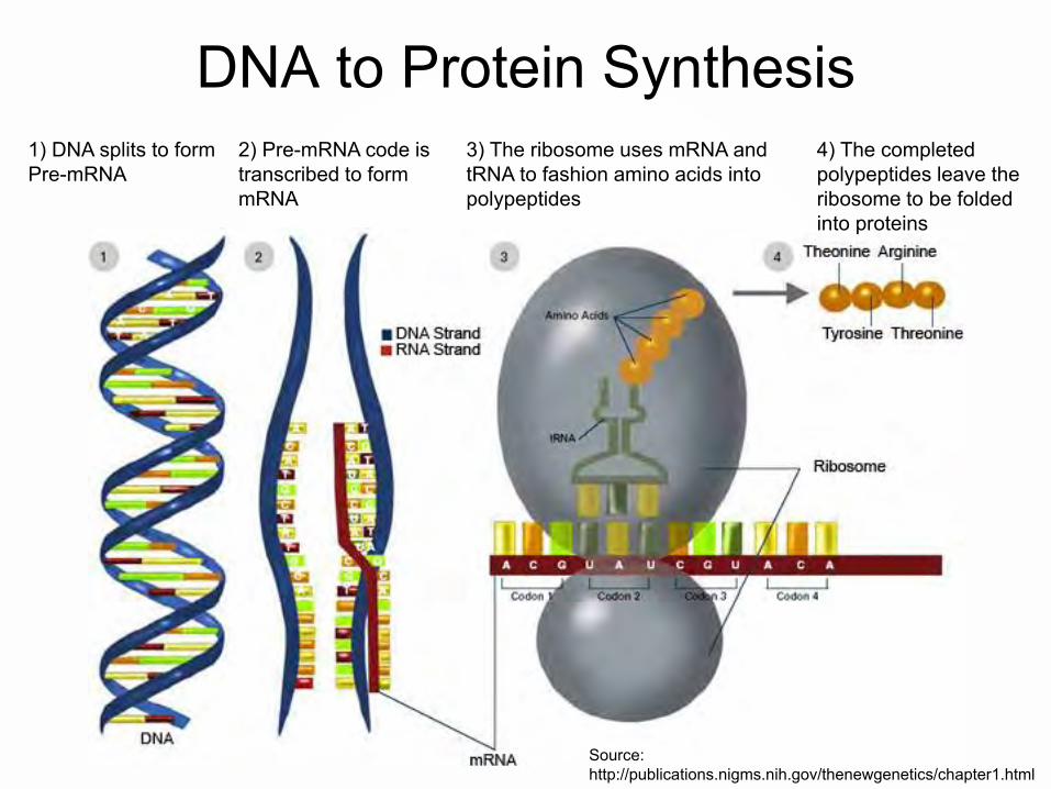
DNA to Protein Synthesis1) DNA splits to form Pre-mRNA
Source: http://publications.nigms.nih.gov/thenewgenetics/chapter1.html
2) Pre-mRNA code is transcribed to form mRNA
3) The ribosome uses mRNA and tRNA to fashion amino acids into polypeptides
4) The completed polypeptides leave the ribosome to be folded into proteins

Protein Background• Proteins are a major class of biomolecules that can
directly connect biology to nanotechnology.• Our bodies are 20% protein: this allows us to think, feel,
move, and function.• Proteins come in many shapes and sizes, giving them
many functions:– Catalysis of chemical reactions within cells– Cellular gatekeepers – Immune system monitoring by recognition of foreign cells– Structural support to cells and tissues
• Compare to DNA: 1 structure and 1 function
Protein Background
• Proteins are built from 20 types of Amino Acids ~1 nm in size (e.g. tryptophan).
• DNA gives the protein information on how to assemble from the amino acid building blocks.
• Proteins have highly variable structures and can change their shapes in response to their surroundings in order to provide a signal.
• Proteins can provide binding sites for chemical reactions to occur.
• Proteins can identify cells. This is important for cancer, because tumors may have identifying proteins.

Membrane Structure/Protein Keys
http://neutrons.ornl.gov/conf/dsm2008/registration.shtml
Lipid Bilayer:Excellent example of how “nature” does self assembly
Protein receptors for cell communicationExtracellular
environment
Intercellular region (cytoplasm)

Hydrophobic/Hydrophilic Interaction
Protein interaction establishes the cell’s response to materials
• Polypeptides contain both hydrophilic and hydrophobic amino acids.
• Polypeptides will fold in aqueous solution. Once folded, polypeptides become proteins, which are a vital part of biological processes.
• So the hydrophobic/hydrophilic nature of amino acids give proteins 3 dimensional complexity.

C
COO-
C
CH2
CH3
+H3N HC
COO-
CH3
+H3N H C
COO-
CH
+H3N H
H3C CH3
C
COO-
CH2
+H3N H
CH
H3C CH3
C
COO-
CH2
+H3N H
CH2
S
CH3
C
COO-
CH2
+H3N H C
COO-
CH2
+H3N H
OH
Special Amino Acids
C
COO-
CH2
+H3N H
SH
C
COO-
H
+H3N H C
H1N CH2
H2C CH2
COO-
Hydrophobic Amino Acids
C
COO-
CH2
+H3N H
C CH
NH
Alanine(Ala or A)
Valine(Val or V)
Isoleucine(Ile or I)
Leucine(Leu or L)
Methionine(Met or M)
Phenylalanine(Pha or F)
Tyrosine(Tyr or Y)
Tryptophan(Trp or W)
Cysteine (Cys or C)
Glycine (Gly or G)
Proline(Pro or P)
C
COO-
CH2
+H3N H
CH2
CH2
CH2
NH3+
C
COO-
CH2
+H3N H
CH2
CH2
NH
C NH2+
NH2
C
COO-
CH2
+H3N H
C
CH
NHCH
N+
H
C
COO-
CH2
+H3N H
OH
C
COO-
C
+H3N H
CH3
OHH
C
COO-
CH2
+H3N H
C
OH2N
C
COO-
CH2
+H3N H
C
OH2N
CH2C
COO-
CH2
+H3N H
COO-
C
COO-
CH2
+H3N H
CH2
COO-
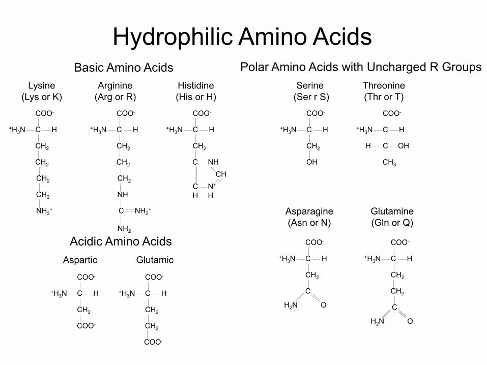
Basic Amino Acids
Hydrophilic Amino Acids
Lysine(Lys or K)
Arginine(Arg or R)
Histidine(His or H)
Polar Amino Acids with Uncharged R GroupsSerine
(Ser r S)Threonine(Thr or T)
Asparagine(Asn or N)
Glutamine(Gln or Q)
Acidic Amino AcidsAspartic Glutamic

Amino Acids – the Twist
• The hydrophobic – hydrophillic nature of amino acids allows polypeptides (proteins), to twist and bend in water.
• This twisting and bending often definesthe functionality of the proteins.
• Material response, and some diseases are defined by lack of a certain twist in a protein…..

Amino Acids – the Twist

Intercellular Junctions• Neighboring cells often adhere, interact, and
communicate through direct physical contact.• There are three main types of intercellular
junctions in animal cells.– Tight junctions- fusing connection, prevent leakage of
extracellular fluid– Desmosomes- anchor cells together– Gap junctions- pass salts, sugars, ammino acids, and
other small molecules between cells

Intercellular Junctions
• It is interesting to note that cancer tumors often do not follow the rules of normal cells.
• Cancer breaks the tight junction rule, and the blood capillaries are often leaky compared to normal tissue.
• We will look at how these attributes impact nanotechnology based drugs later in the presentation.
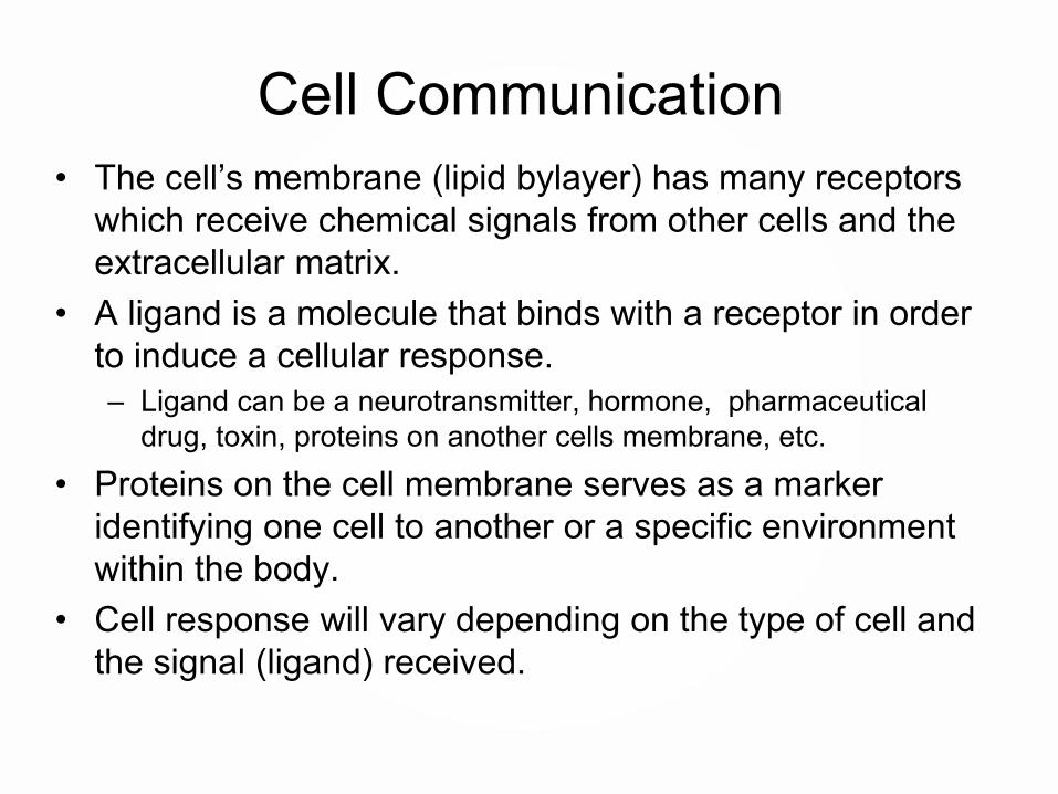
Cell Communication • The cell’s membrane (lipid bylayer) has many receptors
which receive chemical signals from other cells and the extracellular matrix.
• A ligand is a molecule that binds with a receptor in order to induce a cellular response.– Ligand can be a neurotransmitter, hormone, pharmaceutical
drug, toxin, proteins on another cells membrane, etc.• Proteins on the cell membrane serves as a marker
identifying one cell to another or a specific environment within the body.
• Cell response will vary depending on the type of cell and the signal (ligand) received.
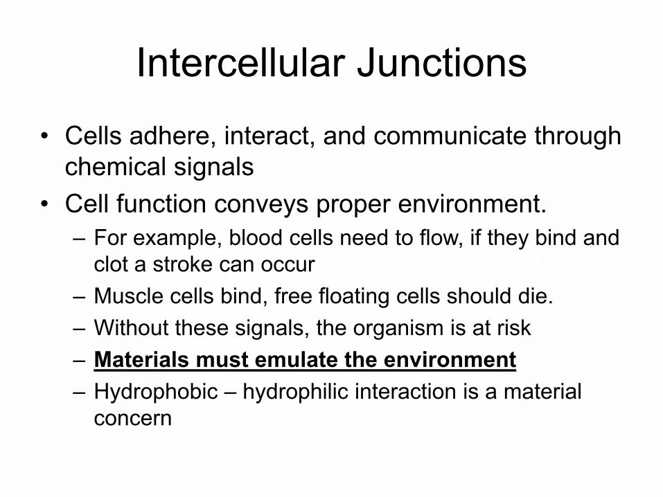
Intercellular Junctions• Cells adhere, interact, and communicate through
chemical signals• Cell function conveys proper environment.
– For example, blood cells need to flow, if they bind and clot a stroke can occur
– Muscle cells bind, free floating cells should die.– Without these signals, the organism is at risk– Materials must emulate the environment– Hydrophobic – hydrophilic interaction is a material
concern
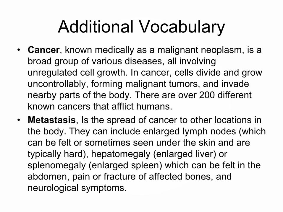
Additional Vocabulary• Cancer, known medically as a malignant neoplasm, is a
broad group of various diseases, all involving unregulated cell growth. In cancer, cells divide and grow uncontrollably, forming malignant tumors, and invade nearby parts of the body. There are over 200 different known cancers that afflict humans.
• Metastasis, Is the spread of cancer to other locations in the body. They can include enlarged lymph nodes (which can be felt or sometimes seen under the skin and are typically hard), hepatomegaly (enlarged liver) or splenomegaly (enlarged spleen) which can be felt in the abdomen, pain or fracture of affected bones, and neurological symptoms.
Additional Vocabulary• Angiogenesis, is the physiological process through
which new blood vessels form from pre-existing vessels. Vascular endothelial growth factor (VEGF) is a signal protein produced by cells that stimulates vasculogenesisand angiogenesis.
• Ligand, is a substance (usually a small molecule), that forms a complex with a biomolecule to serve a biological purpose. Ligands can direct particles. Ligands are a signal triggering molecule, binding to a site on a target protein. Selective ligands have a tendency to bind to a very limited types of receptors, whereas non-selective ligands bind to several types of receptors.

Additional Vocabulary• Cancer expression, data from 22 tumor types has
identified multiple metabolic expression changes associated with cancer. These expressions can be used to identify and attack tumors.
• HER2, epidermal growth factor receptor 2 (HER2), which promotes the growth of cancer cells. This gene mutation and the elevated levels of HER2 that it causes can occur in many types of cancer — not only breast cancer. This is a gene mutation that occurs only in the cancer cells and is not a type of mutation that you can inherit from a parent.
Additional Vocabulary• Prostate-specific antigen (PSA), also known as gamma-
seminoprotein or kallikrein-3 (KLK3), is a glycoprotein enzyme. PSA is often over expressed when prostrate cancer is present.
• Enhanced Permeability and Retention (EPR) effect, is the property by which certain sizes of molecules (typically liposomes, nanoparticles, and macromolecular drugs) tend to accumulate in tumor tissue much more than they do in normal tissues. Particles can preferentially enter tumors because these newly formed tumor vessels are usually abnormal in form and architecture. They have poorly-aligned defective endothelial cells like a roof missing a shingle. Increased retention is do to the lack of lymphaticsaround the tumor region which would filter out such particles under normal conditions.

Materials in Nanotechnology
Nanoparticles for Medical Applications
Part 3
Terence Kuzma

Outline • Biocompatibility• Quick overview of cellular interaction
– Scale, size, generic animal cell• Nanoscale materials for biological
interaction– Liposomes– Metal Nanoparticles– Nanoshells– Examples of bionano applications

Nanoparticles
• Nanoparticles are useful due to the small size and scaling to biology.
• Nanoparticles made from a metal, semiconductor or polymer must interact on the cellular level. Some terms will be defined to understand this interaction.
• Size and scale are important to understanding how nanotechnology can be applied to medicine.
• On the cellular level, a cell is about 10 microns, and pores may be 100 nm, and an amino acid is about 1 nm.
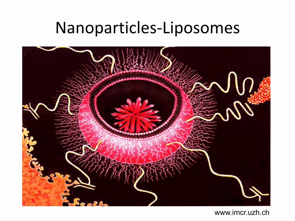
Nanoparticles-Liposomes• The first man made nanoparticle we will look at is the
liposome.• The word liposome is from two Greek words: lipo, "fat"
and soma, "body". Phospholipids are the building block.• Phospholipids in an aqueous environment orient
themselves in a thermodynamically stable form called a bilayer. This bilayer can further orient itself into a sphere known as a liposome
• So a liposome is an artificially-prepared vesicle composed of a lipid bilayer. The liposome can be used as a vehicle for administration of pharmaceutical drugs, DNA/RNA, tags, and nutrients. Liposomes are biodegradable.
Nanoparticles-Liposomes
www.imcr.uzh.ch

HydrophobicTail
HydrophilicHead
Choline
Phosphate
Fatty Acid
OO O
O
O-
O
OOP
N
Phospholipid
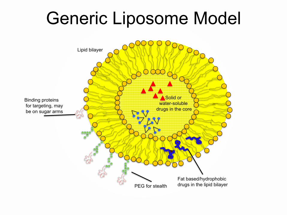
Solid or water-soluble
drugs in the core
Fat based/hydrophobic drugs in the lipid bilayer
Lipid bilayer
Binding proteinsfor targeting, maybe on sugar arms
PEG for stealth
Generic Liposome Model

Nanoparticles-Liposomes• The FDA has set up guidelines to establish quality
control of new therapies based on liposome technology. These chemistry, manufacturing and control guidelines are given in this document: http://www.fda.gov/downloads/Drugs/GuidanceComplianceRegulatoryInformation/Guidances/ucm070570.pdf
• The physicochemical properties of the liposome drug product are critical to ensuring drug product quality.
• These manufacturing guidelines also reflect the interaction of liposomes and show variables that change the functionality of the man made particle.

Nanoparticles-Liposomes• So for cellular uptake, the man made nanoparticle must be
conducive to cellular uptake, and deliver a predictable drug dosage.• The FDA has proposed that properties specific to liposome drug
products that may be useful to assess include:– morphology of the liposome, including lamellarity determination,
if applicable– net charge– volume of entrapment in liposomal vesicles– particle size (mean and distribution profile)– phase transition temperature– spectroscopic data, as applicable– in vitro release of the drug substance from the liposome drug
product– osmotic properties– light scattering index

Nanoparticles-Liposomes• Liposomes can to deliver active molecules to the site of
action, less waste and potential damage to other cells• The major types of liposomes are the multilamellar
vesicle (MLV), the small unilamellar vesicle (SUV), the large unilamellar vesicle (LUV).
LUV
MLV
≥ 100nmOne lipid bilayer
≥ 500nmOne lipid bilayer≥ 20-100nm
One lipid bilayer
SUV

Nanoparticles-Liposomes• Sigma-Aldrich Liposome Kit: SKU L4395-1VL,
Lipid mixtures for the preparation of liposomes Lyophilized powder. 85.00 USD
• Composition: Cholesterol, 9 μmol/package, L-α-Phosphatidylcholine (egg yolk), 63 μmol/package Stearylamine, 18 μmol/package
• http://www.sigmaaldrich.com/catalog/product/sigma/l4395?lang=en®ion=US
• http://www.sigmaaldrich.com/catalog/product/sigma/l4395?lang=en®ion=US

Nanoparticles-Liposomes

Nanoparticles-LiposomesMini-extruder

Nanoparticles-Liposomes
avantilipids.com/
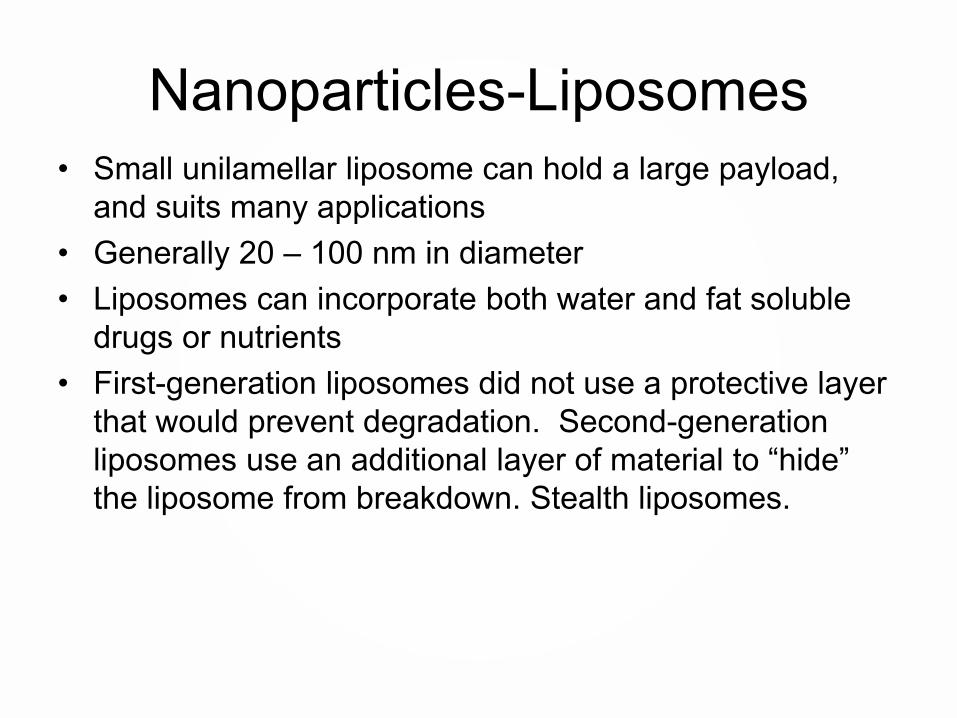
Nanoparticles-Liposomes• Small unilamellar liposome can hold a large payload,
and suits many applications• Generally 20 – 100 nm in diameter• Liposomes can incorporate both water and fat soluble
drugs or nutrients• First-generation liposomes did not use a protective layer
that would prevent degradation. Second-generation liposomes use an additional layer of material to “hide” the liposome from breakdown. Stealth liposomes.

Liposome Uses• This “stealth” allows the liposome to survive in the body
longer, and hopefully deliver the medicine to the desired area. Long-circulating liposomes are obtained by modulating the lipid composition, size, and charge of the vesicle.
• Different methods have been suggested to achieve long circulation of liposomes in vivo, including coating the liposome surface with inert, biocompatible polymers.
• A protective layer over the liposome surface and slows down liposome recognition by opsonins and therefore subsequent clearance of liposomes. Spleen and liver still filter.
• One of the most common coatings is PolyEthyleneGlycol (PEG).
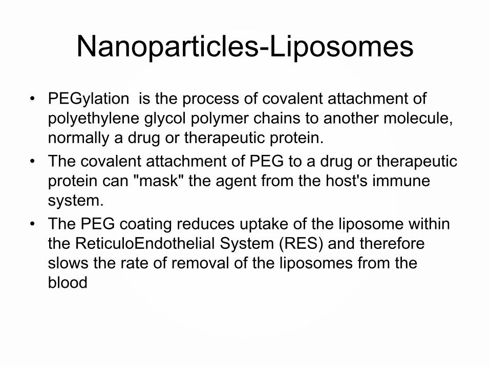
Nanoparticles-Liposomes• PEGylation is the process of covalent attachment of
polyethylene glycol polymer chains to another molecule, normally a drug or therapeutic protein.
• The covalent attachment of PEG to a drug or therapeutic protein can "mask" the agent from the host's immune system.
• The PEG coating reduces uptake of the liposome within the ReticuloEndothelial System (RES) and therefore slows the rate of removal of the liposomes from the blood
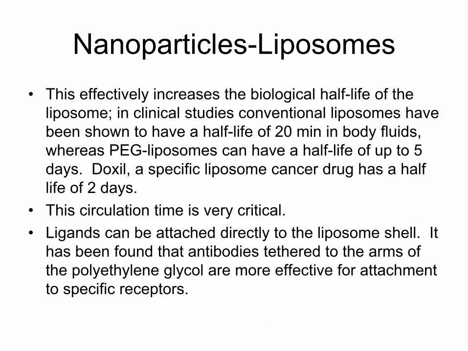
Nanoparticles-Liposomes• This effectively increases the biological half-life of the
liposome; in clinical studies conventional liposomes have been shown to have a half-life of 20 min in body fluids, whereas PEG-liposomes can have a half-life of up to 5 days. Doxil, a specific liposome cancer drug has a half life of 2 days.
• This circulation time is very critical. • Ligands can be attached directly to the liposome shell. It
has been found that antibodies tethered to the arms of the polyethylene glycol are more effective for attachment to specific receptors.

Nanoparticles-Liposomes Uses
• Liposomes can also be used to house tagging molecules. This tagging can be done inside the liposome and/or in the lipid shell of the bilayer, or on polymers bonded to the liposome shell.
• Liposomes can be used to encapsulate and deliver drugs, DNA or RNA, tags, or nutrients for delivery to a specific cell.

Nanoparticles-Liposomes Tagging
• We will be looking at the liposome based cancer drug Doxil. It is interesting that gamma isotopes were used to track the uptake of Doxil in vivo during drug trials. Results looked much like a PET scan.
• These results show that the Doxil has a preference to be absorbed in the tumor area. The reason “why” liposomes can target tumors will be discussed in a few slides.
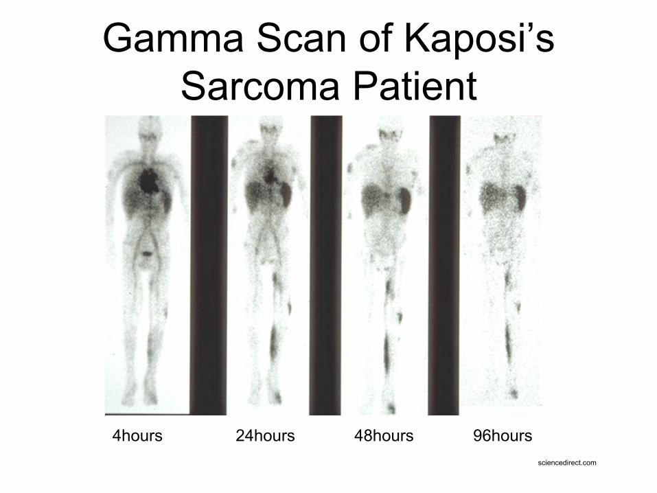
Gamma Scan of Kaposi’s Sarcoma Patient
4hours 24hours 48hours 96hours sciencedirect.com

Doxorubicin in KS Lesions and Normal Skin (Biopsy at 48 Hrs. after Doxil)
1 2 3 4 5 6 70
5
10
15
20
25
Dox
orub
icin
Con
cent
ratio
n (
g/g)
K S LesionNormal Skin
Sample sites

Nanoparticles-Liposomes Cancer Drug
• As seen in the last slide, liposomes have a “natural ability” (size) to target cancer.
• In non cancerous samples the endothelial wall of all healthy human blood vessels are encapsulated by endothelial cells that are bound together by tight junctions. These tight junctions stop any particles in the blood from leaking out of the vessel. So healthy tissue will keep out small particles.

Nanoparticles-Liposomes Cancer Drug
• Tumor vessels do not contain the same level of seal between cells and are diagnostically ''leaky''. This ability is known as the Enhanced Permeability and Retention (EPR) effect. So tumors are like Swiss cheese, and small particles can leak into these defects.

Nanoparticles Breaching Corrupt Endothelial Cells
Nanoparticles-Liposomes Cancer Drug
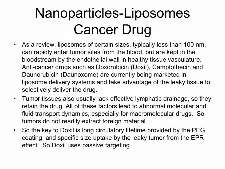
• As a review, liposomes of certain sizes, typically less than 100 nm, can rapidly enter tumor sites from the blood, but are kept in the bloodstream by the endothelial wall in healthy tissue vasculature. Anti-cancer drugs such as Doxorubicin (Doxil), Camptothecin and Daunorubicin (Daunoxome) are currently being marketed in liposome delivery systems and take advantage of the leaky tissue to selectively deliver the drug.
• Tumor tissues also usually lack effective lymphatic drainage, so they retain the drug. All of these factors lead to abnormal molecular and fluid transport dynamics, especially for macromolecular drugs. So tumors do not readily extract foreign material.
• So the key to Doxil is long circulatory lifetime provided by the PEG coating, and specific size uptake by the leaky tumor from the EPR effect. So Doxil uses passive targeting.

Nanoparticles-Liposomes Cancer Drug
• Doxil
Drugs.com
Liposome bilayer
MPEG-DSPE coating
Aqueous core withentrapped doxorubicin HCL
Nanoparticles-Liposomes Cancer Drug
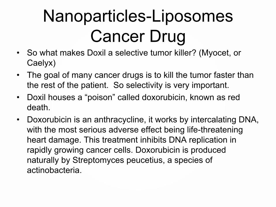
• So what makes Doxil a selective tumor killer? (Myocet, or Caelyx)
• The goal of many cancer drugs is to kill the tumor faster than the rest of the patient. So selectivity is very important.
• Doxil houses a “poison” called doxorubicin, known as red death.
• Doxorubicin is an anthracycline, it works by intercalating DNA, with the most serious adverse effect being life-threatening heart damage. This treatment inhibits DNA replication in rapidly growing cancer cells. Doxorubicin is produced naturally by Streptomyces peucetius, a species of actinobacteria.

© 2013 The Pennsylvania State University
DNA Split Allows Cell Division
http://unit4biology.wikispaces.com/Cell+Division+and+Reproduction

© 2013 The Pennsylvania State University
Stopping Cell Division
By DNA_intercalation.jpegderivative work: Gigillo83 (talk) - DNA_intercalation.jpeg, Public Domain, https://commons.wikimedia.org/w/index.php?curid=10374965

Nanoparticles-Liposomes Cancer Drug
• Anthracyclines are among the most effective anticancer treatments ever developed and are effective against more types of cancer than any other class of chemotherapeutic agents
• But daunorubicin can be deadly to both the tumor and the heart. By design the liposome encapsulation prevents contact with the heart, and preferentially releases the daunorubicin in the tumor.
• As discussed, this selectivity is carried out with the use of the liposome that protects the heart, and at the same time shows passive size preference to the tumor.

Materials in Nanotechnology
Nanoparticles for Medical Applications
Part 4
Terence Kuzma
Outline • Biocompatibility• Quick overview of cellular interaction
– Scale, size, generic animal cell• Nanoscale materials for biological
interaction– Liposomes– Metal Nanoparticles– Nanoshells– Examples of bionano applications
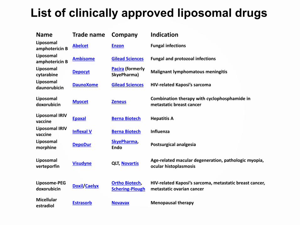
Name Trade name Company IndicationLiposomal amphotericin B
Abelcet Enzon Fungal infections
Liposomal amphotericin B
Ambisome Gilead Sciences Fungal and protozoal infections
Liposomal cytarabine
DepocytPacira (formerly SkyePharma)
Malignant lymphomatous meningitis
Liposomal daunorubicin
DaunoXome Gilead Sciences HIV-related Kaposi’s sarcoma
Liposomal doxorubicin
Myocet ZeneusCombination therapy with cyclophosphamide in metastatic breast cancer
Liposomal IRIV vaccine
Epaxal Berna Biotech Hepatitis A
Liposomal IRIV vaccine
Inflexal V Berna Biotech Influenza
Liposomal morphine
DepoDurSkyePharma, Endo
Postsurgical analgesia
Liposomal verteporfin
Visudyne QLT, NovartisAge-related macular degeneration, pathologic myopia, ocular histoplasmosis
Liposome-PEG doxorubicin
Doxil/CaelyxOrtho Biotech, Schering-Plough
HIV-related Kaposi’s sarcoma, metastatic breast cancer, metastatic ovarian cancer
Micellular estradiol
Estrasorb Novavax Menopausal therapy
List of clinically approved liposomal drugs

© 2013 The Pennsylvania State University
Nanoparticles-Liposomes Tagging
• This visual tagging can allow a surgeon to isolate diseased tissue from healthy tissue.
• Liposomes can also incorporate Quantum dots or plasmons in the core or surface.

© 2013 The Pennsylvania State University
Nanoparticles-Liposomes Tagging
• QDs have been reported to be about 20 times brighter and 100 times more photostable in comparison with organic dyes such as rhodamine
• Utilizing a 50 nm QD, and a liposome of 300 nm, incorporation of the QD is possible.
• Given these dimensions, 3 quantum dots were embedded in each liposome, and the resulting signal was the same as 3 free quantum dots. The authors proposed smaller QDs to incorporate into the liposome.
Liposome encapsulation of fluorescent nanoparticles: Quantum dots and silica nanoparticles Chien-Sheng Chen1, Jie Yao2 and Richard A. Durst1, Cornell University
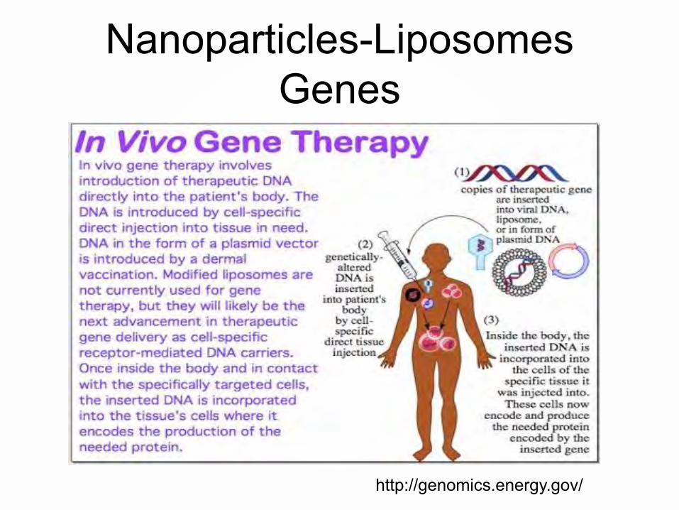
Nanoparticles-Liposomes Genes
http://genomics.energy.gov/

Nanoparticles-Liposomes Gene Delivery

Nanoparticles-Liposomes Nutrition
• Nutritional supplement companies are currently encapsulating nutrients such as vitamin C in liposomes.
• Liposome delivery increases the bioavailability of nutrients compared to traditional oral dietary capsules.
• Liposomes bypass the destructive elements of the gastric system and aid the encapsulated nutrient to be delivered to the cells and tissues
• Hydrophilic drugs/nutrients/tags can be trapped in the central aqueous core of the liposomes, and lipophilic drugs can be solubilized within the lipid bilayer

© 2013 The Pennsylvania State University
Liposomes - review• Liposomes are biocompatible, and feature the same
construction as cells.• Liposomes can be delivered directly to the appropriate
site by passive delivery, ligands, or local injection.• Delivery methods conserve materials and minimize
side effects.• Liposomes can deliver both fat and water soluable
drugs, RNA, and vitamins. • Liposomes are in widespread use today.

Metal Nanoparticles• Since the size of nanoparticles are comparable
to biomolecules, nanoparticles can overcome biological barriers.
• NPs may be 1/100th to 1/10,000th the size of a cell. The cell may uptake the NP as a result of being tricked into ‘thinking’ this NP is a biomolecule (e.g. enzyme, antibody).
• Shape of the nanoparticle also has impact, generally gap junctions accept round shapes more than rods. Generally nanoparticles between 30-100nm seem to be accepted well by most cells

Metal Nanoparticles• Au is often used as the metal of choice because has
been shown to be relatively inert and Au is a good platform to tether chemistry to the particle.
• Au is expensive in bulk, but when using small volumes (e.g. 10 mg) the bulk cost is irrelevant.
• Metal nanoparticles can be used to target and destroy disease cells.
• There are different functionalities of the particles to perform selected tasks. They can be used as tags to identify, used as energy receptors to “cook” (targeted hyperthermia) tumors, may be used as a delivery platform for drug/RNA delivery.

Metal Nanoparticles• Au NPs can be used as a marker to delineate between
healthy tissue and tumors. • In this role the Au NPs are coated with a ligand that will
preferentially adhere to tumor cells.• These Au NPs are grown to a specific size so they will
exhibit plasmon resonance. • A light source is used to stimulate the plasmons during
surgery giving the surgeon a clear boundary between healthy tissue and tumors. This allows the surgeon to minimize trauma to healthy tissue, and locate the unhealthy regions.

Metal Nanoparticles• Au NPs are inert in vivo so side effects are
limited or non existent.• Au NPs are relatively nonreactive and they can
be coated with PEG so they are very good at moving throughout the circulatory system in the “stealth” mode.
• Smaller particles (1-100 nm) are more apt to penetrate into a tumor because they can passively slip between blood vessels (EPR).
Metal Nanoparticles• Au NPs are not good at keeping light in an
excited state: so when illuminated with light the excited state energy can be given off as a vibration (heat).
• Selective antibodies can be/are attached to Au NPs that is designed to latch onto the receptor of the cancer cell
• Up to 150 different types of antibodies can be conjugated to a Au NP through bifunctional PEG linkers to target specific tumors.

Metal Nanoparticles• Antibodies: A class of
proteins that specifically recognize foreign agents in the body and tag them for removal.
• Large libraries of slightly different antibodies are used to probe for a vast array of threats. Hundreds are available commercially.
• Most antibodies have a Y shape:
Metal Nanoparticles• Selection of the antibody is made so healthy
cells will have low levels of the receptor, then Au NPs will selectively seek the cancer cells.
• By illuminating Au NPs with light, they give off heat estimated to be ~70-80°C.
• This local heating results in the local ablation of cancer cells which have been targeted by Au NPs.
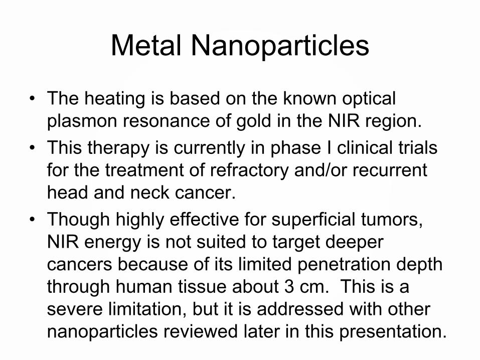
Metal Nanoparticles• The heating is based on the known optical
plasmon resonance of gold in the NIR region. • This therapy is currently in phase I clinical trials
for the treatment of refractory and/or recurrent head and neck cancer.
• Though highly effective for superficial tumors, NIR energy is not suited to target deeper cancers because of its limited penetration depth through human tissue about 3 cm. This is a severe limitation, but it is addressed with other nanoparticles reviewed later in this presentation.
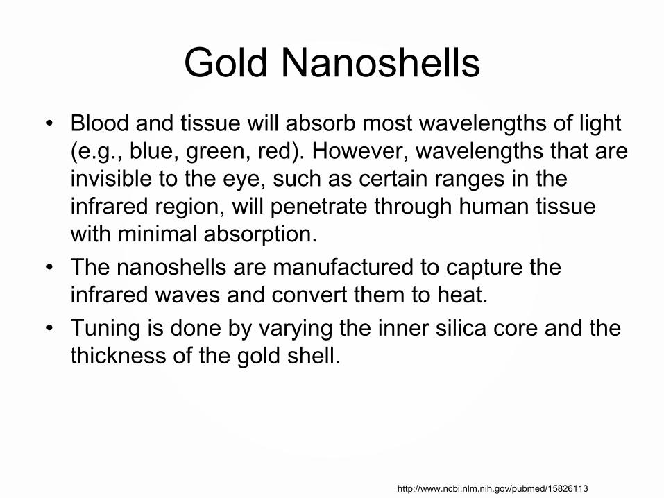
Gold Nanoshells• Blood and tissue will absorb most wavelengths of light
(e.g., blue, green, red). However, wavelengths that are invisible to the eye, such as certain ranges in the infrared region, will penetrate through human tissue with minimal absorption.
• The nanoshells are manufactured to capture the infrared waves and convert them to heat.
• Tuning is done by varying the inner silica core and the thickness of the gold shell.
http://www.ncbi.nlm.nih.gov/pubmed/15826113
Gold Nanoshells• The key innovation with nanoshells is that we can tune
their color from the visible using the dimensions of the core and the shell to different regions of the spectrum. So the particles can be tuned into the near-infrared region of the spectrum. Biomedical engineers call that "the water window," because we're mostly made out of water, and water is most transparent in this region of the spectrum. Light can penetrate tissue by as many as 10 centimeters, depending on tissue type.
http://www.ncbi.nlm.nih.gov/pubmed/15826113

Gold Nanoshells
http://education.mrsec.wisc.edu/220.htm

Gold Nanoshells• Naomi Halas, Professor of Electrical and Computer
Engineering at Rice University, known as the inventor of nanoparticles with tunable optical properties controlled by their shape and structure.
• Nanoshells known commercially as AuroShells are a novel class of optically tunable nanoparticles that consist of a dielectric core surrounded by a thin gold shell that can scatter or absorb energy dependent upon wavelength. So nanoshells can be used for imaging or heat ablation.
• Passive tumor targeting is possible due to a non-specific accumulation of nanoshells at the tumor due to the enhanced permeability and retention (EPR) effect. Again, tumor vasculature is more leaky than normal blood vessels allowing the nanoshells to accumulate.
http://www.ncbi.nlm.nih.gov/pubmed/15826113
Metal Nanoparticles• Quantum dots and plasmons have a wide variety of
applications by visually tagging DNA, RNA, proteins, or organelles.
• These tags allow researchers to see how cells interact. • Allows material to be defined such as the boundary
between healthy tissue and tumor tissue.
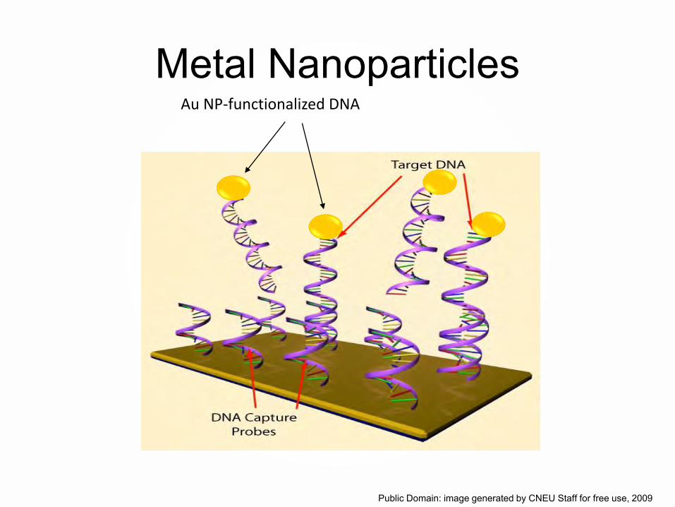
Metal NanoparticlesAu NP-functionalized DNA
Public Domain: image generated by CNEU Staff for free use, 2009
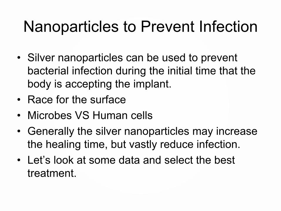
Nanoparticles to Prevent Infection
• Silver nanoparticles can be used to prevent bacterial infection during the initial time that the body is accepting the implant.
• Race for the surface• Microbes VS Human cells• Generally the silver nanoparticles may increase
the healing time, but vastly reduce infection. • Let’s look at some data and select the best
treatment.

Bacteria Growth on Silver Nanoparticle Treated Surface
• Klebsiella pneumoniae adhesion however increases after the coating’s silver content is raised beyond the 3% range. At 9% Ag content the Klebsiella pneumoniaeadhesion is 90% of the untreated Ti
• Staphylococcus epidermis treated samples that the adhesion continues to decline with increasing Ag content, but that the degree of benefit is quite small after a 3% Ag is achieved in the coating.
• DATA from Andrea Ewald, Susanne K Glückermann, Roger Thull , Uwe Gbureck
BioMedical Engineering OnLine 2006, 5:22

Bone Cell Adhesion with Silver Nanoparticles
• Minimal cell adhesion in the 3-4% range
• Higher cell adhesion in the 9% range
BioMedical Engineering OnLine 2006, 5:22

Conclusion
• Biological processes set the scale and material processing needs.
• Nanoparticles such as liposomes, Au NPs, and nanoshells are of appropriate scale to interact on the cellular level.
• Biotech represents a career pathway to use nanotechnology skills.