Embed Size (px)
Citation preview

4MASS SPECTROMETRY FOR QUANTITATIVE IN VITRO ADME ASSAYS
Jun Zhang and Wilson Z. Shou
4.1 INTRODUCTION
In vitro absorption, distribution, metabolism, and excretion (ADME) assays are routinely conducted throughout drug discovery and development to characterize pharmacokinetics-(PK) and toxicity-related properties of new molecular entities (NMEs). These assays usually have demonstrated reasonable in vitro–in vivo cor-relations (IVIVCs), and can serve a number of purposes for in vivo studies such as assessing potential liabilities, selecting/prioritizing compounds to advance, trouble-shooting unexpected results, and predicting parameters in preclinical/clinical studies [1]. Since in vitro experiments are generally less expensive to perform than animal studies, and are also far more amenable to miniaturization and automation in a high-density plate format, they have been utilized extensively in drug discovery to guide lead optimization effort by quickly providing structure–liability relationship (SLR) in parallel to potency optimization efforts.
The most important and therefore frequently performed in vitro ADME assays include metabolic stability, permeability, transporters, drug–drug interaction (DDI), physicochemical property, and cardio- and hepato-toxicity assays (Table 4.1). Briefly, metabolic stability assays are performed by incubating test compounds with an in vitro preparation such as liver microsomes or hepatocytes to assess the metabolic liability of these compounds and also provide prediction of in vivo clearance. Per-meability assays measure the permeability of discovery compounds across in vitro membranes such as parallel artificial membranes and human colonic carcinoma
Mass Spectrometry for Drug Discovery and Drug Development, First Edition. Edited by Walter A. Korfmacher.© 2013 John Wiley & Sons, Inc. Published 2013 by John Wiley & Sons, Inc.
97
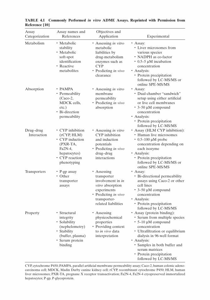
TABLE 4.1 Commonly Performed in vitro ADME Assays. Reprinted with Permission from Reference [10]
Assay Categorization
Assay names and References
Objectives and Application Experimental
Metabolism • Metabolic stability
• Metabolic soft-spot identification
• Reactive metabolites
• Assessing in vitro metabolic liabilities by drug-metabolism enzymes such as CYP
• Predicting in vivo clearance
• Assay:• Liver microsomes from
various species• NADPH as co-factor• 0.5–5 µM incubation
concentration• Analysis:
• Protein precipitation followed by LC-MS/MS or online SPE-MS/MS
Absorption • PAMPA• Permeability
(Caco-2, MDCK cells, etc.)
• Bi-direction permeability
• Assessing in vitro membrane permeability
• Predicting in vivo absorption
• Assay:• Dual-chamber “sandwich”
setup using either artificial or live cell membranes
• 3–50 µM compound concentration
• Analysis:• Protein precipitation
followed by LC-MS/MSDrug–drug
Interaction• CYP inhibition
(rCYP, HLM)• CYP induction
(PXR-TA, Fa2N-4, hepatocytes)
• CYP reaction phenotyping
• Assessing in vitro CYP inhibition and induction potentials
• Predicting in vivo drug–drug interactions
• Assay (HLM CYP inhibition):• Human live microsomes• 0.5–100 µM probe
concentration depending on each isozyme
• Analysis:• Protein precipitation
followed by LC-MS/MS or online SPE-MS/MS
Transporters • P-gp assay• Other
transporter assays
• Assessing transporter involvement in in vitro absorption experiments
• Predicting in vivo transporter-related liabilities
• Assay:• Bi-directional permeability
assays using Caco-2 or other cell lines
• 3–50 µM compound concentration
• Analysis:• Protein precipitation
followed by LC-MS/MSProperty • Structural
integrity• Solubility
(nephelometry)• Stability
(buffer, plasma)• Serum protein
binding
• Assessing physicochemical properties
• Providing context to in vivo data interpretation
• Assay (protein binding):• Serum from multiple species• 5–10 µM compound
concentration• Ultrafiltration or equilibrium
dialysis in 96-well format• Analysis:
• Samples in both buffer and serum matrices
• Protein precipitation followed by LC-MS/MS
CYP, cytochrome P450; PAMPA, parallel artificial membrane permeability assay; Caco-2, human colonic adeno-carcinoma cell; MDCK, Madin Darby canine kidney cell; rCYP, recombinant cytochrome P450; HLM, human liver microsomes; PXR-TA, pregnane X receptor transactivation; Fa2N-4, Fa2N-4 cryopreserved immortalized hepatocytes; P-gp, P-glycoprotein.

INTRODUCTION 99
(Caco-2) cell monolayers, in order to predict NMEs in vivo absorption across intes-tinal epithelium layer. In addition, permeability assays with various cell lines can also be used to assess the involvement of influx and efflux membrane transporters in the absorption of NMEs and predict transporter-related liabilities. In vitro assays with liver microsomes and hepatocytes respectively are used extensively to assess the inhibition and induction of cytochrome P450 (CYP) enzyme activities by NMEs, and predict in vivo DDI potentials. A growing number of in vitro assays such as hERG, sodium and calcium channel assays, as well as human hepatotoxicity assay are being used to assess and predict toxicity. Lastly, physicochemical property assess-ments such as buffer/plasma solubility/stability and serum protein binding assay are important to provide context in interpreting both in vitro and in vivo data. Detailed discussions of the rationale, conduct, and utilities of these in vitro ADME assays are outside the scope of this chapter, and therefore interested readers are encouraged to consult with a number of excellent articles on the subject [2–6].
The analyses of in vitro ADME assay samples require the specific, sensitive, and fast detection of analytes with a lower limit of quantitation (LLOQ) typically in the low nM range. Depending on the analysis required, these assays can be categorized into two types, namely “compound-specific” and “probe-specific.” For assays requir-ing probe-specific analysis, a single “probe” analyte is monitored for all samples—even though there is usually a different NME in the same assay well, its concentration is normally not measured. Examples of probe-specific assays include DDI, trans-porter inhibition, and toxicity assays. For assays requiring compound-specific analy-sis, each sample requires the analysis of a specific NME; therefore it is possible that samples from up to several dozens of different NMEs need to be analyzed from a single ADME assay batch. Such assays include metabolic stability, permeability, transporter substrate, and protein binding assays. For probe-specific assays, a variety of bioanalytical techniques including fluorescence, luminescence, and radioactivity measurement in a plate-reader format are commonly used for sample analysis in a very high throughput (minutes per plate), with the caveat being that the analyte typically needs to be labeled with fluorescent/luminescent/radioactive moieties, which may prevent a native, physiologically relevant substrate from being used in the assay. These techniques, however, are obviously not applicable to compound-specific assays since a different analyte needs to be measured for each sample.
Mass spectrometry is currently the only practical method of detection with ade-quate sensitivity, specificity, and speed for both assay types without the requirement of having a labeled analyte (label-free) in the assays, and naturally it has been the enabling sample analysis method of choice for a majority of in vitro ADME assay support [7–10]. As in other areas of bioanalysis, in vitro ADME bioanalysis follows the same process including method development, sample preparation, sample analy-sis, and data review and reporting, although the sample preparation (usually a simple protein precipitation) is typically incorporated into the assay automation. However, in comparison to other bioanalytical areas, there are several characteris-tics unique to bioanalytical mass spectrometry (MS) in support of in vitro ADME. First, the number of samples generated by the highly automated in vitro ADME assays ranges from hundreds to thousands on a daily basis; therefore the bioanalyti-cal speed and turnaround time are of extremely high importance in order to gener-ate quality data in a timely fashion. Second, for compound-specific assay support, the large number of structurally diverse compounds in each in vitro ADME screen

100 MASS SPECTROMETRY FOR QUANTITATIVE IN VITRO ADME ASSAYS
poses a significant challenge for high-performance liquid chromatography-tandem mass spectrometry (HPLC-MS/MS) method development to achieve the selective and sensitive detection of each of the compounds. Third, samples from in vitro ADME assays are usually cleaner than their in vivo counterparts, and a fit-for-purpose approach is usually used to balance quality with throughput and capacity requirements. Lastly, as a result of these characteristics and requirements, automa-tion and software tools are extensively used in order to maximize productivity and reduce errors. In this chapter, we will divide the discussion of quantitative in vitro ADME support based on the type of mass spectrometric techniques being used, into the following sections: (1) LC coupled with triple quadrupole mass spectrom-eters, (2) LC coupled with high-resolution mass spectrometers, and (3) direct mass spectrometric analysis without chromatographic separation.
4.2 HPLC-MS/MS WITH TRIPLE QUADRUPOLE MASS SPECTROMETERS
As in other areas of bioanalysis, LC coupled with a triple quadrupole mass spec-trometer equipped with an atmospheric pressure ionization (API) source (either electrospray [ESI] or atmospheric pressure chemical ionization [APCI]), operated under selected reaction monitoring (SRM) mode has been the predominant tech-nique for in vitro ADME bioanalysis due to its combination of specificity, sensitivity, speed, robustness, and its general applicability to the majority of discovery com-pounds. A prerequisite for performing SRM analysis for an analyte is the method development (referred to as optimization) effort to find the most suitable MS/MS conditions including ionization polarity, interface region lens voltage, precursor to product ion transitions, and corresponding collision energies. This method develop-ment process has to be performed for each analyte, and is typically conducted manu-ally through infusing compound solution directly into the mass spectrometer. However, compound-specific in vitro ADME experiments are usually performed for several dozens of compounds at a time, and conducting manual optimization for that many compounds would be too time-consuming and also error-prone. To address this challenge in ADME bioanalysis, researchers and vendors developed automated hardware and software solutions to perform automated SRM method development. Earlier efforts include QuanOptimise™ (Waters, Milford, MA) and Automaton™ (AB Sciex, Framingham, MA). In both of these approaches, flow injection analysis (FIA) of compound solutions is performed with an LC system without a column, while the mass spectrometer automatically ramps its parameters to obtain the opti-mized conditions. These FIA-based method development approaches have been used extensively by bioanalytical groups supporting in vitro ADME assays [11–13].
Recently, several even more sophisticated solutions for automated SRM develop-ment have been developed that offer high-quality, manual infusion-like optimiza-tion results in addition to the speed enhancement. For example, QuickQuan™ (Thermo Scientific, Waltham, MA) uses a CTC autosampler to perform automated infusion for SRM method development (Fig. 4.1), and is capable of performing faster optimization (2 min per compound) while obtaining high-quality, reproduc-ible results similar to those obtained by manual infusion [14, 15]. AB Sciex’s new DiscoveryQuant™ Optimize software still uses an FIA mode for automated

Fig
ure
4.1
CT
C a
utos
ampl
er v
alve
con
figur
atio
ns fo
r (A
) M
S/M
S m
etho
d op
tim
izat
ion
by a
utom
ated
infu
sion
; (B
) sa
mpl
e an
alys
is w
ith
mul
tipl
exed
L
C-M
S/M
S. T
he a
ctiv
e flo
w p
ath
is h
ighl
ight
ed in
a d
ark
colo
r. R
epri
nted
wit
h pe
rmis
sion
fro
m R
efer
ence
21.
101

102 MASS SPECTROMETRY FOR QUANTITATIVE IN VITRO ADME ASSAYS
optimization; however, utilizing the fast-scanning ability of its QTrap instruments to perform all MS and MS/MS optimization in one injection provides more number of scans across an FIA peak (Fig. 4.2), and therefore better data quality than using a triple quadrupole MS. The software also offers users the option of performing a further “fine tune” with smaller parameter ramping steps to obtain the most opti-mized SRM conditions [9]. Furthermore, both QuickQuan™ and DiscoveryQuant™ have an elaborate global database feature, which allows multiple users to upload, query and share SRM conditions over network from many locations across the world.
In terms of chromatographic separation methods prior to SRM mass spectromet-ric analysis of in vitro samples, high-performance liquid chromatography (HPLC) or ultra-high pressure liquid chromatography (UHPLC), is still the prevalent tech-nique in ADME support, as in other areas of bioanalysis. A generic, ballistic gradient running on a short (<5 cm) column is a common practice for in vitro ADME bio-analysis [16, 17]. However, in order to meet the requirement of a very high sample load (hundreds to even thousands of samples per day), a number of additional technologies have been developed and implemented specifically for in vitro ADME support. The first of such technologies is multiplexed LC, which involves performing multiple HPLC runs in parallel and directing the eluents into a single mass spec-trometer. In the most popular multiplexing configuration, namely the “staggered
Figure 4.2 Comparison of scan numbers across the flow injection analysis (FIA) peak during SRM optimization by DiscoveryQuant™ Optimize: using trap scan (enhanced product ion [EPI]) for MS/MS optimization (top); and using quadrupole scan (product ion scan) for MS/MS optimization (bottom).
1.5e8
1.0e8
5.0e7
6.0e7
4.0e7
2.0e7
Inte
nsity
, cps
Inte
nsity
, cps
0.0
0.0
0.2 0.4 0.6
0.30
0.32
8 scans with EPI for MS/MS
3 scans with MS2 for MS/MS
Time, min0.8 1.0
0.2 0.4 0.6Time, min
0.8

HPLC-MS/MS WITH TRIPLE QUADRUPOLE MASS SPECTROMETERS 103
parallel” approach (Fig. 4.3), up to four independent (U)HPLC systems are connected to a single MS through a selector valve, with staggered injections made to each LC and the selector valve programmed to direct alternating HPLC streams into the MS [18]. With this multiplexing approach, injection-to-injection cycle time can be sig-nificantly reduced by minimizing “dead time” such as gradient equilibration and autosampler overhead, whereas at the same time the integrity of the HPLC separa-tion is maintained. Multiplexed HPLC-MS/MS has been used to support various in vitro ADME assays [19–21], and an injection cycle time of as fast as 15 s per sample has been reported using this approach [22].
Another high-throughput separation approach commonly employed for in vitro sample analysis is direct online solid-phase extraction (SPE) followed by MS detec-tion. Since many in vitro samples are in “lighter” matrices such as buffers and microsomes where ionization matrix effect is not very severe, online SPE with direct elution into the mass spectrometer can be an adequate alternative to LC separation while offering potentially much higher speed. First pioneered by Janiszewski et al. [23], this method has been used extensively in the analysis of in vitro ADME samples using home-built systems [24, 25]. More recently, two commercial direct online SPE systems, namely Agilent’s RapidFire (Santa Clara, CA) [26] and Apricot Design’s ADDA (Covina, CA) [27], have been introduced based on the same concept, with both offering a cycle time of less than 10 s per sample. A number of groups have reported the use of RapidFire system (Fig. 4.4) for the ultra-fast analy-sis of in vitro ADME samples [28–31]. When using this direct online SPE approach (as well as other high-speed separation mode such as multiplexed HPLC-MS/MS), it is common to acquire multiple injections into the same MS data file (Fig. 4.5), in
Figure 4.3 Schematic of a four-channel, staggered parallel multiplexed HPLC-MS/MS system. Reprinted with permission from Reference 10.
Column 1
Column 2
Column 3
VIv Mass Spec
Column 4
System 4
System 1System 2
System 3
LCSystem 1
LCSystem 2
LCSystem 3
LCSystem 4

104 MASS SPECTROMETRY FOR QUANTITATIVE IN VITRO ADME ASSAYS
order to minimize the time required for the MS instrument software to download acquisition methods. These multiple injections are then “dissected” by custom soft-ware tools to perform peak integration and results review.
A third throughput-enhancing approach commonly used for in vitro ADME is sample reduction through the use of either cassette incubation or cassette analysis. Cassette incubation is performing assays for multiple NMEs in the same well, there-fore reducing the number of sample generated for analysis by multiple folds. Several conditions have to be met in order for the cassette incubation approach to be effec-tive: first the assay itself has to be free from potential drug–drug interaction prob-lems within the cassette; second the compound selection needs to eliminate potential isobaric interferences between compounds in the same cassette during bioanalysis; and lastly a single SRM method and the corresponding data processing method containing all the cassette compounds can be generated with reasonable ease (i.e., automatically) [12, 32–34]. Cassette analysis, also called “sample pooling,” does not incubate multiple compounds in the same well. Instead, samples from discrete assay wells for different compounds are combined after assays and analyzed together with HPLC-MS/MS [35, 36]. Cassette analysis eliminates the assay interaction issue; however sample dilution during pooling requires a more sensitive bioanalytical method in order to analyze the compounds in the pooled samples accurately and reproducibly. For both of these cassette approaches it is typical to combine 4–8 compounds together (and therefore reduce the sample number by 4–8 folds), since beyond these numbers too many SRM transitions would have to be built into a single method, which could result in too long a scan time to be compatible with fast
Figure 4.4 Schematic of the RapidFire direct online SPE-MS/MS system. Reprinted with permission from Agilent.
Sample Loop
pump 2
State # 1: Aspirate State # 2: Wash
State # 3: Elute
pump 1
vacuum pump 3
W1
W2sipper
V1 MS
12
3456
SPE Cartridge
Legend:Port colors
for all 3 valvesV3
V4
V2
pump 2
pump 1
vacuum pump 3
W1
W2sipper
V1 MS
V3
V4
V2
pump 2
pump 1
vacuum pump 3
W1
W2sipper
V1 MS
12
3456
Legend:Port colors
for all 3 valvesV3
V4
V2
pump 2
State # 4: Re-equilibrate
pump 1
vacuum pump 3
W1
W2sipper
V1 MS
12
3456
Legend:Port colors
for all 3 valves
12
3456
Legend:Port colors
for all 3 valves
V3
V4
V2

HPLC-MS WITH HIGH-RESOLUTION MASS SPECTROMETERS 105
Figure 4.5 Mass chromatogram from the RF-MS/MS analysis of a typical P-gp inhibition assay: top panel is the digoxin trace and the bottom panel is the internal standard trace. Reprinted with permission from Reference 30.
Plate 1
Standards
Plate 2
REC A → B
Plate 3
REC B → A
Plate 4
DON A → B
Plate 5
DON B → A
Plate 6
Stock
Plate 1Standards
Plate 2REC A → B
Plate 3REC B → A
Plate 4DON A → B
Plate 5DON B → A
Plate 6Stock
gradients typically employed in sample analysis. A solution to this problem is to use full scan accurate mass for MS analysis instead of SRM on triple quadrupole MS, which will be described in the next section.
4.3 HPLC-MS WITH HIGH-RESOLUTION MASS SPECTROMETERS
Bioanalysis with full scan acquisition on high-resolution mass spectrometers has been previously explored using time-of-flight (TOF) instruments [37]. In compari-son to SRM-based quantitation on triple quads, this approach could potentially offer

106 MASS SPECTROMETRY FOR QUANTITATIVE IN VITRO ADME ASSAYS
a couple of distinctive advantages, namely the complete elimination of SRM method development and the ability to simultaneously collect quantitative and qualitative information, both of which could be very attractive for in vitro ADME bioanalysis. However, those efforts have not resulted in successful implementation due to the limitation in sensitivity, linearity, robustness, and the high cost of the previous gen-eration TOF instruments. Recently, a new generation of high-resolution mass spec-trometers, based on either TOF or Orbitrap™ technology, featuring high sensitivity, wide dynamic range, robust stability, good user-friendliness, and good affordability has become commercially available. As a result, there has been a renewed interest in bioanalysis using high-resolution accurate MS (HRAM) to fully realize its poten-tials in various drug discovery and development drug metabolism and pharmacoki-netics (DMPK) applications including in vitro ADME support [38, 39].
O’Connor and coworkers [40] used a UPLC coupled with a Waters QTOF instru-ment to support a metabolic stability assay and simultaneously perform metabolite identification, at a run time of 2.5–3.5 min per sample. Temesi et al. [41] described the use of a newer TOF instrument (Agilent) to perform the bioanalysis of a high-throughput metabolic stability assay in hepatocytes. They demonstrated that the results for more than 1000 compounds obtained using LC-HRAM were similar to those from LC-SRM from triple-quads, and the data acquisition time was reduced by 20% due to the elimination of SRM method optimization. Using a benchtop Orbitrap instrument, Bateman and coworkers [42] demonstrated the feasibility of simultaneous quantitation of metabolic stability samples and the qualitative assess-ment of potential metabolites. High-speed separation methods mentioned previ-ously, such as multiplexed LC and direct online SPE, have been both successfully used with high-resolution mass spectrometry (HRMS) as the detection method. For instance, Agilent recently launched the RapidFire 360 system, which combines direct online SPE with a TOF mass analyzer to achieve ultrafast ADME sample analysis without the need to perform SRM method optimization. Similarly, Murphy and coworkers reported the high-throughput quantitation of peptides using multi-plexed LC coupled with high-resolution MS on a bench top Orbitrap instrument (Q Exactive) with an 18-s injection-to-injection cycle time [43].
Another advantage of full scan HRAM-based bioanalysis is that theoretically an unlimited number of analytes can be monitored simultaneously, since each scan takes the same time regardless of analyte numbers. Therefore, the common problem of “running out of dwell time” encountered on triple quadrupole-based bioanalysis of multiple analytes can be effectively resolved by using full scan HRAM. Zhang and coworkers [44] demonstrated this utility by performing cassette incubation of up to 32 compounds and analyzing the resulting samples with full scan HRAM (Fig. 4.6). Similar bioanalytical and biological results for parallel artificial membrane permeability assay (PAMPA) and protein binding assays were obtained using this cassette incubation/HRAM analysis approach to those obtained from discrete incu-bation with triple quadrupole MS SRM analysis (Fig. 4.7). In addition, a significant reduction in sample analysis time, as well as cost savings due to the reduced reagent usage, was also reported.
With the development in excellent high-resolution MS hardware and the dem-onstration of full scan bioanalytical feasibility for in vitro ADME support, there has been a pressing need for the development of corresponding software tools for data review and interpretation. While no SRM method development is required upfront

HPLC-MS WITH HIGH-RESOLUTION MASS SPECTROMETERS 107
for sample analysis using HRAM, post-acquisition extraction of accurate masses of analytes is required for quantitation. This step would be especially tedious if carried out manually with instrument software for in vitro ADME assay samples, due to the large number of samples from many different compounds in need of peak extraction and integration [41]. Zhang reported the use of a third-party software tool (GMSU/QC, Gubbs, Alpharetta, GA) to perform automated accurate mass extraction and peak integration from full scan high-resolution data acquired on an Orbitrap instru-ment [44]. Separately, AB Sciex has developed MultiQuant™ software that provides quantitation support for full scan data acquired on its QTOF instruments.
In addition to automated tools to process high-resolution MS data and generate quantitative results, software that facilitates the (preferably automated) data inter-pretation of full scan data to provide qualitative information of the sample (such as metabolites) is also in high demand. For in vitro metabolic stability assays, it has long been the “Holy Grail” of drug metabolism and pharmacokinetics/bioanalytical (DMPK/BA) scientists to perform a single incubation/analysis and obtain both the half-life of parent compound and the identities of metabolites. With the development
Figure 4.6 Extracted ion chromatograms of 50 nM standard in n = 8 cassette PAMPA assay. Mass extraction window ±5 ppm. Reprinted with permission from Reference 44.
c:\xcalibur\...\bms-000006_04Std-4
RT: 0.00–3.52 SM:7G1008060
0.52
1.89
1.81
1.82
1.71
TIC
XICClopipramin
XICCarbamazepine
XICZefazadone
XICBuspirone
4020R
elat
ive
Abu
ndan
ce
010080604020R
elat
ive
Abu
ndan
ce
010080604020
Rel
ativ
eA
bund
ance
010080604020R
elat
ive
Abu
ndan
ce
010080604020R
elat
ive
Abu
ndan
ce
Rel
ativ
eA
bund
ance
Rel
ativ
eA
bund
ance
Rel
ativ
eA
bund
ance
Rel
ativ
eA
bund
ance
Rel
ativ
eA
bund
ance
00.0 1.0 2.0
Time (min)3.00.5 1.5 2.5 3.5
RT: 0.00–3.52 SM:7G1008060
1.75
1.71
1.83
1.88
1.76
XICChlorpheniramine
XICChlorpromazine
XICDiltiazem
XIC, ISAlprenolol
XICImipramine
40200
10080604020
0100806040200
100806040200
1008060402000.0 1.0 2.0
Time (min)3.00.5 1.5 2.5 3.5
8/24/2011 12:42:48 AM
ML:9.63E5M/Z-250.1782-250.1832 MSbms-000006_04
ML:203E5TIC MSbms-000006_04
ML:4.00E4M/Z-275.1287-275.1343 MSbms-000006_04
ML:5.12E4M/Z-315.896-315.660 MSbms-000006_04
ML:5.54E4M/Z-281.890-281.2046 MSbms-000006_04
ML:2.0E3M/Z-237.004-237.052 MSbms-000006_04
ML:1.42E5M/Z-319.004-319.068 MSbms-000006_04
ML:754E4M/Z-470.2276-470.2370MSbms-000006_04
ML:4.74E5M/Z-415.1550-415.1734 MSbms-000006_04
ML:107E5M/Z-396.257-396.2595MSbms-000006_04

108 MASS SPECTROMETRY FOR QUANTITATIVE IN VITRO ADME ASSAYS
of generic data acquisition modes such as MSE [45] and information-dependent analysis (IDA) [46] on newer high-resolution mass spectrometers, it has become possible to capture the necessary information for parent/metabolite quantitation as well as metabolite identification in the data set within a single injection. However, the automated identification of metabolites and subsequent assignment of their structures based on MS (/MS) spectra remain a challenge. Software development in this area has been very active, and included recently the introduction of a number of tools that combine intelligent metabolism prediction (including dealkylation), data reduction (mass defect filter, isotopic pattern matching, etc.), and structural assignment tools to perform metabolite identification and soft spot localization in a semi-automated fashion. Examples of such tools include MetabolitePilot™ from AB Sciex, MetaboLynx XS with MassFragment™ from Waters, and Mass-MetaSite [47] from Molecular Discovery (Perugia, Italy).
4.4 DIRECT MS ANALYSIS WITHOUT CHROMATOGRAPHIC SEPARATION
Direct analysis of samples without any chromatography or sample cleanup is another bioanalytical topic with extensive research over the years, due to its potential to achieve a very high analysis speed, which is attractive to sample-heavy applications such as in vitro ADME support. Earlier efforts include the direct introduction of samples into API sources with flow injection [48] or automated infusion through the use of Nanomate™ instruments (Advion, Ithaca, NY) [49, 50]. However,
Figure 4.7 Correlation of %Free in protein binding assay between results obtained from cassette incubation and high-resolution mass spectrometric (HRMS) analysis and those from discrete incubation and analysis with selected reaction monitoring (SRM) on a triple quad-rupole (QQQ) mass spectrometer. Reprinted with permission from Reference 44.
n=1 vs. n=4, R2 = 0.9499
n=1 vs. n=8, R2 = 0.9733
n=1x vs. n=16, R2 = 0.9862
n=1 vs. n=32, R2 = 0.9701
n=1, HRMS vs. QQQ, R² = 0.9655
-10%
0%
10%
20%
30%
40%
50%
60%
-10%
0%
10%
20%
30%
40%
50%
60%
0% 10% 20% 30% 40% 50%
%F
ree,
n=1
, An
alys
is b
y Q
%F
ree,
Cas
se
tte
Incu
bat
ion
An
alys
is b
y H
RM
S
%Free, n=1, Analysis by HRMS
n=1 vs. n=4
n=1 vs. n=8
n=1 vs. n=16
n=1 vs. n=32
n=1, HRMS vs. QQQ

CONCLUSIONS 109
ionization suppression and interferences from matrix components in the ESI and atmospheric pressure chemical ionization (APCI) sources are common problems, unless extensive sample preparation approaches such as liquid–liquid extraction is performed prior to sample analysis. As a result, a number of alternative ion sources have been explored to perform the direct MS analysis of in vitro ADME samples.
The development of matrix-assisted laser desorption ionization (MALDI) paral-leled that of API sources, and naturally attempts have been made to perform direct bioanalysis of biological samples to take advantage of the speed of MALDI analysis. Gobey et al. [51] used MALDI coupled with a triple quadrupole MS to perform sample analysis for a metabolic stability assay, and achieved a speed of 7 s per sample (limited by the speed of sample stage movement). Similarly, Rathore and coauthors [52] reported the use of MALDI/SRM at a speed of 1.2 s per sample to support an in vitro high-throughput screen of acetylcholinesterase inhibition. Despite the speed advantage, ionization suppression, and interferences from MALDI matrices were also observed. To address these issues, matrix-free desorption ioniza-tion techniques using various inert surfaces are currently being explored [53, 54].
Also recently, a number of ambient sampling/ionization techniques have been developed for MS-based direct analysis. These techniques are usually a two-step ionization process, with the usually solid samples first being introduced into gas phase for ionization, followed by the ambient ionization through an atmospheric pressure ionization source (ESI and APCI) [55]. Desorption electrospray ionization (DESI) [56] and direct analysis in real time (DART) [57] are the first two widely accepted ambient desorption/ionization techniques with many reported applica-tions, and the utility of DART for in vivo bioanalysis has been evaluated with somewhat mixed results [58, 59]. Several other ambient ionization techniques use laser-based sampling methods such as laser ablation or desorption to introduce samples into gas phase, followed by ESI or APCI ionization and mass analysis. These techniques include laser ablation electrospray ionization (LAESI) [60] and laser diode array thermal desorption (LDTD), and LDTD has been demonstrated for in vitro CYP inhibition assay support at a speed of up to 18–28 s per sample [61, 62]. All the ambient sampling/ionization techniques discussed herein are compatible with both triple quadrupole and high-resolution mass spectrometric detections.
It is worth pointing out that while the aforementioned direct analysis techniques could potentially achieve a throughput approaching that of a plate reader (<1 s/sample), they are mostly solid-state ionization methods, which require the samples to be deposited on some types of solid support and dried prior to analysis. This requirement not only increases cost, but more importantly makes it difficult to integrate them easily with in vitro ADME assay automation, since the assays are usually performed in high-density plate format and generate liquid-phase samples. Therefore, corresponding developments in liquid handling that enable the fast, parallel transfer of incubation samples from plates to support materials would be required to truly realize the high-throughput potentials of these MS-based direct analysis methods for in vitro ADME support.
4.5 CONCLUSIONS
MS-based quantitation is the method of choice to support both probe-specific and compound-specific bioanalysis of in vitro ADME assay samples. While HPLC-MS/

110 MASS SPECTROMETRY FOR QUANTITATIVE IN VITRO ADME ASSAYS
MS on triple quadrupole mass spectrometers is utilized extensively for quantitative in vitro ADME support as in other areas of bioanalysis, technologies such as auto-mated SRM MS/MS optimization, multiplexed LC separation, and direct online SPE with corresponding software tools have been developed and implemented to address its unique requirement of quickly analyzing large number of in vitro samples from numerous structurally diverse compounds. Emerging technologies such as high-resolution MS and ambient sampling/ionization are continuously being explored to achieve an even higher throughput in support of in vitro ADME profil-ing, and at the same time provide both quantitative and qualitative information to better serve the needs of drug discovery.
REFERENCES
[1] Kerns EH. Editorial: high throughput in vitro ADME/tox profiling for drug discovery. Curr Drug Metab 2008;9:845–6.
[2] Li AP. Screening for human ADME/Tox drug properties in drug discovery. Drug Discov Today 2001;6:357–66.
[3] Herbst JJ, Dickinson K. Automated high-throughput ADME-Tox profiling for optimiza-tion of preclinical candidate success. Am Pharm Rev 2005;8:96–101.
[4] Hop CE, Cole MJ, Davidson RE, Duignan DB, Federico J, Janiszewski JS, et al. High throughput ADME screening: practical considerations, impact on the portfolio and enabler of in silico ADME models. Curr Drug Metab 2008;9:847–53.
[5] Wang J, Urban L, Bojanic D. Maximising use of in vitro ADMET tools to predict in vivo bioavailability and safety. Expert Opin Drug Metab Toxicol 2007;3:641–65.
[6] Wan H, Holmén AG. High throughput screening of physicochemical properties and in vitro ADME profiling in drug discovery. Combin Chem High Throughput Screen 2009;12:315–29.
[7] Shou WZ, Zhang J. Recent development in high-throughput bioanalytical support for in vitro ADMET profiling. Expert Opin Drug Metab Toxicol 2010;6:321–36.
[8] Carlson TJ, Fisher MB. Recent advances in high throughput screening for ADME prop-erties. Comb Chem High Throughput Screen 2008;11:258–64.
[9] Janiszewski JS, Liston TE, Cole MJ. Perspectives on bioanalytical mass spectrometry and automation in drug discovery. Curr Drug Metab 2008;9:986–94.
[10] Jian W, Shou WZ, Edom RW, Weng N, Zhu M. LC-MS in drug metabolism and phar-macokinetics: a pharmaceutical industrial perspective. In Lee MS, editor. Mass Spec-trometry Handbook. Hoboken, NJ: Wiley, 2012, p. 119.
[11] Fung EN, Chu I, Li C, Liu T, Soares A, Morrison R, et al. Higher-throughput screening for Caco-2 permeability utilizing a multiple sprayer liquid chromatography/tandem mass spectrometry system. Rapid Commun Mass Spectrom 2003;17:2147–52.
[12] Fung EN, Chen YH, Lau YY. Semi-automatic high-throughput determination of plasma protein binding using a 96-well plate filtrate assembly and fast liquid chromatography-tandem mass spectrometry. J Chromatogr B Analyt Technol Biomed Life Sci 2003;795: 187–94.
[13] Chovan LE, Black-Schaefer C, Dandliker PJ, Lau YY. Automatic mass spectrometry method development for drug discovery: application in metabolic stability assays. Rapid Commun Mass Spectrom 2004;18:3105–12.
[14] Kieltyka K, Zhang J, Li S, Vath M, Baglieri C, Ferraro C, et al. A high-throughput bio-analytical platform using automated infusion for tandem mass spectrometric method

REFERENCES 111
optimization and its application in a metabolic stability screen. Rapid Commun Mass Spectrom 2009;23:1579–91.
[15] Smalley J, Xin B, Olah TV. Increasing high-throughput Discovery bioanalysis using automated selected reaction monitoring compound optimization, ultra-high-pressure liquid chromatography, and single-step sample preparation workflows. Rapid Commun Mass Spectrom 2009;23:3457–64.
[16] Plumb RS, Potts Iii WB, Rainville PD, Alden PG, Shave DH, Baynham G, et al. Address-ing the analytical throughput challenges in ADME screening using rapid ultra-performance liquid chromatography/tandem mass spectrometry methodologies. Rapid Commun Mass Spectrom 2008;22:2139–52.
[17] Rainville PD, Wheaton JP, Alden PG, Plumb RS. Sub one minute inhibition assays for the major cytochrome P450 enzymes utilizing ultra-performance liquid chromatography/tandem mass spectrometry. Rapid Commun Mass Spectrom 2008;22:1345–50.
[18] Wu J. The development of a staggered parallel separation liquid chromatography/tandem mass spectrometry system with on-line extraction for high-throughput screening of drug candidates in biological fluids. Rapid Commun Mass Spectrom 2001;15:73–81.
[19] Lindqvist A, Hilke S, Skoglund E. Generic three-column parallel LC-MS/MS system for high-throughput in vitro screens. J Chromatogr A 2004;1058:121–6.
[20] Briem S, Pettersson B, Skoglund E. Description and validation of a four-channel stag-gered LC-MS/MS systems for high-throughput in vitro screens. Anal Chem 2005;77:1905–10.
[21] Zhang J, Shou WZ, Vath M, Kieltyka K, Maloney J, Elvebak L, et al. An integrated bioanalytical platform for supporting high-throughput serum protein binding screening. Rapid Commun Mass Spectrom 2010;24:3593–601.
[22] Peltier JM. Challenges and opportunities in adapting LC/MS/MS to high-throughput screening. Proceedings—58th ASMS Conference on Mass Spectrometry and Allied Topics. Salt Lake City, UT, 2010.
[23] Janiszewski JS, Rogers KJ, Whalen KM, Cole MJ, Liston TE, Duchoslav E, et al. A high-capacity LC/MS system for the bioanalysis of samples generated from plate-based meta-bolic screening. Anal Chem 2001;73:1495–501.
[24] Kerns EH, Kleintop T, Little D, Tobien T, Mallis L, Di L, et al. Integrated high capacity solid phase extraction-MS/MS system for pharmaceutical profiling in drug discovery. J Pharm Biomed Anal 2004;34:1–9.
[25] Yan Z, Lu C, Wu JT, Elvebak L, Brockman A. Validation of a high-throughput absorp-tion, distribution, metabolism, and excretion (ADME) system and results for 60 litera-ture compounds. Rapid Commun Mass Spectrom 2005;19:1191–9.
[26] Miller VP. SPE/MS analysis of ADME assays: a tool to increase throughput and steam-line workflow. Bioanalysis 2012;4:1111–21.
[27] Janiszewski J. Next generation sample delivery platform for HT-LC/MS/MS. Proceedings— 59th ASMS Conference on Mass Spectrometry and Allied Topics. Denver, CO, 2011.
[28] Lim KB, Özbal CC, Kassel DB. Development of a high-throughput online solid-phase extraction/tandem mass spectrometry method for cytochrome P450 inhibition screening. J Biomol Screen 2010;15:447–52.
[29] Luippold AH, Arnhold T, Jörg W, Süssmuth RD. An integrated platform for fully auto-mated high-throughput LC-MS/MS analysis of in vitro metabolic stability assay samples. Int J Mass Spectrom 2010;296:1–9.
[30] Wagner AD, Kolb JM, Özbal CC, Herbst JJ, Olah TV, Weller HN, et al. Ultrafast mass spectrometry based bioanalytical method for digoxin supporting an in vitro P-glycoprotein (P-gp) inhibition screen. Rapid Commun Mass Spectrom 2011;25:1231–40.

112 MASS SPECTROMETRY FOR QUANTITATIVE IN VITRO ADME ASSAYS
[31] Luippold AH, Arnhold T, Jörg W, Krüger B, Süssmuth RD. Application of a Rapid and Integrated Analysis System (RIAS) as a high-throughput processing tool for in vitro ADME samples by liquid chromatography/tandem mass spectrometry. J Biomol Screen 2011;16:370–7.
[32] Bu HZ, Poglod M, Micetich RG, Khan JK. High-throughput Caco-2 cell permeability screening by cassette dosing and sample pooling approaches using direct injection/on-line guard cartridge extraction/tandem mass spectrometry. Rapid Commun Mass Spec-trom 2000;14:523–8.
[33] Youdim KA, Lyons R, Payne L, Jones BC, Saunders K. An automated, high-throughput, 384 well Cytochrome P450 cocktail IC50 assay using a rapid resolution LC-MS/MS end-point. J Pharm Biomed Anal 2008;48:92–9.
[34] Zhao SX, Forman D, Wallace N, Smith BJ, Meyer D, Kazolias D, et al. Simple strategies for reducing sample loads in in vitro metabolic stability high-throughput screening experiments: a comparison between traditional, two-time-point and pooled sample analyses. J Pharm Sci 2005;94:38–45.
[35] Halladay JS, Wong S, Jaffer SM, Sinhababu AK, Khojasteh-Bakht SC. Metabolic stability screen for drug discovery using cassette analysis and column switching. Drug Metab Lett 2007;1:67–72.
[36] Xu R, Manuel M, Cramlett J, Kassel DB. A high throughput metabolic stability screening workflow with automated assessment of data quality in pharmaceutical industry. J Chro-matogr A 2010;1217:1616–25.
[37] Williamson LN, Bartlett MG. Quantitative liquid chromatography/time-of-flight mass spectrometry. Biomed Chromatogr 2007;21:567–76.
[38] Ramanathan R, Jemal M, Ramagiri S, Xia YQ, Humpreys WG, Olah T, et al. It is time for a paradigm shift in drug discovery bioanalysis: from SRM to HRMS. J Mass Spec-trom 2011;46:595–601.
[39] Korfmacher W. High-resolution mass spectrometry will dramatically change our drug-discovery bioanalysis procedures. Bioanalysis 2011;3:1169–71.
[40] O’Connor D, Mortishire-Smith R, Morrison D, Davies A, Dominguez M. Ultra-performance liquid chromatography coupled to time-of-flight mass spectrometry for robust, high-throughput quantitative analysis of an automated metabolic stability assay, with simultaneous determination of metabolic data. Rapid Commun Mass Spectrom 2006;20:851–7.
[41] Temesi DG, Martin S, Smith R, Jones C, Middleton B. High-throughput metabolic stability studies in drug discovery by orthogonal acceleration time-of-flight (OATOF) with analogue-to-digital signal capture (ADC). Rapid Commun Mass Spectrom 2010;24:1730–6.
[42] Bateman KP, Kellmann M, Muenster H, Papp R, Taylor L. Quantitative-Qualitative Data Acquisition Using a Benchtop Orbitrap Mass Spectrometer. J Am Soc Mass Spec-trom 2009;20:1441–50.
[43] Murphy K, Bennett PK, Duczak N. High throughput quantitation of large molecules using Multiplexed Chromatography and High Resolution/Accurate Mass LC/MS. Bio-analysis 2012;4:1013–24.
[44] Zhang J, Maloney J, Drexler D, Cai X, Stewart J, Mayer C, et al. Cassette incubation followed by bioanalysis using high resolution mass spectrometry for in vitro ADME screening assays. Bioanalysis 2012;4:581–93.
[45] Bateman KP, Castro-Perez J, Wrona M, Shockcor JP, Yu K, Oballa R, et al. MSE with mass defect filtering for in vitro and in vivo metabolite identification. Rapid Commun Mass Spectrom 2007;21:1485–96.
[46] Ruan Q, Peterman S, Szewc MA, Li M, Cui D, Humphreys WG, et al. An integrated method for metabolite detection and identification using a linear ion trap/Orbitrap mass

REFERENCES 113
spectrometer and multiple data processing techniques: application to indinavir metabo-lite detection. J Mass Spectrom 2008;43:251–61.
[47] Bonn B, Leandersson C, Fontaine F, Zamora I. Enhanced metabolite identification with MSE and a semiautomated software for structural elucidation. Rapid Commun Mass Spectrom 2010;24:3127–38.
[48] Wang T, Zeng L, Strader T, Burton L, Kassel DB. A new ultra-high throughput method for characterizing combinatorial libraries incorporating a multiple probe autosampler coupled with flow injection mass spectrometry analysis. Rapid Commun Mass Spectrom 1998;12:1123–9.
[49] Balimane PV, Pace E, Chong S, Zhu M, Jemal M, Van Pelt CK. A novel high-throughput automated chip-based nanoelectrospray tandem mass spectrometric method for PAMPA sample analysis. J Pharm Biomed Anal 2005;39:8–16.
[50] Van Pelt CK, Zhang S, Fung E, Chu I, Liu T, Li C, et al. A fully automated nanoelectro-spray tandem mass spectrometric method for analysis of Caco-2 samples. Rapid Commun Mass Spectrom 2003;17:1573–8.
[51] Gobey J, Cole M, Janiszewski J, Covey T, Chau T, Kovarik P, et al. Characterization and performance of MALDI on a triple quadrupole mass spectrometer for analysis and quantification of small molecules. Anal Chem 2005;77:5643–54.
[52] Rathore R, Corr JJ, Lebre DT, Seibel WL, Greis KD. Extending matrix-assisted laser desorption/ionization triple quadrupole mass spectrometry enzyme screening assays to targets with small molecule substrates. Rapid Commun Mass Spectrom 2009;23: 3293–300.
[53] Peterson DS. Matrix-free methods for laser desorption/ionization mass spectrometry. Mass Spectrom Rev 2007;26:19–34.
[54] Greving MP, Patti GJ, Siuzdak G. Nanostructure-initiator mass spectrometry metabolite analysis and imaging. Anal Chem 2011;83:2–7.
[55] Huang MZ, Cheng SC, Cho YT, Shiea J. Ambient ionization mass spectrometry: a tuto-rial. Anal Chim Acta 2011;702:1–15.
[56] Takáts Z, Wiseman JM, Cooks RG. Ambient mass spectrometry using desorption elec-trospray ionization (DESI): instrumentation, mechanisms and applications in forensics, chemistry, and biology. J Mass Spectrom 2005;40:1261–75.
[57] Cody RB, Laramée JA, Durst HD. Versatile new ion source for the analysis of materials in open air under ambient conditions. Anal Chem 2005;77:2297–302.
[58] Zhao Y, Lam M, Wu D, Mak R. Quantification of small molecules in plasma with direct analysis in real time tandem mass spectrometry, without sample preparation and liquid chromatographic separation. Rapid Commun Mass Spectrom 2008;22:3217–24.
[59] Yu S, Crawford E, Tice J, Musselman B, Wu JT. Bioanalysis without sample cleanup or chromatography: the evaluation and initial implementation of direct analysis in real time ionization mass spectrometry for the quantification of drugs in biological matrixes. Anal Chem 2009;81:193–202.
[60] Nemes P, Vertes A. Laser ablation electrospray ionization for atmospheric pressure, in vivo, and imaging mass spectrometry. Anal Chem 2007;79:8098–106.
[61] Wu J, Hughes CS, Picard P, Letarte S, Gaudreault M, Lévesque JF, et al. High-throughput cytochrome P450 inhibition assays using laser diode thermal desorption-atmospheric pressure chemical ionization-tandem mass spectrometry. Anal Chem 2007;79:4657–65.
[62] Beattie I, Smith A, Weston DJ, White P, Szwandt S, Sealey L. Evaluation of laser diode thermal desorption (LDTD) coupled with tandem mass spectrometry (MS/MS) for support of in vitro drug discovery assays: increasing scope, robustness and throughput of the LDTD technique for use with chemically diverse compound libraries. J Pharm Biomed Anal 2012;59:18–28.