Embed Size (px)
Citation preview
CARDIOVASCULAR JOURNAL OF AFRICA • Advance Online Publication, December 2016AFRICA 1
Review Article
The integrated effect of moderate exercise on coronary heart disease Marc J Mathews, Edward H Mathews, George E Mathews
AbstractBackground: Moderate exercise is associated with a lower risk for coronary heart disease (CHD). A suitable integrated model of the CHD pathogenetic pathways relevant to moder-ate exercise may help to elucidate this association. Such a model is currently not available in the literature.Methods: An integrated model of CHD was developed and used to investigate pathogenetic pathways of importance between exercise and CHD. Using biomarker relative-risk data, the pathogenetic effects are representable as measurable effects based on changes in biomarkers.Results: The integrated model provides insight into higher-order interactions underlying the associations between CHD and moderate exercise. A novel ‘connection graph’ was devel-oped, which simplifies these interactions. It quantitatively illustrates the relationship between moderate exercise and various serological biomarkers of CHD. The connection graph of moderate exercise elucidates all the possible inte-grated actions through which risk reduction may occur.Conclusion: An integrated model of CHD provides a summa-ry of the effects of moderate exercise on CHD. It also shows the importance of each CHD pathway that moderate exercise influences. The CHD risk-reducing effects of exercise appear to be primarily driven by decreased inflammation and altered metabolism.
Keywords: moderate exercise, biomarkers, integrated model
Submitted 11/9/15, accepted 5/5/16
Cardiovasc J Afr 2016; 27: online publication www.cvja.co.za
DOI: 10.5830/CVJA-2016-058
Coronary heart disease (CHD) is known to be the major cause of death globally.1 However, it is well documented that regular moderate physical exercise is associated with fewer CHD events in symptomatic2 and asymptomatic3,4 subjects. The precise mechanisms underlying this inverse association are unclear. However, it is apparent that CHD risk may be
substantially mediated, through moderate exercise, by changes in blood pressure, insulin resistance and glucose intolerance, systemic inflammation, triglyceride concentrations, low high-density lipoprotein (HDL) levels and obesity.4,5
It may therefore prove beneficial to quantify and elucidate the underlying pathogenetic effect of moderate exercise on the pathogenesis of CHD. Using a previously described integrated model of CHD,6,7 we investigated the interconnectivity of moderate exercise and the pathogenesis and pathophysiological attributed to CHD.
MethodsAn integrated model was developed as part of a larger research project.6 This project has partially been described in previous articles dealing with certain subsets of the research.7-9 Briefly, a systematic review of the literature post-1998 and including highly cited articles was conducted for CHD pathogenesis, health factors, biomarkers and pharmacotherapeutics. This research was combined to develop the integrated model of CHD.
During the systematic literature review, PubMed, Science Direct, Ebsco Host and Google Scholar were searched for publications with ‘coronary heart disease’ or ‘coronary artery disease’ or ‘cardiovascular disease’ or ‘CHD’ as a keyword and combinations with ‘lifestyle effects’, ‘relative risk prediction’, ‘network analysis’, ‘pathway analysis’, ‘interconnections’, ‘systems biology’, ‘pathogenesis’, ‘biomarkers’, ‘conventional biomarkers’, ‘drugs’, ‘therapeutics’, ‘pharmacotherapeutics’, ‘hypercoagulability’, ‘hypercholesterolaemia’, ‘hyperglycaemia’, ‘hyperinsulinaemia’, ‘inflammation’ and ‘hypertension’ in the title of the study.
Also searched were all major relevant speciality journals in the areas of cardiology, alcohol consumption, nutrition, cigarette smoking, physical exercise, oral health, psychological stress, depression, sleep disorders, endocrinology, psychoneuro-endocrinology, systems biology, physiology, periodontology, CHD, the metabolic syndrome and diabetes.
The health factors in the integrated model were considered as lifestyle effects or co-morbid health disorders that have been associated with statistically significant increases or decreases in CHD risk. This resulted in nine health factors being considered in the model, namely alcohol, food, exercise, smoking, oral health, stress, depression, insomnia and sleep apnoea.
The biomarkers considered for the integrated model were mainly those whose measurement has been associated with statistically significant increases or decreases in CHD risk. This resulted in 23 biomarkers being considered in the model,
Centre for Research and Continued Engineering Development, North-West University, Potchefstroom, South AfricaMarc J Mathews, PhDEdward H Mathews, PhDGeorge E Mathews, [email protected]
CARDIOVASCULAR JOURNAL OF AFRICA • Advance Online Publication, December 20162 AFRICA
namely triglycerides, low-density lipoprotein (LDL), HDL, apolipoprotein-B (Apo B), leptin, high-sensitivity C-reactive protein (hsCRP), interleukin-6 (IL-6), tumour necrosis factor-α (TNF-α), growth-differentiation factor-15 (GDF-15), osteoprotegerin (OPG), myeloperoxidase (MPO), B-type natriuretic peptide (BNP), homocysteine, fibrinogen, troponins, urinary albumin-to-creatinine ratio (ACR), glycosylated haemoglobin (HbA1c), insulin-like growth factor-1 (IGF-1), adiponectin, cortisol, brain-derived neurotrophic factor (BDNF) and insulin resistance.
In brief, the systematic review of the literature revealed the pathological effects of various health factors on the pathogenesis of CHD. This information was combined to form a visual representation of the pathogenesis of CHD as it is affected by these health factors. The biomarkers were included in the visual representation to show functionally measurable aspects of the pathogenesis.6,7 This visual representation presents an integrated model of CHD.
This integrated model of CHD schematically illustrates the complexity of CHD and shows all theoretical pathogenetic pathways between health factors and CHD. The model has been previously used to describe the effects of high-carbohydrate diets on CHD,7 and the possible mechanisms through which antidepressants9 and moderate alcohol consumption8 may reduce CHD risk.
In this study the integrated model was used to describe the integrated effects of exercise on the pathogenesis of CHD. Furthermore, the effect of exercise on CHD was investigated by analysing the effect that exercise has been shown to have on measurable and quantifiable biomarkers.
Statistical analysisIt must be noted that some of the relative risk (RR) values in this article differ from convention. The need for this comes as a result of the visual scaling of the traditional RR. Traditionally, if one plots an RR = 3 and RR = 0.33, respectively, one does not ‘look’ three times worse and the other three times better than the normal RR = 1. The reason is that the scales for the positive and negative effects are not numerically similar. A graph of ‘good’ and ‘bad’ RR can therefore be deceptive for the untrained person, for example a patient.
This article rather uses the method that the conventional RR = 3 is three times worse than the normal RR = 1, while the conventional RR = 0.33 means that the patient’s position is three times better than the normal RR = 1. Therefore, in summary, a conventional RR = 3 is presented as per normal, as a three-fold increase in risk and a conventional RR = 0.33 is presented as a three-fold decrease in risk (1/0.33 = 3).
Results
Integrated model of coronary heart disease
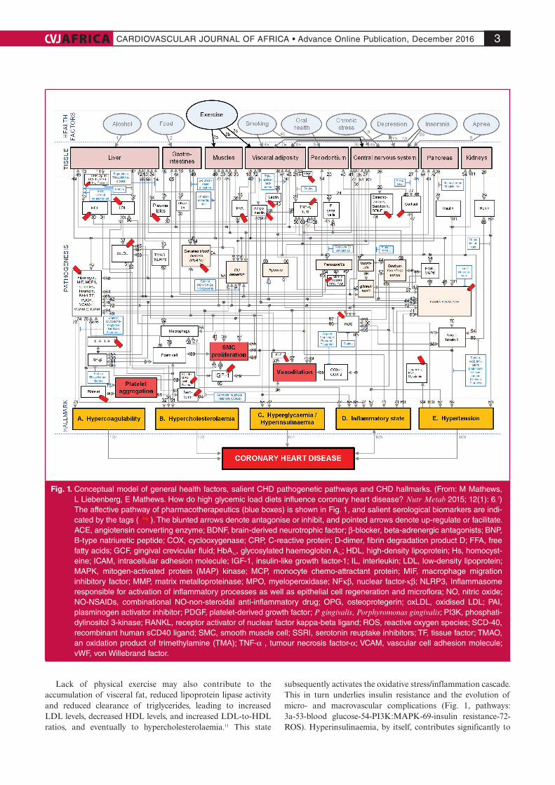
The integrated model of CHD that was developed in previous studies is presented in Fig. 1. The pathways (pathogenesis of CHD) within the integrated model can be tracked from where a chosen health factor influences the relevant tissue, to the end state of CHD. The pathways are therefore a visual representation of previously published knowledge. Salient serological biomarkers (shown in Fig. 1 as ) and pharmacotherapeutics (shown in
Fig. 1 as ) that act on the pathways are further indicated in Fig. 1.
The focus of this review is on using the integrated model to describe the interconnections of moderate exercise on the pathogenesis of CHD. Therefore a more detailed discussion of Fig. 1, relevant to exercise, is given in the next section. This review therefore attempts to quantify the CHD effect of moderate exercise by the connection of these to an array of biomarkers that represent increasing or decreasing CHD risk.
Pathogenetic effects of physical exercise In order to appraise the CHD effects of moderate exercise, the relevant pathogenetic pathways need to be considered. While Fig. 1 also indicates other health factors, only the pathways activated by moderate exercise are summarised in Table 1. It is however important to note that not all the pathways will be relevant to every patient and that all the pathways may not be active simultaneously, or occur in the same patient.
Fig. 1 (pathway: 3a-53-55-hyperglycaemia) shows the pathways involved in a lack of physical exercise (and decreased daily energy expenditure) and how this affects carbohydrate metabolism through changes in muscle glucose transporter (GLUT) protein content. Denervation of skeletal muscle results in rapid decreases in both muscle GLUT-4 contents and insulin-stimulated glucose uptake, therefore resulting in hyperglycaemia and concomitant hyperinsulinaemia (both CHD hallmarks) in non-diabetic patients.10
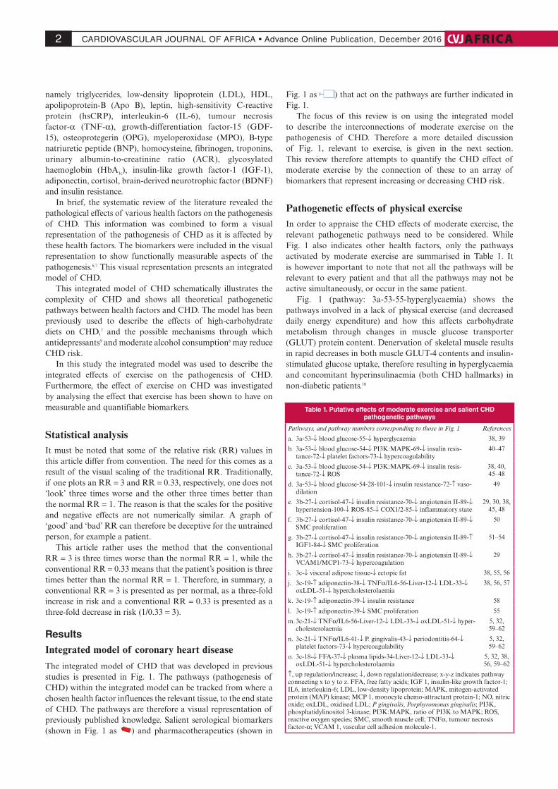
Table 1. Putative effects of moderate exercise and salient CHD pathogenetic pathways
Pathways, and pathway numbers corresponding to those in Fig. 1 References
a. 3a-53-↓ blood glucose-55-↓ hyperglycaemia 38, 39
b. 3a-53-↓ blood glucose-54-↓ PI3K:MAPK-69-↓ insulin resis-tance-72-↓ platelet factors-73-↓ hypercoagulability
40–47
c. 3a-53-↓ blood glucose-54-↓ PI3K:MAPK-69-↓ insulin resis-tance-72-↓ ROS
38, 40, 45–48
d. 3a-53-↓ blood glucose-54-28-101-↓ insulin resistance-72-↑ vaso-dilation
49
e. 3b-27-↓ cortisol-47-↓ insulin resistance-70-↓ angiotensin II-89-↓ hypertension-100-↓ ROS-85-↓ COX1/2-85-↓ inflammatory state
29, 30, 38, 45, 48
f. 3b-27-↓ cortisol-47-↓ insulin resistance-70-↓ angiotensin II-89-↓ SMC proliferation
50
g. 3b-27-↓ cortisol-47-↓ insulin resistance-70-↓ angiotensin II-89-↑ IGF1-84-↓ SMC proliferation
51–54
h. 3b-27-↓ cortisol-47-↓ insulin resistance-70-↓ angiotensin II-89-↓ VCAM1/MCP1-73-↓ hypercoagulation
29
i. 3c-↓ visceral adipose tissue-↓ ectopic fat 38, 55, 56
j. 3c-19-↑ adiponectin-38-↓ TNFα/IL6-56-Liver-12-↓ LDL-33-↓ oxLDL-51-↓ hypercholesterolaemia
38, 56, 57
k. 3c-19-↑ adiponectin-39-↓ insulin resistance 58
l. 3c-19-↑ adiponectin-39-↓ SMC proliferation 55
m. 3c-21-↓ TNFα/IL6-56-Liver-12-↓ LDL-33-↓ oxLDL-51-↓ hyper-cholesterolaemia
5, 32, 59–62
n. 3c-21-↓ TNFα/IL6-41-↓ P. gingivalis-43-↓ periodontitis-64-↓ platelet factors-73-↓ hypercoagulability
5, 32, 59–62
o. 3c-18-↓ FFA-37-↓ plasma lipids-34-Liver-12-↓ LDL-33-↓ oxLDL-51-↓ hypercholesterolaemia
5, 32, 38, 56, 59–62
↑, up regulation/increase; ↓, down regulation/decrease; x-y-z indicates pathway connecting x to y to z. FFA, free fatty acids; IGF 1, insulin-like growth factor-1; IL6, interleukin-6; LDL, low-density lipoprotein; MAPK, mitogen-activated protein (MAP) kinase; MCP 1, monocyte chemo-attractant protein-1; NO, nitric oxide; oxLDL, oxidised LDL; P gingivalis, Porphyromonas gingivalis; PI3K, phosphatidylinositol 3-kinase; PI3K:MAPK, ratio of PI3K to MAPK; ROS, reactive oxygen species; SMC, smooth muscle cell; TNFα, tumour necrosis factor-α; VCAM 1, vascular cell adhesion molecule-1.
CARDIOVASCULAR JOURNAL OF AFRICA • Advance Online Publication, December 2016AFRICA 3
Lack of physical exercise may also contribute to the accumulation of visceral fat, reduced lipoprotein lipase activity and reduced clearance of triglycerides, leading to increased LDL levels, decreased HDL levels, and increased LDL-to-HDL ratios, and eventually to hypercholesterolaemia.11 This state
subsequently activates the oxidative stress/inflammation cascade. This in turn underlies insulin resistance and the evolution of micro- and macrovascular complications (Fig. 1, pathways: 3a-53-blood glucose-54-PI3K:MAPK-69-insulin resistance-72-ROS). Hyperinsulinaemia, by itself, contributes significantly to
Fig. 1. Conceptual model of general health factors, salient CHD pathogenetic pathways and CHD hallmarks. (From: M Mathews, L Liebenberg, E Mathews. How do high glycemic load diets influence coronary heart disease? Nutr Metab 2015; 12(1): 6.7) The affective pathway of pharmacotherapeutics (blue boxes) is shown in Fig. 1, and salient serological biomarkers are indi-cated by the tags ( ). The blunted arrows denote antagonise or inhibit, and pointed arrows denote up-regulate or facilitate. ACE, angiotensin converting enzyme; BDNF, brain-derived neurotrophic factor; β-blocker, beta-adrenergic antagonists; BNP, B-type natriuretic peptide; COX, cyclooxygenase; CRP, C-reactive protein; D-dimer, fibrin degradation product D; FFA, free fatty acids; GCF, gingival crevicular fluid; HbA1c, glycosylated haemoglobin A1c; HDL, high-density lipoprotein; Hs, homocyst-eine; ICAM, intracellular adhesion molecule; IGF-1, insulin-like growth factor-1; IL, interleukin; LDL, low-density lipoprotein; MAPK, mitogen-activated protein (MAP) kinase; MCP, monocyte chemo-attractant protein; MIF, macrophage migration inhibitory factor; MMP, matrix metalloproteinase; MPO, myeloperoxidase; NFκβ, nuclear factor-κβ; NLRP3, Inflammasome responsible for activation of inflammatory processes as well as epithelial cell regeneration and microflora; NO, nitric oxide; NO-NSAIDs, combinational NO-non-steroidal anti-inflammatory drug; OPG, osteoprotegerin; oxLDL, oxidised LDL; PAI, plasminogen activator inhibitor; PDGF, platelet-derived growth factor; P gingivalis, Porphyromonas gingivalis; PI3K, phosphati-dylinositol 3-kinase; RANKL, receptor activator of nuclear factor kappa-beta ligand; ROS, reactive oxygen species; SCD-40, recombinant human sCD40 ligand; SMC, smooth muscle cell; SSRI, serotonin reuptake inhibitors; TF, tissue factor; TMAO, an oxidation product of trimethylamine (TMA); TNF-α , tumour necrosis factor-α; VCAM, vascular cell adhesion molecule; vWF, von Willebrand factor.
CARDIOVASCULAR JOURNAL OF AFRICA • Advance Online Publication, December 20164 AFRICA
atherogenecity, leading to CHD.12
An increase in plasma free fatty acid (FFA) concentrations plays a key role in the pathogenesis of insulin resistance through actions that block insulin signal transduction. An increase in FFA levels results in induction of oxidative stress, low-grade systemic inflammation, and subnormal vascular reactivity, in addition to causing insulin resistance.5 As insulin resistance also results in the relative non-suppression of adipocyte hormone-sensitive lipase,13 there is further enhancement in lipolysis, increased FFA and insulin resistance. As insulin suppresses pro-inflammatory transcription factors, such as nuclear factor-κβ (NF-κβ), and also suppresses reactive oxygen species (ROS) generation, insulin resistance therefore also has a comprehensive pro-inflammatory effect (Fig. 1, pathways: 3c-18-FFA-37-plasma lipids-34-12-LDL-33-oxLDL-51-hypercholesterolaemia).
Fig. 1 therefore shows why an insulin-resistant state may be pro-inflammatory. The origin of the insulin resistance may be traced back to the pro-inflammatory cytokine TNF-α, which is expressed by adipose tissue.14 Adipose tissue has been shown to express not only TNF-α, but also other pro-inflammatory mediators, including CRP. Macrophages residing in the adipose tissue may also be a source of pro-inflammatory factors and they can also modulate the secretory activities of adipocytes15 (Fig. 1, pathway: 3c-21-TNFα/IL6).
During regular moderate exercise, IL-6 is produced by skeletal muscle fibres via a TNF-independent pathway. IL-6 stimulates the appearance in the circulation of anti-inflammatory cytokines, which inhibit the production of pro-inflammatory TNF-α.16 Additionally, IL-6 enhances lipid turnover, stimulating lipolysis as well as fat oxidation. Regular physical exercise therefore induces suppression of TNF-α and thereby offers protection against TNF-α-induced insulin resistance.16 Low-grade systemic inflammation therefore appears to be aetiologically linked to the pathogenesis of CHD,17 countered by moderate exercise with its anti-inflammatory effects5 (Fig. 1, pathway: 3a-53-blood glucose-54-69-insulin resistance-71).
The adipokine adiponectin is anti-inflammatory and potentially anti-atherogenic.5 Low adiponectin levels act as a marker for CHD and are associated with overweight subjects.18 Regular physical exercise (and an energy-controlled diet) reduces visceral fat mass, with a subsequent increased release of anti-inflammatory adiponectin, therefore resulting in reduced risk of CHD19 (Fig. 1, pathway: 3c-19-39-insulin resistance).
Lack of physical exercise may lead to hypertension, another CHD hallmark, through increased vascular and sympathetic tone created by reduced bioavailability of nitrous oxide (NO) and activation of the renin–angiotensin system20, 21 (Fig. 1, pathway: 3a-53-blood glucose-54-60-72-vasodilation). Hypertension is directly correlated with visceral fat mass, which may be decreased by moderate exercise.22
The lower blood glucose levels that result from moderate exercise lead to a reduction in the phosphatidylinositol 3-kinase (PI3K) to mitogen-activated protein kinase (MAPK) ratio, which in turn decreases insulin resistance23 (Fig. 1, pathway: 3a-53-blood glucose-54-69-72-73-hypercoagulabilty). Increased insulin sensitivity decreases serum levels of platelet factors and thus reduces the potential for hypercoagulability.24,25
Moderate exercise also increases coronary blood flow,26 which increases the release of prostaglandins.27 This is important in heart microvasculature, in which prostaglandins are substantially
involved in flow-mediated vasodilation.27
Moderate exercise acts on the central nervous system by decreasing serum cortisol levels.28 This in turn reduces insulin resistance, which decreases angiotensin II levels and results in reduced hypertension. Reactive oxygen species (ROS) and cyclooxygenase (COX) 1/2 levels reduce concomitantly, which lead to a lower inflammatory state20 (Fig. 1, pathway: insulin resistance-85-inflammatory state).
It is apparent that moderate exercise directly and indirectly affects a plethora of interconnected pathogenetic mechanisms. Each CHD hallmark and pathogenetic trait can amplify the patient’s risk of CHD, therefore necessitating an integrated, multi-faceted therapeutic approach.
In this section, the pathogenetic pathways activated by moderate exercise are described, but the effects of these pathways have not been quantified. The next interrogation was therefore whether biomarkers could quantify the CHD effect of moderate exercise. This was accomplished by using connection graphs,
Table 2. Association between biomarkers and prediction of CHD relative risk
Biomarker (class and salient examples)
Prediction of CHD relative risk (95% CI)
Size of studies(N = number of trials,
n = number of patients)Refer-ences
Lipid-related markers
Triglycerides 0.99 (0.94–1.05) (N = 68, n = 302 430) 63
LDL 1.25 (1.18–1.33) (N = 15, n = 233 455) 64
HDL 0.78 (0.74–0.82) (N = 68, n = 302 430) 63
Apo B 1.43 (1.35–1.51) (N = 15, n = 233 455) 64
Leptin 1.04 (0.92–1.17) (n = 1 832) 65
Inflammatory markers
hsCRP 1.20 (1.18–1.22) (N = 38, n = 166 596) 66
IL-6 1.25 (1.19–1.32) (N = 25, n = 42 123) 67
TNF-α 1.17 (1.09–1.25) (N = 7, n = 6 107) 67
GDF-15 1.40 (1.10–1.80) (n = 1 740) 68
OPG 1.41 (1.33–1.57) (n = 5 863) 69
Marker of oxidative stress
MPO 1.17 (1.06–1.30) (n = 2 861) 70
Marker of vascular function and neurohormonal activity
BNP 1.42 (1.24–1.63) (N = 40, n = 87 474) 71
Homocysteine 1.15 (1.09–1.22) (N = 20, n = 22 652) 72, 73
Coagulation marker
Fibrinogen 1.15 (1.13–1.17) (N = 40, n = 185 892) 66
Necrosis marker
Troponins 1.15 (1.04–1.27) (n = 3 265) 58
Renal function marker
Urinary ACR 1.57 (1.26–1.95) (n = 626) 74
Metabolic markers
HbA1c 1.42 (1.16–1.74) (N = 2, n = 2 442) 75
IGF-1 0.76 (0.56–1.04) (n = 3 967) 76
Adiponectin 0.97 (0.86–1.09) (N = 14, n = 21 272) 77
Cortisol 1.10 (0.97–1.25) (n = 2 512) 78, 79
BDNF ? ? 80–82
Insulin resistance (HOMA) 1.46 (1.26–1.69) (N = 17, n = 51 161) 83
From: M Mathews, L Liebenberg, E Mathews. How do high glycemic load diets influence coronary heart disease? Nutr Metab 2015; 12(1): 6.7
n , number of participants; N, number of trials; CI, confidence interval; ACR, albumin-to-creatinine ratio; Apo B, apolipoprotein-B; BDNF, brain-derived neurotrophic factor; BNP, B-type natriuretic peptide; GDF-15, growth-differentiation factor-15; HbA1c, glycated haemoglobin A1c; HDL, high-density lipoprotein; HOMA, homeostasis model assessment; hsCRP, high-sensitivity C-reactive protein; IGF-1, insulin-like growth factor-1; IL-6, interleukin-6; LDL, low-density lipoprotein; MPO, myeloperoxidase; OPG, osteoprotegerin; TNF-α, tumour necrosis factor-α.
CARDIOVASCULAR JOURNAL OF AFRICA • Advance Online Publication, December 2016AFRICA 5
which link the relative effect of a health or pathogenic factor to the individual biomarkers through the pathways that are shown in Fig. 1.
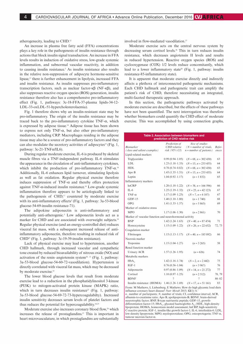
Biomarkers of coronary heart diseaseThe integrated model that was developed is a high-level conceptual model, from which the interconnectedness of CHD is immediately apparent (Fig. 1). The model is however complicated. Biomarkers can be used as indicators of an underlying disorder and the measurement of specific biomarkers enables prediction of the RR for CHD associated with the biomarker.29-31 The relevant biomarkers and their association with CHD risk per one standard deviation increase in said biomarker are given in Table 2. This can allow for the quantification of the effects of moderate exercise on the pathogenesis of CHD.
To simplify the integrated model, serological biomarkers (which can easily be measured) are used to link the effect of exercise to the corresponding RR of CHD. Fig. 2 presents a comparison of the RR associated with an array of serological biomarkers per one standard deviation increase in the biomarker.7
Effects of moderate exerciseUsing the integrated model in Fig. 1, it is possible to account for the impact that moderate exercise would have on the serological biomarkers of CHD. This enables a simplification of
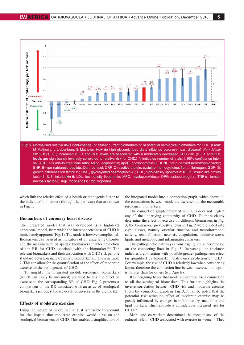
the integrated model into a connection graph, which shows all the connections between moderate exercise and the measurable serological biomarkers.
The connection graph presented in Fig. 3 does not neglect any of the underlying complexity of CHD. To more clearly determine the effect of exercise on different biomarkers in Fig. 3, the biomarkers previously shown in Fig. 2 were divided into eight classes, namely vascular function and neurohormonal activity, renal function, necrosis, coagulation, oxidative stress, lipids, and metabolic and inflammatory markers.
The pathogenetic pathways (from Fig. 1) are superimposed on the connecting lines in Fig. 3. Increasing line thickness indicates a connection with possible greater pathogenetic effect (as quantified by biomarker relative-risk prediction of CHD). For example, the risk of CHD is relatively low when considering leptin, therefore the connection line between exercise and leptin is thinner than for others (e.g. Apo B).
It is intriguing to see that moderate exercise has a connection to all the serological biomarkers. This further highlights the inverse correlation between CHD risk and moderate exercise. From the connection graph in Fig. 3, it can be noted that the potential risk reduction effect of moderate exercise may be greatly influenced by changes in inflammatory, metabolic and lipid markers, which provide a considerable increased risk for CHD.2-4
Mora and co-workers determined the mechanisms of the reduced risk of CHD associated with exercise in women.2 They
Fig. 2. Normalised relative risks (fold-change) of salient current biomarkers or of potential serological biomarkers for CHD. (From: M Mathews, L Liebenberg, E Mathews. How do high glycemic load diets influence coronary heart disease? Nutr Metab 2015; 12(1): 6.7) Increased IGF-1 and HDL levels are associated with a moderately decreased CHD risk. (IGF-1 and HDL levels are significantly inversely correlated to relative risk for CHD.) N indicates number of trials; I, 95% confidence inter-val; ACR, albumin-to-creatinine ratio; Adipo, adiponectin; ApoB, apolipoprotein-B; BDNF, brain-derived neurotrophic factor; BNP, B-type natriuretic peptide; Cort, cortisol; CRP, C-reactive protein; cysteine, homocysteine; fibrin, fibrinogen; GDF-15, growth-differentiation factor-15; HbA1c, glycosylated haemoglobin A1c; HDL, high-density lipoprotein; IGF-1, insulin-like growth factor-1; IL-6, interleukin-6; LDL, low-density lipoprotein; MPO, myeloperoxidase; OPG, osteoprotegerin; TNF-α, tumour necrosis factor-α; Trigl, triglycerides; Trop, troponins.
CARDIOVASCULAR JOURNAL OF AFRICA • Advance Online Publication, December 20166 AFRICA
found that a reduction in inflammatory biomarkers were the largest contributors to lowered risk. These were followed, in order, by blood pressure, lipid levels, body mass index (BMI) and haemoglobin level. In the study, the combination of different individual risk factors quantified only 35.5% of the total risk reduction due to physical exercise on CHD.2
It is therefore clear that the risk factors used by Mora and co-workers, in terms of serological biomarkers, did not fully quantify the risks associated with CHD. In their study, LDL, HDL and Apo B serum levels were recorded to monitor lipid levels, but only hsCRP serum levels were used for deducing inflammatory levels.2 It may therefore be possible that with the addition of the other biomarkers indicated in Fig. 3, the effect of moderate exercise may be better quantified.
In Fig. 3, it is clear from the risk associated with inflammation that reduction in inflammation would prove beneficial to CHD risk. The full extent of the relationship between exercise and inflammation has not been determined but it has been proven that chronic moderate exercise has a systemic anti-inflammatory effect.5,16,32 It has further been shown that the anti-inflammatory effect of exercise provides the largest individual risk-reduction component of moderate exercise in women.2
Naturally there is a strong link to the metabolic process that is manifested in the connection to the metabolic biomarkers,
specifically insulin resistance and glycated haemoglobin level.33,34 This connection may be largely mediated by the increased expenditure of energy, which produces favourable effects on CHD pathogenesis.10, 23 Moderate exercise is also related to changes in lipid factors such as increases in HDL cholesterol and decreases in LDL cholesterol and Apo B levels.33,34
Discussion It is clear that there are a wide variety of effects of exercise on the pathogenesis of CHD, which can be described by the changes in biomarkers. However, from the connection graph in Fig. 3, it is not immediately clear what the overall effect of moderate exercise is on CHD. This effect has been quantified in the RR reduction for CHD, which is observed in those who engage in moderate exercise.
Moderate-intensity physical exercise of 1 100 kcal/week is associated with an average RR of 0.75 (0.71–0.79), based on a large meta-analysis.35 The RR of 0.75 would correlate to a RR reduction of 1.33-fold using the method previously described in the Methods section.
The data from Fig. 3 show that inflammation and metabolic dysregulation may be key aspects in the pathogenesis of CHD.5,10,16,23,32-34 These aspects decrease during exercise and may
Fig. 3. Interconnection of relative risk effects of moderate exercise and serological biomarkers for CHD. ACR, albumin-to-creatinine ratio; Adipo, adiponectin; Apo B, apolipoprotein-B; BDNF, brain-derived neurotrophic factor; BNP, B-type natriuretic peptide; Cort, cortisol; CRP, C-reactive protein; cysteine, homocysteine; fibrin, fibrinogen; GDF-15, growth-differentiation factor-15; HbA1c, glyco-sylated haemoglobin A1c; HDL, high-density lipoprotein; IGF-1, insulin-like growth factor-1; IL-6, interleukin-6; LDL, low-density lipoprotein; MPO, myeloperoxidase; OPG, osteoprotegerin; TNF-α, tumour necrosis factor-α; Trigl, triglycerides; Trop, troponins.
CARDIOVASCULAR JOURNAL OF AFRICA • Advance Online Publication, December 2016AFRICA 7
therefore play a part in the 1.33-fold decreased risk for CHD.Based on the evidence, it is believed that the CHD benefit
associated with exercise is substantial and should garner a similar level of public interest as do other risk factors such as smoking, high cholesterol levels and treatments such as statin therapy. However, while exercise is frequently advised for healthy living,36 it is unfortunate that only 48.9% of Americans meet the physical activity guidelines. It follows from this that 51.1% of Americans do not meet the minimum physical activity guidelines, which results in 162.8 million Americans at a greater risk of CHD due to physical inactivity.37
The individual studies selected unfortunately represent only the risk associated with the cohort studied and cannot accurately be extrapolated to other populations without further research.
Conclusion Although it is well known that moderate exercise is associated with a lower risk of CHD, all the positive effects on CHD pathogenesis were not available in a detailed integrated model. Such a model would help provide further insight. A high-level conceptual model was therefore developed, which links moderate exercise with the pathogenesis, hallmarks and biomarkers of CHD.
The novel connection graph developed from this model shows, at a glance, the positive effect of moderate exercise on certain important aspects of the pathogenesis of CHD. It helps to graphically explain why moderate exercise is associated with lower CHD risk. From this it is apparent that exercise has a wide-ranging impact on the pathogenesis of CHD, with these effects notable in changes in CHD biomarkers.
The integrated high-level CHD model and simplified connection graph provide a summary of evidence for a causal relationship between CHD risk and moderate exercise. We acknowledge the fact that the integrated view is relevant to other lifestyle issues and for full comprehension will have to be replicated in other articles describing these factors.
The angel investor was Dr Arnold van Dyk and the research was later self-
funded. Prof Leon Liebenberg was involved in the initial research.
References1. Mathers CD, Boerma T, Fat DM. Global and regional causes of death.
Br Med Bull 2009; 92(1): 7–32.
2. Mora S, Cook N, Buring JE, Ridker PM, Lee I-M. Physical activity and
reduced risk of cardiovascular events potential mediating mechanisms.
Circulation 2007; 116(19): 2110–2118.
3. Green DJ, Spence A, Halliwill JR, Cable NT, Thijssen DH. Exercise
and vascular adaptation in asymptomatic humans. Exp Physiol 2011;
96(2): 57–70.
4. Thompson PD, Buchner D, Piña IL, Balady GJ, Williams MA, Marcus
BH, et al. Exercise and physical activity in the prevention and treatment
of atherosclerotic cardiovascular disease: A statement from the Council
on Clinical Cardiology (Subcommittee on Exercise, Rehabilitation,
and Prevention) and the Council on Nutrition, Physical Activity, and
Metabolism (Subcommittee on Physical Activity). Circulation 2003;
107(24): 3109–3116.
5. Gleeson M, Bishop NC, Stensel DJ, Lindley MR, Mastana SS, Nimmo
MA. The anti-inflammatory effects of exercise: mechanisms and impli-
cations for the prevention and treatment of disease. Nat Rev Immunol
2011; 11(9): 607–615.
6. Mathews MJ. A systems engineering approach to coronary heart disease
[PhD dissertation]. Potchefstroom: North-West University, 2016.
7. Mathews MJ, Liebenberg L, Mathews EH. How do high glycemic load
diets influence coronary heart disease? Nutr Metab 2015; 12(1): 6.
8. Mathews MJ, Liebenberg L, Mathews EH. The mechanism by which
moderate alcohol consumption influences coronary heart disease. Nutr
J. 2015; 14(1): 33.
9. Mathews MJ, Mathews EH, Liebenberg L. The mechanisms by which
antidepressants may reduce coronary heart disease risk. BMC Cardiovasc
Disord 2015; 15(1): 82.
10. Strasser B. Physical activity in obesity and metabolic syndrome. Ann NY
Acad Sci 2013; 1281(1): 141–159.
11. Warburton DE, Nicol CW, Bredin SS. Health benefits of physical activ-
ity: the evidence. Can Med Assoc J 2006; 174(6): 801–809.
12. Reaven GM. Role of insulin resistance in human disease. Diabetes 1988;
37(12): 1595–1607.
13. Dandona P, Aljada A, Chaudhuri A, Mohanty P, Garg R. Metabolic
syndrome: A comprehensive perspective based on interactions between
obesity, diabetes, and inflammation. Circulation 2005; 111(11): 1448–
1454.
14. Hotamisligil GS, Shargill NS, Spiegelman BM. Adipose expression of
tumor necrosis factor-alpha: direct role in obesity-linked insulin resist-
ance. Science 1993; 259(5091): 87–91.
15. Xu H, Barnes GT, Yang Q, Tan G, Yang D, Chou CJ, et al. Chronic
inflammation in fat plays a crucial role in the development of obesity-
related insulin resistance. J Clin Invest 2003; 112(12): 1821–1830.
16. Petersen AMW, Pedersen BK. The anti-inflammatory effect of exercise.
J Appl Physiol 2005; 98(4): 1154–1162.
17. Libby P. Inflammation in atherosclerosis. Arterioscler Thromb Vasc Biol
2012; 32(9): 2045–2051.
18. Pischon T, Girman CJ, Hotamisligil GS, Rifai N, Hu FB, Rimm EB.
Plasma adiponectin levels and risk of myocardial infarction in men. J
Am Med Assoc 2004; 291(14): 1730–1737.
19. Pedersen BK, Saltin B. Evidence for prescribing exercise as therapy in
chronic disease. Scand J Med Sci Sports 2006; 16(S1): 3–63.
20. Brown NJ, Agirbasli MA, Williams GH, Litchfield WR, Vaughan DE.
Effect of activation and inhibition of the renin-angiotensin system on
plasma PAI-1. Hypertension 1998; 32(6): 965–971.
21. Chobanian AV, Bakris GL, Black HR, Cushman WC, Green LA,
Izzo JL, et al. Seventh report of the joint National Committee on
Prevention, Detection, Evaluation, and Treatment of High Blood
Pressure. Hypertension 2003; 42(6): 1206–1252.
22. Klein S, Burke LE, Bray GA, Blair S, Allison DB, Pi-Sunyer X, et al.
Clinical implications of obesity with specific focus on cardiovascu-
lar disease. A statement for professionals from the American Heart
Association Council on Nutrition, Physical Activity, and Metabolism:
endorsed by the American College of Cardiology Foundation.
Circulation 2004; 110(18): 2952–2967.
23. Rose AJ, Richter EA. Skeletal muscle glucose uptake during exercise:
how is it regulated? Physiology 2005; 20(4): 260–270.
24. Cimenti C, Schlagenhauf A, Leschnik B, Schretter M, Tschakert
G, Gröschl W, et al. Low endogenous thrombin potential in trained
subjects. Thromb Res 2013; 131(6): e281–e285.
25. Rauramaa R, Salonen JT, Seppänen K, Salonen R, Venäläinen J,
Ihanainen M, et al. Inhibition of platelet aggregability by moderate-
intensity physical exercise: a randomized clinical trial in overweight men.
Circulation 1986; 74(5): 939–944.
26. Hambrecht R, Wolf A, Gielen S, Linke A, Hofer J, Erbs S, et al. Effect
CARDIOVASCULAR JOURNAL OF AFRICA • Advance Online Publication, December 20168 AFRICA
of exercise on coronary endothelial function in patients with coronary
artery disease. N Engl J Med 2000; 342(7): 454–460.
27. Hambrecht R, Fiehn E, Weigl C, Gielen S, Hamann C, Kaiser R, et al.
Regular physical exercise corrects endothelial dysfunction and improves
exercise capacity in patients with chronic heart failure. Circulation 1998;
98(24): 2709–2715.
28. Raastad T, Bjøro T, Hallen J. Hormonal responses to high-and
moderate-intensity strength exercise. Eur J Appl Physiol 2000; 82(1–2):
121–128.
29. Libby P. Inflammation in atherosclerosis Nature 2002; 420: 868–874.
30. Packard RR, Libby P. Inflammation in atherosclerosis: from vascular
biology to biomarker discovery and risk prediction. Clin Chem 2008;
54(1): 24–38.
31. Vasan RS. Biomarkers of cardiovascular disease molecular basis and
practical considerations. Circulation 2006; 113(19): 2335–2362.
32. Beavers KM, Brinkley TE, Nicklas BJ. Effect of exercise training on
chronic inflammation. Clin Chim Acta 2010; 411(11): 785–793.
33. Jorge MLMP, de Oliveira VN, Resende NM, Paraiso LF, Calixto A,
Diniz ALD, et al. The effects of aerobic, resistance, and combined
exercise on metabolic control, inflammatory markers, adipocytokines,
and muscle insulin signaling in patients with type 2 diabetes mellitus.
Metabolism 2011; 60(9): 1244–1252.
34. Kelley GA, Kelley KS. Efficacy of aerobic exercise on coronary heart
disease risk factors. Prev Cardiol 2008; 11(2): 71–75.
35. Sattelmair J, Pertman J, Ding EL, Kohl HW, Haskell W, Lee I-M. Dose
response between physical activity and risk of coronary heart disease: a
meta-analysis. Circulation 2011; 124(7): 789–795.
36. Committee PAGA. Physical activity guidelines for Americans.
Washington, DC: US Department of Health and Human Services, 2008.
37. Go AS, Mozaffarian D, Roger VL, Benjamin EJ, Berry JD, Borden WB,
et al. Heart disease and stroke statistics – 2013 update: a report from the
American Heart Association. Circulation 2013; 127(1): e6–e245.
38. Golbidi S, Laher I. Exercise and the cardiovascular system. Cardiol Res
Pract 2012; 2012: e210852.
39. Chan D, Ng LL. Biomarkers in acute myocardial infarction. BMC Med
2010; 8(1): 34.
40. Muniyappa R, Montagnani M, Koh KK, Quon MJ. Cardiovascular
actions of insulin. Endocr Rev 2007; 28(5): 463–491.
41. Myers J, Kaykha A, George S, Abella J, Zaheer N, Lear S, et al. Fitness
versus physical activity patterns in predicting mortality in men. Am J
Med 2004; 117(12): 912–918.
42. Green DJ, O’Driscoll G, Joyner MJ, Cable NT. Exercise and cardiovas-
cular risk reduction: time to update the rationale for exercise? J Appl
Physiol 2008; 105(2): 766–768.
43. Jenkins NT, Martin JS, Laughlin MH, Padilla J. Exercise-induced
signals for vascular endothelial adaptations: implications for cardiovas-
cular disease. Curr Cardiovasc Risk Rep 2012; 6(4): 331–346.
44. Høstmark AT, Ekeland GS, Beckstrøm AC, Meen HD. Postprandial
light physical activity blunts the blood glucose increase. Prev Med 2006;
42(5): 369–371.
45. Boulé NG, Weisnagel SJ, Lakka TA, Tremblay A, Bergman RN,
Rankinen T, et al. Effects of exercise training on glucose homeostasis:
The HERITAGE Family study. Diabetes Care 2005; 28(1): 108–114.
46. Temelkova-Kurktschiev TS, Koehler C, Henkel E, Leonhardt W,
Fuecker K, Hanefeld M. Postchallenge plasma glucose and glycemic
spikes are more strongly associated with atherosclerosis than fasting
glucose or HbA1c level. Diabetes Care 2000; 23(12): 1830–1834.
47. Okutsu M, Suzuki K, Ishijima T, Peake J, Higuchi M. The effects of
acute exercise-induced cortisol on CCR2 expression on human mono-
cytes. Brain Behav Immun 2008; 22(7): 1066–1071.
48. Teixeira de Lemos E, Oliveira J, Páscoa Pinheiro J, Reis F. Regular phys-
ical exercise as a strategy to improve antioxidant and anti-inflammatory
status: benefits in type 2 diabetes mellitus. Oxid Med Cell Longev 2012;
2012: 15.
49. Wang JC, Bennett M. Aging and atherosclerosis mechanisms, functional
consequences, and potential therapeutics for cellular senescence. Circ
Res 2012; 111(2): 245–259.
50. Epstein FH, Ross R. Atherosclerosis – an inflammatory disease. N Engl
J Med 1999; 340(2): 115–126.
51. von der Thüsen JH, Borensztajn KS, Moimas S, van Heiningen S,
Teeling P, van Berkel TJ, et al. IGF-1 has plaque-stabilizing effects in
atherosclerosis by altering vascular smooth muscle cell phenotype. Am J
Pathol 2011; 178(2): 924–934.
52. Shai S-Y, Sukhanov S, Higashi Y, Vaughn C, Rosen CJ, Delafontaine
P. Low circulating insulin-like growth factor I increases atherosclerosis
in ApoE-deficient mice. Am J Physiol Heart Circ Physiol 2011; 300(5):
H1898.
53. Ruidavets J, Luc G, Machez E, Genoux A, Kee F, Arveiler D, et al.
Effects of insulin-like growth factor 1 in preventing acute coronary
syndromes: The PRIME study. Atherosclerosis 2011; 218(2): 464–469.
54. Higashi Y, Sukhanov S, Anwar A, Shai S-Y, Delafontaine P. Aging,
atherosclerosis, and IGF-1. J Gerontol A Biol Sci Med Sci 2012; 67(6):
626–639.
55. Van Gaal LF, Mertens IL, Christophe E. Mechanisms linking obesity
with cardiovascular disease. Nature 2006; 444(7121): 875–880.
56. Mougios V. Exercise Biochemistry. Champaign, IL: Human Kinetics,
2006.
57. Yu Z, Ye X, Wang J, Qi Q, Franco OH, Rennie KL, et al. Associations of
physical activity with inflammatory factors, adipocytokines, and meta-
bolic syndrome in middle-aged and older chinese people. Circulation
2009; 119(23): 2969–2977.
58. Wang TJ, Wollert KC, Larson MG, Coglianese E, McCabe EL, Cheng
S, et al. Prognostic utility of novel biomarkers of cardiovascular stress:
the Framingham Heart Study. Circulation 2012; 126(13): 1596–1604.
59. Autenrieth C, Schneider A, Doering A, Meisinger C, Herder C, Koenig
W, et al. Association between different domains of physical activity and
markers of inflammation. Med Sci Sports Exerc 2009; 41(9): 1706–1713.
60. Lira FS, Rosa JC, Pimentel GD, Souza HA, Caperuto EC, Carnevali
LC, et al. Endotoxin levels correlate positively with a sedentary lifestyle
and negatively with highly trained subjects. Lipids Health Dis 2010;
9(1): 82.
61. Shanely R, Nieman D, Henson D, Jin F, Knab A, Sha W. Inflammation
and oxidative stress are lower in physically fit and active adults. Scand J
Med Sci Sports 2011: 215–223.
62. Pedersen BK. The diseasome of physical inactivity–and the role of
myokines in muscle–fat cross talk. J Physiol. 2009;587(23):5559-68.
63. Di Angelantonio E, Sarwar N, Perry P, Kaptoge S, Ray KK, Thompson
A, et al. Major lipids, apolipoproteins, and risk of vascular disease. J Am
Med Assoc 2009; 302(18): 1993–2000.
64. Sniderman AD, Williams K, Contois JH, Monroe HM, McQueen MJ,
de Graaf J, et al. A meta-analysis of low-density lipoprotein cholesterol,
non-high-density lipoprotein cholesterol, and apolipoprotein B as mark-
ers of cardiovascular risk. Circ Cardiovasc Qual Outcomes 2011; 4(3):
337–345.
65. Luc G, Empana J, Morange P, Juhan-Vague I, Arveiler D, Ferrieres J,
et al. Adipocytokines and the risk of coronary heart disease in healthy
middle aged men: the PRIME study. Int J Obes 2009; 34(1): 118–126.
66. Kaptoge S, Di Angelantonio E, Pennells L, Wood AM, White IR,
Gao P, et al. C-reactive protein, fibrinogen, and cardiovascular disease
prediction. N Engl J Med 2012; 367(14): 1310–1320.
CARDIOVASCULAR JOURNAL OF AFRICA • Advance Online Publication, December 2016AFRICA 9
67. Kaptoge S, Seshasai SRK, Gao P, Freitag DF, Butterworth AS,
Borglykke A, et al. Inflammatory cytokines and risk of coronary heart
disease: new prospective study and updated meta-analysis. Eur Heart J
2014; 35(9): 578–589.
68. Daniels LB, Clopton P, Laughlin GA, Maisel AS, Barrett-Connor E.
Growth-differentiation factor-15 is a robust, independent predictor of
11-year mortality risk in community-dwelling older adults: the Rancho
Bernardo study. Circulation 2011; 123(19): 2101–2110.
69. Mogelvang R, Pedersen SH, Flyvbjerg A, Bjerre M, Iversen AZ,
Galatius S, et al. Comparison of osteoprotegerin to traditional athero-
sclerotic risk factors and high-sensitivity C-reactive protein for diagnosis
of atherosclerosis. Am J Cardiol 2012; 109(4): 515–520.
70. Rana JS, Arsenault BJ, Després J-P, Côté M, Talmud PJ, Ninio E, et al.
Inflammatory biomarkers, physical activity, waist circumference, and
risk of future coronary heart disease in healthy men and women. Eur
Heart J 2009; 32(3): 336–344.
71. Di Angelantonio E, Chowdhury R, Sarwar N, Ray KK, Gobin R,
Saleheen D, et al. B-type natriuretic peptides and cardiovascular
risk systematic review and meta-analysis of 40 prospective studies.
Circulation 2009; 120(22): 2177–2187.
72. Humphrey LL, Fu R, Rogers K, Freeman M, Helfand M. Homocysteine
level and coronary heart disease incidence: a systematic review and
meta-analysis. Mayo Clin Proc 2008; 83(11): 1203–1212.
73. Homocysteine Studies Collaboration. Homocysteine and risk of
ischemic heart disease and stroke: a meta-analysis. J Am Med Assoc
2002; 288(16): 2015–2022.
74. Kistorp C, Raymond I, Pedersen F, Gustafsson F, Faber J, Hildebrandt
P. N-terminal pro-brain natriuretic peptide, C-reactive protein, and
urinary albumin levels as predictors of mortality and cardiovascular
events in older adults. J Am Med Assoc 2005; 293(13): 1609–1616.
75. Pai JK, Cahill LE, Hu FB, Rexrode KM, Manson JE, Rimm EB.
Hemoglobin A1c is associated with increased risk of incident coronary
heart disease among apparently healthy, nondiabetic men and women. J
Am Heart Assoc 2013; 2(2): e000077.
76. Schneider HJ, Wallaschofski H, Volzke H, Markus MR, Doerr M, Felix
SB, et al. Incremental effects of endocrine and metabolic biomarkers
and abdominal obesity on cardiovascular mortality prediction. PloS
One 2012; 7(3): e33084.
77. Kanhai D, Kranendonk M, Uiterwaal C, Graaf Y, Kappelle L, Visseren
F. Adiponectin and incident coronary heart disease and stroke. A
systematic review and meta‐analysis of prospective studies. Obes Rev
2013; 14(7): 555–567.
78. Smith GD, Ben-Shlomo Y, Beswick A, Yarnell J, Lightman S, Elwood P.
Cortisol, testosterone, and coronary heart disease: prospective evidence
from the Caerphilly study. Circulation 2005; 112(3): 332–340.
79. Hamer M, Endrighi R, Venuraju SM, Lahiri A, Steptoe A. Cortisol
responses to mental stress and the progression of coronary artery calci-
fication in healthy men and women. PloS One. 2012; 7(2): e31356.
80. Noble EE, Billington CJ, Kotz CM, Wang C. The lighter side of BDNF.
Am J Physiol Regul Integr Comp Physiol 2011; 300(5): R1053.
81. Karatsoreos IN, McEwen BS. Psychobiological allostasis: resistance,
resilience and vulnerability. Trends Cogn Sci 2011; 15(12): 576–584.
82. Calabrese F, Molteni R, Racagni G, Riva MA. Neuronal plasticity: a
link between stress and mood disorders. Psychoneuroendocrinology 2009;
34: S208-S216.
83. Gast KB, Tjeerdema N, Stijnen T, Smit JW, Dekkers OM. Insulin
resistance and risk of incident cardiovascular events in adults without
diabetes: meta-analysis. PloS One 2012; 7(12): e52036.