Embed Size (px)
Citation preview

Manual of rotavirus detection and
characterization methods
WHO/IVB/08.17 ORIGINAL: ENGLISH
Immunization, Vaccines and Biologicals

Manual of rotavirus detection and
characterization methods
WHO/IVB/08.17 ORIGINAL: ENGLISH
Immunization, Vaccines and Biologicals

ii
The Department of Immunization, Vaccines and Biologicals thanks the donors whose unspecified financial support
has made the production of this document possible.
This document was produced by the Expanded Programme on Immunization
of the Department of Immunization, Vaccines and Biologicals
Ordering code: WHO/IVB/08.17 Printed: October 2009
This publication is available on the Internet at: www.who.int/vaccines-documents/
Copies of this document as well as additional materials on immunization, vaccines and biologicals may be requested from:
World Health Organization Department of Immunization, Vaccines and Biologicals
CH-1211 Geneva 27, Switzerland • Fax: + 41 22 791 4227 • Email: [email protected] •
© World Health Organization 2009
All rights reserved. Publications of the World Health Organization can be obtained from WHO Press, World Health Organization, 20 Avenue Appia, 1211 Geneva 27, Switzerland (tel: +41 22 791 3264; fax: +41 22 791 4857; email: [email protected]). Requests for permission to reproduce or translate WHO publications – whether for sale or for noncommercial distribution – should be addressed to WHO Press, at the above address (fax: +41 22 791 4806; email: [email protected]).
The designations employed and the presentation of the material in this publication do not imply the expression of any opinion whatsoever on the part of the World Health Organization concerning the legal status of any country, territory, city or area or of its authorities, or concerning the delimitation of its frontiers or boundaries. Dotted lines on maps represent approximate border lines for which there may not yet be full agreement.
The mention of specific companies or of certain manufacturers’ products does not imply that they are endorsed or recommended by the World Health Organization in preference to others of a similar nature that are not mentioned. Errors and omissions excepted, the names of proprietary products are distinguished by initial capital letters.
All reasonable precautions have been taken by the World Health Organization to verify the information contained in this publication. However, the published material is being distributed without warranty of any kind, either expressed or implied. The responsibility for the interpretation and use of the material lies with the reader. In no event shall the World Health Organization be liable for damages arising from its use.
The named authors alone are responsible for the views expressed in this publication.
Printed by the WHO Document Production Services, Geneva, Switzerland

iii
Contents
Acknowledgements .............................................................................................................vUsing this manual ............................................................................................................ vii
1. Introduction and overview ......................................................................................1
1.1 Background ..........................................................................................................11.2 Rotavirus Detection ............................................................................................21.3 Rotavirus Characterization .................................................................................41.4 Safety precautions .............................................................................................161.5 Specimen requirements .....................................................................................171.6 Shipping ..............................................................................................................17
2. Laboratory Procedures:Rotavirus Detection .....................................................18
2.1 Virus cultivation .................................................................................................192.2 Detection of rotavirus in cell culture by IF .....................................................212.3 Electron microscopy (EM) ...............................................................................222.4 PAGE and silver staining .................................................................................23
3. Rotavirus characterization: Serological Methods .............................................31
3.1. Serotyping and subgrouping with monoclonal antibodies ............................31
4. Rotavirus characterisation: Molecular methods ................................................47
4.1. Method 1: Manual RNA extraction from stool .............................................474.2 Method 2: RNA extraction using hydroxyapatite .........................................494.3 Method 3: RNA Extraction Using RNAID ..................................................514.4 Method 4: RNA extraction using Trizol ........................................................534.5 Method 5: RNA extraction using the CTAB method ...................................544.6 Method 6: RNA extraction using the Boom method .....................................554.7 Method 7: RNA extraction from cell lysates ..................................................564.8 Method 8: G Genotyping ..................................................................................594.9 Method 9: P Genotyping .................................................................................634.10 Method 10: G Genotyping ................................................................................664.11 Method 11: P Genotyping ...............................................................................704.12 Method 12: G Genotying .................................................................................734.13 Method 13: P Genotyping ...............................................................................774.14 Method 14: G genotyping ................................................................................804.15 Method 15: P Genotyping ...............................................................................854.16 Method 16: G and P genotyping ......................................................................914.17 Trouble shooting ...............................................................................................99
5. References ...............................................................................................................100

iv
Appendix 1: Oligonucleotide primers ........................................................................104
Appendix 2:Cloning and sequencing methods .........................................................108
Appendix 3: Preparation of reagents .........................................................................132
Appendix 4:Suppliers ....................................................................................................138
Appendix 5:Worksheets ................................................................................................143

v
Acknowledgements
This document was produced for the Expanded Programme on Immunization of the Department of Immunization, Vaccines and Biologicals, WHO by the Laboratory Directors of the following Regional Rotavirus Laboratories and the WHO Rotavirus Collaborating Centers:
Jon GentschWHO Collaborating Center (US)Gastroenteritis and Respiratory Viruses Laboratory BranchDivision of Viral DiseasesNational Center for Immunization and Respiratory DiseasesCoordinating Center for Infectious DiseasesCenters for Disease Control and PreventionAtlanta, Georgia, US
Jim Gray and Miren Iturriza-GómaraEuropean Regional Rotavirus LaboratoryEnteric Virus Unit Virus Reference Department Centre for Infections Health Protection Agency London, UK
John Klena Eastern Mediterranean Regional Rotavirus LaboratoryEnteric Diseases Research ProgramU.S. Naval Medical Research Unit No. 3Cairo, Egypt
Carl KirkwoodWHO Collaborating Center (Australia)Enteric Virus group Murdoch Childrens Research Institute, Royal Children’s Hospital Victoria, Australia
George ArmahWest African Rotavirus Regional LaboratoryDepartment of Electron Microscopy and HistopathologyNoguchi Memorial Institute for Medical ResearchUniversity of Ghana, Legon, Ghana

vi
Nicola A. PageMRC Diarrhoeal Pathogens Research Unit (DPRU)University of Limpopo (Medunsa Campus)PO Box 173, Medunsa0204, South Africa
The following members of the “Rotavirus Experts External Advisory Group” kindly reviewed and provided helpful comments on the document: Kari Johansen - European Centre for Disease Prevention and Control, Ana Maria Bispo - PAHO, Duncan Steele - PATH and Timo Vesikari - University of Tampere Medical School.

vii
Using this manual
This manual has been prepared by staff of the WHO Rotavirus Collaborating Centers and Regional Laboratories throughout the world and is a comprehensive collection of methods for the detection and characterisation of rotaviruses. The manual should be used in conjunction with the training provided in a Collaborating Centre or Regional Laboratory and contact with that centre should be maintained in order to provide quality assurance and frequent updates of methods.
For some of the techniques described, a single method is provided, indicating general consensus among the network of rotavirus laboratories. Where multiple methods for a particular analyte or target are included, these have arisen through differences in facilities, access to equipment and reagents and to some extent geographical variation among rotavirus strains. It should be remembered that like the rotaviruses themselves, the methods used to detect and characterise strains are continually evolving and being refined.
To get the best out of this manual, staff should consult with their regional Collaborating Center or Regional Laboratory, take account of the facilities, equipment and reagents available and select the methods that are most suited to their environment. Users can create their own laboratory bench manual by taking the Introduction, References, Appendices and their methods of choice.
This manual explains the most common procedures for rotavirus strain surveillance.
Section 1 presents brief overviews of the methods and discusses implementation •issues.
Sections 2-4 describe each of the procedures in detail, including modifications •to standard reagents or typing strategies that can be applied as needed to achieve optimal results.
Section 5 lists the published scientific papers cited in the manual. •
Appendices provide additional information on reagents, suppliers, primers, •and cloning and sequencing methods.

viii

1WHO/IVB/08.17
1.1 Background
Group A rotaviruses, the most important cause of severe childhood diarrhea, belong to the Reoviridae family of non-enveloped dsRNA-containing viruses. Members of the genus Rotavirus have an 11-segment genome encased within three iscosahedral protein shells and are referred to as triple-layered viruses. The virion consists of an inner VP2 protein layer surrounding the RNA segments and several molecules of VP1 and VP3 proteins, a middle VP6 protein capsid, and an outer layer containing VP4 protein spikes embedded in a VP7 capsid.1
The capsid proteins are responsible for many of the serologic properties of Group A rotaviruses. Host antibodies to the VP6 protein define the rotavirus group antigen, whereas antibodies to VP7 and VP4 define G and P serotypes, respectively. Because antibodies to VP7 and VP4 also elicit protective immunity, vaccines have been targeted to these two proteins. Results from studies of animal models suggest that antibodies to VP6 and a viral nonstructural protein, NSP4, might also be involved in generation of protective immunity.2,3 Current vaccine development strategies are based on oral immunization with live-attenuated rotavirus strains designed to prevent the most severe form of the disease by eliciting serotype-specific, heterotypic, or a combination of serotype-specific and heterotypic immunity to the most common rotavirus serotypes.4,5
In countries considering a rotavirus vaccination program, public health officials need to collect data on the rotavirus disease burden to assess the need for a vaccine.6 Strain surveillance is also needed to determine the most important serotypes against which to provide protection. Countries setting up these projects therefore need laboratory workers trained in methods for rotavirus detection and characterization. Although rotavirus diagnostic procedures are routine, characterization methods include a variety of specialized techniques for the antigenic and molecular identification of rotavirus strains.
A compendium of current methods and protocols and a general strategy for conducting strain surveillance, including a flow chart of possible approaches to strain characterization, have been published elsewhere.7,8
1. Introduction and overview

Manual of Rotavirus detection and characterization methods2
1.2 Rotavirus Detection
Techniques for rotavirus detection include:
Electron microscopy •Antigen detection •
enzyme immunoassay (EIA) –
L a t e x a g g l u t i n a t i o n a n d l a t e r a l - f l o w i m m u n o a s s a y s –(immunochromatography)
Nucleic acid detection (PAGE) and nucleic acid amplification (RT-PCR)•
Most of these methods are relatively efficient at detecting rotaviruses, at least in part because of the large amount of intact rotavirus present in stool specimens of children with gastroenteritis. The methods have been reviewed in detail elsewhere and will be described only briefly here.
1.2.1 Electron microscopy
Electron microscopy is highly specific for detection of rotavirus and is as sensitive as some EIAs. However, the method is too labor intensive for routine detection of rotavirus in large numbers of stool specimens. In addition, EM requires an expensive instrument and highly trained personnel and cannot distinguish between rotaviruses of different groups.
1.2.2 Antigen detection
The most widely used methods for rotavirus diagnosis are based on detection of protein antigens on rotavirus particles in stool specimens. The most appropriate antigen detection format for large-scale surveillance studies is an EIA that uses rotavirus-specific antibodies to capture antigen onto wells of plastic plates. The antigen is then detected in a colorimetric reaction using a second rotavirus-specific antibody coupled to a detector enzyme. The EIA format is highly sensitive and specific and is adaptable to large sample volumes in the 96-well plate format. The optical density (OD) results can be easily recorded with a standard plate reader, permitting analysis of results with standard computer programs. Latex agglutination, utilizing latex particles coated with anti-rotavirus antibodies can be used as an alternative to EIA and rapid near patient tests using immunochromatographic methods are being used widely in consulting rooms.
Because of the importance of rotaviruses in clinical settings, many antigen detection methods have been commercialized, and data are available on their sensitivity and specificity. Two commercial tests with adequate sensitivity and specificity have been recommended for surveillance networks: PremierTM Rotaclone® (Meridian Biosciences; Cincinnati, Ohio) and IDEIATM Rotavirus (Oxoid (Ely) Limited Thermo Fisher Scientific, Cambridgeshire, United Kingdom). Currently, however, organizers of surveillance networks, including the Centers for Disease Control and Prevention (CDC) and the World Health Organization (WHO), recommend the IDEIA kit because of its price (about $US 1 per test) and the willingness of the manufacturer to ship kits anywhere in the world. The recommendation of one kit for use by surveillance networks will also enhance the comparability of data collected in different sites.

3WHO/IVB/08.17
The recommendation should not, however, be interpreted as an endorsement of the IDEIA kit as more sensitive or specific compared to Rotaclone. Laboratories should consult the kit inserts for a detailed laboratory protocol and assay performance characteristics.
Although the commercial EIAs used for rotavirus surveillance are sensitive and specific, quality control (QC) procedures are needed to ensure that different laboratories are performing the commercial assay at high proficiency. A standard panel of rotavirus-positive and -negative stool samples (proficiency panel) can be obtained from any of the rotavirus Regional laboratories listed in this manual. Network laboratories should report results from proficiency panel testing back to the Regional laboratory to confirm the accuracy of the test results. Laboratories should also arrange to send one of their first batches of rotavirus-positive stool specimens collected from children with gastroenteritis to a Regional laboratory for independent confirmation of results. If discrepancies are found in either the proficiency panel or the clinical specimens, the surveillance laboratory should work with the Regional laboratory to identify the source of the discordant results.
1.2.3 Nucleic acid detection
Because of the large quantities of rotavirus present in stool samples from children with gastroenteritis, the viral nucleic acid segments can be visualized directly after extraction from virus particles, by electrophoresis on acrylamide gels, and staining with ethidium bromide or silver nitrate. After electrophoresis, human rotavirus Groups A, B, and C have distinct patterns of gene-segment distribution, designated electropherotypes. The results of electropherotyping correlate with the presence of viruses of a specific group as shown by using other methods. Thus, the presence of distinct electropherotype patterns has long been considered diagnostic for the presence of individual rotaviruses of Groups A, B, and C (Figure 1).9 For Group A rotaviruses, most samples that are positive for rotavirus by EIA will be positive for the characteristic pattern of rotavirus RNA segments after electrophoresis and silver staining. In some cases, silver nitrate staining of viral nucleic acid has roughly the same sensitivity as EIA methods.10
Consequently, the PAGE method has sometimes been used to diagnose Group A rotavirus infections for surveillance studies. However, this method is very labor intensive and time consuming.
A variety of sensitive conventional or real-time reverse-transcription polymerase chain reaction (RT-PCR) methods have been developed based on primers specific for several different rotavirus genes.11-13 These methods have been particularly useful in detecting rotavirus in extra-intestinal tissues, in studies of the duration of viral shedding in stool and the correlation between disease severity and virus load.12,14 RT-PCR is also useful for verifying that RNA extracts contain intact rotavirus RNA. However, because it is relatively expensive and labor intensive and detects low copy numbers of rotavirus RNA, RT-PCR is not suitable for use in routine rotavirus detection studies.

Manual of Rotavirus detection and characterization methods4
1.3 Rotavirus Characterization
1.3.1 Serotyping and subgrouping with monoclonal antibodies
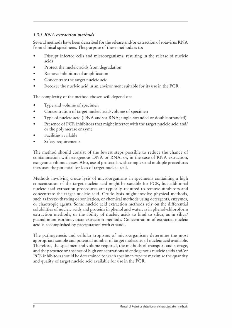
Enzyme immunoassays described by groups in Australia, Japan, and the United States allow determination of rotavirus VP6 subgroup and VP7 serotype using serotype-specific monoclonal antibodies.16,26,43 The five most common rotavirus G serotypes (G1, G2, G3, G4, G9) can be assigned a serotype directly from fecal material using several ELISA formats incorporating monoclonal antibodies (Mabs) that bind in a serotype-specific manner to the VP7 protein. Similarly, VP6 subgroupings I, II, I & II and non-I, II can be assigned using binding specificity of VP6 Mabs. The Mabs can be obtained as Ascites fluids from the individual investigators that isolated them.
Three types of combined subgroup (VP6) and serotype (VP7) ELISAs, described in this manual are commonly used in rotavirus research. One uses a polyclonal antibody to coat the solid phase, which will bind the serotype- or subgroup-specific Mabs to capture the rotaviruses in the fecal samples. The second method uses the serotype- or subgroup-specific Mabs bound directly to the solid phase to capture the virus. The third uses a polyclonal anti-rotavirus VP6 cross-reactive antibody bound to the solid phase to capture the virus, which will then be incubated with the different serotype- or subgroup-specific Mabs.
Studies using serotyping Mabs have typically typed 60%-70% of strains circulating in the community. The method is rapid and inexpensive. It provides G serotype information and, with suitable reagents, might provide information on antigenic differences between rotavirus strains of the same serotype.
VP7 serotyping. In the early to mid 1980s, Mabs against the VP7 (G) serotype antigen of the four most common rotaviruses were isolated and shown to bind in a serotype-specific manner to intact virus particles. Subsequent development of serotype-specific EIA methods with these Mabs permitted the direct serotyping of rotaviruses in fecal specimens.15,16 Large-scale serotyping studies of rotaviruses in stool samples showed that these serotypes (now called G1 to G4) were globally common causes of gastroenteritis in children.17 The antibodies are now used for direct serotyping studies.
The Mab technique provides an antigenic measure of strain serotype rather than the indirect result provided by RT-PCR genotyping. Mabs specific for different variants of common serotypes have also been isolated, and their use in Mab-serotyping EIAs allows the detection of these antigenic variants (designated monotypes) in circulating rotaviruses.18 Other serotype-specific Mabs have proven valuable for detection of less common serotypes such as G5, G6, G8 and G10. Several Mabs to serotype G9 have been important in the detection of this serotype, subsequently determined to be common worldwide.19,20

5WHO/IVB/08.17
A disadvantage of Mab serotyping is that a substantial fraction of the rotaviruses in fecal specimens cannot be serotyped. Reasons include insufficient numbers of intact virus particles, antigenic variation in common serotypes that renders them non-reactive with Mabs, and stool inhibitors that alter the binding of virus to antibody. The problem can be partially overcome by using larger panels of Mabs containing antibodies to different antigenic variants of the various serotypes. However, for some collections of rotavirus specimens, a large percentage of samples will need to be analyzed by RT-PCR to determine the genotypes of the strains not typeable with Mabs.21 Another drawback of Mab serotyping is the need for continual supplies of Mabs and rotavirus hyperimmune antisera, both of which must be produced in animals. These processes require considerable resources in the context of the declining use of animals in research.
VP4 serotyping. Serotyping assays based on Mabs specific for the three most common human rotavirus P (VP4) serotypes/subtypes, P1A[8], P1B[4], and P2A[6], have also been developed.22,23 These assays have proven valuable in defining antigenic variation in serotypes such as the globally common P1A[8].24 However, the many cross-reactive epitopes observed between different P serotypes/subtypes in rotavirus field isolates precludes the use of this assay for routine P typing studies.
VP6 subgrouping. Polyclonal antibodies to the most abundant virion protein, VP6, are cross-reactive among all human and animal rotaviruses and largely define the group reactivity of rotaviruses.25 In contrast, some Mabs to the VP6 protein react specifically with different rotavirus strains. For example, one group of antibodies reacts with the VP6 protein of typical serotype G2 strains with short electropherotypes, but do not react with most serotype G1, G3, G4, and G9 strains with long electropherotypes. Another group of VP6-specific antibodies has reciprocal reactivity (i.e., react with typical long electropherotype strains but not with short strains). These reactivity patterns, referred to as VP6 subgroup I (SGI) and subgroup II (SGII) for Mabs that react with short and long electropherotype strains, respectively, have been important tools in rotavirus epidemiology.26 Less commonly, typical human rotaviruses might react with both SGI and SG II Mabs or with neither type of Mab.
Animal strains and some uncommon human rotaviruses (e.g., G6, G8, and G10) have phenotype SGI specificity and a long electropherotype. When analyzed, these rare human strains usually show strong genetic homology with animal strains and might have derived from reassortment between human and animal rotaviruses. Subgrouping studies have also been useful in detecting human rotavirus strains with relationships to animal rotaviruses. A growing body of evidence suggests that reassortment among animal and human rotaviruses represents an important source for generating genetic diversity in human rotaviruses.27-29

Manual of Rotavirus detection and characterization methods6
1.3.2 Polyacrylamide gel electrophoresis (PAGE)
Rotavirus dsRNA can be detected in clinical specimens by extraction of the viral RNA and analysis by electrophoresis on a polyacrylamide gel followed by silver staining. Rotavirus dsRNA has 11 segments. During electrophoresis through the gel, these negatively charged macromolecules separate according to size. The patterns of dsRNA can be visualized in the gel by staining with silver nitrate. Silver staining is a sensitive procedure to detect small amounts of nucleic acid in polyacrylamide gels. Silver ions form a stable complex with nucleic acids. After staining, the gels can be dried and stored.
The dsRNA extracted from Group A rotaviruses can be split into four size classes: four large segments, two medium-sized segments, three small segments, and the two smallest segments. Group A human and animal rotaviruses also display two electropherotypes: “long” and “short.” Short electrophoretic patterns exhibit a larger segment 11 (encoding NSP5) that migrates more slowly and is located between gene segments 9 and 10.30 Although most Group A rotaviruses have either a short or a long pattern, super-short electropherotypes have been documented. These correlations between RNA patterns and serotypes have been maintained and have become a useful epidemiologic tool. Detailed descriptions of the correlations between electropherotype and viral antigenic and genetic properties have been published.27,31
Although this relatively time-consuming method requires a trained technologist, the main advantage of PAGE is the lack of ambiguity in the results. The genome pattern obtained from a Group A rotavirus can be readily distinguished from, for example, a Group C rotavirus genome pattern. The sample is positive for Group A rotavirus if 11 segments of dsRNA are visible and the pattern is similar to Group A rotavirus control RNA. Uncommon patterns can be tested against Group B and Group C rotavirus controls if necessary (Figure 1).

7WHO/IVB/08.17
Figure 1. PAGE gel showing differences in segment migration configuration of Group A, B, and C rotaviruses
A B B C C C C
Source: Steele AD, Geyer A, Gerdes G. Rotavirus infections. In: Coetzer JT, ed. Infectious diseases of livestock. 2nd ed. Cape Town: Oxford University Press, 2004.
Manual of Rotavirus detection and characterization methods8
1.3.3 RNA extraction methods
Several methods have been described for the release and/or extraction of rotavirus RNA from clinical specimens. The purpose of these methods is to:
Disrupt infected cells and microorganisms, resulting in the release of nucleic •acidsProtect the nucleic acids from degradation•Remove inhibitors of amplification•Concentrate the target nucleic acid •Recover the nucleic acid in an environment suitable for its use in the PCR•
The complexity of the method chosen will depend on:
Type and volume of specimen •Concentration of target nucleic acid/volume of specimen •Type of nucleic acid (DNA and/or RNA; single-stranded or double-stranded)•Presence of PCR inhibitors that might interact with the target nucleic acid and/•or the polymerase enzymeFacilities available •Safety requirements•
The method should consist of the fewest steps possible to reduce the chance of contamination with exogenous DNA or RNA, or, in the case of RNA extraction, exogenous ribonucleases. Also, use of protocols with complex and multiple procedures increases the potential for loss of target nucleic acid.
Methods involving crude lysis of microorganisms in specimens containing a high concentration of the target nucleic acid might be suitable for PCR, but additional nucleic acid extraction procedures are typically required to remove inhibitors and concentrate the target nucleic acid. Crude lysis might involve physical methods, such as freeze-thawing or sonication, or chemical methods using detergents, enzymes, or chaotropic agents. Some nucleic acid extraction methods rely on the differential solubilities of nucleic acids and proteins in phenol and water, as in phenol-chloroform extraction methods, or the ability of nucleic acids to bind to silica, as in silica/guanidinium isothiocyanate extraction methods. Concentration of extracted nucleic acid is accomplished by precipitation with ethanol.
The pathogenesis and cellular tropisms of microorganisms determine the most appropriate sample and potential number of target molecules of nucleic acid available. Therefore, the specimen and volume required, the methods of transport and storage, and the presence or absence of high concentrations of endogenous nucleic acids and/or PCR inhibitors should be determined for each specimen type to maximise the quantity and quality of target nucleic acid available for use in the PCR.

9WHO/IVB/08.17
The original methods for extracting rotavirus dsRNA from fecal specimens were based on standard phenol-chloroform extraction and ethanol precipitation.18 However, when RT-PCR techniques were developed for rotavirus detection and genotyping, RNA prepared by phenol- chloroform extraction could not always be amplified by RT-PCR, even when a large amount of RNA could be identified by PAGE and silver staining.21 This failure was attributed to inhibitors of the RT-PCR enzymes that were not removed by the extraction procedure.
Subsequently, a variety of methods were developed to reduce the amount of stool inhibitors in RNA extracts. These methods incorporated a step in which the extracted RNA was mixed with a substrate known to bind nucleic acid, such as CF11 cellulose, hydroxyapatite, or finely ground silica beads. Several variations of the silica method in which extracted RNA is bound to the glass beads in the presence of guanidinium isothiocyanate (GTC) of high molarity and subsequently eluted have become the most widely used manual method and the method of choice when the RNA will be analyzed by RT-PCR subsequently. The GTC/silica method not only reduces stool inhibitors of RT-PCR but also irreversibly inactivates the RNAse present in stool, likely resulting in more stable RNA preparations. In addition, the extracted RNA can be used in PAGE and staining techniques for direct visualization of rotavirus genome segments. Because the manual method is so labor intensive, automated instruments are available for GTC/silica extraction. Several commercial kits based on a modified phenol- extraction procedure (acid-phenol method) followed by alcohol precipitation of RNA have also been shown to reduce the concentration of stool inhibitors, as indicated by efficient detection or genotyping of viral RNA.
1.3.4 RT-PCR
Rotaviruses in clinical specimens can be detected and G and P types determined by extraction of the viral RNA from fecal specimens and analysis by semi-nested RT-PCR with primers specific for regions of the genes encoding the VP7 (G-type) or VP4 (P-type). The objective is to obtain genotype-specific PCR products for analysis on an agarose gel or sequencing gel. RT-PCR of rotavirus dsRNA has three steps: 1) denaturation of dsRNA, 2) reverse transcription of dsRNA, and 3) amplification of cDNA. PCR consists of these steps: 1) heating the DNA to be amplified to separate the two template strands, 2) adding two primers that are complimentary to the region to be amplified, 3) adding a heat-stable DNA polymerase enzyme that catalyses the extension of the primers using the DNA strand as template, and 4) repeating the cycle, with the newly synthesised cDNA heat-denatured and the enzymes extending the primers attached to the liberated single DNA strands.

Manual of Rotavirus detection and characterization methods10
Preparation of the master mix
The master mix contains all of the components necessary to make new strands of DNA in the PCR process.
Final concentration Component Purpose
Water Provides a diluent for reagents
1 X Buffer Keeps the master mix at the proper pH for the PCR reaction
200 uM Deoxynucleotides
Provide energy and nucleosides for the synthesis of DNA. It is important to add equal amounts of each nucleotide (dATP, dTTP, dCTP, dGTP) to the master mix to prevent mismatches of bases.
0.2-1.0 uM PrimersShort pieces of DNA (20-30 bases) bind to the DNA template allowing Taq DNA polymerase enzyme to initiate incorporation of the deoxynucleotides.
2.5 U/100 ul Taq polymerase Heat-stable enzyme that adds the deoxynucleotides to the DNA template.
0.05-1.0 ug Template DNA that will be amplified by the PCR DNA reaction
RT-PCR genotyping. WHO Rotavirus Collaborating Centers and Regional Laboratories use the methods described in this manual to genotype rotavirus strains.
RT-PCR genotyping methods are used increasingly as a surrogate for serotyping. In the generic protocol for hospital-based surveillance to estimate the burden of rotavirus gastroenteritis in children, laboratories are advised to use one method to characterize the common types of rotavirus, given the cost and time required to set up each method.6 Since RT-PCR genotyping can determine both G and P types, confirm results, and characterize non-typeable strains with nucleotide sequencing, RT-PCR genotyping is the method of choice for most laboratories.
Rotavirus genotyping methods are based on semi-nested RT-PCR, in which viral RNA extracted from fecal specimens is reverse-transcribed and amplified by PCR in the presence of consensus primers for the rotavirus genes specifying G (gene 9) or P (gene 4) serotype.21,32-35 The primers are selected to be homologous to strains from different serotypes, so that one primer pair can be used to amplify most human rotavirus strains. The DNAs from the first amplification cycle are used as a template in a second PCR in the presence of one of the original consensus primers and a mixture of genotype-specific primers of opposite polarity from the consensus primer, each designed to yield a product of different size. The genotypes are then determined based on the size of the product after analysis by agarose gel electrophoresis.

11WHO/IVB/08.17
In an alternative procedure, complementary DNA (cDNA) is made in the presence of random hexamers and then as a template for semi-nested PCR with the consensus and genotype-specific primers described above.36 Results from this method are comparable to those from procedures using specific priming during cDNA synthesis. In addition, because random hexamers prime from any RNA template in the fecal specimen, the cDNA and DNA generated can subsequently be amplified with specific primers for other enteric viruses, bacteria, and parasites.
As described in the protocols for G and P genotyping by RT-PCR, several sets of primers have been developed and used successfully. These include the original set for G genotypes 1-4, 8, and 9, a subsequent set for G types 1-4 and 9, the original P genotyping set for P[4], P[6], P[8], P[9], and P[10], and a second set for P[4], P[6], P[8], and P[9]. Each set has been shown to genotype large collections of rotavirus strains and has been used successfully with isolates from many regions of the world.
Issues in strain genotyping. Although rotavirus genotyping primers have been documented to work well and provide generally accurate results, several issues must be considered regarding their use (Figures 2, 3 and 4).
Non-typeable strains resulting from genetic variation in common strains• . Regardless of the primers used, a fraction of strains (i.e., a few percent to >50%) cannot be typed for P or G genotype or for both P and G genotypes. In several recent studies, genetic variations in the VP4 and VP7 genes of globally common rotavirus strains (e.g., P[8] and G1) have precluded amplification of these isolates with the original genotyping primers that are homologous to rotavirus strains isolated before 1990.35,37 This inability to genotype has been manifested by the detection of strains that produce a high yield of the consensus PCR product in the first-round PCR but do not yield a genotyping PCR product. Sequencing of the VP4 and/or VP7 genes of some of these non-typeable (NT) strains showed that they contained several sequence changes in the region corresponding to the P[8] or G1 primer binding site that prevented amplification with the original genotyping primer. New primers based on the variant sequence of the NT strains were designed at the same genome position as the original primer and subsequently used to genotype the remaining strains, suggesting that the variant strains belonged to the same P[8] or G1 genotype. These studies show the importance of considering genetic variation when using RT-PCR for strain genotyping. Investigators setting up genotyping methods using the originally published genotyping primers are likely to encounter NT strains and will need to develop a strategy (e.g., use of alternative primers) to reduce the number of NT strains detected. A list of alternative primers is provided in Appendix 3.
If the available primers do not work well for given specimen collections, genotyping primers might also need to be tailored to strains circulating in certain countries or regions. Investigators might need to apply molecular techniques such as nucleotide sequencing to characterize NT strains and redesign genotyping primers. Local laboratories might consider collaborating with Regional laboratories to obtain sequence information on circulating rotavirus strains to facilitate modification of primers. All redesigned primers need to be tested against a variety of field isolates and standard strains bearing common genotypes to detect cross-reactivity. In addition, results obtained with new primer sets will need to be selectively confirmed. Finally, because new rotavirus variants might cocirculate with parental strains, the design of degenerate primers capable of binding to both strains should be considered.

Manual of Rotavirus detection and characterization methods12
Non-typeable strains resulting from the presence of novel strains. • Although NT strains are often genetic variants of common genotypes that no longer bind to the original genotype-specific primer, further characterization of NT strains, often by nucleotide sequencing, has demonstrated the presence of novel rotaviruses. Examples include the detection of types G5, G6, G10, G12, P[11], and P[14] among NT strains. The availability of sequence data permitted the design of a specific primer or primer pair for the novel strains that subsequently could be used in monoplex or multiplex PCR to genotype related strains. Although these novel strains have usually been detected at very low frequency, examples of high-incidence detection of such strains have also been reported.27,29
Other reasons for an inability to type strains.• Samples positive for rotavirus antigen might fail to yield any PCR products after amplification with G and P consensus primers and genotyping primers. Untypeable samples might be the result of a false-positive EIA, insufficient or degraded RNA, the presence of residual stool inhibitors in the RNA extract, the presence of novel strains, or technical problems with the assay itself. If untypeable samples represent a significant percentage of the analyzed strains, it is important to design a strategy to identify them. A possible first step might be to confirm the presence of rotavirus particles by EM or rotavirus antigen and RNA by one of several methods, including a repeat of the antigen EIA and subsequent PAGE analysis, or the use of a detection RT-PCR with consensus primers.38 If rotavirus is detected or RNA is present by PAGE, then a repetition of the RNA extract might be considered, followed by a repeat of the typing procedure. If RNA is absent by PAGE and/or RT-PCR, then the samples should be categorized as “RNA not detected” rather than NT. If these additional steps fail to identify a sample with intact RNA, then characterization of such strains might require testing a variety of primers to obtain products for sequencing or using advanced methods.
Confirmation of results. Although genotyping methods have been shown to be >90% accurate, misidentification by RT-PCR methods does occur.21,39 To ensure the accuracy of results, selective confirmation of genotype assignments should be carried out. Although several confirmation methods have been described (e.g., Southern hybridization with cDNA and oligonucleotide probes or serotyping methods), sequence analysis has become the standard for both confirmation and identification of NT strains. For confirmation, sequencing can be performed either on the genotype-specific products or on a fragment of the VP7 or VP4 gene after amplification. The advantage of sequencing the genotype-specific PCR products is the ability to confirm infections by purifying and sequencing different sized products isolated from an agarose gel. For the VP7 gene, a variety of consensus primer-pairs have been described including beg9/end9 and VP7-F/VP7-R and degenerate versions, 9con1/9con2, and 9con1-L/VP7-R deg. Consensus primers for VP4 gene fragments include con2/con3, HumCom5/HumCom3 and VP4-F/VP4-R (see Appendix 3).34 After sequencing, the strain genotype can be determined by comparing the genes of strains with known VP4 or VP7 types from the GenBank database.40
In some cases, the PCR products might need to be cloned before sequence analysis. An advantage of cloning is that only a small amount of PCR product is required, thereby enabling sequencing of strains even when the concentration of product is too low for direct sequencing.

13WHO/IVB/08.17
1.3.5 Other characterization methods
Some strains might require additional characterization techniques. An unusual level of stool inhibitors or a low level of intact virus in some samples might make it difficult to identify strain genotypes by RT-PCR or sequencing, and the samples might not be typeable by EIA. The presence of novel strains might also preclude characterization by routine methods.
Additional characterization techniques include cultivation in cell culture to amplify the amount of virus present and dilute out stool inhibitors, followed by repetition of routine methods or sequencing. If the sequences of the VP4 and/or VP7 genes suggest a novel serotype, it might be necessary to prepare hyperimmune sera to the strains and conduct cross-neutralization tests to determine if the strains are antigenically distinct from known rotavirus serotypes. Such studies have been used in the past to define new rotavirus serotypes.41 If the genotype of the strain is novel or shows relationships to animal strains, additional studies can be carried out to define the potential origin of the strain. These studies might include sequencing additional genes, with comparison to human and animal rotaviruses, or conducting whole-genome hybridization studies to define the relationships to common rotaviruses of animals and humans.
These types of studies have helped demonstrate that some rare human rotaviruses arose through interspecies transmission of an animal rotavirus to humans. Such studies also suggest that some strains, both common and uncommon, probably arose through reassortment between human and animal rotaviruses.28,42 Thus, the analysis of untypeable rotavirus strains from surveillance studies has been important in defining the genetic diversity and possible origin of many human rotaviruses. Because these techniques require a variety of molecular analyses as well as virus cultivation in mammalian cells, collaborations between Regional laboratories and surveillance sites may be considered

Manual of Rotavirus detection and characterization methods14
Figure 2. Rotavirus strain characterization flow chart7
The flow chart is conceived as a step-wise guideline. If results are obtained at one step, the subsequent steps are optional.
2-3 aliquots (1 working, 1-2 storage)
Working aliquot
RT-PCR P-typing
RT-PCR G-typing
Mab G serotyping, VP6 subgroup
Optional: PAGE profile (electropherotype)
Incompletely typed strains
P-completely typedG-completely typed
Confirmation of typed strains by oligonucleotide probe, RT-PCR confirmation primers, or nucleotide sequence
RT-PCR G-typing
Rotavirus antigen test
Rotavirus positives
Stool specimens with adequate sample
A B C

15WHO/IVB/08.17
Figure 3. Characterization of G and P incompletely typed rotavirus strains from stool7
The flow chart is conceived as a step-wise guideline. If results are obtained at one step, the subsequent steps are optional.
Verify presence of rotavirus by ELISA and PAGE.If ELISA positive and PAGE negative,
possible degraded particles.Consider repeating standard typing procedures
before attempting further characterization.
G incompletely typed stool specimens
P incompletely typed stool specimens
Optional use of alternative reagents (e.g., RT-PCR kit with ”Hotstart” Taq pol)
for increased amplification efficiency
Amplification with standard (e.g., beg9/end9 or
9con1/9con2) or degenerate (beg9/end 9) primers or with
individual type-specific primers
Amplification with standard or alternative
con2/con3 primers
Design of new primers/probes for characterization of remaining incompletely typed strains (examples of published alternative primers: P[8], G1, and G8 )
Validation through testing G incompletely typed strains
Validation through testing P incompletely typed strains
Sequencing of a subset of VP7 full-length products or type-specific amplicons. Consider cloning in event of weak PCR products appearing on the gel.
Sequencing of VP4 full-length product
or type-specificamplicon

Manual of Rotavirus detection and characterization methods16
Figure 4. Characterization of incompletely typed rotavirus strains from cell cultivation7
The flow chart is conceived as a step-wise guideline. If results are obtained at one step, the subsequent steps are optional.
Detection of RV antigen in MA104 cell culture-adapted virus+ RV confirmed -
RT-PCR of culture-adapted strain+ RT-PCR product -
Extended cultivation 2-3 cycles + RV confirmed -
Multiplex hemi-nested PCR+ PCR product -
Use of degenerate primers for RT-PCR reaction
Type with standard or rare (incl. animal) primers
Confirmation of typing product:Sequencing or southern blotting
Optional: Genogroup analysis –overall genetic relationship
Optional: VP7 and VP4 serotypes by cross neutralisation
Optional: complete nucleotide sequence characterization of VP4,
VP7, NSP4 genes
1.4 Safety precautions
Laboratories should obtain a copy of the WHO biosafety manual: http://www.who.int/csr/resources/publications/biosafety/Biosafety7.pdf
Review the contents and follow recommended procedures for working with biosafety level 2 (BSL2) agents such as rotavirus. Personnel should be trained in BSL2 procedures and practices before beginning work
Obtain gloves and lab coat before beginning work. •Dispose of all biologic agents and chemicals in accordance with local environmental •and safety regulations.

17WHO/IVB/08.17
1.5 Specimen requirements
All specimens should include information pertaining to the sample as determined by the study protocol.
Collect 1-2 ml or 1-2 gm of fecal specimen from a patient with symptoms of •gastroenteritis. Prepare 1 ml of 10% (w/v) fecal suspension in phosphate-buffered saline (PBS), •balanced salt solution (BSS), M199, or 0.01M Tris solution (pH 7.5, 14.5mM NaCl, 10mM CaCl2). Vortex and clarify by centrifugation. •Collect the clarified supernatant (~1-1.5 ml), and store at 4-8oC for short term •storage and –70oC for the longer term. The faecal sample can be stored at 4 to 8°C provided that oxygen is excluded through the addition a layer of liquid paraffin to the surface of the sample.
1.6 Shipping
In some cases stool samples might need to be shipped from the site of collection to the local laboratory where detection EIA and strain characterization will be conducted. In addition, samples might be sent to Regional laboratories for confirmation testing or more advanced characterization. Shipping procedures for rotavirus should be discussed with Reference laboratories as procedures vary depending on distance and location. When shipping by air carrier, international shipping procedures need to be followed. These procedures can be found in the following WHO document: www.who.int/entity/csr/resources/publications/surveillance/whocdscsredc2004.pdf

Manual of Rotavirus detection and characterization methods18
2. Laboratory Procedures: Rotavirus Detection1
Virus cultivation and Immunofluorescence (IF)2
Reagents and equipment
1• ry or 2ry rhesus monkey kidney (RMK) cells (M.A. Bioproducts)MA104 cells (Low Passage, ATCC)•M199 (balanced salt solution)•Penicillin (25,000 Units/ml) + streptomycin (25,000 µg/ml) stock •Amphotericin B (250 µg/ml)•Fungizone (5 µg/ml) + ceftaxidime (20 µg/ml) + vancomycin (20 µg/ml) (FCV)•Fetal bovine serum (FBS)•Anti-rotavirus VP6 Mabs•FITC-conjugated anti-mouse antibodies•Fluorescent mountant•Shell vials (Trac bottles)•Cell-culture flasks (25 cm• 2 and 75 cm2)Sterile pipettes (10 ml)•Sterile Pasteur pipettes (1 ml)•Sterile universal containers•Tissue culture 8-well chamber slides •Trypsin (10x: 25 mg/ml) •Versene (10x: 2.0 mg/ml NaEDTA)•Crystaline Trypsin Factor IX •Sterile phosphate-buffered saline (PBS) solution 0.1M, pH 7.4•
1 Commercial enzyme immunoassay methods are not included as staff should follow the procedures described in the kit insert. Only “in-house” methods are described.
2 This method was provided by the West African and European Regional Rotavirus Laboratories.

19WHO/IVB/08.17
Reagent preparation3
Medium 199-containing antibiotics*Amphotericin B 1.0 ml Penicillin+streptomycin stock 200.0 µl M199 100.0 ml
Growth medium
Amphotericin B 1.0 mlPenicillian+streptomycin stock 200.0 µlM199 100.0 mlFBS 10.0 ml
Maintenance medium
Amphotericin B 1.0 mlPenicillin+streptomycin stock 200.0 µlM199 100.0 mlFBS 2.0 ml
Virus culture medium Add Trypsin Factor IX to a final concentration of 0.5 µg/ml in M199- containing antibiotics
PBS solution 0.1M, pH 7.4
NaCl 20.2 gNa2HPO4.2H2O 1.15 gKH2PO4 0.2 gDissolve in 800 ml of distilled water; adjust pH to 7.4 with HCl/ NaOH. Make up to 1,000 ml with distilled water.
2.1 Virus cultivation
2.1.1 MA104 cell maintenance
Remove the medium from confluent monolayers of MA104 cells, and rinse with 1) sterile PBS.
Add 5 ml of Trypsin (1:10 dilution) and 5 ml of Versene (1:10 dilution), and leave 2) for 5 min at room temperature.
Remove Trypsin-Versene, and incubate the cells at 373) oC for 5-10 min or until the cells begin to detach from the bottle.
Add 10 ml of growth medium, and resuspend the cells with a sterile pipette.4)
Split the cells into two fresh bottles and add growth medium (to 10-ml or 5) 25-ml final volume for 25-cm2 or 75-cm2 flasks, respectively). Incubate at 37oC in an atmosphere of 5% CO2.
2.1.2 Shell-vial preparation
After trypsinisation and resuspension of MA104 cells, dilute them to a 1) concentration of 5 x 10⁵ cells/ml in growth medium.
Add 500 µl of the cell suspension to each vial, and incubate at 372) oC in an atmosphere of 5% CO2 until the cells are confluent. Seed RMK cells at the same concentration.
3 A comprehensive description of reagent preparation is described in Appendix 3 and suppliers of reagents in Appendix 4

Manual of Rotavirus detection and characterization methods20
2.1.3 Trypsin activation of rotavirus
Treat 100 µl of virus suspension with 100 µl of M199-containing antibiotics + 1) 10µg ml Trypsin Factor IX to activate virus infectivity.
Incubate at 372) oC for 30 min.
2.1.4 Inoculation of shell vials
24 hours before inoculation with rotavirus, remove the growth medium from the 1) confluent shell vials, and replace with serum-free M199-containing antibiotics.
Remove the medium, and add 200 µl of the activated rotavirus solution. Centrifuge 2) the vials at 2,500 rpm for 1 h at 30oC.
Remove the inoculum, and add 1 ml of virus culture medium. Incubate at 373) oC in an atmosphere of 5% CO2.
2.1.5 Culture of rotaviruses from clinical specimens
Clarify the rotavirus-positive 10% fecal specimens (in BSS) by centrifugation at 1) 8,000 rpm for 15 min.
Treat 100 µl of the clarified supernatant with an equal volume of M199-containing 2) 10 µg/ml Trypsin Factor IX as described above.
Inoculate the RMK cells as described above. 3) Note: Always include a negative control.
In the first passage (Po), add 1 drop of FCV antibiotic solution to each virus-4) inoculated vial/tube after removal of the inoculum and addition of the virus culture medium.
When cell lysis is evident, freeze (-705) oC) and thaw the cell-culture medium and use to inoculate a fresh vial of RMKC as described above.
After two passages, inoculate MA104 cells as described above; continue passaging 6) to achieve high viral titers.
Once the rotaviruses are adapted to growth in MA104 cells, the titer can be 7) increased by inoculating the contents of three shell-vials onto one 25-cm2 flask of MA104 cells as described above, except that, after activation of the rotavirus, incubate the inoculated cells at 37oC in an atmosphere of 5% CO2 for 1h.
Notes:
If a CO – 2 incubator is not available, use an ordinary incubator, making sure that the caps on the vials/flasks are tightly screwed.
Using shell-vials for virus inoculation allows centrifugation of the sample to –facilitate attachment. This has been shown to increase infectivity by >30%. Alternatively, use roller tubes that are prepared as described for shell-vials, but carry out inoculation at 37oC for 1 h without centrifugation.
When growing rotavirus from clinical samples, cytopathic effect and –cell lysis can take several days (up to 1 week) and might be difficult to distinguish from the cell death that will also appear in the control vials/tubes. If no lysis is observed after 3 days postinoculation, refresh the medium and incubate 3 more days before passaging.
The presence of virus can be detected by IF using VP6-specific Mabs (see 8) 2.2.).

21WHO/IVB/08.17
2.2 Detection of rotavirus in cell culture by IF
After an 18-hr incubation, wash the infected cells in the shell vials with PBS 1) and fix them with methanol for 15 min. Alternatively, scrape the cells from the vial/cell culture tube and mix well, add a drop of the infected cells to a clean slide, let dry, and then fix in acetone for 15 min.
Cover the fixed cells with anti-VP6 monoclonal antibody at an appropriate 2) dilution in PBS. Note: Optimal dilutions of serologic reagents should be predetermined by titration
Incubate at 373) oC for 30 min. Wash the slides/vials twice with PBS for 10 min each with gentle rocking.
Cover the infected cells with FITC-conjugated anti-mouse antibody at an 4) appropriate dilution4 in PBS-containing counterstain (0.005% Evans Blue). Incubate at 37oC for 30 min.
Wash twice as before. Air dry the slides/vials. Mount slides, or remove the glass 5) cover slip from the shell vial and mount it onto a clean slide.
The cytoplasm of infected cells will be apple green, whereas the nuclei will be red. 6) The specific green fluorescence will have a particulate appearance that is evenly distributed throughout the cell’s cytoplasm.
2.2.1 Rotavirus titration
Inoculate chamber slides with 300 µl of a suspension of MA104 cells containing 1) 5x10⁵ cells/ml in growth medium. Grow at 37oC in an atmosphere of 5% CO2 until they reach confluency.
Remove growth medium and replace with serum-free M199-containing antibiotics 2) 24 h before the cells are to be used for rotavirus titration.
Make serial 10-fold dilutions of the rotavirus suspension, and activate the virus 3) dilutions as described above.
Inoculate each well in the slide with a different activated virus dilution, 4) and incubate at 37oC for 1h.
Remove the inoculum, and add 300 µl of serum-free M199-containing antibiotics 5) without trypsin.
Incubate the chambers at 376) oC in an atmosphere of 5% CO2 for 18 h. Remove the medium and wash with PBS.
Perform IF as described above.7)
Note: The plastic chambers may be removed from the slide after Step 7 and the cells fixed with acetone. Alternatively, they can be removed just before mounting, in which case the cells must be fixed using methanol.
Count the fluorescent focus in at least two wells inoculated with different 8) dilutions, and calculate the Fluorescent Focus Forming Units (FFU)/ml of virus suspension.
4 Check manufacturer’s recommended dilution

Manual of Rotavirus detection and characterization methods22
2.3 Electron microscopy (EM)
Reagents and equipment
Formvar-coated copper grids •Glass slide •Ammonium acetate•Phosphotungstic acid (PTA)•Uranyl acetate (UA) •Precision micropipettes tips to deliver 10-uL and 20-uL volumes•Electron microscope•Fine-tip forceps with grip mechanism•Whatman #1 filter paper•
Reagent preparation
To make 2% PTA dissolve 200 mg of PTA in 10 ml of distilled water. Adjust the •pH to 5-6, filter, and store. Dilute to 1% with filtered distilled water.•To make 1% uranyl acetate, dissolve 100 mg of uranyl acetate in 10 ml of distilled •water. Adjust the pH to 4.5, filter, and refrigerate in a dark bottle. Allow to reach room temperature before use.•
Direct-detection EM procedure5
Mix a small sample of stool with distilled water or 1% ammonium acetate on a 1) glass slide.
Note: Staining due to excess mucus in the specimen can be overcome by allowing the smear to dry on the slide and then resuspending it in distilled water. Alternatively, a 20% suspension can be partially clarified to remove bacteria, followed by concentration by differential centrifugation.
Put a drop of fecal suspension (10-20 µL) on the grid. Blot off excess fluid after 2) 2 min, and allow to air dry.
Float the grid on stain (2% PTA, pH 5-6, or 1% uranyl acetate, pH 4-5, 3) for 1 min), remove and allow to air dry.
Note: For a review of negative-staining technique, see Biel SS, Gelderblom HR. Electron microscopy of viruses. In: Cann AJ, ed. Cell culture: a practical approach. Oxford University Press, 1999.
Observe in the electron microscope, and identify rotavirus particles through their 4) characteristic morphology (Figure 5).
5 This method was provided by the West African Regional Rotavirus Laboratory

23WHO/IVB/08.17
Figure 5. 75nm diameter Rotavirus particles
2.4 PAGE and silver staining6
Reagents and Equipment
Pipettors•Vortex mixer•Water bath (variable temperature)•Microfuge•Refrigerator (-20• oC or -70oC)Electrophoresis apparatus (gel assembly and electrophoresis tank)•Power pack•Side arm flask (degassing)•Vacuum pump•Laboratory scale, spatula, and weighing boats•Plastic/glass dishes•Timer•Orbital rotator•Gel dryer (vacuum or air)•Benchkote™ (Whatman)•Eppendorf tubes•Yellow/blue tips•Marker pen •Distilled water•
6 This method was provided by the south African Regional Rotavirus Laboratory

Manual of Rotavirus detection and characterization methods24
Reagent preparation
RNA extraction
Buffers
10% SDS stock Add 1 g of SDS to 10 ml of distilled water. Dissolve in a 65°C water bath.
1 M sodium acetate (NaAc) containing 1% SDS
Dissolve 8.2 g of sodium acetate in 60 ml of distilled water. Add 10 ml of 10% SDS stock and mix. Adjust the pH to 5.0 with glacial acetic acid, and make up to 100 ml with sterile distilled water. Heat the solution to 42°C if a precipitate is present prior to use.
Phenol-chloroform (1:1)Mix equal volumes of citrate-saturated phenol, pH 4.3, and chloroform. Place in a dark or foil-covered bottle. Store at 4°C.
3 M NaAc, pH 5.0 Dissolve 4.92 g of sodium acetate in 10 ml of distilled water. Make up to 20 ml with distilled water.
PAGE
Buffers
30% acrylamide stock
Dissolve 30 g of acrylamide and 0.8 g of N,N’methylene bis-acrylamide in 100 ml of distilled water. Filter before use. Place the solution in a dark or foil-covered bottle, and store at 4°C.
1N hydrochloric acid (HCl) Add 8.6 ml of concentrated HCl to 91.4 ml of sterile distilled water.
Resolving gel buffer (1.5 M, pH 8.8)Dissolve 18.15 g of Tris base in 40 ml of distilled water. Adjust the pH to 8.8 with 1N HCl. Make up to 100 ml with distilled water.
Stacking gel buffer (0.5 M, pH 6.8)Dissolve 5.98 g of Tris base in 50 ml of distilled water. Adjust the pH to 6.8 with 1N HCl. Make up to 100 ml with distilled water.
10% (w/v) ammonium persulphate (APS)
Dissolve 0.1 g of APS in 1 ml of distilled water just prior to use. Store at 4°C for a maximum of 3 days.
5 x Tris-glycine running buffer Dissolve 15.1 g of Tris base and 94 g of glycine in distilled water, and make up to 1,000 ml with distilled water.
1 x Tris-glycine running buffer Dilute 200 ml 5 x Tris-glycine buffer with 800 ml of distilled water.
PAGE sample running dyeDissolve 10 mg of bromophenol blue and 1 ml of glycerol in 5 ml of stacking gel buffer. Make up to 10 ml with distilled water.

25WHO/IVB/08.17
10% resolving gel:•
Reagent1.5-mm gel 0.75-mm gel
1 X 2 X 1 X 2 X
dH2O 15.8 ml 31.6 ml 9.9 ml 19.8 ml
30% acryl stock 10.0 ml 20.0 ml 6.3 ml 12.5 ml
Resolving buffer, pH 8.8 3.75 ml 7.5 ml 2.4 ml 4.8 ml
TEMED 15 µl 30 µl 10 µl 20 µl
10% APS 450 µl 900 µl 282 µl 564 µl
4% spacer gel: •
Reagent1.5-mm gel 0.75-mm gel
1 X 2 X 1 X 2 X
dH2O 6.8 ml 13.6ml 5.1 ml 10.2 ml
30% acryl stock 1.6 ml 3.2 ml 1.2 ml 2.4 ml
Stacking buffer, pH 6.8 1.25 ml 2.5 ml 0.9 ml 1.8 ml
TEMED 5 µl 10 µl 4 µl 8 µl
10% APS 150 µl 300 µl 112 µl 225 µl
Silver staining
For one 15 cm x 15 cm x 1.5 mm polyacrylamide gel
Buffers
Fixing solution 1 Add 80 ml of ethanol and 10ml acetic acid to 110 ml of dH20.
Fixing solution 2 Add 20 ml of ethanol and 1ml acetic acid to 180 ml of dH20.
Silver nitrate solution Dissolve 0.37 g of AgN03 in 200 ml of dH20.
Developing solution Add 2 ml of 36% formaldehyde to 250 ml of dH20. Just before use, dissolve 7.5g of NaOH in this solution.
Stopping solution Add 10 ml of acetic acid to 200 ml of dH20.

Manual of Rotavirus detection and characterization methods26
2.4.1 Phenol-chloroform extraction of RNA from stool
On ice:
Place 450 µl of a 10% stool suspension (prepared in water) into an eppendorf 1) tube.
Add 50 µl of a pre-warmed solution of 1 M NaAcetate with 1% SDS.2)
Note: At this step, the European Rotavirus Regional Laboratory adds 30 µl of SDS solution (50 mM Tris, 0.3 M EDTA, pH 8, 6% SDS, 0.6% 2-ME) to 200 µl of stool dilution. The WHO Rotavirus Collaborating Center in Melbourne, Australia, adds 1/10 volume of 10% SDS to the specimen suspension, followed by vortexing for 10 sec, before adding 1/10 volume of 1 M NaAcetate.
Vortex for 10 sec, and incubate at 373) oC for 15 min.
Note: The time may be shortened to 5 min at room temperature.
Add an equal volume of phenol-chloroform.4)
Note: The European Rotavirus Regional Laboratory adds 1/24 volume of iso amyl alcohol to the phenol-chloroform.
Vortex for 1 min, and incubate for an additional 15 min at 565) oC.
Open and immediately reseal the tubes before further vortexing.6)
Vortex for 1 min and then centrifuge at 12,000 rpm for 3 min.7)
Note: The centrifugation speed may be reduced to 5,000 rpm and the time extended to 5 min.
Transfer the upper aqueous phase to a fresh tube.8)
Add 250 µl of phenol-chloroform to the dsRNA solution.9)
Repeat Steps 5-8 (phenol extraction).10)
Note: After phenol extraction, the WHO Rotavirus Collaborating Center in Melbourne, Australia, adsorbs the dsRNA to hydroxyapatite before eluting pure dsRNA (see Section 4.2).
To the dsRNA solution, add 1/10 volume (~40 µl) of 3M sodium acetate 11) (pH 5.0) and 700 µl of ice-cold absolute ethanol. Mix gently by inversion 4-6 times, and incubate at -20oC for 2 h and at -70oC for 30 min.
Centrifuge at 12,000 rpm for 15 min at 412) oC. Decant the ethanol immediately, and invert the tube onto a paper towel to dry for >15 min.
Using the pipette, resuspend the dsRNA pellet in 30 µl of loading buffer.13)
Note: The West African Regional Rotavirus Laboratory also uses the Bender Buffer method to isolate dsRNA for PAGE. For details, contact Dr. George Armah at [email protected].

27WHO/IVB/08.17
2.4.2 Polyacrylamide gel electrophoresis
Clean the glass plates with soap and water, and then wipe with 70% or 96% 1) ethanol. Allow the ethanol to evaporate.
Assemble the glass plates for gel casting according to the manufacturer’s 2) instructions. Mark the top level of the resolving gel on the plate with a marker pen, remembering to leave room for the stacking gel above the resolving gel.
Prepare the resolving gel according to the recipe above. Pipette the acrylamide 3) solution between the glass plates to the mark and overlay the gel with a layer of water-saturated iso-butanol (to ensure formation of an even interface and exclusion of oxygen).
Note: Alternatively, use water or ethanol diluted 1:1 with 0,375 M Tris, pH 8.8.
Depending on its size, allow the gel to set for at least 45 min and up to 2 h, until 4) the interface between the gel and the overlay is visible.
Pour the liquid from the top of the resolving gel, wash the top of the gel 3 times 5) with distilled water, and remove excess liquid by inserting a piece of filter paper between the glass plates and allowing the excess liquid to absorb into filter paper
Note: Avoid touching the gel surface.
Place the gel apparatus upright, prepare the stacking gel, and load it on top of 6) the resolving gel. Position the comb immediately.
Allow the gel to polymerize for at least 45 min-1 h before loading the samples.7)
Remove the comb, and assemble the glass plates in the electrophoresis 8) apparatus.
Add running buffer to the bottom reservoir, and insert the glass plates into the 9) electrophoresis tank. Fill the wells with the electrophoresis buffer, and remove air from under the gel bottom.
Load each dsRNA sample in PAGE buffer into the designated gel wells. 10) When using a large-format gel electrophoresis system (e.g., Hoefer SE600), electrophorese at 100 V or 20 mA for 16-20 h. When using a small-format system (e.g., Bio-Rad Mini Protean 3), electrophorese at 150 V for ~2 h.

Manual of Rotavirus detection and characterization methods28
2.4.3 Silver staining of dsRNA in gels7
Pour out the running buffer, and remove the gel from between the glass plates.1)
Cut the bottom right corner for gel orientation. Discard the stacking gel.2)
Add 200 ml of fixing solution 1 to each gel, and rotate at room temperature for 3) 30 min on an orbital shaker.
Aspirate fixing solution 1 and replace with 200 ml of fixing solution 2. Rotate for 4) 30 min at room temperature on the orbital shaker. Notes: The WHO Rotavirus Collaborating Center in Melbourne, Australia, does not use fixing solution 1 and fixes the gel using only fixing solution 2. The European WHO Regional Laboratory fixes the gel only in a solution that contains 25% methanol and 10% glacial acetic acid.
Make up AgNO5) 3 just before use. Work carefully as AgNO3 stains hands and surfaces. Aspirate fixing solution 2, and add 200 ml of silver nitrate staining solution. Rotate for 30 min at room temperature on the orbital shaker.
Note: This step may be performed in the dark.
Prepare developing solution by adding the NaOH to the previously prepared 6) formaldehyde and water solution.
Aspirate the silver nitrate staining solution, and wash the gel twice with distilled 7) water for 2 min each time. Note: Rinsing time is very important. For thin gels (0.75 mm), rinsing can be reduced to 3 times for 15 sec each.
Add approximately 50 ml of developing solution to the gel, and agitate by hand 8) for 30 sec to remove any black precipitate.
Aspirate the developing solution, and then add the remaining developing solution 9) (~200 ml). Rotate for ~5 min at room temperature or until RNA bands are visible.
Drain off the developing solution, and add the stopping solution to prevent 10) further color development.
Rotate for 5-10 min at room temperature before rinsing the gel in distilled 11) water.
Dry the gel in a standard vacuum gel dryer (e.g., Bio-Rad Laboratories, Hercules, 12) CA). Alternatively, cover the gel with cellophane sheets and dry overnight at room temperature (Hoefer Easy Breeze) or simply seal the gel in a plastic bag. The gel can also be temporarily stored in a 20% ethanol/1% glycerol mixture or in a 5% acetic acid solution.
7 The WHO Rotavirus Collaborating Center, Atlanta, Georgia, UsA, uses the Biorad silver stain Kit according to the manufacturer’s instructions.

29WHO/IVB/08.17
2.4.4 Troubleshooting
Gel sandwich leaks while casting: Plates, spacers, and gasket must be completely clean; wash if necessary. Replace chipped plates, especially if near the spacers. Check the plate and spacer alignment, and realign if necessary. Tighten the clamps only as far as needed to create a seal.
Sample wells are damaged or irregular: Remove air bubbles before inserting combs; slide comb into the solution at an angle. Allow acrylamide gels to set for a minimum of 1 h. Rinse out unpolymerized gel with running buffer. Remove the comb at a slight angle and very slowly to prevent damage to the gel.
Gel polymerization is incomplete: Use only recent stock of the highest quality reagents. If the dry ammonium persulphate does not crackle when added to water, replace with fresh stock. Solutions with extreme pH values (especially acidic) might not polymerize. Remove oxygen from the gel environment; de-gas the monomer solution for 5-10 min before pouring, and then overlay the gel surface with water-saturated n-butanol. Adjust the gel solution temperature so that it is at least 20ºC, especially for low-percentage acrylamide gels. Increase the TEMED or APS concentration.
Upper buffer chamber leaks: Check that the glass plates, spacers, and clamps are aligned and fit snugly into the upper chamber gaskets. Check that the gaskets are centered and fit along the upper chamber groove. Check that the gasket is not damaged or pinched.
Stained sample collects near buffer front: Molecules are not sufficiently restricted by the resolving gel pore size; increase the percentage acrylamide concentration.
Stained sample collects near the top of the gel when buffer has reached the bottom: The gel pore size is too small; decrease the resolving (or stacking) percentage acrylamide concentration.
Band resolution is poor: Begin electrophoresis as soon as the sample is loaded to prevent low-molecular-weight species from diffusing. Conduct the separation at a lower current or voltage setting. Allow the gel to polymerize fully.
Reagent quality and gel preparation• : Use only the highest quality reagents. Use only gels that were recently prepared. Add a stacking gel; prepare the separating gel surface by first rinsing it with water, drying with filter paper, and then pouring the stacking gel to ensure continuity between both gels. Check the pH values of the separating and stacking gel solutions. Sample preparation• : Desalt the sample. Adjust the sample volume or concentration. Increase the glycerol or sucrose to increase sample density.
Tracking dye does not sharpen into concentrated zone in stacking gel: Pour a taller stacking gel; for best results, allow the height of the stacking gel to be 2.5 times that of the sample in the well. Dispose of outdated acrylamide solutions, and use only the highest grade of acrylamide. When preparing samples, avoid using solutions with high salt concentrations.

Manual of Rotavirus detection and characterization methods30
Dye front curves up (“smiles”) at the edges: To reduce the running temperature, be sure the lower buffer chamber is filled to the correct level. Circulate the coolant. Pre-chill the buffer. Decrease the current or voltage setting. Run the gel in the cold room
The run is unusually slow (or fast): Check for leaks; plates and spacers must be aligned and free of grease.
Adjust the solutions• : If the required pH of a solution is exceeded, do not back-titrate; prepare fresh buffer. Check recipes, gel concentrations, and buffer dilution. Dispose of outdated acrylamide solutions; use only the highest grade of acrylamide. Decrease salt concentration of samples. Adjust voltage or current settings• : To increase or decrease the migration rate, adjust the voltage or current setting by 25%-50%.
Bands are skewed or distorted:
Check gel preparation and polymerization• : De-gas the stacking gel solution, and avoid trapping air bubbles under the comb teeth. Overlay the running gel with water-saturated n-butanol before polymerization begins to avoid forming an uneven gel surface. Check sample preparation• : Desalt the sample. Centrifuge or filter the sample before loading to remove particulates.

31WHO/IVB/08.17
3.1. Serotyping and subgrouping with monoclonal antibodies
3.1.1 Method 1: Serotypic determination of rotavirus G-type8
Reagents and equipment
Tris-HCl•CaCl• 2
NaCl•NaHCl• 3
Na• 2CO3
KCl•Na• 2HPO4-12H2OKH• 2PO4
Tween 20•Casein•Tetramethyl benzidine (TMB)•Dimethyl sulphoxide (DMSO)•Sodium acetate•Citric acid•Hydrogen peroxide•Sulphuric acid•Horseradish peroxidase-conjugated sheep anti-mouse immunoglobulin•Rabbit anti-rotavirus polyclonal antiserum•Serotype and subgroup-specific anti-rotavirus monoclonal antibodies •(see Table 1)96-well microtiter plates•Plate sealers•Water bath at 37• oCMicrotitre plate reader (spectrophotometer)•Refrigerator•Rotavirus positive control antigens from cell culture•
8 This method was provided by the WHO Rotavirus Collaborating Center, Melbourne, Australia
3. Rotavirus characterization:
Serological Methods

Manual of Rotavirus detection and characterization methods32
Reagent preparation
Diluents for fecal extract
0.01 M Tris-HCl, pH 7.5, + CaCl2 + NaCl 1.21 g Tris base8.5 g NaCl1.54 g CaCl2Adjust to pH 7.5 with HCl, and make up to 1 liter with distilled water.
Buffers
0.6 M carbonate-bicarbonate buffer, pH 9.61.59 g NaHCO32.93 g Na2CO3Dissolve in 800 ml; then adjust pH to 9.6 with acetic acid; make up to 1 liter.PBS, pH 7.240 g NaCl1 g KCl14.5 g Na2HPO4 - 12H201.2 g KH2PO4Dissolve in 4 liters; then adjust pH to 7.2, and make up to 5 liters.
Washing buffer (PBS-T)
1 x PBS with 0.05% (v/v) Tween 20 (PBS-T)Add 0.5ml of Tween 20 to 1 liter of PBS.
Sample diluents 0.5% (w/v) casein in PBS-T: Add 0.5 g of casein to 100 ml of PBS-T; place on stirrer until dissolved.
TMB solution g TMB10 ml DMSO:Store in 500-µl aliquots at 4oC.
TMB Substrate buffer(per plate)
5 ml dH2O 5 ml 0.2M sodium acetate50 ul 0.2M citric acid 1.2 ul 30% hydrogen peroxide (H2O2) 100 ul TMB solutionAdd in order, with TMB solution last and drop-wise. Make fresh when required.
Stop solution 2M H2SO4 Add 50 ml neat H2SO4 slowly to 410 ml dH20.

33WHO/IVB/08.17
Procedure
Day 1
Before starting the ELISA, prepare 10% fecal suspensions in 0.01 M Tris solution, 1) pH 7.5, with NaCl and CaCl2.
Coat a 96-well immunoplate with 100 µl/well of rabbit polyclonal antisera diluted 2) in PBS, pH 7.2. Coat an entire column with each polyclonal antisera as detailed in the plate layout diagram (Table 2). Determine the appropriate dilution of each polyclonal antisera (1:1000 to 1:10,000). Prepare new stock for each assay. Polyclonal antiserum should represent each of the standard rotavirus serotypes (e.g., G1, RV4/Wa; G2, RV5/S2; G3, RV3/P; G4, ST3/Va70; G9, F45/Wi61).
Seal plates with Linbro plate sealer (ICN-Flow), and incubate for 1.5 h at 373) oC in a water bath.
Make a 0.5% casein solution in PBS-T (requires ~200 ml per 5 plates). Make the 4) solution fresh daily.
Wash plates with PBS-T wash buffer 5 times, allowing ~3 min for each wash. 5) Blot plates onto an absorbent towel between each wash to remove excess buffer.
Dilute 10% fecal extracts prepared previously 1:4 in 0.5% casein solution. 6) Similarly dilute positive and negative control fecal specimens. Dilute tissue culture control antigens 1:2.
Add 100 µl of diluted fecal extracts or tissue culture controls to each well in 7) a specific row. See Table 2 for details. Each plate should have positive and negative fecal specimens and one tissue culture strain. Use positive control tissue culture antigens representing the five common rotavirus G serotypes in each assay. Control virus strains should represent the specific G serotype of each Mab included in the assays: RV4 (P[8]G1), RV5 (P[4]G2), RV3 (P[6]G3), ST3 (P[6]G4) and F45 (P[8]G9). Cultivate virus strains in MA104 cells, aliquot into 1ml stocks, and store at –70oC until required.
Seal plates with plate sealer, and incubate overnight at 48) oC.

Manual of Rotavirus detection and characterization methods34
Day 2
Aspirate off fecal supernatant, and wash plates five times with PBS-T wash buffer, 1) allowing ~3 min for each wash. Blot plates onto an absorbent towel between each wash to remove excess buffer.
Add 100 µl/well of mouse Mabs diluted in 0.5% casein solution. Add Mabs to an 2) appropriate polyclonal-coated column as detailed in the plate layout (Table 2). Determine each dilution by titration against control antigens, and prepare fresh before use. A list of Mabs is provided in Table 1.
Seal plates with plate sealer, and incubate for 2.5 h at 373) oC in a water bath.
Wash plates with PBS-T wash buffer 5 times, allowing ~3 min between each 4) wash. Blot plates onto an absorbent towel between each wash to remove excess buffer.
Add 100 µl/well of diluted horseradish peroxidase conjugated sheep anti-mouse 5) immunoglobin (Silenus, DAH) to all wells (diluted in 0.5% casein solution). Determine the appropriate dilution by checkerboard titration; screen a range between 1:5,000 and 1:50,000.
Seal plates with plate sealer, and incubate for 1.5 h at 376) oC in a water bath.
Wash plates with PBS-T wash buffer 5 times, allowing 3 min between each wash. 7) Blot onto an absorbent towel between each wash.
Detection procedure
Make up TMB substrate buffer, and add 100 µl of buffer to all wells. 1)
Incubate plates for 10 min at room temperature; positive wells turn blue. 2)
Stop the reaction by adding 50 µl/well of 2M H3) 2SO4; positive wells turn yellow.
Read plates within 30 min on an EIA plate reader (e.g., Titertek Multiscan 4) MCC/340 MKII) using a 450-nm filter.
Interpretation of results
Specimens are positive if the absorbance exceeds 0.2 and is at least twice the absorbance of the background control. A list of possible results for the G serotyping EIA is provided in Table 3.
Notes:
Wash buffer can be added with a squirt bottle or a microtiter plate washer.1)
Incubations can be conducted at 372) oC either in a water bath or humidified incubator.
Store control samples; either negative or positive for rotavirus antigen by electron 3) microscopy, EIA, and PAGE, at -70oC until required.
Polyclonal antiserum raised against each of the standard rotavirus serotypes G1, 4) G2, G3, G4, and G9 strains are required. For serotyping of human rotavirus strains, at least one Mab specific for serotypes G1, G2, G3, G4, and G9 are required (see Table 1).

35WHO/IVB/08.17
Table 1. G-serotyping and VP6-subgroup monoclonal antibodies used for rotavirus typing by collaborating laboratories
101BC5C7A3M4 (Beards et al., 1984)255/60 631/9
Australia, USA, UK, South Africa.All labsAll labs
Serotype G1 RV4-1 RV4.2 RV4.3 KU-4 5E8
Australia/UKAustralia/UKAustralia/UKUSA/ South AfricaUSA/ South Africa
Serotype G2 RV5.1 RV5.2 S2-SG10 IC10
Australia/UKAustralia/UKUSA/ South AfricaUSA/ South Africa
Serotype G3 RV3.1 YO-1E2 G3-159
Australia/UKUSA/ South AfricaUSA/ South Africa
Serotype G4 ST3.1 ST-2G7
Australia/UKUSA/ South Africa
Serotype G5 5B8 USASerotype G6 IC3 USASerotype G8 B37:1 Australia/USASerotype G9F45.1F45.8
AustraliaAustralia/USA
Serotype G10B223N7 USASubgroup I255/60
Australia/UKUSA/ South Africa
Subgroup II631/9
Australia/UKUSA/ South Africa

Manual of Rotavirus detection and characterization methods36
Tabl
e 2.
Sam
ple
plat
e la
yout
for
poly
clon
al a
ntis
erum
mon
oclo
nal a
ntib
odie
s an
d te
st a
nd c
ontr
ol fe
cal a
nd t
issu
e cu
ltur
e sp
ecim
ens
12
34
56
78
910
1112
Rab
an
ti-SA
11
Pre
Imm
une
Rab
an
ti-R
V4
Rab anti
RV4
Rab anti
RV4
Rab
an
tiR
V5
Rab anti
RV5
Rab anti
RV3
Rab anti
ST3
Rab anti
F45
Rab anti
SA11
Rab anti
SA11
Mab
sA
101B
C5C
710
1BC
5C7
RV
4.1
RV
4.2
RV
4.3
RV
5.1
RV
5.3
RV
3.1
ST3
.1F4
5.8
255/
60
631/
9
B C D E F G H Col
umns
1-1
2 sh
ow t
he p
olyc
lona
l ant
iser
a an
d ap
prop
riat
e m
onoc
lona
l ant
ibod
ies.
Pol
yclo
nal a
ntis
era
are
adde
d in
to w
ells
dow
n ap
prop
riat
e co
lum
ns, f
ollo
wed
by
addi
tion
of
feca
l mat
eria
l add
ed a
cros
s an
app
ropr
iate
row
. Fin
ally
, eac
h of
the
spe
cifi
c m
onoc
lona
l ant
ibod
ies
is a
dded
do
wn
the
indi
cate
d co
lum
n.
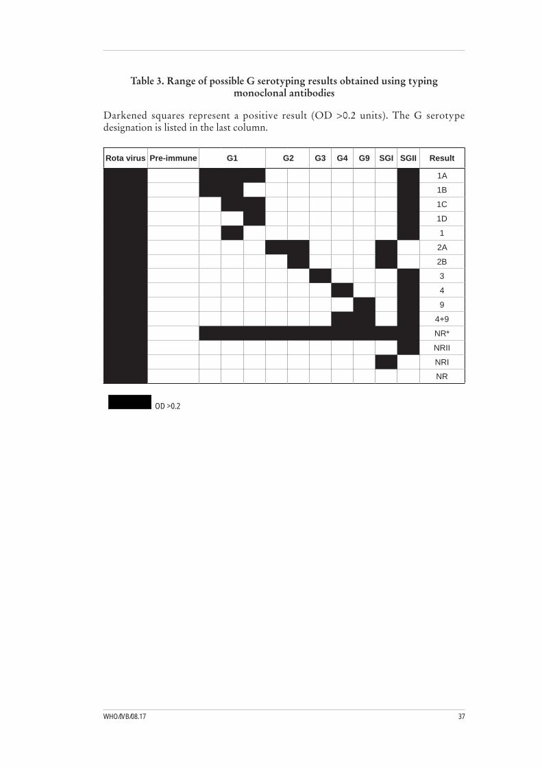
37WHO/IVB/08.17
Table 3. Range of possible G serotyping results obtained using typing monoclonal antibodies
Darkened squares represent a positive result (OD >0.2 units). The G serotype designation is listed in the last column.
Rota virus Pre-immune G1 G2 G3 G4 G9 SGI SGII Result
1A
1B
1C
1D
1
2A
2B
3
4
9
4+9
NR*
NRII
NRI
NR
OD >0.2

Manual of Rotavirus detection and characterization methods38
3.1.2 Method 2: Serotypic determination of rotavirus G and P-types9
Reagents and equipment
NaCl•KCl•Na• 2HPO4-12H2OKH• 2PO4
Tween 20•Skimmed milk powder•Tetramethyl benzidine (TMB)•Dimethyl sulphoxide (DMSO)•Sodium acetate•Citric acid•Hydrogen peroxide•Sulphuric acid•Hyperimmune rabbit anti-rotavirus serum•Horseradish peroxidase-conjugated goat anti-rabbit immunoglobulin•Serotype-specific anti-rotavirus monoclonal antibodies (see Table 1)•96-well microtiter plates (Immulon II)•Plate sealers•Incubator at 37• oCMicrotitre plate washer•Microtitre plate reader (spectrophotometer)•Refrigerator•Rotavirus positive control antigens from cell culture•
9 This method was provided by the WHO Rotavirus Collaborating Center, Atlanta, Georgia, UsA and also used by the Eastern Mediterranean Regional Rotavirus Laboratory

39WHO/IVB/08.17
Reagent preparation
Buffers
Phosphatebufferedsaline pH 7.2
40 g NaCl 1 g KCl 14.5 g Na2HPO4 - 12H20 1 g KH2PO4Dissolve in 4 liters; adjust pH to 7.2, and make up to 5 liters.
PBS-Tween-20 PBS-Tween (0.05% Tween-20) (PBS-T) Add 0.5 ml of Tween 20 to 1 liter of PBS
2.5% skim milk-PBS Dissolve 2.5 g of skim milk powder in 100 ml of PBS.
50X TMB0.1 g TMB 10 ml DMSOStore in 500-µl aliquots at 4oC.
Hydrogen peroxide 3% (v/v) hydrogen peroxide
Substrate solution
10 ml citrate acetate buffer, pH 5.516 µl 3% H202200 µl 50x TMBMake fresh just before use.
Sulfuric acid 2N sulfuric acid
Procedure
Day 1
Add 50 µl of each diluted Mab (in PBS) (typically 1 each for G1 to G4 plus SG 1) I and SG II) to designated wells. For each sample, add 50 µl of PBS to one well to serve as a negative control. Swirl the plate on a lab bench to ensure that Mabs cover the entire well. Cover the plate, and incubate overnight at 4°C.

Manual of Rotavirus detection and characterization methods40
Day 2
Wash the plate 5 times with PBS-T. 1)
Block the plates by adding 200 µl of 2.5% skim milk-PBS to each well. 2) Incubate at 37°C for 1 h.
Prepare 10% fecal extracts in PBS. Dilute each extract 1:4 in 2.5% skim 3) milk-PBS.
Remove blocking agent by dumping from plate. Do not wash the wells. 4) Add 50 µl of diluted fecal specimens (1:4) to each well. Add control virus stock and mock-infected MA104 cells (diluted 1:2 in 2.5% skim milk-PBS) to the control wells. Control strains should represent specific G serotypes for the Mabs included in the assay. For G1–G4, you may include Wa (P1A[8] G1), DS-1 (P1B[4], G2), P (P1A[8], G3), and Va70 (P1A[8], G4).
Incubate at 37°C for 2.5 h. Wash the plates 5 times with PBS-T.5)
Add 50 µl of pooled hyperimmune rabbit serum diluted 1:50,000 in 2.5% skim 6) milk-PBS to each well. For pooled hyperimmune rabbit serum, prepare a mixture of anti-Wa, anti-DS-1, anti-Yo, and anti-ST3 to yield a final concentration for each antiserum of 1:50,000.
Incubate at 37°C for 1.5 h. Wash the plates 5 times with PBS-T.7)
Add 50 µl of diluted peroxidase-conjugated goat anti-rabbit (1:2,000 in 2.5% 8) skim milk-PBS) to each well.
Incubate at 37°C for 1 h. Wash the plates 5 times with PBS-T.9)
Add 100 µl of substrate solution (200 µl of 50x TMB stock/10 ml citrate acetate 10) buffer/16 µl of 3% H2O2), and incubate 10 min at room temperature.
Stop the reaction with 50 µl of H11) 2SO4. Read the OD at a dual wavelength of 450/630 nm.
Interpretation of results
Samples meeting the following criteria are considered positive against relative types:
Positive (P)/Negative (N) >2 and P - N 1) >0.07, when P is the average absorbance of the well coated with the specific Mab and N is the average absorbance of the well coated with PBS for each sample.

41WHO/IVB/08.17
3.1.3 Method 3: Serotypic determination of rotavirus subgroup and G and P-types10
Reagents and equipment
NaHCO• 3
Na• 2CO3
NaCl•KCl•Na• 2HPO4-12H2OKH• 2PO4
Tween 20•Skimmed milk powder•Tetramethyl benzidine (TMB)•Dimethyl sulphoxide (DMSO)•Sodium acetate•Citric acid•30% Hydrogen peroxide•Sulphuric acid•Hyperimmune rabbit anti-rotavirus serum•Horseradish peroxidase-conjugated goat anti-mouse immunoglobulin•Serotype and subgroup-specific anti-rotavirus monoclonal antibodies •(see Table 1)96-well microtiter plates (Falcon flexible plates 3912 [Becton Dickinson])•Plate sealers•Incubator at 37• oCMicrotitre plate washer•Microtitre plate reader (spectrophotometer)•Refrigerator•Rotavirus positive control antigens from cell culture•
10 This method was provided by the European Regional Rotavirus Laboratory, London, UK

Manual of Rotavirus detection and characterization methods42
Reagent preparation
Buffers
0.1M carbonate-bicarbonate buffer, pH 9.6
1.465 g NaHCO30.795 g Na2CO3Dissolve in 800 ml; then adjust pH to 9.6 with acetic acid; make up to 1 liter.
Phosphate buffered saline solution (0.1M), pH 7.2
40 g NaCl1 g KCl4.5 g Na2HPO4 - 12H201 g KH2PO4Dissolve in 4 liters; then adjust pH to 7.2, and make up to 5 liters.
PBS-T Add 0.5 ml of Tween 20 to 1 liter of PBS.2.5% skim milk-PBS Add 2.5 g of skim milk powder to 100 ml of PBS.2.5% skim milk-PBS-T Add 2.5 g of skim milk powder to 100 ml of PBS-TTMB tablets (Sigma) Product T3405: 1 mg/tablet
0.05 M phosphate-citrate buffer, pH 6.0
5.0 ml 1M Sodium Citrate (prepared with 147 gm Na3C6H5O7 and up to 500 ml dH2O) 10 ml 1M Sodium Phosphate (prepared with 60 gm NaH2PO4 and up to 500 ml with dH20)85 ml dH2O
Before beginning, prepare 10%-20% fecal suspensions in balanced salt solution (BSS) in 2-ml screw-cap tubes (Sarstedt).
Procedure
Coat the wells of Falcon flexible plates (3912) with 100 µl of 1/10,000 dilution 1) rabbit anti-rotavirus antibodies (DAKO) in 0.1 M of carbonate-bicarbonate buffer, pH 9.6.
Incubate at 372) oC for 2 h. Wash the plates 3 times with PBS-T.
Block each well with 100 µl of 5% SMP-PBS-T either for 2 h at 373) oC or overnight at 4oC.
Aspirate off the blocking solution, and add 75 µl of 2.5% SMP-PBS-T followed 4) by 25 µl of sample (clarified supernatant from the 10% stool suspension in BSS). Include positive and negative controls as well as sufficient sample duplicates for the number of specific antibodies (e.g., four for serotyping G1-G4).
Aspirate fecal samples, and wash the plates 5 times with PBS-T.5)
Add 100 µl of the appropriate dilution of Mabs (subgroup- or serotype-specific) 6) in 2.5% SMP-PBS-T. Incubate at 37ºC for 2.5 h.
Aspirate the diluted Mabs, and wash the plates 5 times with PBS-T.7)
Add 100 µl of HRPO-conjugated goat anti-mouse IgG (Sigma) at an appropriate 8) dilution in 2.5% SMP-PBST. The optimal dilutions of serologic reagents should be predetermined by chessboard titration. Incubate at 37ºC for 1.5 h.

43WHO/IVB/08.17
Aspirate the HRPO, and wash the plates 5 times with PBS-T.9)
Add 100 µl of freshly prepared substrate: TMB (Sigma) 0.1 mg/ml in 10 ml, 10) 0.05 M phosphate-citrate buffer, pH 6.0, and 0.014% hydrogen peroxide.
Incubate at room temperature for 10 min. Stop the reaction with 50 µl of 11) 2 M H2SO4 .
Read the plates on an EIA plate reader, using the OD at 450 nm (ref. filter 12) 620 nm).
Interpretation of results
The cut-off value is calculated as the mean of the OD450 values + 3 SD of the rotavirus-negative fecal sample controls for each plate. A sample is considered positive if the OD450 is above the cut-off value and if the ratio of the highest OD450 value obtained against the mean of the OD450 values of that sample reacting against Mabs to the other serotypes or subgroup is > 2. When the value of the ratio is between 1.5 and 2, the result is considered equivocal.

Manual of Rotavirus detection and characterization methods44
3.1.4 Method 4: VP6 subgroup ELISA11
Reagents and equipment
NaCl•Na• 2HPO4-2H2OKH• 2PO4
HCl•NaOH•EDTA•0.05M phosphate citrate buffer pH 6.0•Bovine serum albumin (BSA)•Tween 20•Tetramethyl benzidine (TMB)•Dimethyl sulphoxide (DMSO)•30% Hydrogen peroxide•Sulphuric acid•Hyperimmune rabbit anti-rotavirus serum•Horseradish peroxidase-conjugated goat anti-mouse immunoglobulin•Subgroup-specific anti-rotavirus monoclonal antibodies (see Table 1)•96-well microtiter plates (Nunc Immunoplates [Thermo Fisher Scientific Inc])•Plate sealers•Incubator at 37• oCMicrotitre plate washer•Microtitre plate reader (spectrophotometer)•Refrigerator•Rotavirus positive control antigens from cell culture•
11 This method was provided by the south and West African Regional Rotavirus Laboratories

45WHO/IVB/08.17
Reagent preparation
Buffers
0.05M carbonate-bicarbonate buffer, pH 9.6
0.7325 g NaHCO30.3975 g Na2CO3Dissolve in 200 ml distilled water; adjust to pH 9.6 with acetic acid, and then make up to 250 ml with distilled water.
PBS, pH 7.2
20.2 g NaCl1.15 g Na2HPO4.2H2O0.2 g KH2PO4Dissolve in 800 ml distilled water; adjust pH to 7.2 with HCl/ NaOH. Make up to 1,000 ml with distilled water.
PBS-T Add 0.5 ml of Tween 20 to 1,000 ml of PBS.
PBS-T/EDTA Dissolve 0.93 g of EDTA in 250 ml of PBS-T.Adjust the pH to 7.2 with 3M NaOH, and then make up to a final volume of 500 ml with PBS-T.
PBS-T/BSA Dissolve 1 g of bovine serum albumin (BSA) in 199 ml of PBS-T. Adjust the pH to 7.2 with HCl or NaOH. Aliquot, and then store at –20oC.
TMB (Sigma)0.1 g tetramethylbenzidine10 ml dimethyl sulfoxide (DMSO)Store in 500-µl aliquots at 4oC.
0.05 M phosphate-citrate buffer, pH 6.0
5.0 ml 1M Sodium Citrate (prepared with 147 gm Na3C6H5O7 and up to 500 ml dH2O) 10 ml 1M Sodium Phosphate (prepared with 60 gm NaH2PO4 and up to 500 ml with dH20)85 ml dH2O
Procedure
Day 1
Dilute the coating antibody (rabbit anti-human rotavirus #0903, DAKO, 1) Cambridgeshire, United Kingdom) in 0.05 M of carbonate/bicarbonate buffer (the precise dilution needs to determine; MRC use 1:50, West Africa use 1:75). Require 10 ml of diluted coating antibody per plate assayed.
Place 100 µl of the diluted antibody into each well of a 96-well NUNC 2) flat-bottom microtiter plate. Incubate the plates overnight at 4°C.
Day 2
Wash the plate 5 times in PBS-T.1)
Add 100 µl of PBS-T/EDTA to each well, and dispense 50 µl of the 10% stool 2) suspension (vortexed and stored overnight) into three consecutive wells. Incubate the plate overnight at 4°C. Use distilled water or dilution buffer as the negative control and standard rotavirus strains as the positive controls.

Manual of Rotavirus detection and characterization methods46
Day 3
Wash the plate 5 times in PBS-T.1)
Prepare dilutions of VP6 Mabs in PBS-T-BSA (determine appropriate dilutions 2) using checkerboard titrations).
Rotavirus group antigen (A3M4 or 101BC5C7). –
Subgroup I (255/60) –
Subgroup II (631/9). –
Add 100 µl of the Mab solutions to the correct microtiter wells (i.e., every third 3) row for each Mab). Incubate the plates at 37°C for 3 h.
Wash the plates 5 times in PBS-T. 4)
Dilute horseradish peroxidase (HRP)-conjugated goat anti-mouse IgG 5) (Zymed, San Francisco, United States) 1:2000 with PBS-T/BSA (i.e., dilute 5 µl of the anti-mouse IgG in 10 ml PBS-T-BSA).
Dispense 100 µl of this solution into each well. Incubate the plate at 37°C for 6) 2 h.
Wash the plates 5 times in PBS-T. 7)
Add 100 µl of TMB substrate to each well, and incubate for 10 min in the dark.8)
Stop the reaction with the addition of 50 µl of 5% sulphuric acid.9)
Read the plate visually and spectrophotometrically at 450 nm on a microplate 10) reader to obtain an OD reading.
Interpretation of results
Determine the ratio of the OD readings obtained for the two subgroup Mabs. Results are interpreted as follows:
Rotavirus Group A antigen OD >1.0 Subgroup I OD SGI:SGII >1.7 positive Subgroup II OD SGII:SGI >2.0 }

47WHO/IVB/08.17
4.1. Method 1: Manual RNA extraction from stool12
Reagents and equipment
PBS, pH 7.2•50 mM Tris-HCl, pH 7.5•6 M guanidinium isothiocyanate (GITC)•RNAID glass powder•Ethanol•Sodium dodecyl sulphate (SDS)•Phenol-chloroform•Molecular-biology-grade water•Vortexer •Microcentrifuge•Heating block (56• oC)Rocker (Model 1105 Clay Adams Nutator, Becton Dickinson, Parsippany, NJ)•Freezer (-80• oC)Pipettes (20-1,000 µl)•Speed vac (optional)•Screw-cap microcentrifuge tubes•Pipette tips (filter)•
12 This method was provided by the WHO Rotavirus Collaborating Center, Atlanta, Georgia, UsA and also used by the Eastern Mediterranean Regional Rotavirus Laboratory
4. Rotavirus characterisation: Molecular methods

Manual of Rotavirus detection and characterization methods48
Procedure
Prepare 500 µl of a 20% (w/v or v/v) stool extract in PBS, pH 7.2, using a 1) screw-capped microcentrifuge tube. If you are not preparing samples for EIA, extracts can be made in 50 mM Tris-HCl, pH 7.5.Add an equal volume of Vertrel XF, vortex for 1 minute, and centrifuge for 2) 10 min, 8,000 g at RT.Transfer 200 µl of the supernatant to a new screw-capped microcentrifuge 3) tube.Add 400 µl of 6 M GITC dissolved in 50 mM Tris-HCl, pH 7.5, to each tube; 4) vortex, and incubate at 56oC for 10 min.Add 10 µl of RNAID glass powder to each sample; vortex, place on a rocker for 5) 6 min at room temperature, and centrifuge for 60 sec at 4,000 g.Remove supernatant from the pellet, add 400 µl of guanidine wash solution 6) (400 µl of 6 M guanidine:200 µl of Tris-HCl), and then completely resuspend the pellet and centrifuge for 1 min at 4,000 g. Remove the supernatant.Wash each pellet 3 times with ethanol wash solution (400 µl/wash) by completely 7) resuspending the pellet and centrifuging as above. Centrifuge at 4,000 g for the first 2 washes. After the third wash, centrifuge for 2 min at 10,000 g.Pour off the supernatant. Recentrifuge for 10 sec, and remove all residual liquid 8) from the pellet using an eppendorf pipette; dry at room temperature for 30 min or in a vacuum dryer with no heat. Samples may also be dried in a heating block for 10 min at 37oC.Resuspend the pellet in 35 µl of H9) 2O. Incubate for 10 min at 5610) oC, and centrifuge for 2 min at 10,000 g.Transfer the supernatant to a clean centrifuge tube.11) Repeat step 11; combine the supernatants, and store at -8012) oC until used.
Interpretation of results
Successful RNA extraction can be tested by using PAGE and silver staining of the positive control RNA and fecal RNA extracts (about one-fourth to one-third of the total extract).
Notes:
RNA extracts prepared by this method can be used for routine PAGE analysis •of rotavirus dsRNA. The concentration of eluted RNA (to be used for PAGE) can be increased by reducing the elution volume of water added to the RNAID pellet.
If the eluted RNA is concentrated for PAGE by vacuum drying, dry for a minimal •time with low or no heat. Partial degradation of RNA might be an indication that drying conditions are too harsh.
To prepare 6 M of guanidine isothiocyanate, add 100 g of guanidine + 140 ml of •50 mM Tris-HCl, pH 7.5. Store in 10-ml aliquots at -20oC.
For the first two RNAID washes, the centrifugation speed can be reduced so •that the RNAID pellet is less difficult to resuspend.

49WHO/IVB/08.17
Limitations
This procedure cannot detect rotaviruses without additional steps.
4.2 Method 2: RNA extraction using hydroxyapatite13
Reagents
10% sodium dodecyl sulphate (SDS)•0.01M Tris/CaCl2/NaCl pH 7.5•1 M sodium acetate (NaAc), pH 5.6•Phenol-chloroform:isoamyl alcohol (25:24;1; low pH5.2)•Hydroxyapatite•K• 2HPO4
KH• 2PO4
10mM KP•200mM KP•
Equipment and supplies
Vortexer •Microcentrifuge•Pipettes (20-1,000 µl)•Screw-cap microcentrifuge tubes•Pipette tips (filter)•
Procedure (on ice)
Prepare 200 µl of 10 % (w/v) fecal extract in 0.01M Tris/CaCl2/NaCl.1)
Add 20 µl of 10%2) SDS, and vortex for 10 sec.
Add 20 µl of 1M NaAc, pH 5.6, and 200 µl of phenol-chloroform mixture 3) (Amnesco-phenol chloroform II, low pH 5.2, premixed with isoamyl alcohol 25:24:1). Vortex for 30 sec.
Centrifuge at maximum speed (10,000 g) for 1 min in a bench-top centrifuge.4)
Collect aqueous supernatant (RNA).5) Do not collect any of the interface because it can contain inhibitors; if extracting 200 µl of faecal extract, only collect about 150 µl of aqueous supernatant. Transfer to a fresh tube.
Add 50 µl of hydroxyapatite. Vortex for 30 sec to allow RNA to bind to 6) hydroxyapatite.
Centrifuge at full speed (10,000 g) for 1 min. Discard the supernatant.7)
Wash the pellet by resuspending in 500 µl of washing solution (10 mM KP). 8) Vortex for 30 sec.
13 This method was provided by the WHO Rotavirus Collaborating Center, Melbourne, Australia
Manual of Rotavirus detection and characterization methods50
Centrifuge at full speed (10,000 g) for1 min, and discard the supernatant.9)
Repeat steps 9-10.10)
Elute the dsRNA by resuspending the pellet in 50-100 µl of elution solution 11) (200 mM KP).
Vortex for 30 sec. Centrifuge at full speed (10,000 g) for 1 min.12)
Collect the supernatant (contains dsRNA), and transfer to fresh sterile tubes. 13) Store at -20oC or -70oC until required.
Interpretation of results
Extracted RNA is suitable for use in RT-PCR and PAGE analysis. This method does not provide RNA of sufficient quality to use in real-time PCR analysis.

51WHO/IVB/08.17
4.3 Method 3: RNA Extraction Using RNAID14
Reagents
DEPC-treated water•1M sodium acetate (NaAc)•SDS•6 M guanidinium isothiocyanate (GITC)•Phenol-chloroform•RNAID glass powder•
Equipment and supplies
Pipettes (20-1,000 µl)•Water bath•Freezer (-20• oC)Heating block (56• oC)Vortexer•Microcentrifuge•Speed vac (optional)•Pipette tips (filter)•Screw-cap microcentrifuge tubes•
Procedure
Before beginning the extraction, place 1M NaAc containing 1 % SDS into a 37°C 1) water bath. Prepare 6 M GITC.
Add 50 µl of 1M NaAc containing 1% SDS, pH 5.0, to 500 µl of previously 2) prepared 10% fecal suspension. Vortex for 10 sec, and incubate at 37°C for 15 min.
Add 500 µl of phenol/chloroform (1:1), and vortex for 1 min. Incubate at 56°C 3) for 15 min.
Open and immediately reseal the tubes before vortexing. This reduces the air 4) pressure in the tube and prevents the tubes from popping open during mixing. Vortex for 1 min. Centrifuge for 2-3 min at 12,000 rpm.
Carefully remove the upper aqueous phase containing the dsRNA, and place 5) it in a clean eppendorf tube. Note: Avoid any interface material; the material contains protein and DNA that will contaminate the extraction and potentially degrade the RNA.
Repeat from step 3 using 250 µl of phenol-chloroform.6)
14 This method was provided by the south and West African Regional Rotavirus Laboratories
Manual of Rotavirus detection and characterization methods52
Add 500 µl of GITC to the recovered suspension, and vortex. Centrifuge for 7) 5 min at 12,000 rpm. Pour the solution into a clean eppendorf if a pellet forms. Caution: Do not inhale GITC; work in safety cabinet.
Vortex the RNAID matrix well and again just before addition to each sample. 8) Add 10 µl of RNAID matrix (supplied with the RNA extraction kit to each sample, vortex for 30 sec, and incubate on the rocker at room temperature for 15 min.
Ensure that the cold-trap and speed vac (medium temperature) are switched on 9) and ready for use later.
Centrifuge at 5,000 rpm for 10 sec. Discard the supernatant.10)
Add 400 µl of RNAID wash (supplied with the extraction kit) to the pellet and 11) gently resuspend with a pipette. Centrifuge at 12,000 rpm for 30 sec. Discard the supernatant.
Repeat step 12 using only 100 µl of RNAID wash. Centrifuge at 12,000 rpm for 12) 1 min, and discard the supernatant.
Spin the pellet in a speed vac for 10 min to remove excess ethanol.13)
Resuspend the pellet with a pipette in 40 µl of DEPC-treated water 14) (supplied with the extraction kit). Incubate at 65°C in a water bath for 10 min to elute the RNA from the beads.
Centrifuge at 12,000 rpm for 2-3 min.15)
Carefully transfer the supernatant containing the extracted RNA to a sterile 16) eppendorf tube. Store at 4° or -20°C until needed for RT-PCR reactions.

53WHO/IVB/08.17
4.4 Method 4: RNA extraction using Trizol15
Reagents and equipment
TRIzol or TRI-Reagent•Chloroform•Isopropyl alcohol•Molecular-biology-grade water•Vortexer•Microcentrifuge•Pipettes (20-1,000 µl)•Speed vac (optional)•Screw-cap microcentrifuge tubes•Pipette tips (filter)•
Procedure
Vortex the previously prepared 10% stool suspension, and leave it to settle at room 1) temperature for 30-60 min before use. Note: The stool suspension can be clarified by centrifugation for 5 min at RT and 5,000 rpm in a tabletop centrifuge.
Transfer 250 µl of the stool supernatant to a sterile 1.5-ml eppendorf tube, 2) and add 750 µl of TRIzol or TRI-Reagent. Vortex the tube for 30 sec, and incubate at room temperature for 5 min.
Add 200 µl of chloroform to each sample, and vortex for 30 sec. Incubate the 3) sample at room temperature for 3 min. Note: The amount of chloroform can be reduced to 150 µl.
Centrifuge the samples at 12,000 rpm for 5 min at 4°C or room temperature to 4) separate the phases.
Carefully transfer the clear, upper aqueous phase into a sterile eppendorf tube 5) (approximately 450 µl). Note: Avoid the white interface and pink/clear organic phase.
Add 2 volumes (approximately 700 µl) of ice-cold isopropyl alcohol (isopropanol). 6) Mix gently by turning the tube 4-6 times. Incubate at room temperature or at -20°C for 20 min.
Centrifuge at 12,000 rpm at 4°C for 15 min to pellet the dsRNA. Discard the 7) supernatant immediately and very carefully, and allow the pellets to air dry at room temperature.
Resuspend the pellet in 20 µl of sterile deionised water. 8) Note: The volume of water can be reduced to 15 ul.
Store the sample at -20°C until needed for RT-PCR reactions.9)
15 This method was provided by the south and West African Regional Rotavirus Laboratories
Manual of Rotavirus detection and characterization methods54
4.5 Method 5: RNA extraction using the CTAB method16
Reagents
Freon/Genetron•PEG 600•0.4 M NaCl•4 M NaCl•Chloroform•Proteinase K•Na acetate•Ethanol•
Equipment and supplies
Heating block (56• oC)Freezer (-20• oC or –70oC)Vortexer•Microcentrifuge•Pipettes (20-1,000 µl)•Screw-cap microcentrifuge tubes•Pipette tips (filter)•
Procedure
Add 300 µl of a 10%-20% stool suspension to a 1.5-ml eppendorf tube.1) Extract once with an equal volume (300 µl) of Freon/Genetron. Vortex the sample 2) for 30 sec, and centrifuge at 12,000 rpm for 5 min. Transfer the supernatant to a new eppendorf tube.Add 300 µl of PEG 6000 buffer (final concentration of 8% PEG 6000 and 3) 0.4M NaCl). Incubate at 4ºC for 30 min.Centrifuge at 4ºC for 15 min.4) Resuspend the pellet in 150 µl of deionised water and 150 µl of 2 x Proteinase K 5) buffer. Add fresh Proteinase K at 400 µg/ml. Incubate at 37ºC for 30 min.Add 50 µl of 10% CTAB and 50 µl of 4M NaCl. Vortex for 10 sec. Incubate at 6) 56ºC for 30 min.Add 400 µl of phenol-chloroform (equal volume) to the mixture for extraction. 7) Vortex for 1 min, and spin for 10 min at room temperature.Extract once with an equal volume of chloroform. Vortex for1 min, and spin for 8) 10 min at room temperature. To the aqueous phase, add 1 ml of 100% ethanol and 40 µl of NaAc 9) (final concentration of 0.2 M NaAc).Keep at –20ºC for 2 h or at –70ºC for 30 min. Pellet the dsRNA by centrifuging 10) at 12,000 rpm for 15 min at 4ºC.Remove as much residual ethanol as possible. Dry the pellet at room temperature, 11) and resuspend the pellet in 20 µl of sterile deionised water or 1 x TE.Use 1-5 µl for an RT-PCR reaction.12)
16 This method was provided by the south African Regional Rotavirus Laboratory

55WHO/IVB/08.17
4.6 Method 6: RNA extraction using the Boom method17
Reagents and equipment
Guanidinium isothiocyanate (GITC)•0.1M Tris HCl, pH 6.4•Triton X-100•Silicon dioxide•0.2 M EDTA, pH 8.0•HCl•Ethanol•Acetone•Distilled water•DEPC-treated water•RNAsin•Vortexer•Microcentrifuge•Pipettes: (20-1,000 µl)•Heating block (56• oC)Freezer (-20• oC)Screw-cap microcentrifuge tubes•Pipette tips (filter)•Fine-tip pastettes•
Procedure
Add 200 µl of fecal extract to 1 ml of lysis buffer-L6 buffer and 20 µl of size-1) fractionated silica in a 1.5-ml screw-cap microcentrifuge tube. Include RNase-free distilled water in each run as a negative control. Use cell culture grown SA11 or RRV as a positive control.
Vortex for 10 sec, and incubate at RT for 15 min.2)
Pellet by centrifugation (microcentrifuge) for 15 sec (13,000 rpm), and discard 3) the supernatant; store the wash fluids for disposal.
Wash the pellet with 1 ml of lysis buffer L2 twice, 1 ml of 70% ethanol twice, 4) and 1 ml of acetone once; store the wash fluids for disposal.
After removal of the acetone (perform carefully as pellets might become 5) dislodged), centrifuge, and place the tube with lid open at 56oC in a dry heating block for 5 min.
Add 49 µl of RNase-free distilled water and 1 µl of RNasin. Vortex and incubate 6) at 56oC for 15 min to elute the nucleic acid from the silica.
Pellet by centrifugation at 13,000 rpm for 4 min, and extract the supernatant 7) (avoid disturbing the silica). Recentrifuge if the silica becomes resuspended.
Store in a new microfuge tube at 4oC for 24 h or at –70oC for longer.8)
17 This method was provided by the European Regional Rotavirus Laboratory, London, UK

Manual of Rotavirus detection and characterization methods56
4.7 Method 7: RNA extraction from cell lysates18
Reagents and equipment
Phenol-chloroform, 1:1•10% sodium dodecyl sulphate (SDS)•5 M NaCl•95% ethanol•70% ethanol•6 M guanidinium isothiocyanate (GITC)•RNAID beads•50 mM Tris-HCl, pH 7.5•RNAID kit wash buffer•Vortexer•Microcentrifuge•Speed vac (optional)•Pipettors (20-1000 µl)•Screwcap microcentrifuge tubes•Pipette tips (with filter)•Surgical gloves•
Procedure
Day1
Freeze and thaw virus stock three times (-201) oC/RT).
Take 400 µl of supernatant, and centrifuge at 10,000 2) g in a 1.5-ml screw-cap microcentrifuge tube for 10 min.
Transfer the supernatant to a new 1.5-ml microcentrifuge tube; add SDS to a final 3) concentration of 1%, and then mix.
Incubate at 564) oC for 5 min.
Add equal volumes (~400 µl) of phenol-chloroform (1:1) to each sample, 5) and vortex for 60 sec.
Centrifuge for 5 min at 10,000 g. Remove the supernatant to a new 1.5-ml tube, 6) and add 1/10 volume of 5 M NaCl.
Add 2–2.5 volumes of 95% ethanol, and mix. Store overnight at -207) oC.
18 This method was provided by the WHO Rotavirus Collaborating Center, Atlanta, Georgia, UsA and also used by the Eastern Mediterranean Regional Rotavirus Laboratory

57WHO/IVB/08.17
Day 2
Centrifuge the sample for 20 min at 14,000 1) g and 4oC in an eppendorf microcentrifuge tube or equivalent. Remove the supernatant, and drain the pellet.
Wash the pellet by adding 1 ml of cold 70% ethanol.2)
Centrifuge for 5 min at maximum g force at 43) oC.
Pour off the supernatant, drain the pellet, and dry at room temperature or in a 4) vacuum dryer with low or no heat.
Resuspend in 200 µl of 50 mM Tris-HCl, pH 7.5, and incubate at 655) oC for 5–10 min.
Add 400 µl of 6 M GITC (prepared in 50 mM Tris-HCl, pH 7.5) to each sample; 6) mix, and incubate at 56oC for 10 min.
Add 10 µl of RNAID glass powder to each sample; vortex, and then rock on a 7) nutator for 15 min. Centrifuge for 60 sec at 4,000 g in a microcentrifuge.
Remove the supernatant with a separate pipette for each sample. Wash once 8) with 400 µl of guanidine wash solution (400 µl of 6 M guanidine:200 µl of 50 mM Tris-HCl). Centrifuge for 1 min at 4,000 g in a microcentrifuge. Remove the supernatant.
Repeat the wash step 3 times with an RNAID kit wash buffer. For the first 9) 2 washes, centrifuge at 4,000 g; for the third wash, centrifuge for 2 min at 10,000 g, and then remove the supernatant.
Remove all residual liquid from the samples, and dry in a centrifuge dryer with 10) no heat or at room temperature for 30 min.
Extract the RNAID pellet for 10 min at 5611) oC in 35 µl of purified water, and then centrifuge for 2 min at 10,000 g.
Transfer the supernatant to a new tube; use a separate tip for each sample.12)
Repeat the extraction of the RNAID pellet with 35 µl of water, and combine the 13) supernatants. Store the supernatant at -80oC until use.
Interpretation of results
Successful RNA extraction can be tested using PAGE and silver staining of the positive control RNA and cell lysate RNA (approximately one-fourth or one-half of the total extract).

Manual of Rotavirus detection and characterization methods58
Notes
RNA extracts prepared by this method can be used for routine PAGE analysis 1) of rotavirus dsRNA.
Concentration of eluted RNA (to be used for PAGE) may be increased by 2) reducing the elution volume of water added to the RNAID pellet.
If the eluted RNA is concentrated for PAGE by vacuum drying, dry for a minimal 3) time with low or no heat. Partial degradation of RNA can be an indication that drying conditions are too harsh.
To prepare 6 M of guanidine isothiocyanate, add 100 g of guanidine + 140 ml of 4) 50 mM Tris-HCl pH 7.5. Store in 10-ml aliquots at -20oC.
Commercial preparations of phenol-chloroform:isoamyl alcohol (25:24:1) can 5) be substituted in step 5.
For the first two RNAID washes, the centrifugation speed can be reduced so 6) that the RNAID pellet is less difficult to resuspend.
Limitations
This procedure cannot detect rotaviruses without additional steps.

59WHO/IVB/08.17
4.8 Method 8: G Genotyping19
Reagents and equipment
Pipettes (20-1000 µl)•Microcentrifuge•Thermalcycler•Electrophoresis tank•Power pack•UV transiluminator•Microwave oven or boiling water bath•Circulating water bath•Ice bath•Gel documentation system•Gel staining tank•PCR tubes•Pipette tips (filter)•Microcentrifuge tubes•Molecular-grade water •dNTPs •PCR buffer •MgCl• 2 Reverse transcriptase •Taq polymerase •NuSieve GTG agarose •SeaPlaque agarose •TBE •123-bp DNA ladder •Ethidium bromide •Mineral oil •Sucrose •Bromophenol blue •TE buffer (10 mM Tris-HCl, pH 8, 0.1 mM EDTA)•Oliogonucleotide primers• 20: 9con1, 9con2, 9T1, 9T2, 9T3P, 9T4, 9T9B;
con3, con2, 1T1, 2T1, 3T1, 4T1, 5T1, ND2
19 This method was provided by the WHO Rotavirus Collaborating Center, Atlanta, Georgia, UsA and also used by the Eastern Mediterranean Regional Rotavirus Laboratory
20 The nucleotide sequences of the primers are shown in Appendix 1
Manual of Rotavirus detection and characterization methods60
First amplification
In a laboratory room designated for setting up PCR master mixes, prepare 1) the consensus primer pre-mix by adding 1 µl/reaction for each of the primers 9con1 (20 uM) and 9con2 (20 µM) and 3 µl of deionized water per reaction. Note: Use eppendorf pipettes and aerosol pipette tips for all steps.Prepare the RT and AmpliTaq (Taq) mixtures for the desired number of reactions 2) according to the RT-PCR protocol form below. Do not add RT and Taq to the mixtures at this step.
Component RT mixture(µl for 1 reaction)
Taq mixture(µl for 1 reaction)
Water 18.65 36.5
2.5 mM dNTPs 8 8
10 x buffer 5 5
25 mM MgCl2 8 —
RT 10 U (0.35 µl) —
Taq — 0.5
Total 40 50
Transfer the PCR tubes containing the primer/water mixture to the RNA work 3) area, and place the tubes in a second ice bath.Add 5 µl of RNA (from stool RNA extracts or controls) to the 9con1 + 9con2/4) H2O mixture; mix, and heat at 97ºC for 5 min.Rapidly cool the RNA/primer mixture in an ice bath for 1 min, spin for 10 sec 5) at the maximum g force of the microcentrifuge, and return the primer mixture to the ice bath.Add the RT to the RT mixture, mix by gentle pipetting to avoid air bubbles, and 6) add 40 µl of this mixture to each tube. Mix each tube gently to avoid air bubbles. Transfer the tubes to a 42oC circulating water bath. Incubate for 30–60 min.Near the end of the incubation period for the RT reaction, add Taq polymerase 7) to the Taq mixture, and mix by gentle pipetting. Return the samples to the ice bath. Then add 50 µl of Taq mix to each tube, and add 2 drops of mineral oil to each tube (if required for the thermal cycler).Centrifuge for a few seconds. Return the samples to the ice bath before placing 8) them in a preheated (94ºC) Perkin Elmer thermal cycler for 10-20 PCR cycles under the following conditions:
94ºC, 0.5 min 10 cycles 42ºC, 0.5 min 72ºC, 1 min 1 cycle 72ºC, 7 min 1 cycle Soak at 4ºC
Dilute 10 µl of each PCR product in 6X gel loading buffer, and electrophorese in 9) a 1.8% agarose gel (volume = 80-100 ml, ratio of 2 parts Nusieve GTG agarose to 1 part SeaPlaque agarose in 1 x TBE) to detect products. Use 5 µl of the 123-bp DNA ladder + 5 µl of water as a marker to determine the size of the PCR products.
Note: Running the first amplification products is optional and is usually not done when the first amplification is run for only 10 cycles.
{

61WHO/IVB/08.17
Second amplification
Prepare the second amplification PCR mixture as follows: 1)
Component Taq mixture(µl for 1 reaction)
Water 29.5
2.5 mM dNTPs 8
10 x buffer 5
25 mM MgCl2 4
10 uM Gene9 pool* 1
Taq 0.5
Total 48
*Mix equal volumes of 9con1 (20-µM solution) and VP7 genotype-specific primers (9T1, 9T1, 9T3P, 9T4, 9T9B; 20 µM each) so that all primers have final concentrations of 10 µM. The primers and their sequences are listed below:
Primer Sequence (5’ to 3’) Strain/Gtype
nt position/polarity
Accession no.
First-amplification consensus primers
9con1 tag ctc ctt tta atg tat gg Wa/G1 37–56/+ K02033
9con2 gta taa aat act tgc cac ca Wa/G1 922–941/- K02033
Second-amplification genotyping primers
9T-1 tct tgt caa agc aaa taa tg Wa/G1 176–195/- K02033
9T-2 gtt aga aat gat tct cca ct S2/G2 262–281/- M11164
9T-3 gtc cag ttg cag tgt agc Y0/G3 484–501/- D86284
9T-4 ggg tcg atg gaa aat tct ST3/G4 423–440/- X13603
9T-9 tat aaa gtc cat tgc ac 116E/G9 131–147/- L14072
Aliquot 48 µl of the PCR Taq mixture into a new tube for each sample.2) Centrifuge the first amplification products for 5 sec at 10,000 g3) , and transfer 2 µl of each product to the appropriate tube containing the Taq reaction mixture. Vortex each sample.Add two drops of mineral oil (if required for the thermal cycler) to each tube, 4) and centrifuge for 10 sec.Transfer the tubes to the thermal cycler, and run for 30 PCR cycles with the 5) following conditions:
94ºC, 0.5 min 10 cycles 42ºC, 0.5 min 72ºC, 1 min 1 cycle 72ºC, 7 min 1 cycle Soak at 4ºC
{

Manual of Rotavirus detection and characterization methods62
Dilute 10 µl of each PCR product in 6X gel loading buffer, and electrophoese in 6) a 3.0% agarose gel (volume = 80-100 ml, ratio of 2 parts Nusieve GTG agarose to 1 part SeaPlaque agarose in 1 x TBE) to detect products. Use 5 µl of the 123-bp DNA ladder + 5 µl of water as a marker to determine the size of the PCR products. Stain with ethidium bromide, and examine on a UV light box.
Interpretation of results
A sample is positive for G types 1, 2, 3, 4, or 9 if the sample PCR band comigrates with the molecular markers for these G serotypes.
Notes
When setting up genotyping, obtain and test standard strains (at least G1-G4, •G8, and G9, P[4], P[6], P[8]) first to ensure that the assay is working properly. These strains are available through WHO Rotavirus Collaborating Centers and Rotavirus Regional Laboratories.The original Gouvea et al • 21 pool of genotype-specific primers contained an oligonucleotide specific for G8, whereas those described by Das et al 32 did not. The protocol detailed above can be adapted for use with the primer set described by Gouvea 21 or detection of G8 strains. Regardless of the primer set used, strains have been described that are untypeable with, or misclassified by, existing primer sets.8, 45 Thus, selective confirmation of genotyping results by an independent method is an important part of a strain surveillance study. Methods for confirmation of genotyping include nucleotide sequencing, probe hybridization, and typing with Mabs.A 2-min denaturation step (94ºC) is sometimes included• before the first PCR cycle for the first and second PCR amplification steps.In-house reagents (10 x PCR and 25 mM MgCl• 2) have been used in place of the Perkin Elmer reagents. Stock solutions (1 M) are prepared from molecular-biology-grade Tris-HCl, MgCl2, and KCl (Sigma) in ultrapure deionized water and then autoclaved, aliquoted, and stored frozen at –80oC. Working solutions (10 x PCR and 25 mM MgCl2) are prepared in the same type of water, aliquoted in small tubes, and stored frozen at –80oC.
Limitations
This test does not provide serologic proof of the serotypes of the tested strains, although there is a strong correlation between PCR typing and monoclonal-based serotyping results.

63WHO/IVB/08.17
4.9 Method 9: P Genotyping21
First amplification
In a laboratory room designated for setting up PCR master mixes, prepare the 1) consensus primer pre-mix by adding 1 µl/reaction for each of the primers con3 (20 uM) and con2 (20 uM) and 3 µl of deionized water per reaction (use eppendorf pipettes and aerosol pipette tips for all steps).
Prepare the RT and AmpliTaq (Taq) mixtures for the desired number of reactions 2) according to the following RT-PCR protocol form. Do not add the RT and Taq to the mixtures at this step.
Component RT mixture (µl for 1 reaction)
Taq mixture (µl for 1 reaction)
Water 16–19 36.5 2.5 mM dNTPs 8 8 10 x buffer 5 5 25 mM MgCl2 8–10 — RT 10 U (0.35 ul) — Taq — 0.5 Total 40 50
Transfer the tubes with the primer/water mixture to the RNA work area.3)
Add 5 µl of RNA (from RNA extracts) to the con3 + con2/H4) 2O mixture, and mix and heat at 97ºC for 5 min.
Rapidly cool the RNA/primer mixture in an ice bath for 1 min; spin for 10 sec 5) at maximum g force, and return the mixture to the ice bath.
Add the RT to the RT mixture; mix by gentle pipetting to avoid air bubbles, 6) and add 40 µl of this mixture to each tube. Mix each tube gently to avoid air bubbles. Transfer the tubes to a 42oC circulating water bath. Incubate for 60 min.
Near the end of the incubation period for the RT reaction, add7) Taq polymerase to the Taq mixture and mix by gentle pipetting. Return the samples to the ice bath; then add 50 µl of Taq mix to each tube, and add 2 drops of mineral oil to each tube (if required for the thermal cycler).
Centrifuge for10 sec, return the samples to the ice bath, and place in a preheated 8) (94ºC) Perkin Elmer thermal cycler for 20 cycles with the following PCR conditions:
94ºC, 0.5 min 20 cycles 42ºC, 0.5 min 72ºC, 1 min 1 cycle 72ºC, 7 min 1 cycle Soak at 4ºC
21 This method was provided by the WHO Rotavirus Collaborating Center, Atlanta, Georgia, UsA and also used by the Eastern Mediterranean Regional Rotavirus Laboratory
{

Manual of Rotavirus detection and characterization methods64
Using 6 x gel loading buffer, run the PCR products (10 µl) in a 1.8%–3.0% 9) agarose gel (volume, 80–100 ml) (ratio of 2 parts Nusieve GTG agarose to 1 part SeaPlaque agarose in 1x TBE) to detect products. Use 5 µl of the 123-bp DNA ladder + 5 µl of water as a marker to determine the size of the PCR products. Note: Running the first amplification product is optional.
Second amplification
Prepare the second amplification PCR mixture as follows: 1)
Component Taq mixture (µl for 1 reaction)Water 29.52.5 mM dNTPs 810 x buffer 525 mM MgCl2 410 uM gene 4 pool* 1Taq 0.5Total 48
* Prepare a premix by mixing equal volumes of 20 µM con3 and the genotype-specific primer mixture 1T-1, 2T-1, 3T-1, 4T-1, and 5T-1 (each at 20 µM). Thus, the final concentration of each primer is 10 µM. When other primers, such as ND2 (P[11]-specific) are used, add 1 µl of that primer stock (10 µM) and reduce the water volume by 1 µl/reaction.
Primer Sequence (5’ to 3’) Strain/P[type]
nt position/polarity
Accession no.
First amplification consensuscon3 tgg ctt cgc tca ttt ata gac a KU/P[8] 11-32 M21014con2 att tcg gac cat tta taa cc KU/P[8] 868-887 M21014
Second amplification (include VP7-Rdeg)
1T-1 tct act tgg ata acg tgc KU/P[8] 339–356/- M21014 2T-1 cta ttg tta gag gtt aga gtc RV-5/P[4] 474–494/- M32559 3T-1 tgt tga tta gtt gga ttc aa 1076/P[6] 259–278/- M88480 4T-1 tga gac atg caa ttg gac K8/P[9] 385– 402/- D90260 5T-1 atc ata gtt agt agt cgg 69M/P[10] 575–594/- M60600
ND2 agc gaa ctc acc aat ctg 116E/P[11] 116–133/- L07934
Aliquot 48 µl of the Taq mixture into a new tube for each sample.2)
Centrifuge the first amplification products for 5 sec at 10,000 g3) , and transfer 2 µl of each product to the appropriate tube containing the Taq reaction mixture.
Add two drops of mineral oil (if required by the thermal cycler) to each tube, 4) and centrifuge for 10 sec at maximum g force.
Run 30 cycles of PCR with the following conditions:5)
94ºC, 0.5 min 30 cycles 42ºC, 0.5 min 72ºC, 0.75 min 1 cycle 72ºC, 7 min 1 cycle Soak at 4º
{

65WHO/IVB/08.17
Using 6 x gel loading buffer, analyze the DNA products (10 µl) by agarose gel 6) electrophoresis.
Interpretation of results
A sample is positive for P types 4, 6, 8, 9, 10, and 11 if the sample PCR band comigrates with the molecular markers for these P types.
Notes
When setting up genotyping, obtain and test standard strains (at least G1-G4, •G8 and G9, P[4], P[6], P[8]) first to ensure that the assay is working properly. These strains are available through WHO Rotavirus Collaborating Centers and Rotavirus Regional Laboratories.An independent set of P-genotyping primers was developed by Gunasena et al.• 5
Several reports have demonstrated that genetic variation in the VP4-gene primer binding sites of currently circulating strains reduces the percentage of strains that can be typed with the original genotyping primers. Newly designed primers for P[8] strains have been used to address this problem. 45 As for G-genotyping, it is important to confirm P-genotyping results by an independent method (e.g., nucleotide sequencing, probe hybridization, or typing with Mabs).A 2-min denaturation step (94ºC) can be included • before the first PCR cycle for the first and second PCR amplification steps.In-house reagents (10 x PCR and 25 mM MgCl• 2) can be used in place of the Perkin Elmer reagents. Stock solutions (1 M) are prepared from molecular-biology-grade Tris-HCl, MgCl2, and KCl (Sigma) in ultrapure deionized water and then autoclaved, aliquoted, and stored frozen at –20ºC. Working solutions (10x PCR and 25 mM MgCl2) are prepared in the same type of water, aliquoted in small tubes, and stored frozen at –80oC.
Limitations
This test does not provide serologic proof of the serotypes of the tested strains, although there is a correlation between the results of PCR typing and antibody-based serotyping methods. Recently described exceptions to the correlation between P serotypes and P genotypes confirm that nucleic acid-based typing methods such as RT-PCR can be used only to identify the most common VP4 genes in circulation and cannot be used to assign serotypes.

Manual of Rotavirus detection and characterization methods66
4.10 Method 10: G Genotyping22
Reagents and equipment
Pipettes (20 -1000 µl)•Microcentrifuge•Thermalcycler•Electrophoresis tank•Power pack•UV transiluminator•Microwave oven•Gel documentation system•PCR tubes•Pipette tips (filter)•Microcentrifuge tubes•1M TrisHCl, pH 8.3 •1M KCl •100 mM MgCl• 2 DMSO •dNTPs (20 mM each) •AMV-RT •AmpliTaq DNA polymerase •Agarose •0.5 X TBE •100-bp DNA ladder •Ethidium bromide •Sucrose •Bromophenol blue •0.5 M EDTA, pH 8.0•Oliogonucleotide primers• 23: Beg9, End9, RVG9, aBT1, aCT2, aDT4, G3-Aust, aAT8, G9;VP4F, VP4-R, 2T-1, 3T-1, 1T-1, 4T-1, 5T-1
22 This method was provided by the WHO Rotavirus Collaborating Center in Melbourne, Australia23 The nucleotide sequences of the primers are shown in Appendix 1

67WHO/IVB/08.17
First-round amplification
In a laminar flow cabinet and 1) on ice, prepare the RT/PCR master mix for one sample:
77.5 µl dH2O 1.0 µl 1M Tris-HCl, pH 8.3 4.0 µl 1M KCl 1.5 µl 100 mM MgCl2 7.0 µl DMSO 1.0 µl dNTP mix (contains 20 mM each dATP, dCTP, dGTP, dTTP) 1.0 µl Beg9 primer (500 ng/ml) 1.0 µl End9 primer (500 ng/ml)
Make up enough master mix for the total number of tubes (samples, 2) positive control, negative control, + one extra). Quick spin to mix.
Add 94 µl of the master mix to each tube.3)
Add 5 µl of dsRNA. Vortex quickly to mix.4)
Heat the samples at 975) oC for 3 min in the thermocycler, and transfer immediately to wet ice for at least 1 min. Vortex quickly to mix.
Add 0.5 µl/sample of RT AMV (24 U/µl; 12 units per sample).6)
Add 0.5 µl/sample of Amplitaq DNA polymerase (5 U/µl; 2.5 units per 7) sample).
Vortex quickly to mix, and place the tubes in a bench-top centrifuge for a 8) short spin; reach 10,000 rpm and then stop
Place the tubes in the thermocycler and incubate as detailed below:9)
Reverse transcription: 42oC, 60 min PCR (30 cycles): 94oC, 1 min 42oC, 2 min 68oC, 1 min Hold at 4oC
Electrophoresis of full-length product
Set up a gel tray with comb ready for the gel mixture.1)
Fill the electrophoresis tank with 0.5 X TBE buffer.2)
Make 1.2% (w/v) agarose gel in 0.5 X TBE. Dissolve the agarose in the microwave 3) at 100% power for 30 sec. Swirl to mix.
Microwave for a further 7-15 sec. When the agarose boil stops, swirl and heat 4) again until the agarose is crystal clear. Cool under running tap water to ~55oC.
Add ethidium bromide (10 mg/ml) for a final concentration of 24 ng/ml 5) (add 1.2 ml to 50 ml of agarose).
Pour the gel into a tray and allow to set.6)
Submerge the gel in 0.5 X TBE buffer.7)
Combine 10 µl of cDNA and 5 µl of gel loading buffer. For the MW marker, 8) combine 2 µl of MW marker + 8 µl of H2O + 5 µl gel loading buffer.
{

Manual of Rotavirus detection and characterization methods68
Load samples into the wells using the pipette.9)
Run the gel at 100 volts for ~1 h or until the dye front is about 3/4 the length 10) of the gel.
View the gel using a UV light source.11) Note: Running of first-round product is optional.
Second amplification: VP7 genotype-specific PCR
Use genotype-specific primers for G1, G2, G3, G4, G8, and G9 (Table 9). 1) Dilute each primer to 100 ng/ml, and combine as a single primer mix.
In a laminar-flow cabinet and 2) on ice, prepare the master mix for one sample:
79 µl dH2O 1.0 µl 1M Tris-HCl, pH 8.3 4.0 µl 1M KCl 1.5 µl 100 mM MgCl2 7.0 µl DMSO 1.0 µl dNTP mix 1.0 µl RVG 9 primer (100 ng/ml) 4.0 µl genotyping primers for G1-G9 (each at 100 ng/ml). 0.5 µl Taq polymerase (2.5 U)
Make up enough master mix for the total number of tubes (samples, 3) positive control, negative control, + one extra). Quick spin to mix.
Add 99 µl of the master mix to each tube.4)
Add 1 µl of cDNA from the first-round RT-PCR.5)
Vortex quickly to mix, and place the tubes in bench-top centrifuge for a short 6) spin; reach 10,000 rpm, and then stop
Place the tubes in the thermocycler, and incubate as detailed below7)
Denature: 94oC, 2 min 30 cycles 94oC, 1 min 42oC, 2 min 68oC, 1 min Hold at 4oC
Make 1.5% (w/v) agarose gel in 0.5 XTBE, and set up tank as described earlier 8) for full-length VP7 cDNA products.
{

69WHO/IVB/08.17
Interpretation of results
Figure 6. Schematic illustration of the VP7 genotyping PCR (see Appendix 1)
BA C D E F39 50 87 101 120 130 143 152 207 220 233 242
5’mRNA
AA3’
1062nt1
Beg9SBeg9
SBeg9
(G8) 9AT8
(G3) G3 Aus
(G1) 9BT1
(G2) 9CT1
(G4) 9DT4
(G9) G9
End 9RVG 91062bp
End A942bp
885bp
812bp
749bp
652bp
583bp
306bp
Variable regionsPrimers
Compare PCR bands with molecular weight markers. The expected sizes of each VP7 genotype are listed below.
beg9 and end9 product 1062 bp Human rotavirus VP7 genotypes (3’ end primer) VP7 genotype G1 749 bp VP7 genotype G2 652 bp VP7 genotype G3 812 bp VP7 genotype G4 583 bp VP7 genotype G8 885 bp VP7 genotype G9 306 bp

Manual of Rotavirus detection and characterization methods70
4.11 Method 11: P Genotyping24
First round RT-PCR amplification of VP8* subunit of VP4 gene
In a laminar-flow cabinet and 1) on ice, prepare the RT/PCR reaction mix (for one sample) for full-length amplification.
77.5 µl dH2O 1.0 µl 1M Tris-HCl, pH 8.3 4.0 µl 1M KCl 1.5 µl 100 mM MgCl2 7.0 µl DMSO 1.0 µl dNTP mix (contains 20 mM each dATP, dCTP, dGTP, dTTP) 1.0 µl VP4F primer (100 ng/ml) 1.0 µl VP4R primer (100 ng/ml)
Make up enough master mix for the total number of tubes (samples, positive 2) control, negative control, + one extra). Vortex and quick spin to mix.
Add 94 µl of the master mix to each tube.3)
Add 5 µl of dsRNA. Vortex quickly to mix4)
Heat the samples at 975) oC for 3 min in the thermocycler, and transfer immediately to wet ice for at least 1 min. Vortex quickly to mix
Add 0.5 µl/sample of RT AMV (24 U/µl; 12 units per sample).6)
Add 0.5 µl/sample of Amplitaq DNA polymerase (5 U/µl; 2.5 units per 7) sample).
Vortex quickly to mix, and place the tubes in a bench-top centrifuge for a short 8) spin; reach 10,000 rpm, and then stop.
Place the tubes in the thermocycler, and incubate as detailed below:9)
Reverse transcription: 42oC, 60 min PCR (30 cycles) 94oC, 1 min 50oC, 2 min 72oC, 1 min
Hold at 4oC
Make 1.2% (w/v) agarose gel in 0.5 X TBE to analyse full-length products as 10) described previously.
24 This method was provided by the WHO Rotavirus Collaborating Center in Melbourne, Australia
{

71WHO/IVB/08.17
Second amplification: VP4 genotype-specific PCR
In a laminar flow cabinet and 1) on ice, prepare the PCR reaction mix for one sample:
80.75 µl dH2O 1.0 µl 1M Tris-HCl, pH 8.3 4.0 µl 1M KCl 1.75 µl 100 mM MgCl2 1.0 µl dNTP mix (contains 20mM each dATP, dCTP, dGTP, dTTP) 6.0 µl P mix (contains genotyping primers for P4, P6, P8, P9, P10 + VP4F, each at 100 ng/ml) 0.5 µl Amplitaq DNA polymerase (2.5 U per sample)
Make up enough master mix for the total number of tubes (samples, 2) positive control, negative control, + one extra). Vortex and quick spin to mix.
Add 95 µl of the master mix to each tube.3)
Add 5 µl of cDNA (RT/PCR product).4)
Vortex quickly to mix, and place the tubes in bench-top centrifuge for a short 5) spin; reach 10,000 rpm, and then stop.
Place the tubes in the thermocycler, and incubate as detailed below:6)
Denature 94o C, 2 min 40 cycles 94oC, 1 min 42oC, 2 min 68oC, 1 min
Hold at 4oC
Electrophoresis of P-typing product
Prepare a 2% agarose gel as detailed earlier in the G genotyping method. DNA samples are run on agarose gel with molecular weight markers (100 pb) to allow size determination of PCR products. The expected sizes are listed below:
VP4F and VP4R product 663 bp Human rotavirus VP4 genotypes: VP4 genotype P[8] 224 bp VP4 genotype P[4] 362 bp VP4 genotype P[6] 146 bp VP4 genotype P[9] 270 bp VP4 genotype P[10] 462 bp
{

Manual of Rotavirus detection and characterization methods72
Figure 7. Schematic illustration of the VP4 genotyping PCR using Gentsch P typing primers (see Appendix 1)
1 132 278 356 402 492 695 795 2359
VP4F 3T-1 1T-1 4T-1 2T-1 5T-1
VP4R
5’ nt3’
VP4R663bp
146bp
224bp
270bp
362bp
462bp
P[6] 3T-1
P[8] 1T-1
P[9] 4T-1
P[4] 2T-1
P[10] 5T-1

73WHO/IVB/08.17
4.12 Method 12: G Genotying25
Reagents and Equipment
Pipettes (20 -1000 µl)•Microcentrifuge•Thermocycler•Microcentrifuge tubes•Gel documentation system•PCR tubes•Pipette tips (filter)•5X AMV buffer •dNTPs •AMV-RT •Ice bath •10X Taq buffer •Taq polymerase •Agarose •Tris, acetic acid, EDTA •Gel loading buffer •Ethidium bromide •MgCl• 2
Mineral oil (if thermocycler has no heated lid) • Oliogonucleotide primers• 26: Beg9, sBeg9 End9, RVG9, aBT1, aCT2, aDT4, aET3, aAT8, aFT9; Con2, Con3, 2T-1, 3T-1, 1T-1, 4T-1, 5T-1.
25 This method was provided by the West African Regional Rotavirus Laboratory26 The nucleotide sequences of the primers are shown in Appendix 1

Manual of Rotavirus detection and characterization methods74
First amplification
Label 0.5-ml or 0.2-ml thin-walled tubes for each amplification reaction, 1) and include a positive and negative control. Label the top of the lid with the sample number, primers used, and date.
Prepare the RT master mix, and place on ice. 2) 2.0 µl 5xAMV buffer (supplied with RT enzyme) 0.25 µl 10 mM dATP 0.25 µl 10 mM dCTP 0.25 µl 10 mM dGTP 0.25 µl 10 mM dTTP 0.2 µl AMV RT (stock = 25 U/µl)
Dilute each of the VP7 consensus primers to 20 pmol. Then add 1 µl of each 3) primer to all sample tubes.
Add 5 µl of dsRNA to the primer.4)
Add ddH5) 2O to a final volume of 10 µl, and mix by pipetting.
Denature the samples for 5 min at 94ºC, and transfer all sample tubes immediately 6) to an ice-bath.
Add 3.2 µl of RT master mix to each tube, and mix well. Spin for 10 sec, 7) and incubate the tubes at 42ºC for 30 min. Note: Be sure to add the RT mix within 5 min.
Prepare the PCR amplification master mix, and place on ice. Add the Taq 8) polymerase just before use.
1.0 µl 10 mM dATP 1.0 µl 10 mM dCTP 1.0 µl 10 mM dGTP 1.0 µl 10 mM dTTP 4.0 µl 10 x Taq buffer 2.4 µl 25 mM MgCl2 29.3 µl dH20 0.3 µl Taq polymerase
Centrifuge the sample tubes for 10 sec after an incubation at 42ºC, and place in 9) a sample rack on ice.
Add 36.8 µl of amplification master mix to each RT sample tube; mix, and 10) keep on ice.
Add 2-3 drops of mineral oil to each tube (if the thermocycler has no heated lid 11) [105ºC]).
Place the samples in thermocycler, and incubate using cycles the detailed 12) below:
94oC, 1 min 30 cycles 42oC, 2 min 72oC, 1 min 1 cycle 72oC, 7 min Hold at 4oC
Analyze the PCR fragments on 1% TAE agarose gels, and determine the sizes 13) using 100-bp molecular weight markers as described earlier for full-length VP7 cDNA products.
{

75WHO/IVB/08.17
Detailed method for VP7 genotype amplification
Genotyping is conducted using RVG9 and a cocktail of type-specific primers 1) (aAT8, aBT1, aCT2, aDT4, aET3, aFT9). Dilute each genotyping and consensus end primer to contain 20 pmol.
Add 1-2 µl of first-round RT-PCR product to labelled tubes.2)
Prepare the genotype master mix in a 1.5-ml eppendorf tube:3)
1.0 µl 10 mM dATP 1.0 µl 10 mM dCTP 1.0 µl 10 mM dGTP 1.0 µl 10 mM dTTP 7 x 1.0 µl 20 pmol for each genotyping primer and conserved end primer 4.0 µl 10 x Taq buffer 2.4 µl 25 mM MgCl2 0.3 µl Taq polymerase 20.3 or 21.3 µl dH20 (depending on the quantity of cDNA used)
Add 39 or 38 µl of master mix (depending on the amount of cDNA added) to 4) each labelled tube containing first-round RT-PCR products, and mix well; add 2-3 drops of mineral oil if needed.
Place the tubes in the thermocycler, and incubate using the conditions detailed 5) below.
94oC, 1 min 30 cycles 42oC, 2 min 72oC, 1 min 1 cycle 72oC, 7 min
Hold at 4oC
Run the PCR fragments on a 2% TAE agarose gel at 80-90 volts with appropriate 6) molecular weight marker to determine the genotype of the rotavirus strain.
{

Manual of Rotavirus detection and characterization methods76
Figure 8. Schematic illustration of the VP7 genotyping PCR (see Appendix 1)
BA C D E F39 50 87 101 120 130 143 152 207 220 233 242
165 198 309 351 408 438 477 504 672 711 747 7741062
Variable regionsPrimers
5’mRNA
AA
ntBeg9SBeg9
SBeg9(G8) 9AT8
(G1) 9BT1
(G2) 9CT1
(G4) 9DT4
(G9) 9FT9
End 9RVG 91062bp
End A942bp
885bp
749bp
652bp
583bp
306bp
3’
(G3) 9ET3 374bp
Expected VP7 genotypes PCR product sizes:Beg 9/sBeg9 and End9 product 1062 bp
Human rotavirus VP7 genotypes (3’ end primer):
VP7 genotype G1 749 bp VP7 genotype G2 652 bp VP7 genotype G3 374 bp VP7 genotype G4 583 bp VP7 genotype G8 885 bp VP7 genotype G9 306 bp

77WHO/IVB/08.17
4.13 Method 13: P Genotyping27
First-round amplification: RT-PCR of the VP8* subunit of the VP4 gene
Label 0.5-ml or 0.2-ml thin-walled tubes for each amplification reaction, and 1) include a positive and negative control. Label the top of the lid with sample number, primers used, and date.
Prepare the RT master mix, and place on ice. 2)
2.0 µl 5xAMV buffer (supplied with RT enzyme) 0.25 µl 10 mM dATP 0.25 µl 10 mM dCTP 0.25 µl 10 mM dGTP 0.25 µl 10 mM dTTP 0.2 µl AMV RT (stock = 25 U/µl)
Dilute each of the VP4 consensus primers (con2 and con3) to 20 pmol. Then add 3) 1 µl of each primer to all sample tubes.
Add 5 µl of dsRNA to the primers.4)
Add dH5) 2O to a final volume of 10 µl, and mix by pipetting.
Denature the samples for 5 min at 94ºC, and transfer all sample tubes immediately 6) to an ice-bath.
Add 3.2 µl of RT master mix to each tube, and mix well. Spin for 10 sec, and 7) incubate the tubes at 42ºC for 30 min. Note: Be sure to add the RT mix within 5 min.
Prepare the PCR amplification master mix, and place on ice. Add the Taq8) polymerase just prior to use.
1.0 µl 10 mMdATP 1.0 µl 10 mMdCTP 1.0 µl 10 mM dGTP 1.0 µl 10 mM dTTP 4.0 µl 10 x Taq buffer 2.4 µl 25 mM MgCl2 29.7 µl dH20 0.3 µl Taq polymerase
Centrifuge the sample tubes for 10 sec after incubation at 42ºC, and place in a 9) sample rack on ice.
Add 36.8 µl of amplification master mix to each RT sample tube; mix, and 10) keep on ice.
Add 2-3 drops of mineral oil to each tube, if needed. 11)
27 This method was provided by the West African Regional Rotavirus Laboratory

Manual of Rotavirus detection and characterization methods78
Place the samples in the thermocycler, and incubate using the cycles detailed 12) below:
94oC, 1 min 30 cycles 42oC, 1 min 72oC, 1 min 1 cycle 72oC, 7 min
Hold at 4oC
Analyze the PCR fragments on 1% TAE agarose gels, and determine the sizes 13) using 100-bp molecular weight markers as described earlier for full-length VP7 cDNA products.
Detailed method for VP4 genotype amplification
Genotyping of the VP8 subunit of VP4 is conducted using consensus primer 1) con3 and a cocktail of type-specific primers (1T-1, 2T-1, 3T-1, 4T-1, and 5T-1). Dilute each genotyping and consensus end primer to contain 20 pmol.
Add 1-2 µl of first-round RT-PCR product to labelled tubes.2)
Prepare the genotype master mix in a 1.5-ml eppendorf tube.3) 1.0 µl 10 mM dATP 1.0 µl 10 mM dCTP 1.0 µl 10 mM dGTP 1.0 µl 10 mM dTTP 6x 1.0 µl 20 pmol for each genotyping primer and conserved end primer 4.0 µl 10x Taq buffer 2.4 µl 25 mM MgCl2 0.3 µl Taq polymerase 21.3 or 22.3 µl dH20 (depending on the quantity of cDNA used)
Add 38 or 39 µl of master mix (depending on the amount of cDNA added) 4) to each labelled tube containing first-round RT-PCR products, and mix well; add 2-3 drops of mineral oil, if needed.
Place the tubes in the thermocycler, and incubate using the conditions detailed 5) below:
94oC, 1 min 30 cycles 42oC, 2 min 72oC, 1 min 1 cycle 72oC, 7 min
Hold at 4oC
Run the PCR fragments on a 2% TAE agarose gel at 80-90 volts with appropriate 6) molecular weight marker to determine the genotype of the rotavirus strain.
Expected VP4 PCR product sizes: con2 and con3 product 876 bp Human rotavirus VP4 genotypes: VP4 genotype P[8] 345 bp VP4 genotype P[4] 483 bp VP4 genotype P[6] 267 bp VP4 genotype P[9] 391 bp VP4 genotype P[10] 594 bp
{
{

79WHO/IVB/08.17
Figure 9. Schematic illustration of the VP4 genotyping PCR using Gentsch primers (see Appendix 1)
1 11 278 356 402 494 605 887 2359
con3 3T-1 1T-1 4T-1 2T-1 5T-1con2
nt3’
con2876bp
267bp
345bp
391bp
483bp
594bp
P[6] 3T-1
P[8] 1T-1
P[9] 4T-1
P[4] 2T-1
P[10] 5T-1
5’

Manual of Rotavirus detection and characterization methods80
4.14 Method 14: G genotyping28
Reagents and equipment
5X AMV buffer•dNTPs•AMV-RT•10X Taq buffer•Taq polymerase•MgCl• 2Mineral oil•Oliogonucleotide primers• 29: sBeg, End9, RVG9, dAnEnd9, aBT1, aCT2, aDT4, aAT8; 9Con1, 9T1-1, 9T1-2, 9T-3, 9T-4, 9T-9, MW8, mG3, mG10, mG9. Agarose•Tris, Acetic acid, EDTA•Gel loading buffer•Ethidium bromide•100-bp DNA ladder•Pipettes (20-1,000 µl)•Microfuge•Thermocycler•Ice bath•PCR tubes•Pipette tips (filter)•Microcentrifuge tubes•
28 Two methods to determine the VP7 genotype of rotavirus strains are provided by the south African Regional Rotavirus Laboratory:
RT-PCR and genotyping using Gouvea primers21 – The protocol for this method is as described in Method 14 •andprimerssBegandEnd9/dAnEnd9areusedforfirstround RT-PCRandG-specificprimersaAT8,aBT1,aCT2,mG3,aDT4,mG9,mG10andRVG9 are utilized for genotyping. The primer sequences are shown in Appendix 1 and expected PCR sizes are provided in Figure 10. RT-PCR and genotyping using Das/Cunliffe primers32 -- This method is described in Method 14. A list of •primers, with expected PCR sizes, is provided in Table 11. Figure 11 is a schematic illustration of the method.
29 The nucleotide sequences of the primers are shown in Appendix 1

81WHO/IVB/08.17
First-round RT-PCR amplification using the DAS primer set
RT-PCR of the VP7 gene can be carried out using different consensus primers: sBeg-End9/dAnEnd9, 9con1-9con2 and 9con1-End9.
Label 0.5-ml or 0.2-ml thin-walled tubes for each amplification reaction, and 1) include a positive and negative control. Label the top of the lid with the sample number, primers used, and date.
Prepare the RT master mix, and place on ice. 2) 2.0 µl 5x AMV buffer (supplied with RT enzyme) 0.2 µl 10 mM dATP 0.2 µl 10 mM dCTP 0.2 µl 10 mM dGTP 0.2 µl 10 mM dTTP 0.2 µl AMV RT Roche (stock = 25 U/µl)
Dilute each of the VP7 consensus primers (sBeg-End9/dAnEnd9, 9con1-9con2, 3) or 9con1-End9) to 10 pmol. Then add 1 µl of each primer to all sample tubes.
Add 5-8 µl of dsRNA to the primers. 4)
Add ddH5) 2O to a final volume of 10 µl, and mix by pipetting.
Denature the samples for 5 min at 94ºC, and transfer the sample tubes immediately 6) to an ice-bath.
Add 3.0 µl of RT-master mix to each tube, and mix well. Spin for 10 sec, and 7) incubate the tubes at 42ºC for 30 min. Note: Be sure to add the RT mix within 5 min.
Prepare the PCR amplification master mix, and place on ice. Add the Taq 8) polymerase 5 min before use.
1.0 µl 10 mMdATP 1.0 µl 10 mMdCTP 1.0 µl 10 mM dGTP 1.0 µl 10 mM dTTP 4.0 µl 10 x Taq buffer 2.4 µl 25 mM MgCl2 26.3 µl ddH20 0.3 µl Taq polymerase
Centrifuge the sample tubes for 10 sec after incubation at 42ºC, and place in a 9) sample rack on ice.
Add 37 µl of amplification master mix to each RT sample tube; mix, and 10) keep on ice.
Add 2-3 drops of mineral oil to each tube, if needed. 11)

Manual of Rotavirus detection and characterization methods82
Place the samples in the thermocycler, and incubate using either of the following 12) sets of conditions:
Denature 94oC, 2 min 94oC, 1 min 30 cycles 42oC, 2 min 72oC, 3 min 1 cycle 72oC, 7 min
Hold at 4oC
Denature 94oC, 2 min 94oC, 1 min 30 cycles 42oC, 1 min 72oC, 1 min 1 cycle 72oC, 7 min
Hold at 4oC
Analyze the PCR fragments on 1.5% TAE agarose gels, and determine sizes 13) using 100-bp molecular weight markers.
Detailed method for VP7 genotype amplification with DAS primer set
Genotyping is conducted using either Gouvea/Iturriza-Gómara primer set 1) (RVG9 with aAT8, aBT1, aCT2, mG3, aDT4, mG9 and mG10) for genotypes G1-G4, G8, G9 and G10 or the Das/Cunliffe primer set (9con1 with 9T-1, 9T-2, 9T-3, 9T-4, MW-8, and 9T-9) for genotypes G1-G4, G8, and G9). Dilute each genotyping and consensus end primer to contain 10 pmol.
Add 1-2 µl of first-round RT-PCR product to labelled tubes.2)
Prepare the genotype master mix in a 1.5-ml eppendorf tube.3)
1.0 µl 10 mM dATP 1.0 µl 10 mM dCTP 1.0 µl 10 mM dGTP 1.0 µl 10 mM dTTP 7/8x1.0 µl 10 pmol for each genotyping primer and conserved end primer 5.0 µl 10x Taq buffer 1.5 µl 50 mM MgCl2 0.3 µl Taq polymerase 31.2 or 29.2 µl ddH20 (depending on the quantity of cDNA used and number of primers)
Add 48 or 49 µl master mix to each tube, for a total volume of 50 µl 4) (including cDNA), and mix well; add 2-3 drops of mineral oil, if needed.
{
{

83WHO/IVB/08.17
Place the tubes in the thermocycler for 30 cycles using either of the following 5) sets of conditions:
Denature 94oC, 2 min 94oC, 1 min 30 cycles 42oC, 2 min 72oC, 3 min 1 cycle 72oC, 7 min
Hold at 4oC
Denature 94oC, 2 min 94oC, 1 min 30 cycles 42oC, 1 min 72oC, 1 min 1 cycle 72oC, 7 min
Hold at 4oC
Run PCR fragments on a 1.5% TAE agarose gel at 80-90 volts with appropriate 6) molecular weight marker to determine genotype of rotavirus strain.
Figure 10. Schematic illustration of the VP7 genotyping PCR using RVG9 and Gouvea/Iturriza-Gómara primers
Variable regionsPrimers
39 50 87 101 120 130 143 152 207 220 233 242 AA3’nt1062
Beg
mRNA
5’1 178 198 314 335 411 435 480 499 666 687 757 776
250 269
sBeg
sBeg
aAT8(G8)
mG3(G3)
aBT1(G1)
aCT(G2)
aDT4(G4)
mG10(G10)
mG9(G9)
End9RVG91062bp
942bp
885bp
812bp
749bp
652bp
583bp
396bp
305bp
EndA
{
{

Manual of Rotavirus detection and characterization methods84
Figure 11. Schematic illustration of the VP7 genotyping PCR using 9con1 and Das/Cunliffe primers
37 1062
End99 con 1
110bp
158bp
244bp
403bp
466bp
651bp
9T - 9B (G9)
9T - 1 (G1)
9T - 2 (G2)
9T - 4 (G4)
9T - 3P (G3)
MW - 8 (G8)

85WHO/IVB/08.17
4.15 Method 15: P Genotyping30
Reagents and equipment
Oliogonucleotide primers• 31:Con2, Con3, VP4F, VP4R, 2T-1, 3T-1, 1T-1D, 4T-1, 5T-1, mP[11], p4943; pB2223, pGott, pOSU, pUK, pNCDV.
Rest as for G genotyping•
First-round amplification: RT-PCR of the VP8* subunit of the VP4 gene
Label 0.5-ml or 0.2-ml thin-walled tubes for each amplification reaction, 1) and include a positive and negative control. Label the top of the lid with the sample number, primers used, and date.
Prepare the RT master mix, and place on ice: 2)
2.0 µl 5 x AMV buffer (supplied with RT enzyme) 0.2 µl 10 mM dATP 0.2 µl 10 mM dCTP 0.2 µl 10 mM dGTP 0.2 µl 10 mM dTTP 0.2 µl AMV RT (stock = 25 U/ul)
Dilute each of the VP4 consensus primers (con2 and con3, or VP4F and VP4R) 3) to 10 pmol/µl. Add 1 µl of each primer to all sample tubes.
Add 5-8 µl of dsRNA to the primers. 4)
Add ddH5) 2O to a final volume of 10 µl, and mix by pipetting.
Denature the samples for 5 min at 94ºC, and transfer all sample tubes immediately 6) to an ice-bath.
Add 3.0 µl of RT master mix to each tube, and mix well. Spin for 10 sec, 7) and incubate the tubes at 42ºC for 30 min. Note: Be sure to add the RT mix within 5 min.
Prepare the PCR amplification master mix, and place on ice. Add the Taq8) polymerase just prior to use.
1.0 µl 10 mMdATP 1.0 µl 10 mMdCTP 1.0 µl 10 mM dGTP 1.0 µl 10 mM dTTP 4.0 µl 10 x Taq buffer 2.4 µl 25 mM MgCl2 26.3 µl ddH20 0.3 µl Taq polymerase
30 The south African Regional Rotavirus Laboratory RT-PCR protocol for the VP4 (VP8*) gene uses the Gentsch/Iturriza-Gómara primer sets.33 RT-PCR using outer primers con2 and con3 (Figure 9) will result in the VP8 subunit of 876 bp. Human genotyping is done using a cocktail of primers con3 and primers 1T-1D, 2T-1, 3T-1, 4T-1, 5T-1, mP[11] and p4943 (Appendix 1 and Figure 12). Animal genotyping is done using con2 and primers OsU, B223, Gottfried, UK, and NCDV (Appendix 1).
31 The nucleotide sequences of the primers are shown in Appendix 1

Manual of Rotavirus detection and characterization methods86
Centrifuge the sample tubes for 10 sec after incubation at 42ºC, and place in a 9) sample rack on ice.
Add 37 µl of amplification master mix to each RT sample tube; mix, and 10) keep on ice.
Add 2-3 drops of mineral oil to each tube, if needed. 11)
Place the samples in the thermocycler, and incubate as described below:12)
Denature 94oC, 2 min 94oC, 1 min 30 cycles 42oC, 1 min 72oC, 1 min 1 cycle 72oC, 7 min
Hold at 4oC
If this PCR is unsuccessful, the 4213) oC and 72oC temperature steps can be extended to 2 min and 3 min, respectively.
Analyze PCR fragments on 1.5% TAE agarose gels, and determine sizes using 14) 100-bp molecular weight markers.
Detailed method for VP4 genotype amplification
Genotyping of the VP8 subunit of VP4 is conducted using consensus primer con3 1) or VP4F and a cocktail of type-specific primers (1T-1D, 2T-1, 3T-1, 4T-1, 5T-1, mP[11] and p4943). For animal genotyping, use the consensus primer Con2 or VP4R and primers OSU, B223, Gottfried, UK and NCDV.
Dilute each genotyping and consensus end primer to contain 10 pmol. 2)
Add 1-2 µl of first-round RT-PCR product to labelled tubes.3)
Prepare genotype master mix in a 1.5-ml eppendorf tube:4)
1.0 µl 10 mM dATP 1.0 µl 10 mM dCTP 1.0 µl 10 mM dGTP 1.0 µl 10 mM dTTP 8x1.0 µl 10 pmol for each genotyping primer and conserved primer 5.0 µl 10 x Taq buffer 1.5 µl 50 mM MgCl2 0.3 µl Taq polymerase 30.2 to 29.2 µl dH20 (depending on quantity of cDNA used)
Add 49 or 48µl master mix to each tube to make a total volume of 50 µl 5) (including cDNA). Mix tubes well; add 2-3 drops of mineral oil if needed.
Place tubes in thermocycler for 30 cycles as described below:6)
Denature 94oC, 2 min 94oC, 1 min 30 cycles 42oC, 1 min 72oC, 1 min 1 cycle 72oC, 7 min
Hold at 4oC
{
{

87WHO/IVB/08.17
If this PCR is unsuccessful, the 427) oC and 72oC temperature steps can be extended to 2 min and 3 min, respectively.
Run the PCR fragments on a 1.5% agarose gel at 80-90 volts.8)
Expected VP4 PCR product sizes
con2 and con3 product 876bp or VP4F and VP4R product 663bp
Human rotavirus VP4 genotypes (con3 or VP4F and Gentsch genotype primers):
VP4 genotype P[8] 345bp or 224bp VP4 genotype P[4] 483bp or 362bp VP4 genotype P[6] 267bp or 146bp VP4 genotype P[9] 391bp or 270bp VP4 genotype P[10] 594bp or 462bp VP4 genotype P[11] 312bp or 180bp VP4 genotype P[14] 546bp or 414bp
Animal VP4 genotypes (con2 or VP4R and animal genotype primers):
VP4 genotype P[1] 622bp or 530bp VP4 genotype P[5] 555bp or 463bp VP4 genotype P[6] 423bp or 331bp VP4 genotype P[7] 502bp or 410bp VP4 genotype P[11] 314bp or 222bp

Manual of Rotavirus detection and characterization methods88
Figure 12: Schematic illustration of the VP4 genotyping PCR using con3 and Gentsch/Iturriza-Gómara primers
5’ nt3’
1 11 132 278 323 356 402 494 554 594 795 887 2359
con3
VP4F
P[4]
P[11]
P[8]
P[9]
P[4]
P[14]
P[10]
267bp
312bp
345bp
391bp
483bp
546bp
594bp
876bp
663bp
con2
VP4R
2T-1
mP[11]
1T-1D
4T-1
2T-1
p4943
5T-1
Interpretation of results
PCR bands are compared with molecular weight markers. The expected sizes of the VP7 and VP4 genotyping PCR products after electrophoresis are shown in Figures 11, 12, and 13.
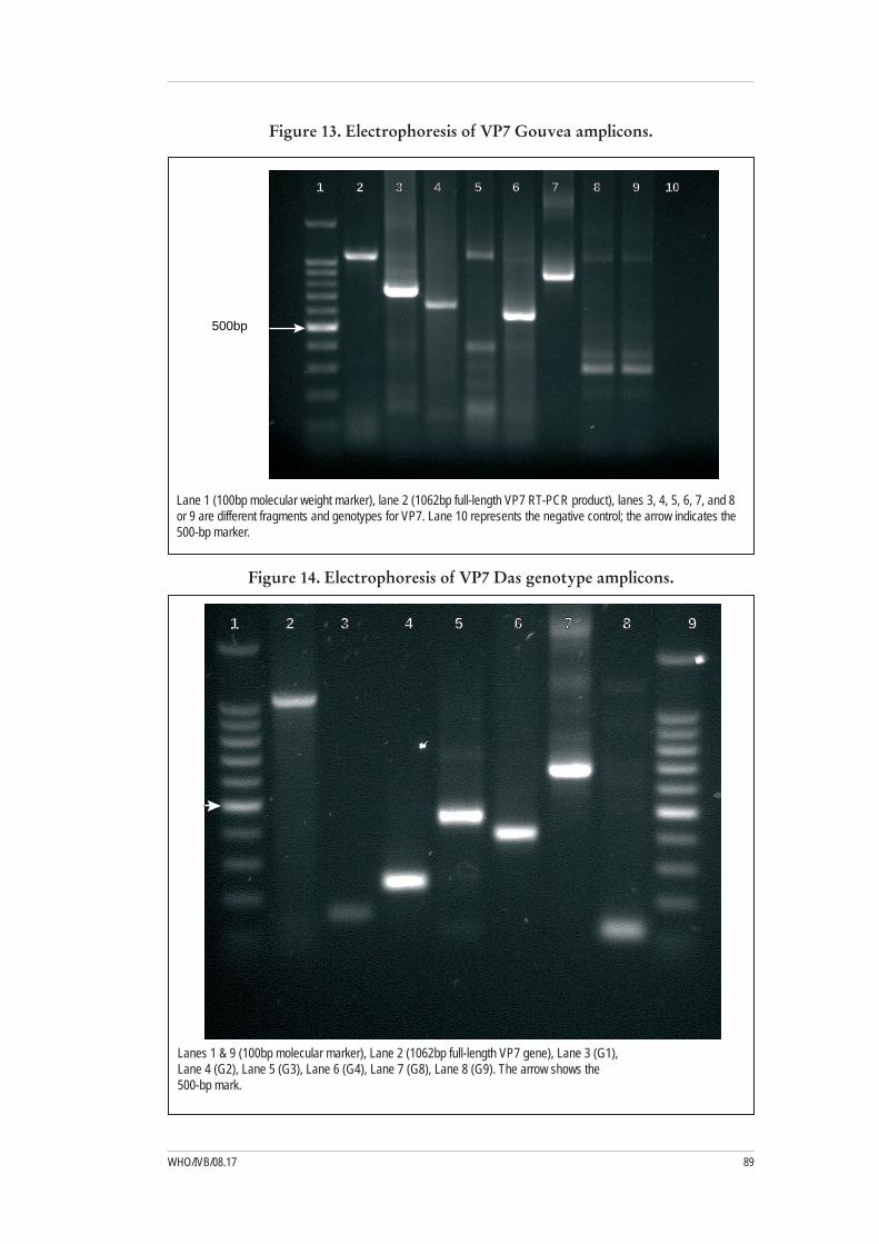
89WHO/IVB/08.17
Figure 13. Electrophoresis of VP7 Gouvea amplicons.
500bp
1 2 3 4 5 6 7 8 9 10
Lane 1 (100bp molecular weight marker), lane 2 (1062bp full-length VP7 RT-PCR product), lanes 3, 4, 5, 6, 7, and 8 or 9 are different fragments and genotypes for VP7. Lane 10 represents the negative control; the arrow indicates the 500-bp marker.
Figure 14. Electrophoresis of VP7 Das genotype amplicons.
1 2 3 4 5 6 7 8 9
Lanes 1 & 9 (100bp molecular marker), Lane 2 (1062bp full-length VP7 gene), Lane 3 (G1), Lane 4 (G2), Lane 5 (G3), Lane 6 (G4), Lane 7 (G8), Lane 8 (G9). The arrow shows the 500-bp mark.

Manual of Rotavirus detection and characterization methods90
Figure 15. Electrophoresis of Gentsch VP4 genotype amplicons.
1 2 3 4 5 6
MW FL P[8] P[6] P[4] NC
Lane 1 (100bp molecular weight marker), lane 2 (876bp full-length VP4 RT-PCR product), lanes 3, 4, and 5 represent P[8], P[6], and P[4]. Lane 6 is the negative control. The arrow indicates the 500-bp marker.
91WHO/IVB/08.17
4.16 Method 16: G and P genotyping32
Reagents and equipment
Random hexamers•dNTPs•M-MLV-RT•10X PCR buffer•Taq polymerase•MgCl• 2Oliogonucleotide primers• 33: VP7-F, VP7-R, VP7-RINT, G1, G2, G3, G5, G8, G9, G10, G12, VP4-F, VP4-R, P[4], P[6], P[8], P[9], P[10], P[11] Agarose•TBE•Gel loading buffer•Ethidium bromide•100-bp DNA ladder•Pipettes (20-1,000 µl)•Microfuge•Thermocycler•Ice bath•PCR tubes•Pipette tips (filter)•Microcentrifuge tubes•Electrophoresis tank and power pack•Gel imager or UV transilluminator•
32 This method was provided by the European Regional Rotavirus Laboratory, London, UK33 The nucleotide sequences of the primers are shown in Appendix 1

Manual of Rotavirus detection and characterization methods92
Random priming RT
Transfer 40 µl of extracted nucleic acid to a PCR tube. Denature the dsRNA at 1) 97oC for 5 min. Chill the tubes on ice for 2 min.
Prepare the RT mix for N + 2 (N = number of tubes in test) 2)
10X buffer II (Invitrogen) 7.0 µl 50 mM MgCl2 7.0 µl Random primers 18 (see Appendix 3) 1.0 µl dNTPs (10 mM) 2.0 µl M-MLV (200 U/µl) Invitrogen 2.0 µl RNase-free H2O 11.0 µl Total volume 30.0 µl
Add 30 µl of RT mix to each tube containing the extracted RNA.3)
Incubate the tubes at 374) oC for 1h. Incubate the tubes at 95oC for 5 min. Chill the tubes on ice for 2 min.
The total volume should be 70 µl. The cDNA can be used directly in the PCR 5) or stored at -20oC for further use.
Note: RT and PCR conditions have been optimized using Invitrogen reagents. Re-optimisation might be required if other reagents are used
G-typing consensus PCR (VP7)
Prepare the first-round PCR mix for N + 2 (N = number of tubes in test) 1)
10X buffer II (Invitrogen) 4.5 µl 50 mM MgCl2 2.0 µl dNTPs (10 mM) 1.0 µl Taq polymerase (5 U/µl) (Invitrogen) 0.2 µl Primer VP7-F (20 pmoles/µl) 1.0 µl Primer VP7-R (20 pmoles/µl) 1.0 µl RNase-free H2O 35.3 µl Total volume 45.0 µl
Note: Primers provide increased sensitivity and sufficient DNA for direct sequencing if the genotype is not determined in the second-round multiplex reaction. See Appendix 1 for primer sequences.
Add 45 µl of PCR mix to each PCR tube. Add 5 µl of cDNA (from the 2) RT reaction).
Briefly spin in the microcentrifuge (pulse for 5 sec). Transfer the tubes to the PCR 3) machine. Add the tubes to thermocycler, and cycle at the following temperatures for the following times:
94oC, 2 min, X1 94oC, 1 min 52oC, 1 min, X35 72oC, 1 min 72oC, 7 min, X1
Hold at 15oC
{

93WHO/IVB/08.17
G-typing multiplex PCR
Prepare second-round PCR mix for N + 2 (N = number of tubes in test) 1)
10X buffer II (Invitrogen) 4.8 µl 50 mM MgCl2 2.5 ul dNTPs (10 mM) 1.0 u Taq polymerase (5 U/µl) (Invitrogen) 0.2 u Primer VP7-R (20 pmoles/µl) 1.0 ul Primer G1 (20 pmoles/µl) 1.0 u Primer G2 (20 pmoles/µl) 1.0 u Primer G3 (20 pmoles/µl) 1.0 u Primer G4 (20 pmoles/µl) 1.0 u Primer G8 (20 pmoles/µl) 1.0 u Primer G9 (20 pmoles/µl) 1.0 u Primer G10 (20 pmoles/µl) 1.0 u Primer G12 (20 pmoles/µl) 1.0 u RNase-free H2O 30.5 u Total volume 48.0 ul
Notes:
See Appendix 1 for primer sequences. –
The position and sequence of the G3 primer differ from those published –previously (Gouvea et al,1990).These changes were introduced to prevent cross-reactivity between G3 and G10 primers and targets.8
The G9 primer has been modified to account for genetic drift of G9 strains –from the prototype strain.8
A G10 primer has been included because of isolation of increased numbers –of G10 strains in India in recent years.8
A G12 primer has been included because of the increasing number of –G12 strains detected globally.45
Add 48 µl of second-round mix to each PCR tube. Add 2 µl of first-round 2) product.
Briefly spin in microcentrifuge (pulse for 5 sec). Transfer tubes to the PCR 3) machine.
Add tubes to the thermocycler and cycle using the following program:4)
94oC, 4 min, X1 94oC, 1 min 42oC, 2 min, X30 72oC, 1 min 72oC, 7 min, X1
Hold at 15oC
{
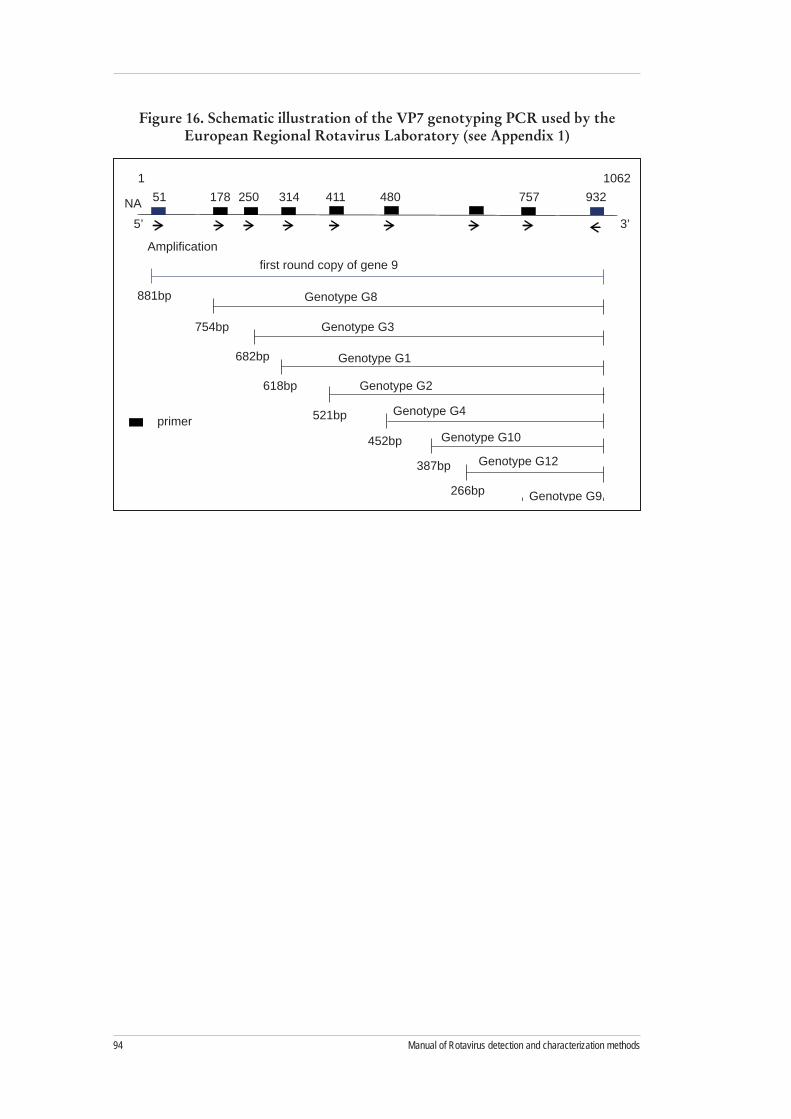
Manual of Rotavirus detection and characterization methods94
Figure 16. Schematic illustration of the VP7 genotyping PCR used by the European Regional Rotavirus Laboratory (see Appendix 1)
51NA5’
1178 250 314 411 480 757 932
1062
3’
Amplificationfirst round copy of gene 9
Genotype G8
Genotype G3
Genotype G1
Genotype G2
Genotype G4
Genotype G10
Genotype G12
Genotype G9
881bp
754bp
682bp
618bp
521bp
452bp
387bp
266bp
179bp
primer

95WHO/IVB/08.17
VP7 semi- nested consensus PCR (for sequence typing)
Note: This PCR provides increased sensitivity and should be used in conjunction with DNA sequencing for samples that failed to G-type in the multiplex genotyping assay
Prepare PCR mix for N + 2 (N = number of tubes in test) 1)
10X buffer II (Invitrogen) 4.5 µl 50mM MgCl2 2.0 µl dNTPs (10 mM) 1.0 µl Taq polymerase (5 U/µl) (Invitrogen) 0.2 µl Primer VP7-F (20 pmoles/ul) 1.0 ul Primer VP7-RINT (40 pmoles/ul) 1.0 ul RNase-free H2O 35.3 ul Total volume 45.0 ul
Note: See Appendix 1 for primer sequences.
Add 45 µl of PCR mix to each tube. Add 2 µl of first-round amplicon from 2) 4.16.2..
Briefly spin in the microcentrifuge (pulse for 5 sec). Transfer tubes to the PCR 3) machine.
Add tubes to the thermocycler and cycle as described below:4)
94oC, 2 min, X1 94oC, 1 min 50oC, 1 min, X35 72oC, 1 min 72oC, 7 min, X1
Hold at 15oC
{
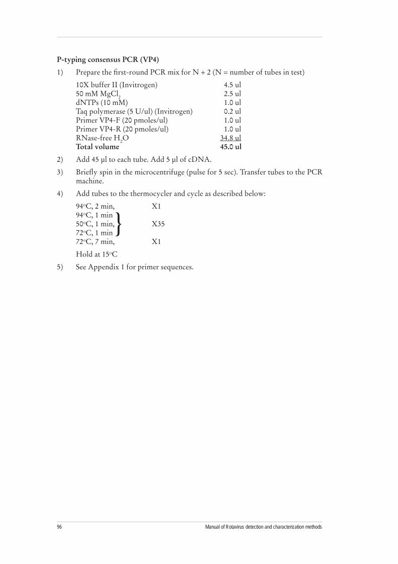
Manual of Rotavirus detection and characterization methods96
P-typing consensus PCR (VP4)
Prepare the first-round PCR mix for N + 2 (N = number of tubes in test) 1)
10X buffer II (Invitrogen) 4.5 ul 50 mM MgCl2 2.5 ul dNTPs (10 mM) 1.0 ul Taq polymerase (5 U/ul) (Invitrogen) 0.2 ul Primer VP4-F (20 pmoles/ul) 1.0 ul Primer VP4-R (20 pmoles/ul) 1.0 ul RNase-free H2O 34.8 ul Total volume 45.0 ul
Add 45 µl to each tube. Add 5 µl of cDNA.2)
Briefly spin in the microcentrifuge (pulse for 5 sec). Transfer tubes to the PCR 3) machine.
Add tubes to the thermocycler and cycle as described below:4)
94oC, 2 min, X1 94oC, 1 min 50oC, 1 min, X35 72oC, 1 min 72oC, 7 min, X1
Hold at 15oC
See Appendix 1 for primer sequences.5)
{

97WHO/IVB/08.17
P-typing multiplex PCR
Prepare second-round mix for N + 2 (N = number of tubes in test) 1)
10X buffer II (Invitrogen) 4.8 ul 50 mM MgCl2 2.5 ul dNTPs (10 mM) 1.0 ul Taq polymerase (5 U/ul) (Invitrogen) 0.2 ul Primer VP4-F (20 pmoles/ul) 1.0 ul Primer P[4] (20 pmoles/ul) 1.0 ul Primer P[6] (20 pmoles/ul) 1.0 ul Primer P[8] (20 pmoles/ul) 1.0 ul Primer P[9] (20 pmoles/ul) 1.0 ul Primer P[10] (20 pmoles/ul) 1.0 ul Primer P[11] (20 pmoles/ul) 1.0 ul RNase-free H2O 32.5 ul Total volume 48.0 ul
Add 48 µl of second-round mix to a new 0.2-ml tube. Add 2-µl of first-round 2) product.
Briefly spin in microcentrifuge (5 sec).3)
Transfer tubes to PCR machine room.4)
Add tubes to thermocycler and cycle as described below:5)
94oC, 4 min, X1 94oC, 1 min 45oC, 2 min, X30 72oC, 1 min 72oC, 7 min, X1
Hold at 15oC
Notes:
See Appendix 1 for primer sequences. –
The P[8] primer has been modified account for genetic drift of P[8] strains –from the prototype strain.46
A P[11] primer has been included because of isolation of increased numbers –of P[11] strains in India in recent years.8
{

Manual of Rotavirus detection and characterization methods98
Agarose-gel electrophoresis
Note: Avoid the use of mini-gels for genotyping second-round PCR because the resolution might not be sufficient to differentiate genotypes.
Add 2 g of Nusieve 3:1 agarose (or an appropriate gel for analysis of nucleic acid 1) <1 Kb) to 100 ml of 1X TBE for a midi-gel to give a 2% gel.
Melt in the microwave at full power for 2 min.2)
Caution3) : Any microwaved solution can become superheated and boil vigorously when moved or touched. Use extreme care in handling. Remove the boiling solution from the microwave oven, allow to stand for a few seconds at room temperature, and release the air by gentle swirling. Heat, using several short, 20- to 30-second intervals with gentle swirling between pulses to resuspend the powder.
Cool to 454) oC. Pour into a gel plate (14 cm wide x 16 cm long) 32 fitted with two 22-slot combs. Add 10 µl of PCR product to 10-µl sample buffer in a microtiter plate.
Remove the comb and end pieces, and add 40 µl of size markers or diluted sample 5) to the appropriate well.
Place the gel plate in the gel tank; add 1X TBE level with the gel (do not flood the 6) plate). Run the products into the gel for 5 min at a constant voltage of 150V.
Flood the gel with 1X TBE, making sure it is fully submerged.7)
Electrophorese at constant voltage (7-8 V/cm). 8)
Figure 17. Schematic illustration of the VP4 genotyping PCR used by the European Regional Rotavirus Laboratory (see Appendix 1)
Amplification
first round copy of gene 9
132-149 278NT5’ 3’
328 356 402 494 594 775-795 23591
663bp
146bp
191bp
224bp
270bp
362bp
462bp
P[6]
P[11]
P[8]
P[9]
P[4]
P[10]

99WHO/IVB/08.17
4.17 Trouble shooting
Too much dNTP, or degraded dNTP: Too much dNTP can inhibit the PCR reaction. The optimal range is 40-200 µM. Also, dNTPs are sensitive to repeated freeze-thaw cycles. Make small aliquots when you get a fresh batch, and turn over your stock frequently since dNTPs frozen at -20°C will eventually go bad.
Not mixing MgCl2: Magnesium chloride solutions form a concentration gradient when frozen and need to be vortexed before use.
Wrong MgCl2 concentration: Every PCR reaction has an optimal MgCl2 concentration range, usually between 1 and 4 mM. Mg2+ ions form complexes with dNTPs and can also act as a cofactor for polymerases, so try several conditions to optimize your concentration.
Inhibitors in the reaction: Make sure to know the origin of your source DNA or RNA. Substances such as chloroform, phenol, EDTA, ionic detergents (SDS), xylene cyanol, bromophenol blue, and ethanol can inhibit PCR. An extra clean-up step on your template might be sufficient. Also, certain polymerases are more susceptible to certain substances, so check your polymerase for possible inhibitors.
Poor-quality mineral oil: Low-grade preparations can contain nucleases that can kill the PCR. Also, avoid autoclaving the mineral oil, if possible. Exposure to high heat can cause formation of reaction-inhibiting hydrocarbons. Do not irradiate mineral oil with UV for long periods.
Too much enzyme: Excess enzyme can smear PCR products. Depending on the concentration of the supplied enzyme, most researchers use 0.5 µl of the stock Taq per reaction, but this might be too much for your reaction.
Wrong primer concentration: If the primer concentration is too low, no product will be visible. A concentration that is too high can cause primer dimerization and inadequate amplification. Use 0.1–1.0 µM of primer.
Wrong PCR program: Make sure the program you select is the correct one. A slip of a finger can alter your personal program on a PCR machine. Check your program while it is cycling to be sure you have chosen correctly.
Excess or insufficient template: Excess template can bind the primers and inhibit the PCR. With insufficient template, amplification might not be detectable. For 25–30 cycles, 104 copies of the target sequence are sufficient.
Poor primer design: Avoid obvious errors such as self-complementarity, complementarity between paired primers, or excessively long oligos (>30 bp). Often, making a new primer next to a suspect one can solve the problem and can be faster and cheaper than trying numerous variations in reaction conditions.

Manual of Rotavirus detection and characterization methods100
Estes MK. Rotaviruses and their replication. In: Knipe DM, Howley PM, eds. 1) Fields virology, 4th ed. Philadelphia: Lippincott Williams & Wilkins, 2001; p. 1747-86.
Ball JM, Tian P, Zeng CQY, Morris AP, Estes MK. Age-dependent diarrhea 2) induced by a rotaviral nonstructural glycoprotein. Science 1996;272:101-4.
Burns JW, Majid SP, Krishnaney AA, Greenberg HB. Protective effect of 3) rotavirus VP6-specific IgA monoclonal antibodies that lack neutralizing activity. Science 1996;272:104-7.
Bernstein DI, Sack DA, Rothstein E, Reisinger K, Smith VE, O’Sullivan D, 4) Spriggs DR, Ward RL. Efficacy of live, attenuated, human rotavirus vaccine 89-12 in infants: a randomised placebo-controlled trial. Lancet 1999;354: 287-90.
Clark HF, Offit PA, Ellis RW, Eiden JJ, Krah D, Shaw AR, Pichichero M, 5) Treanor JJ, Borian FE, Bell LM, Plotkin SA. The development of multivalent bovine rotavirus (strain WC3) reassortant vaccine for infants. J Infect Dis 1996;174 (Suppl 1):S73-80.
Bresee J, Parashar U, Holman R, Gentsch J, Glass RI, Ivanoff B, Linhares 6) AC, Molbak K. (Generic protocols (i) hospital-based surveillance to estimate the burden of rotavirus gastroenteritis in children and (ii) a community based survey on utilization of health care services for gastroenteritis in children. Geneva:World Health Organization, 2002; p.1-67.
Fischer TK, Gentsch JR. Rotavirus typing methods and algorithms. 7) Rev Med Virol 2004;14:71-82.
Iturriza-Gómara M, Kang G, Gray J. Rotavirus Genotyping: 2004. Keeping up 8) with an Evolving Population of Human Rotaviruses. J Clin Virol. 31, 259-265
Steele AD, Geyer A, Gerdes G. Rotavirus infections. In: Coetzer JT, ed. 9) Infectious diseases of livestock, 2nd ed. Cape Town: Oxford University Press, 2004; p.1256-64.
Herring AJ, Inglis NF, Ojeh CK, Snodgrass DR, Menzies JD. Rapid diagnosis 10) of rotavirus infection by direct detection of viral nucleic acid in silver-stained polyacrylamide gels. J Clin Microbiol 1982;16:473-7.
Gouvea V, Allen JR, Glass RI, Fang ZY, Bremont M, Cohen J, McCrae MA, 11) Saif LJ, Sinarachatanant P, Caul EO. Detection of group B and C rotaviruses by polymerase chain reaction. J Clin Microbiol 1991;29:519-23.
5. References

101WHO/IVB/08.17
Kang G, Iturriza-Gomara M, Wheeler JG, Crystal P, Monica B, Ramani S, 12) Primrose B, Moses PD, Gallimore CI, Brown DW, Gray J. Quantitation of group A rotavirus by real-time reverse-transcription-polymerase chain reaction: correlation with clinical severity in children in South India. J Med Virol 2004;73:118-22.
Wilde J, Yolken R, Willoughby R, Eiden J. Improved detection of rotavirus 13) shedding by polymerase chain reaction. Lancet 1991;337:323-6.
Richardson S, Grimwood K, Gorrell R, Palombo E, Barnes G, Bishop R. 14) Extended excretion of rotavirus after severe diarrhoea in young children. Lancet 1998;351:1844-8.
Coulson B, Unicomb LE, Putson GA, Bishop RF. Simple and specific enzyme 15) immunoassay using monoclonal antibodies for serotyping human retroviruses. J Clin Microbiol 1987;25:509-15.
Taniguchi K, Urasawa T, Morita Y, Greenberg HB, Urasawa S. Direct serotyping 16) of human rotavirus in stools using serotype 1-, 2-, 3-, and 4-specific monoclonal antibodies to VP7. J Infect Dis 1987;155:1159-66.
Koshimura Y, Nakagomi T, Nakagomi O. The relative frequencies of G 17) serotypes of rotaviruses recovered from hospitalized children with diarrhea: a 10-year survey (1987-1996) in Japan with a review of globally collected data. Microbiol Immunol 2000;44:499-510.
Coulson BS, Kirkwood C. Relation of VP7 amino acid sequence to 18) monoclonal antibody neutralization of rotavirus and rotavirus monotype. J Virol 1991;65:5968-74.
Coulson BS, Gentsch JR, Das BK, Bhan MK, Glass RI. Comparison of 19) enzyme immunoassay and reverse transcriptase PCR for identification of serotype G9 rotaviruses. J Clin Microbiol 1999;37:3187-93.
Kirkwood C, Bogdanovic-Sakran N, Palombo E, Masendycz P, Bugg H, 20) Barnes G, Bishop R. Genetic and antigenic characterization of rotavirus serotype G9 strains isolated in Australia between 1997 and 2001. J Clin Microbiol 2003;41:3649-54.
Gouvea V, Glass RI, Woods P, Taniguichi K, Clark HF, Forrester B, Fang ZY. 21) Polymerase chain reaction amplification and typing of rotavirus nucleic acid from stool specimens. J Clin Microbiol 1990;28:276-82.
Coulson BS. Typing of human rotavirus VP4 by an enzyme immunoassay using 22) monoclonal antibodies. J Clin Microbiol 1992;31:1-8.
Padilla-Noriega L, Werner-Eckert R, Mackow ER, Gorziglia M, Larralde G, 23) Taniguchi K, Greenberg HB. Serologic analysis of human rotavirus serotypes P1A and P2 by using monoclonal antibodies. J Clin Microbiol 1993;31:622-8.
Padilla-Noriega L, Mendez-Toss M, Menchaca G, Contrearas JF, 24) Romero-Guido P, Puerto FI, Guiscafre H, Mota F, Herrera I, Cedillo R, Munoz O, Calva J, Guerrero ML, Coulson BS, Greenberg HB, Lopez S, Arias CF. Antigenic and genomic diversity of human rotavirus VP4 in two consecutive epidemic seasons in Mexico. J Clin Microbiol 1998;36:1688-92.

Manual of Rotavirus detection and characterization methods102
Kapikian A, Hoshino Y, Chanock R. Rotaviruses. In: Knipe DM, Howley PM, 25) eds. Fields Virology. Vol. 2. 4th ed. Philadelphia: Lippincott-Raven Publishers, 2001; p.1787-1834.
Greenberg H, McAuliffe V, Valdesuso J, Wyatt R, Flores J, Kalica A, 26) Hoshino Y, Singh N. Serological analysis of the subgroup protein of rotavirus using monoclonal antibodies. Infect Immun 1983;39:91-9.
Desselberger U, Iturriza-Gomara M, Gray JJ. Rotavirus epidemiology and 27) surveillance. Novartis Foundation Symposium 2001;238:125-47.
Nakagomi O, Nakagomi T. Genomic relationships among rotaviruses 28) recovered from various animal species as revealed by RNA-RNA hybridization assays. Res Veterin Sci 2002;73:207-14.
Santos N, Hoshino Y. Global distribution of rotavirus serotypes/genotypes and 29) its implication for the development and implementation of an effective rotavirus vaccine. Rev Med Virol 2005;15:29-56.
Matsui SM, Mackow ER, Matsuno S, Paul PS, Greenberg HB. Sequence analysis 30) of gene 11 equivalents from “short” and “super short” strains of rotavirus. J Virol 1990;64:120-4.
Gentsch JR, Woods PA, Ramachandran M, Das BK, Leite JP, Alfieri A, 31) Kumar R, Bhan MK, Glass RI. Review of G and P typing results from a global collection of strains: implications for vaccine development. J Infect Dis 1996;174(Suppl 1):S30-6.
Das BK, Gentsch JR, Cicirello HG, Woods PA, Gupta A, Ramachandran 32) M, Kumar R, Bhan MK, Glass RI. Characterization of rotavirus strains from newborns in New Delhi, India. J Clin Microbiol 1994;32:1820-2.
Gentsch JR, Glass RI, Woods P, Gouvea V, Gorziglia M, Flores J, Das BK, 33) Bhan MK. Identification of group A rotavirus gene 4 types by polymerase chain reaction. J Clin Microbiol 1992;30:1365-73.
Gunasena S, Nakagomi O, Isegawa Y, Kaga E, Nakagomi T, Steele AD, 34) Flores J, Ueda S. Relative frequency of VP4 gene alleles among human rotaviruses recovered over a 10 year period (1982-1991) from Japanese children with diarrhea. J Clin Microbiol 1993; 31:2195-7.
Iturriza-Gomara M, Green J, Gray J. Methods of rotavirus detection, sero- and 35) genotyping, sequencing, and phylogenetic analysis. In: Gray J, Desselberger U, eds. Rotaviruses: methods and protocols. Human Press, Totowa, New Jersey, 2000; p. 189-216.
Iturriza-Gomara M, Green J, Brown DWG, Desselberger U, Gray JJ. 36) Comparison of specific and random priming in the reverse transcription polymerase chain reaction for genotyping group A rotaviruses. J Virol Meth 1999;78:93-103.
Cunliffe NA, Gondwe JS, Graham SM, Thindwa BD, Dove W, Broadhead 37) RL, Molyneux ME, Hart CA. Rotavirus strain diversity in Blantyre, Malawi, from 1997 to 1999. J Clin Microbiol 2001;39:836-43.

103WHO/IVB/08.17
Iturriza-Gomara M, Wong C, Blome S, Desselberger U, Gray J. 38) Molecular characterization of VP6 genes of human rotavirus isolates: correlation of genogroups with subgroups and evidence of independent segregation. J Virol 2002;76:6596-601.
Masendycz PJ, Palombo EA, Gorrell RJ, Bishop RF. Comparison of enzyme 39) immunoassay, PCR, and type-specific cDNA probe techniques for identification of group A rotavirus gene 4 types. J Clin Microbiol 1997;35:3104-8.
Green KY, Sears JF, Taniguchi K, Midthun K, Hoshino Y, Gorziglia M, 40) Nishikawa K, Urasawa S, Kapikian AZ, Chanock RM, Flores J. Prediction of human rotavirus serotype by nucleotide sequence analysis of the VP7 protein gene. J Virol 1988;62:1819-23.
Hoshino Y, Kapikian AZ. Rotavirus serotypes: classification and importance 41) in epidemiology, immunity, and vaccine development. J Health Pop Nutr 2000;18:5-14.
Palombo EA. Genetic reassortment and interspecies transmission of rotaviruses. 42) In: Kobayashi N, ed. Research signpost. 2003: 37, p661. Kerala.
Boom R, Sol CJA, Salismans MMM, Jansen CL, Werthiem-van Dillen PME, 43) van den Noordaa J. Rapid and simple method for the purification of nucleic acids. J Clin Microbiol, 1990, 28:495-503.
Indrani Banerjee, Sasirekha Ramani, Beryl Primrose, Miren Iturriza-Gomara, 44) James J. Gray, Mary K. Estes , David W. Brown and Gagandeep Kang. Modification of Rotavirus Multiplex RT-PCR for the Detection of G12 strains Based on Characterization of Emerging G12 Rotavirus Strains from South India. J Med Virol, 2007, in press.
Iturriza-Gomara M, Green J, Brown DWG, Desselberger U, Gray JJ. 45) Diversity within the VP4 gene of rotavirus P[8] strains: Implications for RT-PCR genotyping. J Clin Microbiol 2000, 38: 898-901.

Manual of Rotavirus detection and characterization methods104
VP7 consensus oligonucleotide primers
Primer Sequence (5’→3’) Position Recommended by
Forward
9con1 TAG CTC CTT TTA ATG TAT GG nt 37-56 Americas, South Africa
9con2 GTA TAA AAT ACT TGC CAC CA Nt 922–941 Americas
9con1-L TAG CTC CTT TTA ATG TAT GGT AT nt 37-59 Americas
Beg 9 GGC TTT AAA AGA GAG AAT TTC CGT CTG G nt 1-28
Americas, Asia, West Africa, South Africa
sBeg9 GGCTTTAAAAGAGAGAATTTC nt 1-21 West Africa, South Africa
VP7-F ATG TAT GGT ATT GAA TAT ACC AC nt 51-71 Europe
Reverse
End 9 GGT CAC ATC ATA CAA TTC TAA TCT AAG nt 1062-1036
Americas, Asia, West Africa, South Africa
RVG9 GGTCACATCATACAATTCT nt 1062-1044 West Africa, South Africa, Asia
dAnEnd9 GGTCACATCWWACARYTCTA nt 1062-1045 South Africa
VP7-R AAC TTG CCA CCA TTT TTT CC 3 nt 914-932 Europe
VP7-RINT ANA YNG ANC CWG TYG GCC A nt 331-344 Europe
VP7-Rdeg AAC TTG CCA CCA TYT YTT CC Nt 914-33 Americas
Appendix 1: Oligonucleotide primers

105WHO/IVB/08.17
VP4 consensus oligonucleotide primers
Primer Sequence (5’→3’) Position Recommended by
Forward con3 TGG CTT CGC TCA TTT ATA GAC A nt 11–32 Americas, Asia, West Africa, South Africa, Europe
VP4-F TAT GCT CCA GTN AAT TGG nt 132-149 Europe, Asia and South Africa
Reverse con2 ATT TCG GAC CAT TTA TAA CC nt 868–887 Americas, Asia, West Africa, South Africa, Europe
VP4-R ATT GCA TTT CTT TCC ATA ATG nt 775-795 Europe, Asia and South Africa
G type-specific oligonucleotide primers
Primer Sequence (5’→3’) Position Type Recommended by
9T-1 TCT TGT CAA AGC AAA TAA TG nt 176–195 G1 Americas, South Africa
aBT1 CAA GTA CTC AAA TCA ATG ATG G nt 314-335 G1Americas, Asia, West Africa, South Africa, Europe
9T-2 GTT AGA AAT GAT TCT CCA CT nt 262–281 G2 Americas, South Africa
aCT2 CAA TGA TAT TAA CAC ATT TTC TGT G nt 411-435 G2Americas, Asia, West Africa, South Africa, Europe
9T-3 GTC CAG TTG CAG TGT AGC nt 484–501 G3 Americas, South Africa
G3 or mG3 ACG AAC TCA ACA CGA GAG G nt 250-269 G3 Europe and
South Africa
G3-Aust ACG AAC TCA ACA CGA GAR G nt250-269 G3 Asia
aET3 CGT TTG AAG AAG TTG CAA CAG nt 689-709 G3 Americas, West Africa
9T-4 GGG TCG ATG GAA AAT TCT nt 423–440 G4 Americas, South Africa
aDT4 CGT TTC TGG TGA GGA GTT G nt 480-498 G4Americas, Asia, West Africa, South Africa, Europe
aAT8 GTC ACA CCA TTT GTA AAT TCG nt 178-198 G8Americas, Asia, West Africa, South Africa, Europe
MW-8 TCTTCAAAAGTCGTAGTG nt 670 - 688 G8 South Africa

Manual of Rotavirus detection and characterization methods106
Primer Sequence (5’→3’) Position Type Recommended by
9T-9 TAT AAA GTC CAT TGC AC nt 131–147 G9 Americas, South Africa
aFT9 CTA GAT GTA ACT ACA ACT AC nt 757-776 G9 Americas, West Africa
G9 or mG9 CTT GAT GTG ACT AYA AAT AC nt 757-776 G9 Europe, Asia and
South Africa
G10 or mG10 ATG TCA GAC TAC ARA TAC TGG nt 666-687 G10 Europe and
South Africa
G12 CCG ATG GACGTAACGTTGTA nt 548 - nt567 G12 Europe and South Africa
P type-specific oligonucleotide primers
Primer Sequence (5’→3’) Position Type Recommended by
pNCDV CGAACGCGGGGGTGGTAGTTG nt 269-289 P[1] South Africa
2T-1 CTA TTG TTA GAG GTT AGA GTC nt 474–494 P[4]Americas, Asia, West Africa, South Africa, Europe
pUK GCCAGGTGTCGCATCAGAG nt 336-354 P[5] South Africa
3T-1 TGT TGA TTA GTT GGA TTC AA nt 259–278 P[6]Americas, Asia, West Africa, South Africa, Europe
pGott GCTTCAACGTCCTTTAACATCAG nt 465-487 P[6] South Africa
pOSU CTTTATCGGTGGAGAATACGTCAC nt 389-412 P[7] South Africa
1T-1 TCT ACT TGG ATA ACG TGC nt 339 –356 P[8] Americas, Asia, West Africa
1T-1Wa TCT ACT GGG TTA ACG TGC Nt 339-356 P[8] Americas
1T1-VN TCT ACT GGA TCG ACG TGC Nt 339-356 P[8] Americas
1T-1D TCT ACT GGR TTR ACN TGC nt 339-356 P[8] Europe and South Africa
4T-1 TGA GAC ATG CAA TTG GAC nt 385–402 P[9]Americas, Asia, West Africa, South Africa, Europe
5T-1 ATC ATA GTT AGT AGT CGG nt 575–594 P[10]Americas, Asia, West Africa, South Africa, Europe

107WHO/IVB/08.17
Primer Sequence (5’→3’) Position Type Recommended by
ND2 AGC GAA CTC ACC AAT CTG nt 116– 133 P[11] Americas
P[11] or mP[11] GTA AAC ATC CAG AAT GTG nt 305-323 P[11] Europe and
South Africa
pB223 GGAACGTATTCTAATCCGGTG nt 574-594 P[11] South Africa
p4943 GGTGTAGTTCCTGCGTA nt 538-554 P[14] South Africa
SE-1 CTCTGCTACTCTACCTATTTG nt 271-291 P[14] South Africa

Manual of Rotavirus detection and characterization methods108
Method 1: Cloning PCR amplicons34
Reagents and equipment
500-ml conical flasks•0.6-ml PCR tubes•Petri dishes•Disposable spreaders•Disposable 1-ul and 10-ul loops•Glass plate•Glass bijoux•Disposable scalpels•Microcentrifuge•Thermocycler•Vortexer•Microwave oven•Water bath (56• oC)Water bath (42• oC)Incubator (37• oC)Shaking incubator (37• oC)Magnetic stirrer•Gel imaging system•Heating block•pH meter•Weighing balance•Ampicillin •Xgal •IPTG •Dimethyformamide •TAE or TBE •Ethidium bromide •DNA ladder •TR buffer •
34 This method was provided by the European Regional Rotavirus Laboratory
Appendix 2: Cloning and sequencing
methods

109WHO/IVB/08.17
L agar •Dorset egg slopes •Screw-cap microfuge tubes•TOPO TA cloning kit •Competent cells (TOP10F’) •SOC medium •Expand HF Taq Polymerase •dNTPs •Geneclean• TM (GC) spin kits Gel-loading buffer •Orange G •Ficoll •Trizma/Tris •EDTA •Oligonucleotide primers•Power pack•PTAG5’, PTAG3’ •Molecular-biology-grade agarose •Electrophoresis tanks•Gel staining tank•Pipettes (20-1,000 ul)•Ice bath•UV transiluminator•
PCR amplicon purification in solution
Select an amplicon of the correct size for purification.1)
Purify DNA using GC spin columns.2)
Add 30 µl of PCR amplicon to 400 µl of GC spin glassmilk to spin filter. 3) Invert to mix, and incubate at room temperature for 5 min, mixing every minute.
Centrifuge for 1 min at 14,000 x g.4)
Pour off liquid from the catch tube into a GTC waste bottle.5)
Add 500 µl of GC Spin New Wash, and centrifuge for 30 sec at 14,000 g.6)
Pour off liquid from the catch tube into an ethanol waste bottle. Add 500 µl of 7) GC Spin New Wash. Centrifuge for 30 sec at 14,000 g.
Pour off liquid from the catch tube into an ethanol waste bottle. Centrifuge for 8) 1 min at 14,000 g.
Place filter in a new catch tube. Add 30 µl of nuclease-free water; vortex at 9) half speed briefly. Note: The volume of eluate can be varied to concentrate or dilute the purified product.
Centrifuge for 30 sec at 14,000 g. Discard the spin filter.10)

Manual of Rotavirus detection and characterization methods110
PCR amplicon purification by gel electrophoresis
Run 30 µl of PCR amplicon on an agarose gel (usually 2%, but depends on 1) amplicon size) in 1X TAE or 1X TBE until dye front reaches the end of the gel.
Note: Determine the concentration of the gel according to the expected size of the product and the electrophoresis buffer used:
500-2500 bp 2.0% gel in TAE 1.0% gel in TBE 60-700 bp 4.0% gel in TAE 3.0% gel in TBE 20-250 bp 6.0% gel in TAE 5.0% gel in TBE
Add the gel to the 1X TAE/EtBr staining solution in the staining tank for 2) 15-30 min.
Rinse the gel with water in a sandwich box. Place the gel on a glass plate. 3) Examine the PCR amplicons under a longwave light.
Cut amplicons from the gel with a disposable scalpel, and add a gel slice to a 4) 1.5-ml screw-top centrifuge tube.
Purify DNA from the gel slice using GC spin columns.5)
Add 400 ul of Geneclean GTC/silica solution. Place in a heating block at 566) oC until the gel slice has melted. Centrifuge for 30 sec at 14,000 g.
Pipette off liquid from the catch tube into a GTC waste bottle. Add 500 ul of 7) GC Spin New Wash. Vortex, and add solution to the spin filter. Centrifuge for 30 sec at 14,000 g.
Pour off liquid from the catch tube into an ethanol waste bottle. Add 500 ul of 8) GC Spin New Wash. Pour off liquid from the catch tube into an ethanol waste bottle.
Centrifuge for 1 min at 14,000 g.9)
Place a filter in a new catch tube. Add 30 µl of nuclease-free water, and vortex at 10) half speed briefly. Centrifuge for 30 sec at 14,000 x g. Discard the spin filter.
Date-label the tubes, and store them in a cloning/sequencing box at –2011) oC until required.
Preparation of L agar plate for cloning
Melt the L agar with the lid loosened in a microwave on high for 5-15 min, 1) depending on the number of bottles. Place the bottles in a water bath at 56oC for ~30 min.
Add an aliquot of ampicillin (200 µl of 50 mg/ml aliquot) to the liquid agar, 2) and mix by swirling gently.
Pour the agar into 9-cm diameter petri dishes (5 dishes per 100-ml bottle 3) L agar). After the agar sets, invert the dishes to dry, and store the plates at 4oC until needed.
Add 100 µl of IPTG solution and 20 µl of X gal solution4) to an agar plate, and spread across the surface with a disposable spreader. Place in a 37oC incubator inverted to dry. Prepare IPTG/X gal plates fresh.
111WHO/IVB/08.17
Preparation of X-gal and IPTG solutions
To make X gal solution (50 mg/ml), add 150 mg of X gal to 3 ml of dimethyformamide in a glass bijoux. Store at –20oC until needed.
To make IPTG solution (24 mg/ml), add 120 mg of IPTG to 5 ml of water in glass bijoux. Store at –20oC until needed.
Cloning
A PCR amplicon for cloning can be an unpurified amplicon, a Geneclean purified amplicon in solution, or a GC purified amplicon by gel electrophoresis, as appropriate
Set up a 4.5-µl reaction in 0.5-ml screw-top tubes. 1)
Add 4 µl of nuclease-free water and 1 ul of pCR® TOPO TA vector to 1 tube, 2) and mix.
Add 2.5 ul of the above to the next tube. Repeat, depending on the number of 3) ligations to be performed.
Add 2 ul of PCR amplicon, and spin briefly.4)
If using purified amplicons, set up a single reaction with 4 µl of purified amplicon 5) and 1 µl of pCR® TOPO TA vector per ligation, or use the method described above.
Incubate at room temperature for 5 min.6)
Transformation of TOPO TOP10F’ cells
Remove TOPO TOP10F’ cells from –801) oC and place on ice. Place the SOC medium to thaw to room temperature, or use SOC medium stored at room temperature (8 cloning reactions require 4 cell vials, unless performing single-tube reactions). Label the cell vials (50 ul of cells is sufficient for two reactions). Add 25 ul to each tube/vial for transformation.
Add 2 ul of PCR amplicon/ pCR® TOPO TA vector ligation reaction, and mix 2) by flicking on the side and then flicking the mixture down. Incubate the tubes on ice for 30 min.
Heat shock the cells for 30 sec at 423) oC. Incubate the vials on ice for 2 min.
Add 250 ul of SOC medium, and shake the tubes horizontally in an 4) incubator at 37oC for 30 min. Spread 100 ul of each transformant onto a separate L agar/ampicillin plate (containing Xgal/IPTG). Incubate inverted at 37oC overnight.
Store the plates at 45) oC, and screen the clones. White colonies indicate plasmid containing an insert.
Note: Some inserts might be unstable in TOP10F’ cells. If so, use a recombination- deficient cell line such as MAX Efficiency Stbl2 competent cells (Invitrogen), following the manufacturer’s instructions. Incubate at 30oC after transformation.

Manual of Rotavirus detection and characterization methods112
Clone screening: heat denaturation preparation
Pick up to 10 white colonies (blue colonies contain no insert) for each transformant, 1) and prepare a subculture on an L agar/ampicillin plate (dividing the plate into a checkerboard by marking the back of the plate). Incubate at 37oC overnight, and store the plates inverted at 4oC.
Aliquot 100 ul of nuclease-free water into 0.6-ml sequencing tubes (1 tube for 2) each subculture).
Use a 1-ul loop, and nick the subcultured colony. Add to the water and mix.3)
Incubate the tubes at 954) oC for 5 min, and hold on ice for 2 min. Centrifuge for 15 sec at 17,000 x g.
Prepare 49 ul of PCR mix with PTAG5’/PTAG3’ primers in 0.6-ml PCR 5) tubes.
For a 1X mix, add 5 ul of Expand HF 10X PCR buffer, 1 ul of 25mM MgCl6) 2, 1mM of each dNTP, 20 pmols of PTAG5’ primer, 20 pmols of PTAG3’ primer, and water to 49 ul.
Add 1 ul of denatured colony preparation to a 49-ul PCR mix. Centrifuge for 7) 15 sec at 17,000 g.
Add tubes to the thermocycler and use the following program:8)
94oC, 4 min, X1 94oC,1 min 55oC, 1 min X30 72oC, 2 min 72oC, 7 min, X1 Hold at 15oC
Note: The extension time can be adjusted depending on the length of the amplicon.
Run 15 ul of PCR amplicon on an agarose gel (usually 2%, but depends on 9) amplicon size) in 1X TAE or 1X TBE until the dye front reaches the end of the gel.
Add the gel to the EtBr staining solution for 15-30 min. Rinse the gel with water 10) in a sandwich box. Photograph the gel.
The size of the amplicon is 200 bp derived from the vector plus the size of the 11) insert (the original PCR amplicon).
Archiving positive clones
Store positive clones:
At room temperature by inoculating them onto Dorset Egg slopes; or•
At –20• oC inoculated on LB medium containing glycerol; or
At –70• oC by storing in cryovials containing porous beads.
}

113WHO/IVB/08.17
Method 2: Beckman CEQ Capillary Sequencing35
Reagents and equipment
Microcentrifuge•Refrigerated microcentrifuge•Thermocycler•Speed vac (optional)•Microwave oven•Electrophoresis tank•Gel staining tank•Pipettes (20-1,000 ul)•Beckman CEQ8000 capillary sequencer•Geneclean• TM (GC) spin kits 0.6-ml thin walled PCR •Nuclease-free water tubes •0.2-ml thin walled PCR •Molecular-biology-grade ethanol •Disposable scalpels•Molecular-biology-grade water plates •Beckman sequencing •70% ethanol •Beckman buffer plates •3M NaCl solution, pH 5.3 •100 mM EDTA, pH 8.0 •Molecular-biology-grade agarose •DNA ladder •TE •Ficoll •Ethidium bromide •Beckman CEQ DTCS Quick Start Kit •Beckman CEQ sequencing separation buffer •Beckman CEQ separation gel (608010) •
35 This method was provided by the European Regional Rotavirus Laboratory

Manual of Rotavirus detection and characterization methods114
PCR amplicon purification by gel electrophoresis
Run 30 µl of PCR amplicon on an agarose gel (usually 2%) in 1X TAE or 1X 1) TBE until the dye front reaches the end of the gel.
Add the gel to the 1X TAE/EtBr staining solution in the staining tank for 2) 15 min.
Rinse the gel with water in a sandwich box, and place the gel on a glass plate. 3) Examine the PCR amplicons under a longwave UV light.
Cut amplicons from the gel with a disposable scalpel, and add gel slice to a 4) 1.5-ml screw-top centrifuge tube.
Purify DNA from the gel slice using the GC spin kit.5)
Add 400 µl of GC GTC/silica solution. Place in a heating block at 566) oC until the gel slice has melted. Centrifuge for 30 sec at 14,000 g.
Pipette off liquid from the catch tube into a GTC waste bottle. Add 500 ul of 7) GC Spin New Wash. Vortex and add solution to the spin filter. Centrifuge for 30 sec at 14,000 g.
Pour off liquid from the catch tube into an ethanol waste bottle. Add 500 ul of 8) GC Spin New Wash. Pour off liquid from the catch tube into an ethanol waste bottle.
Centrifuge for 1 min at 14,000 g.9)
Place filter in a new catch tube. Add 30 µl of nuclease-free water, and vortex at 10) half speed briefly Centrifuge for 30 sec at 14,000 g. Discard the spin filter.
Date-label the tubes, and store them in the current cloning/sequencing box or 11) other appropriate box at –20oC until required.
PCR amplicon purification in solution
Purify DNA using GC spin columns.1)
Add 30 µl of PCR amplicon to 400 µl of GC spin glassmilk to spin filter. 2) Invert to mix, and incubate at room temperature for 5 min, mixing every minute.
Centrifuge for 1 min at 14,000 g.3)
Pour off liquid from the catch tube into a GTC waste bottle. Add 500 µl of GC 4) Spin New Wash, and centrifuge for 30 sec at 14,000 g.
Pour off liquid from the catch tube into an ethanol waste bottle. 5)
Add 500 µl of GC Spin New Wash, and centrifuge for 30 sec at 14,000 g.6)
Pour off liquid from the catch tube into an ethanol waste bottle. Centrifuge for 7) 1 min at 14,000 g.
Place filter in a new catch tube. Add 30 µl of nuclease-free water, and vortex at 8) half speed briefly.
Centrifuge for 30 sec at 14,000 g. Discard the spin filter.9)
Date-label the tubes, and10) store in a cloning/sequencing box at –20oC until required.

115WHO/IVB/08.17
Quantitation of purified amplicon
Run 5 µl of the purified products on a 2% NuSieve (3:1) agarose–TBE gel alongside a PCR marker of known concentration. Estimate the concentration of each amplicon to allow optimum sequencing (25-100 fmol):
Size (Kb) ng for 25 fmol
ng for 50 fmol
ng for 100 fmol
0.2 3.3 6.5 130.3 4.9 9.8 200.4 6.5 13 260.5 8.1 16 331 16 33 652 33 65 1303 50 100 1954 65 130 260
Beckman sequencing PCR
Set up individual reactions to provide sequence in both directions using the forward 1) primer in one and the reverse primer in the other, and label appropriately.
Add 2 µl to 10 µl of purified DNA (see table above) template, depending on 2) original quantity of DNA
Add 8 µl of Beckman CEQ master mix. Add 2 µl of primer (5 pmoles/µl).3)
Add nuclease-free water to give a final volume of 20 µl.4)
Centrifuge for 10 sec at 17,000 g.5)
Add tubes to the thermocycler using BECKSEQ program: 30 cycles at 966) oC for 20 sec, 50oC for 20 sec, 60oC for 4 min, then hold at 4oC.
Post-sequencing PCR purification and precipitation
Remove tubes from the thermocycler and place in a rack.1)
Add 5 µl of stop solution (2 vols 3M NaOAc solution, pH 3.5; 2 vols 100 mM 2) EDTA solution, and 1 vol glycogen). Close lids, and mix by inversion several times.
Centrifuge at 17,000 g for 15 min at 43) oC in a refrigerated centrifuge with the tube hinges facing out.
Remove the supernatant using an extended fine-tip pastette, and discard into an 4) ethanol waste bottle. Add 200 µl of cold 70% ethanol stored at 4oC,
Centrifuge at 17,000 g for 2 min at 45) oC in a refrigerated centrifuge with the tube hinges facing out. Remove the supernatant using an extended fine-tip pastette, and discard into an ethanol waste bottle. Add 200 µl of cold 70% ethanol.
Centrifuge at 17,000 g for 2 min at 46) oC in a refrigerated centrifuge with the tube hinges facing out. Remove the supernatant using an extended fine-tip pastette, and discard into an ethanol waste bottle
Dry in a DNA pellet in a vacuum centrifuge for ~15 min or by placing the tubes 7) with lids open in a heating block at 56oC for a maximum of 15 min.

Manual of Rotavirus detection and characterization methods116
Resuspend the DNA pellet by adding 40 µl of SLS from the kit (stored aliquoted 8) at –20oC), and briefly vortex. Transfer samples to a 96-well Beckman sample plate. Add a drop of Beckman mineral oil to each well.
Store plates at -209) oC, or use immediately.
Beckman capillary sequencer
Add a plate to the Beckman capillary sequencer.1)
Add the corresponding buffer plate with ¾ of each well containing buffer 2) (Beckman) and the same number of rows as in the sample plate.
Add fresh water (Sigma) to the cleaned wetting tray; replace the lid, and make 3) sure the holes in the lid are clear. Place lid on buffer plate.
Load plates according to the on-screen instructions. Prepare a sample template 4) form on the PC.
Check the level of gel and replace if needed; follow the on-screen instructions. 5) Run the sequencing plate.
After completion of the run, download 6) *SCF files from the analysis module.
Analysis of sequencing using Genebuilder
Open the appropriate database in Bionumerics.1)
Add a new entry, type the sample reference number as the key field (bottom of 2) the new window), and open the “entry edit” window by clicking twice on the “new entry” key.
Enter the epidemiologic information in the appropriate fields.3)
Double click on the appropriate experiment icon (e.g., VP7, VP6, VP4, NSP4).4)
Click “yes” to create a new experiment. Click on the Genebuilder icon on the 5) new empty window.
A new window will appear. To import chromatograms, double click “import 6) sequences from file.” Find the sequences to be analysed in the appropriate folder, select “Beckman sequence file (*SCF)” as the file type, select the sense and antisense sequences, and click “open.”
Trim the ends of each sequence to remove any peaks of poor quality by placing 7) the cursor at the point from which the sequence is to be cut and clicking on the “cut left from cursor” or “cut right from cursor” icons.
Align the two sequences and check the orientation. Scroll along the sequence 8) contig, and edit if necessary. Edit point insertion/deletions and mismatches.
Save the 9) contig, and proceed to phylogenetic analysis.

117WHO/IVB/08.17
Phylogenetic analysis
Phylogenetic analysis can be performed using any of a number of software packages (e.g., GCG, Phylip, Bionumerics, Bioedit, DNAStar). First, compare any new sequence to sequences of prototype strains in the database. Once the type has been determined, compare it to more recent representative strains of each of the genotypes to account for drift due to accumulation of point mutations. A genotype can be assigned when the query sequence is >90% homologous at the nt level.
For the construction of phylogenetic trees, the topology should be confirmed using at least two different methods. Neighbour joining and Maximum parsimony are recommended. When the number of sequences in the comparison is not too large (<15), Maximum likelihood may be performed. When the number of sequences in the comparison ≥15, the algorithm will require too much computer memory. Genetic lineages or clades should be confirmed with bootstrapping.

Manual of Rotavirus detection and characterization methods118
Method 3. Cloning PCR fragments using the pGEM®-T EASY vector systems36
Reagents and equipment
Water bath (50• oC, 42oC)UV transiluminator•Balance•Microcentrifuge•Shaking incubator (37• oC) Scalpel•Microcentrifuge tubes•Isopropanol •3M sodium acetate, pH 5 •Low-melt agarose •Ethidium bromide •QIAquick gel extraction kit •Molecular-grade agarose •GENEQUANT •pGEM- T Easy Vector cloning kit •Competent cells (JM109) •SOC medium •LB plates •IPTG •X-Gal •Ampicillin •LB broth •QIAprep spin miniprep kit •
36 This method was provided by the south African Regional Rotavirus Laboratory

119WHO/IVB/08.17
Gel purification of PCR fragments using the QIAquick gel extraction kit
Note: All reagents required for gel purification are supplied with the gel extraction kit.
Ensure that the water bath is at 501) oC and that 100% isopropanol and 3M Na Ac, pH 5.0, are available.
Run the PCR fragment on a 2% low-melting agarose gel containing ethidium 2) bromide.
Visualize the fragment under UV light, and excise the band needed with a clean 3) sharp scalpel. Make the gel slice as small as possible without losing fragment yield.
Weigh the gel slice in a colorless 1.5-ml eppendorf tube, and add 300ul of 4) buffer QG for every 100 mg of gel. For >2% gels, add 600ul of buffer QG. Note: Maximum gel slice per column is 400 mg. If more, use <1 column.
Incubate at 505) oC for 10 min; vortex the tube every 2-3 minutes during incubation. Ensure that the gel slice has melted completely before continuing. For >2% gels, increase the incubation time.
Check that the color of the mixture is yellow (i.e., similar to buffer QG without 6) dissolved agarose). If the mixture is orange or violet, add 10ul of 3M sodium acetate, pH 5.0, and mix. The mixture should turn yellow.
Add 100ul of isopropanol for every 100 mg of gel, and mix thoroughly. Do not 7) centrifuge!
Place QIAquick spin column in a 2-ml collection tube, and apply the sample to 8) the column. Centrifuge at 12,000 rpm for 1 min. Note: The maximum volume of the column is 800ul. For larger volumes, load and spin again.
Discard the flow-through, and replace the column in the same collection tube.9)
Add 500ul of buffer QG to the column, and centrifuge again for 1 min. Discard 10) the flow-through.
Add 750ul of buffer PE to the column, and centrifuge for 1 min. Discard the flow-11) through and column. Centrifuge for another 1 min. Note: Residual ethanol from buffer PE will not be completely removed unless the flow-through is discarded before the additional centrifugation.
Place the column in a sterile 1.5 ml-eppendorf tube.12)
Add 30ul of buffer EB or sterile distilled water to the center of the column 13) membrane.
Incubate the column at room temperature for 1 min and centrifuge for 1 min. 14) The average eluate volume is 28ul from 30ul elution buffer.
Run 3ul of the cleaned PCR product on a 1% agarose gel to confirm a successful 15) extraction.

Manual of Rotavirus detection and characterization methods120
Insertion of purified PCR fragments into pGEM®-T Easy Vector ligation reaction
Determine amount of insert DNA in the elute using the 1) GENEQUANT system.
Calculate the insert:vector ratio and ng of the insert using the following 2) formula:
ng of vector x kb size of insert X ratio insert = ng of insert kb size of vector vector
Note: The vector is supplied at 50 ng/ul.
Centrifuge the Easy Vector tube for 5 sec to collect the contents at the bottom 3) of the tube.
Vortex the 2 x rapid ligation buffer and T4 DNA ligase before use.4)
Prepare the ligation mix. 5)
1 x reaction 2 X Rapid Ligation buffer 5 ul pGEM®-T Easy Vector (50 ng/l) 1ul T4 DNA Ligase (3 Weiss units/l) 1ul Deionized water (10ul-[7ul+insert]) 3ul Total 10ul
Add ligation mix to each PCR fragment tube, and mix the reactions by pipetting. 6) Incubate overnight at 4oC.
Transformation of pGEM®-T Easy Vector clones
Set the water bath to exactly 421) oC, and shake the incubator to 37oC at 150 rpm.
Centrifuge the ligation reaction tubes briefly to collect the contents at the bottom 2) of the tube.
Transfer 2ul of each ligation reaction to a sterile 1.5-ml eppendorf tube, and place 3) on ice. Store the remaining ligation reaction at -20oC.
Thaw JM109 high-efficiency competent cells in an ice bath for +/- 5 min, 4) and mix by gently flicking the tube.
Add 50ul of thawed JM109 cells to each ligation reaction tube. Gently flick the 5) tubes to mix, and incubate in an ice bath for 20 min.
Heat shock by placing the tubes in a 426) oC water bath for 45-50 sec, and chill on an ice bath for 2 min. Do not shake the tubes!
Add 950ul of room-temperature SOC medium to each tube containing cells 7) transformed with ligation reaction mixture. Incubate for 90 min in a 37oC orbital shaker incubator at 150 rpm.
Cover each LB-plate with 100ul IPTG and 20ul X-Gal 30 min before plating the 8) transformation culture.
Plate 100ul of each transformation culture onto duplicate LB/Amp/IPTG/X-Gal 9) plates. Incubate overnight (16-24 h) at 37oC.
Pick white colonies, and inoculate into 5 ml of LB broth + amp.10)
Incubate at 3711) oC with vigorous shaking (200 rpm) for 12-16 h.

121WHO/IVB/08.17
Extraction of plasmid (clone) DNA using Qiagen QIAprep spin miniprep kit
Dispense 5 ml of the LB-broth/ampicillin (100 g/ml) into each tube (at least 1) 2 tubes/sample). Inoculate each tube with a single white colony.
Incubate tubes at 372) oC in an orbital shaker incubator at 200 rpm overnight (16-24 h).
Pellet cells by centrifuging at 5,000 rpm for 5 min. Discard the supernatant.3)
Resuspend the cells in 250ul of buffer P1 and transfer to a sterile 1.5-ml eppendorf 4) tube.
Note: No cell clumps should be visible after resuspension of the pellet.
Add 250ul of buffer P2 (lysis buffer), and gently invert the tube 4-6 times to mix. 5) Continue to mix until the solution becomes viscous and slightly clear.
Note: Do not allow lysis reaction to proceed for more than 5 min.
Add 350ul of buffer N3 (neutralization buffer) and gently invert tube immediately 6) 4-6 times to mix. The solution should turn cloudy.
Centrifuge at 12,000 rpm for 10 min. A compact white pellet should form.7)
Place the QIAprep spin column in a 2-ml collection tube and apply supernatant 8) to the column.
Centrifuge for 1 min at 12,000 rpm and discard the flow-through.9)
Add 500ul of buffer PB to the column, and centrifuge for 1 min. Discard the 10) flow-through.
Add 750ul buffer PE to the column, and incubate for 5 min at room temperature. 11) Centrifuge for 1 min and discard the flow-through. Centrifuge for an additional minute to remove residual wash buffer.
Place the column in a sterile 1.5-ml eppendorf tube, and add 50ul of buffer EB or 12) sterile distilled water to the center of the membrane in the column. Let it stand for 1 min and centrifuge for 3 min.
Run 2ul of eluate on a 1.2% gel, and visualize under UV light.13)

Manual of Rotavirus detection and characterization methods122
Confirmation of correct insert: restriction digestion of clone
Determine the concentration of the DNA plasmid (clone) using the 1) GENEQUANT System.
Add 1ul DNA (1 g/l) to a clean eppendorf tube.2)
Prepare the restriction digestion mixture as follows: 3)
1 x reaction RE 10 x buffer 2.0ul Acetylated BSA (10 g/l) 0.2ul NotI (10 U/l) 0.5ul Deionized water 16.3ul Total 20ul
Transfer 19ul of the restriction digestion mixture to each tube, and mix gently by 4) pipetting. Centrifuge to collect the contents of the tube at the bottom.
Incubate at 375) oC for 60 min; run on a 2% agarose gel, and visualize under UV light.
Sequencing of pGEM plasmid DNA
Sequencing of plasmid insert is conducted by private companies including Gene Care Molecular Genetics, Cape Town or Inqaba Biotec, Pretoria.

123WHO/IVB/08.17
Method 4: ABI sequencing and analysis37
Reagents and Equipment
Microcentrifuge (protocol uses an Eppendorf 5417C)•Thermocycler with heated lid•Vacuum centrifuge•ABI PRISM 3100 genetic analyzer (Applied Biosystems)•96-well optical reaction plate (Catalog # 4306737; Applied Biosystems)•3100 genetic analyzer plate, septa 96-well (Catalog # 4315933; •Applied Biosystems)3100 capillary array, 50 cm (Catalog # 4315930; Applied Biosystems)•Microwave oven•Weighing balance•Horizontal electrophoresis tank •Horizontal electrophoresis tray and comb•Power pack for electrophoresis tank•Covered dish for gel staining•UV light box•1.7-ml centrifuge tubes•0.6-ml PCR tubes•0.2-ml PCR tubes•96-well ABI plate•Laboratory pipettes (10-µl, 20-µl, 200-µl, and 1,000-µl)•Filtered pipette tips•Disposable scalpels•Heat block•Vortexer•Sequencher Version 4.0.5 sequencing analyzation software (Gene Codes •Corporation)SeaKem GTG agarose (catalog # 50070; Cambrex)•DNA marker (123-bp ladder; catalog # 15613-011, Invitrogen; OR ladder •appropriate to expected product size)Sample loading buffer•1X TAE or 1X TBE•EtBr staining solution•Distilled water •QIAquick gel extraction kit (Catalog # 28706; Qiagen)•Nuclease-free water•
This method was provided by WHO Rotavirus Collaborating Center, Atlanta, Georgia, UsA

Manual of Rotavirus detection and characterization methods124
ABI PRISM BigDye Terminator v1.1 Cycle Sequencing Kit (Catalog •# 4336774; Applied Biosystems)•Centri Sep spin columns (Catalog # CS-901; Princeton Separations)•Type 1 water•Hi-Di formamide (Catalog # 4311320; Applied Biosystems)•3100- POP6 performance optimized polymer (Catalog # 4316357; Applied •Biosystems)ABI PRISM 10 buffer with EDTA (Catalog # 402824; Applied Biosystems)•Methanol (reagent grade)•Ethanol (reagent grade)•3M sodium acetate, pH 5.0•Isopropanol (reagent grade)•
PCR amplicon purification by gel electrophoresis
Run 40 µl of PCR amplicon on an agarose gel (usually 1%) in 1X TAE or 1X 1) TBE until the dye runs to the end of the gel (if the amplicon is <300 bp, allow the dye to run 75% of the gel)
Add the gel to the EtBr staining solution in a covered dish and gently shake for 2) 15-20 min.
Rinse the gel with distilled water. Place the gel on UV light box, and take a picture 3) for the record.
Cut amplicons from the gel with a disposable scalpel. Place the gel slice in a 4) 1.7-ml tube.
Purify the gel slice using the QIAquick gel extraction kit. 5)
Weigh the gel slice in a 1.7-ml tube (use empty tubes as reference). Add 1 gel-6) weight buffer QC, and place in a heating block at 50oC for 10 min, vortexing occasionally, until the gel slice has melted.
Verify that the color of the gel slice mixture is yellow. If it is orange or violet, add 7) 10 µl of 3M sodium acetate, pH 5.0, and mix until the mixture turns yellow
Add 1 gel volume of isopropanol to the sample and mix. 8)
Place a QIAquick spin column in a provided 2-ml collection tube. Apply sample 9) to the QIAquick column, and centrifuge at 13,000 rpm for 1 min. Discard flow-through and place the QIAquick column back in the same collection tube.
Add 0.5 ml of Buffer QG to QIAquick column and centrifuge for 1 min at 10) 13,000 rpm. Discard the flow-through and place the QIAquick column back in the same collection tube.
Add .75 ml of buffer PE to the QIAquick column and centrifuge for 1 min at 11) 13,000 rpm. Discard the flow-through and centrifuge again for 1 min at 13,000 rpm to completely remove the buffer PE.
Place the QIAquick column into a clean 1.7-ml tube. Add 50 µl of buffer EB to 12) the center of the membrane and centrifuge for 1 min at 13,000 rpm.

125WHO/IVB/08.17
Cycle sequencing
Add an appropriate amount of nuclease-free water to all vials so that DNA + 1) water equals 10 µl.
Add 2.0 µL of Big Dye 5x buffer to all tubes.2)
Add 4.0 µl of Big Dye Terminator Cycle Sequencing Ready Reaction Kit to all 3) tubes.
Prepare a 1.6-µM dilution of the primer to be used (dilute 1.6 µl of the 100 µM 4) stock in 98.4 µl of H2O). Add 4 µl of primer to all tubes.
Add the purified DNA to the tubes.5)
Cycle using the following conditions:6)
25 cycles 96oC 10 sec 50oC 5 sec 60oC 4 min
Hold at 4oC
Preparation of cycle-sequencing products using Centri-Sep columns
Ensure that the cap is on tightly, and add 800 µl of type 1 water to the 1) columns.
Invert and vortex to remove all air bubbles. Let the columns stand at room 2) temperature for 30 min.
Drain column fluid into the supplied wash tube by removing the top column cap 3) and the removing column end stopper from the bottom. The fluid should begin to drip almost immediately. If it does not, put the top column back on and remove again. The forced air from this movement should get the column to drip.
Let the column drain until approximately 200 µl have drained through. 4) The column will stop draining on its own. When draining is finished, the inside of the column will look opaque rather than glassy and wet.
Spin the column, and wash the tube at 750 g for 2 min to remove the remaining 5) fluid.
Add a cycle sequencing sample directly to the center of the gel. Do not touch 6) the gel with the pipette tip.
Place the column in a sample collection tube (1.5-ml or supplied microcentrifuge 7) tube) and spin at 750 g for 2 min. The purified sample will be in the bottom of the sample collection tube.
Dry the sample with 8) no heat for approximately 45 min in a vacuum centrifuge.

Manual of Rotavirus detection and characterization methods126
Preparation of samples for sequencer
Add 15 µl of Hi-Di Formamide to each sample tube. Shake for 10 min.1)
Denature the sample by heating at 952) oC for 4 min. Place on ice for 1 min.
Load onto a clean or partially used 96-well optical reaction plate. 3)
Add 15 µl of Hi-Di to the unused wells to make the total number of samples 4) divisible by 16.
Quick spin the plate down at maximum speed to get all samples down.5)
Cover the plate with plate septa.6)
Put the plate into the ABI plate holder, and place in the sequencer.7)
Protocol for use of ABI 3100 sequencer
Turn on the computer (login with Vgs3100A, password 1) xxxxxx), and wait for all icons to show. Turn on the sequencer, and wait for green light to come on.
Open “Run 3100 data collection V2.0.” The service console will appear with 2) red circles. As the software boots up, the red circles will change to green boxes. Wait until all boxes are green!
Open ga3100 (list on the left side).3)
Open Plate Manager. Select “New.” This gives you a page to fill out:4)
Plate Name: Enter the run # (Runxxxx) Description: You may enter something here if you like Application: Select “Sequencing Analysis” Owner Name: Enter your name Operator Name: Enter “VGS”
Click “OK.” The Plate Editor will now appear. Enter your sample names. 5)
Under Comments, enter the primer name. Under Results, choose “BD_Results_6) Group.” Under Instrument, choose “BD_StdSeg50_Pop6_1.” Under Analysis choose “Analysis Protocol 1.” To fill in all spaces with the same choice, highlight the column and hit “ctrl+d.” Click “OK.”
To link the plate, under 3100 in the ga3100 menu (left side of screen), click on 7) “Run Scheduler.” Type in Run# and hit “Search.” Your plate should appear. Highlight your plate name and click on the yellow 96-well plate on the right. The plate should change from yellow to green.
To Run, hit the play button (small green triangle) in the left corner of the 8) screen.

127WHO/IVB/08.17
After the run:
Fill polymer using the “Fill Polymer Wizard” (located under “Wizards” on the 1) top row of the screen).
Use the Pop6 polymer when prompted. Ensure that the capillaries are sitting in 2) the buffer tray . If the autosampler is not in the correct position, go to Manual Controls and select “Autosampler.” Select Move to Site 1 (buffer).
Close the main screen when finished.3)
Click “Stop All” on the service console. Do not shut down the computer until 4) ALL boxes have turned back to red.
To analyze:
Open Seq Analysis v5.1 (password xxxxxx)1)
Under File, select “Add Samples.”2)
The files are located in:3)
My computer Local Disk E App. Bio UDC Data Collection DATA
After the samples are added, hit “Play,” and the samples will be analyzed. When 4) finished, hit “Save,” and exit..
To transfer the files, open Windows Explorer, and transfer the files from Local 5) Disk E to the S drive\VGS3100A\Runxxxx.

Manual of Rotavirus detection and characterization methods128
ABI 3100 sequencer maintenance
(This should be done once a week if the machine is being used)
Remove the POP6 polymer and 1xEDTA buffer (1:10 dilution of ABI Prism 1) 10X buffer with EDTA) from the refrigerator. Allow the reagents to reach room temperature before use.
Turn on the computer and log in (password: xxxxxx). Allow the computer to 2) boot up (all icons showing on the desktop), and then turn on the sequencer. Once a steady green light is showing, open the “Run 3100 Data Collection v2” icon on the desktop. Wait until all boxes turn green. Under Wizards, open the “Change Polymer Wizard.” Follow the instructions through “Refill Polymer Reserve Syringe.”
Push the “Plate” button on the front of the machine, and allow the auto sampler 3) to come to a complete stop. Open the doors, and remove the three water chambers and the one buffer chamber. Rinse them with hot water and then with Type 1 water. Put them aside and allow them to dry.
Unscrew the tubing from the lower polymer block. Remove the lower buffer 4) chamber from the lower polymer block. Remove the lower polymer block. Thoroughly rinse the lower buffer chamber with hot water and then with Type 1 water. Using the syringe-washing tool, thoroughly rinse the lower polymer block twice with hot water and then twice with type 1 water. Set these aside to dry.
Remove (unscrew) the syringes from the top of the upper polymer block. 5) Discard any remaining polymer, and rinse the syringes thoroughly with hot water and then with type 1 water. If any polymer residue remains on the syringes, allow them to soak in hot water for about 30 min and then rinse with type 1 water. Set these aside to dry.
Open the door to the capillaries and unscrew the back cover to the laser reading 6) area. Unscrew the valve closest to the upper polymer block. Slowly but firmly pull the upper polymer block out about halfway. You should feel it click. Uunscrew the plastic piece that holds the capillaries in the upper polymer block. Remove the upper polymer block by firmly and slowly pulling. The capillaries should slide out as you are pulling. If any residue remains on the plastic pieces attached to the capillary array, remove these and soak them in hot water. Be careful when doing this step! If not done correctly it will break the capillaries and destroy the array! Close the reading window while washing the polymer blocks and syringes.
Using the same syringe-washing tool, thoroughly rinse the upper polymer 7) chamber twice with hot water and then twice with DI water. Shake all excess water from the upper polymer block and set aside to dry.
Using canned air and a Kay-dry, remove excess water from the blocks and the 8) syringes. Allow these to dry thoroughly.
Get the small syringe and open the POP6 polymer. Suck a small amount of the 9) polymer up into the syringe and gently move the plunger up and down until the polymer has coated the inside. Discard that polymer.

129WHO/IVB/08.17
Slowly and gently fill the small syringe with polymer. 10) Be sure there are no bubbles!! Set the syringe plunger-side-down until all bubbles reach the tip.
Follow the same procedure (steps 9 and 10) for the large syringe.11)
Once all excess water is removed, replace both polymer blocks. Remember to 12) replace the capillaries in their original positions inside the upper polymer block. Screw the plastic piece back to the upper polymer block. Replace the laser reader door, and screw it closed. Attach the brown tubing to the lower polymer block.
Expel any bubbles from the tips of the syringes, and replace them in their original 13) positions. Push the plunger down on the large syringe until all of the air is out of the blocks and lines. Then push the plunger down on the small syringe to remove any remaining air bubbles. Replace the plastic piece that holds the syringes in place.
Continue with the “Change Polymer Wizard” instructions. You might already 14) have completed some of the steps.
After completing the Wizard instructions, go to the 3100 menu and open the 15) Spatial Run Scheduler window. Choose the “Spatial/Fill” procedure, and then “Start.” This will take about 20 minutes.
After the spatial calibration is complete, look at the calibration results. If they are 16) all uniform (or very close to uniform), choose “Accept.” If not, choose “Reject,” select the “Spatial/NoFill” procedure, and run the calibration again. Continue until you have a satisfactory spatial calibration. This usually takes 2 or 3 tries.
If after ~3 tries, you have not seen a satisfactory calibration, open the doors, 17) unscrew the plastic piece closest to the upper polymer block, unscrew the reader window, and pull out the block far enough for the reader window to be accessible. Place ~15 ul of methanol into the center section of the reader window, and allow it to air dry. Push the block back on, screw the reader window back on, and screw the plastic piece back onto the upper polymer block. Remove any bubbles by pushing more polymer through, and close the doors.
Perform spatial calibration as before. There is no need to refill the capillaries.18)
Once you have an acceptable calibration reading, accept it, and close down the 19) computer or begin the appropriate actions to load the sequencer.

Manual of Rotavirus detection and characterization methods130
Sequencing analysis using the Sequencher program
Open the Sequencher. Under File, choose “New Project.”1)
Under File – Import, choose “Sequences.”2)
The “Open” box will appear. Be sure to change Type of Files to “All Files.” Find 3) the saved folder under “Look In.” Once you locate the file, select Sequence (or multiple sequences using the control button). Select “Open,” and the sequence file (labelled an “Auto Sequence File, ABI”) will be imported.
Prepare the sequences for analysis. Open each file. A text of the selected sequence 4) will appear. To modify and clean the sequence, select “Show Chromatogram.” A graphic with colored peaks will appear. Edit the sequence using the following steps:
Clean the end of the sequence: Usually, the sequence will become faint –and even invisible toward the end of the fragment, but the sequencer will continue to try to call the bases. Most of the time, there are plenty of called “Ns” at the end of the sequence. Unless you can see a strong visible peak, delete the end portion of the sequence by highlighting that section and hitting “Delete.”
Clean the beginning of the sequence: At the beginning of the sequence, –the opposite problem is true. The signal tends to be so strong that many times the first 10 or so bases are difficult for the machine to call. Find the best-looking area at the beginning of the sequence and delete the bases before it.
Cleaning the rest of the sequence: Verify the remaining sequence. Check –the base called with the graphic. Sometimes a base will cause background noise and give a straight line above 2 or more bases. For example, a string of Cs might give a signal strong enough to cause a straight line over an area that consists of strong visible peaks for an A or T or G. This can be corrected by highlighting the base and changing it to the correct base. Occasionally, the sequence will not call a base. Go to “Edit” and select “Insert when editing.” This will place the base before the highlighted base. Likewise, the sequencer will occasionally add bases that are not there. This usually occurs when there is a long streak of one base (e.g., a string of Cs), and the sequencer will call 6 Cs when there are only 5 peaks. Delete one of the Cs by highlighting it and pressing Delete. Occasionally, the sequencer will place an N in an area where there is no place. Most of the time, this should be deleted as well.
Once all sequences have been cleaned, select the sequences to be joined by using 5) the Shift key and the mouse.
Select “Align Automatically” to align the sequence. A contig of the selected 6) sequences will be made. If the contig is not made (the computer will say “0 alignments made”) or if not all sequences were included in the alignment, change the assembly parameters to be less stringent by selecting “Assembly Parameters” and modifying the parameters.
Once the contig has be made, open it by double-clicking the mouse and selecting 7) “Bases.”

131WHO/IVB/08.17
At this point, the bases can be confirmed by the complementary strand(s). 8) To change any bases, select “Show Chromatogram,” and edit as described previously.
Once the contig has been verified, select “Contig” from the top menu, and select 9) “Create new sequence from contig.” This will create a .txt version of the sequence from the consensus of the contig.
To trim the vectors, select “Windows” from the main menu. Select 10) “Specify Vector Insertion Sites.” Here you can input the primers to be trimmed from the end. Once you have selected the vector insertion site, go to Sequence under the main menu and choose “Trim Vector.” .
After the sequence is cleaned and trimmed, it can be exported for further analysis. 11) To export, choose “Export/ Sequences” under the “File” menu. The sequence will be saved in a .gcg rich-text format, in the folder of choice. The text file can then be moved to the Hummingbird Network for analysis on the SeqLab program or copied for use with NCBI BLAST
Sequence analysis using NCBI BLAST
Go to the NCBI BLAST website 1) http://www.ncbi.nlm.nih.gov/blast/.
Select Nucleotide-nucleotide BLAST (blastn).2)
Enter the sequence (copy and paste is OK; numbers are excluded in the 3) search).
Select BLAST.4)
In the next Window, select “Format.”5)
A new window with results on the closest match will appear.6)
Sequence analysis using SeqLab (in Genetics Computer Group Sequence Analysis Software Suite)
Under File, select “New List.” Name the list, and enter “OK.”1)
Under File, select “Add new sequences from/ Sequence files.” The file list will 2) appear. To add the test files, the filter must be set to see all files, which can be accomplished by deleting all characters after the * in the box and selecting the “Filter” button.
Add multiple files using the control key. Be sure to add appropriate reference 3) strains!
Once files have been added to the list, select all the files and move them to the 4) Editor view by selecting “Editor” under the “Mode” button.
To align, select all sequences and under Functions/Multiple Comparison, select 5) “pile up.”
Note: Once the sequences have been aligned, a variety of other analysis can be performed, such as using the “Pretty” function (under Function/Multiple Comparison) to create a file that will locate all bases deviate from the consensus, or using the “Grow Tree” function (under Function/Evolution) to create a phylogenetic tree.
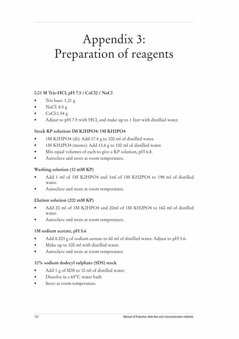
Manual of Rotavirus detection and characterization methods132
0.01 M Tris-HCl, pH 7.5 / CaCl2 / NaCl
Tris base: 1.21 g •NaCl: 8.5 g •CaCl:1.54 g •Adjust to pH 7.5 with HCl, and make up to 1 liter with distilled water.•
Stock KP solution: IM K2HPO4: 1M KH2PO4
1M K2HPO4 (di): Add 17.4 g to 100 ml of distilled water.•1M KH2PO4 (mono): Add 13.6 g to 100 ml of distilled water.•Mix equal volumes of each to give a KP solution, pH 6.8. •Autoclave and store at room temperature.•
Washing solution (10 mM KP)
Add 1 ml of 1M K2HPO4 and 1ml of 1M KH2PO4 to 198 ml of distilled •water. Autoclave and store at room temperature.•
Elution solution (200 mM KP)
Add 20 ml of 1M K2HPO4 and 20ml of 1M KH2PO4 to 160 ml of distilled •water. Autoclave and store at room temperature.•
1M sodium acetate, pH 5.6
Add 8.203 g of sodium acetate to 60 ml of distilled water. Adjust to pH 5.6.•Make up to 100 ml with distilled water. •Autoclave and store at room temperature•
10% sodium dodecyl sulphate (SDS) stock
Add 1 g of SDS to 10 ml of distilled water.•Dissolve in a 65• °C water bath.Store at room temperature.•
Appendix 3: Preparation of reagents

133WHO/IVB/08.17
1M sodium acetate (NaAc) containing 1% SDS, pH 5.0
Add 8.2 g of sodium acetate to 60 ml of distilled water. Add 10 ml of 10% SDS •stock, and mix.Adjust the pH to 5.0 with glacial acetic acid, and make up to 100 ml with distilled •water. Before use, heat the solution to 42oC if precipitate is present.•
3M sodium acetate (NaAc), pH 5.0
Add 4.92 g of sodium acetate to 10 ml of distilled water.•Make up to 20 ml with distilled water.•
Phenol-chloroform (1:1)
Mix equal volumes of saturated phenol (pH 4.3) and chloroform.•Place in a dark or foil-covered bottle (solution is light-sensitive), and store at •4°C.
Phenol:chloroform (using phenol crystals)
Dissolve 14.74 g of phenol in 10 ml of chloroform.•
7M guanidine isothiocyanate (GITC)
Add 7.09 g of guanidine isothiocyanate to 4 ml of distilled water.•Make up to a final volume of 10 ml.•Heat in a 56°C water bath to dissolve crystals.•
Caution: GITC emits cyanide gas on contact with acids.
2 x polyethylene glycol (PEG 6000)
Combine 16 g of PEG 6000 and 4.86 g of 0.8M NaCl.•Dissolve and make up to 100 ml with distilled water.•
10 x TE buffer, pH 7.4
Combine 1.21 g of Tris and 0.372 g of EDTA. •Dissolve in 80 ml of distilled water. Adjust the pH to 7.4 with 1N HCl.•Make up to 100 ml with distilled water.•
1 x TE buffer, pH 7.4
Mix 10 ml of 10 x TE buffer with 90 ml of distilled water.•
10% CTAB
Dissolve 10 g of CTAB in 80 ml of distilled water.•Make up to 100 ml with distilled water.•

Manual of Rotavirus detection and characterization methods134
4M NaCl
Dissolve 11.7 g of NaCl in 30 ml of distilled water.•Make up to 50 ml with distilled water.•
2M sodium acetate (NaAc)
Dissolve 8.2 g of NaAc in 30 ml of distilled water.•Make up to 50 ml with distilled water.•
Agarose gel: sample loading buffers
Americas buffer
4 g sucrose •2.5 mg bromophenol blue •6 ml TE buffer (10 mM Tris-HCl, pH 8.0/1 mM EDTA)•
Asian buffer
5 g sucrose •4 ml 0.5M EDTA, pH 8.0 •100 µl 10% bromophenol blue •Make up to 10 ml with distilled water.•
European buffer
10% Ficoll •0.25% Orange G •Make up in TE buffer.•
African buffer
0.001 g bromophenol blue•4 g sucrose •9 ml TAE buffer•1 ml distilled water•
Ethidium bromide (EtBr) stock (10 mg/ml)
1 EtBr tablet (100 mg)•10 ml distilled water •
Caution: EtBr is a powerful mutagen and is moderately toxic. Always wear gloves when working with solutions or gels containing this dye.
10 x PCR buffer
0.1 M Tris-HCl, pH 8.3•0.5 M KCl•

135WHO/IVB/08.17
5 x Tris-borate EDTA buffer (TAE buffer), pH 7.8
54 g Tris base•27.5 g boric acid •20 ml 0.5M EDTA, pH 8.0•800 ml distilled water•
0.5M EDTA, pH 8.0
18.61 g EDTA.2H2O•70 ml distilled water•Adjust pH with NaOH and make up to 100 ml with distilled water.•
1M Tris
12.11 g Tris base •80 ml distilled water•Adjust pH with concentrated HCl and make up to 100 ml with distilled water. •
pH Volume concentrated HCl
7.4 7.0 ml7.6 6.0 ml8.0 4.2 ml
Caution: Wear visor and globes when handling concentrated acids.
L6 nucleic acid extraction buffer
60 g guanidinium isothiocyanate •50 ml 0.1M Tris-HCL, pH 6.4•11 ml 0.2M EDTA, pH 8.0•1.3 g Triton X-100 •Store at room temperature in a dark bottle or cover with foil.•
Caution: Guanidinium isothyocyanate emits cyanide gas on contact with acids.
L2 nucleic acid extraction buffer
180 g guanidinium isothiocyanate •150 ml 0.1M Tris-HCl, pH 6.4 •Store at room temperature in a dark bottle or cover with foil.•
70% ethanol
70 ml ethanol•30 ml molecular-biology-grade distilled water •

Manual of Rotavirus detection and characterization methods136
Random primers (hexamers)
Reconstitute in 2.5 ml of RNase-free water.•Use 1 ul per 50-ul reaction volume.•
Size-fractionated silica
Add 60 g of silicon dioxide, SiO2 (Sigma; S-5631), to distilled water to a total •volume of 500 ml in a glass measuring cylinder.Allow the silica to sediment under gravity for 24 h at room temperature.•Extract 430 ml of supernatant, and add demineralised water to 500 ml. •Shake vigorously.Sediment for 24 h at room temperature.•Extract 440 ml of supernatant, and add 600 ul of concentrated HCl (32%, w/v) •to adjust the silica suspension to pH 2.0.
Caution: Wear visor, apron and gloves when handling concentrated acids.Aliquot the silica suspension in 4-ml volumes in glass bijoux, and sterilize by •autoclaving.Store at –20°C.•
RNase-free deionised water
Add 100 ul of diethylpyrocarbonate (DEPC; Sigma: D-5758) to 100 ml of cell-•culture-grade distilled water. Caution: Perform the addition in a fume cupboard.•Incubate for >12 h at 37°C.•Autoclave for 15 min at 15 lb/sq. in. at 121°C.•
1M sodium chloride (NaCl)
To 5.844 g of NaCl, add distilled water to give a final volume of 100 ml.•
1M potassium chloride (KCl)
To 3.728 g of KCl, add distilled water to give a final volume of 50 ml.•
2M glucose
To 36.032 g of glucose, add distilled water to give a final volume of 100 ml.•Sterilize by filtration.•
2M magnesium stock
20.33 g magnesium chloride (MgCl2.6H2O)•24.65 g magnesium sulphate (MgSO4.7H2O)•Add distilled water to give a final volume of 100 ml. •Sterilize by filtration•

137WHO/IVB/08.17
Ampicillin (50 mg/ml)
To 0.5 g of ampicillin, add distilled water to give a final volume of 10ml. Aliquot •and store at –20°C.
0.1M IPTG
To 1.2 g of IPTG, add distilled water to give a final volume of 50ml. Sterilize by •filtration.
Luria-Bertani (LB) broth
10 g Tryptone•5 g yeast extract•10 g NaCl •
Dissolve in distilled water and adjust pH to 7.0 with NaOH. Make up to a final volume of 1,000 ml with distilled water. Sterilize by autoclaving.
SOC medium, pH 7.0
2 g Tryptone•0.5 g yeast extract •1 ml 1M NaCl •250 ul 1M KCl •
Dissolve in 97 ml of distilled water, autoclave, cool to room temperature, and add:
1 ml 2M magnesium•1 ml 2M glucose •
Sterilize by filtration. Check that pH is 7.0.
1.5% LB-agar plates with ampicillin, IPTG, and X-Gal
Add 1.5 g of agar to 100 ml of LB broth. Autoclave to sterilize.•Allow to cool to 50oC. •Add 0.2 ml of ampicillin stock, 0.5 ml of 0.1M, and 0.16 ml of 50 mg/ml •X-Gal.Mix before pouring plates. and allow to set for at least 30 min•Store upside down at 4oC.•
Note: Alternatively, spread 100 ul of 0.1m IPTG and 20 ul of 50 mg/ml of X-Gal over the surface of an LB/Amp plate. Incubate the plates at 37°C for 30 min before use.
2 x proteinase K buffer
20 ml 1M Tris-HCl, pH 7.5 •5 ml 0.5M EDTA •1.75 g NaCl•20 ml 10% SDS •
Make up to 100 ml with distilled water. Store at room temperature.

Manual of Rotavirus detection and characterization methods138
United States
Material Manufacturer/Address Catalog No.
Guanidine isothiocyanate Acros Organics (Fisher Scientific)600 Business Center DrivePittsburgh, PA 15205https://www1.fishersci.com Phone: 800-766-7000Fax: 800-926-1166
AC4111-1000
Phenol-chloroform:isoamyl alcohol
United States Biochemical Corp. (Amersham International)P.O. Box 22400Cleveland, OH 44122http://www4.amershambiosciences.comPhone: 800-321-5000Fax: 800-535-5072
US75831-400ml
RNAID matrix (silica powder) Bio 101, Inc.P.O. Box 2284La Jolla, CA 92038-2284Phone: 800-424-6101Fax: 760-598-0116
1001-204
RNAID kit wash buffer Bio101, Inc. (see above) 1007-203
PBS, pH 7.2 (GIBCO cell-culture product)10% SDS
Invitrogen Corporation1600 Faraday AvenueP.O. Box 6482Carlsbad, California 92008Phone: (760) 603-7200FAX: (760) 602-6500
20012-04315553-035
Ethanol, 100% Pharmco Products58 Vale RoadBrookfield, CTPhone: 203-740-3471Fax: 203-740-3481
64-17-5
Vertrel XF(1,1,1,2,3,4,4,5,5,5-decafluoropentane)
Miller-Stephenson Chemical Co., Inc. George Washington HighwayDanbury, CT 06810Phone:203-743-4447Fax: [email protected]://www.miller-Stephenson.com
MS-782
Appendix 4: Suppliers

139WHO/IVB/08.17
Material Manufacturer/Address Catalog No.
Phenol-chloroform (1:1) United States Biochemical Corp.P.O. Box 22400Cleveland, Ohio 44122Phone: 800-321-9322Fax: 800-535-0898
75831
NaCl J.T. Baker222 Red School LanePhillipsburg, NJ 08865Phone: (800) 582-2537 Fax: (908) 859-9318
3624-05
Super reverse transcriptase Molecular Genetics Resources621 Johnson Road, Suite 8Tampa, FL 33634Phone: 800-255-8142Fax: 813-881-1589
MG101
AmpliTaq DNA polymerase Applied Biosystems805 Lincoln Center DriveFoster City, CAPhone: 800-345-8224 or 1-650-638-5800Fax: 1 415 572 2743
N808-0160
100 mM deoxynucleotide triphosphates
Amersham Biosciences Corp P.O. Box 1327800 Centennial Ave.Piscataway, NJ 08855-1327Phone: 800-526-3593Fax: 800-329-3593
27-2035-01
Mineral oil Sigma-AldrichP.O. Box 14508St. Louis, MO 63178Phone: 800-325-3010Fax: 800-325-5052
M-3516
Nusieve GTG agarose Cambrex Bioscience191 Thomaston StreetRockland, ME 04841Phone: 800-341-1547Fax: 800-362-5552
50084
SeaPlaque agarose Cambrex Bioscience (see above) 50100
10 x TBE Invitrogen Corporation1600 Faraday AvenuePO Box 6482Carlsbad, CA 92008Phone: 760-603-7200FAX: 760-602-6500
15581-044
123-bp DNA ladder Invitrogen (see above) 15613-029
10 x PCR buffer, 25 mM MgCl2 buffer set
Applied Biosystems (see above) N808-0130

Manual of Rotavirus detection and characterization methods140
Europe
Material Manufacturer/Address Catalog No.
0.5M EDTA pH8 Sigma-Aldrich Co., LtdFancy RoadPooleDorset BH12 4QH UKPhone: 44(0)1201 733114
E7889
1 Kb DNA ladder Invitrogen, LtdPO Box 35, 3 Fountain DriveInchinnan Business ParkPaisley PA4 9RF UKPhone: 44(0)141 8146100
15615-016
100 bp DNA ladder Roche Diagnostics, LtdBell LaneLewesEast Sussex BN7 1LG UKPhone: 0808 100 9998
1-721-933
10X PCR buffer Invitrogen 18067-017
10X TAE Invitrogen 15558-026
10X TBE Invitrogen 15581-044
3M NaCl solution, pH 5.3 Sigma S7899
Acetone VWR InternationalMerck HousePooleDorset, UKPhone: 44(0)1202 660444
100033P
Ampicillin Sigma A-0166
Beckman buffer plates Beckman Coulter UK, LtdOakley CourtKingsmead Business ParkLondon RoadHigh WycombeBucks HP11 1JU UKPhone: 44(0) 1494 441181
609844
Beckman CEQ DTCS Quick Start kit
Beckman Coulter 608120
Beckman CEQ separation capillary array
Beckman Coulter 608087
Beckman CEQ separation gel 608010
Beckman Coulter 608010
Beckman CEQ sequencing separation buffer
Beckman Coulter 608012
Competent cells (TOP10F’) Invitrogen KNM4550-40
DEPC-treated water PromegaDelta HouseChilworth Science ParkSouthampton SO16 7NS UKPhone: 44(0)123 8076 0225
P1193

141WHO/IVB/08.17
Material Manufacturer/Address Catalog No.
Dimethyformamide Sigma D4551
dNTPs Invitrogen 10297-018
EDTA Sigma E5134
Ethidium bromide Invitrogen E3565
Expand HF Taq Polymerase Roche Molecular Biochemicals 1732650)
Ficoll Sigma F2637
Geneclean spin kits Anachem, Ltd.20 Charles StreetLutonBeds LU2 0EB UKPhone: 44(0)1582 456666
1101-600
Guanidinium isothiocyanate Sigma G6639
HCl VWR International 2964582X
IPTG Fisher Scientific UK, LtdBishop Meadow RoadLoughboroughLeicestershire LE11 5RG UKPhone: 44(0)1509 231166
BPE1260-10
L2 buffer Severn Biotech, LtdUnit 2, Park LaneKidderminsterWorcs DY11 6TJ UKPhone: 44(0)1562 825286
20-8200
L6 buffer Severn Biotech 20-8600
MgCl2 Invitrogen 18067-017
M-MLV Invitrogen 28025-013
Molecular-biology-grade agarose
Roche 1-388-991
Molecular-biology-Grade ethanol
VWR International 427385A
Molecular-biology-grade water Sigma W4502
NuSieve 3:1 agarose Cambrex Bioscience Wokingham, LtdAshville WayWokinghamBerkshire RG41 2PL UKPhone: 44(0)118 9795234
50094
Oligonucleotide primers Invitrogen
Orange G Sigma O-3756
Random hexamers Amersham 27-2166-01
RNasin Promega N2511
Sequencing plates Beckman Coulter 609801
Costar WWR Int. Ltd. UK 6551
Silicon dioxide Sigma S5631

Manual of Rotavirus detection and characterization methods142
Material Manufacturer/Address Catalog No.
Size-fractionated silica Severn Biotech 20-8000
SOC medium Invitrogen 15544-034
Taq polymerase Invitrogen 10342-053
TOPO TA cloning kit Invitrogen KNM4550-40
Triton X-100 Sigma T8787
Trizma/Tris Sigma T1507
Xgal Fisher Scientific BPE1615-1

143WHO/IVB/08.17
Appendix 5: Worksheets

Manual of Rotavirus detection and characterization methods144
Worksheet 1. RT-PCR amplification of rotavirus dsRNA
dsRNA: ____________________________________Date: ______________________Extraction method: _____________________________________________________
A. Denature (dsRNA)
____________ l dsRNA____________ l primer 1 _____ VP7 ______ VP4 _____ Other ___________________________ l primer 2 _____ VP7 ______ VP4 ______ Other _______________
7-10 µl total (Make up to 10µl with ddH2O)Denature for 5 min in boiling water bath. Chill immediately in an ice bath.
B. Reverse transcription (dsRNA)
1x reaction _______________ x reactions0.2 µl 10 mM dATP _______________ l 0.2 µl 10 mM dCTP _______________ l 0.2 µl 10 mM dGTP _______________ l0.2 µl 10 mM dTTP _______________ l 0.2 µl AMV RT _______________ l Roche (25 U/µl)2.0 µl AMV buffer _______________ l
Work on ice. Add 3.0 µl of RT-master mix to each tube, and mix with pipette.Quick spin. Incubate for 25-30 min at 42oC.Note: Check manufacturer’s concentration of AMV. Use 3-5 U of RT per reaction.
C. PCR amplification (cDNA)
1x reaction _______________ x reactions1.0 µl dATP _______________ µl 1.0 µl dCTP _______________ µl 1.0 µl dGTP _______________ µl 1.0 µl dTTP _______________ µl4.0 µl 10 x Taq buffer _______________ µl (Invitrogen)2.4 µl 25 mM MgCl2 _______________ µl 26.3 µl dH2O _______________ µl 0.3 µl Taq polymerase _______________ µl (5U/µl)
Add 37 µl of PCR-MM to each tube and 2 drops of mineral oil if needed. Keep on ice!
D. Amplification cycles
...........oC .......min
...........oC ........min ......... oC .......min ......... oC ........min x 30 cycles
...........oC

145WHO/IVB/08.17
Worksheet 2. Genotyping or re-amplification of cDNA
Samples: ____________________________________Date: ______________________ Primers: ________________________________ Operator ______________________
1x Reaction __________ xReactions __________ x Reactions0.5-6.0 µl cDNA __________ µl __________ µl1.0 µl 10 mM dATP __________ µl __________ µl1.0 µl 10 mM dCTP __________ µl __________ µl1.0 µl 10 mM dGTP __________ µl __________ µl1.0 µl 10 mM dTTP __________ µl __________ µl1.0 µl (10 pmol) Primer 1 __________ µl __________ µl1.0 µl (10 pmol) Primer 2 __________ µl __________ µl1.0 µl (10 pmol) Primer 3 __________ µl __________ µl1.0 µl (10 pmol) Primer 4 __________ µl __________ µl1.0 µl (10 pmol) Primer 5 __________ µl __________ µl1.0 µl (10 pmol) Primer 6 __________ µl __________ µl1.0 µl (10 pmol) Primer 7 __________ µl __________ µl1.0 µl (10 pmol) Primer 8 __________ µl __________ µl5.0 µl 10 x Taq buffer __________ µl __________ µl1.5 µl 50 MgCl2 __________ µl __________ µl0.3 µl Taq pol. (5 U/µl) __________ µl __________ µl
Add sufficient ddH2O to make up to 50µl
__________ µl __________ µl
Add 49.5 - 44 µl of PCR-MM to each tube and 2 drops of mineral oil if needed. Keep on ice!

Manual of Rotavirus detection and characterization methods146

The World Health Organization has provided technical support to its Member States in the field of vaccine-preventable diseases since 1975. The office carrying out this function at WHO headquarters is the Department of Immunization, Vaccines and Biologicals (IVB).
IVB’s mission is the achievement of a world in which all people at risk are protected against vaccine-preventable diseases. The Department covers a range of activities including research and development, standard-setting, vaccine regulation and quality, vaccine supply and immunization financing, and immunization system strengthening.
These activities are carried out by three technical units: the Initiative for Vaccine Research; the Quality, Safety and Standards team; and the Expanded Programme on Immunization.
The Initiative for Vaccine Research guides, facilitates and provides a vision for worldwide vaccine and immunization technology research and development efforts. It focuses on current and emerging diseases of global public health importance, including pandemic influenza. Its main activities cover: i ) research and development of key candidate vaccines; ii ) implementation research to promote evidence-based decision-making on the early introduction of new vaccines; and iii ) promotion of the development, evaluation and future availability of HIV, tuberculosis and malaria vaccines.
The Quality, Safety and Standards team focuses on supporting the use of vaccines, other biological products and immunization-related equipment that meet current inter-national norms and standards of quality and safety. Activities cover: i ) setting norms and standards and establishing reference preparation materials; ii ) ensuring the use of quality vaccines and immunization equipment through prequalification activities and strengthening national regulatory authorities; and iii ) monitoring, assessing and responding to immunization safety issues of global concern.
The Expanded Programme on Immunization focuses on maximizing access to high quality immunization services, accelerating disease control and linking to other health interventions that can be delivered during immunization contacts. Activities cover: i ) immunization systems strengthening, including expansion of immunization services beyond the infant age group; ii ) accelerated control of measles and maternal and neonatal tetanus; iii ) introduction of new and underutilized vaccines; iv ) vaccine supply and immunization financing; and v ) disease surveillance and immunization coverage monitoring for tracking global progress.
The Director’s Office directs the work of these units through oversight of immunization programme policy, planning, coordination and management. It also mobilizes resources and carries out communication, advocacy and media-related work.
Family and Community Health
World Health Organization
20, Avenue Appia
CH-1211 Geneva 27
Switzerland
E-mail: [email protected]
Web site: http://www.who.int/immunization/en/
Department of Immunization, Vaccines and Biologicals
Generic protocol for monitoring impact of rotavirus vacci-
nation on gastroen-teritis disease bur-
den and viral strains