Embed Size (px)
Citation preview

Endodontic management of traumatic dental injuries
Dr Réka FazekasDepartment of Conservative Dentistry
Semmelweis University
Based on Mahmoud Torabinejad, Richard E. Walton, ENDODONTICS: PRINCIPLES AND PRACTICE 4th edition

Epidemiology of traumatic dental injuries
The oral region • 1 % of the total body area
Injuries of the oral region• 5 % of all body injuries
17 % in preschool children
Incidence: 1-3 % Prevalence: 20 -30 % (permanent, primary dentition)• Most frequent during the first 10 years of life

Etiologic factors
• Falls
• Sports
• Hits by another person
• Traffic accidents
dr Fazekas
Vary with age groups:

dr Fazekas

History• chief complaint• present injury
• date and time• how the injury took place• any other injuries in the past• actual problems with the tooth/teeth -pain, mobility,
occlusal interference• medical historyTetanus prohylaxis depends on the circumstances of theinjury, and the tetanus immunisation status of the patient.
• deep, destructed, contaminated injuries• active / passive Tetanus immunisation
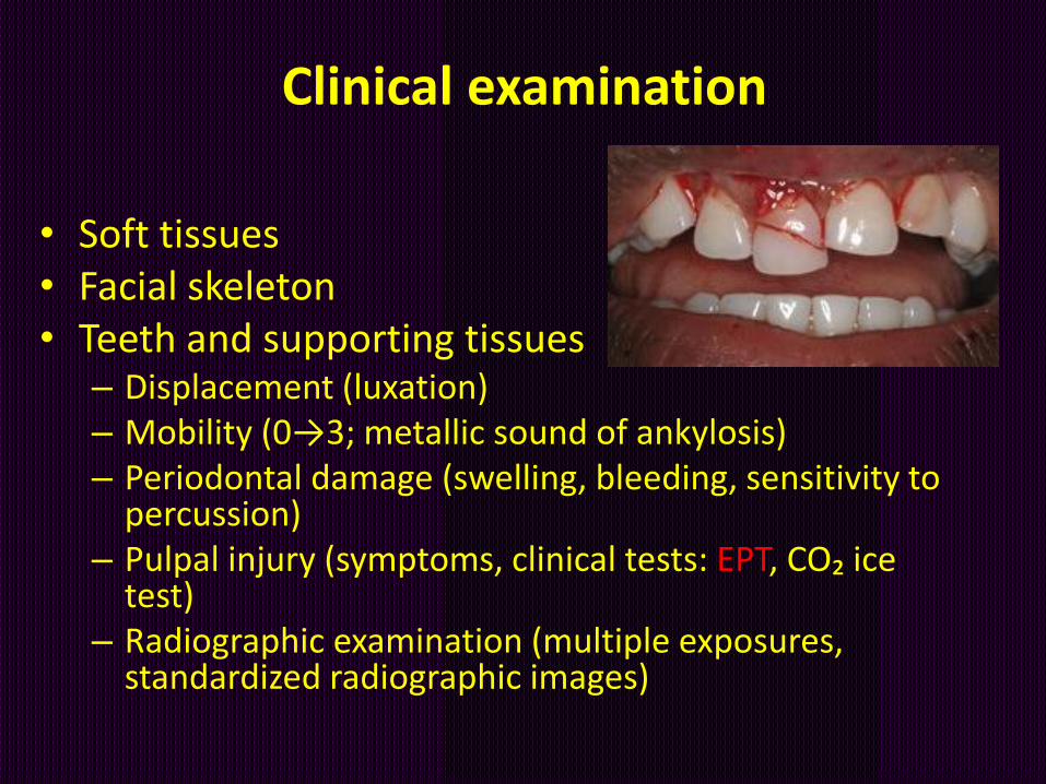
Clinical examination
• Soft tissues• Facial skeleton• Teeth and supporting tissues
– Displacement (luxation)– Mobility (0→3; metallic sound of ankylosis)– Periodontal damage (swelling, bleeding, sensitivity to
percussion)– Pulpal injury (symptoms, clinical tests: EPT, CO₂ ice
test)– Radiographic examination (multiple exposures,
standardized radiographic images)

Diagnosis
• Enamel fractures
• Crown fractures without pulp exposure
• Crown fractures with pulp exposure
• Crown-root fractures
• Root fractures
• Luxation injuries
• Avulsions
• Alveolar fractures

Classification of dental injuries
I. FracturesII. LuxationsIII. AvulsionIV. Alveolar fracture
dr Fazekas

Enamel fracture
• Involves the enamel only
• Includes enamel chipping
• Incomplete fractures
• Enamel cracks

Crown fracture without pulp exposure
• Uncomplicated crown fracture involving enamel and dentin with no pulp exposure

Crown fracture without pulp exposure
• Treatment: – Reattachment of separated enamel-dentin fragment
– Restoration with composite resin
– Indirect veneer (at a later date,
↑esthetics, function)
• Prognosis: good

Crown fracture without pulp exposure

Cusp fracture

Crown fracture with pulp exposure
Complicated fracture involving enamel and dentin and exposure of the pulp

Crown fracture with pulp exposure
• Extent of fracture– Vital pulp th. + acid-etched composite
restauration– Root canal treatment + post and core-supported
crown
• Stage of root development– Immature teeth: shallow (partial) pulpotomy
+ acid-etched composite restauration / reattaching the fractured segment
• Lenght of time since injury– Time ↑ prognosis ↓

Treatment of crown fractures with pulp exposure
• Vital pulp therapy
– Pulp cupping
– Pulpotomy: Cvek technique (shallow) / conventional technique (to or below the cervical level)
• Root canal therapy
– Accommodating to prosthetic requirements
168 hours2mm

Vital pulp therapy
Indication:
• immature teeth that can subsequently be restored with acid-etched composite
• mature teeth* that can subsequently be restored with acid-etched composite
* age ? compliance? date and circumstances of the accident?

Technique of shallow (Cvek) pulpotomy

Technique of shallow (Cvek) pulpotomy
1. Anesthesia2. Rubber dam isolation3. Exposed dentine is washed with saline or
sodium hypochlorite solution4. Extruding granulation tissue is removed with a
spoon excavator →determination of the size and location of the exposure
5. Pulp tissue is removed to a depth of about 2 mm below the exposure (shallow)-water cooled small round diamond in the high-speed handpiece

Technique of shallow (Cvek) pulpotomy
6. The wound is washed with sterile saline
7. Haemostasis can be expected within 5 minutes
8. The wound is washed again to remove the clot
9. The wound is dressed with calcium hydroxide or MTA cement
10. Cavity is sealed with a hard-setting glass ionomer cement (when using Ca(OH)2)
11. Acid-etched composite restauration

Calcium hydroxide versus MTA
Calcium hydroxide
• Desintegration with time
• After 6-12 month the tooth should be reentered
• Replace calcium hydroxide with a dentin bonding material
MTA
• Can be placed directly onto the pulp tissue
• Long curing time (-)
• Non-resorbable
• No need to reenter the tooth

Pulpotomy , apexogenesis

Vital pulp therapy
• Treatment evaluation: after 6 month, than yearly
• Criteria for successful shallow pulpotomy:
• Asymptomatic tooth, proper function• No radiographic evidence of apical periodontitis• No indication of root resorption• Tooth responds to pulp testing• Continued root development is evident
radiographically
Necrotic pulp or arrested root formation → apexification
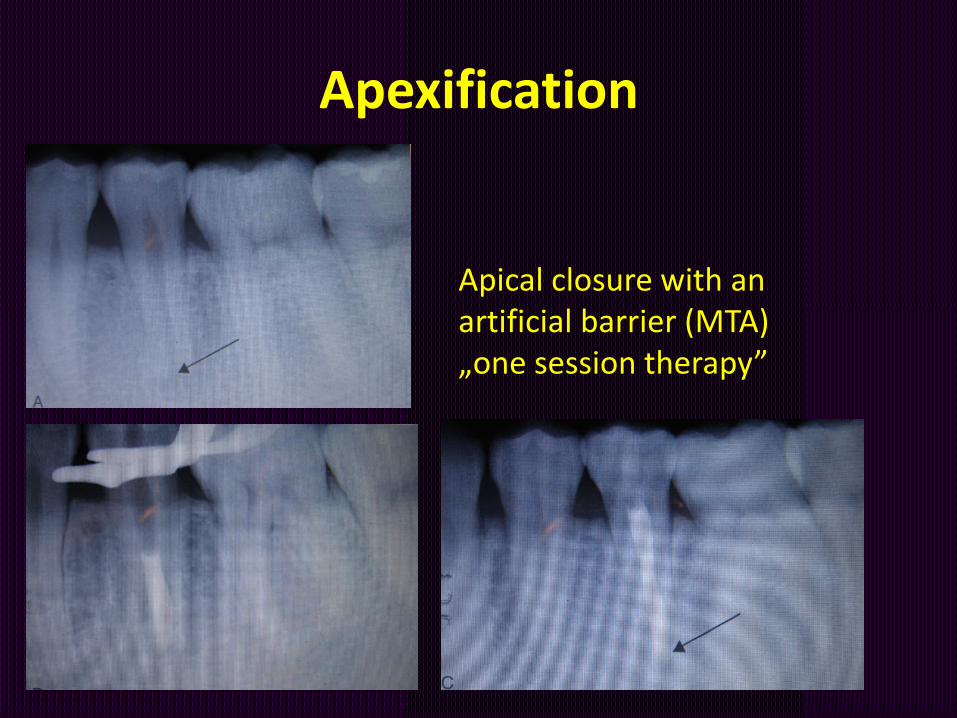
Apexification
Apical closure with an artificial barrier (MTA)„one session therapy”

irreversiblepulpitis
closed apicalforamen
vital pulp therapy
• pulpacapping• pulpotomy
apexogenesis
openedapical
foramen
vital pulp therapy(?!)or
RCT
apexification+
RCT
dr Fazekas
pulp explosure
pulpanecrosis
toothremains vital
pulpanecrose
irreversiblepulpitis pulpanecrosis
RCT

Crown-root fracture
• Includes enamel, dentin, and root cementum and may or may not include the pulp
– Chisel-type fracture: anterior teeth
– Shattered crown: pieces are held in place only by the part of a fractured segment still attached to the periodontal ligament
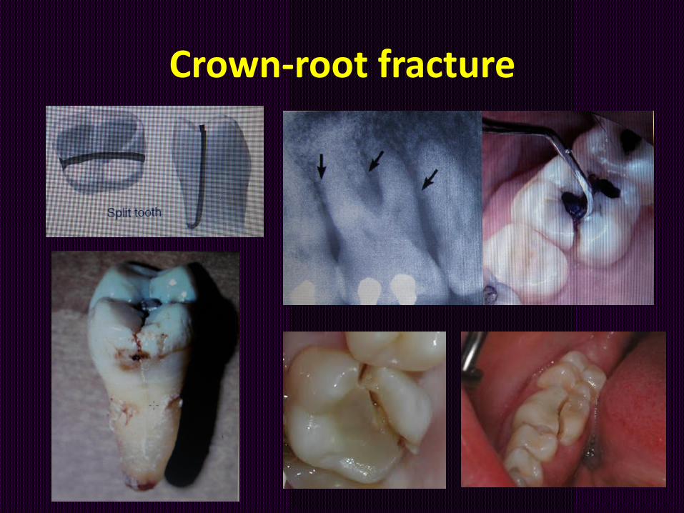
Crown-root fracture

Crown-root fracture
Examination:• Loose fragments • Additional radiographs
Remove all loose fragments ???
Emergency care:• Bonding loose tooth fragments, at least
temporary (immature teeth!)• Pulp therapy

Urgent care for crown-root fracture

Crown-root fracture
Treatment planning:
• Pulpotomy or pulpectomy
• Remaining tooth structure
• Level of the subgingival fracture: root extrusion, gingivoplasty, alveoplasty
• Extraction: bridge, implant, orthodontic treatment

Definitive treatment of crown-root fracture
Fragment removal
• gingivectomy (ostectomy), endodontic treatment and restoration with a post-retained crown
• Orthodontic extrusion of apical fragment
Decoronation (Root submergence)maintaining the volume of the alveolar process for later optimal implant installation
Extraction
• with immediate or delayed implant-retained crown restoration or a conventional bridge.

A technique for managing certain split teeth and cusp fractures

Root fracture
• Involve cementum, dentin and pulp
• Also referred to as
– intraalveolar
– horizontal
– transverse root fracture

Root fracture
Generally mild symptoms:
• Mobile or displaced tooth
• Pain on biting
X-ray: additional angle!
Emergency care:
• Repositioning
• stabilization: 4-6 weeks of splinting

Root fracture
Treatment:
• Cervical, middle third: splinting
• apical: observation
Sequelae of root fractures:
• Calcific metamorphosis (EPT!)
Root canal treatment: when pathosis is evident
Prognosis:
• ↑ heal spontaneously or after splint therapy

Stabilization of a root fracture

Luxation injuries
• Trauma to the supporting structures of teeth
• Often affect the neural and vascular supply to the pulp
• ↑ luxation, ↑ displacement, ↑damage

Luxation injuries
concussion
subluxation extrusive luxation lateral luxation intrusive luxation

Luxation injuries


Treatment of luxation injuries
• Concussio: no immediate treetment is necessery; allow thetooth to „rest” (avoid biting), pulpal status is monitored
• Subluxation: no treatment / stabilization for 1 to 2 weeks
• Extrusion and lateral luxation: repositioning and splinting, RCT in case of irreversibile pulpitis or pulp necrosis
• Intrusion:
– tooth with an open apex: it may reposition spontaneously
– fully developed tooth: active extrusion and RCT
Luxated teeth in which the pulps become necrotic are indicated forroot canal therapy.
dr Fazekas

Monitoring the pulpal status
• signs and symptoms
• pulp response to pulp testing (CO2 Ice test and EPT): retesting in 4 to 6 weeks
• radiographic examination : repeating in 4 to 6 weeks
• discoloration of the crown
• pink/ grayish/ yellow to brown
• may be reversed
dr Fazekas

Avulsion
Complete displacement of a tooth out of its socket
• Time out of socket
• Storage media

Treatment of avulsions
1. Someone may telephone for advice, presenting an opportunity for immediate replantation
2. The patient may be brought to the office with a tooth that has been out of the alveolus for less than 1 hour
3. The tooth has been out for more than 1 hour and not kept in a suitable storage medium

First aid for avulsed teeth
The prognosis is improved by replantation immediately after avulsion
• Rinse the tooth in cold, running tap water (10 seconds)
• Do not scrub the tooth• Replace the tooth in the socket using gentle
finger pressure• Hold the tooth in position• Seek dental care immediately

Suitable storage medium
• special storage solutions
• saline
• milk
• saliva
• water is inadequate!

Replantation within 1 hour of avulsion –tooth with a closed apex

Replantation more than 1 hour after avulsion –tooth with closed apex
• soak the tooth in a 2.4 % solution of sodium-fluoride for 5-20 minutes
• RCT
• Splint for 4 weeks

Sequelae to replantation
• Surface „repair-related” resorption– transient
• Inflammatory, „infection-related” resorption– loss of tooth structure
– loss of adjacent alveolar bone
– prevention: RCT

Sequelae to replantation
• Replacement „ankylosis-related” resorption
– Tooth structure is resorbed and replaced by bone
– Bone fuses directly to the root surface

Fracture of the alveolar process
• Initial, urgent need is splinting (surgeons)
• Examination, pulp testing, X-ray, follow up
• RCT if pulp necrosis
Fracture or comminution of the alveolar socket, or of the alveolar process

Take home messages
• traumatic dental injuries need a complex treatment
• every effort should make to keep alive an immature tooth
• lack of sensitivity byself is no evident sign of pulp necrosis
• a 5 year follow-up