Embed Size (px)
Citation preview
56International Journal of Scientific Study | December 2015 | Vol 3 | Issue 9
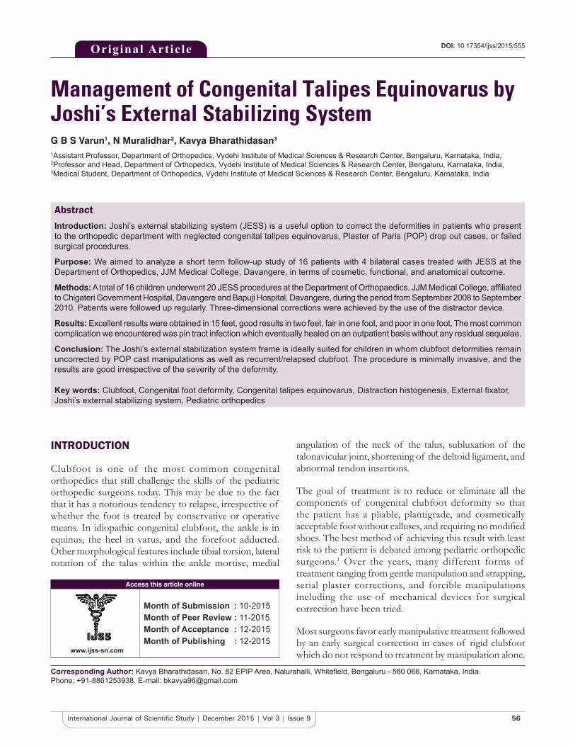
Management of Congenital Talipes Equinovarus by Joshi’s External Stabilizing SystemG B S Varun1, N Muralidhar2, Kavya Bharathidasan3
1Assistant Professor, Department of Orthopedics, Vydehi Institute of Medical Sciences & Research Center, Bengaluru, Karnataka, India, 2Professor and Head, Department of Orthopedics, Vydehi Institute of Medical Sciences & Research Center, Bengaluru, Karnataka, India, 3Medical Student, Department of Orthopedics, Vydehi Institute of Medical Sciences & Research Center, Bengaluru, Karnataka, India
angulation of the neck of the talus, subluxation of the talonavicular joint, shortening of the deltoid ligament, and abnormal tendon insertions.
The goal of treatment is to reduce or eliminate all the components of congenital clubfoot deformity so that the patient has a pliable, plantigrade, and cosmetically acceptable foot without calluses, and requiring no modified shoes. The best method of achieving this result with least risk to the patient is debated among pediatric orthopedic surgeons.1 Over the years, many different forms of treatment ranging from gentle manipulation and strapping, serial plaster corrections, and forcible manipulations including the use of mechanical devices for surgical correction have been tried.
Most surgeons favor early manipulative treatment followed by an early surgical correction in cases of rigid clubfoot which do not respond to treatment by manipulation alone.
INTRODUCTION
Clubfoot is one of the most common congenital orthopedics that still challenge the skills of the pediatric orthopedic surgeons today. This may be due to the fact that it has a notorious tendency to relapse, irrespective of whether the foot is treated by conservative or operative means. In idiopathic congenital clubfoot, the ankle is in equinus, the heel in varus, and the forefoot adducted. Other morphological features include tibial torsion, lateral rotation of the talus within the ankle mortise, medial
Original Article
AbstractIntroduction: Joshi’s external stabilizing system (JESS) is a useful option to correct the deformities in patients who present to the orthopedic department with neglected congenital talipes equinovarus, Plaster of Paris (POP) drop out cases, or failed surgical procedures.
Purpose: We aimed to analyze a short term follow-up study of 16 patients with 4 bilateral cases treated with JESS at the Department of Orthopedics, JJM Medical College, Davangere, in terms of cosmetic, functional, and anatomical outcome.
Methods: A total of 16 children underwent 20 JESS procedures at the Department of Orthopaedics, JJM Medical College, affiliated to Chigateri Government Hospital, Davangere and Bapuji Hospital, Davangere, during the period from September 2008 to September 2010. Patients were followed up regularly. Three-dimensional corrections were achieved by the use of the distractor device.
Results: Excellent results were obtained in 15 feet, good results in two feet, fair in one foot, and poor in one foot. The most common complication we encountered was pin tract infection which eventually healed on an outpatient basis without any residual sequelae.
Conclusion: The Joshi’s external stabilization system frame is ideally suited for children in whom clubfoot deformities remain uncorrected by POP cast manipulations as well as recurrent/relapsed clubfoot. The procedure is minimally invasive, and the results are good irrespective of the severity of the deformity.
Key words: Clubfoot, Congenital foot deformity, Congenital talipes equinovarus, Distraction histogenesis, External fixator, Joshi’s external stabilizing system, Pediatric orthopedics
Access this article online
www.ijss-sn.com
Month of Submission : 10-2015 Month of Peer Review : 11-2015 Month of Acceptance : 12-2015 Month of Publishing : 12-2015
Corresponding Author: Kavya Bharathidasan, No. 82 EPIP Area, Nalurahalli, Whitefield, Bengaluru - 560 066, Karnataka, India. Phone: +91-8861253938. E-mail: [email protected]
DOI: 10.17354/ijss/2015/555
Varun, et al.: Management of CTEV by JESS
57 International Journal of Scientific Study | December 2015 | Vol 3 | Issue 9
There has been much debate in the past as to whether a conservative or operative treatment was more effective in the treatment of clubfoot. Those feet which have had numerous manipulations and operations are stiff, deformed, and rigid due to scar tissue formation; thus, many patients are not suitable candidates for management by soft tissue release procedures.
Joshi et al. devised a simple controlled differential distraction system and stabilization in 1988. Joshi’s external stabilizing system (JESS) is a simple, versatile, and light fixator system with tremendous potential.2 It includes a bloodless, semi-invasive procedure that avoids fibrous tissue formation, further shortening (unlike bony procedures), post-operative complications, and scarring. JESS ensures proper control of all the components of correction by causing actual physiological lengthening and histogenesis of soft tissues thereby reducing the pressure on the growing epiphysis. However, meticulous post-correction care is crucial for success.
We aimed to assess the efficacy of controlled differential distraction as a method of treatment in idiopathic clubfoot (neglected, recurrent, and relapsed cases) and critically assess the results based on the clinical and radiological findings. Furthermore, we hoped to evaluate the various technical problems and complications of the JESS technique and suggest ways to overcome them.
MATERIALS AND METHODS
This study includes 20 congenital talipes equinovarus (CTEV) feet in 16 patients from the Department of Orthopedics, Bapuji Hospital and Chigateri Government General Hospital affiliated to JJM Medical College, Davangere, comprising 8 patients from each hospital. The study was conducted between September 2008 and September 2010. Out of the 16 patients, 7 patients were neglected cases, 3 patients were recurrent or relapsed cases, and 6 patients were plaster of Paris (POP) dropout cases of idiopathic clubfoot and were surgically treated by JESS fixator. The patients were between 1 and 3 years old, and those who were medically unfit for surgery were excluded from the study.
On admission of the patient, a thorough history was elicited from the parents/attendants to reveal the duration and previous treatment of the deformed foot. A careful marital history was elicited where four patients’ parents were found to have a history of second degree consanguinity. No other associated congenital abnormalities were detected.
Feet receiving a score of ≤7 by clinical examination by Carroll’s assessment were included in the study. The feet were radiologically evaluated, and the following values were calculated: Talo - calcaneal angle (in anteroposterior
[AP] and stress dorsiflexion views), talo - first metatarsal angle (in AP view), tibio - calcaneal angle (in lateral view), and talo - calcaneal index. Routine blood and urine investigations were performed regularly. Following approval of fitness for surgery, the patients in this study were operated under general anesthesia with the patient in supine position. No tourniquet was used in this procedure.
Insertion of K-Wires• Tibial: Two parallel transfixing wires were passed in
the tibia about 2.5 cm below and lateral to the tibial tuberosity, perpendicular to the longitudinal axis. The length of the middle segment of the Z’ bar was marked below the first wire. The second wire was passed parallel to the first wire at this level
• Metatarsal: One transfixing wire was passed from the fifth to first metatarsal at the level of the neck. 2 separate wires, one from the medial and the other from the lateral aspects were inserted parallel to the first wire. It was made sure that all the metatarsals had been impaled by at least one of the wires
• Calcaneal: Two transfixing parallel wires were passed into the tuber of the calcaneum from the medial side. The axial calcaneal wire was passed posterior to anterior just distal to the insertion of the Achilles tendon in the longitudinal axis of the calcaneum.
Attachment of “Z” and “L” Rods• Tibial attachment: The tibial wires were attached to
the middle segment of the “Z” rods by link joints on the medial and lateral aspects. One connecting rod was used to span the anterior limbs of “Z” rod and another to span the posterior limbs
• Metatarsal attachment: Two small “L” rods were attached to the metatarsal wires on the medial and lateral aspect of the foot
• Calcaneal attachment: Two large “L” rods were attached to the transfixing calcaneal wires on either side of the heel. Behind the foot, these rods were connected to each other by a connecting rod to which the axial calcaneal wire was clamped.
Connecting the Segmental Hold• Calcaneo-metatarsal connection: A pair of appropriately
sized distractors was attached to the calcaneal and metatarsal wires on either side of the foot
• Tibio-calcaneal connection: Posterior limbs of the “Z” rods were attached to “L” rods of the calcaneal hold by a distraction on either side. Distractors were attached near the transfixing pins
• Tibio-metatarsal connection: The anterior limbs of the “Z” rods were connected by a pair of rods to the small “L” rods anterior to the attachment of the metatarsal wires.
Varun, et al.: Management of CTEV by JESS
58International Journal of Scientific Study | December 2015 | Vol 3 | Issue 9
Connection of Anterior Stabilizing RodsTwo anterior connecting rods were connected on the medial and lateral aspects of the assembly from the transverse connecting rod of the superior limbs of the Z rods (proximally) to the metatarsal wires/inferior limbs of the metatarsal L rods (distally).
Sterile dressing was applied to the pin tract sites, and a foot plate was applied to prevent clawing of the toes. Distal pulsations (dorsalis pedis and posterior tibial arteries) were checked manually using a pulse oximeter. Capillary filling time was noted. The patient was shifted to the post-operative ward, monitored for a day, and then shifted to the wards. The dressings were changed on alternate days during the hospital stay for a week with spirit and betadine lotion. Pin sites were covered with dry gauze, and the patients were advised to report immediately if there was any discharge from the pin tracts.
On the 3rd post-operative day, differential fractional calcaneo-metatarsal distraction on the medial side was started at twice the rate than that on the lateral side (medial - 0.25 mm every 6 h; lateral - 0.25 mm every 12 h). The tibio-calcaneal distraction was carried out in two positions: (1) The distractors mounted between the inferior limbs of the “Z” rods and posterior limbs of the calcaneal “L” rods lying parallel to the leg and just posterior to the transfixing calcaneal wires (medial - 0.25 mm every 6 h; lateral - 0.25 mm every 12 h) and (2) the distractors shifted posteriorly and connected above to the transverse bar connecting the posterior limbs of “Z” rods and below to the posterior calcaneal bars connecting the posterior limbs of “L” rods and axial calcaneal pin (both - 0.25 mm every 6 h). The end point for distraction was assessed clinically and radiologically. The above explained distraction was very clearly demonstrated to the patient’s attender and supervised for 2 days. 7 days following the surgery, the patient was fit enough to be discharged and was advised for a regular follow-up at weekly intervals for 6 weeks to look for a progressive correction of the deformity, persistent edema, rule out pin tract infections, and tighten the loosened link joints.
Following the correction, the assembly was held in static position for a further 3-6 weeks to allow soft tissue maturation in the elongation position. Single stage removal of the whole assembly was done under general anesthesia and a well molded above-knee plaster cast was applied in maximum correction for 2 weeks. Once the pin tracts healed completely, a below knee cast was applied, and the patient was asked to ambulate with full weight bearing in the plaster. It was removed after 4 weeks.
Full correction of forefoot adduction, varus, and equinus was achieved, usually at the end of 6 weeks. X-ray of the
operated foot with ankle AP and stress dorsiflexion views were taken finally after the removal of the below knee plaster and talocalcaneal index calculated (>40°). For all patients, CTEV corrective shoes were advised for 5 years to maintain the correction and prevent recurrence. Using the Hospital for Joint Diseases Orthopedic Institute Functional Rating System for clubfoot (Lehman; Atar et al.) and Carroll’s assessment, the results were classified as excellent 85-100, good 70-84, fair 60-69, and poor <60 (out of a total score of 100) at follow-up intervals of 3, 6, and 9 months. The parents care and compliance played an important role in the success of this procedure.
The parents care and compliance played an important role in the success of this procedure. (Figures 1-8)
RESULTS
The age of these patients ranged from 1 to 3 years with an average of 1.9 years. Out of 20 feet, 14 feet (70%) were male and 6 feet (30%) were female patients. There were 12 feet (60%) unilateral and 8 feet (40%) bilateral cases. There were 8 feet (40%) belonging to neglected cases, 8 feet (40%) to POP dropout cases, and 4 feet (20%) to relapsed/recurrent cases (Table 1). Out of 20 feet, 12 feet (60%) underwent the previous procedure in the form of manipulation and serial casting and for the remaining 8 feet (40%), no treatment was given. In this study of 20 feet treated by JESS, there were 4 feet (20%) pin tract infections, 1 foot (5%) skin necrosis, 1 foot (5%) persistent edema, 1 foot (5%) flexion contractures of toes, and 1 foot (5%) loosening of the pin (Graph 1). 15 feet (75%) were excellent, 2 feet (10%) were good, 2 feet (10%) were fair, and 1 foot (5%) was poor as graded by the Hospital for Joint Diseases Orthopedic Institute Functional Rating System for clubfoot (Graph 2). Radiological assessment was done using talocalcaneal index and it was compared with other case series showing good radiological correction (Table 2).
DISCUSSION
External fixators are a versatile method of correcting complex three-dimensional deformities of the foot such as clubfoot. The basic principle of external fixation (JESS) in this study was the same as advocated by
Table 1: Distribution of casesType of clubfoot Number of cases Number of feet %Neglected 7 8 40POP drop out 6 8 40Recurrent/relapsed 3 4 20Total 16 20 100POP: Plaster of paris
Varun, et al.: Management of CTEV by JESS
59 International Journal of Scientific Study | December 2015 | Vol 3 | Issue 9
In this study, excellent results were obtained due to the fact that except for a few cases which had superficial pin tract infection, no other complications occurred. Of the two cases with scores between 84 and 70, flexion contracture of toes was noted in one case, and forefoot adduction persisted due to the decrease in the rate of metatarso-calcaneal distraction. However, it was treated with physiotherapy and corrective shoes. By 6 months, the flexion contracture was corrected and pain free. Fair results were because of skin necrosis in one foot and persistent edema in another, which lead to temporary cessation of correction for a week with gradual and supervised distraction. In one foot, the results were poor due to the loosening of the axial calcaneal pin due to improper hold in the calcaneum. Hence, the pin was removed, and the scoring was <60 due to persistant equinus and varus deformities at the end of correction phase.
Post-operative assessment yielded results that were comparable to those of other external fixator systems of
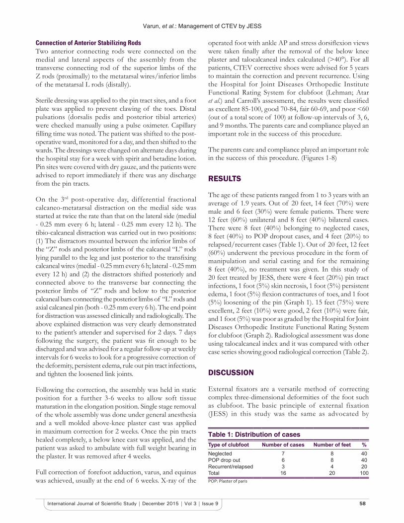
Figure 1: Unilateral neglected pre-operated foot (Case 1)
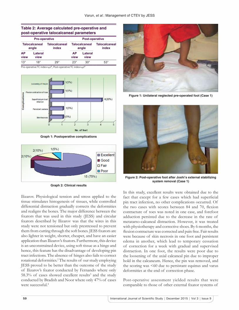
Figure 2: Post-operative foot after Joshi’s external stabilizing system removal (Case 1)
Graph 1: Postoperative complications
Graph 2: Clinical results
Table 2: Average calculated pre-operative and post-operative talocalcaneal parameters
Pre-operative Post-operativeTalocalcaneal
angleTalocalcaneal
indexTalocalcaneal
angleTalocalcaneal
indexAP view
Lateral view
AP view
Lateral view
13° 18° 29° 23° 30° 53°Pre‑operative TC index<40°, Post‑operative TC index>40°
Ilizarov. Physiological tension and stress applied to the tissue stimulates histogenesis of tissues, while controlled differential distraction gradually corrects the deformities and realigns the bones. The major difference between the fixators that was used in this study (JESS) and circular fixators described by Ilizarov was that the wires in this study were not tensioned but only prestressed to prevent them from cutting through the soft bones. JESS fixators are also lighter in weight, shorter, cheaper, and have an easier application than Ilizarov’s fixators. Furthermore, this device is an unconstrained device, using soft tissue as a hinge and hence, this feature has the disadvantage of developing pin tract infections. The absence of hinges also fails to correct rotational deformities.2 The results of our study employing JESS proved to be better than the outcome of the study of Ilizarov’s fixator conducted by Fernando where only 58.3% of cases showed excellent results3 and the study conducted by Bradish and Noor where only 47% of cases were successful.4
Varun, et al.: Management of CTEV by JESS
60International Journal of Scientific Study | December 2015 | Vol 3 | Issue 9
Oganesian and Istomina (75.7% good results).5 Our study seemed to show better results than that of Anwar and Arun (59.7% excellent and good results)6 and Shrivastava et al. (40% excellent results).7 In the study by Suresh et al. of 44 feet treated by JESS, there were 77% excellent, 13% good, 0% fair, and 9% poor results.8 Their results may have been better because of the younger study population. A recent study by Manjappa shows 14 satisfactory corrected feet out 15 CTEV cases operated by JESS as per Simon’s Criteria.9
Eight cases were presented with complications in this study. Out of 20 feet, four feet with superficial pin tract
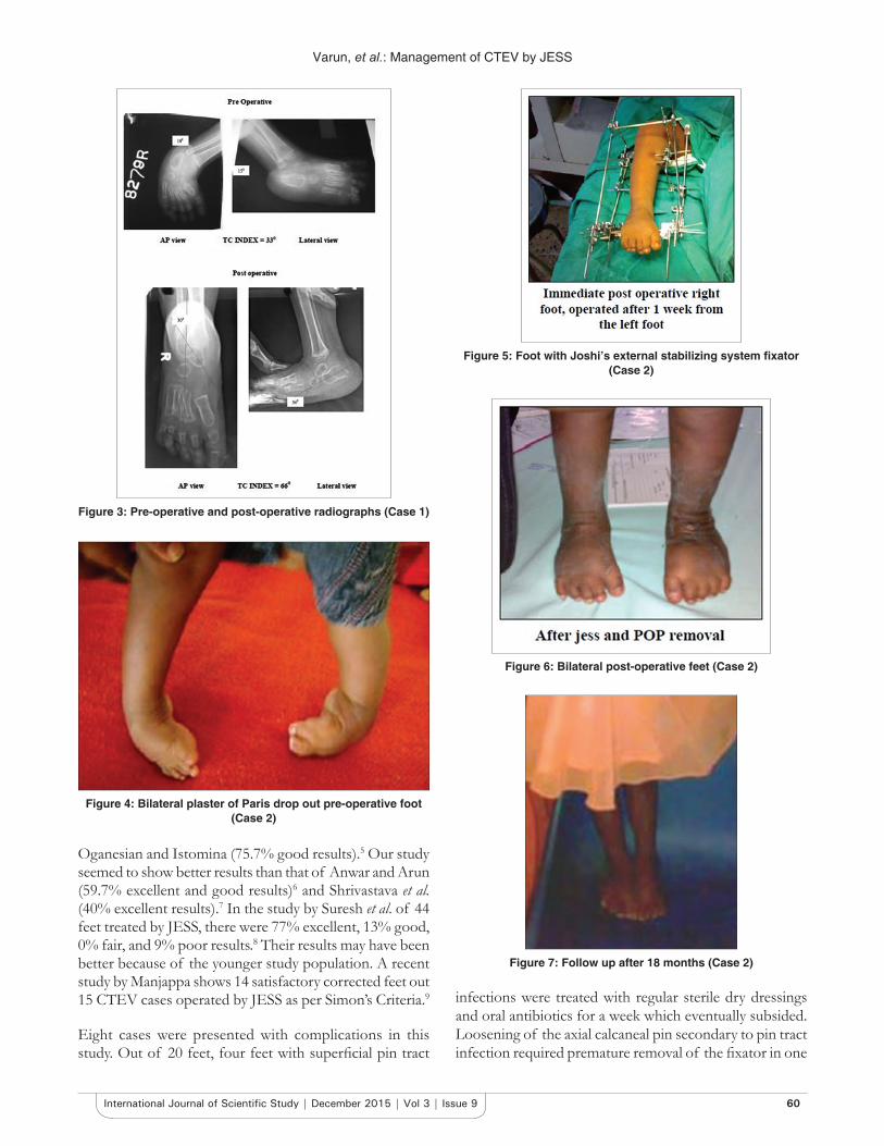
Figure 3: Pre-operative and post-operative radiographs (Case 1)
Figure 4: Bilateral plaster of Paris drop out pre-operative foot (Case 2)
Figure 5: Foot with Joshi’s external stabilizing system fixator (Case 2)
Figure 6: Bilateral post-operative feet (Case 2)
Figure 7: Follow up after 18 months (Case 2)
infections were treated with regular sterile dry dressings and oral antibiotics for a week which eventually subsided. Loosening of the axial calcaneal pin secondary to pin tract infection required premature removal of the fixator in one
Varun, et al.: Management of CTEV by JESS
61 International Journal of Scientific Study | December 2015 | Vol 3 | Issue 9
foot which lead to poor results. One foot with flexion contracture of toes may have occurred due to relative inelasticity of the flexor tendons. Skin necrosis in one foot was attributed to the rapid rate of correction of deformities. In this case, the distraction was stopped and reversed until tension relieved. The distraction was continued after a few days under supervision. In one foot, persistent edema was observed due to the same cause mentioned above and was treated similarly with an elevation of the limb and anti-edema measures. Similarly, in the studies by Suresh et al. and Anwar and Arun, the predominant complication was pin tract infections.6,8 Manjappa, however, reported significant edema as the leading complication and only one case of pin-tract infection.9
CONCLUSION
The goal of any clubfoot surgery is to obtain a cosmetically acceptable, pliable, functional, painless, and plantigrade foot, and to spare the parent and the child from the ordeal of frequent hospitalization and years of treatment with casts and braces. The best treatment for clubfoot that does not respond to conventional treatment remains controversial. The procedure used in the current study holds promise for fulfilling the above-mentioned goals. This procedure is ideally suited for children in whom the clubfoot deformities remain uncorrected by POP casts and manipulation, as well as for recurrent clubfoot. If performed at round 9 months of age, the procedure enables the child to walk with a plantigrade foot by the time he or she reaches the walking age group.10
Functional distraction using JESS apparatus is an easy method, which does not require any sophisticated instrumentation and minimal image intensifier. Parents learn the distraction technique easily and comply with the procedure. Pin tracks should be cared meticulously. An adequate period of static phase is necessary before removal of the apparatus. Strict postoperative management and follow-up are mandatory.
Differential distraction technique gives good result in children, but results are excellent in younger children and those who have not undergone any previous operative procedure. All cases of CTEV are not amenable to this technique; only those cases which are neglected, recurrent, and POP drop out cases should be operated. In relatively mild and moderate varieties of clubfoot, probably traditional soft tissue surgery still holds good. Motivated and compliant parents were a pivotal factor on which the success of the study depended. Although the technique has many advantages, one should not forget that injudicious and unsupervised distraction may lead to catastrophic results in the small developing foot. Long-term studies (10 years) are required to accurately assess the functional outcome of treatment of clubfoot by JESS.
REFERENCES
1. Cummings J, Lovel WW. Current concept operative treatment of congenital idiopathic club foot. J Bone Joint Surg 1988;70-A:1108.
2. Joshi BB. Correction of congenital talipes equino varus (CTEV) by controlled differential fractional distraction using Joshi's external stabilization system (JESS). 1st edition. JESS Research and Development Centre, Mumbai, India, 2001:1-53.
3. De La Huerta F. Correction of neglected club foot by llizarov method. Clinical Orthopedics and Related Research. Vol. 201. Philadelphia: J. B. Lippincott Co.; 1994. p. 89-93.
4. Bradish CF, Noor S. The ilizarov method in the management of relapsed club feet. J Bone Joint Surg Br 2000;82:387-91.
5. Oganesian OV, Istomina IS. Talipes equinocavovarus deformities corrected with the aid of a hinged-distraction apparatus. Clin Orthop Relat Res 1991:42-50.
6. Marthya AH, Arun B. Short term results of results of correction of CTEV with JESS distractor. J Orthop 2004;1:e3.
7. Shrivatsava S, Das R, Shukla J, Shrivatsava N. Our experience with JESS in the management of CTEV. Indian J Orthop 2000;34:88-91.
8. Suresh S, Ahmed A, Sharma VK. Role of Joshi’s external stabilisation system fixator in the management of idiopathic clubfoot. J Orthop Surg (Hong Kong) 2003;11:194-201.
9. CN M. Joshi’s external stablization system (JESS) application for correction of resistant club-foot. Internet J Orthop Surg 2009;18:1.
10. McKay DW. New concept of and approach to clubfoot treatment: Section II – Correction of the clubfoot. J Pediatr Orthop 1983;3:10-21.
How to cite this article: Varun GBS, Muralidhar N, Bharathidasan K. Management of Congenital Talipes Equinovarus by Joshi’s External Stabilizing System. Int J Sci Stud 2015;3(9):56-61.
Source of Support: Nil, Conflict of Interest: None declared.
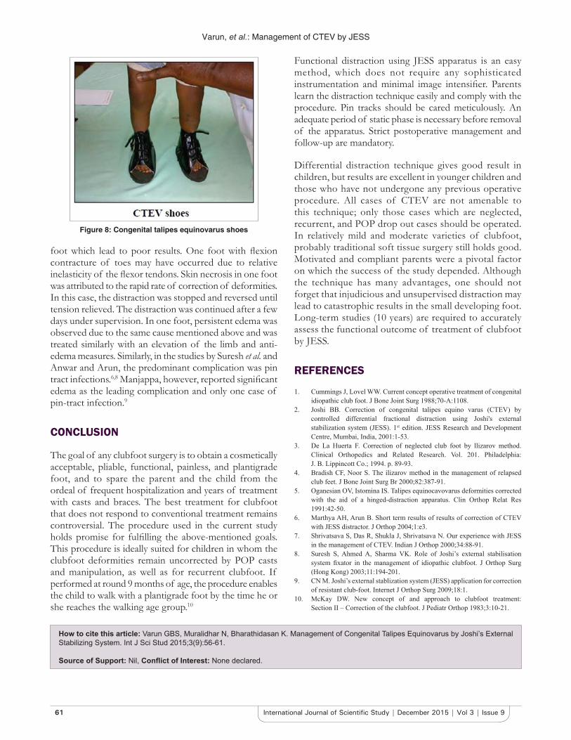
Figure 8: Congenital talipes equinovarus shoes