Embed Size (px)
Citation preview
ClubfootAHAMED MOHAIDEEN, M.D.
ORTHOPEDIC CENTER OF PALM BEACH COUNTY
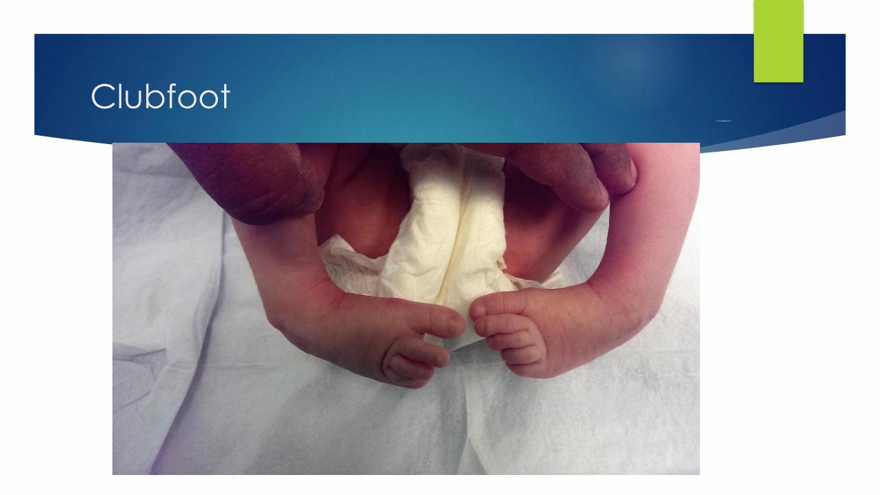
Clubfoot
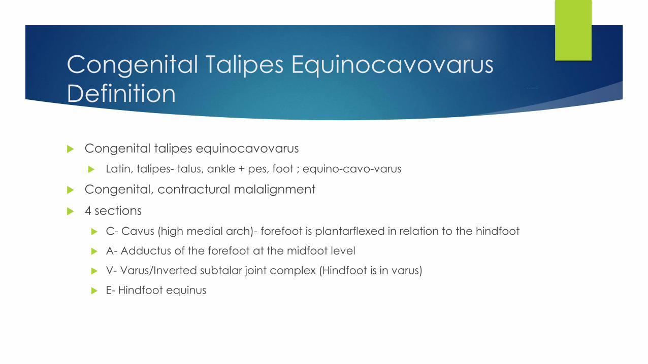
Congenital Talipes Equinocavovarus
Definition
Congenital talipes equinocavovarus
Latin, talipes- talus, ankle + pes, foot ; equino-cavo-varus
Congenital, contractural malalignment
4 sections
C- Cavus (high medial arch)- forefoot is plantarflexed in relation to the hindfoot
A- Adductus of the forefoot at the midfoot level
V- Varus/Inverted subtalar joint complex (Hindfoot is in varus)
E- Hindfoot equinus
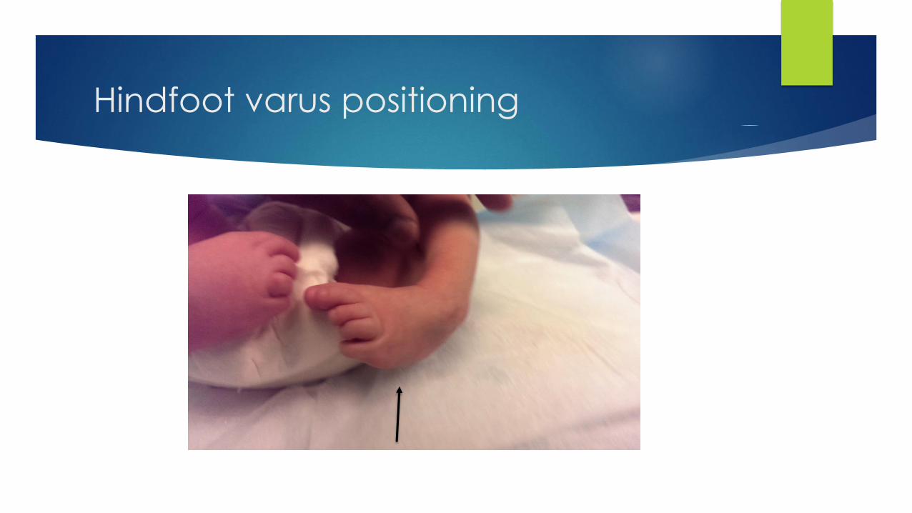
Hindfoot varus positioning
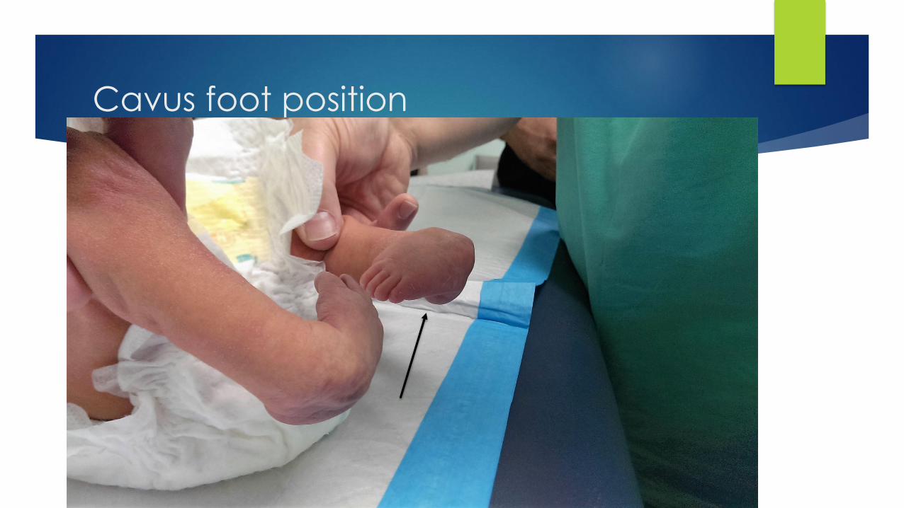
Cavus foot position
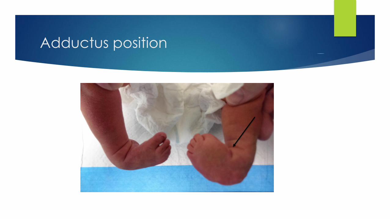
Adductus position

Hindfoot in varus position
Types
Idiopathic- normal child with clubfoot issues only
Postural- resolves spontaneously with no Tx or with 1 to 2 casts
Can probably watch and see natural progression
Neurogenic- related to myelomeningocele
Syndromic- clubfoot seen with other anomalies as well
Neurogenic and Syndromic are less likely to respond to non-operative
techniques or tend to recur
Syndromes associated with clubfoot
Arthrogryposis
Constriction Bands
Prune Belly
Tibial hemimelia
Mobius syndrome
Freeman-Sheldon Syndrome- Whistling face
Diastrophic dwarfism
Larsen Syndrome
Opitz Syndrome
Pierre Robin Syndrome
Epidemiology
1-2 per 1000 commonly quoted
Variable by population groups
Nordic (e.g., Sweden)- 0.93 to 1.5
Asian- 0.6 per 1000
Western Australia- 0.9
Polynesians, Hawaiians, Maori- 6.8
Family relations
What is the chance that it can happen in a family that already has a case of clubfoot?
Occurs 17 times higher in first-degree relatives
Sharing 50% of genetic material- parents, children, siblings
Occurs 6 times higher for second- degree relatives
Aunts, Uncles, grandparents, grandchildren, nieces, nephews, half-siblings
Occurs same incidence as general population for third degree relatives
Great-grandparents, Great grandchildren, Great uncles/aunts, First cousins
Wynn-Davies, JBJS (Br) 1964, Clin Orthop Relat Res 1972
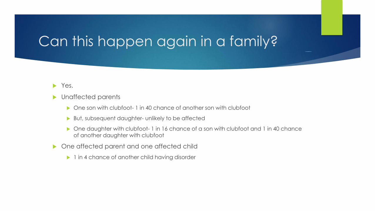
Can this happen again in a family?
Yes.
Unaffected parents
One son with clubfoot- 1 in 40 chance of another son with clubfoot
But, subsequent daughter- unlikely to be affected
One daughter with clubfoot- 1 in 16 chance of a son with clubfoot and 1 in 40 chance of another daughter with clubfoot
One affected parent and one affected child
1 in 4 chance of another child having disorder
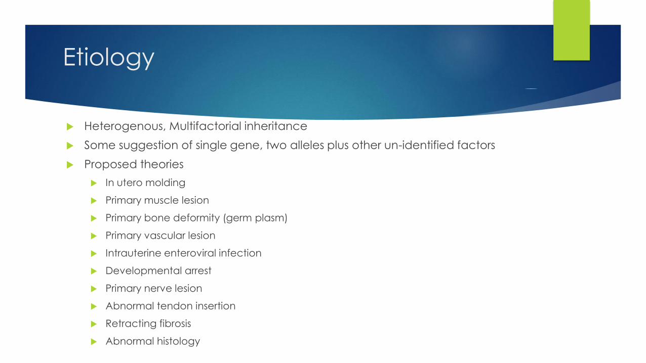
Etiology
Heterogenous, Multifactorial inheritance
Some suggestion of single gene, two alleles plus other un-identified factors
Proposed theories
In utero molding
Primary muscle lesion
Primary bone deformity (germ plasm)
Primary vascular lesion
Intrauterine enteroviral infection
Developmental arrest
Primary nerve lesion
Abnormal tendon insertion
Retracting fibrosis
Abnormal histology
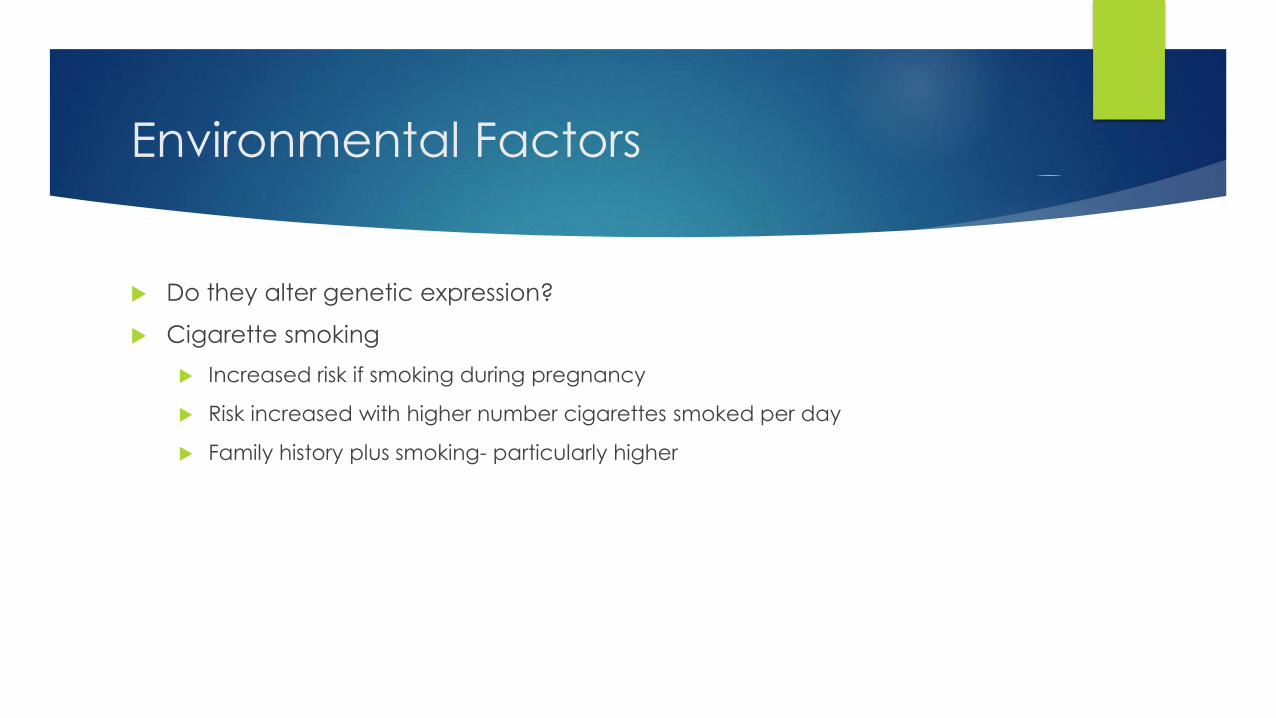
Environmental Factors
Do they alter genetic expression?
Cigarette smoking
Increased risk if smoking during pregnancy
Risk increased with higher number cigarettes smoked per day
Family history plus smoking- particularly higher
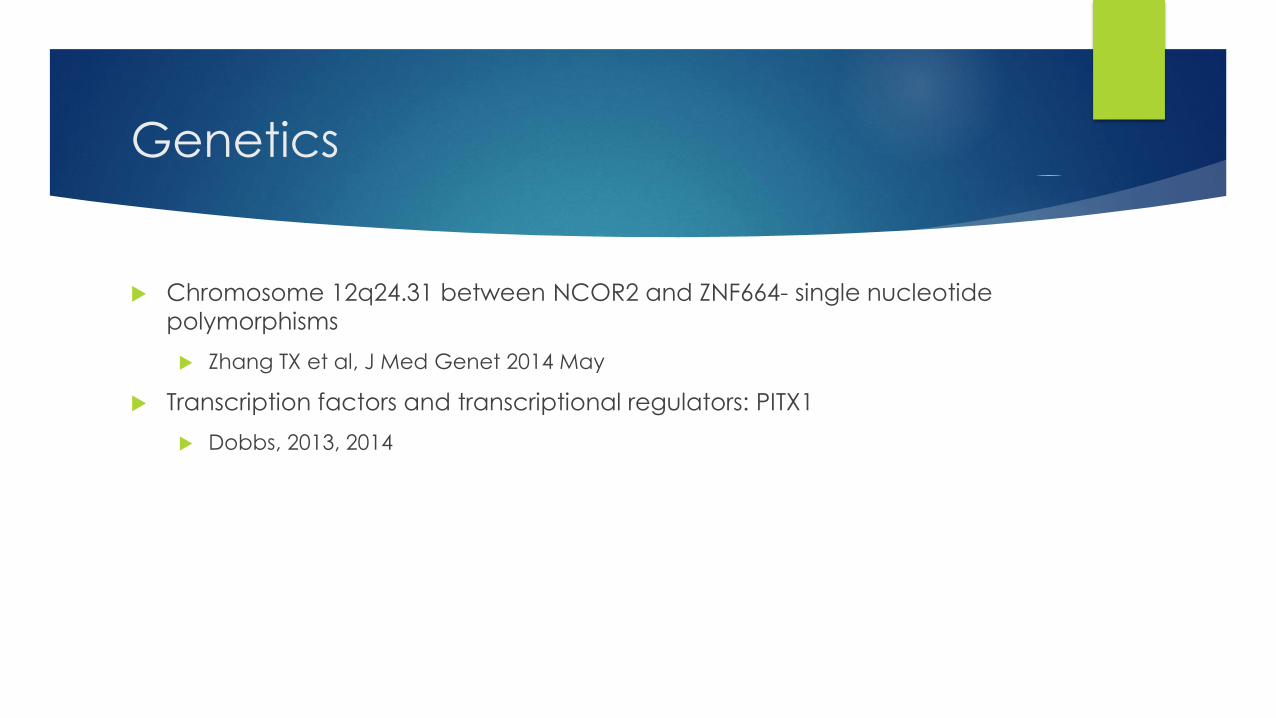
Genetics
Chromosome 12q24.31 between NCOR2 and ZNF664- single nucleotide
polymorphisms
Zhang TX et al, J Med Genet 2014 May
Transcription factors and transcriptional regulators: PITX1
Dobbs, 2013, 2014
Signs
Cavus
Adductus (Inversion of subtalar joint- between talus and calcaneus)
Varus
Equinus
Varying severity- flexible to rigid
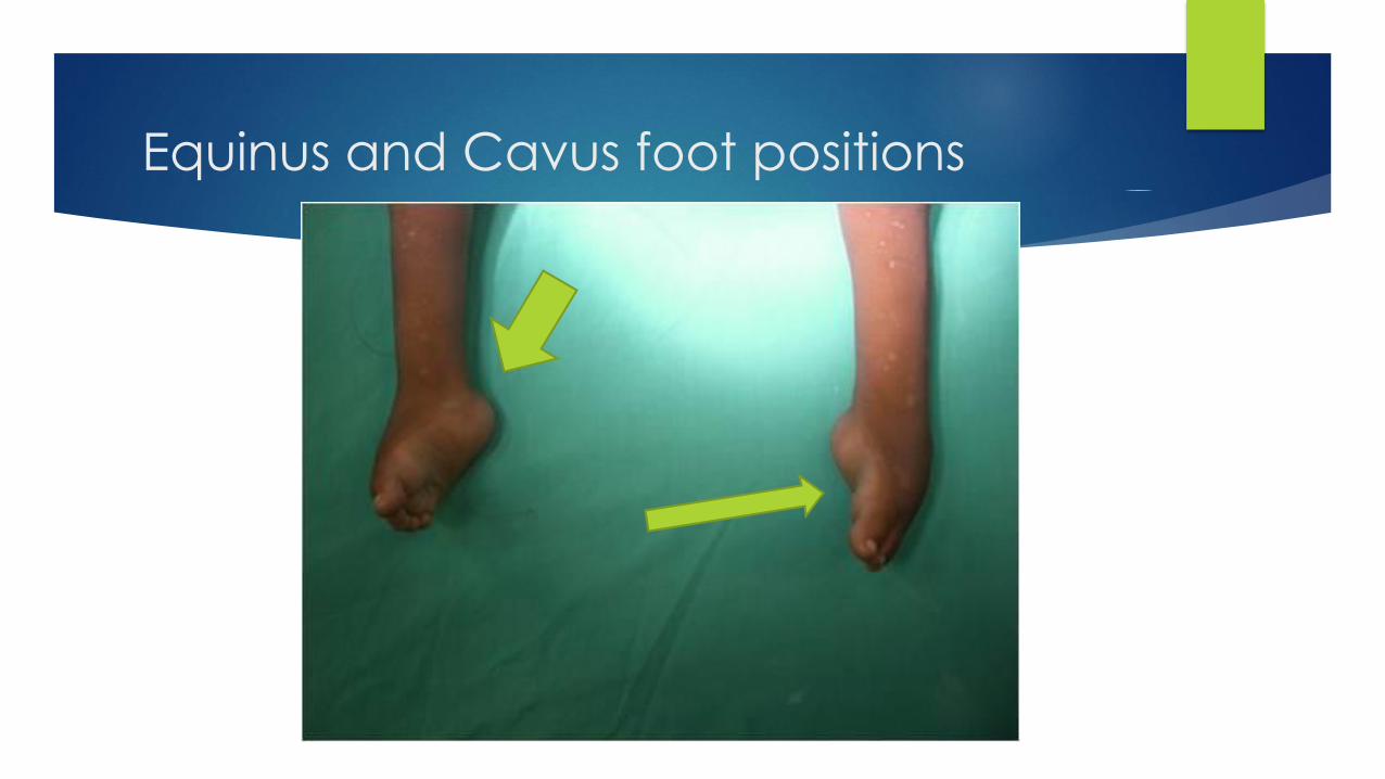
Equinus and Cavus foot positions
Signs (continued)
Single large (or Double) posterior ankle crease
Empty heel pad sign- cannot feel calcaneus
Transverse medial crease (midfoot)
Palpable head of talus- dorsolateral over midfoot region just distal to
ankle joint
Smaller foot and calf – “clubleg”
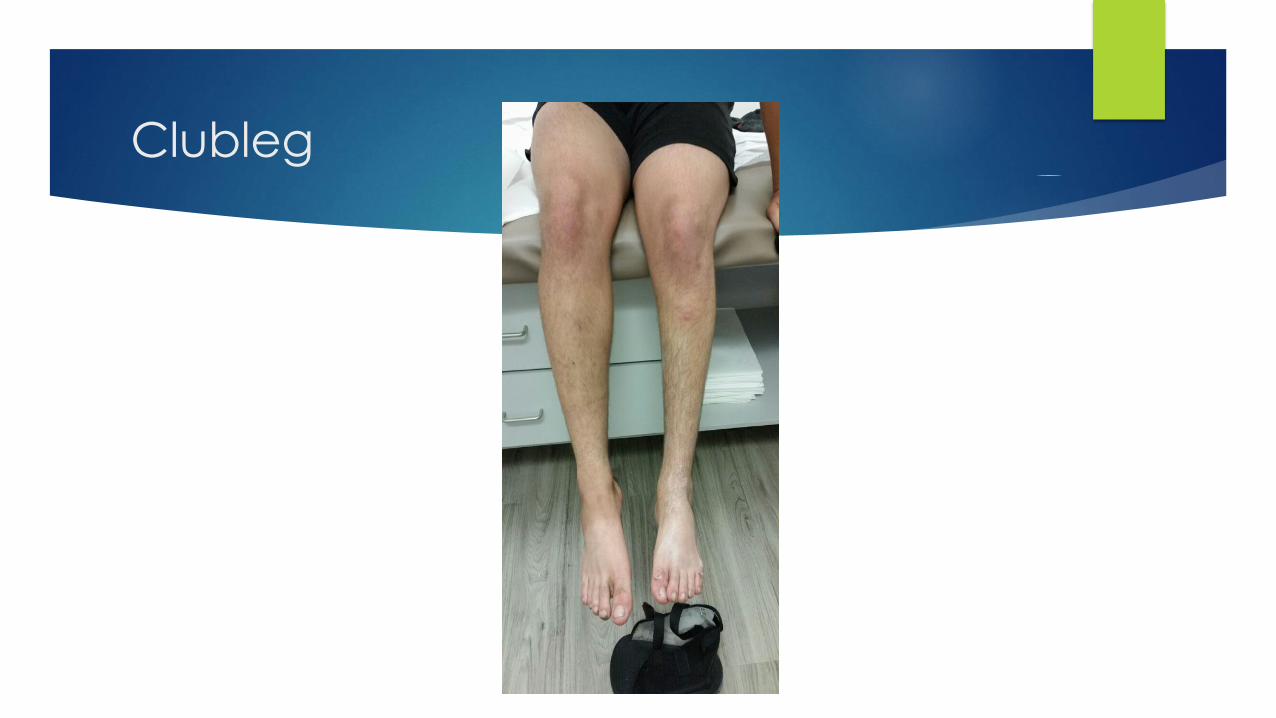
Clubleg
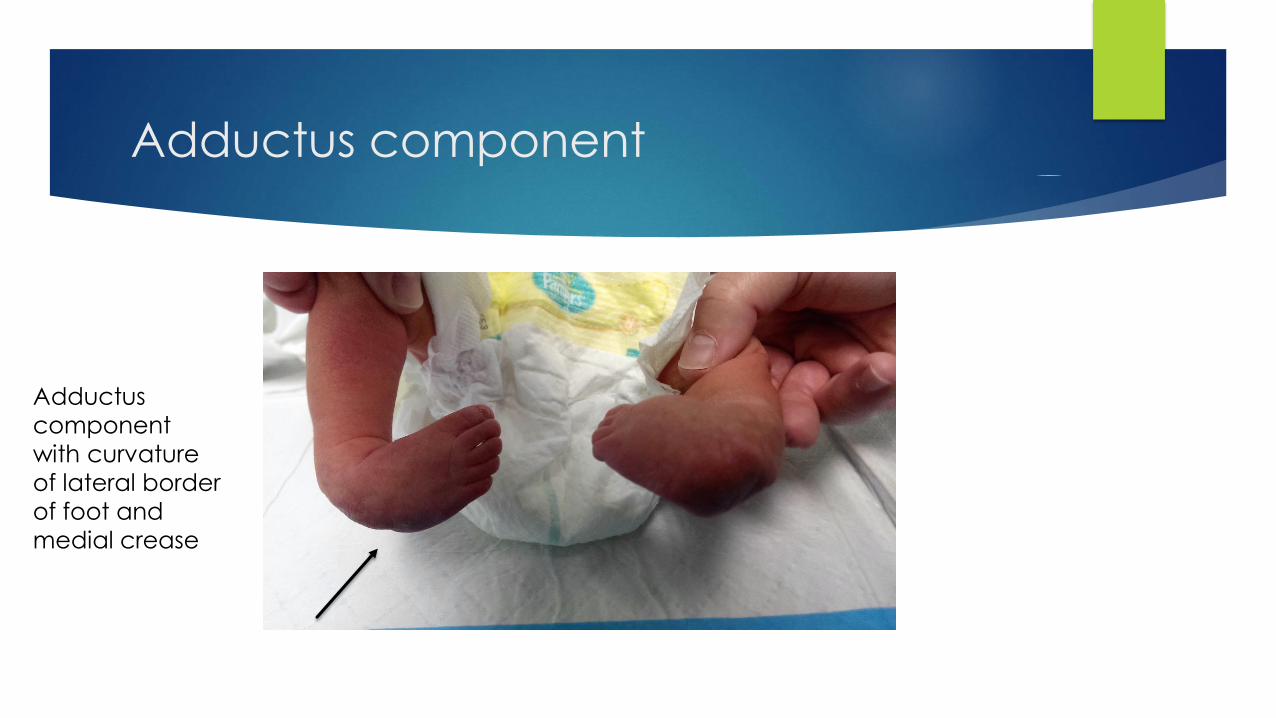
Adductus component
Adductus
component
with curvature
of lateral border
of foot and
medial crease
Signs (continued)
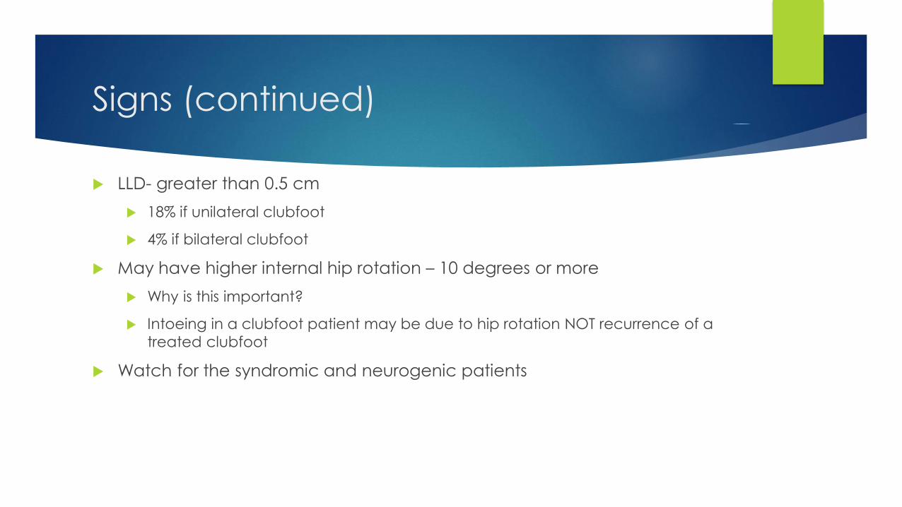
LLD- greater than 0.5 cm
18% if unilateral clubfoot
4% if bilateral clubfoot
May have higher internal hip rotation – 10 degrees or more
Why is this important?
Intoeing in a clubfoot patient may be due to hip rotation NOT recurrence of a
treated clubfoot
Watch for the syndromic and neurogenic patients
Classifications
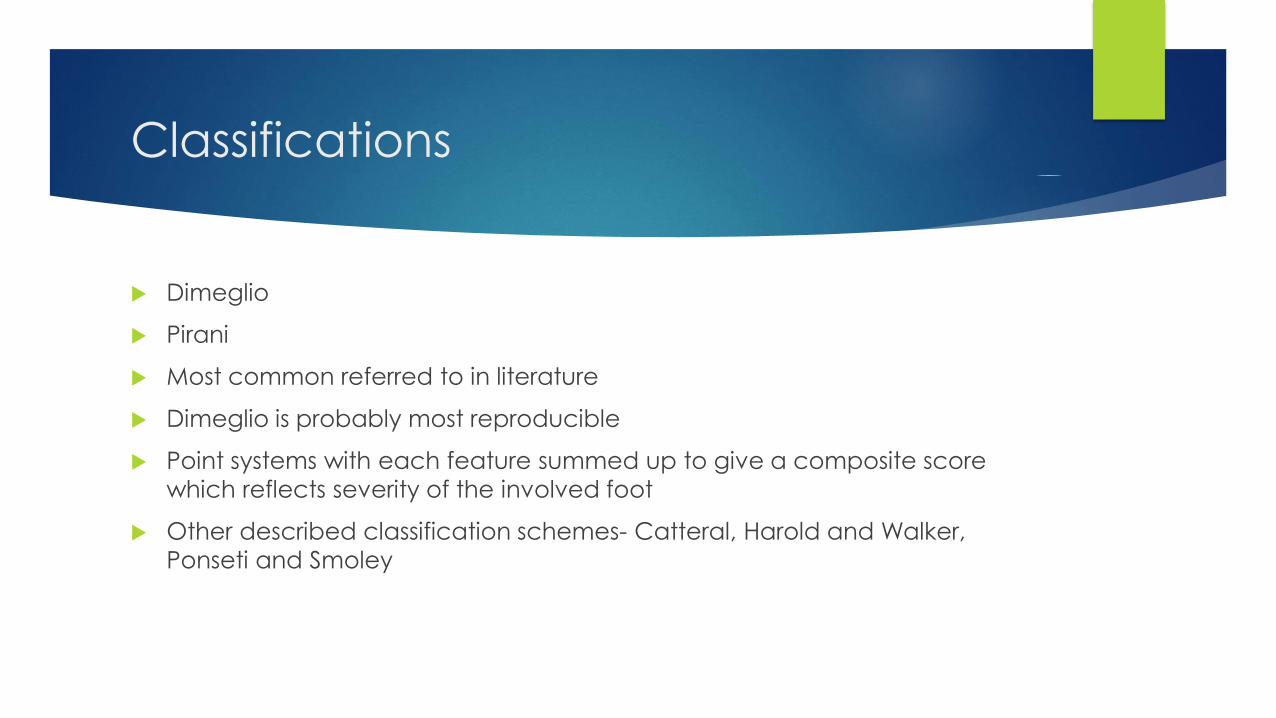
Dimeglio
Pirani
Most common referred to in literature
Dimeglio is probably most reproducible
Point systems with each feature summed up to give a composite score
which reflects severity of the involved foot
Other described classification schemes- Catteral, Harold and Walker,
Ponseti and Smoley
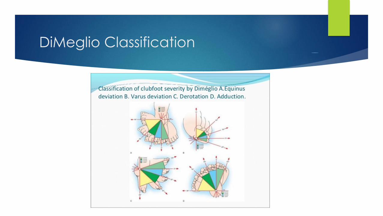
DiMeglio Classification
Radiographic Studies
Not required for diagnosis and management
Poor reproducibility of foot positioning
“Forced dorsiflexion” AP and lateral
Foot is held in maximum dorsiflexion while x-rays are taken
In utero ultrasound
Recognition is 0.1% to 0.4%
Low false-negative
False-positive- 30 to 40%
Functional false-positive- correctible passively after delivery (Positional clubfoot)
Further intrauterine evaluation
Controversial if amniocentesis is required with identified, isolated clubfoot
deformity
To identify syndromic presentation
Recognize additional abnormalities
Orthopedic consultation
For family counseling regarding etiology, treatment, prognosis

Pathoanatomy
Malalignment of bones at the joints
How?
Partly deformation of the bones themselves
Natural History
Untreated clubfoot
Rigid
Callus with bursa develops on the dorsolateral aspect
Hyperflexed midfoot
Extreme case- toes point backwards during ambulation
May be functional with a prosthesis
Pain more with hard floors and sidewalks
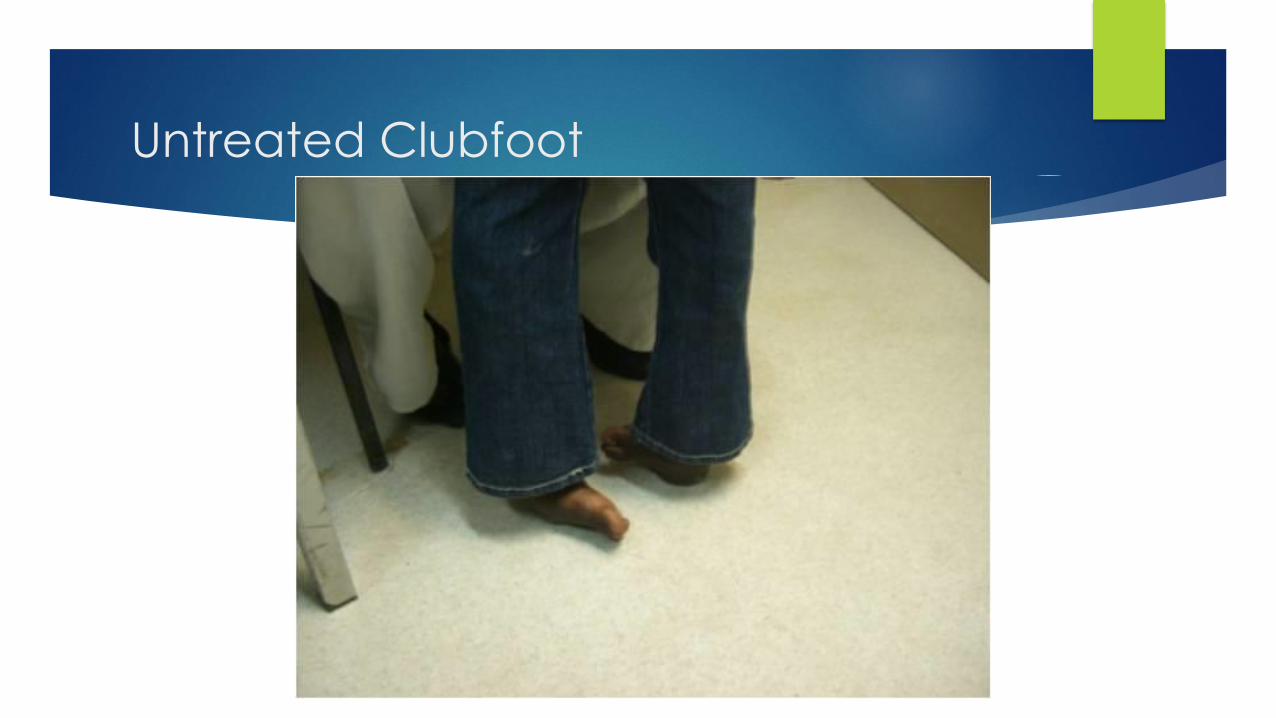
Untreated Clubfoot
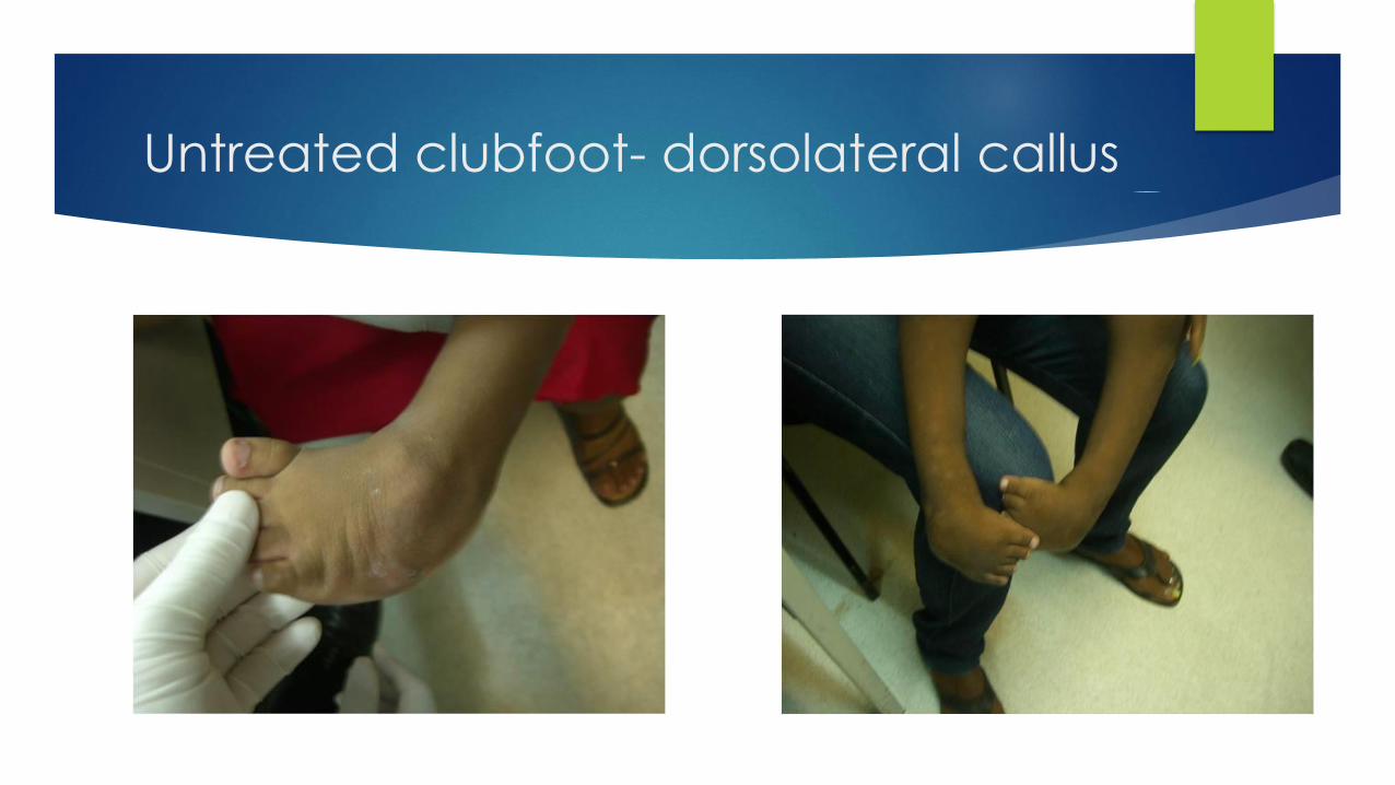
Untreated clubfoot- dorsolateral callus
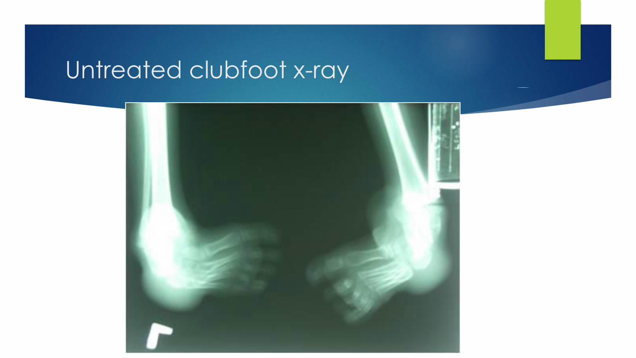
Untreated clubfoot x-ray
Treatment
Non-operative Treatment
To achieve a plantigrade, supple, painless foot with normal appearance
To avoid special shoe wear
Hiram Kite (Kite casting technique)
Poor long term results
Long-term immobilization
Ignacio Ponseti (Ponseti casting technique)
“French” Method
Inpatient admission
Serial strapping and physical therapy manipulations
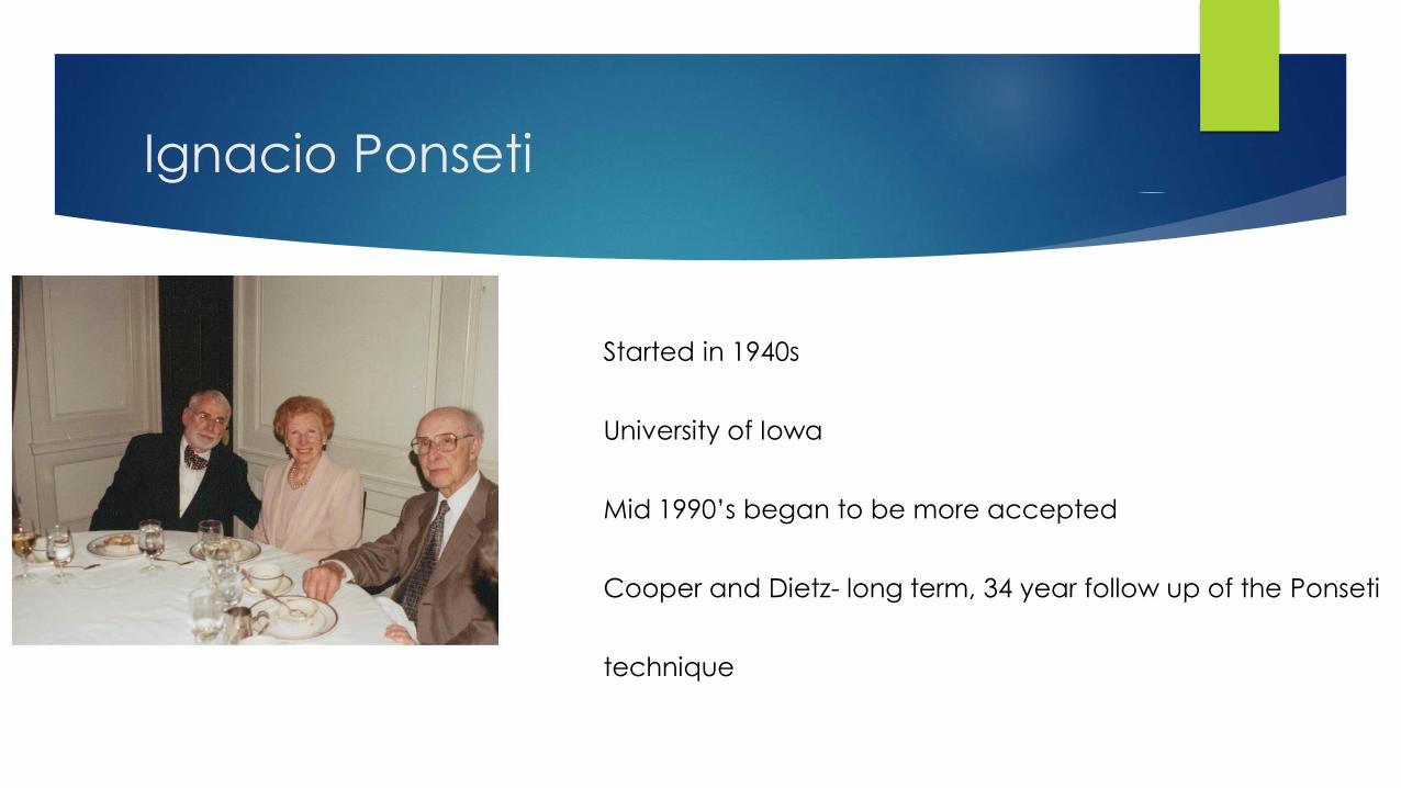
Ignacio Ponseti
• Started in 1940s
• University of Iowa
• Mid 1990’s began to be more accepted
• Cooper and Dietz- long term, 34 year follow up of the Ponseti
technique
Treatment- Ponseti technique
Serial casting
Average 4 to 8 casts
Has been shown to be effective even up to age 9!
Gentle manipulations/castings
Starting even several months after birth may lead to similar outcomes
Fiberglass shown to be superior to plaster of Paris for:
Durability, convenience, performance, ease of removal.
Has been performed by lay individuals (no formal medical training)
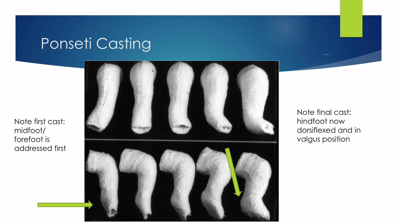
Ponseti Casting
Note first cast:
midfoot/
forefoot is
addressed first
Note final cast:
hindfoot now
dorsiflexed and in
valgus position
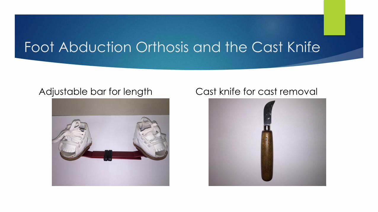
Foot Abduction Orthosis and the Cast Knife
Adjustable bar for length Cast knife for cast removal
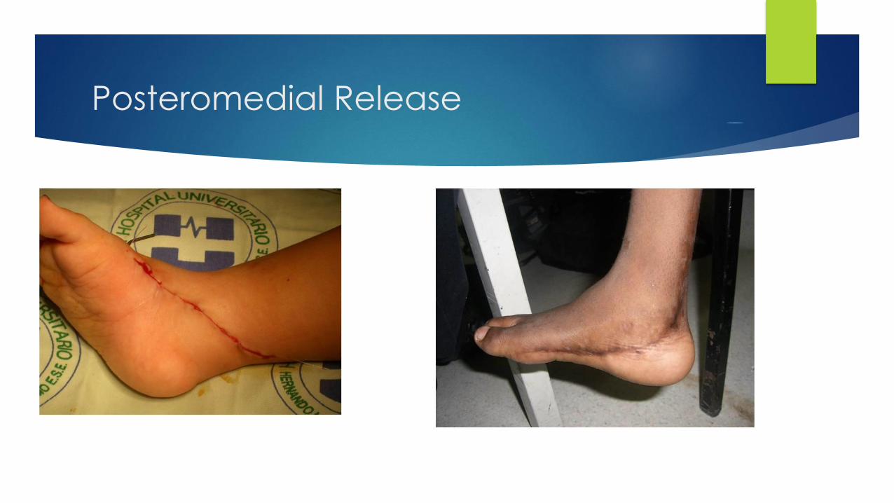
Posteromedial Release
Bibliography
www.genome.gov (NIH- NHGRI- National Institutes of Health- National Human Geome Research Institute)
www.cancer.gov (NIH- NCI- National Institutes of Health- National Cancer Institute)
www.law.cornell.edu- 29 CFR 1635.3- Definitions specific to GINA.
A method for the early evaluation of the Ponseti (Iowa) technique for the treatment of idiopathic clubfoot. Lehman WB, Mohaideen A, Madan S, ScherDM, Van Bosse HJ, Iannacone M, Bazzi JS, Feldman DS. J Pediatr Orthop B. 2003 Mar; 12(2): 133-40
Copy number analysis of 413 isolated talipes equinovarus patients suggests role for transcriptional regulators of early limb development. Alvarado DM, Buchan JG, Frick SL, Herzenberg JE, Dobbs MB, Gurnett CA. Eur J Hum Genet. 2013 Apr; 21(4):373-80
Bibliography
Genome-wide association study identifies new disease loci for isolated
clubfoot. Zhang TX, Haller G, Lin P, Alvarado DM, Hecht JT, Blanton SH,
Stephens Richards B, Rice JP, Dobbs MB, Gurnett CA. J Med Genet. 2014
May;51(5):334-9.
Lovell and Winter Pediatric Orthopaedics, 7th ed., editors Stuart Weinstein,
Jack Flynn
Photos and DiMeglio chart courtesy of:
Personal collections of Ahamed Mohaideen, M.D., Ezra Berkowitz, M.D.,
Wallace B. Lehman, M.D., Alain DiMeglio, M.D.
Special Thanks to:
Dr. Ezra Berkowitz