Embed Size (px)
Citation preview
Journal of
Clinical Medicine
Article
Malocclusion of Molar Teeth Is Associated with Activities ofDaily Living Loss and Delirium in Elderly Critically IllOlder Patients
Yoshihisa Fujinami 1, Toru Hifumi 2, Yuko Ono 1 , Masafumi Saito 1, Tomoya Okazaki 2 , Natsuyo Shinohara 2,Kyoko Akiyama 2, Misa Kunikata 2, Shigeaki Inoue 1,* , Joji Kotani 1 and Yasuhiro Kuroda 2
�����������������
Citation: Fujinami, Y.; Hifumi, T.;
Ono, Y.; Saito, M.; Okazaki, T.;
Shinohara, N.; Akiyama, K.;
Kunikata, M.; Inoue, S.; Kotani, J.;
et al. Malocclusion of Molar Teeth Is
Associated with Activities of Daily
Living Loss and Delirium in Elderly
Critically Ill Older Patients. J. Clin.
Med. 2021, 10, 2157. https://doi.org/
10.3390/jcm10102157
Academic Editor: Ray C. Williams
Received: 2 April 2021
Accepted: 13 May 2021
Published: 17 May 2021
Publisher’s Note: MDPI stays neutral
with regard to jurisdictional claims in
published maps and institutional affil-
iations.
Copyright: © 2021 by the authors.
Licensee MDPI, Basel, Switzerland.
This article is an open access article
distributed under the terms and
conditions of the Creative Commons
Attribution (CC BY) license (https://
creativecommons.org/licenses/by/
4.0/).
1 Department of Disaster and Emergency and Critical Care Medicine, Kobe University Graduate School ofMedicine, Kobe 650-0017, Japan; [email protected] (Y.F.); [email protected] (Y.O.);[email protected] (M.S.); [email protected] (J.K.)
2 Department of Disaster and Emergency and Critical Care Medicine, Kagawa University Graduate School ofMedicine, Kagawa 761-0701, Japan; [email protected] (T.H.); [email protected] (T.O.);[email protected] (N.S.); [email protected] (K.A.);[email protected] (M.K.); [email protected] (Y.K.)
* Correspondence: [email protected]; Tel.: +81-78-382-6521
Abstract: A single-center retrospective cohort study examined the association between molar mal-occlusion status at ICU admission and loss of activities of daily living (ADL) at hospital dischargeamong acutely ill patients. Patients were assigned to the bilateral occlusion group or malocclusiongroup (N = 227 and 93, respectively). The following data were collected from electronic medicalrecords: age, sex, Clinical Frailty Scale (CFS) on admission, Acute Physiology and Chronic HealthEvaluation (APACHE) II score, confirmed diagnosis (neurological disorders or others), CFS at hospi-tal discharge, and occlusion condition. Patients who were frail at admission (CFS > 5) were excludedfrom analysis, and ADL loss was defined as CFS > 5 at hospital discharge. Multivariate analysisshowed malocclusion was independently associated with ADL loss [OR, 2.03; 95% CI, 1.13–3.64;p = 0.02]. For those aged 65 and older, malocclusion was significantly associated with both ADL loss[OR, 3.25; 95% CI, 1.44–7.32; p < 0.01] and the incidence of delirium [OR, 2.61; 95% CI, 1.14–5.95;p = 0.02]. Malocclusion on ICU admission was associated with ADL loss in critically ill patients, andwas associated with ADL loss and the incidence of delirium in the elderly. Poor oral health was apoor prognostic factor among critically ill patients.
Keywords: poor oral health; frailty; ICU prognosis
1. Introduction
The increasing number of elderly patients admitted to ICUs is a major concern forhealthcare management, as aging is a prognostic factor for mortality among ICU patients [1].Indeed, a Canadian multicenter prospective cohort study revealed that 30% of ICU patientsover the age of 80 remained in the unit for more than seven days, and 22% died while beingthere [2]. Therefore, clinicians have been searching for an intervening factor through whichto improve the prognosis of aging patients in ICUs.
Because aging itself is inevitable, frailty has been recently regarded as importantby clinicians. Frailty is the most challenging manifestations of the aging population [3].It is defined as a clinically recognizable state of increased vulnerability resulting fromaging-associated functional decline in multiple physiological systems [4]. A meta-analysisshowed that the frailty of ICU patients was associated with higher hospital and long-termmortality, with lower discharges [5]. Notably, delirium—a neurobehavioral syndromecharacterized by impaired cognition with nonspecific manifestations—is one of the reasonsfor the poor prognosis; it also affects long-term activities of daily living (ADL) and cognitiveimpairment [6].
J. Clin. Med. 2021, 10, 2157. https://doi.org/10.3390/jcm10102157 https://www.mdpi.com/journal/jcm
J. Clin. Med. 2021, 10, 2157 2 of 13
Several studies have reported an association between oral health and the pathogenesisof frailty [7]. Elderly people with frailty have significantly poorer oral function than prefrailand robust individuals. Moreover, the risk of frailty has been associated with lower occlusalforce, masseter muscle thickness, and oral diadochokinetic rate [8]. Patients with poor oralhealth have more comorbidities such as cardiovascular diseases and strokes [9], chronicobstructive pulmonary disease [10], and type 2 diabetes [11]. Furthermore, they tend toexperience malnutrition [12] and cognitive dysfunction [13]. Beyond this, poor oral healthis associated with low socioeconomic status [14].
When accompanied by impaired malocclusion status with tooth loss and periodontaldisease, poor oral health is associated with mortality [15–17]. However, the associationbetween malocclusion status (a principal component of oral frailty) and ADL loss ordelirium among acutely ill patients remains unclear. Thus, this study serves to determinethe relevance of malocclusion on ADL loss and delirium development among acutelyill patients.
2. Materials and Methods2.1. Study Design and Setting
This retrospective cohort study was based in an emergency medical center at KagawaUniversity Hospital, Japan. The study was conducted according to the guidelines of theDeclaration of Helsinki, and the protocol and statistical analyses plan were approved bythe review board at Kagawa University Hospital (IRB number: H30-173).
2.2. Participants and Data Sources
The study included all patients aged ≥18 years who were admitted to the facility’semergency ICU between 1 November 2017 and 31 October 2018. Patients who had beendischarged from the hospital within 48 h, and patients who received palliative care wereexcluded. Patients who were frail on admission [i.e., Clinical Frailty Scale (CFS) > 5] werealso excluded from analysis. Finally, we also excluded those with missing data, usingcomplete data sets alone.
The following data were collected from the hospital’s electronic medical records: age,gender, CFS on admission, Acute Physiology and Chronic Health Evaluation (APACHE) IIscore, confirmed diagnosis (neurological disorders or others), CFS at hospital discharge,and occlusion condition.
2.3. Definitions2.3.1. Bilateral Occlusion and Malocclusion
Occlusion condition was assessed according to the number of teeth, chewing ability,articulatory oral motor skill, tongue pressure, as well as subjective difficulty when eatingtough foods and swallowing in general. Our database captured the information about thecondition of molar teeth occlusion but did not record other items.
Since occlusal contact area and occlusal force show similar results [18,19], occlusionsof the premolars and the molars were assessed. Bilateral occlusion status was defined ashaving at least one set of the molars on the same side of the upper and lower jaw. Althougha similar evaluation method has not been used in the past, we set the current definitionof malocclusion based on the Eichner index [20], which evaluates malocclusion based onthe number of occlusal supports provided by the molars and premolars. Based on thisdefinition, subjects were divided into a bilateral occlusion group and a malocclusion group.Subsequently, we excluded from the bilateral occlusion group when (1) the tooth crownwas lost, even if the root of the tooth was still present; (2) the mobility of the occlusal toothwas Grade 3, shaky in three dimensions; and (3) there was an existing traumatic toothinjury. The occlusion condition was assessed by X-ray images or the dentist examinationsheld once a week as an ICU round.
J. Clin. Med. 2021, 10, 2157 3 of 13
2.3.2. ADL Loss
To evaluate ADL loss, this study employed the CFS [21] (Appendix A). Specifically,ADL loss was defined as patients with a CFS > 5 at the time of hospital discharge, that is,they were completely dependent outside the facility. A score of 6 was deemed moderatelyfrail, while 7 = severely frail, 8 = extremely frail, and 9 = terminally ill. Other studies haveused this definition of ADL loss [22]. We chose not to include the patients with a CFS > 5 atthe time of admission because it is difficult to distinguish hospital-acquired ADL loss fromunderlying etiologies.
Patients who died in hospital were categorized as an ADL loss group. Notably, CFSwas scored using nursing records of patient activity and interviews with patient families.The CFS was independently estimated by the author, two physicians (T.O. and N.S.), andtwo nurses (K.Y. and M.K.). The consistency was 88% with a maximum difference of twopoints and Cohen’s kappa coefficient was 0.82. These raters independently determinedthe CFS score of each patient, and a third adjudicated any remaining conflicts after thescore reconciliation.
2.3.3. Neurological Disorders
The classification of neurological disorders was applied if the subject had cerebralhemorrhage, subarachnoid hemorrhage (SAH), cerebral infarction, epilepsy, encephalitis,hepatic encephalopathy, brain sarcoidosis, post resuscitation encephalopathy, or trau-matic brain injury (TBI). Notably, TBI included traumatic SAH, brain contusion, epiduralhematoma, and subdural hematoma. This category did not include head injury devoid ofbrain injury.
2.3.4. Delirium
We diagnosed delirium based on the confusion assessment method for the ICU (CAM-ICU) [23] up to 14 days after admission. During endotracheal intubation, sedation wasmanaged with a goal of RASS −2 to −1. CAM-ICU was assessed for each shift, anddelirium was considered present if observed once during the ICU stay for up to two weeksafter admission. Patients who died in hospital were categorized as a Delirium group. Thepatients in a comatose state with Glasgow Coma Scale (GCS) < 9 from admission onwardwere excluded. Patients with severe dementia were already excluded by the “CFS > 5 onadmission” criterion. Patients who died in hospital were excluded from analysis.
2.4. Exposure and Outcome Measurement
The primary exposure was malocclusion status. The primary outcome was ADL lossand the secondary outcome was the occurrence of delirium.
2.5. Statistical Analysis
Differences in the baseline clinical characteristics of the bilateral occlusion group andthe malocclusion group were evaluated. The Mann-Whitney U test was used to comparedifferences in the continuous variables between the two groups. Where appropriate, eitherthe Fisher exact test or chi-squared test were used to compare differences in categoricalvariables between the two groups. A multivariate logistic regression analysis was used toadjust for the potential confounders of age, gender, CFS on admission, APACHE II score,and neurological disorders on admission. This yielded an adjusted odds ratio (OR) forADL loss and the occurrence of delirium after malocclusion as the primary exposure. Aset of these variables was chosen a priori, based on previous reports [16,17,21,22,24] andbiological plausibility. Although recent studies on ICU patients and frailty have oftenapplied the CFS to those over 18 years of age [5] as the present study, the CFS is originallydesigned as a tool to assess frailty in patients over 65 years of age. Therefore, we conductedsubgroup analysis for the patients 65 years or older. All statistical analyses were performedusing JMP software version 13 (SAS Institute, Cary, NC, USA). A two-sided probabilityvalue of <0.05 was considered statistically significant for all analyses.
J. Clin. Med. 2021, 10, 2157 4 of 13
3. Results
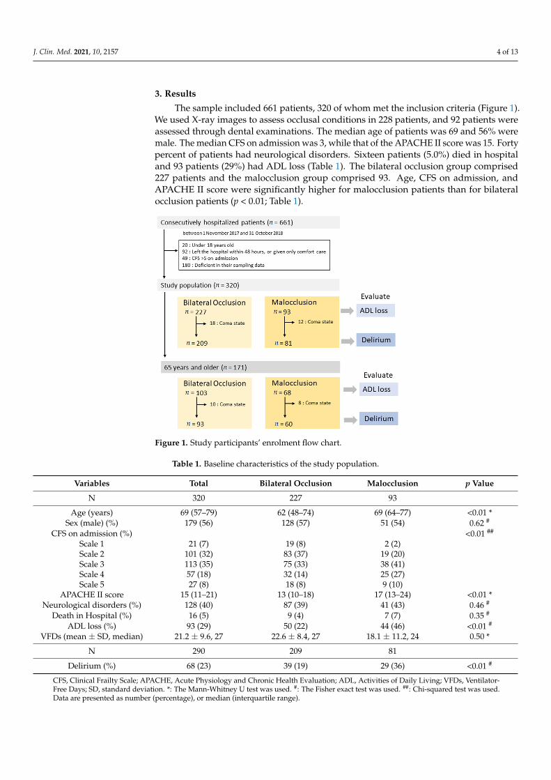
The sample included 661 patients, 320 of whom met the inclusion criteria (Figure 1).We used X-ray images to assess occlusal conditions in 228 patients, and 92 patients wereassessed through dental examinations. The median age of patients was 69 and 56% weremale. The median CFS on admission was 3, while that of the APACHE II score was 15. Fortypercent of patients had neurological disorders. Sixteen patients (5.0%) died in hospitaland 93 patients (29%) had ADL loss (Table 1). The bilateral occlusion group comprised227 patients and the malocclusion group comprised 93. Age, CFS on admission, andAPACHE II score were significantly higher for malocclusion patients than for bilateralocclusion patients (p < 0.01; Table 1).
J. Clin. Med. 2021, 10, x FOR PEER REVIEW 4 of 13
the CFS is originally designed as a tool to assess frailty in patients over 65 years of age. Therefore, we conducted subgroup analysis for the patients 65 years or older. All statisti-cal analyses were performed using JMP software version 13 (SAS Institute, Cary, NC, USA). A two-sided probability value of <0.05 was considered statistically significant for all analyses.
3. Results The sample included 661 patients, 320 of whom met the inclusion criteria (Figure 1).
We used X-ray images to assess occlusal conditions in 228 patients, and 92 patients were assessed through dental examinations. The median age of patients was 69 and 56% were male. The median CFS on admission was 3, while that of the APACHE II score was 15. Forty percent of patients had neurological disorders. Sixteen patients (5.0%) died in hos-pital and 93 patients (29%) had ADL loss (Table 1). The bilateral occlusion group com-prised 227 patients and the malocclusion group comprised 93. Age, CFS on admission, and APACHE II score were significantly higher for malocclusion patients than for bilat-eral occlusion patients (p < 0.01; Table 1).
Figure 1. Study participants’ enrolment flow chart.
Table 1. Baseline characteristics of the study population.
Variables Total Bilateral Occlusion Malocclusion p Value N 320 227 93
Age (years) 69 (57–79) 62 (48–74) 69 (64–77) <0.01 * Sex (male) (%) 179 (56) 128 (57) 51 (54) 0.62 #
CFS on admission (%) <0.01 ## Scale 1 21 (7) 19 (8) 2 (2) Scale 2 101 (32) 83 (37) 19 (20) Scale 3 113 (35) 75 (33) 38 (41) Scale 4 57 (18) 32 (14) 25 (27) Scale 5 27 (8) 18 (8) 9 (10)
Figure 1. Study participants’ enrolment flow chart.
Table 1. Baseline characteristics of the study population.
Variables Total Bilateral Occlusion Malocclusion p Value
N 320 227 93
Age (years) 69 (57–79) 62 (48–74) 69 (64–77) <0.01 *Sex (male) (%) 179 (56) 128 (57) 51 (54) 0.62 #
CFS on admission (%) <0.01 ##
Scale 1 21 (7) 19 (8) 2 (2)Scale 2 101 (32) 83 (37) 19 (20)Scale 3 113 (35) 75 (33) 38 (41)Scale 4 57 (18) 32 (14) 25 (27)Scale 5 27 (8) 18 (8) 9 (10)
APACHE II score 15 (11–21) 13 (10–18) 17 (13–24) <0.01 *Neurological disorders (%) 128 (40) 87 (39) 41 (43) 0.46 #
Death in Hospital (%) 16 (5) 9 (4) 7 (7) 0.35 #
ADL loss (%) 93 (29) 50 (22) 44 (46) <0.01 #
VFDs (mean ± SD, median) 21.2 ± 9.6, 27 22.6 ± 8.4, 27 18.1 ± 11.2, 24 0.50 *
N 290 209 81
Delirium (%) 68 (23) 39 (19) 29 (36) <0.01 #
CFS, Clinical Frailty Scale; APACHE, Acute Physiology and Chronic Health Evaluation; ADL, Activities of Daily Living; VFDs, Ventilator-Free Days; SD, standard deviation. *: The Mann-Whitney U test was used. #: The Fisher exact test was used. ##: Chi-squared test was used.Data are presented as number (percentage), or median (interquartile range).
J. Clin. Med. 2021, 10, 2157 5 of 13
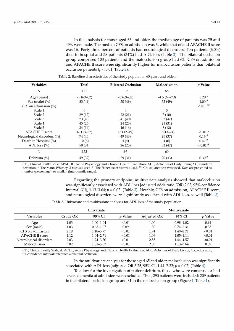
In the analysis for those aged 65 and older, the median age of patients was 75 and49% were male. The median CFS on admission was 3, while that of and APACHE II scorewas 16. Forty three percent of patients had neurological disorders. Ten patients (6.0%)died in hospital and 58 patients (34%) had ADL loss (Table 2). The bilateral occlusiongroup comprised 103 patients and the malocclusion group had 63. CFS on admissionand APACHE II score were significantly higher for malocclusion patients than bilateralocclusion patients (p < 0.01; Table 2).
Table 2. Baseline characteristics of the study population 65 years and older.
Variables Total Bilateral Occlusion Malocclusion p Value
N 171 103 68
Age (years) 75 (69–82) 76 (69–82) 74.5 (69–79) 0.20 *Sex (male) (%) 83 (49) 50 (49) 33 (49) 1.00 #
CFS on admission (%) <0.01 ##
Scale 1 0 0 0Scale 2 29 (17) 22 (21) 7 (10)Scale 3 73 (43) 41 (40) 32 (47)Scale 4 45 (26) 24 (23) 21 (31)Scale 5 24 (14) 16 (16) 8 (12)
APACHE II score 16 (13–22) 15 (12–19) 19 (13–24) <0.01 *Neurological disorders (%) 74 (43) 49 (48) 25 (37) 0.16 #
Death in Hospital (%) 10 (6) 4 (4) 4 (6) 0.42 #
ADL loss (%) 58 (34) 26 (25) 32 (47) <0.01 #
N 153 93 60
Delirium (%) 49 (32) 29 (31) 20 (33) 0.30 #
CFS, Clinical Frailty Scale; APACHE, Acute Physiology and Chronic Health Evaluation; ADL, Activities of Daily Living; SD, standarddeviation. *: The Mann-Whitney U test was used. #: The Fisher exact test was used. ##: Chi-squared test was used. Data are presented asnumber (percentage), or median (interquartile range).
Regarding the primary endpoint, multivariate analysis showed that malocclusionwas significantly associated with ADL loss [adjusted odds ratio (OR) 2.03; 95% confidenceinterval (CI), 1.13–3.64; p = 0.02] (Table 3). Notably, CFS on admission, APACHE II score,and neurological disorders were significantly associated with ADL loss, as well (Table 3).
Table 3. Univariate and multivariate analyses for ADL loss of the study population.
Univariate Multivariate
Variables Crude OR 95% CI p Value Adjusted OR 95% CI p Value
Age 1.03 1.00–1.04 <0.01 1.00 0.98–1.02 0.94Sex (male) 1.03 0.63–1.67 0.89 1.30 0.74–2.31 0.35
CFS on admission 2.19 1.48–5.77 <0.01 1.94 1.40–2.71 <0.01APACHE II score 1.12 1.04–2.71 <0.01 1.09 1.05–1.14 <0.01
Neurological disorders 2.03 1.24–3.30 <0.01 2.55 1.44–4.57 <0.01Malocclusion 3.02 1.81–5.03 <0.01 2.03 1.13–3.64 0.02
CFS, Clinical Frailty Scale; APACHE, Acute Physiology and Chronic Health Evaluation; ADL, Activities of Daily Living; OR, odds ratio;CI, confidence interval; reference = bilateral occlusion.
In the multivariate analysis for those aged 65 and older, malocclusion was significantlyassociated with ADL loss [adjusted OR 3.25; 95% CI, 1.44–7.32; p = 0.02] (Table 4).
To allow for the investigation of patient delirium, those who were comatose or hadsevere dementia at admission were excluded. Thus, 290 patients were included: 209 patientsin the bilateral occlusion group and 81 in the malocclusion group (Figure 1; Table 1).
J. Clin. Med. 2021, 10, 2157 6 of 13
Table 4. Univariate and multivariate analyses for ADL loss of the study population 65 years and older.
Univariate Multivariate
Variables Crude OR 95% CI p Value Adjusted OR 95% CI p Value
Age 1.09 1.04–1.14 <0.01 1.08 1.02–1.15 <0.01Sex (male) 0.89 0.47–1.67 0.71 1.11 0.51–2.40 0.80
CFS on admission 2.37 1.60–3.50 <0.01 2.20 1.38–3.49 <0.01APACHE II score 1.09 1.03–1.14 <0.01 1.06 1.00–1.12 0.04
Neurological disorders 1.87 0.99–3.55 0.06 2.86 1.32–6.23 <0.01Malocclusion 2.63 1.37–5.05 <0.01 3.25 1.44–7.32 <0.01
CFS, Clinical Frailty Scale; APACHE, Acute Physiology and Chronic Health Evaluation; ADL, Activities of Daily Living; OR, odds ratio;CI, confidence interval; reference = bilateral occlusion.
Age, CFS on admission, APACHE II score, the percentage of neurological disorders,and the percentage of malocclusion were significantly higher among delirium patients thanthey were among non-delirium patients (Table 5). However, multivariate analysis revealedthat malocclusion was not associated with the incidence of delirium among the acutely illpatients [adjusted OR, 1.33; 95% CI, 0.76–2.34; p = 0.32]. (Table 5).
Table 5. Univariate and Multivariate analysis for Delirium of the study population.
Univariate Multivariate
Variables Crude OR 95% CI p Value Adjusted OR 95% CI p Value
Age 1.03 1.01–1.05 <0.01 1.01 0.99–1.03 0.37Sex (male) 0.89 0.56–1.43 0.64 1.04 0.61–1.76 0.88
CFS on admission 1.78 1.39–2.26 <0.01 1.40 1.05–1.95 0.02APACHE II score 1.11 1.07–1.15 <0.01 1.09 1.05–1.13 <0.01
Neurological disorders 1.82 1.13–2.94 <0.01 1.97 1.16–3.36 0.01Malocclusion 2.14 1.29–3.53 <0.01 1.33 0.76–2.34 0.32
CFS, Clinical Frailty Scale; APACHE, Acute Physiology and Chronic Health Evaluation; ADL, Activities of Daily Living; OR, odds ratio;CI, confidence interval; reference = bilateral occlusion.
In the multivariate analysis for those aged 65 and older, malocclusion was signifi-cantly associated with the incidence of delirium [adjusted OR, 2.61; 95% CI, 1.14–5.95;p = 0.02], (Table 6).
Table 6. Univariate and Multivariate analysis for Delirium of the study population 65 years and older.
Univariate Multivariate
Variables Crude OR 95% CI p Value Adjusted OR 95% CI p Value
Age 1.10 1.05–1.16 <0.01 1.13 1.06–1.20 <0.01Sex (male) 0.89 0.48–1.67 0.72 1.18 0.55–2.54 0.68
CFS on admission 1.32 0.94–1.87 0.11 1.01 0.65–1.58 0.97APACHE II score 1.06 1.01–1.10 0.01 1.07 1.01–1.13 0.02
Neurological disorders 4.03 2.07–7.84 <0.01 5.82 2.63–12.90 <0.01Malocclusion 1.92 1.01–3.64 0.05 2.61 1.14–5.95 0.02
CFS, Clinical Frailty Scale; APACHE, Acute Physiology and Chronic Health Evaluation; ADL, Activities of Daily Living; OR, odds ratio;CI, confidence interval; reference = bilateral occlusion.
4. Discussion
This study found that malocclusion at admission was significantly associated withthe development of ADL loss in ICU patients and the incidence of delirium in elderlypeople aged 65 years and older. This is the first study to demonstrate the impact that poororal health has on ADL loss, leading to quality-of-life issues such as frailty and cognitiveimpairment in acutely ill patients. Notably, patients with malocclusion were older, frailer,and more critically ill at the time of admission.
Although ADL loss is well known to be associated with aging, frailty, and a highAPACHE score [5], our study revealed that ADL loss was also associated with neurological
J. Clin. Med. 2021, 10, 2157 7 of 13
disorders. There were significant differences between the bilateral occlusion and malocclu-sion groups regarding several of the baseline characteristics measured during admission,such as age, APACHE score, and CFS (Tables 1 and 2). This study demonstrated thatpatients with malocclusion score significantly higher on these items than the bilateral occlu-sion group. Since they are well known as poor prognostic factors [5], such differences areoften noteworthy problems for clinicians, especially during emergency medical treatment.Moreover, multivariate analysis revealed that malocclusion itself was an independentlypoor prognostic factor for acutely ill patients (Table 3). In this study, we excluded patientswith CFS > 5, and they were mostly over 80 years old. Because moderately or severelyfrail patients at admission were excluded, the average age of the subjects was lower thanthat in other observational studies, and age was not associated with outcome. We haveadditionally analyzed the difference in ADL using the difference of CFS from at admissionto at discharge. Furthermore, we conducted subgroup analysis by stratifying the levelof CFS at admission to observe the difference of CFS in each level of ADL (Appendix B),because the situation is very different between CFS 2 to 4 and CFS 4 to 6 even thoughthe CFS went up by 2. Even though the statistical power was decreased by stratification,we could observe the significant difference of CFS in CFS 3 group at admission (p = 0.02),and the trend of decrease in CFS difference of CFS 2 and 4 groups at admission (p = 0.09,0.14 respectively). These data are consistent with our original data that malocclusion isassociated with the development of ADL loss.
As previously noted, patients with poor oral health have more comorbidities [9], andpoor oral health is associated with low socioeconomic status, as evidenced by educationand income [14]. Notably, for some diseases, socioeconomic status is associated withprognosis [25,26]. Even in Japan, where health insurance is provided largely withoutexception, low socioeconomic status is correlated with major health disadvantages.
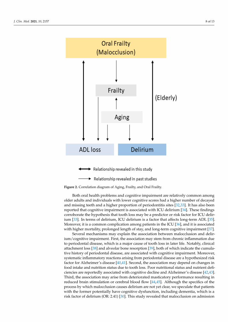
Studies have shown that malocclusion is associated with frailty, and frailty is a poorprognostic factor [5,27]. Notably, our study showed that malocclusion was an independentfactor of poor prognosis (Table 3; Figure 2). We provided information on quantificationsof the interaction between the variables of the multivariate analyses (Appendix C), whichdemonstrated that interaction of malocclusion on age and severity was observed in bothADL loss and delirium. These results suggest that malocclusion has both direct and indirecteffects on ADL loss. Although the relationship between malocclusion and ADL loss isstill unclear, we speculate that malocclusion contributed to (1) recovery delay from apathological condition, (2) secondary infection, (3) malnutrition, and (4) delirium. Asthe evidence for (1): There is a report that occlusal disharmonies cause immune systemdysfunction and the malfunction of central catecholaminergic neurotransmission [28]. For(2): Many researchers have reported that poor oral health is associated with the occurrenceof ventilator-associated pneumonia among ICU patients [24]. For (3): Fewer teeth arepositively related to swallowing dysfunction, and poor oral health causes a decrease insaliva secretion; these mechanisms could contribute to malnutrition [29]. For (4): Thisstudy assessed the contribution to delirium as the secondary outcome because it is wellknown as a risk factor for poor prognosis in ICU patients [23].
Delirium in the ICU is often assessed with CAM-ICU [23] and predicted using theEarly Prediction Model for Delirium in ICU Patients (E-PRE-DELITIC) model [30], whichwas proposed by Boogaard and colleagues. With reference to this model, this studyadopted urgent admission, age, APACHE score, and neurological disorders for evaluation.Moreover, it was reported that frailty on admission was the risk factor for delirium inurgently admitted patients [31]; therefore, we adopted frailty as an item. In addition, weadopted malocclusion as an item to substantiate the hypothesis that malocclusion wasassociated not only indirectly but also directly.
J. Clin. Med. 2021, 10, 2157 8 of 13
J. Clin. Med. 2021, 10, x FOR PEER REVIEW 8 of 13
secretion; these mechanisms could contribute to malnutrition [29]. For (4): This study as-sessed the contribution to delirium as the secondary outcome because it is well known as a risk factor for poor prognosis in ICU patients [23].
Figure 2. Correlation diagram of Aging, Frailty, and Oral Frailty.
Delirium in the ICU is often assessed with CAM-ICU [23] and predicted using the Early Prediction Model for Delirium in ICU Patients (E-PRE-DELITIC) model [30], which was proposed by Boogaard and colleagues. With reference to this model, this study adopted urgent admission, age, APACHE score, and neurological disorders for evalua-tion. Moreover, it was reported that frailty on admission was the risk factor for delirium in urgently admitted patients [31]; therefore, we adopted frailty as an item. In addition, we adopted malocclusion as an item to substantiate the hypothesis that malocclusion was associated not only indirectly but also directly.
Both oral health problems and cognitive impairment are relatively common among older adults and individuals with lower cognitive scores had a higher number of decayed and missing teeth and a higher proportion of periodontitis sites [32,33]. It has also been reported that cognitive impairment is associated with ICU delirium [34]. These findings corroborate the hypothesis that tooth loss may be a predictor or risk factor for ICU delir-ium [33]. In terms of delirium, ICU delirium is a factor that affects long-term ADL [35]. Moreover, it is a common complication among patients in the ICU [36], and it is associated with higher mortality, prolonged length of stay, and long-term cognitive impairment [37].
Several mechanisms may explain the association between malocclusion and delir-ium/cognitive impairment. First, the association may stem from chronic inflammation due to periodontal disease, which is a major cause of tooth loss in later life. Notably, clinical attachment loss [38] and alveolar bone resorption [39], both of which indicate the cumu-lative history of periodontal disease, are associated with cognitive impairment. Moreover, systematic inflammatory reactions arising from periodontal disease are a hypothesized risk factor for Alzheimer’s disease [40,41]. Second, the association may depend on changes in food intake and nutrition status due to tooth loss. Poor nutritional status and nutrient deficiencies are reportedly associated with cognitive decline and Alzheimer’s disease [42,43]. Third, the association may arise from deteriorated masticatory performance re-sulting in reduced brain stimulation or cerebral blood flow [44,45]. Although the specifics of the process by which malocclusion causes delirium are not yet clear, we speculate that
Figure 2. Correlation diagram of Aging, Frailty, and Oral Frailty.
Both oral health problems and cognitive impairment are relatively common amongolder adults and individuals with lower cognitive scores had a higher number of decayedand missing teeth and a higher proportion of periodontitis sites [32,33]. It has also beenreported that cognitive impairment is associated with ICU delirium [34]. These findingscorroborate the hypothesis that tooth loss may be a predictor or risk factor for ICU delir-ium [33]. In terms of delirium, ICU delirium is a factor that affects long-term ADL [35].Moreover, it is a common complication among patients in the ICU [36], and it is associatedwith higher mortality, prolonged length of stay, and long-term cognitive impairment [37].
Several mechanisms may explain the association between malocclusion and delir-ium/cognitive impairment. First, the association may stem from chronic inflammation dueto periodontal disease, which is a major cause of tooth loss in later life. Notably, clinicalattachment loss [38] and alveolar bone resorption [39], both of which indicate the cumula-tive history of periodontal disease, are associated with cognitive impairment. Moreover,systematic inflammatory reactions arising from periodontal disease are a hypothesized riskfactor for Alzheimer’s disease [40,41]. Second, the association may depend on changes infood intake and nutrition status due to tooth loss. Poor nutritional status and nutrient defi-ciencies are reportedly associated with cognitive decline and Alzheimer’s disease [42,43].Third, the association may arise from deteriorated masticatory performance resulting inreduced brain stimulation or cerebral blood flow [44,45]. Although the specifics of theprocess by which malocclusion causes delirium are not yet clear, we speculate that patientswith the former potentially have cognitive dysfunction, including dementia, which is arisk factor of delirium (OR: 2.41) [30]. This study revealed that malocclusion on admission
J. Clin. Med. 2021, 10, 2157 9 of 13
was significantly associated with the incidence of delirium in the elderly aged 65 years andolder. Further studies are needed to clarify the detail association and mechanisms.
Notably, there are some study limitations. First, this was a retrospective study con-ducted in an acutely ill population from a single tertiary care center. Accordingly, theresults may not be generalizable to less severely ill populations. In addition, we excluded180 patients due to deficiencies in their sampling data. Of these, 100 were excluded dueto deficiencies in their data concerning occlusal condition. These individuals tended tobe younger and most of them were limb trauma patients; they were not examined byhead-to-neck X-rays and oral assessments. Regarding the delirium analysis, we excluded30 patients (9.4% in enrolled patients) because they were comatose (GCS < 9) and/or hadsevere dementia (CFS > 5) on admission, leading to less power in multivariate analysis.Second, we limited our focus to ADL loss and delirium as outcome variables; future studiesmay wish to explore other relevant outcomes. Third, a longer period of observation maybe needed to assess the relationship between malocclusion and associated outcomes.
Despite these limitations, this is the first study to assess the relationship betweenmalocclusion and ADL and the development of delirium for which poor oral health isa measure of short-term outcome in acutely ill patients. Moreover, poor oral health isfrequently used as a bedside indicator of this clinical state. Furthermore, we believe itmay be the intervenable factor in the ICU. Interventions for oral health can improve theclinical course by reducing malnutrition, secondary infections such as pneumonia, anddelirium. Preventing tooth loss and encouraging denture wearing once teeth are lostmay indirectly contribute to maintaining or improving ADL, mediated by recovery ofswallowing function and nutritional status [29]. Further intervention studies may beneeded to prove the association between oral frailty and not only short-term but alsolong-term ICU outcomes.
5. Conclusions
Malocclusion with molar teeth loss is associated with the loss of ADL loss and delir-ium in elderly critically ill patients. By recognizing this association, we hope to raiseawareness of oral dysfunction prevention and strengthen medical and dental collaborationafter hospitalization.
Author Contributions: Y.F. and T.H. contributed to the acquisition of data, conceived of and designedthis study, interpreted the data, drafted the manuscript, and revised the manuscript for importantintellectual content. Y.O. and M.S. elaborated on the text. T.O., N.S., K.A., and M.K. contributed to theacquisition of data. S.I., J.K., and Y.K. interpreted the data, and revised the manuscript for importantintellectual content. All the authors contributed to the acquisition of data, reviewed, discussed. Allauthors have read and agreed to the published version of the manuscript.
Funding: This research received no external funding.
Institutional Review Board Statement: The study was conducted according to the guidelines of theDeclaration of Helsinki, and the protocol and statistical analyses plan were approved by the reviewboard at Kagawa University Hospital (IRB number: H30-173).
Informed Consent Statement: Patient consent was waived due to the retrospective cohort studywith de-identified data.Data availability Statement: The datasets generated and/or analyzed duringthe current study are available from the corresponding author on reasonable request.
Acknowledgments: The authors are grateful to biostatisticians from the Clinical &TransrationalResearch Center, Kobe University Hospital for valuable advice concerning the statistical analysis.
Conflicts of Interest: The authors declare no conflict of interest.

J. Clin. Med. 2021, 10, 2157 10 of 13
Appendix A
Table A1. Clinical Frailty Scale.
1. Very Fit—People who are robust, active, energetic, and motivated. People in this group exercise regularly and are among thefittest for their age.
2. Well—These people have no active disease symptoms but are less fit compared to those in category 1. They occasionally exerciseor engage in activity, e.g., seasonally.
3. Managing well—People whose medical problems are well controlled; however, they are not regularly active beyondroutine walking.
4. Vulnerable—While these people are not dependent on others for daily help, their symptoms often limit their activities. Acommon complaint is feeling “slowed down” and/or being tired during the day.
5. Mildly frail—These people often have more apparent slowing and need help with high order instrumental ADLs (finances,transportation, heavy housework, medications). Mild frailty progressively impairs the ability to shop, walk outside alone, preparemeals, and do housework.
6. Moderately frail—These people need help with all outside activities and with keeping house. They often have problems withstairs, need help with bathing, and might need minimal assistance (cueing, standby) with dressing.
7. Severely frail—These people are completely dependent on others for their personal care impairment. Even so, they appear to bestable and not at high risk of dying (within 6 months).
8. Very Severely frail—These people are completely dependent on others for their personal care as they are approaching the end oflife. Typically, they could not even recover from a minor illness.
9. Terminally ill—These people are approaching the end of life. This category applies to people with a life expectancy <6 monthswho are not otherwise obviously frail.
Appendix B
Table A2. Subgroup analysis by stratifying the level of CFS at admission to observe the difference of CFS in each levelof ADL.
Bilateral Malocclusion
CFSon Admission n
CFSat Discharge
(Difference of CFS)
Event(%) n
CFSat Discharge
(CFS Different)
Event(%) p Value
1 0.34
21 1 (0) 0 2 1 (0) 02 (1) 7 (33) 2 (1) 03 (2) 8 (38) 3 (2) 1 (50)4 (3) 3 (14) 4 (3) 05 (4) 1 (5) 5 (4) 1 (50)
6–9 (5–8) 2 (10) 6–9 (5–8) 0
2 0.09
83 2 (0) 16 (19) 19 2 (0) 1 (5)3 (1) 34 (41) 3 (1) 4 (21)4 (2) 14 (17) 4 (2) 6 (32)5 (3) 8 (10) 5 (3) 4 (21)
6–9 (4–7) 11 (13) 6–9 (4–6) 4 (21)
3 0.02
75 3 (0) 23 (31) 38 3 (0) 3 (8)4 (1) 22 (29) 4 (1) 11 (29)5 (2) 14 (19) 5 (2) 8 (21)
6–9 (3–6) 16 (21) 6–9 (3–5) 16 (42)
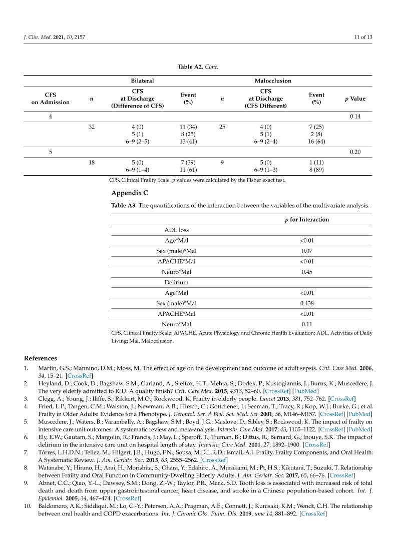
J. Clin. Med. 2021, 10, 2157 11 of 13
Table A2. Cont.
Bilateral Malocclusion
CFSon Admission n
CFSat Discharge
(Difference of CFS)
Event(%) n
CFSat Discharge
(CFS Different)
Event(%) p Value
4 0.14
32 4 (0) 11 (34) 25 4 (0) 7 (25)5 (1) 8 (25) 5 (1) 2 (8)
6–9 (2–5) 13 (41) 6–9 (2–4) 16 (64)
5 0.20
18 5 (0) 7 (39) 9 5 (0) 1 (11)6–9 (1–4) 11 (61) 6–9 (1–3) 8 (89)
CFS, Clinical Frailty Scale. p values were calculated by the Fisher exact test.
Appendix C
Table A3. The quantifications of the interaction between the variables of the multivariate analysis.
p for Interaction
ADL loss
Age*Mal <0.01
Sex (male)*Mal 0.07
APACHE*Mal <0.01
Neuro*Mal 0.45
Delirium
Age*Mal <0.01
Sex (male)*Mal 0.438
APACHE*Mal <0.01
Neuro*Mal 0.11CFS, Clinical Frailty Scale; APACHE, Acute Physiology and Chronic Health Evaluation; ADL, Activities of DailyLiving; Mal, Malocclusion.
References1. Martin, G.S.; Mannino, D.M.; Moss, M. The effect of age on the development and outcome of adult sepsis. Crit. Care Med. 2006,
34, 15–21. [CrossRef]2. Heyland, D.; Cook, D.; Bagshaw, S.M.; Garland, A.; Stelfox, H.T.; Mehta, S.; Dodek, P.; Kustogiannis, J.; Burns, K.; Muscedere, J.
The very elderly admitted to ICU: A quality finish? Crit. Care Med. 2015, 4313, 52–60. [CrossRef] [PubMed]3. Clegg, A.; Young, J.; Iliffe, S.; Rikkert, M.O.; Rockwood, K. Frailty in elderly people. Lancet 2013, 381, 752–762. [CrossRef]4. Fried, L.P.; Tangen, C.M.; Walston, J.; Newman, A.B.; Hirsch, C.; Gottdiener, J.; Seeman, T.; Tracy, R.; Kop, W.J.; Burke, G.; et al.
Frailty in Older Adults: Evidence for a Phenotype. J. Gerontol. Ser. A Biol. Sci. Med. Sci. 2001, 56, M146–M157. [CrossRef] [PubMed]5. Muscedere, J.; Waters, B.; Varambally, A.; Bagshaw, S.M.; Boyd, J.G.; Maslove, D.; Sibley, S.; Rockwood, K. The impact of frailty on
intensive care unit outcomes: A systematic review and meta-analysis. Intensiv. Care Med. 2017, 43, 1105–1122. [CrossRef] [PubMed]6. Ely, E.W.; Gautam, S.; Margolin, R.; Francis, J.; May, L.; Speroff, T.; Truman, B.; Dittus, R.; Bernard, G.; Inouye, S.K. The impact of
delirium in the intensive care unit on hospital length of stay. Intensiv. Care Med. 2001, 27, 1892–1900. [CrossRef]7. Tôrres, L.H.D.N.; Tellez, M.; Hilgert, J.B.; Hugo, F.N.; Sousa, M.D.L.R.D.; Ismail, A.I. Frailty, Frailty Components, and Oral Health:
A Systematic Review. J. Am. Geriatr. Soc. 2015, 63, 2555–2562. [CrossRef]8. Watanabe, Y.; Hirano, H.; Arai, H.; Morishita, S.; Ohara, Y.; Edahiro, A.; Murakami, M.; Pt, H.S.; Kikutani, T.; Suzuki, T. Relationship
between Frailty and Oral Function in Community-Dwelling Elderly Adults. J. Am. Geriatr. Soc. 2017, 65, 66–76. [CrossRef]9. Abnet, C.C.; Qiao, Y.-L.; Dawsey, S.M.; Dong, Z.-W.; Taylor, P.R.; Mark, S.D. Tooth loss is associated with increased risk of total
death and death from upper gastrointestinal cancer, heart disease, and stroke in a Chinese population-based cohort. Int. J.Epidemiol. 2005, 34, 467–474. [CrossRef]
10. Baldomero, A.K.; Siddiqui, M.; Lo, C.-Y.; Petersen, A.A.; Pragman, A.E.; Connett, J.; Kunisaki, K.M.; Wendt, C.H. The relationshipbetween oral health and COPD exacerbations. Int. J. Chronic Obs. Pulm. Dis. 2019, ume 14, 881–892. [CrossRef]

J. Clin. Med. 2021, 10, 2157 12 of 13
11. Leite, R.S.; Marlow, N.M.; Fernandes, J.K.; Hermayer, K. Oral Health and Type 2 Diabetes. Am. J. Med. Sci. 2013, 345, 271–273.[CrossRef] [PubMed]
12. Shwe, P.S.; Ward, S.A.; Thein, P.M.; Junckerstorff, R. Frailty, oral health and nutrition in geriatrics inpatients: A cross-sectionalstudy. Gerodontology 2019, 36, 223–228. [CrossRef] [PubMed]
13. Daly, B.; Thompsell, A.; Sharpling, J.; Rooney, Y.M.; Hillman, L.; Wanyonyi, K.L.; White, S.; Gallagher, J.E. Evidence summary:The relationship between oral health and dementia. Br. Dent. J. 2017, 223, 846–853. [CrossRef] [PubMed]
14. Farmer, J.; Phillips, R.C.; Singhal, S.; Quiñonez, C. Inequalities in oral health: Understanding the contributions of education andincome. Can. J. Public Health 2017, 108, e240–e245. [CrossRef] [PubMed]
15. Tu, Y.-K.; Galobardes, B.; Smith, G.D.; McCarron, P.; Jeffreys, M.; Gilthorpe, M.S. Associations between tooth loss and mortalitypatterns in the Glasgow Alumni Cohort. Heart 2007, 93, 1098–1103. [CrossRef]
16. Brown, D.W. Complete Edentulism Prior to the Age of 65 Years is Associated with All-Cause Mortality. J. Public Health Dent. 2009,69, 260–266. [CrossRef]
17. Ansai, T.; Takata, Y.; Soh, I.; Awano, S.; Yoshida, A.; Sonoki, K.; Hamasaki, T.; Torisu, T.; Sogame, A.; Shimada, N.; et al.Relationship between tooth loss and mortality in 80-year-old Japanese community-dwelling subjects. BMC Public Health 2010,10, 386. [CrossRef]
18. Imamura, Y.; Sato, Y.; Kitagawa, N.; Uchida, K.; Osawa, T.; Omori, M.; Okada, Y. Influence of occlusal loading force on occlusalcontacts in natural dentition. J. Prosthodont. Res. 2015, 59, 113–120. [CrossRef] [PubMed]
19. Ikebe, K.; Matsuda, K.-I.; Kagawa, R.; Enoki, K.; Yoshida, M.; Maeda, Y.; Nokubi, T. Association of masticatory performance withage, gender, number of teeth, occlusal force and salivary flow in Japanese older adults: Is ageing a risk factor for masticatorydysfunction? Arch. Oral Biol. 2011, 56, 991–996. [CrossRef]
20. Ikebe, K.; Matsuda, K.-I.; Murai, S.; Maeda, Y.; Nokubi, T. Validation of the Eichner index in relation to occlusal force andmasticatory performance. Int. J. Prosthodont. 2011, 23, 521–524.
21. Rockwood, K.; Song, X.; Macknight, C.; Bergman, H.; Hogan, D.B.; McDowell, I.; Mitnitski, A. A global clinical measure of fitnessand frailty in elderly people. Can. Med. Assoc. J. 2005, 173, 489–495. [CrossRef] [PubMed]
22. Cheung, A.; Haas, B.; Ringer, T.J.; McFarlan, A.; Wong, C.L. Canadian Study of Health and Aging Clinical Frailty Scale: Does ItPredict Adverse Outcomes among Geriatric Trauma Patients? J. Am. Coll. Surg. 2017, 225, 658–665. [CrossRef]
23. Ely, E.W.; Shintani, A.; Truman, B.; Speroff, T.; Gordon, S.M.; Harrell, J.F.E.; Inouye, S.K.; Bernard, G.R.; Dittus, R.S. Delirium as aPredictor of Mortality in Mechanically Ventilated Patients in the Intensive Care Unit. JAMA 2004, 291, 1753–1762. [CrossRef] [PubMed]
24. Haghighi, A.; Shafipour, V.; Bagheri-Nesami, M.; Baradari, A.G.; Charati, J.Y. The impact of oral care on oral health status andprevention of ventilator-associated pneumonia in critically ill patients. Aust. Crit. Care 2017, 30, 69–73. [CrossRef] [PubMed]
25. Capasso, B.; Pezzatini, M.; Cinquepalmi, M.; Antonelli, M.S.; Garaceni, G.; Rampini, A.; Cardella, S.; Castagnola, G.; Maggi, S. Isthe social status a new prognostic factor in the Fournier’s gangrene? G. Chir. 2019, 40, 141–144. [PubMed]
26. Giannico, O.V.; Ambrosino, I.; Patano, F.; Germinario, C.; Quarto, M.; Moretti, A.M. Educational level, marital status and sex associal gender discharge determinants in chronic obstructive pulmonary disease exacerbations: A time-to-event analysis. MonaldiArch. Chest Dis. 2019, 89. [CrossRef]
27. Tanaka, T.; Takahashi, K.; Hirano, H.; Kikutani, T.; Watanabe, Y.; Ohara, Y.; Furuya, H.; Tetsuo, T.; Akishita, M.; Iijima, K. OralFrailty as a Risk Factor for Physical Frailty and Mortality in Community-Dwelling Elderly. J. Gerontol. Ser. A Boil. Sci. Med. Sci.2018, 73, 1661–1667. [CrossRef]
28. Areso, M.P.; Giralt, M.T.; Sainz, B.; Prieto, M.; García-Vallejo, P.; Gómez, F.M. Occlusal disharmonies modulate central catechola-minergic activity in the rat. J. Dent. Res. 1999, 78, 1204–1213. [CrossRef]
29. Furuta, M.; Komiya-Nonaka, M.; Akifusa, S.; Shimazaki, Y.; Adachi, M.; Kinoshita, T.; Kikutani, T.; Yamashita, Y. Interrelationshipof oral health status, swallowing function, nutritional status, and cognitive ability with activities of daily living in Japanese elderlypeople receiving home care services due to physical disabilities. Community Dent. Oral Epidemiol. 2012, 41, 173–181. [CrossRef]
30. van den Boogaard, M.; Pickkers, P.; Slooter, A.J. Development and validation of PRE-DELIRIC (PREdiction of DELIRium in ICupatients) delirium prediction model for intensive care patients: Observational multicentre study. BMJ 2012, 344, e420. [CrossRef]
31. Choutko-Joaquim, S.; Tacchini-Jacquier, N.; Pralong D’Alessio, G.; Verloo, H. Associations between Frailty and Delirium amongOlder Patients Admitted to an Emergency Department. Dement. Geriatr. Cogn. Dis. Extra 2019, 9, 236–249. [CrossRef] [PubMed]
32. Wu, B.; Plassman, B.; Crout, J.R.; Liang, J. Cognitive function and oral health among community-dwelling older adults. J. Gerontol.A Biol. Sci. Med. Sci. 2008, 63, 495–500. [CrossRef] [PubMed]
33. Tsakos, G.; Watt, R.G.; Rouxel, P.L.; De Oliveira, C.; Demakakos, P. Tooth Loss Associated with Physical and Cognitive Decline inOlder Adults. J. Am. Geriatr. Soc. 2015, 63, 91–99. [CrossRef]
34. Lucke, J.A.; De Gelder, J.; Blomaard, L.C.; Fogteloo, A.J.; Alsma, J.; Schuit, S.C.; Brink, A.; De Groot, B.; Blauw, G.J.; Mooijaart, S.P.CAM-ICU may not be the optimal screening tool for early delirium screening in older emergency department patients: Aprospective cohort study. Eur. J. Emerg. Med. 2019, 26, 428–432. [CrossRef]
35. Needham, D.M.; Davidson, J.; Cohen, H.; Hopkins, R.O.; Weinert, C.; Wunsch, H.; Zawistowski, C.; Bemis-Dougherty, A.;Berney, S.C.; Bienvenuet, O.J.; et al. Improving long-term outcomes after discharge from intensive care unit: Report from astakeholders’ conference. Crit. Care Med. 2012, 40, 502–509. [CrossRef]
36. Milbrandt, E.B.; Deppen, S.; Harrison, P.L.; Shintani, A.K.; Speroff, T.; Stiles, R.A.; Truman, B.; Bernard, G.R.; Dittus, R.S.; Ely, E.W.Costs associated with delirium in mechanically ventilated patients. Crit. Care Med. 2004, 32, 955–962. [CrossRef] [PubMed]

J. Clin. Med. 2021, 10, 2157 13 of 13
37. Girard, T.D.; Jackson, J.C.; Pandharipande, P.P.; Pun, B.T.; Thompson, J.L.; Shintani, A.K.; Gordon, S.M.; Canonico, A.E.;Dittus, R.S.; Bernard, G.R.; et al. Delirium as a predictor of long-term cognitive impairment in survivors of critical illness. Crit.Care Med. 2010, 38, 1513–1520. [CrossRef] [PubMed]
38. Gil-Montoya, J.A.; Sanchez-Lara, I.; Carnero-Pardo, C.; Fornieles, F.; Montes, J.; Vilchez, R.; Burgos, J.S.; Gonzalez-Moles, M.A.;Barrios, R.; Bravo, M. Is Periodontitis a Risk Factor for Cognitive Impairment and Dementia? A Case-Control Study. J. Periodontol.2015, 86, 244–253. [CrossRef] [PubMed]
39. Shin, H.-S.; Shin, M.-S.; Ahn, Y.-B.; Choi, B.-Y.; Nam, J.-H.; Kim, H.-D. Periodontitis Is Associated with Cognitive Impairment inElderly Koreans: Results from the Yangpyeong Cohort Study. J. Am. Geriatr. Soc. 2016, 64, 162–167. [CrossRef] [PubMed]
40. Holmes, C.; Butchart, J. Systemic inflammation and Alzheimer’s disease. Biochem. Soc. Trans. 2011, 39, 898–901. [CrossRef][PubMed]
41. Watts, A.; Gatz, M.; Crimmins, E.M. Inflammation as a potential mediator for the association between periodontal disease andAlzheimer’s disease. Neuropsychiatr. Dis. Treat. 2008, 4, 865–876. [CrossRef] [PubMed]
42. Harrison, F.E. A critical review of vitamin C for the prevention of age-related cognitive decline and Alzheimer’s disease.J. Alzheimers Dis. 2012, 29, 711–726. [CrossRef]
43. Larrieu, S.; Letenneur, L.; Helmer, C.; Dartigues, J.F.; Barberger-Gateau, P. Nutritional factors and risk of incident dementia in thePAQUID longitudinal cohort. J. Nutr. Health Aging 2004, 8, 150–154.
44. Onozuka, M.; Fujita, M.; Watanabe, K.; Hirano, Y.; Niwa, M.; Nishiyama, K.; Saito, S. Mapping brain region activity duringchewing: A functional magnetic resonance imaging study. J. Dent. Res. 2002, 81, 743–746. [CrossRef]
45. Momose, T.; Nishikawa, J.; Watanabe, T.; Sasaki, Y.; Senda, M.; Kubota, K.; Sato, Y.; Funakoshi, M.; Minakuchi, S. Effect ofmastication on regional cerebral blood flow in humans examined by pos-itron-emission tomography with 15O-labelled water andmagnetic resonance imaging. Arch. Oral. Biol. 1997, 42, 57–61. [CrossRef]