Embed Size (px)
Citation preview

Magnetization Transfer Study of HIV Encephalitis and ProgressiveMultifocal Leukoencephalopathy
Vincent Dousset, Jean-Pierre Armand, Denis Lacoste, Stephane Mieze, Luc Letenneur, Jean-Francois Dartigues,Jean-Marie Caille, and the Groupe d’Epidemiologie Clinique du SIDA en Aquitaine
PURPOSE: To ascertain whether the use of magnetization transfer (MT) in MR imaging cancharacterize tissue destruction in human immunodeficiency virus (HIV)-positive patients withpresumed progressive multifocal leukoencephalopathy (PML) or HIV encephalitis. METHODS:Brain MR studies that included MT were obtained in three groups: 11 healthy control subjects, 10HIV-positive patients with clinical and radiologic findings of PML, and 13 HIV-positive patients withHIV encephalitis. MT ratios (MTRs) were calculated in PML and HIV encephalitis lesions and innormal-appearing white matter in the patients and control subjects. RESULTS: PML lesionsrevealed a dramatic decrease in MTR (22% 6 2.3). HIV encephalitis lesions had statisticallysignificantly higher MTR values (40% 6 3.8) than PML lesions. The MTR of normal-appearing whitematter was significantly higher in the control subjects (47% 6 2.3) than in the PML group (46% 6
3.3) or the HIV encephalitis group (44% 6 2.6). CONCLUSION: MTR determinations suggest thepossibility of distinguishing PML from HIV encephalitis and of indicating whether HIV encephalitisis involved in white matter that appears normal on conventional MR images.
Index terms: Magnetic resonance, magnetization transfer; Encephalitis; Brain, diseases; Acquiredimmunodeficiency syndrome (AIDS)
AJNR Am J Neuroradiol 18:895–901, May 1997
9
Among magnetic resonance (MR) imagingtechniques that have been proposed to improvein vivo tissue characterization, magnetizationtransfer (MT) has appeared to be particularlysensitive to tissue destruction. MT effects aredue to dipole-dipole interactions between waterprotons and protons at the surface of macro-molecules (1). Proteins or lipids in cell mem-branes are the molecules that probably partici-pate in this exchange (2). In normal whitematter, the exchange rate of MT is high (1, 3, 4),
Received June 22, 1996; accepted after revision November 14.Presented at the annual meetings of the American Society of Neurora-
diology, Seattle, Wash, June 1996, and the Society of Magnetic Resonance,Nice, France, August 1995.
From the Service de Neuroradiologie, Hopital Pellegrin-Tripode (V.D.,J-P.A., S.M., J-M.C.), CHU de Bordeaux (D.L., G.E.C.S.A.), and the De-partement d’Epidemiologie et Statistique Medicale, INSERM U330, Univer-site de Bordeaux II (L.L., J-F.D.), Bordeaux, France.
Address reprint requests to Vincent Dousset, MD, Service de Neurora-diologie, Hopital Pellegrin-Tripode, 33076 Bordeaux, France.
AJNR 18:895–901, May 1997 0195-6108/97/1805–0895
© American Society of Neuroradiology
8
whereas in experimentally induced lesions ofthe central nervous system and in multiple scle-rosis lesions, the decrease in the lesion MT ratio(MTR) was found to be proportional to the de-gree of macromolecular destruction (myelinand axonal loss) (4–9). Furthermore, the MTRof multiple sclerosis lesions and normal-ap-pearing white matter of patients seem to corre-late with the clinical disability score (4, 6, 7,10). The aim of this work was to evaluate theability of MT to differentiate a pure demyelinat-ing process, progressive multifocal leukoen-cephalopathy (PML), from a much less destruc-tive disease, human immunodeficiency virus(HIV) encephalitis. We also investigated thenormal-appearing white matter of HIV-positivepatients with or without PML in order to deter-mine whether there is a widespread diffusion ofHIV infection in the central nervous system.
Subjects and MethodsImaging was performed with a 1.5-T MR unit and a
transmit-receive head coil. The MT imaging sequence was
5

performed using the pulsed saturation method (11). Im-ages were acquired with a gradient-echo sequence withparameters of 600/12/2 (repetition time/echo time/exci-tations) and a 60° flip angle. An off-resonance pulse wasapplied before each excitation in order to achieve MT bysaturating the macromolecular matrix in the steady state.The frequency off-set was established at 1.5 kHz below thewater frequency, the duration of the gaussian pulse was8.192 milliseconds and the power was 15.5 3 1026 T. Areference image was also obtained with the amplitude ofthe off-resonance pulse set at zero. Both the referenceimage and the image with the MT saturation pulse wereobtained with the gain and image scale maintained con-stant. An MTR was calculated for a given region of interest(ROI) using the equation
MTR 5 ~1 2 Ms/Mo! 3 100
where Ms is the ROI measurement on the saturation imageand Mo is the ROI measurement on the unsaturated image(4). For seeing MTR directly pixel by pixel, the two sets ofimages were transferred to a workstation where a home-made application made it possible to calculate an MTR mapwith a color-encoding scale of 10% from cold colors (black,0% to 10%; purple, 11% to 21%) to hot colors (yellow, 22% to32%; orange, 33% to 43%; red, 44% to 54%).
Other conventional imaging acquisitions included a T2-weighted spin-echo sequence (2500/22–90/1) and aT1-weighted spin-echo sequence (500/22/2) after intra-venous injection of 0.1 mmol/kg gadopentetate dimeglu-mine. For each of the sequences, the section orientationwas axial and the section thickness was 5 mm, with a gapbetween sections of 1 mm. The overall duration of theexamination was 17 minutes.
HIV-positive patients included in this study were se-lected by physicians who specialize in infectious diseases.For the purpose of the study, diagnosis of HIV encephalitisand PML were based on the following criteria. For group 1(HIV encephalitis), criteria were cognitive impairment(without any other concurrent brain infection) associatedwith atrophy and deep symmetric white matter hyperin-tensities on MR images. For group 2 (PML), criteria were afocal neurologic deficit that slowly worsened over severalweeks (without any other concurrent brain infection), pos-itive polymerase chain reaction for JC papovavirus withinthe cerebrospinal fluid, and focal subcortical hyperinten-sity, of which the location on the MR studies correlated withthe clinical deficit. If one of the criteria was missing, PMLdiagnosis was confirmed by histologic examination aftercerebral biopsy. Healthy volunteers (group 3) constitutedthe control group. They were neither high-risk people forHIV infection nor had a history of any neurologic disease,and their MR studies appeared normal. In this group, thepostcontrast sequence was not performed.
According to the criteria described above, groups werecomposed as follows: group 1 included 13 HIV-positivepatients (mean age, 36 6 8 years) with presumed HIVencephalitis; group 2 included 10 HIV-positive patients(mean age, 39 6 14 years) with presumed PML; and group 3comprised 11 healthy volunteers (mean age, 35 6 14 years).
896 DOUSSET
Imaging Analysis
Sites of HIV encephalitis and PML lesions were identifiedon T2-weighted images by two trained neuroradiologists.The HIV encephalitis was seen as diffuse bilateral andsymmetric high signal intensity located in the periventricu-lar areas and as focal subcortical high signal intensity forPML lesions. On contrast-enhanced T1-weighted images,there was an absence of enhancement in these lesions.None of the patients with PML lesions had evidence of HIVencephalitis, and vice versa. For both HIV encephalitis andPML lesions, two MTR values were calculated and aver-aged at the sites of hyperintense white matter signal (Figs1 and 2). MTR values were compared by using a Mann-Whitney nonparametric test with type 1 error risk a 5 .05to assess significant differences.
In the control group, mean MTR values of normal whitematter were calculated from nine ROIs in the pons, cen-trum semiovale, internal capsule, and frontal and occipitalwhite matter (Fig 3). Each ROI included 32 pixels. Themean MTR of lesion-free normal-appearing white matterwas calculated for each of the patients from groups 1 and2 by using the same protocol as for group 3. MTR values ofthe normal-appearing white matter of the three groupswere compared by using the Kruskal-Wallis analysis ofvariance.
Results
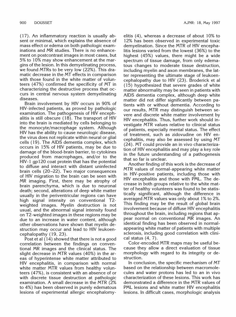
All HIV-positive patients with HIV encephalitishad cognitive dysfunction and deep white mat-ter hyperintensities on MR images (Table 1).The clinical, biological, and imaging features ofHIV-positive patients with PML are summarizedin Table 2. In one patient, PML was confirmedhistologically, although clinical and MR imageswere highly suggestive of PML. The typical MRcharacteristics of HIV encephalitis and PML le-sions are displayed in Figures 1 and 2, respec-tively.
The mean MTR value of hyperintensities inthe deep white matter in HIV encephalitis was40% 6 3.8% (mean 6 standard deviation). Thehighest MTR value of an HIV encephalitis lesionwas 45% (patient 9, group 1). The lowest valuefound was 36% (patient 8, group 1). For PMLlesions, the mean MTR value was 22% 6 2.3%.Highest and lowest MTR values in PML lesionswere, respectively, 29% (patient 2, group 2) and14% (patient 7, group 2). The difference be-tween the MTR of HIV and PML lesions wassignificant, with P 5 .0001. Figure 4 shows col-or-encoding MTR maps in both HIV encephalitisand PML lesions.
The mean MTR value of the normal-appear-ing white matter from group 3 (healthy volun-
AJNR: 18, May 1997

Fig 1. HIV encephalitis (patient 4).A, T2-weighted image shows deep white
matter hyperintensity in the periventricularareas (arrows).
B, Magnetization transfer image with cur-sors indicating ROI in one lesion site (arrow-head) and normal-appearing white matter inanother site (arrow).
Fig 2. PML (patient 8).A T2-weighted image shows a parietooc-
cipital lesion attributed to PML (arrow).B, Magnetization transfer image with one
cursor at the lesion site and one cursor innormal-appearing white matter (arrow-head).
AJNR: 18, May 1997 MAGNETIZATION TRANSFER 897
teers, 99 sites) was 47% 6 2.3%. For group 1,the mean MTR value of normal-appearing whitematter averaged from 110 sites, because sevensites were involved by HIV encephalitis, was44% 6 2.6%. For group 2, the mean MTR valuecalculated in 82 sites, eight sites being involvedby PML, was 46% 6 3.3%. Statistical analysiswith Kruskal-Wallis analysis of variance re-vealed a difference between the three groups,with P 5 .009.
Discussion
The imaging features of HIV encephalitis andPML lesions in HIV-positive patients have been
described extensively (12–16). Usually, suchlesions are detected by MR imaging and do notrequire histologic confirmation by cerebral bi-opsy (16). Nevertheless, there are cases ofatypical presentation and, in addition, superim-posed disease processes are frequently presentin infected patients. In our series, we obtainedpathologic proof in only one patient with PML;however, in both groups, the clinical and imag-ing features of these diseases were typical. Forthe purpose of this study, we ensured the diag-nosis of PML with a polymerase chain reactiontest for the JC virus either in the cerebrospinalfluid or in blood. All patients from group 2 diedof PML, whereas four patients in group 1 died of

Fig 3. Magnetization transfer imagesin healthy volunteer. Cursors indicate theROIs for MTR measurement in the nor-mal-appearing white matter.
898 DOUSSET AJNR: 18, May 1997
cachexia or nonneurologic infections. Differen-tiating between PML and HIV encephalitis is crit-ical because the prognosis is much better forthe latter and there is the possibility of reversalof neurologic dysfunction with the use ofzidovudine. In PML, on the other hand, there isno treatment and death usually occurs within 6months.
The pathogenesis of PML and HIV encephali-tis is very distinct. PML, which has a prevalenceof 4% among patients with the acquired immu-nodeficiency syndrome (AIDS), is due to anopportunistic agent, JC papovavirus, that growsin oligodendrocytes. The virus destroys oligo-dendroglial cells and therefore the myelinsheaths. Demyelination develops first at thejunction of the cortex and white matter and, inmost cases, in the posterior areas of the brain,in the parietal, occipital, and cerebellar lobes
TABLE 1: Magnetization transfer ratio (MTR) values for sites ofHIV encephalitis lesions and normal-appearing white matter in 13HIV-positive patients (group 1)
Patient Sex/Age, y
MTR Values, %
HIVEncephalitis
Lesions
Normal-AppearingWhite Matter
1 F/52 43 452 M/35 40 453 M/40 39 424 F/32 41 465 F/31 41 436 F/26 39 427 M/30 40 438 M/47 36 439 M/31 45 47
10 F/39 40 4611 F/47 38 4212 M/26 39 4413 M/37 43 46

Fig 4. Color-encoded MTR maps.A, In a healthy volunteer, a majority of red spots within the white matter indicates MTR values of over 44%.B, In patient 9, with a biopsy-proved PML lesion in the right parietal lobe, cold colors indicate low MTR within the core of the
demyelinating process.C, In patient 10, with HIV encephalitis, diffuse abnormality is seen as orange spots within the white matter, an indicator of a much less
destructive process than that in patient 9 (Fig 4B).
TABLE 2: Clinical and imaging features and magnetization transfer ratio (MTR) values for sites of HIV encephalitis and normal-appearingwhite matter in 10 progressive multifocal leukoencephalopathy (PML) (group 2)
Patient Sex/Age, y Neurologic DeficitCSF JC
Virus PCR*PML Lesion Site
MTR Values, %
PMLLesions
Normal-AppearingWhite Matter
1 F/34 R hemiparesia 1 L frontal parietal 17 482 M/50 L hemiparesia 1 R parietal occipital 28 48
L hemianopsia3 M/61 R hemiparesia 1 L frontal parietal 25 444 M/34 L hemianopsia 1 R occipital 23 41
Sensitive disturbance5 M/30 Gait disturbance 1 L parietal 18 44
Sensitive disturbance6 M/30 R hemiparesia 1 L parietal occipital 29 44
Sensitive disturbance7 M/24 Ataxia 1 Cerebellum 14 48
Sensitive disturbance8 M/63 L hemianopsia 1 R parietal occipital 22 489 M/32† L hemiparesia 2 R frontal parietal 22 44
10 M/31 R hemianopsia 1 L occipital temporal 26 47
* Polymerase chain reaction for JC papovavirus within the cerebrospinal fluid.† Patient with biopsy-proved PML.
AJNR: 18, May 1997 MAGNETIZATION TRANSFER 899
(17). An inflammatory reaction is usually ab-sent or minimal, which explains the absence ofmass effect or edema on both pathologic exam-inations and MR studies. There is no enhance-ment on postcontrast images in most cases, but5% to 10% may show enhancement at the mar-gins of the lesion. In this demyelinating process,we found MTRs to be very low (22%). This dra-matic decrease in the MT effects in comparisonwith those found in the white matter of volun-teers (47%) confirmed the specificity of MT incharacterizing the destructive process that oc-curs in central nervous system demyelinatingdiseases.
Brain involvement by HIV occurs in 90% ofHIV-infected patients, as proved by pathologicexamination. The pathogenesis of HIV enceph-alitis is still obscure (18). The transport of HIVinto the brain is mediated by cells belonging tothe monocyte/macrophage system. AlthoughHIV has the ability to cause neurologic disease,the virus does not replicate within neural or glialcells (19). The AIDS dementia complex, whichoccurs in 15% of HIV patients, may be due todamage of the blood-brain barrier, to cytokinesproduced from macrophages, and/or to theHIV-1 gp120 coat protein that has the potentialto diffuse and interact with distant uninfectedbrain cells (20–22). Two major consequencesof HIV migration to the brain can be seen withMR imaging. First, there may be atrophy ofbrain parenchyma, which is due to neuronaldeath; second, alterations of deep white matterusually in the periventricular regions can havehigh signal intensity on conventional T2-weighted images. Myelin destruction is notusual, and the abnormal signal intensity foundon T2-weighted images in these regions may bedue to an increase in water content, althoughother observations have shown that myelin de-struction may occur and lead to HIV leukoen-cephalopathy (19, 23).
Post et al (14) showed that there is not a goodcorrelation between the findings on conven-tional MR images and the clinical status. Theslight decrease in MTR values (40%) in the ar-eas of hyperintense white matter attributed toHIV encephalitis, in comparison with normalwhite matter MTR values from healthy volun-teers (47%), is consistent with an absence of orwith discrete tissue destruction at pathologicexamination. A small decrease in the MTR (2%to 4%) has been observed in purely edematouslesions of experimental allergic encephalomy-
900 DOUSSET
elitis (4), whereas a decrease of about 10% to12% has been observed in experimental toxicdemyelination. Since the MTR of HIV encepha-litis lesions varied from the lowest (36%) to thehighest (45%) values, there might be a widespectrum of tissue damage, from only edema-tous changes to moderate tissue destruction,including myelin and axon membranes, the lat-ter representing the ultimate stage of leukoen-cephalopathy due to HIV (23). Broderick et al(15) hypothesized that severe grades of whitematter abnormality may be seen in patients withAIDS dementia complex, although deep whitematter did not differ significantly between pa-tients with or without dementia. According toour results, MTR may distinguish between se-vere and discrete white matter involvement byHIV encephalitis. Thus, further work should in-vestigate MTR values relative to clinical statusof patients, especially mental status. The effectof treatment, such as zidovudine on HIV en-cephalitis, may also be assessed by using MT(24). MT could provide an in vivo characteriza-tion of HIV encephalitis and may play a key rolein the future understanding of a pathogenesisthat so far is unclear.
Another finding of this work is the decrease ofMTR values of normal-appearing white matterin HIV-positive patients, including those withHIV encephalitis and those with PML. The de-crease in both groups relative to the white mat-ter of healthy volunteers was found to be statis-tically significant, although the difference inaveraged MTR values was only about 1% to 2%.This finding may be the result of global braininvolvement because of diffuse HIV encephalitisthroughout the brain, including regions that ap-pear normal on conventional MR images. Anidentical finding has been observed in normal-appearing white matter of patients with multiplesclerosis, including good correlation with clini-cal status (4, 7).
Color-encoded MTR maps may be useful be-cause they allow a direct evaluation of tissuemorphology with regard to its integrity or de-struction.
In conclusion, the specific mechanism of MTbased on the relationship between macromole-cules and water protons has led to an in vivocharacterization of these lesions. This work hasdemonstrated a difference in the MTR values ofPML lesions and white matter HIV encephalitislesions. In difficult cases, morphologic analysis
AJNR: 18, May 1997

AJNR: 18, May 1997 MAGNETIZATION TRANSFER 901
of the lesion by means of MT may contribute tothe proper diagnosis.
AcknowledgementWe thank Ray Cooke for his invaluable linguistic help.
References1. Wolff SD, Balaban RS. Magnetization transfer contrast (MTC) and
tissue water proton relaxation in vivo. Magn Reson Med 1989;10:135–144
2. Morrison C, Henkelman RM. A model for magnetization transfer intissues. Magn Reson Med 1995;33:475–482
3. Harrisson R, Bronskill MJ, Henkelman RM. Magnetization transferand T2 relaxation components in tissue. Magn Reson Med 1995;33:490–496
4. Dousset V, Grossman RI, Ramer KN, et al. Experimental allergicencephalomyelitis and multiple sclerosis: lesion characterizationwith magnetization transfer imaging. Radiology 1992;182:483–491
5. Dousset V, Brochet B, Vital A, et al. Lysolecythin- induced myeli-nation in primates: in vivo study by magnetic resonance imagingand magnetization transfer. AJNR Am J Neuroradiol 1995;16:225–231
6. Gass A, Barker GJ, Kidd D, et al. Correlation of magnetizationtransfer ratio with clinical disability in multiple sclerosis. AnnNeurol 1994;36:62–67
7. Hiehle JF, Lenkinski RE, Grossman RI, et al. Correlation of spec-troscopy and magnetization transfer in the evaluation of demyeli-nating lesions and normal appearing white matter in multiplesclerosis. Magn Reson Med 1994;32:285–293
8. Loevner LA, Grossman RI, McGowan JC, Ramer KN, Cohen JA.Characterization of multiple sclerosis plaques with T1-weightedMR and quantitative magnetization transfer. AJNR Am J Neurora-diol 1995;16:1473–1479
9. Tomiak MM, Rosenblum JD, Prager JM, Metz CE. Magnetizationtransfer: a potential method to determine the age of multiplesclerosis lesions. AJNR Am J Neuroradiol 1994;15:1569–1574
10. Filippi M, Campi A, Dousset V, et al. A magnetization transferimaging study of normal-appearing white matter in multiple scle-rosis. Neurology 1995;45:478–481
11. Wolff SD, Balaban RS. Magnetization transfer imaging: practicalaspects and clinical applications. Radiology 1994;192:593–599
12. Guilleux MH, Steiner RE, Young IR. MR imaging in progressivemultifocal encephalopathy. AJNR Am J Neuroradiol1986;7:1033–1035
13. Hansman Witheman ML, Post MJ, Berger JR, Tate LG, Bell MD,Limonte LP. Progressive multifocal leukoencephalopathy in 47HIV-seropositive patients: neuroimaging with clinical and patho-logic correlation. Radiology 1993;187:233–240
14. Post MJD, Berger JR, Quencer RM. Asymptomatic and neurolog-ically symptomatic HIV-seropositive individuals: prospectiveevaluation with cranial MR imaging. Radiology 1991;178:131–139
15. Broderick DF, Wippold FJ II, Clifford DB, Kido D, Wilson BS.White matter lesions and cerebral atrophy on MR images in pa-tients with and without AIDS dementia complex. AJR Am J Roent-genol 1993;161:177–181
16. Sarrazin JL, Soulie D, Derosier C, Lescop J, Schill H, CordolianiYS. MRI patterns of progressive multifocal leukoencephalopathy.J Neuroradiol 1995;22:172–179
17. Richardson EP, Webster HF. Progressive multifocal leukoenceph-alopathy: its pathological features. Prog Clin Biol Res 1983;105:183–190
18. Brew BJ. The clinical spectrum and pathogenesis of HIV enceph-alopathy, myelopathy, and peripheral neuropathy. Curr OpinNeurol 1994;7:209–216
19. Artigas J, Grosse, Niedobitek F. Neuropathology of AIDS. In:Artigas J, Grosse, Niedobitek F, eds. The Central Nervous Systemin AIDS. Berlin, Germany: Springer-Verlag; 1993:79–200
20. Power C, Kong PA, Crawford TO, et al. Cerebral white matterchanges in acquired immunodeficiency syndrome dementia: al-terations of the blood-brain-barrier. Ann Neurol 1993;34:339–350
21. Dawson VL, Dawson TM, Uhl GR, Snyder H. Human immunode-ficiency virus type 1 coat protein neurotoxicity mediated by nitricoxide in primary cortical cultures. Proc Natl Acad Sci U S A1993;90:3256–3259
22. Toggas SM, Masliah E, Rockenstein EM, Rall GF, Abraham CR,Mucke L. Central nervous system damage produced by expres-sion of HIV-1 coat protein gp120 in transgenic mice. Nature1994;367:188–193
23. Budka H. Human immunodeficiency virus (HIV)–induced diseaseof the central nervous system: pathology and implications for thepathogenesis. Acta Neuropathol 1989;77:225–236
24. Sidtis JJ, Gatsonis C, Price RW, et al. Zidovudine treatment of theAIDS dementia complex: results of a placebo-controlled trial. AnnNeurol 1993;33:343–349