Embed Size (px)
Citation preview
“RISK STRATIFICATION OF PATIENTS WITH HYPERTROPHIC
CARDIOMYOPATHY AND ASSESSMENT OF BIVENTRICULAR
DIASTOLIC FUNCTION BY PULSE/TISSUE DOPPLER
ECHOCARDIOGRAPHIC IMAGING”
Dissertation submitted to
THE TAMIL NADU DR. M.G.R. MEDICAL UNIVERSITY
In partial fulfillment of the requirements for the award of the degree of
D.M. BRANCH - II CARDIOLOGY
MADRAS MEDICAL COLLEGE
And
RAJIV GANDHI GOVERNMENT GENERAL HOSPITAL
CHENNAI-600003
THE TAMIL NADU DR.M.G.R MEDICAL UNIVERSITY
CHENNAI – 600 032
AUGUST 2013
CERTIFICATE
This is to certify that the dissertation entitled “RISK
STRATIFICATION OF PATIENTS WITH HYPERTROPHIC
CARDIOMYOPATHY AND ASSESSMENT OF
BIVENTRICULAR DIASTOLIC FUNCTION BY PULSE/TISSUE
DOPPLER ECHOCARDIOGRAPHIC IMAGING” is the bonafide
original work of Dr.A.MURALIDHARAN in partial fulfillment of the
requirements for D.M. Branch-II (CARDIOLOGY) examination of
THE TAMILNADU DR.M.G.R. MEDICAL UNIVERSITY to be held
in August 2013.The period of post-graduate study and training was from
August 2010 to July 2013.
Dr.V. KANAGASABAI, M.DDeanMadras Medical College &RGGGHChennai - 600003.
Prof.V. E. DHANDAPANI, DMProfessor & Head, Department of Cardiology,Madras Medical College & RGGGH,Chennai- 600003.
DECLARATION
I, Dr.A.MURALIDHARAN, solemnly declare that this
dissertation entitled, “RISK STRATIFICATION OF PATIENTS
WITH HYPERTROPHIC CARDIOMYOPATHY AND
ASSESSMENT OF BIVENTRICULAR DIASTOLIC FUNCTION
BY PULSE/TISSUE DOPPLER ECHOCARDIOGRAPHIC
IMAGING” is a bonafide work done by me at the department of
Cardiology, Madras Medical College and Government General Hospital
during the period 2010 – 2013. under the guidance and supervision of
the Professor and Head of the department of Cardiology of Madras
Medical College and Government General Hospital, Professor
V.E.DHANDAPANI M.D.D.M. This dissertation is submitted to The
Tamil Nadu Dr.M.G.R Medical University, towards partial fulfilment of
requirement for the award of D.M. Degree (Branch-II) in Cardiology.
Place : Chennai Dr.A.MURALIDHARANDate:
ACKNOWLEDGEMENT
I wish to express my respect and sincere gratitude to my beloved
teacher Prof. V. E. DHANDAPANI., D.M (CARDIOLOGY)
Professor and Head of Department of Cardiology for his valuable
guidance and encouragement throughout the study.
I am extremely thankful to our Prof. Dr.M.S RAVI, D.M,
PROF. DR. K. MEENAKSHI, D.M; Prof. Dr. D. MUTHUKUMAR,
D.M., Prof. Dr. N. SWAMINATHAN, D.M,
Prof. Dr. G. RAVISHANKAR, D.M; for their support and guidance
during the study.
I am also expressing my gratitude to my Assistant Professors
Dr.PALANISAMY, Dr. S. VENKATESAN, DR. MOORTHY,
Dr.MURUGAN, Dr .G. MANOHAR, Dr. RAJASEKAR RAMESH,
Dr. ELANGOVAN,Dr. PRATHAPKUMAR,for their valuable
guidance.
Last but not least, my sincere thanks to all the patients who
cooperated for the study.

INDEX
1 Introduction 1
2 Aim of the Study 3
3 Review of Literature 4
4 Materials and Methods 20
5 Results and Data Analysis 27
6 Discussion 49
7 Conclusion 60
Appendix
Bibliography
Master Chart
Proforma
Abbreviations & Acronyms
Ethical Committee Approval Letter
Consent Form
Plagiarism Certificate
1
INTRODUCTION
Hypertrophic cardiomyopathy is a common autosomal dominant
cardiovascular disease .
Dynamic obstruction to LV outflow can occur under resting or
physiologically provocable conditions in patients with HCM. various
phenotypic and genotypic heterogeneity are observed in HCM. HCM is
the most common genetically transmitted cardiovascular disease which
affects 1 in 500 people. The most frequent cause of sudden cardiac
death in young athletes is HCM1. HCM patients may have very severe
to negligible hypertrophy.HCM patients can have fibrosis and disarray
of cardiac myocytes which may be minimal to extensive .
HCM patients have a natural history which may range from being
aymptomatic to having protracted dyspnea and angina.
Sudden cardiac death may be the presenting event in some
hypertrophic cardiomyopathy patients 2 . Hypertrophic cardiomyopathy
was reported by Teare in 1958 as ‘asymmetrical hypertrophy of the
heart in young adults’1.
2
The WHO defined cardiomyopathies as ‘diseases of different and
often unknown etiology in which the dominant feature is cardiomegaly
and heart failure’1. In 1980 this definition was updated as ‘heart muscles
diseases of unknown cause’1.This differentiated HCM from heart
muscle diseases of known etiology, such as myocarditis.
Idiopathic hypertrophic subaortic stenosis, muscular subaortic
stenosis have been the alternate old nomenclature for hypertrophic
obstructive cardiomyopathy2 .
In 1995, a WHO/International Society and Federation of
Cardiology Task Force defined HCM as ‘left and/or right ventricular
hypertrophy, usually asymmetric and involving the interventricular
septum with predominant autosomal dominant inheritance involving
sarcomeric contractile proteins’3.

3
AIM OF THE STUDY:
1.Risk stratification of patients with Hypertrophic
Cardiomyopathy .
2.Assessment of Biventricular diastolic function in patients with
Hypertrophic cardiomyopathy by pulse/tissue doppler
echocardiographic imaging
4
REVIEW OF LITERATURE
HCM results by mutations encoding contractile proteins- β-
myosin heavy chain and myosin binding protein C , troponin T and I,
regulatory and essential myosin light chains, titin, α-tropomyosin, α-
actin, α-myosin heavy chain, and muscle LIM protein 4.
The clinical manifestations vary among patients, from
asymptomatic, to heart failure. Asymmetric hypertrophy of the
interventricular septum, with or without left ventricular outflow tract
obstruction is mostly diagnosed by echocardiography . We have to
exclude other causes of hypertrophy, like hypertension, coarctation of
aorta and aortic stenosis 5.
Athletes with physiological hypertrophy of left ventricle
confound the diagnosis of HCM . The prevalence of HCM is 1:500 in
young adults.
Genetic screening of all first degree relatives is recommended.
Most of the Mutations in HCM happen as single nucleotide substitutions
or “missense” mutations5.
5
Pathology
Severe left ventricular hypertrophy and small LV size are gross
features seen in HCM.
The ventricular septum is mainly hypertrophied.Hypertrophy
may also be symmetrical or affect posterior wall, apex or middle part of
the LV .Asians have a prevalence of about 10 % in midventricular
obstruction.
Involvement of right ventricle can also be found in infants and
children .End stage dilated cardiomyopathy is seen in 5% of patients
with HCM. Dilated cardiomyopathy found in end stage of HCM has
widespread fibrosis, heart wall thinning and LV dilation 6.
Raised LVEDP causes dilation of left atrium.Left atrium is also
dilated by diastolic relaxation abnormalities and mitral insufficiency
resulting from left ventricular outflow tract obstruction or related mitral
valve abnormalities . The cause of mitral regurgitation in hypertrophic
obstructive cardiomyopathy was originally ascribed to be due to the
Venturi effect created by systolic flow acceleration in the left ventricular
outflow tract pulling the anterior mitral leaflet of mitral valve towards
the ventricular septum causing obstruction and regurgitation7 .
6
A study suggested, that at the beginning of ejection the mitral
valve leaflets are projecting into a narrow left ventricular outflow tract
causing fast forward flow which is the main force that drags the leaflets
toward the septum causing obstruction8. The systolic anterior motion of
the mitral leaflets prevents the closing of mitral valve causing mitral
insufficiency.
Differences between physiologic and pathologic hypertrophy
Left Ventricular Hypertrophy
Blood pressure and volume increase results in increase in muscle
mass of heart.cardiac myocyte replication rate is not rapid, hence its
mostly results by an enlargement in size of the cardiac myocyte than a
rise in number .
During growth,pregnancy and in athlete’s heart physiological
hypertrophy occurs. Pathological cardiac hypertrophy occurs in
hypertension. There are many differences among physiological and
pathological myocyte hypertrophy LV hypertrophy is an adaptive
reaction that helps the myocardium to preserve cardiac output 9. Cardiac
remodelling involves left ventricular dilation, accumulation of collagen
and cardiac myocyte loss.

7
Athlete’s heart is a form of physiologic cardiac hypertrophy
which is not associated with fibrosis or dysfunction
Concentric and Eccentric myocyte Hypertrophy :
Physiological hypertrophy induced by exercise may be concentric
or eccentric. Increased volume load of ventricle produces eccentric
hypertrophy. Swimming and running are examples of endurance
exercise which increase volume overload. In Eccentric cardiac
hypertrophy there is a raise in the length of myocytes, which results in
dilation of Left Ventricle.
Pressure overload causes concentric hypertrophy.There is left
ventricle wall thickening and LV dilatation is mininal. Isometric
exercises cause pressure overload .
Stimulus for Hypertrophy Resulting From Exercise :
Exercise results in the discharge of growth factors and
neurotransmitters, which stimulate receptors on different cell types to
cause a biological reaction.
Insulin-like growth factor 1 concentrations are increased in
professional football players. IGF1 levels have a good correlation with
LV mass index and LV end-diastolic dimension index10.
8
During exercise norepinephrine a neurotransmitter is released.
Norepinephrine levels did not have a correlation with thickness of left
ventricle myocardium or LV cavity dimensions .Hence norepinephrine
does not increase cardiac myocyte size
Stimuli For Pathological Hypertrophy
G Protein-Coupled Receptors
In cardiac failure patients Angiotensin II and endothelin-1 are
upregulated. Heart myocytes release Angiotensin II and endothelin-1
during mechanical stress . Angiotensin II and endothelin-1 signal by
combining to G protein-coupled receptors .
Calcineurin
The key enzyme involved in pathological hypertrophy is
calcineurin.In patients with hypertrophy of LV and cardiac failure
calcineurin activity is increased,
HCM- signalling pathways
In HCM signalling pathways have not been clearly defined. It has
been suggested that Mutant proteins in HCM could stimulate
hypertrophy by signalling pathways included in pressure overload type
of hypertrophy.
9
Angiotensin II and endothelin I are increased in individuals with
HCM.
Hypertrophy in HCM
Hypertrophy seen mostly marked in the area of the anterior
papillary muscle and Superior part of septum.Systolic anterior motion of
mitral leaflet causes dynamic obstruction in HCM. Increased afterload
stress results from dynamic obstruction.
Genetically determined sarcomeric protein regional dysfunction
causes abnormal stresses on nearby regions of normal myocardium..
Clinical findings
Cardiomegaly usually found on a chest x-ray, or
electrocardiographic changes can be the initial presentation .Fatigue ,
breathlessness on exertion, palpitations, angina, syncope or sudden
death may be the initial symptomology of presentation.
On physical examination, in pulse there is a rise and rapid
descent. Bulge of precordium and outward displacement of apical
impulse are present when there is cardiac enlargement. S4 causing a
bifid apical impulse is frequently seen. Mid systolic murmur with

10
variable intensity is present in the left sternal border and apex
.Pansystolic murmur of mitral regurgitation may be present at the apex
The midsystolic murmurs vary with maneuvers that result in
increase or decrease left ventricle outflow gradient.
Softer Mid systolic murmurs along the left sternal border may be
present in non-obstructive HCM also.The intensity of this murmur is
softer. Atrial fibrillation occurs in endstage dilated hypertrophic
cardiomyopathy. Wolff-Parkinson-White syndrome may be present in
patients with hypertrophic cardiomyopathy rarely.
Sudden cardiac death can be the only presenting symptom in
hypertrophic cardiomyopathy 11 .End stage is seen in about 5 % of
patients with HCM and is characterized by systolic failure associated
with left ventricular wall thinning and increased ventricular volume 12 .
Diastolic Dysfunction in HCM
Most of HCM patients have abnormalities in relaxation and
filling.In diastole ,isovolumic relaxation stage is considerably
lengthened and it causes a reduced rate and volume of filling. The
contribution of atrial systole to ventricular filling is considerably
increased13.
11
Laboratory studies
Electrocardiogram
In nearly every individual with HCM there are
electrocardiographic abnormalities seen. left ventricular hypertrophy
can be assessed by voltage criteria and Left atrial enlargement can be
seen. ST-T changes are present. Q waves which are pathological can be
seen.
Wolff-Parkinson-White pattern or syndrome can be detected in
electrocardiogram by the presence of delta wave.Genetic Mutations
encoding AMP-activated protein kinase (PRKAG2) and lysosome
associated membrane protein 2 are associated with WPW syndrome .
24 hour Holter monitoring and treadmill stress testing can be
used for finding out arrhythmias and stratification of risk . Nonsustained
ventricular tachycardia detected by 24 hrs Holter monitoring and a
blood pressure response which is abnormal to treadmill testing are risk
factors for sudden death7.
Echocardiographic evaluation of HCM
For the diagnosis of hypertrophic cardiomyopathy echocardiography
is a very important tool. The diagnosis can be established by
12
echocardiography,it is also very useful for followup and stratification of
risk for sudden death. Probable or definite echocardiographic evidence
of HCM is detected on the basis of recognition of a hypertrophied left
ventricle with inter ventricular septum /posterior wall thickness ≥1.3
.When the right ventricular wall thickness exceeds 4 mm the right
ventricle is considered to be involved too . Asymmetric left ventricular
hypertrophy is the hallmark of HCM, the hypertrophy is more marked in
the interventricular septum .Concentric hypertrophy has also been seen
in some individuals. Apical hypertrophy and isolated hypertrophy of
anterior wall or the mid part of left ventricle can also be seen. The mid
ventricular obstruction may lead to the development of an apical left
ventricular aneurysm.Left atrial enlargement can be seen due to result of
mitral regurgitation and diastolic dysfunction. Systolic anterior motion
of anterior mitral leaflet found in patients with hypertrophic obstructive
cardiomyopathy is seen . Color flow Doppler allows to find the location
of obstruction. Continuous wave Doppler can be used to find the left
ventricular outflow tract gradient . The severity of Mitral regurgitation
can be found out by continuous wave doppler. The primary mitral valve
abnormalities are better detected by Transesophageal echocardiography
.The primary mitral valve abnormalities can cause mitral regurgitation
in patients with HCM.
13
When there is no resting gradient ,exercise Stress echo can be
used to bring out the latent gradient in HCM . Doppler flow velocities
across miral inflow and pulmonary vein can be used to find out diastolic
dysfunction in hypertrophic cardiomyopathy .
For the detection of diastolic dysfunction in HCM ,tissue Doppler
is very useful and more sensitive than pulse doppler .Using tissue
Doppler we can detect HCM in carriers of the genetic mutation even
before the development of hypertrophy
For identification of hypertrophic cardiomyopathy carriers
reduced systolic and diastolic tissue doppler velocities have a high
sensitivity and specificity
The prominent diastolic abnormality seen in patients with HCM is
impaired LV relaxation . For the detection of high filling pressures,
Mitral inflow and pulmonary venous flow assessments are not
sensitive. LV filling pressure, peak E velocity of mitral inflow and
mitral annular velocities (Ea) detected by tissue doppler have a good
correlation.The realaxation abnormalities can be detected even before
the start of hypertrophy in individuals with HCM . The response to
septal ablation and surgery can be monitored by tissue doppler
14
Computed tomography and Cardiac MRI :
CT and Cardiac MRI are very useful when thoracic deformities
prevent satisfactory cardiac visualization by transthoracic
echocardiography. Cardiac MRI has also demonstrated that mitral
leaflet elongation is present in hypertrophic cardiomyopathy
independently of other phenotypic variants indicating that the mitral
abnormalities are primary, thus, their important role in the
pathophysiology of the obstruction in LVOT .
Gadolinium magnetic resonance imaging late enhancement
allows detection of the amount of myocardial fibrosis and is a predictor
of systolic dysfunction
Cardiac catheterization :
Cardiac catheterization helped in the initial understanding of the
physiopathology of hypertrophic cardiomyopathy .Noninvasive imaging
techniques like Doppler-echocardiography, computed tomography, and
magnetic resonance imaging have replaced cardiac catheterization for
diagnostic purposes. Nowadays, this method is only used before planned
surgical treatment or percutaneous interventions for septal reduction.
15
Complications of HCM :
Main complications in children with HCM are syncope and sudden
death. Sudden cardiac death may be initial presentation of the disease
and is considered to be secondary to ventricular tachycardia and
fibrillation caused by myocardial fibrosis and ischemia . Sudden death
occurs more often in older children with hypertrophic cardiomyopathy
either during strenuous sport activities or at rest but is infrequent in
infants who are more prone to present and die with congestive heart
failure specially in secondary forms .
Arrhythmias: Supraventricular and nonsustained ventricular
tachycardias, were found in almost a third of pediatric and adolescent
patients with hypertrophic cardiomyopathy studied by ambulatory
electrocardiography Rarely, 3rd degree atrioventricular block is found
in children with hypertrophic cardiomyopathy. They could present with
near syncope or syncopal attacks as the first manifestation of the disease
Nonsustained ventricular tachycardia:
Most of the patients in this age group do not have nonsustained
ventricular tachycardia in holter monitoring.Nonsustained ventricular
tachycardia can be seen during 24 hrs holter monitoring in 20% to 30%
of adults14.
16
Evolving phenotype
Children with hypertrophic cardiomyopathy may evolve to dilated
or restrictive cardiomyopathy phenotypes in 5% of the cases. In both
circumstances they have a poor prognosis and become candidates for
heart transplantation .
Infectious endocarditis
Infectious endocarditis is a rare complication in hypertrophic
cardiomyopathy Infectious endocarditis can affect the anterior mitral
valve leaflet in the ventricular side.
Stroke
Ischemic stroke is somewhat frequent and a cause of death in
adult hypertrophic cardiomyopathy but has not been seen in children .
Medical treatment in HCM:
Beta-blockers and verapamil are used on an empirical basis.
Most physicians use beta-blockers as the firstline drug in the treatment
of HCM.Verapamil is not preferred as the initial drug.
Beta-blockers or verapamil do not have evidence for prevention
of sudden cardiac death.
17
Symptom relief can be seen in most of patients who are on beta-
blockers or verapamil.Medical therapy is used for only symptomatic
patients.Medical therapy cannot be used prophylactically.
Surgery in HCM :
Surgical indication for patients with HCM are a very high
gradient, severe symptoms and failure of drug treatment .Myectomy has
become the primary option for these patients.five to ten grams of muscle
are resected from tha basal septum by surgery.
The LVOT gradient is reduced in more than 90% of patients and
provides symptomatic improvement in functional status15.
DDD pacing to relieve LVOT obstruction
DDD pacing with shortened A-V delay was used to reduce the
outflow gradient and improve quality of life in patients with HCM who
are failures of medical treatment. The mechanism of action causing this
lowering of left ventricular outflow gradient is not known. Several trials
have found out that DDD pacing is not effective in the treatment of
HCM16.They have suggested only a placebo role in the symptomatic
improvement seen in patients who have been treated with DDD pacing.
Now DDD pacing is not recommended for treatment of patients with
obstructive HCM.
18
Alcohol septal ablation :
The first septal artery when temporarily occluded by an
angioplasty balloon caused a reduction in LVOT gradient. This formed
the basis for alcohol septal ablation as a treatment option for
HCM17.Contrast echocardiography is utilised for assessing the perfusion
bed supplied by first septal artery and it is marked out and it predicts
the infarct size following alcohol injection.
RISK STRATIFICATION IN HCM
Hypertrophic cardiomyopathy is the most frequent etiology of
sudden cardiac death in young . Male preponderance is seen among
athletes .
The primary prevention risk factors for SCD in HCM include
family history of SCD, unexplained recent syncope, runs of
nonsustained ventricular tachycardia on 24 hrs Holter monitoring,
hypotensive response to exercise and maximum left ventricular wall
thickness greater than 30 mm18 .
Sustained ventricular tachycardia and ventricular fibrillation are
the most important causes of SCD . Increased SCD risk is caused due to
excess sympathetic activation during exercise.

19
SUDDEN CARDIAC DEATH PREVENTIVE MEASURES IN
HCM
Betablockers and verapamil do not prevent sudden cardiac death
in hypertrophic cardiomyopathy
Implantation of ICD is the most effective procedure to prevent
sudden cardiac death in HCM. ICD is primarily used for secondary
prevention and in primary prevention of sudden cardiac death in HCM
and this has been proved in many studies19.
ICD implantation in HCM is a class I indication for secondary
prevention and classIIB for primary prevention as given in guidelines of
AHA/ACC/NASPE 20.
The disadvantage of ICD is its high cost.History of prior cardiac
arrest or spontaneous sustained ventricular tachycardia in patients is a
mandatory indication for ICD implantation.
For primary prevention of sudden cardiac death in children, ICD
implantation is not preferred.Amiodarone can be used as bridge to ICD
implantation in children till they reach adulthood.
20
MATERIALS AND METHODS
This was a retrospective cross-sectional study done between
March 2012 and March 2013 at the department of cardiology,
Government General Hospital Chennai. The study cohort comprises 40
patients with Hypertrophic cardiomyopathy who are referred to the
department of cardiology, for evaluation or follow up.
SETTING : Department of Cardiology, Rajiv Gandhi
Government General,
Madras Medical College, Chennai.
DESIGN OF STUDY : PProspective observational study
PERIOD OF STUDY : 1 Year
SAMPLE SIZE : 40
INCLUSION CRITERIA
• Probable or definite echocardiographic evidence of HCM
on the basis of recognition of a hypertrophied left ventricle with inter
ventricular septum /posterior wall thickness ≥1.3

21
EXCLUSION CRITERIA
1) Non-diagnostic echocardiographic studies
2) Other concomitant loading or systemic conditions that may lead
to left ventricular hypertrophy -Systemic hypertension ,Aortic
stenosis,Coarctation, or Structural LVOT obstruction
3) HCM patients who have undergone surgical or catheter septal
myectomy
22
STUDY PROCEDURE
All patients referred for echocardiography during this period were
included.
1) 2D, M-mode,Pulseand Tissue Doppler measurements taken.
2) Diastolic function of both Left ventricle and Right ventricle
evaluated.
3) Detecting and grading of the systolic anterior motion of the mitral
valve, LV outflow tract obstruction, intracavitary obstruction and
mitral regurgitation assessed.
4) Describing the pattern of left ventricular hypertrophy
DATA COLLECTION and ANALYSIS
• Interviewer will administer the proforma. Past medical
history and relevant history will be collected as per hospital record after
explaining aim of the study & getting written consent
• Demographic data such as age, gender, and clinical
presentation; ECG data and echocardiographic data will be described
and analysed.
• Echocardiography done on Philips HD7XE machine .
• 24 hour HOLTER study done for patients.

23
ECHOCARDIOGRAPHIC EVALUATION OF
BIVENTRICULAR DIASTOLIC FUNCTION IN HCM
Transthoracic Echocardiography was performed in all
hypertrophic cardiomyopathy patients with Philips HD7XE machine .
Interventricular septum thickness, left ventricular posterior wall
thickness, left ventricular end diastolic diameter, left ventricular end
systolic diameter, right ventricular free wall thickness, right ventricular
end diastolic diameter, fractional shortening of left ventricle were the
variables assessed .
The left ventricular diastolic function was determined from the
transmitral pulse doppler inflow patterns taken from the apical four-
chamber view by positioning sample volume at the tips of the mitral
leaflets during diastole.
Right ventricular diastolic function was assessed from the apical
4 chamber view by sample volume at the tips of tricuspid leaflets
during diastole.
peak velocity of E wave of tricuspid inflow , peak velocity of A
wave of tricuspid inflow, E/A ratio, deceleration time of E wave and
right ventricular isovolumic relaxation time were calculated for
assessment of right ventricular diastolic function.

24
TISSUE DOPPLER IMAGING
The tissue doppler velocity of myocardium was measured at
septal side of mitral valve annulus to determine Septal mitral annular
tissue Doppler velocity MV Ea and MV Aa . The tissue doppler velocity
of myocardium was measured at lateral side of tricuspid valve annulus
to determine lateral tricuspid annular tissue Doppler velocity TV Ea and
TVAa. In our study the patients were divided into two groups according
to their left ventricular outflow gradient gradient less than 30 mmHg
were the non-obstructive HCM group, and LV outflow gradient more
than 30 mmhg were the obstructive HCM group.
25
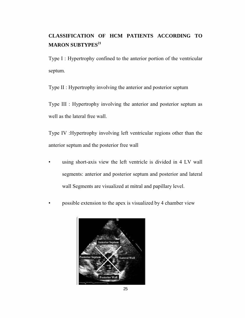
CLASSIFICATION OF HCM PATIENTS ACCORDING TO
MARON SUBTYPES21
Type I : Hypertrophy confined to the anterior portion of the ventricular
septum.
Type II : Hypertrophy involving the anterior and posterior septum
Type III : Hypertrophy involving the anterior and posterior septum as
well as the lateral free wall.
Type IV :Hypertrophy involving left ventricular regions other than the
anterior septum and the posterior free wall
• using short-axis view the left ventricle is divided in 4 LV wall
segments: anterior and posterior septum and posterior and lateral
wall Segments are visualized at mitral and papillary level.
• possible extension to the apex is visualized by 4 chamber view
26
Statistical Analysis
All the obtained data were analyzed by Medcalc
software.Continuous data are expressed as mean values ± SD.Student’s
t-test was used to assess the significant differences of mean values
between patients with non-obstructive and obstructive HCM. A p-value
≤0.05 was considered statistically significant.
Pearson correlation coefficient was used to assess the correlation
between right Ventricle and left ventricle diastolic parameters.
27
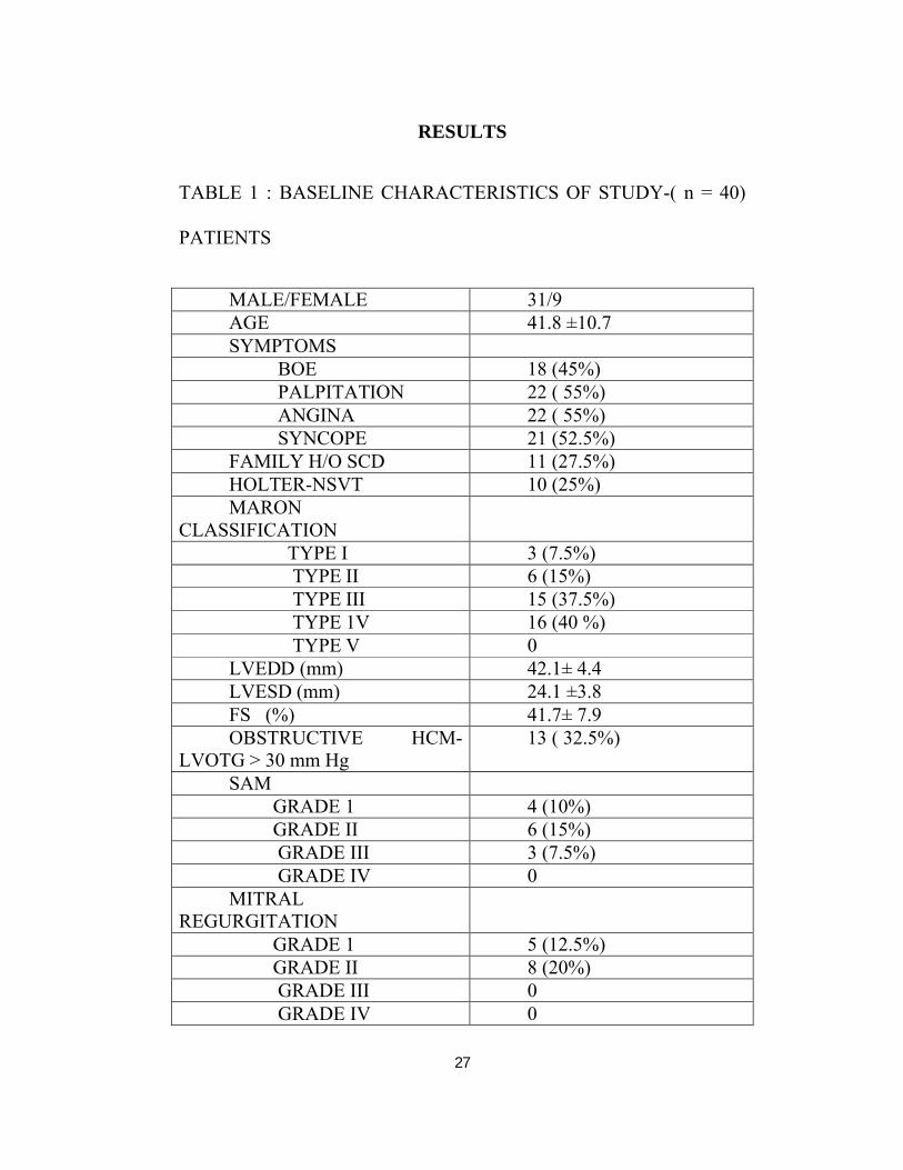
RESULTS
TABLE 1 : BASELINE CHARACTERISTICS OF STUDY-( n = 40)
PATIENTS
MALE/FEMALE 31/9AGE 41.8 ±10.7SYMPTOMS BOE 18 (45%) PALPITATION 22 ( 55%) ANGINA 22 ( 55%) SYNCOPE 21 (52.5%)FAMILY H/O SCD 11 (27.5%)HOLTER-NSVT 10 (25%)MARON
CLASSIFICATION TYPE I 3 (7.5%) TYPE II 6 (15%) TYPE III 15 (37.5%) TYPE 1V 16 (40 %) TYPE V 0LVEDD (mm) 42.1± 4.4LVESD (mm) 24.1 ±3.8FS (%) 41.7± 7.9OBSTRUCTIVE HCM-
LVOTG > 30 mm Hg13 ( 32.5%)
SAM GRADE 1 4 (10%) GRADE II 6 (15%) GRADE III 3 (7.5%) GRADE IV 0MITRAL
REGURGITATION GRADE 1 5 (12.5%) GRADE II 8 (20%) GRADE III 0 GRADE IV 0
28
The study cohort comprises 40 consecutive patients with
Hypertrophic cardiomyopathy who were referred to our department for
evaluation or follow up. There were 31 male and 9 female patients in
our study.
Mean age (± SD) was 41.8 years (± 10.7), for males 43.3 (± 9.6)
yrs, and for females 36.7 (± 13.5) yrs.
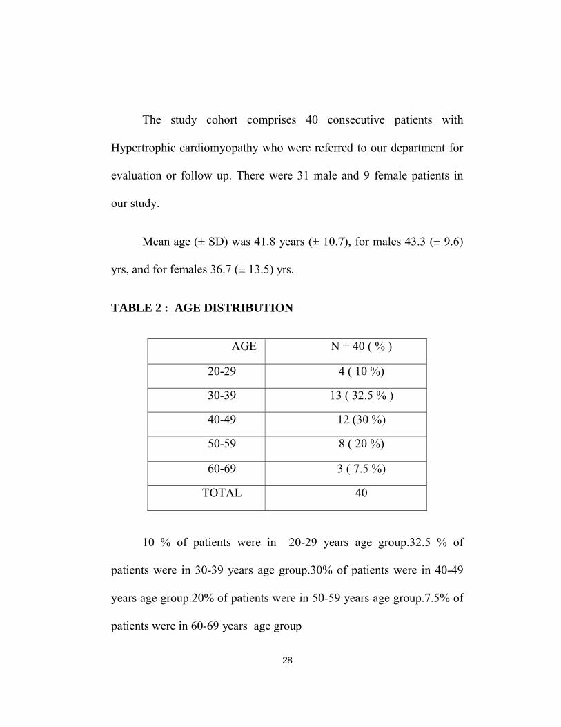
TABLE 2 : AGE DISTRIBUTION
AGE N = 40 ( % )
20-29 4 ( 10 %)
30-39 13 ( 32.5 % )
40-49 12 (30 %)
50-59 8 ( 20 %)
60-69 3 ( 7.5 %)
TOTAL 40
10 % of patients were in 20-29 years age group.32.5 % of
patients were in 30-39 years age group.30% of patients were in 40-49
years age group.20% of patients were in 50-59 years age group.7.5% of
patients were in 60-69 years age group
29
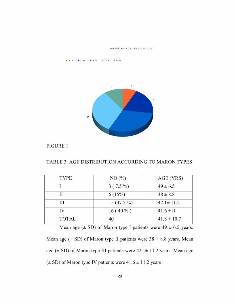
FIGURE 1
TABLE 3: AGE DISTRIBUTION ACCORDING TO MARON TYPES
TYPE NO (%) AGE (YRS)
I 3 ( 7.5 %) 49 ± 6.5
II 6 (15%) 38 ± 8.8
III 15 (37.5 %) 42.1± 11.2
IV 16 ( 40 % ) 41.6 ±11
TOTAL 40 41.8 ± 10.7
Mean age (± SD) of Maron type I patients were 49 ± 6.5 years.
Mean age (± SD) of Maron type II patients were 38 ± 8.8 years. Mean
age (± SD) of Maron type III patients were 42.1± 11.2 years. Mean age
(± SD) of Maron type IV patients were 41.6 ± 11.2 years .
30
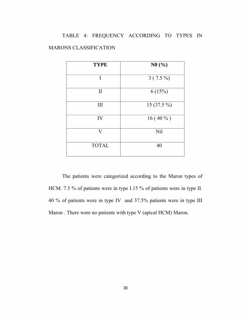
TABLE 4: FREQUENCY ACCORDING TO TYPES IN
MARONS CLASSIFICATION
TYPE N0 (%)
I 3 ( 7.5 %)
II 6 (15%)
III 15 (37.5 %)
IV 16 ( 40 % )
V Nil
TOTAL 40
The patients were categorized according to the Maron types of
HCM. 7.5 % of patients were in type I.15 % of patients were in type II.
40 % of patients were in type IV and 37.5% patients were in type III
Maron . There were no patients with type V (apical HCM) Maron.
FIGURE 2 :N0 OF PTS ACCORDING TO MARON TYPES
TABLE 5 : CATEGORIZATION OF OBSTRUCTIVE HCM
ACCORDING TO MARON TYPES
TYPE
1
II
111
1V
TOTAL
Left ventricular obstruction
No obstruction was found in 27 (67.5%) patients. 32.5 % of
patients had obstructive HCM (>30 mmHg LV outflow obstruction)
was found in 13 (32.5 %) cases,with a rest gradient mean(+SD) of 65 (
31.5) mmHg.
16( 40%)
31
N0 OF PTS ACCORDING TO MARON TYPES
TABLE 5 : CATEGORIZATION OF OBSTRUCTIVE HCM
ACCORDING TO MARON TYPES
NO (%) NO OF
OBSTRUCTIVE TYPE
3 ( 7.5 %) 0
6 (15%) 0
15 (37.5 %) 7 (46.7%)
16 ( 40 % ) 6 (40%)
40 ( 100%) 13 (32.5%)
Left ventricular obstruction
obstruction was found in 27 (67.5%) patients. 32.5 % of
patients had obstructive HCM (>30 mmHg LV outflow obstruction)
was found in 13 (32.5 %) cases,with a rest gradient mean(+SD) of 65 (
3(7%)6(15%)
15( 38%)
16( 40%)TYPE I
TYPE II
TYPE III
TYPE 1V
N0 OF PTS ACCORDING TO MARON TYPES
TABLE 5 : CATEGORIZATION OF OBSTRUCTIVE HCM
OBSTRUCTIVE TYPE
obstruction was found in 27 (67.5%) patients. 32.5 % of
patients had obstructive HCM (>30 mmHg LV outflow obstruction)
was found in 13 (32.5 %) cases,with a rest gradient mean(+SD) of 65 (±
TYPE I
TYPE II
TYPE III
TYPE 1V
32
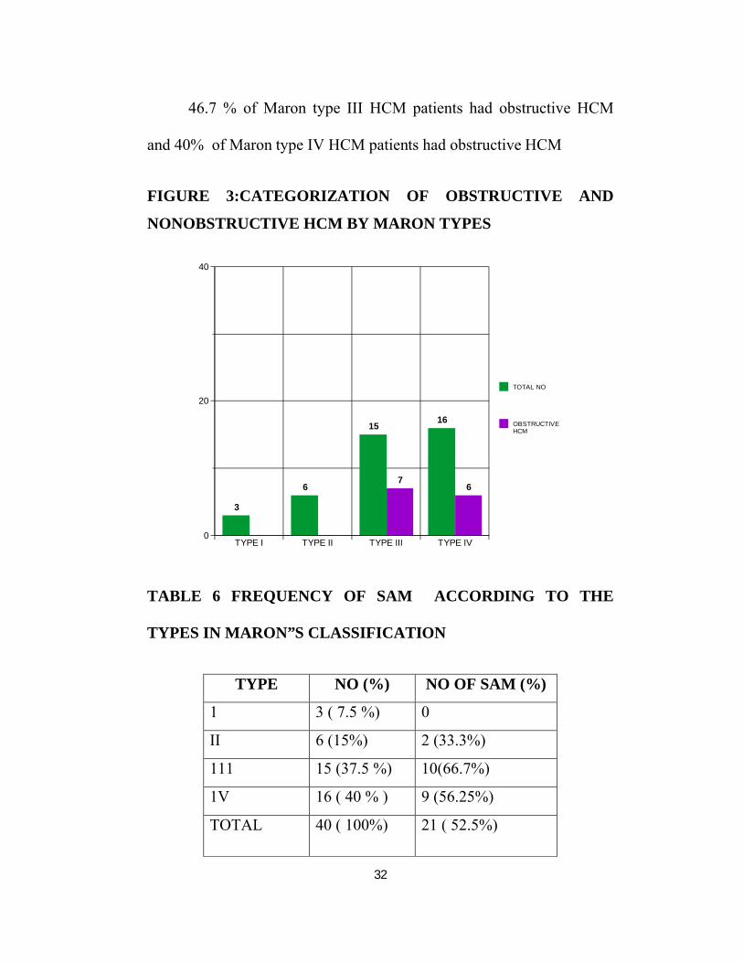
46.7 % of Maron type III HCM patients had obstructive HCM
and 40% of Maron type IV HCM patients had obstructive HCM
FIGURE 3:CATEGORIZATION OF OBSTRUCTIVE AND
NONOBSTRUCTIVE HCM BY MARON TYPES
TABLE 6 FREQUENCY OF SAM ACCORDING TO THE
TYPES IN MARON”S CLASSIFICATION
TYPE NO (%) NO OF SAM (%)
1 3 ( 7.5 %) 0
II 6 (15%) 2 (33.3%)
111 15 (37.5 %) 10(66.7%)
1V 16 ( 40 % ) 9 (56.25%)
TOTAL 40 ( 100%) 21 ( 52.5%)
0
20
40
TYPE I TYPE II TYPE III TYPE IV
3
6
15
7
16
6
TOTAL NO
OBSTRUCTIVEHCM
33
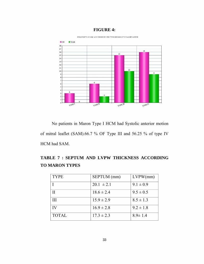
FIGURE 4:
No patients in Maron Type I HCM had Systolic anterior motion
of mitral leaflet (SAM).66.7 % OF Type III and 56.25 % of type IV
HCM had SAM.
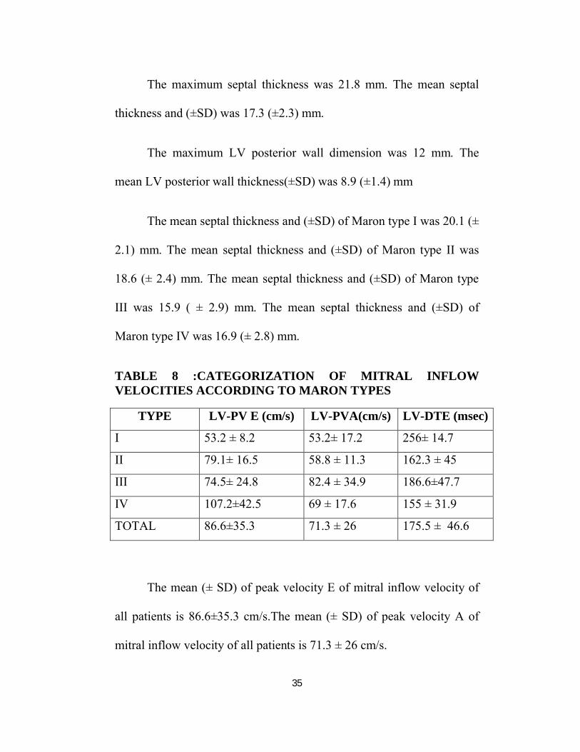
TABLE 7 : SEPTUM AND LVPW THICKNESS ACCORDING
TO MARON TYPES
TYPE SEPTUM (mm) LVPW(mm)
I 20.1 ± 2.1 9.1 ± 0.9
II 18.6 ± 2.4 9.5 ± 0.5
III 15.9 ± 2.9 8.5 ± 1.3
IV 16.9 ± 2.8 9.2 ± 1.8
TOTAL 17.3 ± 2.3 8.9± 1.4
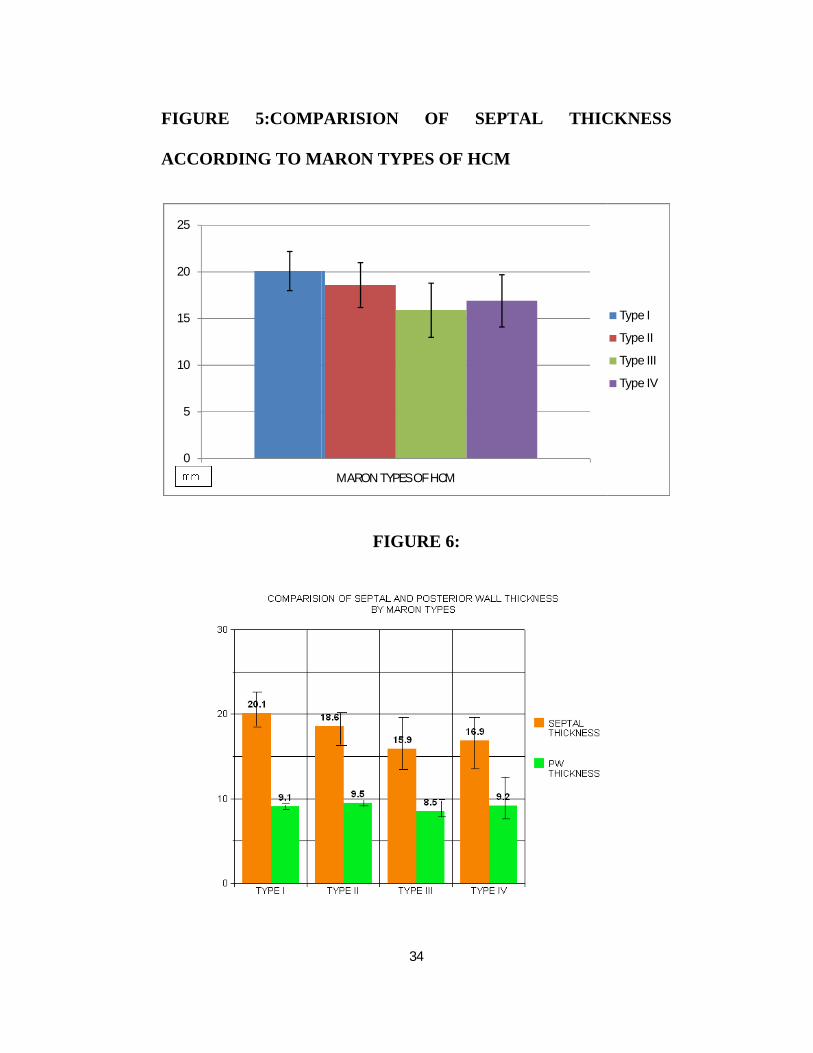
FIGURE 5:COMPARISION OF SEPTAL THICKNESS
ACCORDING TO MARON TYPES OF HCM
0
5
10
15
20
25
34
COMPARISION OF SEPTAL THICKNESS
ACCORDING TO MARON TYPES OF HCM
FIGURE 6:
MARON TYPES OF HCM
COMPARISION OF SEPTAL THICKNESS
Type I
Type II
Type III
Type IV
35
The maximum septal thickness was 21.8 mm. The mean septal
thickness and (±SD) was 17.3 (±2.3) mm.
The maximum LV posterior wall dimension was 12 mm. The
mean LV posterior wall thickness(±SD) was 8.9 (±1.4) mm
The mean septal thickness and (±SD) of Maron type I was 20.1 (±
2.1) mm. The mean septal thickness and (±SD) of Maron type II was
18.6 (± 2.4) mm. The mean septal thickness and (±SD) of Maron type
III was 15.9 ( ± 2.9) mm. The mean septal thickness and (±SD) of
Maron type IV was 16.9 (± 2.8) mm.
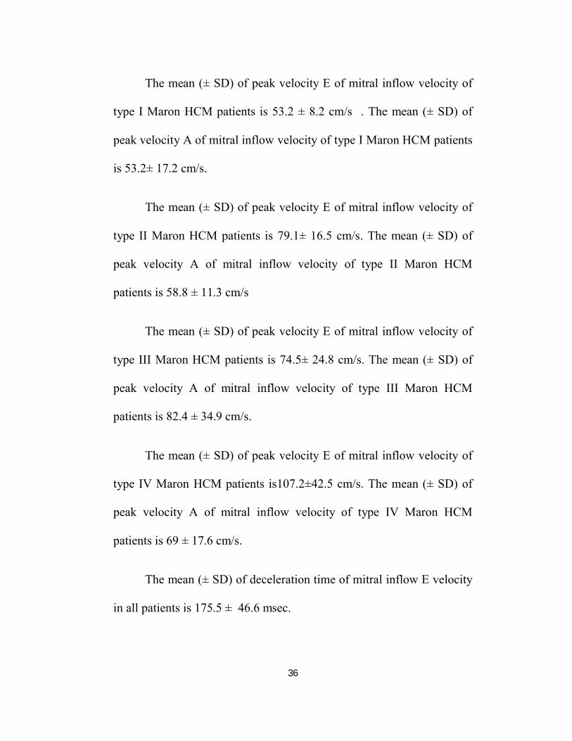
TABLE 8 :CATEGORIZATION OF MITRAL INFLOW VELOCITIES ACCORDING TO MARON TYPES
TYPE LV-PV E (cm/s) LV-PVA(cm/s) LV-DTE (msec)
I 53.2 ± 8.2 53.2± 17.2 256± 14.7
II 79.1± 16.5 58.8 ± 11.3 162.3 ± 45
III 74.5± 24.8 82.4 ± 34.9 186.6±47.7
IV 107.2±42.5 69 ± 17.6 155 ± 31.9
TOTAL 86.6±35.3 71.3 ± 26 175.5 ± 46.6
The mean (± SD) of peak velocity E of mitral inflow velocity of
all patients is 86.6±35.3 cm/s.The mean (± SD) of peak velocity A of
mitral inflow velocity of all patients is 71.3 ± 26 cm/s.
36
The mean (± SD) of peak velocity E of mitral inflow velocity of
type I Maron HCM patients is 53.2 ± 8.2 cm/s . The mean (± SD) of
peak velocity A of mitral inflow velocity of type I Maron HCM patients
is 53.2± 17.2 cm/s.
The mean (± SD) of peak velocity E of mitral inflow velocity of
type II Maron HCM patients is 79.1± 16.5 cm/s. The mean (± SD) of
peak velocity A of mitral inflow velocity of type II Maron HCM
patients is 58.8 ± 11.3 cm/s
The mean (± SD) of peak velocity E of mitral inflow velocity of
type III Maron HCM patients is 74.5± 24.8 cm/s. The mean (± SD) of
peak velocity A of mitral inflow velocity of type III Maron HCM
patients is 82.4 ± 34.9 cm/s.
The mean (± SD) of peak velocity E of mitral inflow velocity of
type IV Maron HCM patients is107.2±42.5 cm/s. The mean (± SD) of
peak velocity A of mitral inflow velocity of type IV Maron HCM
patients is 69 ± 17.6 cm/s.
The mean (± SD) of deceleration time of mitral inflow E velocity
in all patients is 175.5 ± 46.6 msec.
37
The mean (± SD) of deceleration time of mitral inflow E velocity
in type I Maron is 256± 14.7 msec. The mean (± SD) of deceleration
time of mitral inflow E velocity in type II Maron is 162.3 ± 45 msec.
The mean (± SD) of deceleration time of mitral inflow E velocity in
type III Maron is 186.6±47.7 msec. The mean (± SD) of deceleration
time of mitral inflow E velocity in type IV Maron is 155 ± 31.9 msec.
TABLE 9 :CATEGORIZATION OF TRICUSPID INFLOW
VELOCITIES ACCORDING TO MARON TYPES
TYPE RV-PV E(cm/s) RV-PV A(cm/s) RV-DTE (msec)
I 41.8 ±1 42.3 ± 6.9 161.7 ± 55.5
II 56.9 ±12.7 43.7 ±7.1 203 ± 63.4
III 50 ± 11.7 46.2 ± 10.6 221.8 ± 60.4
IV 60.4± 13.6 46.7 ±22.7 193.6 ±60.4
TOTAL 54.6 ±13.1 45.7± 15.6 203.3± 60.6
FIGURE 7

38
The mean (± SD) of peak velocity E of tricuspid inflow velocity
of all patients is 54.6±13.1 cm/s.The mean (± SD) of peak velocity A of
tricuspid inflow velocity of all patients is 45.7 ± 15.6 cm/s.
The mean (± SD) of peak velocity E of tricuspid inflow velocity
of type I Maron HCM patients is 41.8 ± 1 cm/s . The mean (± SD) of
peak velocity A of tricuspid inflow velocity of type I Maron HCM
patients is 42.3± 6.9 cm/s.
The mean (± SD) of peak velocity E of tricuspid inflow velocity
of type II Maron HCM patients is 56.9± 12.7 cm/s. The mean (± SD) of
peak velocity A of tricuspid inflow velocity of type II Maron HCM
patients is 43.7± 7.1 cm/s
The mean (± SD) of peak velocity E of tricuspid inflow velocity
of type III Maron HCM patients is 50± 11.7 cm/s. The mean (± SD) of
peak velocity A of tricuspid inflow velocity of type III Maron HCM
patients is 46.2 ± 10.6 cm/s.
The mean (± SD) of peak velocity E of tricuspid inflow velocity
of type IV Maron HCM patients is 60.4± 13.6 cm/s. The mean (± SD) of
peak velocity A of tricuspid inflow velocity of type IV Maron HCM
patients is 46.7 ± 22.7 cm/s.
39
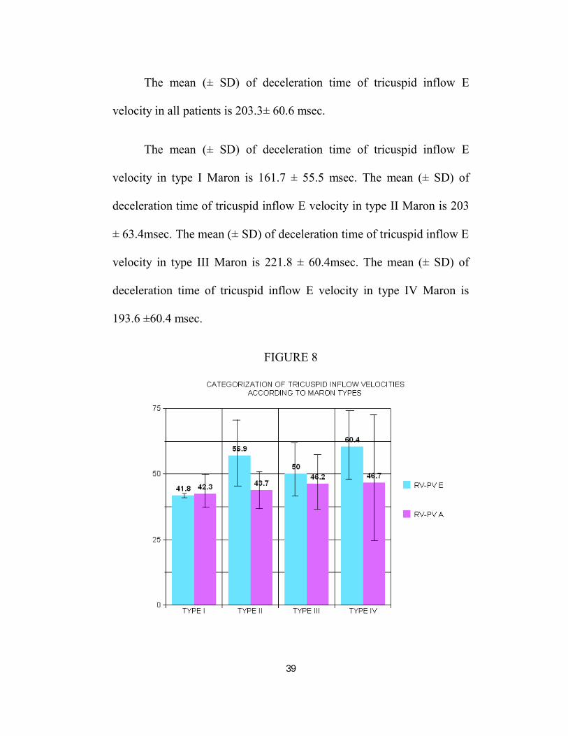
The mean (± SD) of deceleration time of tricuspid inflow E
velocity in all patients is 203.3± 60.6 msec.
The mean (± SD) of deceleration time of tricuspid inflow E
velocity in type I Maron is 161.7 ± 55.5 msec. The mean (± SD) of
deceleration time of tricuspid inflow E velocity in type II Maron is 203
± 63.4msec. The mean (± SD) of deceleration time of tricuspid inflow E
velocity in type III Maron is 221.8 ± 60.4msec. The mean (± SD) of
deceleration time of tricuspid inflow E velocity in type IV Maron is
193.6 ±60.4 msec.
FIGURE 8
40
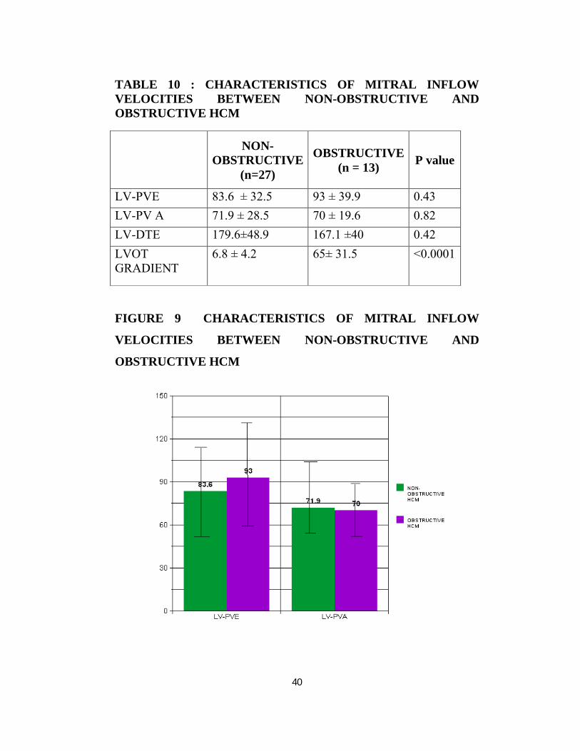
TABLE 10 : CHARACTERISTICS OF MITRAL INFLOW VELOCITIES BETWEEN NON-OBSTRUCTIVE AND OBSTRUCTIVE HCM
NON-OBSTRUCTIVE
(n=27)
OBSTRUCTIVE(n = 13)
P value
LV-PVE 83.6 ± 32.5 93 ± 39.9 0.43
LV-PV A 71.9 ± 28.5 70 ± 19.6 0.82
LV-DTE 179.6±48.9 167.1 ±40 0.42
LVOT GRADIENT
6.8 ± 4.2 65± 31.5 <0.0001
FIGURE 9 CHARACTERISTICS OF MITRAL INFLOW
VELOCITIES BETWEEN NON-OBSTRUCTIVE AND
OBSTRUCTIVE HCM

41
The mean (± SD) of peak velocity E of mitral inflow velocity of
patients with non-obstructive HCM is 83.6 ± 32.5 cm/s.The mean (±
SD) of peak velocity A of mitral inflow velocity of patients with non-
obstructive HCM is 71.9 ± 28.5 cm/s. The mean (± SD) of deceleration
time of mitral inflow E velocity in non-obstructive HCM is 179.6±48.9
msec.
The mean (± SD) of peak velocity E of mitral inflow velocity of
patients with obstructive HCM is 93 ± 39.9 cm/s.The mean (± SD) of
peak velocity A of mitral inflow velocity of patients with obstructive
HCM is 70 ± 19.6 cm/s. The mean (± SD) of deceleration time of mitral
inflow E velocity in patients with obstructive HCM is 167.1 ±40 msec.
There was no significant statistical difference in mitral and tricuspid
inflow velocities between non-obstructive and obstructive HCM.
TABLE 11. Tissue Doppler Imaging velocities in patients with
Hypertrophic cardiomyopathy
Patients ( n = 40) cm/s
MV Ea (septal) 5.8 ±2.4
MV Aa (septal) 6.5 ± 1.8
TV Ea (lateral) 13.4 ± 7.9
TV Aa (lateral) 18.8 ± 10.1

42
TABLE 12 Comparison the results of Tissue doppler velocities
according to left ventricle outflow gradient
NON-OBSTRUCTIVE
(n=27)
OBSTRUCTIVE(n=13)
p-value
MV Ea (septal) 5.7± 2.3 5.8 ±2.7 0.9
MV Aa (septal) 6.6± 1.9 6.5± 1.8 0.87
TV Ea (lateral) 13.3± 7.6 13.5± 8.4 0.94
TV Aa (lateral) 17.8± 9.6 20.9± 10.7 0.36
FIGURE 10 : Comparison the results of Tissue doppler velocities
according to left ventricle outflow gradient
43
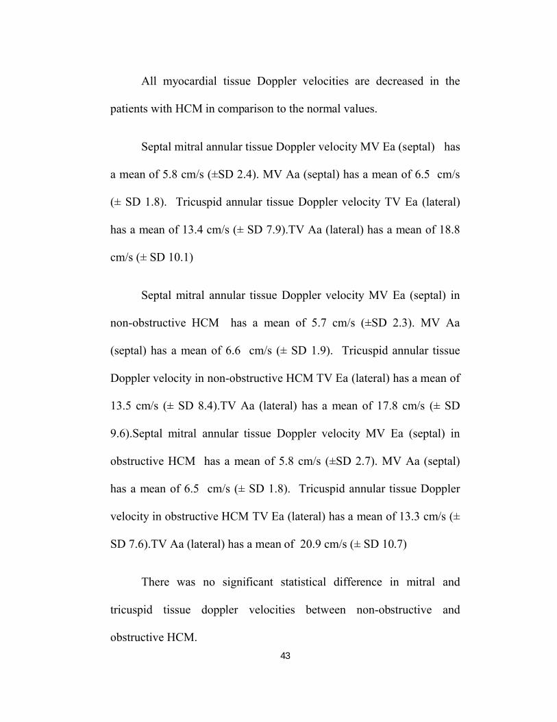
All myocardial tissue Doppler velocities are decreased in the
patients with HCM in comparison to the normal values.
Septal mitral annular tissue Doppler velocity MV Ea (septal) has
a mean of 5.8 cm/s (±SD 2.4). MV Aa (septal) has a mean of 6.5 cm/s
(± SD 1.8). Tricuspid annular tissue Doppler velocity TV Ea (lateral)
has a mean of 13.4 cm/s (± SD 7.9).TV Aa (lateral) has a mean of 18.8
cm/s (± SD 10.1)
Septal mitral annular tissue Doppler velocity MV Ea (septal) in
non-obstructive HCM has a mean of 5.7 cm/s (±SD 2.3). MV Aa
(septal) has a mean of 6.6 cm/s (± SD 1.9). Tricuspid annular tissue
Doppler velocity in non-obstructive HCM TV Ea (lateral) has a mean of
13.5 cm/s (± SD 8.4).TV Aa (lateral) has a mean of 17.8 cm/s (± SD
9.6).Septal mitral annular tissue Doppler velocity MV Ea (septal) in
obstructive HCM has a mean of 5.8 cm/s (±SD 2.7). MV Aa (septal)
has a mean of 6.5 cm/s (± SD 1.8). Tricuspid annular tissue Doppler
velocity in obstructive HCM TV Ea (lateral) has a mean of 13.3 cm/s (±
SD 7.6).TV Aa (lateral) has a mean of 20.9 cm/s (± SD 10.7)
There was no significant statistical difference in mitral and
tricuspid tissue doppler velocities between non-obstructive and
obstructive HCM.
44
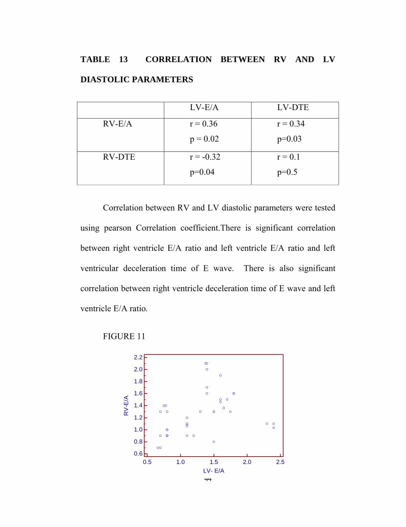
TABLE 13 CORRELATION BETWEEN RV AND LV
DIASTOLIC PARAMETERS
LV-E/A LV-DTE
RV-E/A r = 0.36
p = 0.02
r = 0.34
p=0.03
RV-DTE r = -0.32
p=0.04
r = 0.1
p=0.5
Correlation between RV and LV diastolic parameters were tested
using pearson Correlation coefficient.There is significant correlation
between right ventricle E/A ratio and left ventricle E/A ratio and left
ventricular deceleration time of E wave. There is also significant
correlation between right ventricle deceleration time of E wave and left
ventricle E/A ratio.
FIGURE 11
0.5 1.0 1.5 2.0 2.50.6
0.8
1.0
1.2
1.4
1.6
1.8
2.0
2.2
LV- E/A
RV
-E/A
45
100 150 200 250 3000.6
0.8
1.0
1.2
1.4
1.6
1.8
2.0
2.2
LV-DTE
RV
-E/A
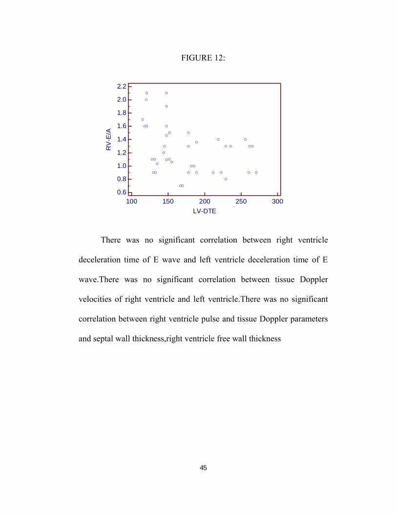
FIGURE 12:
There was no significant correlation between right ventricle
deceleration time of E wave and left ventricle deceleration time of E
wave.There was no significant correlation between tissue Doppler
velocities of right ventricle and left ventricle.There was no significant
correlation between right ventricle pulse and tissue Doppler parameters
and septal wall thickness,right ventricle free wall thickness
46
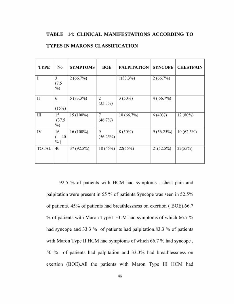
TABLE 14: CLINICAL MANIFESTATIONS ACCORDING TO
TYPES IN MARONS CLASSIFICATION
TYPE No. SYMPTOMS BOE PALPITATION SYNCOPE CHESTPAIN
I 3(7.5 %)
2 (66.7%) 1(33.3%) 2 (66.7%)
II 6
(15%)
5 (83.3%) 2 (33.3%)
3 (50%) 4 ( 66.7%)
III 15(37.5
%)
15 (100%) 7 (46.7%)
10 (66.7%) 6 (40%) 12 (80%)
IV 16 ( 40 % )
16 (100%) 9 (56.25%)
8 (50%) 9 (56.25%) 10 (62.5%)
TOTAL 40 37 (92.5%) 18 (45%) 22(55%) 21(52.5%) 22(55%)
92.5 % of patients with HCM had symptoms . chest pain and
palpitation were present in 55 % of patients.Syncope was seen in 52.5%
of patients. 45% of patients had breathlessness on exertion ( BOE).66.7
% of patients with Maron Type I HCM had symptoms of which 66.7 %
had syncope and 33.3 % of patients had palpitation.83.3 % of patients
with Maron Type II HCM had symptoms of which 66.7 % had syncope ,
50 % of patients had palpitation and 33.3% had breathlessness on
exertion (BOE).All the patients with Maron Type III HCM had

47
symptoms of which 66.7 % had palpitation, 80 % of patients had chest
pain,40 % of patients had syncope and 46.7 % had breathlessness on
exertion (BOE).All the patients with Maron Type IV HCM had
symptoms of which 50 % had palpitation, 62.5% of patients had chest
pain,56.25 % of patients had syncope and breathlessness on exertion
(BOE).
TABLE 15:RISK STRATIFICATION OF HCM ACCORDING TO MARON TYPES
TYPE FAMILY H/O SCD
LV WALL THICKNESS > 30 mm
SYNCOPE HOLTER –NSVT
I 3 ( 7.5 %)
0 0 2 (66.7%) 0
II 6 (15%) 4 (66.7%) 0 4 ( 66.7%) 0
III 15 (37.5 %)
3 (20%) 0 6 (40%) 4(26.7%)
IV 16 ( 40 % )
4 (25%) 0 9 (56.25%) 6(37.5%)
TOTAL 40 11(27.5%) 0 21(52.5%) 10 (25%)
Family H/O sudden cardiac death was present in 27.5% of
patients with HCM.66.7% of patients with Maron Type II had a family
history of sudden cardiac death.20% of patients with Maron Type III
had a family history of sudden cardiac death and 25 % of patients with
Maron Type II had a family history of sudden cardiac death.
48
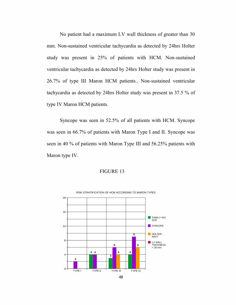
No patient had a maximum LV wall thickness of greater than 30
mm. Non-sustained ventricular tachycardia as detected by 24hrs Holter
study was present in 25% of patients with HCM. Non-sustained
ventricular tachycardia as detected by 24hrs Holter study was present in
26.7% of type III Maron HCM patients.. Non-sustained ventricular
tachycardia as detected by 24hrs Holter study was present in 37.5 % of
type IV Maron HCM patients.
Syncope was seen in 52.5% of all patients with HCM. Syncope
was seen in 66.7% of patients with Maron Type I and II. Syncope was
seen in 40 % of patients with Maron Type III and 56.25% patients with
Maron type IV.
FIGURE 13
0
4
8
12
16
20
TYPE I TYPE II TYPE III TYPE IV
2
4 43
6
4 4
9
6
FAMILY H/OSCD
SYNCOPE
HOLTER -NSVT
LV WALLTHICKNESS> 30 mm
RISK STRATIFICATION OF HCM ACCORDING TO MARON TYPES
49
DISCUSSION
The prevalence of HCM in the adult general population is about
0.2% (1:500) based on several epidemiological studies21,22. Many
patients having the mutant gene for HCM can go undetected clinically.
Our study patients were those referred for echocardiography to our
cardiology OPD.Our study is not a community screening study
.Therefore one would expect it to underestimate the phenotypic
prevalence in the community. The population harbouring the genetic
defect is also underestimated.
Similar prevalence of HCM was found in no more than 1% of
outpatients in Other studies which had patients as referrals to hospital22.
The prevalence in females was significantly less than expected.
Male prevalence ranged from 55% to 78% in previous studies23,24.
There were 31 male and 9 female patients in our study.The
female population is significantly lower in our study.only 22.5% of the
study patients were female.
Reduced patient awareness,26,lesser indications for medical
screening programs25,27, and referral bias of clinicians26 or clinical
presentation which is delayed, which can result from genetic and
50
endocrine factors affecting expression of phenotype of the disease.
Estrogens may have a protective effect on development of secondary
hypertrophy28,29 .
The nature of the study population was predominantly male in
our study.
32.5 % of patients were in the 30-39 years age group and 30%
were in the 40-49 years age group.Hence majority of patients studied in
our study were in the 30-49 years age group.
our population were younger (mean age 41.8 ± 10.7 yrs) and it is
to be noted that male predominance were seen from adolescence to mid-
life in different studies.
Presence of LVOT obstruction in HCM
For categorization of HCM patients into different groups various
cutoff based on LV outlow gradient and site of hypertrophy have been
used. Strict categorization into different categories according to LV
outflow gradient is difficult because of the unpredictability of dynamic
changes that may occur in different patients. In our study we used 30
mmHg as a rest cut-off and /or a 50 mmnHg as a provocative induced
LV outflow gradient. No obstruction was found in twenty seven (67.5%)
51
patients. Thirteen (32.5 %) of patients had obstructive HCM (>30
mmHg LV outflow obstruction) was found in 13 (32.5 %) cases,with a
rest gradient mean(+SD) of 65 (± 31.5) mmHg.
46.7 % of Maron type III HCM patients had obstructive HCM
and 40% of Maron type IV HCM patients had obstructive HCM .There
was no obstructive HCM in Maron type I and II.
LV outflow obstruction prevalence have been from 23 – 77% in
different studies30-32. Patients with obstructive HCM were older than
other groups. Many of our patients where on medical therapy which
may account for the low gradient in them.
LV hypertrophy patterns :
The classification of the left ventricular hypertrophy pattern in
HCM has not been universally formalised. The most commonly used is
the Maron classification21.
The patients in our study were categorized according to the
Maron types of HCM.
7.5 % of patients were in type I.15 % of patients were in type II.
40 % of patients were in type IV and 37.5% patients were in type III
Maron .
52
There were no patients with type V (apical HCM) Maron.Type IV
Maron was the predominant group in our study,which is in contrast to
other studies were type III Maron was the predominant group21.
The four patterns of Maron types of LV hypertrophy showed no
difference in age. 92.5 % of patients with HCM had symptoms . chest
pain and palpitation were present in 55 % of patients.Syncope was seen
in 52.5% of patients. 45% of patients had breathlessness on exertion (
BOE).
66.7 % of patients with Maron Type I HCM had symptoms of
which 66.7 % had syncope and 33.3 % of patients had palpitation.83.3
% of patients with Maron Type II HCM had symptoms of which 66.7 %
had syncope , 50 % of patients had palpitation and 33.3% had
breathlessness on exertion (BOE).
All the patients with Maron Type III HCM had symptoms of
which 66.7 % had palpitation, 80 % of patients had chest pain,40 % of
patients had syncope and 46.7 % had breathlessness on exertion (BOE).
All the patients with Maron Type IV HCM had symptoms of
which 50 % had palpitation, 62.5% of patients had chest pain,56.25 %
of patients had syncope and breathlessness on exertion (BOE).
53
Syncope was present in all groups.The prevalence of syncope was
higher in Maron type I and II.Majority of patients had chestpain in type
III and IV. Chest pain as a symptom was not seen in type I and II.
Biventricular diastolic dysfunction in HCM
As shown in table 8 & 9, all myocardial velocities decreased in
the patients with HCM in comparison to the normal values. Recent
studies have shown that tissue doppler is a reliable method for early
detection of HCM in genotype-positive patients before the onset of
hypertrophy. Reduction in systolic and diastolic Tissue doppler
velocities have been shown in various studies to have a high sensitivity
and specificity for identifying mutation in HCM carriers.
Serial echocardiographic evaluation of patients with clinically
normal phenotype but carrying the mutation of HCM have shown
reduction in tissue doppler velocities and development of hypertrophy
and diastolic dysfunction during follow up after many years.Impaired
LV relaxation is a prominent diastolic abnormality in patients with
HCM. Mitral inflow and pulmonary venous pulse Doppler flow
assessments may not be sensitive for the detection of high filling
pressures of the left ventricle. Simultaneous echocardiographic and
invasive homodynamic studies have revealed no significant correlation
54
between mitral inflow or pulmonary venous flow parameters and filling
pressures in patients with LV systolic dysfunction.Good correlation
between LV filling pressure, mitral inflow E velocity, and tissue
Doppler MV annular velocities (Ea) have been observed in studies.
All myocardial tissue Doppler velocities are decreased in the
patients with HCM in comparison to the normal values in our
study.Reduced tissue doppler early diastolic velocities that have been
detected in our study indicate that diastolic abnormalities may precede
the onset of hypertrophy in HCM.
Patients in Type IV showed maximum peak velocity of E in
mitral inflow velocities. Patients in Type III showed maximum peak
velocity of A in mitral inflow velocities.Deceleration time of Mitral
inflow E velocity was maximum in Maron Type I HCM
patients.Patients in Type IV showed maximum peak velocity of E and
A in tricuspid inflow velocities. Deceleration time of tricuspid inflow
E velocity was maximum in Maron Type III HCM patients.There was
no statistically significant relationship between myocardial tissue
Doppler velocities of obstructive and non-obstructive HCM.This is in
contrast to a study by Rezvaneh Salehi et al which has observed positive
correlation between reduced mitral annular Ea septal tissue Doppler
velocity and LVOT gradient .
55
In all our study patients right ventricular diastolic function is
impaired, with prolonged isovolumic relaxation time, prolonged
deceleration time of E wave reversed E/A ratio and reduced tricuspid
annular tissue Doppler velocities.
Different studies using cardiac MRI have found out diastolic
bnormalities of the right ventricle like reduced rate of early filling and
increased late filling in patients with HCM.
The observations in our study indicate that both left and right
ventricles exhibit similarities in abnormal diastolic filling patterns. Left
ventricular and right ventricular diastolic abnormalities are not related to
the magnitude and severity of left and right ventricular hypertrophy
respectively in our study.
A study by Spirito et al has observed that abnormalities in left
ventricular relaxation in hypertrophic cardiomyopathy patients is not
related to the proportion and severity of left ventricular hypertrophy32.
The possible explanation for right ventricular diastolic
dysfunction is Ventricular interdependence33.The ventricles even though
are separated are connected anatomically and functionally in both
series and parallel.

56
The pulmonary circulation provides the serial connection hence
loading conditions of one ventricle. common anatomical structures like
interventricular septum,pericardium and muscle fibres maintain the
parallel connection between the ventricles.This results in ventricular
interdependence during systole and diastole. In our study there was a
statistically significant correlation between several diastolic indexes of
left ventricle and right ventricle. There is significant correlation between
right ventricle E/A ratio and left ventricle E/A ratio and left ventricular
deceleration time of E wave. There is also significant correlation
between right ventricle deceleration time of E wave and left ventricle
E/A ratio Right ventricular hypertrophy can be seen in more than 50%
of patients with HCM33.
The disarray of myocardial fibres in hypertrophic cardiomyopathy
is not limited to the left ventricle but also affects the right ventricle .
Hence the right ventricular diastolic dysfunction observed in
hypertrophic cardiomyopathy in all our patients may be contributed by
the disarray of myocardial fibres rather than the severity of right
ventricular hypertrophy.
57
RISK STRATIFICATION OF HCM
Family H/O Sudden cardiac death
Family H/O sudden cardiac death was present in 27.5% of
patients with HCM.This is in contrast to a study by Maron et al where
10-20% had a family H/O sudden cardiac death and 5 % had more than
two sudden cardiac deaths in the family which suggests a malignant
form of HCM34. 66.7% of patients with Maron Type II had a family
history of sudden cardiac death.20% of patients with Maron Type III
had a family history of sudden cardiac death and 25 % of patients with
Maron Type II had a family history of sudden cardiac death.
Maximum LV Wall thickness
No patient had a maximum LV wall thickness of greater than 30
mm.This is in contrast to study by Maron et al where maximum LV wall
thickness of greater than 30 mm was seen in < 3% of patients35.spirito et
al and olivetto etal have found that maximum LV wall thickness of
greater than 30 mm is a sudden cardiac death risk factor mainly in
younger patients32.

58
Non-sustained ventricular tachycardia
Non-sustained ventricular tachycardia as detected by 24hrs Holter
study was present in 25% of patients with HCM.This is similar to study
by Monserrat et al where 19.6% of patients had NSVT36.They have
concluded that sudden cardiac death is significantly increased in young
patients with HCM and NSVT. Non-sustained ventricular tachycardia
as detected by 24hrs Holter study was present in 26.7% of type III
Maron HCM patients.. Non-sustained ventricular tachycardia as
detected by 24hrs Holter study was present in 37.5 % of type IV Maron
HCM patients.
SYNCOPE
Syncope was seen in 52.5% of all patients with HCM. Syncope
was seen in 66.7% of patients with Maron Type I and II. Syncope was
seen in 40 % of patients with Maron Type III and 56.25% patients with
Maron type IV.This is similar to a study by Maron et al where syncope
was seen in greater than 50% of patients and where syncope was the
only marker in more than one third of patients35.
59
Study limitations
1. Our study evaluated only the prevalence among patients referred
for echocardiography and not among the community.
2. Our study evaluated the patients during the time period of the
study,rather than those who were first diagnosed.
3. Provocative tests were not standardized and was not attempted in
all patients.
4. Many patients were on medications, which might alter the
measured LV outflow gradient during the study or might have
even abolished such a gradient in some and thus the prevalence
of the obstructive type of HCM may have been underestimated.
60
CONCLUSION
1. Majority of patients were in Type III and Type IV of Maron
classification of hypertrophic cardiomyopathy .
2. Syncope was the most common sudden cardiac death risk factor
seen in our study.
3. Maximum LV wall thickness of greater than 30 mm , a sudden
cardiac death risk factor was not present in our study.
4. There was no statistically significant relationship between
myocardial tissue Doppler velocities of obstructive and non-
obstructive HCM.
5. There was no statistically significant relationship between pulse
Doppler mitral inflow velocities of obstructive and non-
obstructive HCM.
6. Biventricular diastolic dysfunction is seen in most patients with
HCM.
7. Right ventricular diastolic dysfunction may be contributed by the
disarray of myocardial fibres rather than the severity of right
ventricular hypertrophy
8. There is a significant correlation between several right ventricular
and left ventricular diastolic parameters

61
Figure A : shows measurement of LVOT gradient in HCM
62
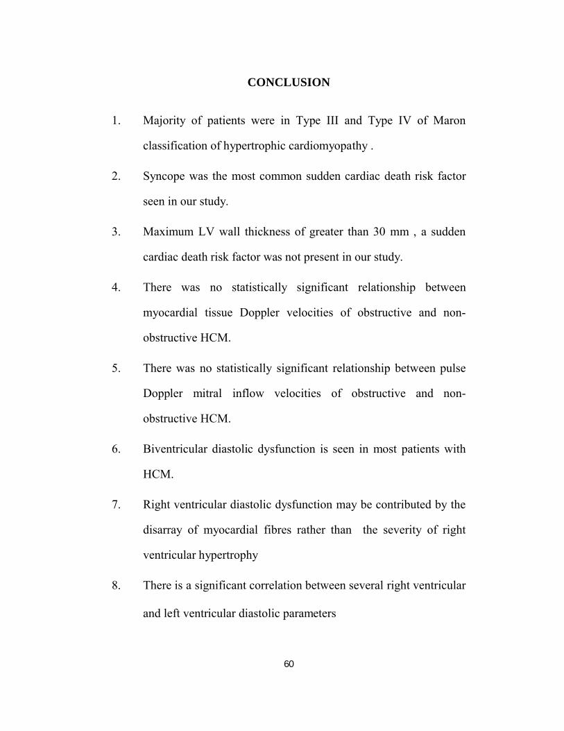
Figure B:shows Mitral inflow pulse wave Doppler tracing
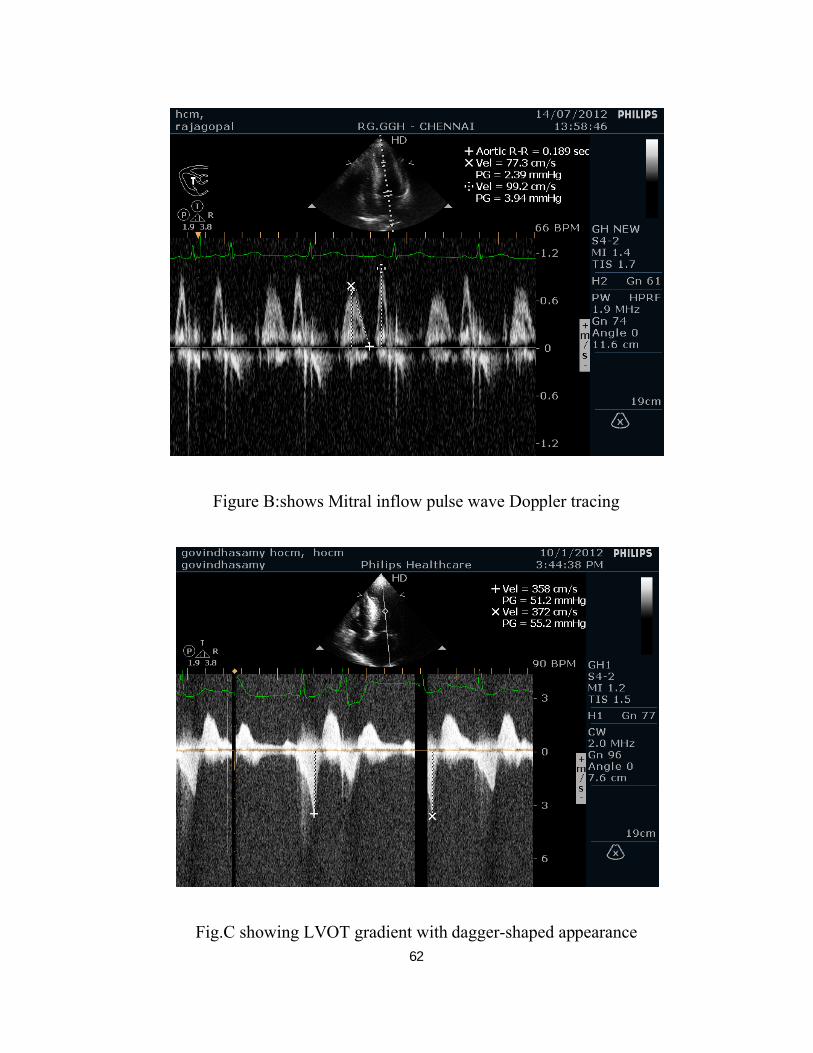
Fig.C showing LVOT gradient with dagger-shaped appearance

63
Figure.D showing measurement of RP interval
Fig.E showing categorization of HCM according to Maron types
64
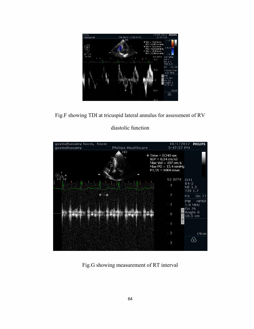
Fig.F showing TDI at tricuspid lateral annulus for assessment of RV
diastolic function
Fig.G showing measurement of RT interval
65
BIBLIOGRAPHY
1. J Martijn Bos. "Genetics of hypertrophic cardiomyopathy: one,
two, or more diseases?", Current Opinion in Cardiology, 05/2007
2. Riding, N. R., O. Salah, S. Sharma, F. Carre, R. O'Hanlon, K. P.
George, B. Hamilton, H. Chalabi, G. P. Whyte, and M. G.
Wilson. "Do big athletes have big hearts? Impact of extreme
anthropometry upon cardiac hypertrophy in professional male
athletes", British Journal of Sports Medicine, 2012.
3. Sheikh, F.. "Mouse models for cardiomyopathy research",
Progress in Pediatric cardiology, 200711
4. G Frisso. "A child cohort study from southern Italy enlarges the
genetic spectrum of hypertrophic cardiomyopathy", Clinical
Genetics, 07/2009
5. Dewar, Laura, Bo Liang, Yueh Li, Shubhayan Sanatani, and Glen
F.. "Familial Hypertrophic Cardiomyopathy-Related Troponin
Mutations and Sudden Cardiac Death", Cardiomyopathies - From
Basic Research to Clinical Management, 2012.
6. Olivotto, I.. "Maximum left ventricular thickness and risk of
sudden death in patients with hypertrophic cardiomyopathy",
Journal of the American College of Cardiology,
7. E., Luis, and Eduardo Moreyr. "Hypertrophic Cardiomyopathy in
Infants and Children", Cardiomyopathies - From Basic Research
66
to Clinical Management, 2012 8.Panza, J.; Maris, T. & Maron, B.
(1992).
8. Development and determinants of dynamic obstruction to left
ventricular outflow in young patients with
hypertrophiccardiomyopathy. Circulation, Vol.85, pp. 1398-405.
9. K. L. Weeks. "The Athlete's Heart vs. the Failing Heart: Can
Signaling Explain the Two Distinct Outcomes?", Physiology,
04/01/2011
10. Neri Serneri GG, Boddi M, Modesti PA, Cecioni I,Coppo M,
Padeletti L, Michelucci A, Colella A,Galanti G. Increased cardiac
sympathetic activityand insulin-like growth factor-I formation are
associated with physiological hypertrophy in athletes. Circ Res
89: 977–982, 2001.
11. Basso, C.; Thiene, G.; Corrado, D.; et al. (2000). Hypertrophic
cardiomyopathy andsudden death in the young: pathologic
evidence of myocardial ischemia. HumPathol, Vol.31, pp. 988-98
12. Biagini, E.; Coccolo, F.; Ferlito, M.; et al. (2005) Dilated-
hypokinetic evolution ofhypertrophic cardiomyopathy.
Prevalence, incidence, risk factors, and prognostic implications in
pediatric and adult patients. J Am Coll Cardiol, Vol. 46: 1543-50.
13. Liviu C. Poliac. "Hypertrophic Cardiomyopathy",
Anesthesiology, 01/2006
14. J Martijn Bos. "Genetics of hypertrophic cardiomyopathy: one, two,
or more diseases?", Current Opinion in Cardiology, 05/2007
67
15. McCully RB, Nishimura RA, Tajik AJ, et al. Extent of clinical
improvement after surgicaltreatment of hypertrophic obstructive
cardiomyopathy. Circulation 1996;94:467-71
16. Maron BJ, Nishimura RA, McKenna WJ, et al. Assessment of
permanent dual-chamber pacing asa treatment for drug-
refractory symptomatic patients with obstructive hypertrophic
cardiomyopathy. A randomized, double-blind, crossover study
(M-PATHY). Circulation 1999;99:2927-2933
17. Sigwart U. Non-surgical myocardial reduction of hypertrophic
obstructive cardiomyopathy. Lancet1995;346:211-4.
18. Decker, J.; Rossano, J.; O’Brian Smith, E.; et al. (2009). Risk
factors and mode of death in isolated hypertrophic
cardiomyopathy in children. J Am Coll Cardiol, Vol.54, pp.250-4.
19. Maron, B.; Shen, W-K.; Links, M.; et al. (2000b). Efficacy of
implantable cardioverterdefibrillators for the prevention of
sudden death in patients with hypertrophic cardiomyopathy. N
Engl J Med, Vol.342, pp. 365-73.
20. Epstein, A.; Dimarco, J.; Ellenbogen, K.; et al. (2008).
ACC/AHA/HRS 2008 guidelines for device-based therapy of
cardiac rhythm abnormalities. Heart Rhythm, Vol.5, pp.e1-62.
21. Maron BJ,GottdienerJC,Epstein SE.Patterns and significance of
distribution of left ventricular hypertrophy in hpertrophic
cardiomyopathy: A wide-angle two-dimensional
echocardiographic study of 125 patients.Am J Cardiol
1981;48:418-28

68
22. Maron BJ, Gardin JM, Flack JM, Gidding SS, Kurosaki TT, Bild
DE. Prevalence of hypertrophic cardiomyopathy in a general
population of young adults. Echocardiographic analysis of 4111
subjects in the CARDIA Study. Coronary Artery Risk
Development in (Young) Adults. Circulation 1995;92:785-9.
23. Maron BJ, Peterson EE, Maron MS, Peterson JE. Prevalence of
hypertrophic cardiomyopathy in an outpatient population
referredfor echocardiographic study. Am J Cardiol
1994;73:577-80.
24. Braunwald E, Lambrew CT, Rockoff SD, Ross J Jr., Morrow
AG.Idiopathic hypertrophic subaortic stenosis. I. A description of
the disease based upon an analysis of 64 patients. Circulation
1964;30Suppl 4:3-119.
25. Wigle ED, Rakowski H, Kimball BP, Williams WG.
Hypertrophic cardiomyopathy. Clinical spectrum and treatment.
Circulation 1995;92:1680-92.
26. Maron BJ, Casey SA, Hurrell DG, Aeppli DM. Relation of left
ventricular thickness to age and gender in
hypertrophiccardiomyopathy. Am J Cardiol 2003;91:1195-8.
27. Nistri S, Thiene G, Basso C, Corrado D, Vitolo A, Maron
BJ.Screening for hypertrophic cardiomyopathy in a young
malemilitary population. Am J Cardiol 2003;91:1021-3.
28. Olivotto I, Maron MS, Adabag AS, Casey SA, Vargiu D, Link
MS,et al. Gender-related differences in the clinical presentation
andoutcome of hypertrophic cardiomyopathy. J Am Coll Cardiol
69
29. Malhotra A, Buttrick P, Scheuer J. Effects of sex hormones on
development of physiological and pathological cardiac
hypertrophy in male and female rats. Am J Physiol
1990;259:H866-71.
30. Kiziblash AM, Heinle SK, Grayburn PA. Spontaneous variability
in left ventricular outflow tract gradient in hypertrophic
obstructive cardiomyopathy Circulation 1998;97:461-6.
31. Panza JA, Maris TJ, Maron BJ. Development and determinants of
dynamic obstruction to left ventricular outflow in young patients
with hypertrophic cardiomyopathy. Circulation 1992;85:1398-
405.
32. Spirito P, Maron BJ. Relation between extent of left ventricular
hypertrophy and diastolic filling abnormalities in hypertrophic
cardiomyopathy. J Am Coll Cardiol 1990; 15: 808–813.
33. Ferlinz J. Right ventricular diastolic performance: compliance
characteristics with focus on pulmonary hypertension, right
ventricular hypertrophy, and calcium channel blockade. Cathet
Cardiovasc Diagn 1998; 43: 206–243.
34. Clinical Course of HypertrophicCardiomyopathy With Survival
to Advanced Age Barry J. Maron, MD, FACC, Susan A. Casey,
RN, Robert G. Hauser, MD, FACC, Dorothee M. Aeppli, PHD J
Am Coll Cardiol 2003;42:882– 8
35. Risk Stratification and Outcome of Patients With Hypertrophic
Cardiomyopathy ≥60 Years of AgeBarry J. Maron, MD; Ethan J.
Rowin, MD; Susan A. Casey, RN; Tammy S. Haas, RN;
70
Raymond H.M. Chan, MD; James E. Udelson, MD Circulation.
2013;127:585-593
36. Non-Sustained Ventricular Tachycardia in Hypertrophic
Cardiomyopathy: An Independent Marker of Sudden Death Risk
in Young Patients Lorenzo Monserrat, MD,† Perry M. Elliott,
MD, MRCP, FACC, Juan R. Gimeno, MD, Sanjay Sharma, BSC
37. Tissue Doppler Imaging Values in Hypertrophic Cardiomyopathy
According to Left Ventricular Outflow Gradient Rezvaneh Salehi
MD, Azin Alizadehasl MD J Cardiovasc Thorac Res 2011; Vol.2
(4): 19- 22
71
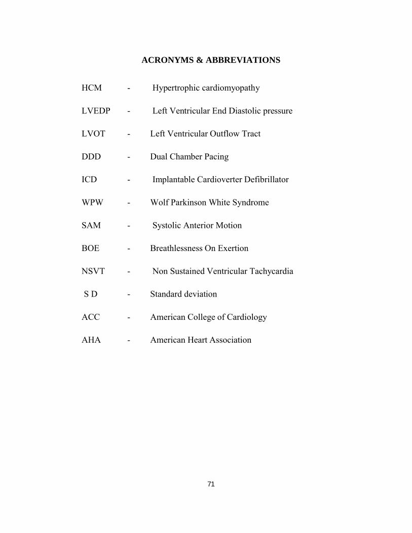
ACRONYMS & ABBREVIATIONS
HCM - Hypertrophic cardiomyopathy
LVEDP - Left Ventricular End Diastolic pressure
LVOT - Left Ventricular Outflow Tract
DDD - Dual Chamber Pacing
ICD - Implantable Cardioverter Defibrillator
WPW - Wolf Parkinson White Syndrome
SAM - Systolic Anterior Motion
BOE - Breathlessness On Exertion
NSVT - Non Sustained Ventricular Tachycardia
S D - Standard deviation
ACC - American College of Cardiology
AHA - American Heart Association
72
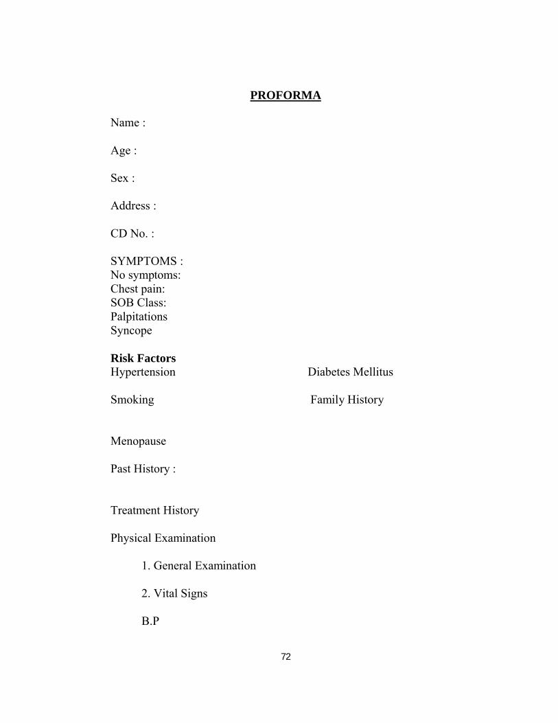
PROFORMA
Name :
Age :
Sex :
Address :
CD No. :
SYMPTOMS :No symptoms:Chest pain:SOB Class:PalpitationsSyncope
Risk FactorsHypertension Diabetes Mellitus
Smoking Family History
Menopause
Past History :
Treatment History
Physical Examination
1. General Examination
2. Vital Signs
B.P
73
Pulse
Respiration JVP Height cm Waveform
3. Systemic Examination
CVS
Inspection / Palpation
Apex Parasternal Heave
Palpable Sounds Thrills
Auscultation
S1 S2 Murmurs
Extra Heart Sounds
Other System
RS: PA: CNS :
74
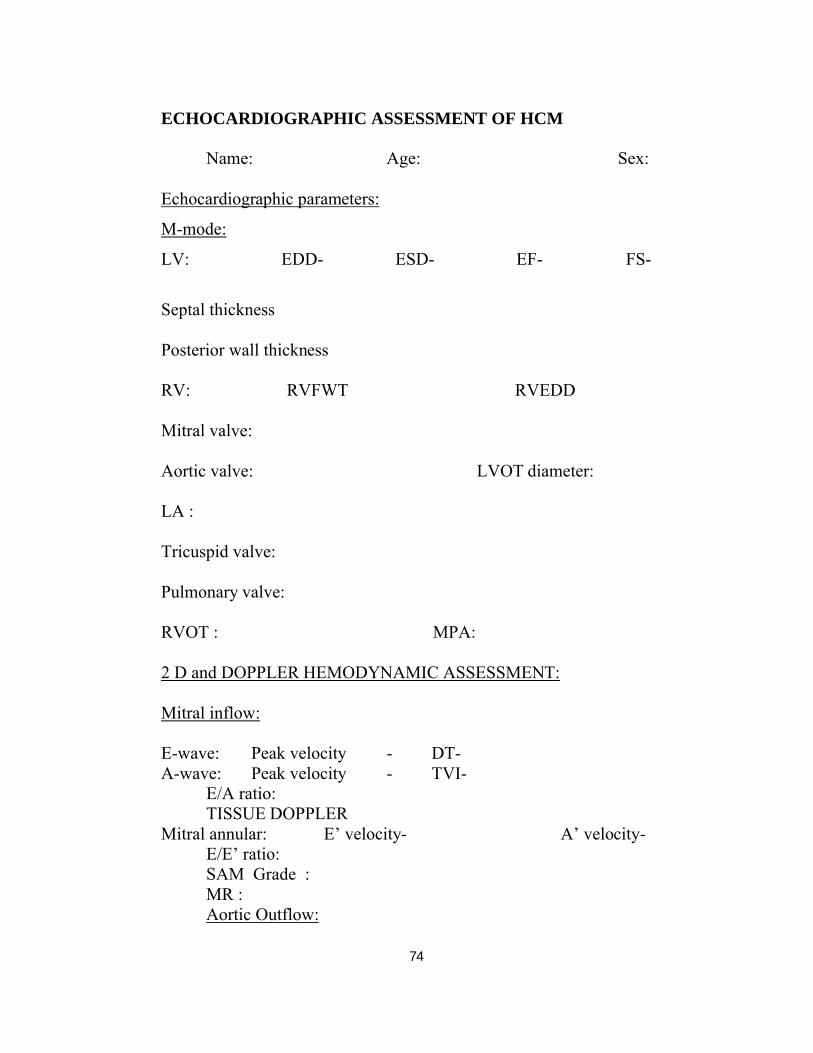
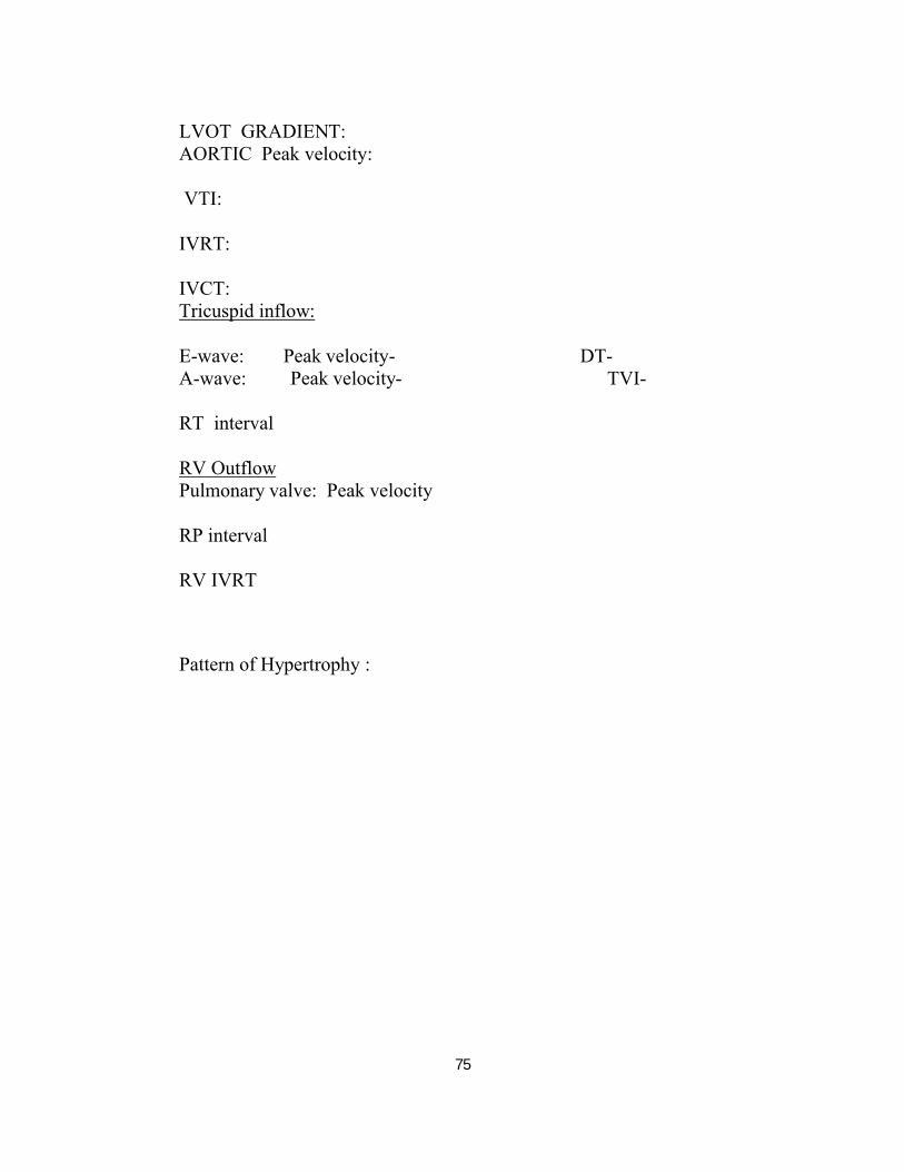
ECHOCARDIOGRAPHIC ASSESSMENT OF HCM
Name: Age: Sex:
Echocardiographic parameters:
M-mode:
LV: EDD- ESD- EF- FS-
Septal thickness
Posterior wall thickness
RV: RVFWT RVEDD
Mitral valve:
Aortic valve: LVOT diameter:
LA :
Tricuspid valve:
Pulmonary valve:
RVOT : MPA:
2 D and DOPPLER HEMODYNAMIC ASSESSMENT:
Mitral inflow:
E-wave: Peak velocity - DT-A-wave: Peak velocity - TVI-
E/A ratio:TISSUE DOPPLER
Mitral annular: E’ velocity- A’ velocity-E/E’ ratio:SAM Grade :MR :Aortic Outflow:
75
LVOT GRADIENT: AORTIC Peak velocity:
VTI:
IVRT:
IVCT:Tricuspid inflow:
E-wave: Peak velocity- DT-A-wave: Peak velocity- TVI-
RT interval
RV OutflowPulmonary valve: Peak velocity
RP interval
RV IVRT
Pattern of Hypertrophy :

76
PATIENT CONSENT FORM
STUDY TITLE :
Risk stratification of patients with Hypertrophic Cardiomyopathy
and assessment of Biventricular diastolic function by pulse/tissue
doppler echocardiographic imaging ”
PARTICIPANT NAME : DATE:
AGE: SEX: I.P.NO. :
The details of the study have been provided to me in writing and explained to me in my own language.
I confirm that I have understood the purpose of the above study.
I have the opportunity to ask the question and all my questions and doubts have been answered to my complete satisfaction.
I understand that my participation in the study is voluntary andthat I am free to withdraw at any time without giving any reason, without my legal rights being affected.
I understand that investigator, the institution, regulatory authorities and the ethical committee will not need my permission to look at my health records both in respect to the current study and any further research that may be conducted in relation to it, even if I withdraw from the study. I understand that my identity will not be revealed in any information released to third parties or published, unless as required under the law. I agree not to restrict the use of any data or results that arise from this study.
I hereby consent to,undergo complete physical examination ,and diagnostic tests including hematological, biochemical,radiological and urine examinations
I have been given an information sheet giving details of the study .I hereby consent to participate in the above study
Signature of the Participant
77
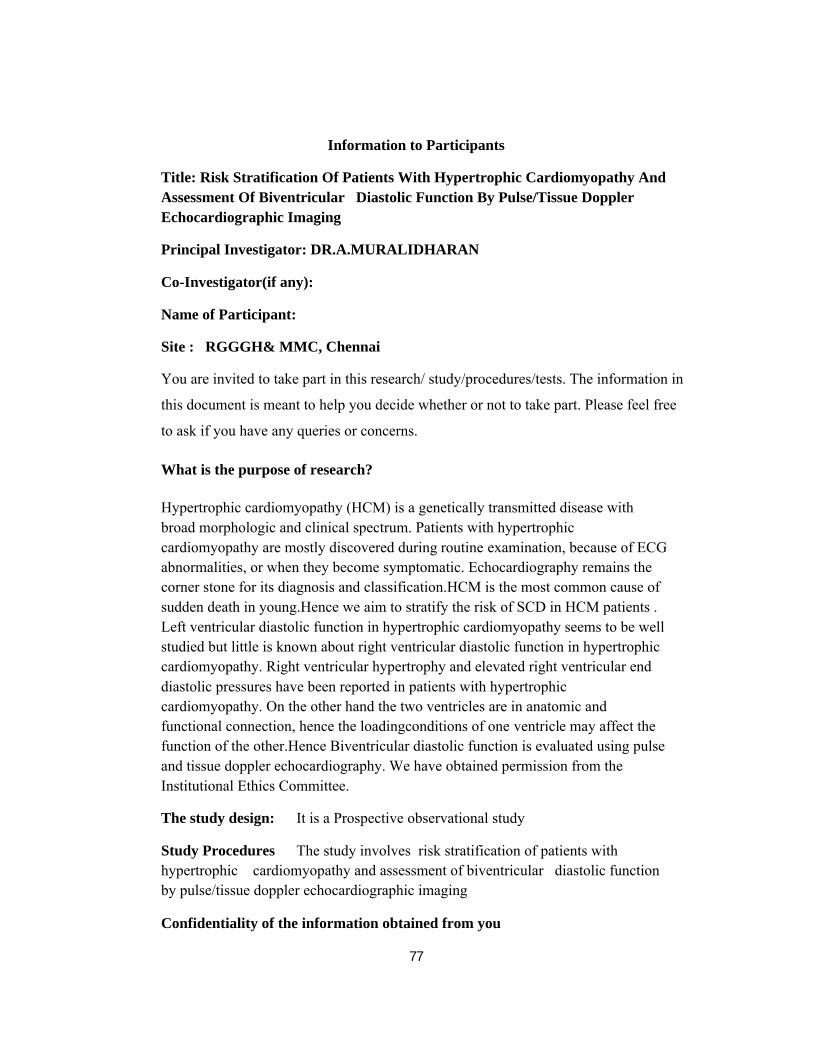
Information to Participants
Title: Risk Stratification Of Patients With Hypertrophic Cardiomyopathy And Assessment Of Biventricular Diastolic Function By Pulse/Tissue Doppler Echocardiographic Imaging
Principal Investigator: DR.A.MURALIDHARAN
Co-Investigator(if any):
Name of Participant:
Site : RGGGH& MMC, Chennai
You are invited to take part in this research/ study/procedures/tests. The information in
this document is meant to help you decide whether or not to take part. Please feel free
to ask if you have any queries or concerns.
What is the purpose of research?
Hypertrophic cardiomyopathy (HCM) is a genetically transmitted disease with broad morphologic and clinical spectrum. Patients with hypertrophic cardiomyopathy are mostly discovered during routine examination, because of ECG abnormalities, or when they become symptomatic. Echocardiography remains the corner stone for its diagnosis and classification.HCM is the most common cause of sudden death in young.Hence we aim to stratify the risk of SCD in HCM patients .Left ventricular diastolic function in hypertrophic cardiomyopathy seems to be well studied but little is known about right ventricular diastolic function in hypertrophic cardiomyopathy. Right ventricular hypertrophy and elevated right ventricular end diastolic pressures have been reported in patients with hypertrophic cardiomyopathy. On the other hand the two ventricles are in anatomic and functional connection, hence the loadingconditions of one ventricle may affect the function of the other.Hence Biventricular diastolic function is evaluated using pulse and tissue doppler echocardiography. We have obtained permission from the Institutional Ethics Committee.
The study design: It is a Prospective observational study
Study Procedures The study involves risk stratification of patients with hypertrophic cardiomyopathy and assessment of biventricular diastolic function by pulse/tissue doppler echocardiographic imaging
Confidentiality of the information obtained from you

78
You have the right to confidentiality regarding the privacy of your medical
information (personal details, results of physical examinations, investigations, and
your medical history). By signing this document, you will be allowing the research
team investigators, other study personnel, sponsors, Institutional Ethics Committee
and any person or agency required by law like the Drug Controller General of India
to view your data, if required.The information from this study, if published in
scientific journals or presented at scientific meetings,will not reveal your identity.
How will your decision to not participate in the study affect you?
Your decision not to participate in this research study will not affect your medical
care or your relationship with the investigator or the institution. You will be taken
care of and you will not loose any benefits to which you are entitled.
Can you decide to stop participating in the study once you start?
The participation in this research is purely voluntary and you have the right to
withdraw from this study at any time during the course of the study without giving
any reasons. However, it is advisable that you talk to the research team prior to
stopping the treatment/discontinuing of procedures etc.
Signature of Investigator Signature of Participant
Date :
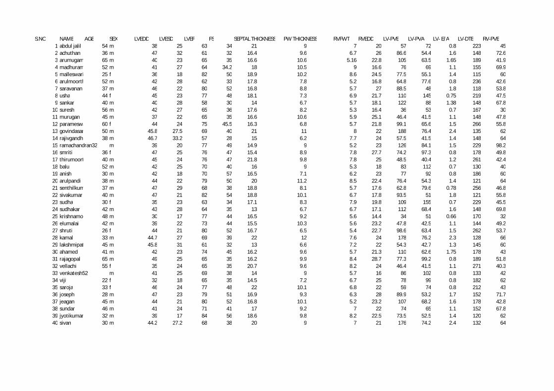
S.NO NAME AGE SEX LVEDD LVESD LVEF FS SEPTAL THICKNESS PW THICKNESS RVFWT RVEDD LV-PVE LV-PVA LV- E/A LV-DTE RV-PVE1 abdul jalil 54 m 38 25 63 34 21 9 7 20 57 72 0.8 223 452 achuthan 36 m 47 32 61 32 16.4 9.6 6.7 26 86.6 54.4 1.6 148 72.63 arumugam 65 m 40 23 65 35 16.6 10.6 5.16 22.8 105 63.5 1.65 189 41.94 madhuram 52 m 41 27 64 34.2 18 10.5 9 16.6 76 69 1.1 155 69.95 malleswari 25 f 36 18 82 50 18.9 10.2 8.6 24.5 77.5 55.1 1.4 115 606 arulmoorthy 52 m 42 28 62 33 17.8 7.8 5.2 16.8 64.8 77.6 0.8 236 42.67 saravanan 37 m 46 22 80 52 16.8 8.8 5.7 27 88.5 48 1.8 118 53.88 usha 44 f 45 23 77 48 18.1 7.3 6.9 21.7 110 145 0.75 219 47.59 sankar 40 m 40 28 58 30 14 6.7 5.7 18.1 122 88 1.38 148 67.8
10 suresh 56 m 42 27 65 36 17.6 8.2 5.3 16.4 36 53 0.7 167 3011 murugan 45 m 37 22 65 35 16.6 10.6 5.9 25.1 46.4 41.5 1.1 148 47.812 parameswari 60 f 44 24 75 45.5 16.3 6.8 5.7 21.8 99.1 65.6 1.5 266 55.813 govindasamy 50 m 45.8 27.5 69 40 21 11 8 22 188 76.4 2.4 135 6214 rajivgandhi 38 m 46.7 33.2 57 28 15 6.2 7.7 24 57.5 41.5 1.4 148 6415 ramachandran32 m 39 20 77 49 14.9 9 5.2 23 126 84.1 1.5 229 98.216 smriti 36 f 47 25 76 47 15.4 8.9 7.8 27.7 74.2 97.3 0.8 178 49.817 thirumoorthy 40 m 45 24 76 47 21.8 9.8 7.8 25 48.5 40.4 1.2 261 42.418 balu 52 m 42 25 70 40 16 9 5.3 18 83 112 0.7 130 4019 anish 30 m 42 18 70 57 16.5 7.1 6.2 23 77 92 0.8 186 6020 arulpandi 38 m 44 22 79 50 20 11.2 8.5 22.4 76.4 54.3 1.4 121 6421 senthilkumar 37 m 47 29 68 38 18.8 8.1 5.7 17.6 62.8 79.6 0.78 256 46.822 sivakumar 40 m 47 21 82 54 18.8 10.1 6.7 17.8 93.5 51 1.8 121 55.823 sudha 30 f 35 23 63 34 17.1 8.3 7.9 19.8 109 155 0.7 229 45.524 sudhakar 42 m 43 28 64 35 13 6.7 6.7 17.1 112 68.4 1.6 148 69.825 krishnamoorthy 48 m 30 17 77 44 16.5 9.2 5.6 14.4 34 51 0.66 170 3226 elumalai 42 m 39 22 73 44 15.5 10.3 5.6 23.2 47.8 42.5 1.1 144 49.227 shruti 26 f 44 21 80 52 16.7 6.5 5.4 22.7 98.6 63.4 1.5 262 53.728 kamal 33 m 44.7 27 69 39 22 12 7.6 24 178 76.2 2.3 128 6629 lakshmipathy 45 m 45.8 31 61 32 13 6.6 7.2 22 54.3 42.7 1.3 145 6030 ahamed 41 m 42 23 74 45 16.2 9.6 5.7 21.3 110 62.6 1.75 178 4331 rajagopal 65 m 49 25 65 35 16.2 9.9 8.4 28.7 77.3 99.2 0.8 189 51.832 vellachi 55 f 35 24 65 35 20.7 9.6 8.2 24 46.4 41.5 1.1 271 40.333 venkatesh52 m 41 25 69 38 14 9 5.7 16 86 102 0.8 133 4234 viji 22 f 32 18 65 35 14.5 7.2 6.7 25 78 99 0.8 182 6235 saroja 33 f 46 24 77 48 22 10.1 6.8 22 59 74 0.8 212 4336 joseph 28 m 47 23 79 51 16.9 9.3 6.3 28 89.9 53.2 1.7 152 71.737 jeagan 45 m 44 21 80 52 16.8 10.1 5.2 23.2 107 68.2 1.6 178 42.838 sundar 46 m 41 24 71 41 17 9.2 7 22 74 65 1.1 152 67.839 jyotikumar 32 m 39 17 84 56 18.6 9.8 8.2 22.5 73.5 52.5 1.4 120 6240 sivan 30 m 44.2 27.2 68 38 20 9 7 21 176 74.2 2.4 132 64

RV-PVA RV-E/A RV-DTE MV-Ea MV-Aa TV-Ea TV-Aa FAMILY H/O SCD syncope NSVT-HOLTER SAM MR LVOT GR E/Ea Maron type RV-IVRT48 0.9 243 3.3 6.9 45.3 48.5 x x 170/11.6 17.3 2 94
49.6 1.46 121 7.1 4.5 7.6 33.7 x x vpc,1stdegreeavblock Gr1 1+ 106/4.6 12 2 12730.7 1.36 236 10.1 5.9 16.4 16.3 x x Gr2 1+ 138/7.62 10.3 4 17065.5 1.06 202 4.3 6.6 5.9 8.4 vpc 150/9.4 17.6 3 139
34 1.7 202 6.7 4.9 16.8 15.5 108/4.8 11.6 4 24032.5 1.3 240 6 8.1 7.5 12.7 106/4 12.6 1 6833.5 1.6 246 8.5 4.1 8.5 4.1 110/5 10.4 2 102
34 1.4 192 4.8 7.7 12.7 23.4 x x nsvt Gr3 440/77 23 3 7732.6 2.1 159 9.7 7.5 23.8 15.6 110/5 10.4 4 86
41 0.7 120 5 8 7 11 112/5 7.2 3 7843.8 1.09 277 3.72 4.19 13.4 18.8 Gr2 140/7.87 12.4 3 14041.2 1.3 302 3.3 5 14.3 11.1 vpcs Gr2 249/24.8 33 3 74
60 1.03 101 7.3 9.7 11 17 nsvt Gr3 2+ 331/43.7 25.7 4 13440 1.6 223 4.3 6.6 8.7 16.3 84.4/2.85 13.37 4 248
119 0.8 115 6.1 9 15.3 26.1 x x nsvt Gr2 2+ 468/87.5 20.6 4 24954 0.9 276 4.9 7.7 11.1 13.4 x nsvt Gr3 1+ 368/54 15.1 4 76
47.6 0.9 124 2.3 4.8 11 31 x 122/6 21 1 22045 0.9 260 5.6 8.1 5.7 8.2 x vpcs Gr1 1+ 289/33 14.8 3 27258 1 178 2.6 4.2 11 31 x nsvt Gr2 2+ 549/121 29.6 3 7030 2.1 210 5.5 4.2 14.7 16.2 120/6 13.9 4 250
33.5 1.4 270 5 9.1 9.5 13.8 106/4.6 12.6 3 6835.6 1.6 256 9.5 4.3 9.5 4.3 119/5.69 9.8 2 93
36 1.3 202 5.8 8.7 14.7 25.4 x x nsvt Gr3 463/85.6 18.8 3 6735.6 1.9 169 10.7 9.5 21.8 18.6 120/5.7 10.5 4 88
42 0.7 110 6 8 9 13 104/4.3 3 7841.8 1.2 274 3.42 4.1 12.7 17.5 Gr2 130/7 14 3 13840.4 1.3 288 3.5 6 13.7 10.9 vpc Gr2 279/32 28 3 72
60 1.1 111 7.5 9.4 12 18 nsvt Gr3 2+ 351/49 24 4 13845 1.3 211 4.2 6.9 8.3 16.6 94.4/3 13 4 232
31.6 1.3 228 11.1 5.6 16.6 17.3 x x Gr2 1+ 148/9 10.6 4 16856 0.9 297 4.7 7.6 10.2 13 x nsvt Gr3 1+ 358/51.8 16.4 4 70
46.8 0.9 121 2.2 4.5 13 32 x 133/7 21 1 24047 0.9 270 5.9 8.4 5.9 8.4 x vpcs Gr1 1+ 279/31 14.5 3 27061 1 182 2.2 4.5 13 32 x nsvt Gr2 2+ 569/130 35 3 6447 0.9 233 3.1 6.7 40.2 46.4 x x 176/12 19 2 98
48.3 1.5 124 6.7 4.2 7.2 31 x x vpc,1stdegreeavblock Gr1 1+ 103/4 13 2 12328.9 1.5 239 10.6 5.6 16.2 16.3 x x Gr2 1+ 128/7 10 4 17063.2 1.1 201 4.4 6.8 5.7 8.1 143/8 16.8 3 132
30 2 210 6.4 4.7 15.9 14.2 102/4 11.5 4 27059 1.1 110 7.1 9.5 12 19 nsvt Gr3 2+ 343/50 24.8 4 137
Your digital receiptThis receipt acknowledges that Turnitin received your paper. Below you will find the receipt informationregarding your submission.
Paper ID 311732839
Paper title
RISK STRATIFICATION OF PATIENTS WITH HYPERTROPHICCARDIOMYOPATHY AND ASSESSMENT OF BIVENTRICULARDIASTOLIC FUNCTION BY PULSE/TISSUE DOPPLERECHOCARDIOGRAPHIC IMAGING
Assignmenttitle Medical
Author Muralidharan Azhakesan 16101507 D.M. CardiologyE-mail [email protected]
Submissiontime 24-Mar-2013 05:11PM
Total words 8207
First 100 words of your submission
“RISK STRATIFICATION OF PATIENTS WITH HYPERTROPHIC CARDIOMYOPATHY ANDASSESSMENT OF BIVENTRICULAR DIASTOLIC FUNCTION BY PULSE/TISSUE DOPPLERECHOCARDIOGRAPHIC IMAGING” Dissertation submitted for D.M. DEGREE EXAMINATIONBRANCH II – CARDIOLOGY MADRAS MEDICAL COLLEGE AND GOVERNMENT GENERALHOSPITAL CHENNAI – 600 003 INTRODUCTION Hypertrophic cardiomyopathy is a commonautosomal dominant cardiovascular disease . Dynamic obstruction to LV outflow can occur underresting or physiologically provocable conditions in patients with HCM. various phenotypic andgenotypic heterogeneity are observed in HCM. HCM is the most common genetically transmittedcardiovascular disease which affects 1 in 500 people...
Copyright 2012 Turnitin. All rights reserved.