Embed Size (px)
Citation preview

104:2532-2542, 2010. First published Sep 15, 2010; doi:10.1152/jn.01039.2009 J NeurophysiolVogel Maciej Dominik Pietr, Per Magne Knutsen, David I. Shore, Ehud Ahissar and Zvi
You might find this additional information useful...
80 articles, 23 of which you can access free at: This article cites http://jn.physiology.org/cgi/content/full/104/5/2532#BIBL
including high-resolution figures, can be found at: Updated information and services http://jn.physiology.org/cgi/content/full/104/5/2532
can be found at: Journal of Neurophysiologyabout Additional material and information http://www.the-aps.org/publications/jn
This information is current as of November 27, 2010 .
http://www.the-aps.org/.American Physiological Society. ISSN: 0022-3077, ESSN: 1522-1598. Visit our website at (monthly) by the American Physiological Society, 9650 Rockville Pike, Bethesda MD 20814-3991. Copyright © 2010 by the
publishes original articles on the function of the nervous system. It is published 12 times a yearJournal of Neurophysiology
on Novem
ber 27, 2010 jn.physiology.org
Dow
nloaded from

Cannabinoids Reveal Separate Controls for Whisking Amplitude and Timingin Rats
Maciej Dominik Pietr,1✠ Per Magne Knutsen,1,2 David I. Shore,3 Ehud Ahissar,1 and Zvi Vogel1,4
1Department of Neurobiology, The Weizmann Institute of Science, Rehovot, Israel; 2Department of Physics, University of California,La Jolla, California; 3Department of Psychology, Neuroscience and Behaviour, McMaster University, Hamilton, Ontario, Canada;and 4The Dr. Miriam and Sheldon Adelson Center for the Biology of Addictive Diseases, Sackler Faculty of Medicine,Tel Aviv University, Tel Aviv, Israel
Submitted 30 November 2009; accepted in final form 14 September 2010
Pietr MD, Knutsen PM, Shore DI, Ahissar E, Vogel Z. Cannabi-noids reveal separate controls for whisking amplitude and timing inrats. J Neurophysiol 104: 2532–2542, 2010. First published Septem-ber 15, 2010; doi:10.1152/jn.01039.2009. Whisking is controlled bymultiple, possibly functionally segregated, motor sensory-motorloops. While testing for effects of endocannabinoids on whisking, weuncovered the first known functional segregation of channels control-ling whisking amplitude and timing. Channels controlling amplitude,but not timing, were modulated by cannabinoid receptor type 1(CB1R). Systemic administration of CB1R agonist �9-tetrahydrocan-nabinol (�9-THC) reduced whisking spectral power across all testeddoses (1.25–5 mg/kg), whereas whisking frequency was affected atonly very high doses (5 mg/kg). Concomitantly, whisking amplitudeand velocity were significantly reduced in a dose-dependent manner(25–43 and 26–50%, respectively), whereas cycle duration and bilat-eral synchrony were hardly affected (3–16 and 3–9%, respectively).Preadministration of CB1R antagonist SR141716A blocked �9-THC–induced kinematic alterations of whisking, and when administeredalone, increased whisking amplitude and velocity but affected neithercycle duration nor synchrony. These findings indicate that whiskingamplitude and timing are controlled by separate channels and thatendocannabinoids modulate amplitude control channels.
I N T R O D U C T I O N
Active whisking and touch are complex functions that in-volve various motor and sensory variables (Ahissar and Zack-senhouse 2001; Berg and Kleinfeld 2003; Bermejo et al. 1998;Brecht et al. 1997; Carvell and Simons 1990; Diamond et al.2008; Knutsen et al. 2006; Mitchinson et al. 2007; Semba andKomisaruk 1984; Vincent 1912; Welker 1964). While explor-ing and palpating objects, the rat motor system controls theamplitude, velocity, and duration of each whisking cycle aswell as those of subcycle components, the rhythm and durationof each whisking bout, and the synchronization and coordina-tion between different whiskers in both mystacial pads (Gao etal. 2003 Mitchinson et al. 2007). The neuronal circuits that areassociated with whisking control are arranged in a complexnetwork of parallel and nested motor-sensory-motor loops(Kleinfeld et al. 1999, 2006). Whether these networks con-trol whisking variables collectively or whether differentvariables are controlled by different neuronal channels is notyet known.
Cannabis sativa (e.g., marijuana) is a source of medicinaland recreational psychotropic preparations (Croxford 2003;Earleywine 2002; Iversen 2003). The major psychoactive com-pound of cannabis is �9-tetrahydrocannabinol (�9-THC)(Mechoulam et al. 1970). �9-THC, and other structurallyrelated compounds (termed cannabinoids), exert a range ofphysiological, behavioral, and cognitive effects (Abel 1970b;Compton et al. 1992; Isbell et al. 1967; Manno et al. 1970;Reeve et al. 1983; Wilson et al. 1994). Neuronal effects ofcannabinoids are mediated by the G protein–coupled cannabi-noid receptor type 1 (CB1R) (Devane et al. 1988; Matsuda etal. 1990), which is typically located on presynaptic terminalsand inhibits neurotransmitter release when activated (Katona etal. 1999, 2000; Kreitzer and Regehr 2001).
The physiological roles of endocannaboids are suggested inpart by the specificity of CB1R expression throughout thebrain. Whereas the basal ganglia, cerebellum, neocortex, andhippocampus contain high levels of CB1R (Egertova andElphick 2000; Herkenham et al. 1990; Marsicano and Lutz1999; Morales et al. 2007; Tsou et al. 1998), CB1R expressionin the thalamus and brain stem is relatively sparse (Tsou et al.1999). Furthermore, CB1R expression varies along the corticalsurface and between cortical layers (Bodor et al. 2005;Lafourcade et al. 2007). The expression patterns of CB1R mayexplain sensory, motor, and cognitive effects of cannabinoids,such as modulation of hippocampal-dependent memory pro-cesses (Robbe et al. 2006; Sullivan 2000) and basal ganglia–related control of locomotion (Sanudo-Pena et al. 1999, 2000b;Shi et al. 2005).
The effects of exogenously administered cannabinoids andthe presence of CB1 receptors throughout motor regions of themammalian brain suggest that endocannabinoids are involvedin motor control. By virtue of the closed-loop architecture ofthe vibrissal motor-sensory system, and the spatial specificityof CB1R expression, it is possible that endocannabinoidsmodulate behavior in a parameter-specific manner. Indeed,CB1R activation alters specific kinematic variables duringlever pressing in rats (McLaughlin et al. 2000), and the CB1Rantagonist SR141716A enhances the amplitude of sensoryevoked activity in S1 (Patel et al. 2002). Here, we testedwhether cannabinoid signaling is associated with specific ki-nematic alterations of whisking, as suggested by CB1R expres-sion patterns in the whisker system and by known region-specific functional involvement in whisker movement control.Using high-speed video and systemic administration of CB1Ragonists and antagonists, we found that the amplitude of
✠ Deceased August 16, 2009.Address for reprint requests and other correspondence: P. M. Knutsen, Dept.
of Physics, Univ. of California–San Diego, 9500 Gilman Dr., La Jolla, CA92093-0374 (E-mail: [email protected]).
J Neurophysiol 104: 2532–2542, 2010.First published September 15, 2010; doi:10.1152/jn.01039.2009.
2532 0022-3077/10 Copyright © 2010 The American Physiological Society www.jn.org
on Novem
ber 27, 2010 jn.physiology.org
Dow
nloaded from

spontaneous, free-air whisking in head-restrained rats wasdifferentially modulated by CB1R activation or inactivation,suggesting that CB1R may be part of an endogenous signalingsystem involved in dynamically controlling whisking move-ments. Furthermore, the specific effect of CB1R activation andinactivation on whisking amplitudes suggests that amplitude iscontrolled separately from timing in this system. We proposethat the anatomical nature of this functional separation can beexplained in part by the spatial distribution of CB1R through-out the rodent brain.
M E T H O D S
Drugs
�9-THC and SR141716A were obtained from the National Instituteon Drug Abuse (Baltimore, MD). �9-THC from its ethanol stocksolution of 100 mg/ml was mixed with vehicle, a PBS containing 25%(wt/vol) 2-hydroxypropyl-1-�-cyclodextrin (Cavasol W7 HP, Wacker),to a final concentration of 1 mg/ml (Jarho et al. 1996). SR141716 wasdiluted with a vehicle containing ethanol, surfactant (Cremophor, Sigma),and saline (0.9% NaCl) in a 1:1:18 mixture to produce a final concen-tration of 1 mg/ml. Drugs or vehicles were administered intraperitoneally(IP) at constant final volume, i.e., when a lower dose had to be injectedthe 1 mg/ml was diluted with the vehicle to give the desired dose alwaysin a constant volume. The same vehicles were used for injections incontrol animals.
Animals
Female Wistar rats, 3–5 mo of age and weighing 200–300 g, served assubjects. Rats were housed individually on a 12:12-h light-dark cycle.Experimental sessions were run once a week or once every 2 wk.
Surgery
Rats were fitted with head mounts for head immobilization aspreviously described (Knutsen et al. 2008). Briefly, animals wereanesthetized (pentobarbitone, 35 mg/kg, ip), the skull was exposed,and holes were drilled to fit stainless steel screws (MX-0080-02B,Small Parts, Miami Lakes, FL). Bone and screws were covered with4-META–containing resin cement (Chemice II, Sun Medical,Moriyama, Japan) and acrylic cement (Jet Acrylic, Lang Dental Mfg,Wheeling, IL), in which two screws were embedded for head immo-bilization. Postoperative care included antibiotics (penicillin andstreptomycin; Pen-Strep, 2 ml/kg, sc), analgesics (carprofen; 5 mg/kg,sc), and ad libitum food and water.
Drug administration schedule
Rats were injected intraperitoneally with �9-THC, SR141716A, ora combination of the two. When co-administered, SR141716A wasinjected 45 min before �9-THC. Vehicle controls mimicked drugadministration schedules and their volumes. Experiments using dif-ferent drugs and/or doses on the same rats were separated by aminimum of 7 days.
Measuring whisker movements
After at least 1 wk of surgical recovery, rats were manuallyhandeled for 30–60 min daily over a 2- to 3-wk period and graduallyacclimatized to the experimental environment and head-fixing proce-dure. Small amounts of mango juice were given to the rats throughoutthese acclimatization sessions. Experimental sessions started whenrats remained calm over a period of �20 min in the head-restrainingdevice.
Although blood plasma levels of �9-THC drop rapidly (min), thedrug can persist in fat and brain tissue for much longer (Kreuz andAxelrod 1973). �9-THC also has significant effects on the corticalEEG (Buonamici et al. 1982), hippocampal local field potentials, andlocomotion several hours after injection (Hajos et al. 2008; Robbe etal. 2006). Therefore data acquisition sessions were repeated in thefirst, fourth, and seventh hours after drug administration. In eachsampling interval, whisking was recorded for 30 min while rats wereinserted into a plastic tube and head restrained. Rats were securelyfixed in place by an articulated manipulator (NF60101, Noga Engi-neering, Schlomi, Israel), which was attached to two mounting screwscemented to the skull (Fig. 1A).
Spontaneous whisker movements were captured at 1,000 frames/sat 640 � 640 pixels using a high-speed video system (MotionScopePCI, Redlake, San Diego, CA) positioned above the rat. The videoacquisition system has previously been described by Knutsen et al.(2005). In each acquisition session (30 min), 5–12 movies �13 s induration were acquired. Whisker tracking was facilitated by removingmost whiskers during brief (5–10 min) isoflurane anesthesia, leavingonly the C2 whisker on each side of the face. Small drops of metallicmarker dye (Artline 990XF, Shachihata, Nagoya City, Japan) wereapplied at two locations along the intact whisker shafts, separated by15–20 mm (Fig. 1A). Movements of these dye labels were trackedoff-line, using the MATLAB based WhiskerTracker application(http://code.google.com/p/whiskertracker). Whisker angles in individ-ual frames were defined by the angle of a line drawn through the twolocations of the dye labels with respect to the eye–nose axis (Fig. 1B),such that a retracted whisker parallel to this line has zero angle. Allexperimental procedures were conducted in accordance with NationalInstitutes of Health guidelines and with approval of the WeizmannInstitute of Science Institutional Animal Care and Use Committee.
Kinematic and spectral analysis of whisking
Whisking movements were analyzed during bouts of continuousfree-air whisking. Whisking bouts were manually selected from timeseries of tracked whisker movements. Individual high-speed moviescontained one or more bouts (Fig. 1C). Individual protraction–retrac-tion cycles were extracted by an automatic peak detection algorithmthat identified protraction onsets, peak protractions, and peak retrac-tions. These events were used to determine the amplitude, duration,and peak velocity of individual protraction and retraction movements(Fig. 1D).
Power spectral densities (PSDs) of angular velocity were estimatedfor each bout of whisking using Welch’s averaged, modified perio-dogram method as implemented in the MATLAB function pwelch(Fig. 1E). These PSDs measures the distribution of frequencies andthe power at each frequency, with the power reflecting the intensity oramplitude of whisking at each frequency. Spectrograms of whiskingbouts were computed by dividing individual bouts into overlappingsegments windowed with a Kaiser window and calculating the PSD of350-ms segments in steps of 20 ms. The instantaneous whiskingfrequency was estimated from the mode of the PSD in each segment.The spectral purity of whisking was estimated by averaging all PSDwindows from the periodogram of a particular bout and normalizingthe result by the whisking bout duration. We define the whisking boutfrequency as the mode of the averaged periodogram (f0). The spectralpurity of whisking is expressed by the spectral width (�f0), corre-sponding to the half-width of the PSD at half-amplitude of f0 (Fig.1F). Whisking power was estimated as the area under the averagedperiodogram in the range 2–12 Hz.
Statistical analysis
Biometric whisking parameters were examined across the popula-tion of rats according to standard methods for within-subject design(Loftus and Masson 1994). The significance of effects induced by
2533CANNABINOIDS AND WHISKING CONTROL
J Neurophysiol • VOL 104 • NOVEMBER 2010 • www.jn.org
on Novem
ber 27, 2010 jn.physiology.org
Dow
nloaded from

different drug treatments was evaluated by a two-way repeated-measures ANOVA (RM-ANOVA), followed by Fisher’s protectedleast significant difference (Fisher’s PLSD) test for multiple compar-isons. F values were compared with Huynh-Feldt epsilon-corrected Fvalues to detect departures from ANOVA assumptions of sphericity.Thus in all instances detected, we report the corrected probabilityvalue as Phf instead of P.
R E S U L T S
Differential effects of �9-THC on whisking powerand frequency
For each video recording of whisker movements, we mea-sured the whisker angles relative to the mystical pad andextracted continuous epochs of motion (Fig. 1, A–C). Whiskingwithin each epoch was characterized by a stable frequency
(Fig. 1, D and E), in agreement with previous reports onhead-restrained rats (Berg and Kleinfeld 2003; Gao et al.2001b). No significant differences in frequencies between left-and right-side whiskers were observed. Thus unless mentionedspecifically, all analysis was restricted to movements of whis-kers on one side of the snout (usually the right).
Systemic administration of �9-THC (1.25, 2.5, and 5 mg/kg)significantly attenuated spontaneous whisking movements inall rats in a monotonic, dose-dependent manner. Typicalwhisking patterns within the fourth hour after control injectionof vehicle or increasing doses of �9-THC are shown in Fig. 2,A and B, for two different rats. Although �9-THC induced aclear reduction in whisker movement amplitudes, the numberof whisk cycles per second did not change. To quantify this, wecomputed the PSD for individual whisking epochs (Fig. 2, Cand D). The PSDs showed a clear dose-dependent reduction in
High-speedcamera Head bolts
Backlight
A
C2
Whisker markers
B
θ
1 2 3 4 5 6 7 850
100
150
Angle(deg)
Time (sec)
Whisking bout
Hz
Time (sec)
12
6
1 2 3 4
0
Max
Min
Rel
ativ
e P
ow
er
C
FE
Angle(deg)
Velocity(deg/s)
−4000
0
4000
50
100
150
Duration
Am
plit
ud
ePe
akve
loci
ty
D
0
.1
.2
ces/rewoP (db
s/ zH/
)
Frequency (Hz)
Protraction Retraction
0
0 0 4 8 12
∆f0
f0
Power Spectral Density
FIG. 1. Kinematic and spectral analysis of whisker movements. A: experimental setup for measuring spontaneous whisker movements in awake,head-restrained rats. A high-speed video camera was used to simultaneously image whiskers on both sides of the head. Whisker visibility was enhanced byilluminating with a strobed backlight. Whiskers of interest were marked with small drops of metallic marker dye at 2 locations. B: head position was markedmanually (white dots), and a rectangular region of interest (dashed rectangle) parallel to a line intersecting the ipsilateral eye and nose was extracted. In eachframe, whisker labels were automatically tracked in head-centered coordinates and a line connecting the labels used as an approximation of whisker location(white dashed line). C: time-varying trace of right C1 whisker angle throughout a 8.5-s video capturing several episodes of whisking (bouts). The gray boxindicates the duration of a single bout of whisking (3.8 s). D: only whisker movements occurring within whisking bouts were selected for further analysis.Individual whisk cycles were automatically extracted from a bout and characterized by duration (horizontal arrows), amplitude (vertical arrows), and peakvelocity (dotted lines) for protraction (blue) and retraction (red). E: the continuous spectrogram of whisking velocity (the 1st derivative of whisker angle)was computed in piecewise steps using Welch’s averaged, modified periodogram method. F: all time bins of spectrogram were averaged to obtain thepower spectral density (PSD). The whisking frequency (f0) was defined as the mode of the PSD. The whisking power corresponds to the area under thePSD curve in the range 2–12 Hz. The spectral purity of whisking was assessed by the spectral width (�f0), defined as the half-width at half-maximumof the PSD curve. In the example shown, f0 was 7.1 Hz and �f0 was 0.6 Hz.
2534 PIETR, KNUTSEN, SHORE, AHISSAR, AND VOGEL
J Neurophysiol • VOL 104 • NOVEMBER 2010 • www.jn.org
on Novem
ber 27, 2010 jn.physiology.org
Dow
nloaded from

power (Fig. 2, C and D) but no change in the average or spreadof the frequency distribution. In agreement with previousreports (Gao et al. 2001b), PSDs typically peaked between 6and 8 Hz, regardless of the �9-THC dose administered (Fig. 2,C and D).
To test the significance of �9-THC effect on whiskingkinematics, we computed PSDs for each whisking bout. Foreach PSD, we measured the power of whisking (area under thePSD between 2 and 12 Hz) and the mode frequency (the peakof the PSD). For each drug dose (control, 1.25, 2.5, and 5mg/kg) and acquisition time point (1st, 4th, and 7th hour), weaveraged all power and frequency measurements obtained forany single rat (n � 6). These averages were compared usingRM-ANOVA with dose and time after injection as factors. For
each �9-THC dose, there was a clear reduction in net power ofwhisking [F(3,15) � 11.26, Phf � 0.01, � � 0.84, MSe �0.003; Fig. 3A]. There was also a trend for reduced poweracross the three acquisition times tested [F(2,10) � 11.26, P �0.05, MSe � 0.001], consistent with previous observations(Gao et al. 2001b). The interaction between dose and acquisi-tion time was not significant [F(6,30) � 1.21, Phf � 0.3, � �0.57, MSe � 0.001]. Post hoc analysis indicated that thewhisking power was significantly attenuated across all dosesand time points and on average (across all time points) de-creased by 64–87% for �9-THC doses of 1.25–5 mg/kg,respectively (Fig. 3A).
Overall, changes in whisking frequency were minor andwere reduced by a significant value only at the highest dose (5
4 80
.005
.01
.015
Rat 202
woP
d( ces/r e)s/z
H/b
Frequency (Hz)
40°
1 sec
0
.0025
.005
.01
Rat 196A C
Wh
iske
r An
gle
Wh
iske
r An
gle
Control1.25 2.5 5
mg/kg∆9-THC }
es/r ew
oP/z
H/b
d( cs)
0 12
4 8Frequency (Hz)
0 12
B D
FIG. 2. Effects of �9-tetrahydrocannabinol (�9-THC) onspontaneous whisking patterns. A and B: whisker movements(angles) executed during representative bouts by 2 different rats(rats 196 and 202; each bout plotted for 6 s). Protractions andretractions are plotted as upward and downward deflections,respectively. All measurements were made within the 4th hourafter injection of vehicle or �9-THC at indicated concentra-tions. C and D: PSDs computed for the whisking traces of Aand B (rats 196 and 202, respectively).
Control 1.25 ∆9-THC mg/kg 2.5 ∆9-THC mg/kg 5 ∆9-THC mg/kg
A
B
C
D
F
Time after injection
0
.1
.2
1h 4h 7h
Rats (n=6)
0
.75
1.5
Freq
uen
cy (H
z)
4
7
10
# # # ## ##
#
#4h
4h
E
4h
∆f0 (H
z)Σ 2-
12 H
z Po
wer
/sec
Σ2-12 Hz
Power/sec
Frequency (Hz)
∆f0 (Hz)
0 0.1 0.2 0.3
4 6 8 10
0 2 4
CD
F
.5
1
CD
F
.5
1
CD
F
.5
1
Control1.25 2.5 5
mg/kg∆9-THC }
1h 4h 7h
1h 4h 7h
FIG. 3. �9-THC–mediated changes inspectral properties of whisker movements.A: bar graphs of net whisking power, com-puted for individual whisking bouts executedwithin the 1st, 4th, and 7th hour after �9-THC injection (n � 6 rats). �9-THC induceda robust reduction in whisking power at alltested concentrations. B and C: distributionsof whisking frequencies and spectral widthscomputed for the same data as in A. Boxesindicate mean values and error bars the 95%CI computed from the within-subject MSeterm of the ANOVA at the level of relevantinteraction. Fisher’s protected leas signifi-cant difference (PLSD) post hoc test formultiple comparisons was performed withinevery time point after injection of each �9-THC dose vs. vehicle control. *P � 0.05 and#P � 0.01. D–F: cumulative probability dis-tribution functions (CDFs) of (D) whiskingpower, (E) whisking frequencies, and (F)spectral widths. The number of whiskingbouts included in all analysis, across all 6rats, was 75 (control), 48 (�9-THC 1.25mg/kg), 44 (�9-THC 2.5 mg/kg), and 32(�9-THC 5.0 mg/kg).
2535CANNABINOIDS AND WHISKING CONTROL
J Neurophysiol • VOL 104 • NOVEMBER 2010 • www.jn.org
on Novem
ber 27, 2010 jn.physiology.org
Dow
nloaded from

mg/kg) in the fourth and seventh hours after injection [F(3,15) �9.63, P � 0.01, MSe � 0.348; Fig. 3B]. A reduction of 1.4 Hz,after 5 mg/kg �9-THC, was the maximal shift in whiskingfrequencies. We did not observe any effect on whisking fre-quency either as a function of the acquisition time [F(2,10) �3.44, Phf � 0.09, � � 0.75, MSe � 0.146] or as a dose � timeinteraction [F(6,30) � 0.85, P � 0.54, MSe � 0.187].
The instantaneous whisking frequency remained relativelyconstant within bouts of whisking (Berg and Kleinfeld 2003;Gao et al. 2001b). This high spectral fidelity, quantified by thespectral width of the PSD (�f0; Fig. 1F), was not affected by�9-THC dose [F(3,15) � 2.75, P � 0.08, MSe � 0.122; Fig.3C] or by the acquisition time [F(2,10) � 0.23, Phf � 0.09,� � 0.75, MSe � 0.146]. In addition, we found no interactionsbetween dose and time [F(6,30) � 1.03, P � 0.4, MSe �0.054].
We validated our RM-ANOVA with nonparametric [Kol-mogorov-Smirnov (K-S)] comparisons between the cumulativeprobability functions (CDFs) of whisking power and frequencyobtained from all rats in the fourth hour after injection of�9-THC. At all tested �9-THC doses, whisking power wassignificantly attenuated compared with control at the 95%rejection limit (Fig. 3D). Whisking frequency, however, wassignificantly affected only at the highest �9-THC dose of 5mg/kg (Fig. 3E). In contrast to the RM-ANOVA, nonparamet-ric testing showed a significant increase of spectral widths (i.e.,a larger scatter of whisking frequencies) at the highest �9-THCdose in the fourth hour after injection (P � 0.002; Fig. 3F).
Whisking activity recovered to control levels, and no effectsof acute �9-THC treatment were detected on any measuredkinematic parameter 24 h after injection of 5 mg/kg �9-THC(data not shown).
Differential effects of �9-THC on whisk amplitudeand duration
The differential effects of �9-THC on whisking powerversus frequency could be caused by a temporal disruption ofwhisking cycles, such as a reduction of the whisk duty cycle,which could thereby shorten and possibly slow down thehigh-velocity retraction phase. To characterize whether such,or other similar, temporal effects were taking place, we com-pared the effects of �9-THC on whisking amplitudes anddurations separately for whisker protractions and retractions(Fig. 4).
In control experiments, protraction and retraction amplitudeswere 34.6 � 5.9 and 34.3 � 5.9°, respectively. In �9-THC–injected animals, protraction and retraction amplitudes werereduced by an average of 9° (25% reduction) at 1.25 mg/kg to15° (43%) at 5 mg/kg. Using RM-ANOVA, we found that�9-THC led to a significant, and dose-dependent, reduction ofboth protraction [F(3,15) � 11.26, Phf � 0.01, � � 0.56,MSe � 67.10] and retraction [F(3,15) � 11.26, Phf � 0.01,� � 0.54, MSe � 66.66] amplitudes (Fig. 4, A and B). Theseamplitude reductions were significantly attenuated with in-creasing time after injections, both with respect to protraction[F(2,10) � 1.22, Phf � 0.26, � � 0.55, MSe � 28.15] andretraction [F(2,10) � 6.28, P � 0.02, MSe � 14.99]. We foundno significant dose � time interaction for protractions [F(6,30) �2.00, P � 0.15, MSe � 16.35] or for retractions [F(6,30) �2.09, P � 0.12, MSe � 24.12]. Post hoc analysis showed that
�9-THC effects were somewhat weaker during the seventhhour after injection, suggesting some drug excretion or desen-sitization.
Effects of �9-THC on the durations of the protraction and ofthe retraction were very small and followed a different dose–response curve (Fig. 4, C and D). In control experiments,protraction and retraction durations were 79.5 � 9.3 and 54.7 � 2.1ms, respectively. In �9-THC–injected animals, protraction du-
0
40
80
120
0
20
40
60
80
Am
plit
ud
e (d
eg)
Protraction
Protraction
Retraction
1h 4h 7h
Time after injection 1h 4h 7h
Retraction
25
50
0
25
50
Du
rati
on
(mse
c)
Du
rati
on
(mse
c)
A
Amplitude (deg)
.5
1
50 1000
100 200
4h
4h
4h
4h
CD
FDuration (msec)
#
# #
#
##
##
#
#
#
##
Am
plit
ud
e (d
eg)
n=6
E
GC
FB
HD1h 4h 7h
1h 4h 7h
Duration (msec)
.5
1
100 2000
CD
F
Amplitude (deg)
.5
1
50 1000
CD
F
.5
1
0C
DF
0
Control1.25 2.5 5
mg/kgΔ9-THC }
Control 1.25 Δ9-THC mg/kg 2.5 Δ9-THC mg/kg 5 Δ9-THC mg/kg
FIG. 4. �9-THC–mediated changes in kinematic properties of whiskerprotractions and retractions. A and B: average protraction and retractionamplitudes (n � 6 rats). C and D: average protraction and retraction durations.Bars indicate the means, and error bars indicate the 95% CIs computed fromthe within-subject MSe term of the ANOVA. Fisher’s PLSD post hoc test formultiple comparisons was carried out for every time point and for each�9-THC concentration vs. control. *P � 0.05 and #P � 0.01. E and F: CDFsof protraction and retraction amplitudes in the 4th hour after administration ofvehicle (n � 1,186), 1.5 mg/kg �9-THC (n � 1,146), 2.5 mg/kg �9-THC (n �1,029), and 5 mg/kg �9-THC (n � 588 whisks). CDFs obtained with all drugtreatments were significantly different from the control CDF at the 95%rejection limit (Kolmogorov-Smirnov test). G and H: CDFs of protraction andretraction durations. Only the CDFs obtained after administration of 5 mg/kg�9-THC were significantly different from the control CDF at the 95% rejectionlimit (Kolmogorov-Smirnov test).
2536 PIETR, KNUTSEN, SHORE, AHISSAR, AND VOGEL
J Neurophysiol • VOL 104 • NOVEMBER 2010 • www.jn.org
on Novem
ber 27, 2010 jn.physiology.org
Dow
nloaded from
ration increased by an average of 12 ms (16%) at 5 mg/kg.Although small, the effect of �9-THC dose on protractionduration was significant [F(3,15) � 4.62, Phf � 0.04, � �0.64, MSe � 113.93] but only for the highest dose (Fisher’spost hoc test). The acquisition time after injection had asignificant influence on protraction duration [F(2,10) � 4.57,Phf � 0.04, � � 0.96, MSe � 31.63], but this was notmodulated by the dose [F(6,30) � 2.00, P � 0.15, MSe �16.35; Fig. 4B]. There was no significant change in retractionduration [F(3,15) � 2.71, Phf � 0.12, � � 0.64, MSe � 81.05],or acquisition time [F(2,10) � 1.65, Phf � 0.24, � � 0.87, MSe �40.51] at all �9-THC doses.
We validated the RM-ANOVA results with nonparametric(K-S) comparisons between the CDFs of whisk amplitudes anddurations measured for all rats in the fourth hour after �9-THCinjection. In agreement with the effect on power of whisking,both protraction and retraction amplitudes were significantlydecreased after �9-THC administration at any dose (Fig. 4, Eand F); only protraction duration was significantly changed(increased) and only at the highest �9-THC dose (5 mg/kg; Fig.4, G and H).
�9-THC–mediated amplitude reduction is accompanied bydecrease in peak velocity
Amplitude reductions were accompanied by reductions inpeak velocity (Fig. 5, A and B). In control animals, the averagepeak velocity during protraction and retraction was 769 � 129and 1432 � 299°/s, respectively. In the fourth hour after�9-THC administration, protraction peak velocity reduced by26% at 1.25 mg/kg and by 50% at 5.0 mg/kg. RM-ANOVAanalysis showed a significant effect of dose [F(3,15) � 21.20,Phf � 0.01, � � 0.91, MSe � 21,997; F(3,15) � 13.00, Phf �0.01, � � 0.63, MSe � 118,673], a marginally significant effectof time [F(3,15) � 4.66, Phf � 0.06, � � 0.75, MSe � 8,090;F(3,15) � 6.64, Phf �0.05, � � 0.83, MSe � 35,593], and nointeraction between the two factors [F(6,30) � 1.40, P � 0.25,MSe � 8,401; F(6,30) � 1.53, Phf � 0.20, � � 0.72, MSe �50,363] on peak protraction and retraction velocities, respec-tively. Although protraction peak velocities were significantlyreduced at all doses and time points, retraction peak velocitieswere reduced at all doses and all but one time-point (7 h). Thismarginally weaker effect on retraction peak velocity could be
-THC mg/kg 2.5
A
250
500
750
1000
Vel
ocity
(de
g/se
c)
500
1000
1500
2000
Vel
ocity
(de
g/se
c)
Time after injection 1h 4h 7h
Protraction Retraction
## #
# ##
###
## ## #
Time after injection 1h 4h 7h
B
Control 1.25 Δ9 Δ9-THC mg/kg 5 Δ9-THC mg/kg
6=n6=n
00
G H I
.25
.5
.75
1
50 1000Amplitude (deg)
1250 25000Peak velocity (deg/sec)
100 2000Duration (msec)
F
0 2 4 6 8 10
15
30
45
60
Am
plit
ud
e (d
eg)
EC
600
300
900
1200D
60
30
90
120
55.252.155.252.1 1.25 2.5 5
Peak
vel
oci
ty (d
eg/s
ec)
Du
rati
on
(mse
c)
Control ControlControlΔ9-THC (mg/kg)Δ9-THC (mg/kg)Δ9-THC (mg/kg)
SR141716A
Time (sec)
Control
n=4
Δ9-THC 2.5 mg/kg
SR141716A 2 mg/kg
.25
.5
.75
1
.25
.5
.75
1
CD
F
Control
2.5 mg/kg Δ9-THC
SR141716A
CD
F
CD
F
FIG. 5. Effects of a cannabinoid receptor type 1 (CB1R)agonist and antagonist on whisking kinematics. A and B: the bargraph represents mean protraction and retraction peak velocitiesfor a group of subjects (n � 6). Error bars displayed at the 95%CI calculated from the within-subject MSe term of the ANOVAat the level of relevant interaction. Fisher’s PLSD post hoc testfor multiple comparisons was carried out within every timepoint for each �9-THC dose vs. control. *P � 0.05 and #P �0.01. C–E: effects of SR141716A (light gray bars) on protrac-tion amplitudes (C), peak velocity (D), and duration (E) com-pared with control (black bars) and �9-THC administered atdifferent doses 4 h after injection. Thick bars indicate means,and error bars are 95% within-subject CIs as reported byrepeated measures (RM)-ANOVA. Fisher’s PLSD post hoc testwas performed separately for each �9-THC dose betweenSR141716A and vehicle pretreatment. *P � 0.05. F: typicaltraces of whisker angle executed by the same rat in the 4th hourafter administration of �9-THC (2.5 mg/kg; dark gray), vehicle(black), or SR141716A (2 mg/kg; light gray). Vertical calibra-tion bar corresponds to 50°. G–I: CDFs of protraction ampli-tudes (G), peak velocities (H), and durations (I) for control (n �1886 whisks), �9-THC (n � 564 whisks), and SR141716A(n � 592 whisks) treatments.
2537CANNABINOIDS AND WHISKING CONTROL
J Neurophysiol • VOL 104 • NOVEMBER 2010 • www.jn.org
on Novem
ber 27, 2010 jn.physiology.org
Dow
nloaded from
caused partly by the passive component of whisker retraction(Berg and Kleinfeld 2003).
�9-THC modulates whisking via CB1 receptors
To determine the involvement of CB1 receptors in �9-THCeffects on whisking behavior, we administered the selectiveCB1R antagonist SR141716A (Rinaldi-Carmona et al. 1994)45 min before the administration of �9-THC or control vehicleand measured whisking behavior in the fourth hour after�9-THC/vehicle injection (n � 4). Reductions in protractionamplitudes and peak velocities caused by �9-THC were com-pletely abolished by SR141716A (Fig. 5, C and D). RM-ANOVA analysis showed a significant effect on protractionamplitude of �9-THC dose [F(3,9) � 11.03, Phf � 0.01, MSe �22.80] and SR141716A [F(1,3) � 23.55, P � 0.05, MSe �109.28], but not of the interaction between these two factors[F(2, 6) � 0.55, P � 0.66, MSe � 29.95]. Moreover, we founda significant effect of SR141716A treatment on protractionvelocity [F(2,3) � 22.40, P � 0.05, MSe � 62,097] but noeffect of the �9-THC � SR141716A interaction [F(2, 6) �1.81, Phf � 0.21, MSe � 19,884]. Fisher’s post hoc testperformed for each �9-THC dose showed that SR141716Apretreatment completely counteracted �9-THC–mediated re-ductions in protraction amplitudes (Fig. 5C) and peak veloci-
ties (Fig. 5D). The same effects were observed for retractionphase of the whisk cycle (data not shown).
The increase in protraction duration observed with 5 mg/kg�9-THC (Fig. 4C) was completely abolished by SR141716Apretreatment (Fig. 5E). RM-ANOVA showed no significanteffects on protraction duration of �9-THC dose [F(1,3) � 1.28,Phf � 0.34, MSe � 96.44], SR141716A treatment [F(2,6) �2.74, P � 0.19, MSe � 105.25], or the �9-THC � SR141716Ainteraction [F(2, 6) � 4.72, Phf � 0.07, MSe � 87.28].
Blocking endogenous cannabinoids increases whiskingamplitude and does not affect whisking duration
By comparing individual epochs of whisking, it was evidentthat �9-THC or SR141716A applied alone affected whiskermovement amplitudes in opposite directions (Fig. 5F). Thesame animals that were treated with SR141716A (n � 4) werealso injected with �9-THC on other days. As was the case inother experiments (Figs. 2–5), �9-THC treatment resulted in asignificantly decreased protraction amplitudes (40%; Fig. 5G)and peak velocities (36%; P � 0.01, K-S test; Fig. 5H), as wellas in a minor, but significant, increase in protraction duration(4%; P � 0.01, K-S test; Fig. 5I). In contrast, SR141716A at2 mg/kg significantly increased the protraction amplitudes by41% (14°) and the protraction peak velocities by 30% relative
Control
∆9-THC (2.5 mg/kg)
Right
Left
Right
Left
SR141716A (2 mg/kg)
Right
Left
2 3 4 5Time (sec)
0 1
A B
F G
C
E
R
-0.2 -0.1 0 0.1 0.2
Control
∆9-THC
-.5
Time (sec)
SR141716A
D
-10
0
10D
elay
(mse
c)
10
20
0
Du
rati
on
(sec
)
0.2
0.4
0.6
0.8
1
Max
R
0
-.5
0
.5
1
R
-0.2 -0.1 0 0.1 0.2
-.5
0
.5
1
R
-0.2 -0.1 0 0.1 0.2
-.5
0
.5
1
0.2
0.4
0.6
0.8
1
CV
0
Amplitude
0.2
0.4
0.6
0.8
1
0
Duration
0.2
0.4
0.6
0.8
1
0
Peak Velocity
0.2
0.4
0.6
0.8
1
CV
0
Amplitude
0.2
0.4
0.6
0.8
1
0
Duration
0.2
0.4
0.6
0.8
1
0
Peak Velocity
noitcarteRnoitcartorP
Control
∆9 -TH
C
SR141716A
FIG. 6. Effects of CB1R agonist and antagonist on whisking coordination. A: right and left C2 whisker movements executed simultaneously (by the same rat)in the 4th hour after administration of vehicle, �9-THC (2.5 mg/kg), or SR141716A (2 mg/kg). Calibration bar corresponds to 50° angle. B: cross-correlationsof the angle traces in A. Peaks were always centered close to 0, showing a high degree of synchrony between right and left whiskers, regardless of drug treatment.C: distributions of peak correlation coefficients obtained from cross-correlations between right and left whisker movements, such as those in A and B, executedin the 4th hour after administration of vehicle (n � 36 whisking bouts), �9-THC (2.5 mg/kg, n � 29), or SR141716A (2 mg/kg, n � 16). D: distributions oftemporal delays of the peak correlation coefficients obtained from cross-correlations. E: distributions of whisking bout durations. None of the differences in C–Ewere significantly different (P � 0.05; 1-way ANOVA). F: distributions of within-bout CVs of amplitude, peak velocities, and durations for protraction and (G)retraction phase of the whisk cycle [*P � 0.05; 1-way ANOVA followed by Tukey-Kramer honestly significant difference (HSD) post hoc test]. Box plots inC–G denote lower and upper quartiles (gray box), medians (horizontal line inside boxes), and range of data (vertical lines).
2538 PIETR, KNUTSEN, SHORE, AHISSAR, AND VOGEL
J Neurophysiol • VOL 104 • NOVEMBER 2010 • www.jn.org
on Novem
ber 27, 2010 jn.physiology.org
Dow
nloaded from

to control experiments (P � 0.05, K-S test; Fig. 5, C, D, G, andH). However, SR141716A administration did not significantlyaffect protraction durations (P � 0.18; K-S test; Fig. 5I).
Cannabinoids do not affect duration, synchrony, andcoordination of whisking bouts
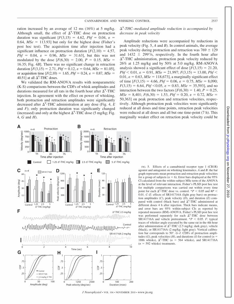
We examined whether �9-THC or SR141716A affected thebilateral control of whisker movements. Free-air whiskermovements of right and left whiskers were typically synchro-nous and amplitude coordinated in control animals (Fig. 6A).We found that, regardless of treatment (�9-THC or SR141716A),bilateral whisker movements remained temporally synchronous(Fig. 6B), as indicated by high correlations (Fig. 6C) and smalltime lags of peak correlations between movements of differentwhiskers (Fig. 6D). Cross-correlations between the same whiskeron the left or right side always peaked close to zero andaveraged �9 ms (r � 0.45–0.75; Fig. 6D). In our analysis,time lags above zero imply that the right whisker was phase-leading the left whisker. Peak cross-correlation coefficients,regardless of treatment condition, were distributed across abroad range of values with sample medians varying between0.6 and 0.8 (Fig. 6C).
Amplitude coordination was estimated by the instantaneousangle difference divided by the average angle of right and leftwhiskers
(�LEFT � �RIGHT) ⁄ �(�LEFT � �RIGHT) ⁄ 2�This formula expresses the ratio of the angle difference
relative to the absolute angle amplitude and thus is an estimate
of motor error (assuming the target is to produce identicalmovements on both sides). The frame-by-frame, instantaneousmeasurements of amplitude coordination were averaged across allframes in each trial, and the distributions of such averages werecompared between the different drug administration conditions.The results of these comparisons show that, in control animals, themotor error was 19.5 � 0.9% and in �9-THC– or SR141716A-injected animals was 19.7 � 0.7%. These small differences werenot significantly different (P � 0.7; 1-way ANOVA).
Whisking epochs ranged from short (�1 s) to tens ofseconds long and were on average 8.3 � 0.8 s long in controlanimals. We did not observe any significant change in boutdurations in the fourth hour after administration during either�9-THC or SR141716A treatments (1-way ANOVA; Fig. 6E).
Finally, we examined whether cannabinoid treatment affectedvariability of whisking kinematics (Fig. 6, F and G). We foundthat protraction amplitudes were more irregular, expressed byCVs, 4 h after treatment with 2.5 mg/kg �9-THC (P � 0.05,1-way ANOVA; Fig. 6F). Within-bout fluctuations of protractionpeak velocity and duration were unaffected regardless or treat-ment compared with vehicle controls. Variability of retractionamplitude was affected in the fourth hour after either �9-THC orSR141716A administration (Fig. 6G). SR141716A treatment,however, exerted an opposite effect to the one by �9-THC byreducing within-bout fluctuations of retraction amplitudes withrespect to control. In addition to increasing irregularity of retrac-tion amplitudes, �9-THC affected peak retraction velocities.
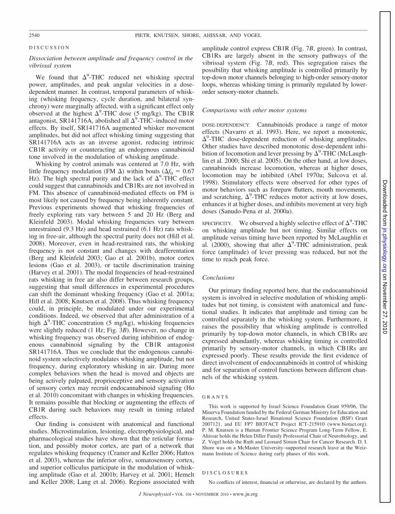
Whisker & ExternalEnvironment
Timing Amplitude
Movement
EndogenousInputs
A
B
CB1R +CB1R -
ExogenousInputs
MCtxSCtx
RF
Whisker &External
Environment
TN
Rn
CbVpm
VL
Pom
12/345a5b
6
12/345
6
Pom
TGFn
Pn
IOSC
BG
FIG. 7. Whisking is controlled by different signal sources. A: duringfree-air whisking, amplitude can be modulated by endogenous signals (i.e.,signals generated internally) and does not require exogenously derived signals,such as contact and proprioception. However, timing of whisker movements ismodulated during contact-dependent tasks and when proprioceptive signalschange (e.g., restrained vs. unrestrained). Thus timing control is not affected byendogenous signals but its change requires exogenous signals, such as thosegenerated by head/body movement or contact. B: CB1R is primarily expressedin motor circuits involved in control of whisker movement (CB1R�; green).CB1R expression is abundant, but highly regional or layer specific, throughoutthe rat nervous system. CB1Rs are absent in sensory regions that processwhisker-related sensory inputs (CB1R�; red). Cells in trigeminal ganglion(TG) express CB1Rs, but primarily those located in the maxillary and non-vibrissal representation of the mandibular division of the TG (Price et al.2003). Cells in the trigeminal nuclei (TN), the ventroposteromedial complex(Vpm), and posteromedial thalamic nucleus (Pom) of the thalamus do notexpress CB1Rs (Tsou et al. 1998). The primary somatosensory (S1) and motor(M1) cortices express CB1Rs primarily in layers 2/3 and 5a (Bodor et al. 2005;Eggan and Lewis 2007; Hill et al. 2007; Trettel et al. 2004). CB1R is notexpressed in layer 5b of S1. The basal ganglia contain high levels of CB1R(Egertova and Elphick 2000; Herkenham et al. 1990; Marsicano and Lutz1999; Morales et al. 2007; Tsou et al. 1998) and receive inputs both fromsomatosensory (S1) and motor (M1) cortices (Alloway et al. 2006). Thepontine nuclei (Pn), which contain CB1R expressing neurons (Cristino et al.2006), receive inputs from CB1R regions of S1 (layer 5b; Mercier et al. 1990)and M1 (layer 5; Legg et al. 1989). Pn neurons project to the cerebellum (Cb),which also contain CB1R expressing neurons (Moldrich and Wenger 2000;Suarez et al. 2008; Tsou et al. 1998). Cerebellar Purkinje cells project to theinferior olive (IO), a CB1R-containing region (Suarez et al. 2008). Cbprojections back to layer 3 of M1 via the ventral lateral thalamus and completea cortico-cerebellar loop (VL; Yamamoto et al. 2004), and a sensory-motorloop via the IO, red nucleus (Rn) and SC (Suarez et al. 2008). CB1R has beenproposed to be present in Rn (Tsou et al. 1998). The lateral facial nucleus (Fn)receive projections from a large number of regions, including M1 (Hattox et al.2003), TN, and the deep gray layer of the SC (Miyashita and Mori 1995;Miyashita et al. 1994). The basal ganglia (BG), which express high amounts ofCB1R, have not been included in this scheme because little is known about theinvolvement of this structure in whisking. The BG is mostly innervated by S1pyramidals of CB1R expressing layer 5a (Mercier et al. 1990).
2539CANNABINOIDS AND WHISKING CONTROL
J Neurophysiol • VOL 104 • NOVEMBER 2010 • www.jn.org
on Novem
ber 27, 2010 jn.physiology.org
Dow
nloaded from
D I S C U S S I O N
Dissociation between amplitude and frequency control in thevibrissal system
We found that �9-THC reduced net whisking spectralpower, amplitudes, and peak angular velocities in a dose-dependent manner. In contrast, temporal parameters of whisk-ing (whisking frequency, cycle duration, and bilateral syn-chrony) were marginally affected, with a significant effect onlyobserved at the highest �9-THC dose (5 mg/kg). The CB1Rantagonist, SR141716A, abolished all �9-THC–induced motoreffects. By itself, SR141716A augmented whisker movementamplitudes, but did not affect whisking timing suggesting thatSR141716A acts as an inverse agonist, reducing intrinsicCB1R activity or counteracting an endogenous cannabinoidtone involved in the modulation of whisking amplitude.
Whisking by control animals was centered at 7.0 Hz, withlittle frequency modulation (FM �) within bouts (�f0 � 0.67Hz). The high spectral purity and the lack of �9-THC effectcould suggest that cannabinoids and CB1Rs are not involved inFM. This absence of cannabinoid-mediated effects on FM ismost likely not caused by frequency being inherently constant.Previous experiments showed that whisking frequencies offreely exploring rats vary between 5 and 20 Hz (Berg andKleinfeld 2003). Modal whisking frequencies vary betweenunrestrained (9.3 Hz) and head restrained (6.1 Hz) rats whisk-ing in free-air, although the spectral purity does not (Hill et al.2008). Moreover, even in head-restrained rats, the whiskingfrequency is not constant and changes with deafferentation(Berg and Kleinfeld 2003; Gao et al. 2001b), motor cortexlesions (Gao et al. 2003), or tactile discrimination training(Harvey et al. 2001). The modal frequencies of head-restrainedrats whisking in free air also differ between research groups,suggesting that small differences in experimental procedurescan shift the dominant whisking frequency (Gao et al. 2001a;Hill et al. 2008; Knutsen et al. 2008). Thus whisking frequencycould, in principle, be modulated under our experimentalconditions. Indeed, we observed that after administration of ahigh �9-THC concentration (5 mg/kg), whisking frequencieswere slightly reduced (1 Hz; Fig. 3B). However, no change inwhisking frequency was observed during inhibition of endog-enous cannabinoid signaling by the CB1R antagonistSR141716A. Thus we conclude that the endogenous cannabi-noid system selectively modulates whisking amplitude, but notfrequency, during exploratory whisking in air. During morecomplex behaviors when the head is moved and objects arebeing actively palpated, proprioceptive and sensory activationof sensory cortex may recruit endocannabinoid signaling (Hoet al. 2010) concomitant with changes in whisking frequencies.It remains possible that blocking or augmenting the effects ofCB1R during such behaviors may result in timing relatedeffects.
Our finding is consistent with anatomical and functionalstudies. Microstimulation, lesioning, electrophysiological, andpharmacological studies have shown that the reticular forma-tion, and possibly motor cortex, are part of a network thatregulates whisking frequency (Cramer and Keller 2006; Hattoxet al. 2003), whereas the inferior olive, somatosensory cortex,and superior colliculus participate in the modulation of whisk-ing amplitude (Gao et al. 2001b; Harvey et al. 2001; Hemeltand Keller 2008; Lang et al. 2006). Regions associated with
amplitude control express CB1R (Fig. 7B, green). In contrast,CB1Rs are largely absent in the sensory pathways of thevibrissal system (Fig. 7B, red). This segregation raises thepossibility that whisking amplitude is controlled primarily bytop-down motor channels belonging to high-order sensory-motorloops, whereas whisking timing is primarily regulated by lower-order sensory-motor channels.
Comparisons with other motor systems
DOSE-DEPENDENCY. Cannabinoids produce a range of motoreffects (Navarro et al. 1993). Here, we report a monotonic,�9-THC dose-dependent reduction of whisking amplitudes.Other studies have described monotonic dose-dependent inhi-bition of locomotion and lever pressing by �9-THC (McLaugh-lin et al. 2000; Shi et al. 2005). On the other hand, at low doses,cannabinoids increase locomotion, whereas at higher doses,locomotion may be inhibited (Abel 1970a; Sulcova et al.1998). Stimulatory effects were observed for other types ofmotor behaviors such as forepaw flutters, mouth movements,and scratching, �9-THC reduces motor activity at low doses,enhances it at higher doses, and inhibits movement at very highdoses (Sanudo-Pena et al. 2000a).
SPECIFICITY. We observed a highly selective effect of �9-THCon whisking amplitude but not timing. Similar effects onamplitude versus timing have been reported by McLaughlin etal. (2000), showing that after �9-THC administration, peakforce (amplitude) of lever pressing was reduced, but not thetime to reach peak force.
Conclusions
Our primary finding reported here, that the endocannabinoidsystem is involved in selective modulation of whisking ampli-tudes but not timing, is consistent with anatomical and func-tional studies. It indicates that amplitude and timing can becontrolled separately in the whisking system. Furthermore, itraises the possibility that whisking amplitude is controlledprimarily by top-down motor channels, in which CB1Rs areexpressed abundantly, whereas whisking timing is controlledprimarily by sensory-motor channels, in which CB1Rs areexpressed poorly. These results provide the first evidence ofdirect involvement of endocannabinoids in control of whiskingand for separation of control functions between different chan-nels of the whisking system.
G R A N T S
This work is supported by Israel Science Foundation Grant 959/06, TheMinerva Foundation funded by the Federal German Ministry for Education andResearch, United States-Israel Binational Science Foundation (BSF) Grant2007121, and EU FP7 BIOTACT Project ICT-215910 (www.biotact.org).P. M. Knutsen is a Human Frontier Science Program Long-Term Fellow, E.Ahissar holds the Helen Diller Family Professorial Chair of Neurobiology, andZ. Vogel holds the Ruth and Leonard Simon Chair for Cancer Research. D. I.Shore was on a McMaster University–supported research leave at the Weiz-mann Institute of Science during early phases of this work.
D I S C L O S U R E S
No conflicts of interest, financial or otherwise, are declared by the authors.
2540 PIETR, KNUTSEN, SHORE, AHISSAR, AND VOGEL
J Neurophysiol • VOL 104 • NOVEMBER 2010 • www.jn.org
on Novem
ber 27, 2010 jn.physiology.org
Dow
nloaded from

R E F E R E N C E S
Abel EL. Effects of the marihuana-homologue, pyrahexyl, on open fieldbehaviour in the rat. J Pharm Pharmacol 22: 785, 1970a.
Abel EL. Marijuana and memory. Nature 227: 1151–1152, 1970b.Ahissar E, Zacksenhouse M. Temporal and spatial coding in the rat vibrissal
system. Prog Brain Res 130: 75–88, 2001.Aldes LD. Thalamic connectivity of rat somatic motor cortex. Brain Res Bull
20: 333–348, 1988.Alloway KD, Lou L, Nwabueze-Ogbo F, Chakrabarti S. Topography of
cortical projections to the dorsolateral neostriatum in rats: multiple overlap-ping sensorimotor pathways. J Comp Neurol 518: 33–48, 2006.
Berg RW, Kleinfeld D. Rhythmic whisking by rat: retraction as well asprotraction of the vibrissae is under active muscular control. J Neurophysiol89: 104–117, 2003.
Bermejo R, Houben D, Zeigler HP. Optoelectronic monitoring of individualwhisker movements in rats. J Neurosci Methods 83: 89–96, 1998.
Bodor AL, Katona I, Nyiri G, Mackie K, Ledent C, Hajos N, Freund TF.Endocannabinoid signaling in rat somatosensory cortex: laminar differencesand involvement of specific interneuron types. J Neurosci 25: 6845–6856,2005.
Brecht M, Preilowski B, Merzenich MM. Functional architecture of themystacial vibrissae. Behav Brain Res 84: 81–97, 1997.
Buonamici M, Young GA, Khazan N. Effects of acute delta 9-THC admin-istration on EEG and EEG power spectra in the rat. Neuropharmacology 21:825–829, 1982.
Carvell GE, Simons DJ. Biometric analyses of vibrissal tactile discriminationin the rat. J Neurosci 10: 2638–2648, 1990.
Compton DR, Johnson MR, Melvin LS, Martin BR. Pharmacologicalprofile of a series of bicyclic cannabinoid analogs: classification as canna-bimimetic agents. J Pharmacol Exp Ther 260: 201–209, 1992.
Cramer NP, Keller A. Cortical control of a whisking central pattern gener-ator. J Neurophysiol 96: 209–217, 2006.
Cristino L, de Petrocellis L, Pryce G, Baker D, Guglielmotti V, Di MarzoV. Immunohistochemical localization of cannabinoid type 1 and vanilloidtransient receptor potential vanilloid type 1 receptors in the mouse brain.Neuroscience 139: 1405–1415, 2006.
Croxford JL. Therapeutic potential of cannabinoids in CNS disease. CNSDrugs 17: 179–202, 2003.
Devane WA, Dysarz FA III, Johnson MR, Melvin LS, Howlett AC.Determination and characterization of a cannabinoid receptor in rat brain.Mol Pharmacol 34: 605–613, 1988.
Diamond ME, von Heimendahl M, Knutsen PM, Kleinfeld D, Ahissar E.‘Where’ and ‘what’ in the whisker sensorimotor system. Nat Rev Neurosci9: 601–612, 2008.
Earleywine M. Understanding Marijuana. Oxford University Press: Oxford,UK, 2002.
Egertova M, Elphick MR. Localisation of cannabinoid receptors in the ratbrain using antibodies to the intracellular C-terminal tail of CB. J CompNeurol 422: 159–171, 2000.
Eggan SM, Lewis DA. Immunocytochemical distribution of the cannabinoidCB1 receptor in the primate neocortex: a regional and laminar analysis.Cereb Cortex 17: 175–191, 2007.
Gao P, Bermejo R, Zeigler HP. Whisker deafferentation and rodent whiskingpatterns: behavioral evidence for a central pattern generator. J Neurosci 21:5374–5380, 2001.
Gao P, Hattox AM, Jones LM, Keller A, Zeigler HP. Whisker motor cortexablation and whisker movement patterns. Somatosens Mot Res 20: 191–198,2003.
Hajos M, Hoffmann WE, Kocsis B. Activation of cannabinoid-1 receptorsdisrupts sensory gating and neuronal oscillation: relevance to schizophrenia.Biol Psychiatry 63: 1075–1083, 2008.
Harvey MA, Sachdev RN, Zeigler HP. Cortical barrel field ablation andunconditioned whisking kinematics. Somatosens Mot Res 18: 223–227,2001.
Hattox A, Li Y, Keller A. Serotonin regulates rhythmic whisking. Neuron 39:343–352, 2003.
Hemelt ME, Keller A. Superior colliculus control of vibrissa movements. JNeurophysiol 100: 1245–1254, 2008.
Herkenham M, Lynn AB, Little MD, Johnson MR, Melvin LS, de CostaBR, Rice KC. Cannabinoid receptor localization in brain. Proc Natl AcadSci USA 87: 1932–1936, 1990.
Hill DN, Bermejo R, Zeigler HP, Kleinfeld D. Biomechanics of the vibrissamotor plant in rat: rhythmic whisking consists of triphasic neuromuscularactivity. J Neurosci 28: 3438–3455, 2008.
Hill EL, Gallopin T, Ferezou I, Cauli B, Rossier J, Schweitzer P, LambolezB. Functional CB1 receptors are broadly expressed in neocortical GABAergicand glutamatergic neurons. J Neurophysiol 97: 2580–2589, 2007.
Ho WS, Patel S, Thompson JR, Roberts CJ, Stuhr KL, Hillard C.Endocannabinoid modulation of hyperaemia evoked by physiologicallyrelevant stimuli in the rat primary somatosensory cortex. Br J Pharmacol130: 736–746, 2010.
Isbell H, Gorodetzsky CW, Jasinski D, Claussen U, von Spulak F, KorteF. Effects of (–)delta-9-trans-tetrahydrocannabinol in man. Psychopharma-cologia 11: 184–188, 1967.
Iversen L. Cannabis and the brain. Brain 126: 1252–1270, 2003.Jarho P, Urtti A, Jarvinen K, Pate DW, Jarvinen T. Hydroxypropyl-beta-
cyclodextrin increases aqueous solubility and stability of anandamide. LifeSci 58: PL181–PL185, 1996.
Katona I, Sperlagh B, Magloczky Z, Santha E, Kofalvi A, Czirjak S,Mackie K, Vizi ES, Freund TF. GABAergic interneurons are the targets ofcannabinoid actions in the human hippocampus. Neuroscience 100: 797–804, 2000.
Katona I, Sperlagh B, Sik A, Kafalvi A, Vizi ES, Mackie K, Freund TF.Presynaptically located CB1 cannabinoid receptors regulate GABA releasefrom axon terminals of specific hippocampal interneurons. J Neurosci 19:4544–4558, 1999.
Kleinfeld D, Ahissar E, Diamond ME. Active sensation: insights from therodent vibrissa sensorimotor system. Curr Opinion Neurobiol 16: 435–444,2006.
Kleinfeld D, Berg RW, O=Connor SM. Anatomical loops and their electricaldynamics in relation to whisking by rat. Somatosens Mot Res 16: 69–88,1999.
Knutsen PM, Biess A, Ahissar E. Vibrissal kinematics in 3D: tight couplingof azimuth, elevation, and torsion across different whisking modes. Neuron59: 35–42, 2008.
Knutsen PM, Derdikman D, Ahissar E. Tracking whisker and head move-ments in unrestrained behaving rodents. J Neurophysiol 93: 2294–2301,2005.
Knutsen PM, Pietr M, Ahissar E. Haptic object localization in the vibrissalsystem: behavior and performance. J Neurosci 26: 8451–8464, 2006.
Kreitzer AC, Regehr WG. Retrograde inhibition of presynaptic calciuminflux by endogenous cannabinoids at excitatory synapses onto Purkinjecells. Neuron 29: 717–727, 2001.
Kreuz DS, Axelrod J. Delta-9-tetrahydrocannabinol: localization in body fat.Science 179: 391–393, 1973.
Lafourcade M, Elezgarai I, Mato S, Bakiri Y, Grandes P, Manzoni OJ.Molecular components and functions of the endocannabinoid system inmouse prefrontal cortex. PLoS ONE 2: e709, 2007.
Lang EJ, Sugihara I, Llinas R. Olivocerebellar modulation of motor cortexability to generate vibrissal movements in rat. J Physiol 571: 101–120, 2006.
Legg CR, Mercier B, Glickstein M. Corticopontine projection in the rat: thedistribution of labelled cortical cells after large injections of horseradishperoxidase in the pontine nuclei. J Comp Neurol 286: 427–441, 1989.
Loftus GR, Masson MEJ. Using confidence intervals in within-subjectdesigns. Psychonomic Bull Rev 1: 476–490, 1994.
Manno JE, Kiplinger GF, Haine SE, Bennett IF, Forney RB. Comparativeeffects of smoking marihuana or placebo on human motor and mentalperformance. Clin Pharmacol Ther 11: 808–815, 1970.
Marsicano G, Lutz B. Expression of the cannabinoid receptor CB1 in distinctneuronal subpopulations in the adult mouse forebrain. Eur J Neurosci 11:4213–4225, 1999.
Matsuda LA, Lolait SJ, Brownstein MJ, Young AC, Bonner TI. Structureof a cannabinoid receptor and functional expression of the cloned cDNA.Nature 346: 561–564, 1990.
McLaughlin PJ, Delevan CE, Carnicom S, Robinson JK, Brener J. Finemotor control in rats is disrupted by delta-9-tetrahydrocannabinol. Pharma-col Biochem Behav 66: 803–809, 2000.
Mechoulam R, Shani A, Edery H, Grunfeld Y. Chemical basis of hashishactivity. Science 169: 611–612, 1970.
Mercier BE, Legg CR, Glickstein M. Basal ganglia and cerebellum receivedifferent somatosensory information in rats. Proc Natl Acad Sci USA 87:4388–4392, 1990.
Mitchinson B, Martin CJ, Grant RA, Prescott TJ. Feedback control inactive sensing: rat exploratory whisking is modulated by environmentalcontact. Proc Biol Sci 274: 1035–1041, 2007.
2541CANNABINOIDS AND WHISKING CONTROL
J Neurophysiol • VOL 104 • NOVEMBER 2010 • www.jn.org
on Novem
ber 27, 2010 jn.physiology.org
Dow
nloaded from

Miyashita E, Keller A, Asanuma H. Input-output organization of the ratvibrissal motor cortex. Exp Brain Res 99: 223–232, 1994.
Miyashita E, Mori S. The superior colliculus relays signals descending fromthe vibrissal motor cortex to the facial nerve nucleus in the rat. Neurosci Lett195: 69–71, 1995.
Moldrich G, Wenger T. Localization of the CB1 cannabinoid receptor in therat brain. An immunohistochemical study. Peptides 21: 1735–1742, 2000.
Morales M, Hein K, Vogel Z. Hippocampal interneurons co-express tran-scripts encoding the alpha7 nicotinic receptor subunit and the cannabinoidreceptor 1. Neuroscience 152: 70–81, 2007.
Navarro M, Fernandez-Ruiz JJ, De Miguel R, Hernandez ML, Cebeira M,Ramos JA. Motor disturbances induced by an acute dose of delta 9-tetra-hydrocannabinol: possible involvement of nigrostriatal dopaminergic alter-ations. Pharmacol Biochem Behav 45: 291–298, 1993.
Patel S, Gerrits R, Muthian S, Greene AS, Hillard CJ. The CB1 receptorantagonist SR141716 enhances stimulus-induced activation of the primarysomatosensory cortex of the rat. Neurosci Lett 335: 95–98, 2002.
Price TJ, Helesic G, Parghi D, Hargreaves KM, Flores CM. The neuronaldistribution of cannabinoid receptor type 1 in the trigeminal ganglion of therat. Neuroscience 120: 155–162, 2003.
Reeve VC, Grant JD, Robertson W, Gillespie HK, Hollister LE. Plasmaconcentrations of delta-9-tetrahydrocannabinol and impaired motor func-tion. Drug Alcohol Depend 11: 167–175, 1983.
Rinaldi-Carmona M, Barth F, Héaulme M, Alonso R, Shire D, Congy C,Soubrié P, Brelière JC, Le Fur G. Biochemical and pharmacologicalcharacterization of SR141716A, the first potent and selective brain canna-binoid receptor antagonist. Life Sci 56: 1941–1947, 1995.
Robbe D, Montgomery SM, Thome A, Rueda-Orozco PE, McNaughtonBL, Buzsaki G. Cannabinoids reveal importance of spike timing coordina-tion in hippocampal function. Nat Neurosci 9: 1526–1533, 2006.
Sanudo-Pena MC, Romero J, Seale GE, Fernandez-Ruiz JJ, Walker JM.Activational role of cannabinoids on movement. Eur J Pharmacol 391:269–274, 2000a.
Sanudo-Pena MC, Tsou K, Romero J, Mackie K, Walker JM. Role of thesuperior colliculus in the motor effects of cannabinoids and dopamine. BrainRes 853: 207–214, 2000b.
Sanudo-Pena MC, Tsou K, Walker JM. Motor actions of cannabinoids in thebasal ganglia output nuclei. Life Sci 65: 703–713, 1999.
Semba K, Komisaruk BR. Neural substrates of two different rhythmicalvibrissal movements in the rat. Neuroscience 12: 761–774, 1984.
Shi LH, Luo F, Woodward DJ, Chang JY. Dose and behavioral contextdependent inhibition of movement and basal ganglia neural activity byDelta-9-tetrahydrocannabinol during spontaneous and treadmill locomotiontasks in rats. Synapse 55: 1–16, 2005.
Suarez J, Bermudez-Silva FJ, Mackie K, Ledent C, Zimmer A, CravattBF, de Fonseca FR. Immunohistochemical description of the endogenouscannabinoid system in the rat cerebellum and functionally related nuclei. JComp Neurol 509: 400–421, 2008.
Sulcova E, Mechoulam R, Fride E. Biphasic effects of anandamide. Phar-macol Biochem Behav 59: 347–352, 1998.
Sullivan JM. Cellular and molecular mechanisms underlying learning andmemory impairments produced by cannabinoids. Learn Mem 7: 132–139,2000.
Thong IG, Dreher B. The development of the corticotectal pathway in thealbino rat. Brain Res 390: 227–238, 1986.
Trettel J, Fortin DA, Levine ES. Endocannabinoid signalling selectivelytargets perisomatic inhibitory inputs to pyramidal neurones in juvenilemouse neocortex. J Physiol 556: 95–107, 2004.
Tsou K, Brown S, Sanudo-Pena MC, Mackie K, Walker JM. Immunohis-tochemical distribution of cannabinoid CB1 receptors in the rat centralnervous system. Neuroscience 83: 393–411, 1998.
Tsou K, Mackie K, Sanudo-Pena MC, Walker JM. Cannabinoid CB1receptors are localized primarily on cholecystokinin-containing GABAergicinterneurons in the rat hippocampal formation. Neuroscience 93: 969–975,1999.
Veinante P, Lavallee P, Deschenes M. Corticothalamic projections fromlayer 5 of the vibrissal barrel cortex in the rat. J Comp Neurol 424: 197–204,2000.
Vincent S. The function of the vibrissae in the behavior of the white rat. BehavMonogr 1: 7–81, 1912.
Welker WI. Analysis of sniffing of the albino rat. Behavior 22: 223–244,1964.
Wilson WH, Ellinwood EH, Mathew RJ, Johnson K. Effects of marijuanaon performance of a computerized cognitive-neuromotor test battery. Psy-chiatry Res 51: 115–125, 1994.
Wise SP, Jones EG. Somatotopic and columnar organization in the cortico-tectal projection of the rat somatic sensory cortex. Brain Res 133: 223–235,1977.
Yamamoto T, Nishimura Y, Matsuura T, Shibuya H, Lin M, Asahara T.Cerebellar activation of cortical motor regions: comparisons across mam-mals. Prog Brain Res 143: 309–317, 2004.
2542 PIETR, KNUTSEN, SHORE, AHISSAR, AND VOGEL
J Neurophysiol • VOL 104 • NOVEMBER 2010 • www.jn.org
on Novem
ber 27, 2010 jn.physiology.org
Dow
nloaded from