Embed Size (px)
Citation preview

M.A. Al-Bayati/Medical Veritas 5 (2008) 1775–1796 1775
Analysis of causes that led to toddler Steven Young’s respiratory arrest, intracranial and retinal bleeding, bronchopneumonia, peritonitis, and death
Mohammed Ali Al-Bayati, PhD, DABT, DABVT
Toxicologist & Pathologist Toxi-Health International
150 Bloom Drive, Dixon, CA 95620 Phone: +1 707 678 4484 Fax: +1 707 678 8505
Abstract Steven, a 25-month-old white male, suffered from respiratory arrest and was resuscitated and taken to St. Joseph’s Hospital in Phoenix, Arizona. He was placed on mechanical ventilation and treated with epinephrine, IV fluids, antibiotics, ulcer prophylaxis, and sodium bicarbonate. No im-provement in Steven’s neurological condition was observed and he was pronounced dead 20 hours following admission. An autopsy was performed and the medical examiner alleged that Steven died as a result of multiple blunt force injuries to the head and other regions of his body, and that the manner of death was homicide. Armando Castillo, Steven’s caretaker was accused of killing Steven. He was arrested and indicted. A jury convicted him in May of 1999 and sentenced him to 27 years in prison for killing Steven. My review of the medical evidence reveals that Steven was suffering from severe lymphocytopenia and immune depression, acute bronchopneu-monia, bacterial infections, acute and chronic peritonitis, kidney infections, and liver damage. He had blood lymphocyte counts of 568-693 cells/µL, which is well below normal. Gram-stain study and blood culture of Steven’s blood sample taken at 30 minutes following admission revealed the presence of Gram-positive cocci and Staphalococcus coagulase negative. Steven suffered from septic shock and vomited after he ate and the vomit blocked his airways and caused respiratory arrest. His intracrainal and retinal bleeding, other bleeding, and minor bruises were caused by infections, liver damage, and medications. The factual causes of Steven’s illness, bleeding, and death were not revealed in court by the state and the jury convicted Armando based on a false theory. © Copyright 2008, Medical Veritas International Inc. All rights reserved. Keywords: Antibiotics, bacteremia, bronchopneumonia, brain edema, diffuse axonal injuries, epinephrine, Gram-positive bacteria, hematourea, intracranial bleeding,
ischemia, lymphocytopenia, peritonitis, protein urea, Staphalococcus coagulase negative, septicemia, subdural bleeding, subarchnoid hemorrhage, respira-tory arrest
1. Summary of the case and findings Steven R. Young was 25 months old when he suffered from respiratory arrest on June 13, 1998 in his home in Phoenix, Ari-zona. He was living with his mother and her boyfriend, Ar-mando Castillo. Armando was 27-years old in 1998, divorced, with 3 children ages 3, 4, and 9 years. Armando and Steven’s mother had lived together from January of 1998 until June 13th. Steven also spent two days a week with his biological father. On June 13th, Steven’s mother went to work at about 0700, leaving Steven in the care of Armando. When she returned home around 1600, Armando left to pick up his children from his ex-wife’s house and Steven was sleeping on the coach. She looked at Steven after spending a few minutes in the apartment and she smelled vomitus. She touched him and she found him unconscious, unresponsive, and he appeared to be choking on some vomit. She carried him next door and asked her neighbor for help. Her neighbor called 911 at 1620 and performed CPR. The neighbor opened Steven’s mouth and a significant amount of vomit came out. The paramedics arrived on the scene at 1624 and found Steven unresponsive, unconscious, and with vomit in his airway. They suctioned his airway and oral cavity and re-moved secretions and particles. The paramedics performed CPR on Steven and recorded a pulse rate of 134/minute and a respiratory rate of 10/minute. They intubated Steven, placed an interosseous line in his left tibia, and gave him 250 mL of normal saline (IV). Steven was transported to St. Joseph’s Hospital emergency room (ER).
At the ER, Steven was hand bagged and placed on mechani-cal ventilation. He was flaccid and had a plus rate of 57 per minute and a systolic blood pressure of 30’s mm Hg. He was given 0.5 mg of epinephrine via endotracheal tube, then, an epinephrine IV infusion was started at 0.3 µg/kg per minute. Steven’s blood pressure was increased to 90’s /40’s. A chest X-ray taken at 10 minutes following admission showed the presence of fluid in Steven’s lungs. His CT scan head exam taken at 1 and 5 hours following admission showed scalp swelling and a small subdural hematoma on the left side. An MRI performed at about 16 hours following admission re-vealed the presence of a scalp hematoma on the left side, hy-poxic brain injury, and bilateral infarcts involving the posterior cerebral artery territories. In addition, his eye exams revealed bilateral retinal hemorrhage. Steven’s abdominal CT scan and MRI exams revealed the presence of fluid collected in his abdominal cavity. Dr. Toraya Augusto operated on Steven’s abdomen at 0245 on June 14th and collected 500-1000 mL of dirty-looking fluid from the ab-dominal cavity. His careful exploration of the abdomen failed to reveal evidence of bowel perforation, injuries in the liver and other organs that indicate trauma, or evidence of bleeding. However, he found a very deep old-looking laceration of the root of the mesentery at the level of the angle of Treitz and the third portion of the duodenum. The tissues around the third and fourth portions of the duodenum were greatly indurated. In ad-dition, he saw a great deal of inflammation at the root of the mesentery.
doi: 10.1588/medver.2008.05.00186

M.A. Al-Bayati/Medical Veritas 5 (2008) 1775–1796 1776
Steven was maintained on maximal support efforts with me-chanical ventilation, epinephrine infusion, and intravenous flu-ids. He was also treated with antibiotics, ulcer prophylaxis, and sodium bicarbonate. On hospital day two, no improvement in Steven’s neurological examination was observed. An electroen-cephalogram was performed and it showed Steven was brain dead. Steven was pronounced dead at St. Joseph’s Hospital at 1444 on June 14, 1998. Dr. Mark A. Fischione performed the autopsy on Steven’s body (Case # 98-01612) at 1100 on June 15, 1998. Fischione alleged that Steven died as a result of mul-tiple blunt force injuries to the head and other regions of his body and the manner of death was homicide. Fischione based his allegations on the following findings. 1) Bilateral subdural bleeding, subarachnoid hemorrhage, and cerebral ischemia; 2) bilateral retinal and optic nerve hemor-rhage; 3) an old area of induration and fibrous scarring at the root of the mesentery; 4) hemorrhage and contusion of cecum and ascending colon; and 5) multiple old and new minor abra-sions and contusions of the head, face, trunk, and extremities. Armando was accused of killing Steven, arrested, and in-dicted by grand jury in July of 1998. Armando denied the charges. He stated that he loved Steven and took good care of him. During the week prior to Steve’s hospitalization on June 13th, his father took him on Monday June 8th and brought him back to his mother at 2000 on Wednesday, June 10th. On June 10th, Steven had flu like symptoms; he slept, and vomited when he woke up at 2300. Steven was coughing, de-veloped a fever, and vomited again on June 11th. On June 10-13th, Steven ate little, appeared tired, and slept a lot. Armando was tried in Superior Court of the State of Ari-zona, County of Maricopa in May of 1999. Dr. Fischione, the medical examiner and Dr. Kay Rauth-Farley, the director of the local child abuse program, testified as experts in support of the state’s case against Armando. They alleged that Steven’s death was caused by multiple blunt force trauma and vigorous shak-ing (Shaken Baby Syndrome) and the manner of death was homicide. The defense did not present any medical expert to challenge their allegations. Armando was found guilty by a jury of child abuse and murder in the second degree. He was sentenced to 27 years in prison. Armando’s family contacted me in February of 2008 and requested that I evaluate the medical evidence in Steven’s case to find the likely causes that led to his illness, bleeding, and death. I am a toxicologist and pathologist with over 20 years experience in these fields. I have evaluated many cases of chil-dren who died suddenly from unexplained causes and cases of children and adults who suffered from acute and/or chronic illnesses. I was able to explain the causes of illnesses and death in these cases using differential diagnosis. I have also served as an expert witness in many medical-legal cases involving children and adults. I have published over 45 articles in medical and scientific journals. I evaluated Steven’s medical records, autopsy report, the testimony of witnesses, and the medical articles cited in this report using differential diagnosis. Approximately 300 hours were required to evaluate the medical evidence, perform an
analysis, and write this report. My investigation in this case reveals the following: 1. Steven was suffering from severe lymphocytopenia on June 13th. His blood samples taken at 0.5 and 7.5 hours following admission revealed that he had lymphocyte counts of 568 cells/µL and 693/µL, respectively. Lymphocytopenia in chil-dren is defined as a blood lymphocyte count of less than 3000/µL. Steven’s blood CD4+ T cell count was not measured. However, it is expected that his CD4+ T cell count would be less than 300 per µL of blood as Steven had severe immune depression. For example, Schottstaedt et al. evaluated a case of a man who had a total blood lymphocytopenia of 896/µL. His blood analysis showed that he had a CD4+ T cell count of 215/µL and a CD4 T/ CD8 T cell ratio of 0.7. Fischione and the treating physicians did not investigate the cause(s) that led to Steven’s severe lymphocytopenia or consider that serious immune de-pression would lead to his health problems (Sections 3, 4). 2. Steven was suffering from acute bronchopneumonia. His chest X-ray, CT scan, and MRI exams performed during the 16 hours following admission showed fluid and consolidations in both lungs. At autopsy, the weight of Steven’s right and left lungs were 157% and 197% of the average expected weight for age, respectively. In addition, I examined the H & E stained section of Ste-ven’s lung microscopically. I observed bleeding covering a wide area of the lung section and evidence of acute inflamma-tion involving the alveoli and the airways as shown in Figures 1-4 (Section 4). We obtained this slide from the medical examiner office (98-1612-R/C). Fischione did not report any microscopic descrip-tion of Steven’s lungs in his autopsy report that was issued on November 18, 1998. He also did not present any information in court concerning microscopic changes in Steven’s lungs. 3. Steven was suffering from acute bacterial infections as shown by the following clinical tests and biomarkers, and the medical studies described in Section 5 of this report:
a. Gram-stain study performed on Steven’s blood sample taken at 30 minutes following admission revealed the pres-ence of Gram-positive cocci. His blood culture was posi-tive for Staphalococcus coagulase negative (SCN). SCN has been known to cause serious and fatal infections, espe-cially in children with severe immune depression similar to Steven’s case (Section 5). For example, Wisplinghoff et al., 2003 evaluated data collected from 49 US hospitals during a 6-year period re-lated to individuals with bloodstream infections (BSI). They detected 22,609 bloodstream infections, of which 3,432 occurred in individuals < or =16 years of age. Gram-positive organisms accounted for 65% of these cases and the most common organisms were coagulase-negative staphylococci (43%). The overall crude mortality was 14% (Section 5).
doi: 10.1588/medver.2008.05.00186

M.A. Al-Bayati/Medical Veritas 5 (2008) 1775–1796 1777
b. A blood analysis performed at 30 minutes following Steven’s admission revealed a high band neutrophil count of 25% of total white blood cell count and serum creatinine level of 1.0 mg/dL (twice the upper normal level). His urine analysis revealed high levels of protein and moderate amount of blood in urine. Steven’s treatment with antibiot-ics reduced his creatinine level by 20%. In addition, a urine sample taken at about 2 hours following Steven’s treatment with antibiotics showed only a trace amount of protein and blood. These data indicate that Steven was suffering from bacterial infections of the kidney.
c. Steven suffered from acute and chronic peritonitis. Dr. Toraya Augusto operated on Steven’s abdomen on June 14th and collected 500-1000 mL of dirty-looking fluid from the abdominal cavity. He observed an old mesenteric healed lesion at the root of the mesentery and the tissues around the third and fourth portions of the duodenum. He also observed a great deal of inflammation at the root of the mesentery. Augusto’s examination of the abdominal cavity did not reveal evidence of bowel perforation, injuries of the liver, spleen, and other organs that indicates trauma, or bleeding.
4. Steven’s blood analysis performed at 30 minutes following admission showed that his serum glutamic oxaloacetic transa-minase (SGOT) was elevated, which indicates liver damage. In addition, Steven’s prothrombine time (PT) and his international normalized ratio (INR) were elevated on June 14th. The major-ity of clotting factors are synthesized in the liver and acute liver injuries are usually associated with coagulation disorders. (Sec-tion 5). 5. Steven received high doses of epinephrine at the hospital that have been shown to cause intracranial bleeding in children and adults. He was given 0.5 mg of epinephrine via endotracheal tube following admission and then an epinephrine IV infusion was started at 0.3 µg/kg per minute and stopped at 1200 on June 14th (Section 5). 6. The focal area of subcalpular hemorrhage over Steven’s left temporoparietal region, observed by the medical examiner on June 15th, was not noted on Steven’s two CT scan head exams taken during the five hours following admission to the hospital. These observations indicate that the bleeding outside Steven’s skull occurred after 2259 on June 13th. 7. Steven’s two CT scans and the MRI head exams performed during the 16 hours following his admission to the hospital showed a diffused subdural hemorrhage involving the left side only and they did not reveal the presence of bleeding in the su-barachnoid space. However, Fischione reported that Steven had bilateral subdural hemorrhage and a diffuse subarachnoid hem-orrhage. These observations indicate that the subdural bleeding on the right side and the subarachnoid hemorrhage were devel-oped after Steven’s MRI head exam of June 14th. 8. Steven’s CT scan head exams taken at 1 and 5 hours follow-ing admission did not show evidence of axonal damage, infarc-
tions, or necrosis in the brain. Infarctions, ischemia, and axonal damage in Steven’s brain were first observed on an MRI head exam performed at about 16 hours following admission. These clinical observations indicate that the axonal damage observed in Steven’s brain was caused by ischemia resulting from the blockage of arteries by blood clots (Section 7). 9. Steven had the following risk factors that led to bleeding in his retina and other locations and these factors were not consid-ered in the differential diagnosis in this case:
a. Bacterial infections that led to disseminated intravascular coagulation and bleeding problems.
b. Liver damage that led to a reduction in the synthesis of clotting factors.
c. Treatment with high doses of epinephrine that caused significant increases in his heart rate and blood pressure.
d. Steven’s intracranial pressure (ICP) increased from 10 mm Hg on June 13th to > 80 mm Hg on June 14th and a sud-den rise in the ICP has caused intraocular bleeding in some individuals.
e. Steven was suffering from anemia, and some individuals with anemia have developed retinopathy and bleeding in the retina. His hemoglobin level and hematocrit value were 27% and 29% below the normal lower limit value, respec-tively.
10. Fischione reported 14 minor external bruises and abrasions on Steven’s body. However, the treating physicians noted only three minor bruises and two minor areas of petechial hemor-rhage on Steven’s body. These observations indicate that at least 11 of these minor skin lesions occurred after Steven’s ad-mission to the hospital (Section 8). 11. The old bruises observed on Steven’s abdomen and scrotum were caused by blood clotting problems resulting from bacterial infections. It made Steven more susceptible to bruising. Steven was 25 months old and he was very active prior to his illness. 12. My review of Fischione and Rauth-Farley’s testimonies reveals that crucial clinical and medical data that point to the likely causes of Steven’s illness, bleeding, and death were not presented to the jury in Armando’s trial. These include:
a. Steven was suffering from severe lymphocytopenia and immune depression, acute bronchopneumonia and pulmo-nary bleeding, kidney infections, and liver damage.
b. Steven’s Gram-stain and blood bacterial culture studies of blood sample taken at 30 minutes following admission revealed Gram-positive cocci and Staphalococcus coagu-lase negative (SCN).
c. The 110 mL of the dirty looking fluid collected by Fis-chione from Steven’s abdominal cavity on June 15th accu-mulated after Augusto operated on Steven’s abdomen on June 14th as described in # 3c above. In addition, Augusto’s
doi: 10.1588/medver.2008.05.00186

M.A. Al-Bayati/Medical Veritas 5 (2008) 1775–1796 1778
examination of Steven’s abdominal cavity on June 14th did not reveal evidence of bowel perforation, injuries of the liver, spleen, and other organs that indicates trauma, or bleeding.
d. Steven had several risk factors for internal and external bleeding as described in # 9 above.
e. The subcalpular hemorrhage over the left temporoparie-tal region was not noted on Steven’s two CT scan head ex-ams taken during the five hours following Steven’s admis-sion to the hospital. It occurred in the hospital after 2259 on June 13th.
f. The two CT scans and the MRI head exams performed during the 16 hours following Steven’s admission to the hospital showed subdural bleeding on the left side only and did not show Steven had subarachnoid hemorrhage.
g. Steven’s brain ischemia and axonal injuries were not ob-served on CT scan exams performed at 1 and 5 hours fol-lowing admission to the hospital. Ischemia was noted on his MRI head exam performed at 16 hours post admission and resulted from blockage of the arteries with blood clots. Ischemia has been known to lead to nerve damage and ax-onal injuries as shown by medical studies described in Sec-tion 7.
h. The treating physicians noted only three minor bruises and two minor areas of petechial hemorrhage on Steven’s body of 14 minor external bruises and abrasions described by Fischione. These observations indicate that at least 11 of these minor skin lesions occurred after Steven’s admission to the hospital as a result of the treatment with epinephrine, liver damage, and infections.
The medical evidence described above clearly shows that (a) Steven illness was caused by bacterial infection. He suffered from septic shock and vomited after he ate around 1500 on June 13th. The vomit blocked his airways and caused respiratory ar-rest; (b) the internal and external bleeding was caused by infec-tions and medications; and (c) the factual causes of Steven’s illness, bleeding, and death were not revealed in court by the state and the jury convicted Armando based on a false theory. 2. Steven’s heath condition prior to his hospitalization on June 13, 1998 Steven R. Young was born at full term on May 11, 1996 in Maryvale Samaritan Hospital in the state of Arizona. His medi-cal records from birth to his hospitalization on June 13, 1998 was not available for review, except for his visit to the emer-gency room at St. Joseph’s Hospital on November 16, 1996. He had an ear infection and was treated with antibiotic [1]. In addition, Steven appeared sick on June 10-13, 1998. He was coughing, developed fever, appeared lethargic, threw up twice, and he eat little. Steven suffered from respiratory arrest on June 13th and his mother called 911. The paramedics resusci-tated Steven and transported him to St. Joseph Hospital emer-gency room [2-8].
2.1 Steven’s visit to the hospital on November 16, 1996 and treatment given Steven was taken to the emergency room (ER) at St. Jo-seph’s Hospital on November 16, 1996. He had a 3-day history of fever, congestion, and had developed rash on his torso and the top of his head. His temperature was approximately 101oF. He did not eat well and he was fussy. His mother treated him with Tylenol but it did not bring his temperature down. Then, his temperature was brought down with Motrin. A doctor examined Steven at the ER and observed an inflammation and congestion in his left middle ear. He was treated with 10-day course of amoxicillin [1]. 2.2 Steven’s illness during the week prior to his respiratory arrest on June 13, 1998 Steven was 25 months old when he suffered from respiratory arrest on June 13, 1998. He was living with his mother and her boyfriend, Armando Castillo in Phoenix, Arizona and spent two days a week with his biological father. Armando and Steven’s mother had lived together from January of 1998 until June 13, 1998. During the week prior to Steve’s hospitalization on June 13th, his father took him on Monday, June 8th and brought him back to his mother at 2000 on Wednesday, June 10th. Steven had flu like symptoms, he slept, and vomited when he woke up at 2300 on June 10th. Steven was coughing, developed fever, and vomited again on June 11th. On June 11-13th, Steven ate little, appeared tired, and slept a lot. In addition, Steven’s parents and Armando no-ticed old bruises on Steven’s scrotum and abdomen. On Saturday, June 13th, Steven’s mother went to work at about 0700, leaving Steven in the care of Armando. Armando was the sole caregiver for Steven between 0700 and 1600. Ar-mando stated that Steven was tired, slept most of the time, and he did not eat the noodles that he cooked for him at lunch. He drank only water. Steven’s mother returned home at approximately 1445 PM for a few minutes to let Armando know that she had meeting after work and she would be coming back late from work. Ar-mando asked her to go to McDonald’s and bring cheeseburgers and fries for Steven and himself because Steven did not eat all day. Steven’s mother went to McDonald’s and brought eight cheeseburgers and fries to them and then returned to her work at about 1500. Armando stated that shortly after Steven’s mother left home at 1500, Steven fell from a chair and hit his head on the wheel caster of another chair. Steven did not appear to be hurt. He got up and said, “I bumped my head.” The estimated chair height was less than two feet. Steven ate ½ of a cheeseburger with fries and went to sleep on the coach around 1530 facing Ar-mando. Armando was sitting on the bed watching television. Steven’s mother returned home from work around 1600. Then Armando left to pick up his children from his ex-wife’s house. Armando was divorced with three children ages 3, 4, and 9 years in June of 1998. After spending a few minutes in the apartment, Steven’s mother looked at Steven. He appeared to be sleeping on the
doi: 10.1588/medver.2008.05.00186

M.A. Al-Bayati/Medical Veritas 5 (2008) 1775–1796 1779
couch and she smelled vomit. She tried to wake hip up but he was unresponsive. He was not breathing well, and it appeared to her that Steven vomited and choked on some of the vomit. She got scared, picked Steven up, and went to her neighbor asking for help. She laid Steven on a sofa in her neighbor’s apartment and her neighbor called 911 at 1620. The paramedics gave the neighbor instructions to perform CPR. The neighbor opened Steven’s mouth and a significant amount of vomit came out. 2.3 Treatments given by the paramedics on June 13, 1998 The paramedics arrived on the scene at 1624 and found Ste-ven unresponsive and unconscious. They found vomit in his airway and his mouth. They suctioned his airway and oral cav-ity and removed secretions and particles using a suctioning ma-chine. The paramedics performed CPR on Steven and recorded a pulse of 134/minute and a respiratory rate of 10/minute. They intubated Steven with orotracheal tube and placed an interosse-ous line in his left tibia. They gave Steven 250 mL of normal saline (IV) and transported him to St. Joseph’s Hospital emer-gency department. They arrived at 1642. The paramedics no-ticed en route to the hospital a redness and swelling over Ste-ven’s eyelids [7, 8]. 3. Steven’s hospitalization on June 13-14, 1998, clinical tests, diagnoses, and treatments given Steven R. Young was brought to the emergency room (ER) of St. Joseph’s Hospital (SJH) by ambulance at 1642 on June 13, 1998. He was hand bagged and placed on mechanical venti-lation at the ER. Steven had an agonal, gasping respiratory ef-fort with a rate of 20-30 per minute. He was flaccid. Breath sounds were equal with good chest expansion on hand bagging. Bruising was noted on Steven’s scrotum [8]. Steven’s puls was 57 per minute and his systolic blood pres-sure was in the 30’s mm Hg. He was given 0.2 mg and 0.3 mg of epinephrine via endotracheal tube at 1723 and 1725, respec-tively. Then, an epinephrine IV infusion was started at 0.3 µg/kg per minute. Steven’s blood pressure increased to 90’s /40’s and good distal pulses were palpated. A blood analysis performed at 1715 on June 13th revealed the following: 1. Steven had a blood pH of 7.18, a PCO2 of 50 mm Hg, and a bicarbonate level of 18.1 mEq/L. He was suffering from respi-ratory and metabolic acidosis. He was treated with sodium bi-carbonate IV (Table 1). 2. He had a high serum glucose level of 220 mg/dL. In addition, his serum glucose increased to 439 mg/dL at 0225 on June 14th (Table 2). 3. He had low levels of serum potassium and calcium (Table 2). 4. He had a high serum creatinine level of 1.0 mg/dL indicating that Steven was suffering from kidney problems.
5. He had a high SGOT level of 94 U/L (normal rage: 0-40) indicating liver damage. 6. He had albumin and protein levels below the normal lower limit (Table 3). 7. He had a high band neutrophil count of 25% of the total white blood cell count (Table 5). 8. His lymphocyte count was 568 cells/µL and he was suffering from severe lymphocytopenia (Table 5). 9. His hemoglobin level and hematocrit value were 27% and 29% below the lower normal limit value, respectively (Table 6). He was suffering from anemia and was given blood transfu-sions. A blood sample collected at 30 minutes following Steven’s admission (1715) was cultured for bacterial growth and Gram stained. His blood culture was positive for Staphalococcus co-agulase negative. The microscopic examination of the Gram stained blood smear revealed the presence of Gram-positive cocci. In addition, a urine analysis performed at 1715 on June 13th revealed high levels of protein and moderate amount of blood in urine (Table 4). Steven was treated with broad-spectrum antibiotics (Cefo-taxime and Vancomycin), mannitol (diuretic), and Pepcid. The treatment with antibiotics reduced his serum creatinine level by 20%. In addition, a urine sample taken at about 2 hours follow-ing his treatment with antibiotics showed only a trace amount of protein and blood (Tables 3-4). A chest X-ray taken at 10 minutes following admission showed the presence of fluid in Steven’s lungs. His X-ray, CT scan, and the MRI exams performed at later times also showed fluid in the lungs. No fractures or traumatic injuries were seen on his X-ray and CT scan exams (Table 7). The CT scan head exams taken at 1 and 5 hours following admission showed scalp swelling and small subdural hematoma on the left side. A ventriculostomy was performed and his in-tracranial pressure (ICP) was measured. He had an ICP of 10 mm Hg. An MRI performed at about 16 hours following admission revealed the presence of scalp hematoma on the left side, hy-poxic brain injury, and bilateral infarcts involving the posterior cerebral artery territories (Table 8). Steven’s cerebral spinal fluid (CSF) collected at 2220 on June 13th revealed the presence of blood and it contained high levels of glucose (297 mg/dL) and protein (4800 mg/dL). Steven’s abdomen was examined by CT scan at about 2 hours following admission and it revealed the presence of a small amount pf fluid collected in his abdominal cavity. The amount of fluid in Steven’s abdomen increased significantly as shown by his CT scan and the MRI exams performed at later time (Table 9). Dr. Toraya Augusto operated on Steven’s abdomen at 0245 on June 14th and he collected a large amount of dirty-looking fluid (500-1000 mL) from the abdominal cavity. Gross exami-nation of the fluid did not reveal the presence of blood. Au-gusto’s careful exploration of the abdomen failed to reveal evi-dence of bowel perforation.
doi: 10.1588/medver.2008.05.00186

M.A. Al-Bayati/Medical Veritas 5 (2008) 1775–1796 1780
Augusto found a very deep, old-looking laceration of the root of the mesentery at the level of the angle of Treitz and the third portion of the duodenum. The tissues around the third and fourth portions of the duodenum were greatly indurated. In ad-dition, he saw a great deal of inflammation at the root of the mesentery. Steven was maintained on maximal support efforts with me-chanical ventilation, epinephrine infusion, and intravenous flu-ids. Antibiotics and ulcer prophylaxis were continued. His aci-dosis was corrected with sodium bicarbonate. On hospital day two, there was no improvement in Steven’s neurological examination. An electroencephalogram was per-formed and showed Steven was brain dead. His intracranial pressure increased to > 80 mm Hg on June 14th. Steven was pronounced dead at 1444 on June 14, 1998. The clinical data collected in the hospital are described in Section 3.1-12. An autopsy was performed on Steven’s body at 1100 on June 15th. 3.1 Respiratory and metabolic acidosis Steven’s blood gases measured on June 13th and 14th are presented in Table 1. A blood analysis performed following his admission to the hospital revealed that he was suffering from respiratory and metabolic acidosis. He had a blood pH of 7.18, a PCO2 of 50 mm Hg and a bicarbonate level of 18.1 mEq/L. He was treated with sodium bicarbonate IV. Table 1. Steven’s blood gases
Date Time PH
PCO2 mm Hg
PO2 mm Hg
O2% Sat.
HCO3 mEq/L
BE mEq/L
June 13th 1715 7.18 50 362 99.8 18.1 -10.3 1730 7.38 37 593 99.9 21.4 -2.9 1845 7.24 28 421 99.8 12.2 -15.4 2000 7.18 32 385 99.8 11.7 -16.2 2200 7.28 37 135 98.5 17.6 -8.6 June 14th 0250 7.39 27 196 99.5 16.3 -7.3 Reference Range
7.34-7.45
35-45 60-70
>94 21-28 2.0+/-
2.0 3.2 Hyperglycemia, hypernatremia, hypokelemia, and hy-pocalcemia A blood analysis performed at 30 minutes following Ste-ven’s admission to the hospital showed that he had a high se-rum glucose level of 220 mg/dL and low levels of potassium and calcium. Steven’s serum glucose increased by twofold of the initial level within 7 hours. In addition, he developed hyper-natremia (Table 2). Table 2. Steven’s serum glucose and electrolytes levels
Measurements June 13th
1715 June 14th
0245 Reference
Range Glucose (mg/dL) 220 439 70-110 Sodium (mEq/L) 146 153 135-145 Chloride (mEq/L) 114 110 100-110 Potassium (mEq/L) 3.2 3.9 3.5-5.5 Calcium (mg/dL) 8.0 9.1 8.5-10.5
3.3 Lymphocytopenia, bacterial infection, and kidney dam-age A blood analysis performed at 30 minutes following Ste-ven’s admission to the hospital showed that he had a high se-rum creatinine level of 1.0 mg/dL (twice the upper normal level). His serum albumin and protein levels were below the normal lower limit (Table 3). His urine analysis revealed high levels of protein and moderate amount of blood in his urine (Table 4). A blood sample taken at 30 minutes following Steven’s ad-mission also revealed that he was suffering from severe lym-phocytopenia and had a high band neutrophil count (Table 5). Microscopic examination of a blood smear stained with Gram-stain revealed the presence of Gram-positive cocci. His blood culture was positive for Staphalococcus coagulase negative. Treatment with antibiotics reduced Steven’s creatinine level in serum by 20% and his serum levels of albumin and total pro-tein increased. In addition, a urine sample taken at about 2 hours following treatment with antibiotics showed only a trace amount of protein and blood (Tables 3-4). Table 3. BUN, creatinine, and protein levels in Steven’s se-rum
Measurements June 13th
1715 June 14th
0245 Reference
Range BUN (mg/dL) 13 9 5-25 Creatinine (mg/dL) 1.0 0.8 0.3-0.5 Albumin (g/dL) 2.9 3.3 3.5-5.0 T. Protein (g/dL) 4.7 5.3 6.0-8.0
Table 4. Steven’s urine analyses on June 13th
Measurements June 13th
1715 June 13th
1850 Reference
Range Color Yellow Straw Yellow Clarity Hazy Clear Clear Specific gravity (g/mL) 1.020 1.010 1.003-1.040 PH 7.5 7.0 5-8 Glucose (mg/dL) Negative 0.5 Negative Ketones Negative Negative Negative Protein (mg/dL) > 300 Trace Negative Occult blood 2+ Trace Negative RBC/HPE 0-2 0-2 0-2 WBC/ HPE 0-2 o-2 0-2
Table 5. Steven’s white blood cell and differential counts measured June 13-14th
Measurements June 13th
1715 June 14th
0245 Reference
Range White blood cell /µL 7000 9900 4,500-13,500 Neutrophil, polys% 59 57 26-30 Neutrophil, polys/µL 4,130 5,643 1,500-7,500 Neutrophil, Bands % 25 27 1-5 Neutrophil, Bands/µL 1,750 2,673 270 Lymphocytes % 8 7 55-65 Lymphocytes/µL 560 693 4,000-10,500 Moncytes % 6 8 1-10 Moncytes/µL 420 798 50-800 Eosinophils % 2 - 0-5% Eosinophils/µL 140 - 20-650
doi: 10.1588/medver.2008.05.00186

M.A. Al-Bayati/Medical Veritas 5 (2008) 1775–1796 1781
3.4 Anemia A blood analysis performed at 30 minutes following admis-sion showed that Steven was suffering from anemia (Table 6). His hemoglobin level and hematocrit value were 27% and 29% below the normal lower limit value, respectively. He was given blood transfusions. Table 6. Steven’s hematology values June 13-14th
Measurements June 13th at 1715
June 14th at 0245
Reference Range
RBC x 106/µL 3.28 4.76 4.41-4.85 Hemoglobin (g/dL) 8.6 12.6 11.7-14.4 Hematocrit % 26.8 38.9 34-42 MCV (FL) 81.5 81.6 75-94 MCH (PG) 26.3 26.4 25-31 MCHC (g/dL) 32.2 32 28-35 RDW 14.8 14.4 11.5-14.5 Platelet count x 103/µL 200 359 150-40
3.5 Fluid and consolidation observed in Steven’s lungs The results of Steven’s chest X-ray, CT scan, and MRI ex-ams taken during the 16 hours following his admission to the hospital are presented in Table 7. A chest X-ray taken at 10 minutes following admission showed the presence of fluid in the lungs. His X-ray, CT scan, and the MRI exams performed at later times also showed fluid in the lungs. In addition, his MRI exam revealed consolidation in both lungs. No fractures or inju-ries that indicate trauma were seen on his X-ray and CT scan exams. Table 7. Steven’s chest X-ray, CT scan, and MRI exams
Date & time
Study type Findings
June 13th
at 1700 X-ray Bilateral perihilar pulmonary infiltrates,
slight butterfly pattern, consistent with noncardiogenic edema/fluid overload.
June 13th
at 1900 X-ray Bilateral perihilar pulmonary infiltrates,
though with marked interval improvement since the prior exam.
June 13th
at 2325 X-ray Diffuse extensive parahilar lung infilter-
ates.
June 14th
at 0100 CT
scan Development of lung infiltrates on the left
since the prior exam.
June 14th
at 0730 X-ray Bilateral bihilar pulmonary infiltrates and
slight butterfly configuration, though with marked interval improvement as compared to June 13th.
June 14th at 1200
MRI Small pleural fluid seen on the left. Consolidation seen in both lungs.
3.6 Scalp lesion, subdural hematoma, and brain lesions The results of Steven’s CT scan and MRI head exams taken during the 16 hours following admission are presented in Table 8. The CT scans taken at 1 and 5 hours following admission
showed scalp swelling and subdural hematoma on the left side. In addition, the MRI performed at about 16 hours following admission revealed the presence of a scalp hematoma on the left side, hypoxic brain injury, and bilateral infarcts involving the posterior cerebral artery territories. Table 8. Steven’s CT scan and MRI head exams
Date & time Exam Findings June 13th at 1745
CT Scalp swelling, left parietal calvarium with no underlying calvarial fracture.
Diffuse, relatively, left sided falcine and tentorial subdural hematoma causing mass effect on the left cerebral hemisphere.
June 13th at 2259
CT Scalp swelling, left parietal calvarium. Left subdural hematoma. No evidence of dural sinus thrombosis Brain swelling, left parietal convexity. Com-
pression of the gyri and sulci by the left hemispheric subdural hematoma.
No evidence of infarction.
June 14th at 1200
MRI Scalp swelling and hematoma seen in the left parietal scalp.
Blood-fluid noted in the right maxillary si-nus.
A small subdural hematoma on the left cere-bral convexities.
Contusions in the let frontal and temporal lobes, and bilateral infarction involving the posterior cerebral artery territories.
Hyperintensity was seen in the basal ganglia and thalami bilaterally, arguing for diffuse hypoxic injury.
Multiple gradient-echo hypointense foci in subcortical white matter diffusely and bilat-erally, arguing for diffuse axonal injuries.
3.7 Examination of the CSF A two mL sample of Steven’s cerebral spinal fluid (CSF) was collected at 2220 on June 13th by lumbar puncture and ana-lyzed. It was collected at about 5.5 hours following Steven’s admission to the hospital and treatment with antibiotics. His CSF contained a significant amount of blood. It also contained high levels of glucose of 297 mg/dL (normal range: 40-70) and a protein level of 4800 mg/dL (normal range: 15-45). His CSF culture for bacterial growth was negative. 3.8 Abdominal lesions observed by the surgeon and fluid collected Steven’s abdomen was examined by CT scan at about 2 hours following admission and it revealed the presence of small amount of fluid collected in his abdomen. Steven’s CT scan and the MRI exams of the abdomen taken after 6 hours following admission showed the amount of fluid in his abdominal cavity was increased significantly (Table 9). Dr. Toraya Augusto operated on Steven’s abdomen at 0245 on June 14th to find the cause(s) that led to the fluid problem. Examination of the skin revealed ecchymosis in the scrotum
doi: 10.1588/medver.2008.05.00186
M.A. Al-Bayati/Medical Veritas 5 (2008) 1775–1796
doi: 10.1588/medver.2008.05.00186
1782
and umbilical areas. Under adequate general anesthesia, Ste-ven’s abdomen was entered through a midline incision from below the xyphoid to below the umbilicus. Dr. Augusto found a large amount of dirty-looking fluid (500-100 mL) in Steven’s abdominal cavity. Examination of the fluid grossly did not reveal the presence of blood and a Gram stain of the fluid did not reveal microorganisms. Bacterial cul-ture of the fluid also revealed negative bacterial growth. Augusto’s careful exploration of the abdomen failed to re-veal evidence of bowel perforation. However, Augusto ob-served the following lesions: 1) A very deep, old-looking lac-eration of the root of the mesentery at the level of the angle of Treitz and third portion of the duodenum; 2) the tissues around the third and fourth portions of the duodenum were greatly in-durated; and 3) a great deal of inflammation at the root of the mesentery. Augusto irrigated the area thoroughly with saline solution and suctioned out all the fluid. Steven’s abdomen was closed with sutures. Table 9. Steven’s CT scan and MRI exams of the abdomen
Date & time Exam Findings 06/13/98 1830
CT scan
A small collection of fluid anteriorly over the liver within the abdomen.
June 14th at 0100
CT scan
Moderate-size perihepatic and perisplenic fluid collections.
Some edema noted of the third portion of the duodenum and proximal small bowel with blurring of the wall margins.
Findings have increased since previous CT scan.
06/14/98 at 1200
MRI Diffuse fluid noted in the peritoneal cavity.
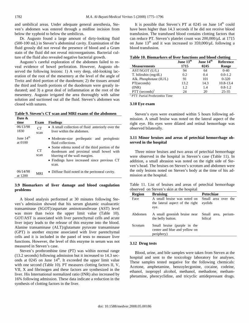
3.9 Biomarkers of liver damage and blood coagulation problems A blood analysis performed at 30 minutes following Ste-ven’s admission showed that his serum glutamic oxaloacetic transaminase (SGOT)/aspartate aminotransferase (AST) level was more than twice the upper limit value (Table 10). GOT/AST is associated with liver parenchymal cells and acute liver injury leads to the release of this enzyme into the blood. Alanine transaminase (ALT)/glutamate pyruvate transaminase (GPT) is another enzyme associated with liver parenchymal cells and it is included in the panel of tests to measure liver functions. However, the level of this enzyme in serum was not measured in Steven’s case. Steven’s prothrombine time (PT) was within normal range (13.2 seconds) following admission but it increased to 14.3 sec-onds at 0245 on June 14th. It exceeded the upper limit value with one second (Table 10). PT measures clotting factors II, V, VII, X and fibrinogen and these factors are synthesized in the liver. His International normalized ratio (INR) also increased by 16% following admission. These data indicate a reduction in the synthesis of clotting factors in the liver.
It is possible that Steven’s PT at 0245 on June 14th could have been higher than 14.3 seconds if he did not receive blood transfusion. The transfused blood contains clotting factors that can reduce PT. Steven’s platelet count was 200,000/µL at 1715 on June 13th and it was increased to 359,000/µL following a blood transfusion. Table 10. Biomarkers of liver functions and blood clotting
Measurements June 13th
1715 June 14th
0245 Reference
Range AST/SGOT (U/L) 94 64 0-40 T. bilirubin (mg/dL) 0.2 0.4 0.0-1.2 Alk. Phosphotase (IU/L) 91 101 0-320 PT(seconds) 13.2 14.3 10.8-13.4 (INR) 1.2 1.4 0.8-1.2 PTT (seconds)1 24 20 25-35
1 PTT: Partial Prothrombin Time
3.10 Eye exam Steven’s eyes were examined within 5 hours following ad-mission. A small bruise was noted on the lateral aspect of the right eye. His eyes were dilated and retinal hemorrhage was observed bilaterally. 3.11 Minor bruises and areas of petechial hemorrhage ob-served in the hospital Three minor bruises and two areas of petechial hemorrhage were observed in the hospital in Steven’s case (Table 11). In addition, a small abrasion was noted on the right side of Ste-ven’s head. The bruises on Steven’s scrotum and abdomen were the only lesions noted on Steven’s body at the time of his ad-mission at the hospital. Table 11. List of bruises and areas of petechial hemorrhage observed on Steven’s skin at the hospital
Region Bruising Petechiae Face A small bruise was noted on
the lateral aspect of the right eye.
Small area over the eyelids
Abdomen A small greenish bruise near the belly button.
Small area, perium-bilical
Scrotum Small bruise (purple in the center and blue and yellow on periphery)
3.12 Drug tests Blood, urine, and bile samples were taken from Steven at the hospital and sent to the toxicology laboratory for analyses. These samples tested negative for the following chemicals: Acetone, amphetamine, benzoylecgonine, cocaine, codeine, ethanol, isopropyl alcohol, methanol, methadone, metham-phetamine, phencyclidine, and tricyclic antidepressant drugs.

M.A. Al-Bayati/Medical Veritas 5 (2008) 1775–1796 1783
4. Autopsy findings, the medical examiner’s opinions, and clinical data overlooked by the medical examiner Steven was pronounced dead at St. Joseph’s Hospital at 1444 on June 14, 1998. He was 25 months old. Dr. Mark A. Fischione performed the autopsy on Steven’s body at 1100 on June 15, 1998 (Case # 98-01612). The autopsy was conducted in Maricopa County, Phoenix, Arizona [9]. Dr. Fischione alleged that Steven died as a result of multiple blunt force injuries to the head and other regions of his body, and that the manner of death was homicide. He based his alle-gations on the following findings. 1) Bilateral subdural bleed-ing, subarachnoid hemorrhage, and cerebral ischemia; 2) bilat-eral retinal and optic nerve hemorrhage; 3) an old area of indu-ration and fibrous scarring at the root of the mesentery; 4) hem-orrhage and contusion of cecum and ascending colon; and 5) multiple old and new minor abrasions and contusions on the head, face, trunk, and extremities. The medical data described below clearly show that Steven was suffering from severe lymphocytopenia, bacterial infec-tions, and septicima. He had severe acute bronchopnemonia; acute and chronic inflammation of the peritoneum; and kidney and liver damage. He developed septic shock and vomited. Some of his vomit blocked his airways and resulted in respira-tory arrest that led to hypoxia, acidosis, hypotension, and coma. Steven’s intracrainal bleeding, retinal hemorrhage, and his minor bruises and skin lesions were caused by infections and septicemia, liver damage, and the high doses of epinephrine given in the hospital (Sections 5, 6). Steven’s brain edema and axonal injuries were caused by anoxia, ischimea, and sodium bicarbonate given to Steven in the hospital (Section 7). CT scan sand X-rays exams did not show Steven had any bone fractures or injuries of the internal organs that indicate trauma (Section 8). 1. Steven was admitted to the hospital on June 13th and blood samples taken at 0.5 and 7.5 hours following admission re-vealed that he had lymphocyte counts of 568 cells/µL and 693/µL, respectively. His lymphocyte counts indicate that he was suffering from severe lymphocytopenia and immune de-pression. Lymphocytopenia in children is defined as a blood lymphocyte count of less than 3000/µL [10]. Steven’s blood CD4+ T cell count was not measured. How-ever, it is expected that his CD4+ T cell count would be less than 300 per µL of blood. For example, Schottstaedt et al. evaluated a case of a man who had a total blood lymphocyto-penia of 896/µL and he was suffering from chronic illness. The blood CD4+ T cell count of this individual was 215/µL and he had a CD4 T/ CD8 T cell ratio of 0.7 [11]. Fischione and the treating physicians did not investigate the cause(s) that led to Steven’s severe lymphocytopenia or consider his serious im-mune depression as leading to his health problems. 2. Steven was suffering from acute bronchopneumonia at the time of his admission to the hospital as shown by the following clinical testes and pathology studies. The likely cause of his lung infection was Staphaylococcus coagulase-negative (Sec-tion 5).
a. A chest X-ray taken at 10 minutes following Steven’s admission to the hospital showed the presence of fluid in his lungs. His chest X-ray, CT scan, and MRI exams per-formed during the 16 hours following admission also showed fluid and consolidations in both lungs (Table 7).
b. At autopsy, Steven’s right and left lungs weighted 140 g and 152 g, respectively. The weights of his right and left lungs were 157% and 197% of the average expected weight for age, respectively (Table 12). The medical examiner (ME) stated that sectioning of the lungs disclosed a dark red-blue, moderately congested lung parenchyma.
c. The ME did not report any microscopic description of Steven’s lungs in his autopsy report issued on November 18, 1998 [9]. We requested the H & E stained slides of sec-tions prepared from Steven’s lungs at the time of autopsy and we received one slide (98-1612-R/C). I examined this slide microscopically and I found bleeding involving a wide area of the lung section and inflammation involving the alveoli and the airways (Figures 1-4). The bronchioles and the alveolar lumens surrounding the inflammed bronchioles are filled with inflammatory cells (macrophages, lymphocytes, and neutrophils). The majority of the cells are macrophages (Figures 3, 4). The alveolar lumens in the major section of the slides are also filled with blood and fluids (Figure 2). These lesions indi-cate that Steven was suffering from acute bronchopneu-monia and bleeding. Figure 5 shows the normal structure of a lung in a 3.5-year-old child (Eliza Jane) [12].
3. Steven was suffering from acute bacterial infections as shown by the clinical tests and biomarkers described blow.
a. A Gram-stain study performed on Steven’s blood taken at 30 minutes following admission revealed the presence of Gram-positive cocci. His blood culture was positive for Staphalococcus coagulase negative (SCN). SCN have been known to cause serious and fatal infections, especially in children with a severe immune depression similar to Ste-ven’s case (Section 5).
b. Blood analysis of a sample taken at 30 minutes follow-ing admission to the hospital revealed that Steven had a high band neutrophil count of 25% of the total white blood cell count (Table 5).
c. A blood analysis performed at 30 minutes following ad-mission to the hospital showed that Steven had a high se-rum creatinine level of 1.0 mg/dL (twice the upper normal level). His serum albumin and protein levels were below the normal lower limit (Table 4). His urine analysis re-vealed high levels of protein and a moderate amount of blood in his urine (Table 4).
d. Treatment with antibiotics reduced Steven’s serum creatinine level by 20%. In addition, Steven’s urine sample taken at about 2 hours following treatment with antibiotics showed only a trace amount of protein and blood (Tables 3-4). These data indicate that Steven was suffering from kid-ney damage caused by bacterial infections.
doi: 10.1588/medver.2008.05.00186

M.A. Al-Bayati/Medical Veritas 5 (2008) 1775–1796 1784
Furthermore, Steven’s weight measured in the hospital on June 13th was 12.0 kg (26.4 pounds) and his weight measured at autopsy on June 14th was 13.2 Kg (29 pounds). His weight increased by 1.2 kg (2.6 pounds) in one day. These data indicate that his body was not eliminating water in a normal fashion because of kidney problems.
e. Steven had acute and chronic peritonitis. Dr. Augusto performed a laparotomy on June 14th and found a large amount of dirty-looking fluid in the abdominal cavity (500-1000 mL). He did not see bleeding or perforation of the bowel that indicates trauma.
Augusto irrigated the area thoroughly with saline solution and suctioned out all the fluid. However, Fischione found ap-proximately 110 mL of cloudy tan-white liquid within the ab-dominal cavity on June 15th and alleged that this fluid resulted from injury caused by trauma. The clinical evidence clearly shows that the 110 mL of fluid accumulated in Steven’s abdo-men after the laparotomy of June 14th and resulted from in-flammation. 4. A blood analysis performed at 30 minutes following Steven’s admission showed that his serum glutamic oxaloacetic transa-minase (SGOT) was elevated (Table 10). GOT is associated with liver parenchymal cells, and liver injury leads to the re-lease of this enzyme to the blood. In addition, Steven’s prothrombine time (PT) and his international normalized ratio (INR) were elevated on June 14th as a result of liver problems (Table 10). The majority of the clotting factors are synthesized in the liver and acute liver injuries are usually associated with coagu-lation disorders. 5. Steven received high doses of epinephrine at the hospital that have been shown to cause intracranial bleeding in children and adults. He was given 0.2 mg and 0.3 mg of epinephrine via en-dotracheal tube at 1723 and 1725, respectively. Then, an epi-nephrine IV infusion was started at 0.3 µg/kg per minute and stopped at 1200 on June 14th. Intracranial bleeding has been reported in some children and adults treated with high therapeutic doses of epinephrine [13-18]. Fischione did not consider the high doses of epinephrine given to Steven in causing bleeding in this case (Section 6). 6. The focal area of subcalpular hemorrhage over Steven’s left temporoparietal region observed by the medical examiner on June 15th was not noted on Steven’s two CT scan head exams taken during the five hours following his admission to the hos-pital (Table 8). These observations indicate the bleeding outside Steven’s skull occurred after 2259 on June 13th. 7. The two CT scans and the MRI head exams performed dur-ing the 16 hours following Steven’s admission to the hospital showed a diffused subdural hemorrhage involving the left side only and they did not reveal the presence of bleeding in the su-barachnoid space (Table 8). However, Fischione reported that Steven had bilateral subdural hemorrhage and a diffuse su-barachnoid hemorrhage. These observations indicate that the
subdural bleeding on the right side and the subarachnoid hem-orrhage developed after Steven’s MRI head exam on June 14th. 8. Fischione alleged that Steven’s brain ischemia and axonal injuries were caused by blunt trauma but Steven’s CT scan and MRI head exams showed that the ischemia developed in the hospital. Ischemia was not observed on Steven’s CT scan ex-ams performed at 1 and 5 hours following admission to the hospital. Ischemia was noted on the MRI head exam performed at 16 hours post admission. The ischemia in this case resulted from blockage of the arteries with blood clots (Table 8). Ische-mia has been known to lead to nerve damage and axonal inju-ries as shown by medical studies described in Section 7. 9. Fischione alleged that Steven’s retinal bleeding was caused by trauma. My review of the medical evidence in this case re-veals that Steven had the following risk factors that led to bleeding in the retina, and these factors were not considered in a differential diagnosis in this case (Section 6):
a. Steven was suffering from bacterial infections that led to disseminated intravascular coagulation and bleeding prob-lems.
b. He had liver damage that led to a reduction in the syn-thesis of clotting factors.
c. He was treated with high doses of epinephrine that cause bleeding due to the significant increases in the heart rate and blood pressure.
d. Steven had a significant increase in the intracranial pres-sure (ICP) following his admission to the hospital. He had an ICP of 10 mm Hg on June 13th and his ICP increased to > 80 mm Hg on June 14th. A sudden rise in the ICP has caused intraocular bleeding in some individuals (Section 6).
e. Steven was suffering from anemia and some individuals with anemia have developed retinopathy and bleeding in the retina as shown by the study described in Section 6 of this report. Steven’s blood analysis performed at 30 min-utes following admission showed that he had a hemoglobin level of 8.6 g/dL and a hematocrit value of 26.8%. His he-moglobin level and hematocrit value were 27% and 29% below the normal lower limit value, respectively.
10. Fischione reported 14 minor external bruises and abrasions on Steven’s body (8.1-4). However, the treating physicians noted only three minor bruises and two minor areas of petechial hemorrhage on Steven’s body (Table 11). These observations indicate that at least 11 of these minor skin lesions occurred after Steven’s admission to the hospital. Table 12. Steven’s lung weight and as % of normal weight
Lung
Steven’s lung
weight (g)
Average normal lung weight (g)
Steven’s lung weight as %
of normal wt. R. Lung 140 89 157 L. Lung 152 77 197 Total lungs 292 166 176
doi: 10.1588/medver.2008.05.00186

M.A. Al-Bayati/Medical Veritas 5 (2008) 1775–1796
doi: 10.1588/medver.2008.05.00186
1785
Figure 1. Microscopic image of Steven’s lung section (H & E, x 20) showing bleeding in a wide area of the lung.
Figure 3. Microscopic image of Steven’s lung section (H & E, x 84) showing that the bronchioles and the alveolar lumens surrounding the inflammed bronchioles are filled with inflammatory cells (macro-phages, lymphocytes, and neutrophils).
Figure 2. Microscopic image of Steven’s lung section (H & E, x 120) showing the alveolar lumens filled with blood and fluid.
Figure 4. Microscopic image of Steven’s lung section (H & E, x 120) showing the majority of the inflammatory cells in the bronchioles and are macrophages.
Figure 5. Microscopic image of Eliza Jane’s lung (H & E, x 52) showing normal structures of the lung in a 3.5-year-old child [12].

M.A. Al-Bayati/Medical Veritas 5 (2008) 1775–1796 1786
5. Steven’s severe lymphocytopenia, Staphalococcus infec-tions, and inflammations observed in tissues and organs Steven was admitted to the hospital on June 13, 1998 and blood samples taken at 0.5 and 7.5 hours following admission revealed that he had lymphocyte counts of 568 cells/µL and 693/µL, respectively. His lymphocyte counts indicate that he was suffering from severe lymphocytopenia and immune de-pression. Lymphocytopenia in children is defined as a blood lymphocyte count of less than 3000/µL [10]. The CD4+ T cell count was not measured in Steven’s case. However, it is expected that his CD4+ T cell count would be less than 300 per µL of blood. For example, Schottstaedt et al. evaluated a case of a man who had a total blood lymphocyto-penia of 896/µL and suffered from chronic illness. The blood CD4+ T cell count of this individual was 215/µL and he had a CD4+ T/ CD8+ T cell ratio of 0.7 [11]. In addition, I evaluated a case of a man with lung fibrosis who had a total blood lymphocyte count of 483/µL. A blood analysis revealed that he had CD4+ T cell count of 255/µL and CD4+ T cells /CD8+ T cells ratio of 0.6. This man also developed pneumonia and fungal infections. The clinical data and medical studies described below show that Steven was suffering from chronic and acute bacterial in-fections involving his lungs, liver, kidneys, peritoneum, and intestine. Steven’s blood culture of a sample taken at 30 min-utes following admission tested positive for Staphalococcus coagulase negative (SCN). A Gram stain study of this blood sample revealed the presence of Gram-positive cocci. Furthermore, blood analysis of the sample taken at 30 min-utes following admission revealed that he had a high band neu-trophil count of 25% of total white blood cell count. It indicates that Steven was suffering from acute bacterial infections. Sta-phalococcus and SCN have been known to cause serious and fatal infections, especially in children with severe immune de-pression similar to Steven’s case (Section 5.1). 5.1 Staphalococcus coagulase negative infections in humans Coagulase-Negative Staphalococi (CNS), practically S. epi-dermidis are a prominent cause of bacteremia in immunosup-pressed individuals like Steven. Steven had acute broncho-pneumonia, a chronic and acute peritonitis, and renal and liver damage. The following clinical studies outline the significance of CNS in causing serious infections and death among children and adults: 1. Wisplinghoff et al., 2003 evaluated data collected from 49 US hospitals during a 6-year period related to individuals with bloodstream infections (BSI). They detected 22,609 blood-stream infections, of which 3,432 occurred in individuals < or =16 years of age. Gram-positive organisms accounted for 65% of these cases and the most common organisms were coagulase-negative staphylococci (43%). The mean interval between ad-mission to the hospital and infection averaged 21 days for co-agulase-negative staphylococci. The overall crude mortality was 14% (475 of 3432) [19].
2. Wisplinghoff et al., 2004 conduced a study involving data collected from 24,179 individuals with bloodstream infections (BSI) collected from 49 US hospitals over a 7-year period (1995-2002). Eighty-seven percent of these cases were monomicrobial infection. Gram-positive organisms caused 65% of these BSI. The most common organisms causing BSI were coagulase-negative staphylococci (CNS) (31% of isolates). The crude mortality rate was 27% [20]. 3. The SENTRY Antimicrobial Surveillance Program has moni-tored bloodstream infections (BSI) from individuals in medical centers worldwide since 1997. During 1997-2002, a total of 81,213 BSI pathogens from North America, Latin America, and Europe were tested for antimicrobial susceptibility. S. aureus, E. coli, and coagulase-negative staphylococci were the three most common BSI pathogens in all three regions each year. Individual age analysis showed the most common BSI pathogen among neonates was coagulase-negative staphylococci [21]. 4. Tantracheewathorn et al. reviewed the records of 3,747 cases of children with bacterial infections diagnosed at BMA Medical College and Vajira Hospital between January 2000 and Decem-ber 2005. The common sites of infections were bloodstream (28.6%), lower respiratory tract (15.3%), skin and soft tissue (14.9%), and urinary tract (12.5%). The major isolated Gram-positive pathogens were S. aureus, coagulase negative Staphy-lococcus, and Enterococus. All Gram-positive cocci remained sensitive to vancomycin and Linezolid antibiotics [22]. 5. Pérez-González et al. evaluated the medical records of 868 children with bloodstream infections. The organisms isolated most commonly were Klebsiella pneumoniae, Candida species, and coagulase-negative staphylococci. The mortality rates for children with a Gram-positive bacterial bloodstream infection was 19.2% [23]. 6. Nimri et al. assessed the clinical data obtained from 210 cases of children under 10 years of age with fever. These chil-dren were admitted to the hospital because of gastroenteritis, respiratory tract infections, or suspected sepsis. Most of the children with septicemia (71.3 per cent) were less than 1 year old. Focal source of bacteremia was gastroenteritis (40.4 per cent), pneumonia or bronchopneumonia (20 per cent), meningi-tis (7.4 per cent), and urinary tract infections (7.4 per cent). The predominant pathogens isolated from blood or stool specimens were Gram-positive bacteria (53.3 per cent), mainly Streptococ-cus pneumoniae and coagulase-negative Staphylococcus spp. The mortality rate was 4 per cent, mostly from septicemia cases [24]. 7. Babay et al. evaluated the clinical data obtained from 220 children with blood stream infection (BSI) hospitalized in Saudi Arabia. Two hundred and ten (95.4%) had single blood culture isolate. One hundred and seventy-three (78.6%) of the isolates were Gram-positive bacteria that included coagulase-negative Staphylococcus epidermidis (55.4%) and Staphylococcus
doi: 10.1588/medver.2008.05.00186

M.A. Al-Bayati/Medical Veritas 5 (2008) 1775–1796 1787
aureus (9.5%). None of the Gram-positive isolates were vanco-mycin resistant. Fever was the most common presentation of children (26%) with positive blood culture with no apparent focus of infection. Respiratory tract infections 26 (12%) were the next most com-mon. Sepsis was seen in (7.7%) children between 8 days and 6 months of age. [25]. 8. Urrea et al., 2003 examined the medical records of 39 chil-dren with bacterial infections that included bacteremia (51.7%), respiratory infections (19.0%) and urinary tract infections (17.2%). Coagulase-negative staphylococci (39%) and Pseu-domonas aeruginosa (24%) were the most common organisms isolated [26]. 9. Urrea et al., 2004 reviewed the clinical data collected from 100 children with acute lymphoblastic leukemia and other ill-nesses admitted to the pediatric hematology/oncology unit at the University Hospital in Barcelona. 55.5% of these children had bacteremia. The most frequently isolated microorganisms were Gram-positive bacteria (78.6%). Coagulase-negative Staphylococci were the most common isolates in bacteremias (70%) [27]. 10. Gayvallet-Montredon et al. evaluated 20 children with bloodstream infections. The most common isolated pathogens were Gram-positive cocci in 10 cases: 5 methicillin-sensible Staphylococcus aureus, 4 methicillin-resistant coagulase-negative staphylococci and 1 Streptococcus milleri [28]. 5.2 Steven’s bronchopneumonia A chest X-ray taken at 10 minutes following Steven’s admis-sion to the hospital showed the presence of fluid in his lungs. A chest X-ray, CT scan, and MRI exams performed during the 16 hours following admission also showed fluid and consolidations in both lungs (Table 7). At autopsy, Steven’s right and left lungs weighted 140 g and 152 g, respectively. The weight of his right and left lungs were 157% and 197% of the average expected weight for age, respec-tively (Table 12). The medical examiner (ME) stated that sec-tioning of the lungs disclosed a dark red-blue, moderately con-gested lung parenchyma. I examined an H & E stained tissue section of Steven’s lung microscopically and I found bleeding in a wide area of the lung section and inflammation involving the alveoli and the airways (Figures 1-4). The bronchioles and the alveolar lumens sur-rounding the inflammed bronchioles are filled with inflamma-tory cells (macrophages, lymphocytes, and neutrophils). The majority of the cells are macrophages (Figures 3, 4). The alveo-lar lumens in the major section of the slides are also filled with blood and fluids (Figure 2). These lesions indicate that Steven was suffering from an acute bronchopneumonia and bleeding. A blood analysis performed following Steven’s admission to the hospital revealed that he was suffering from respiratory aci-dosis. He had a blood pH of 7.18, a PCO2 of 50 mm Hg, and a bicarbonate level of 18.1 mEq/L. He also had serum potassium and calcium levels lower than normal and he developed hyper-natremia following his admission in the hospital (Table 2). The
following clinical studies show that children who suffer from pneumonia and bronchopneumonia also develop hypoxia, aci-dosis, electrolyte imbalances, peripheral circulatory collapse, and convulsions: 1. Simpson and Flenley studied the arterial blood-gas and pH changes in 32 children under three years of age with acute lower-respiratory tract infections. Hypoxia was common, the PO2 being below 80 mm Hg in 14 (67%) of the 21 cases in which it was measured and below 50 mm Hg in 5 of these (24%). Carbon-dioxide retention, with a PCO2 over 50 mm Hg was present in 16 cases (50%). There were 8 cases in which the blood pH was less than 7.20 or the PCO2 greater than 65 mm Hg. In the deaths that occurred during the study, the blood pH values on admission were 7.14, 7.19, and 7.25. The respective PCO2 levels were 52, 110, and 68 mm Hg [29]. 2. Bhushan and Gupta evaluated 40 cases of children (29 males and 11 females) who suffered from bronchopneumonia (n=32), bronchiolits (n=4), and pneumonia (n=3). They found uncom-pensated acidosis in 75% of the cases at admission. PCO2 was elevated in 65% of cases and hypoxemia was an almost univer-sal finding. PCO2 above 65 mm Hg was associated with bad prognosis [30]. 3. Simpson et al. evaluated 11 infants who suffered from respi-ratory failure due to severe lower respiratory tract infections. Progressive respiratory difficulties leading to exhaustion, pe-ripheral circulatory collapse, recurrent apnoeic attacks or gener-alized convulsions were the main clinical presentations result-ing in severe ventilatory failure. In 9 infants preventilation PCO2 exceeded 65 mm Hg [31]. 4. Singhi and Dhawan studied 264 hospitalized children with pneumonia for serum sodium and potassium concentration, and plasma osmolality (Posm) on the day of admission. Hyperna-tremia and hypokalemia (serum potassium less than or equal to 3.5 mEq/L) were found in 3.7% and 19% of the children, re-spectively [32]. 5. Poddar et al. studied 20 infants (3.6 +/- 2.9 months), hospital-ized consecutively with acute bronchiolitis for water and elec-trolyte changes during the acute stage and compared them to those on recovery. Ten infants each were assigned alternatively to study body water compartment or renal water handling on the day of hospitalization and after recovery. There was a significant decrease in urinary sodium from 54 +/- 39 mEq/L to 20 +/- 18 mEq/L and urinary osmolality from 415 +/- 213 mOsm/kg to 252 +/- 204 mOsm/kg at admission and at recovery, respectively. All 10 infants showed significant increase in total body water (mean +/- SD; 22.8 +/- 7.5 ml/kg) at admission as compared to that at recovery [33].
doi: 10.1588/medver.2008.05.00186

M.A. Al-Bayati/Medical Veritas 5 (2008) 1775–1796 1788
5.3 Locations and stages of the inflammations observed in Steven’s abdomen Steven was admitted to the hospital at 1642 on June 13th and Dr. Toraya Augusto operated on Steven’s abdomen at 0245 on June 14th. He found a large amount of dirty-looking fluid (500-1000 mL) in the abdominal cavity. The following clinical data and observations indicate that the fluid in Steven’s abdomen resulted from acute inflammation caused by infections. It was not caused by trauma as was alleged by the medical examiner (ME). 1. Most of the fluid found in Steven’s abdominal cavity was formed after his admission to the hospital as indicated by the following observations:
a) An abdominal CT scan taken at about 2 hours following admission revealed the presence of a small amount of fluid collected in his abdomen. His CT scan and the MRI exams performed after 6 hours following admission showed that the amount of fluid in Steven’s abdomen increased signifi-cantly with time (Table 9).
b) Dr. Augusto operated on Steven’s abdomen at 0245 on June 14th and collected all the fluid found in the abdomen (500-100 ml), irrigated the area thoroughly with saline so-lution, and suctioned out all the fluid. The medical exam-iner found approximately 110 mL of cloudy tan-white liq-uid within the abdominal cavity on June 15th.
2. There was no blood in the fluid collected from Steven’s ab-domen on June 13th. In addition, Augusto’s examination of the abdominal cavity did not reveal the presence of bowel perfora-tion or injuries in the liver, spleen, and other organs. 3. Augusto reported the presence of an old mesenteric healed lesions at the root of the mesentery and the tissues around the third and fourth portions of the duodenum. The presence of fibrosis indicates that these lesions were more than 6 days old and Steven had chronic peritonitis. 4. Augusto stated that there was a great deal of inflammation at the root of the mesentery in Steven’s case. The likely causes of the fluid released to the abdomen are the acute inflammation of the peritoneum. 5. The ME stated “there is focal acute hemorrhage and contu-sion around the cecum and ascending colon including the proximal part of the appendeix.” Augusto examined Steven’s intestine carefully at 0245 on June 14th and he did not see these lesions. These observations indicate that the lesions developed in the hospital after Augusto examined the intestine. 5.4 Evidence of kidney bacterial infections The following clinical biomarkers and medical studies indi-cate that Steven was suffering from acute kidney bacterial in-fections:
1. A blood analysis performed at 30 minutes following Steven’s admission to the hospital showed that he had a high serum creatinine level of 1.0 mg/dL (twice the upper normal limit). Treatment with antibiotics reduced the creatinine level in serum by 20%. Creatinine (Crn) is a small and freely filtered solute by the glomeruli of the kidney. Crn is produced from the break down of creatine in muscle. A reduced glomerular filteration rate (GFR) leads to retention of Crn in the blood. If we assume that Crn is produced at a constant rate in an individual, then a 50 percent reduction in GFR results in proximate doubling of the plasma Crn concentration [10]. Steven’s serum Crn level following admission on June 13th was 1.0 mg/dL and the normal range in a toddler is 0.3-0.5 mg/dL. Steven’s serum level of Crn is 200% of the upper limit value. These data indicate that the glomerular filteration in Ste-ven’s case was reduced by 50% of the normal rate due to kid-ney damage. 2. At the time of admission, Steven’s urine analysis revealed that he had a high level of protein > 300 mg/dL and a moderate amount of blood in urine which indicates kidney damage. In addition, his urine sample taken at about 2 hours following his treatment with antibiotics showed only a trace amount of pro-tein and blood (Table 4). These observations indicate that Ste-ven had acute renal infections. Large quantities of plasma proteins normally flow through the glomerular capillaries but do not enter the urinary space. Both charge and size selectivity prevents virtually all of albu-min, globulin, and other large-molecular-weight proteins from crossing the glomerular wall. The glomerular basement mem-branes trap most large proteins except in cases of damage to the membranes, which allow the passage of proteins into the urine [10]. Finding high levels of protein in urine indicates kidney damage. 3. Steven’s weight following admission to the hospital on June 13th was 12.0 kg (26.4 pounds) and his weight at autopsy on June 14th was 13.2 Kg (29 pounds). His weight increased by 1.2 kg (2.6 pounds) in one day. These data indicate that his body was not able to eliminate water in a normal fashion because of severe kidney problems. 4. Steven developed hypernatremia following admission to the hospital. He had a serum sodium level of 146 mEq/L at 30 min-utes following admission and his serum sodium level increased to 153 mEq/L at 7.5 hours following admission. These observa-tions indicate that his kidney was not able to eliminate sodium at a normal rate. 6. The likely causes of Steven’s intracranial, retinal, and pulmonary bleeding, and the bleeding observed in other sites Steven was admitted to St. Joseph’s Hospital at 1642 on June 13, 1998 and pronounced dead at 1444 on June 14th. Dr. Mark A. Fischione performed the autopsy on Steven’s body at 1100 on June 15, 1998. He described bleeding in the following regions: 1) A diffuse left subdural hemorrhage occupying the
doi: 10.1588/medver.2008.05.00186

M.A. Al-Bayati/Medical Veritas 5 (2008) 1775–1796 1789
entire hemisphere with extension into the right occiput. 2) A diffuse subarachnoid hemorrhage. 3) Bilateral retinal hemor-rhage and marked hemorrhage surrounding the optic nerves. 4) A focal area of subcalpular hemorrhage over the left tem-poroparietal region (1 x ½ inch) Fischione alleged that the bleeding in these regions was caused by blunt trauma to the head. The clinical observations and studies described below show that 1) most of the bleeding in the regions described above was developed after Steven’s admission to the hospital; 2) the likely causes of the bleeding were the high doses of epinephrine given to Steven in the hospi-tal, liver failure, and infections. 6.1 Clinical tests and observations that show the progression of Steven’s bleeding 1. Two CT scans and the MRI head exams performed during the 16 hours following Steven’s admission to the hospital showed a diffused subdural hemorrhage on the left side (Table 8). However, Fischione reported that Steven had bilateral sub-dural hemorrhage. These observations indicate that the subdural bleeding on the right side developed after Steven’s MRI head exam of June 14th. 2. Fischione reported that Steven had a diffuse subarachnoid hemorrhage (SAH). Two CT scan and the MRI head exams performed during the 16 hours following Steven’s admission to the hospital did not show bleeding in this region (Table 8). These observations indicate that the SAH developed after his MRI head exam of June 14th. 3. Fischione stated that Steven had a focal area of subcalpular hemorrhage over the left temporoparietal region. This bleeding was not noted on Steven’s CT scan head exams taken during the 5 hours following his admission to the hospital (Table 8). These observations indicate that the bleeding described above occurred after 2259 on June 13th. 4. Fischione stated that “there is focal acute hemorrhage and contusion around the cecum and ascending colon including the proximal part of the appendeix.” Dr. Augusto operated on Ste-ven’s abdomen at 0245 on June 14th and examined Steven’s intestine carefully. He did not see acute hemorrhage and contu-sion in the cecum and colon. These observations indicate that these lesions developed in the hospital after Augusto’s exami-nation of the intestine. 5. I examined the H & E stained section of Steven’s lung mi-croscopically and observed fresh bleeding involving a major portion of the section. This indicates that Steven had a systemic bleeding problem. 6.2 High doses of epinephrine cause bleeding Steven received high doses of epinephrine at the hospital that have been shown to cause intracranial bleeding in children and adults. He was given 0.2 mg and 0.3 mg of epinephrine via endotracheal tube at 1723 and 1725, respectively. Then, an epi-
nephrine IV infusion was started at 0.3 µg/kg per minute and stopped at 1200 on June 14th. Intracranial bleeding has been reported in some children and adults treated with high therapeutic doses of epinephrine [13-18]. For example, bleeding (intracerebral, subdural and/or su-barachnoid hemorrhage) was reported as one of the serious ad-verse reactions of epinephrine, even when given to individuals at a low dosage level of 0.05 mg subcutaneously, which is less than 10% of the dosage of epinephrine given to Steven in the hospital [13]. In addition, Horowitz et al. reported the development of acute cardiac arrest and fatal subarachnoid hemorrhage in an individual who suffered from an allergic reaction and was treated with epinephrine subcutaneously [14]. I also evaluated the medical records and other medical evidence involving three cases of children who suffered from cardiac arrest and were treated with high doses of epinephrine. These children also had intracranial bleeding and differential diagnosis identified epi-nephrine as important factor in causing bleeding in these chil-dren [16-18]. Epinephrine causes bleeding because it increases heart rate and blood pressure. Steven’s heart rate and systolic blood pres-sure were 57 per minute and 30’s mm Hg prior to receiving epinephrine. His heart rate and blood pressure were raised to 148/min and 130/78 mm Hg due to the treatment with epineph-rine (Table 13). Table 13. Steven’s heart rate and blood pressure measure-ments taken at the hospital
Date Time Heart
rate/min Blood pressure
mm Hg June 13th 1700 57 30’s systolic 1900 110 110/60 2000 125 105/57 2100 136 112/58 2200 110 130/78 2300 123 112/59 2400 131 123/64 June 14th 0100 121 100/52 0200 131 99/59 0400 146 96/56 0500 154 100/55 0600 149 104/54
6.3 Sepsis causes clotting problems and bleeding The clinical tests and observations described in Sections 3-5 of this report show that Steven was suffering from an acute bronchopneumonia, peritonitis, kidney and liver damage, and septicemia. Gram stain and blood culture studies performed on Steven’s blood collected at 30 minutes following admission revealed the presence of Gram-positive cocci and coagulase negative Staphylococcus, respectively. Septicemia is frequently accompanied by changes in the plasmatic as well as cellular coagulation systems and by micro-clot formation. The activation of coagulation by endotoxin is mediated by synthesis of tissue factor by monocytes and endo-thelial cells. Some microorganisms have specific properties, which affect individual components of hemostasis and thus in-crease their virulence. Furthermore, thrombocytopenia, throm-
doi: 10.1588/medver.2008.05.00186

M.A. Al-Bayati/Medical Veritas 5 (2008) 1775–1796 1790
bocytopathy and endothelial cell damage caused by a direct effect of the toxic agents contribute to the bleeding diathesis [34, 35]. The occurrence of a hemorrhagic diathesis and microthrom-bosis is best explained by the syndrome disseminated intravas-cular coagulation (DIC). Widespread intravascular coagulation and hemostatic defect are common in individuals with sepsis. The main cause of hypercoagulation state during sepsis seems to be the inhibition of fibrinolysis as a result of overproduction of plasminogen activator inhibitor-1 in later stages of the dis-ease [34-38]. Levi et al. reviewed articles and published peer-reviewed abstracts on the mechanism of the initiation of disseminated intravascular coagulation (DIC) in sepsis. They found a signifi-cant coagulation activation detected after the appearance of endotoxin in the circulation. This activation is preceded by an increase in the serum levels of various cytokines, such as tumor necrosis factor and interleukins. The activation of coagulation seems to be amplified by impaired function of the protein C-protein S inhibitory pathway [39]. The two CT scan head exams performed during the first 6 hours following Steven’s admission to the hospital did not show any evidence of infarction in the brain. However, Steven’s MRI brain exam performed at about 16 hours following admission showed bilateral infarctions involving the posterior cerebral artery territories. Infarction in tissue is caused by the ischemia resulting from the blockage of an artery by blood clots. 6.4 Acute liver damage causes bleeding A blood analysis performed at 30 minutes following Ste-ven’s admission showed that his serum glutamic oxaloacetic transaminase (SGOT) was more than twice the upper limit value (Table 10). This enzyme is associated with liver paren-chymal cells and acute liver injury leads to the release of this enzyme to the blood. The liver plays a central role in the clotting process. Injuries and diseases of the liver are usually associated with coagulation disorders due to multiple processes. These include reducing the synthesis of clotting and inhibitor factors, decreasing the clear-ance of activated factors, and producing quantitative and quali-tative platelet defects. Some of these abnormalities may lead to hyperfibrinolysis and the acceleration of the intravascular co-agulation process [40-46]. Steven’s prothrombine time (PT) and international normal-ized ratio (INR) were 14.3 seconds (normal range: 10.8-13.4) and 1.4 (normal range 0.8-1.2) at 0245 on June 14th, respec-tively. PT measures clotting factors II, V, VII, X and fibrinogen and these factors are synthesized in the liver. These data indi-cate that Steven’s liver was unable to make clotting factors due to liver problems. 6.5 Retinal bleeding and risk factors observed in Steven’s case The medical examiner evaluated Steven’s eyes at the time of the autopsy and noted the presence of bilateral retinal hemor-rhages and marked hemorrhage surrounding the optic nerves. He alleged that the bleeding in Steven’s eyes was caused by
trauma. My review of the medical evidence in this case reveals that Steven had the following risk factors that led to bleeding in the retina and these factors were not considered in the differen-tial diagnosis in this case: 1. Steven was suffering from bacterial infections that led to disseminated intravascular coagulation and bleeding problems (Section 6.3). 2. Steven was suffering from an acute liver injury that led to a significant reduction in the synthesis of clotting factors. His prothrombin time and international normalized ratio (INR) were elevated (Section 6.1). 3. Steven was treated with high doses of epinephrine that cause bleeding due to the significant increase in the heart rate and blood pressure. Steven’s heart rate and systolic blood pressure were 57 per minute and 30’s mm Hg prior to receiving epineph-rine. His heart rate and blood pressure were raised to 148/min and 130/78 mm Hg due to the treatment with epinephrine (Sec-tion 6.2). 4. Steven had a significant increase in intracranial pressure (ICP) following his admission to the hospital. He had an ICP of 10 mm Hg on June 13th and his ICP was increased to > 80 mm Hg on June 14th. A sudden rise in ICP has caused intraocular bleeding in some individuals. For example, Medele et al. performed prospective ophthal-mological examination in 22 consecutive individuals with su-barachnoid hemorrhage (SAH) or severe brain injury and ele-vated ICP. Thirteen individuals were admitted to the hospital for SAH and 9 for severe brain injury. Monitoring of ICP was performed at the time of admission via a ventricular catheter. Initial ICP exceeded 20 mm Hg in all individuals. Indirect ophthalmoscopy without induced mydriasis was performed within the 1st week after the acute event. Retinal or vitreous hemorrhage was seen in 6 (46%) of 13 individuals with SAH and in 4 (44%) of 9 individuals with severe brain injury. Ocular bleeding was found bilaterally in 3 individuals with SAH and in 1 individual with severe brain injury (18%)[47]. Furthermore, Stiebel-Kalish et al. evaluated the medical re-cords of 70 individuals with subarachnoid hemorrhage resulting from ruptured cerebral aneurysms. They found that 30 eyes of 19 individuals had intraocular hemorrhages; 14 eyes had a vit-reous hemorrhage; 12 eyes had subhyaloid blood without a vit-reous hemorrhage; and 4 eyes had retinal hemorrhages alone [48]. 5. Steven was suffering from anemia and some individuals with anemia have developed retinopathy and bleeding in the retina as shown by the study described below. A blood analysis per-formed at 30 following admission showed that Steven had a hemoglobin level of 8.6 g/dL and a hematocrit value of 26.8%. His hemoglobin level and hematocrit value were 27% and 29% below the normal lower limit value, respectively. Asien et al. evaluated the occurrence of clinically apparent retinal changes in 35 anemic individuals and 35 age-and sex-
doi: 10.1588/medver.2008.05.00186

M.A. Al-Bayati/Medical Veritas 5 (2008) 1775–1796 1791
matched healthy control individuals. Retinal photographs of all subjects were obtained and all vascular and extra vascular reti-nal lesions were recorded. No retinal abnormalities were ob-served in the control subjects. Seven (20%) of the anemic individuals exhibited extra vas-cular lesions (flame-shaped hemorrhages, hard exudates, and cotton-wool spots). Within the group of anemic individuals, the mean hematocrit reading for those with extravascular lesions (N=7) was 24.7%. A significant negative correlation was de-termined between venous length and the level of hematocrit, thereby implying that retinal venous tortuosity is directly re-lated to severity of anemia [49]. 7. The likely causes of Steven’s brain edema, ischemia, and axonal damage At autopsy, the medical examiner (ME) found Steven’s brain was markedly edematous. The examination of the formalin-fixed brain revealed that Steven had bilateral ischemic areas involving the inferior occipitoparietal lobes. The maximum dimensions of the areas with ischemia were 4 cm on the right side and 2.5 cm on the left side. The ME alleged that these le-sions were caused by trauma. The clinical tests and the medical studies described below indicate that the lesions observed in Steven’s brain were mostly developed following his admission to the hospital. Anoxia, medications, bleeding, and infarctions caused edema, ischemia, necrosis, and axonal damage. 7.1 The causes of the brain edema observed in Steven’s case Steven’s brain edema was induced by anoxia, acidosis, hy-potension, treatment with sodium bicarbonate, and irritation resulting from bleeding. Steven was suffering from an acute bronchopneumonia and septicemia. Steven’s heart rate and sys-tolic blood pressure following admission to the hospital were 57 per minute and 30’s mm, respectively. The use of sodium bicarbonate at high therapeutic levels causes hypoxia and brain edema. Steven’s blood pH was 7.18 following admission and it was raised to 7.39 as a result of his treatment with sodium bicarbonate (Table 1). Fauci et al. re-ported that alkalinization of the blood with sodium bicarbonate increases the avidity of hemoglobin to bind oxygen, thus im-pairing the release of oxygen in peripheral tissues [10] Steven’s first head CT scan taken at 30 minutes following admission showed mild brain edema. His CT scan and MRI head exams taken at later revealed a significant increase in the level of fluid in the brain and the surrounding areas. Steven’s intracranial pressure (ICP) was 10 mm Hg on June 13th and increased to > 80 mm Hg on June 14th. In addition, Steven had intracranial bleeding, and blood causes irritation, edema, inflammation, and necrosis in the sur-rounding tissues [50-53]. For example, Mayer et al. performed paired consecutive CT and 99mTc-hexamethylpropylenamine oxime single-photon emission computed tomography (SPECT) scans during the acute (mean, 18 hours) and subacute (72 hours) phase of intracerebral hemorrhage (ICH) in 23 individu-als.
Hematoma and edema volumes were traced and calculated from CT images. They found that the ICH volume (18 mL) did not change but the mean edema volume increased by 36% (from 19 to 25 mL, P<0.0001). Perilesional edema on CT al-ways corresponded topographically with perfusion deficits on SPECT [51]. In addition, Mehdiratta et al. retrospectively reviewed pro-spectively-collected clinical and laboratory data from 23 con-secutive individuals with acute spontaneous ICH. These indi-viduals had a CT scan checked on admission and a follow-up CT scan 3 to 4 days afterward. They measured hematoma and edema volumes on admission and follow-up scans, and calculated the relative edema volume to correct for hematoma volume. They used Spearman correla-tion coefficient to determine the association of various variables with relative perihematoma edema volume. They found that the median hematoma volume increased by approximately 28% from baseline by day 3 to 4. However, the relative edema vol-ume almost doubled during this time period [52]. 7.2. Infarctions and ischemia The medical examiner (ME) cut Steven’s formalin-fixed brain and observed areas with ischemia involving the inferior occipitoparietal lobes bilaterally. The ischemia dimensions were 4 cm on the right side and 2.5 cm on the left side. No area with ischemia was noted on Steven’s CT scan exams performed at 1 and 5 hours following his admission to the hospital. Bilateral infarctions involving the posterior cerebral artery territories were observed on Steven’s MRI head exam per-formed at 16 hours post admission. These observations indicate that the ischemia developed after Steven’s second CT head exam performed at 5 hours following admission (Table 8). The ischemia resulted from blockage of the arteries with blood clots. 7.3 Necrosis and axonal damage Steven’s CT scan head exams taken at 1 and 5 hours follow-ing admission did not show evidence of axonal damage, infarc-tions, or necrosis in the brain. Infarctions, ischemia, and axonal damage in Steven’s brain were first observed on his MRI head exam performed at about 16 hours following admission. It de-tected multiple gradient-echo hypointense foci in subcortical white matter diffusely and bilaterally, arguing for diffuse ax-onal injuries. Theses clinical observations indicate that the axonal damage observed in Steven’s brain was caused by ischemia resulted from the blockage of arteries by blood clots. Petty and Wett-stein stated that brain and spinal cord white matter are vulner-able to the effects of ischemia. Reduction of the energy supply leads to a cascade of events that include depolarization, influx of Na(+), and the subsequent reverse operation of the mem-brane protein the Na(+)/Ca(2+) exchanger. These events ulti-mately lead to intracellular Ca(2+) overload and irreversible axonal injury [54]. White et al. also reported that ischemia results in rapid loss of high-energy phosphate compounds, generalized depolariza-tion, cell death, and axonal damage. They explained the se-quence of events that leads to axonal injuries as follows: Loss
doi: 10.1588/medver.2008.05.00186

M.A. Al-Bayati/Medical Veritas 5 (2008) 1775–1796 1792
of high-energy phosphate compounds induces the release of glutamate and, in selectively vulnerable neurons, opening of both voltage-dependent and glutamate-regulated calcium chan-nels. This results in a large increase in cytosolic Ca(2+) associ-ated with activation of calcineurin and phospholipases. These events lead to proteolysis of calpain substrates, accu-mulation of free arachidonic acid, and depletion of Ca(2+) from the endoplasmic reticulum (ER) lumen. Adenosine degradation products or depletion of ER lumenal Ca(2+) can activate the enzyme kinase that shuts off translation initiation by phos-phorylating the alpha-subunit of eukaryotic initiation factor-2. These events eventually lead to the death of the nerve cell and axonal injuries [55]. Furthermore, Stys stated that white matter of the brain and spinal cord is susceptible to anoxia and ischemia. Myelinated axons of the CNS are critically dependent on a continuous sup-ply of energy largely generated through oxidative phosphoryla-tion [56]. They explained the mechanisms that lead to axonal injuries in cases of anoxia and ischemia as follows: (1) They lead to rapid energy depletion, failure of the Na(+)-K(+)-ATPase, and accumulation of axoplasmic Na+ through non-inactivating Na+ channels, with concentrations approaching 100 mmol/L after 60 minutes of anoxia. (2) They cause severe K+ depletion that re-sults in large membrane depolarization. 3) They lead to exces-sive accumulation of Ca2+ that activates various Ca(2+)-dependent enzymes, such as calpain, phospholipases, and pro-tein kinase C, resulting in irreversible injury [56]. Garthwaite et al. studied the mechanism of ischemic injury to white matter axons in vitro, using transiently depriving rat optic nerves of oxygen and glucose. They evaluated the changes developed in the axons using light and electron microscopes. Their study showed that increasing periods of oxy-gen/glucose deprivation (up to 1 h) caused, after a 90-min re-covery period, the appearance of increasing numbers of swollen axons. The ultrastructure of these axons indicated that they were irreversibly damaged. The damage persisted after a longer recovery period (3 h) [57]. Furthermore, Stys et al. also evaluated the mechanisms of anoxic injury using the in vitro rat optic nerve. Functional in-tegrity of the nerves was monitored electrophysiologically by quantitatively measuring the area under the compound action potential. It recovered to 33.5 +/- 9.3% of control after a stan-dard 60 min anoxic insult. Their study also revealed that anoxia caused rapid depletion of ATP and membrane depolarization leading to Na+ influx through incompletely inactivated Na+ channels. The resulting rise in the intracellular [Na+] coupled with membrane depolari-zation caused damaging levels of Ca2+ to be admitted into the intracellular compartment. It entered the cell through reverse operation of the Na (+)-Ca2+ exchanger [58]. 8. The likely causes of the minor external bruises observed in Steven’s case Steven was admitted to St. Joseph’s Hospital on June 13, 1998 and he was pronounced dead on June 14th. Dr. Mark A. Fischione performed the autopsy on Steven’s body on June 15th and observed 14 minor external bruises and abrasions on Ste-
ven’s body. He alleged that these minor external lesions were caused by trauma. These minor lesions are described in Section 8.1-4. My review of the clinical data reveals that the treating phy-sicians noted only three minor bruises and two minor areas of petechial hemorrhage on Steven’s body (Table 11). Steven’s CT scan and X-ray exams did not reveal bone fracture or injury caused by trauma (Tables 7-9). These observations indicate that Fischione’s allegation that Steven’s injuries were caused by trauma prior to his admission to the hospital is not medically valid. 8.1 Head and face The treating physicians at the hospital observed only a small bruise on the lateral aspect of Steven’s right eye and small area of petechial hemorrhage over his eyelids. However, the medical examiner (ME) noted 8 minor contusions and abrasions (listed below) on Steven’s head and face. These observations indicate that minor skin lesions observed at the time of autopsy occurred after Steven’s admission to the hospital. 1. Two focal contusions on the right side of the head as well as near the right eyebrow; each approximately measuring ¼ x ¼ inch in greatest dimension. 2. A focal abrasion on the top of the head on the right side measuring ¼ inch in length. 3. A thin abrasion overlying the right forehead area measuring ¾ inch in length. 4. Two focal abrasions each overlying the eyebrows at the same level; each approximately measuring ¼ x ¼ inch in greatest dimension. 5. A focal abrasion on the left side of the head measuring ¼ x ¼ inch in greatest dimension. 6. A focal contusion on the undersurface of the chin, near the midline, measuring ¼ x ¼ inch in greatest dimension. 8.2 Abdomen Dr. Augusto operated on Steven’s abdomen at 0245 on June 14th and he noted a small greenish bruise and small area of petechial hemorrhage near the belly button. However, the ME observed the 3 minor contusions listed below on the skin of Steven’s abdomen. These observations indicate that 2 of these contusions occurred after Augusto’s operation. (1) A faint, old contusion located in the left lower quadrant of the abdomen measuring 4 x 2 inches in greatest dimension. (2) An old, faint contusion in the right lower quadrant of the abdomen measuring 1 x 1 inch in greatest dimension.
doi: 10.1588/medver.2008.05.00186
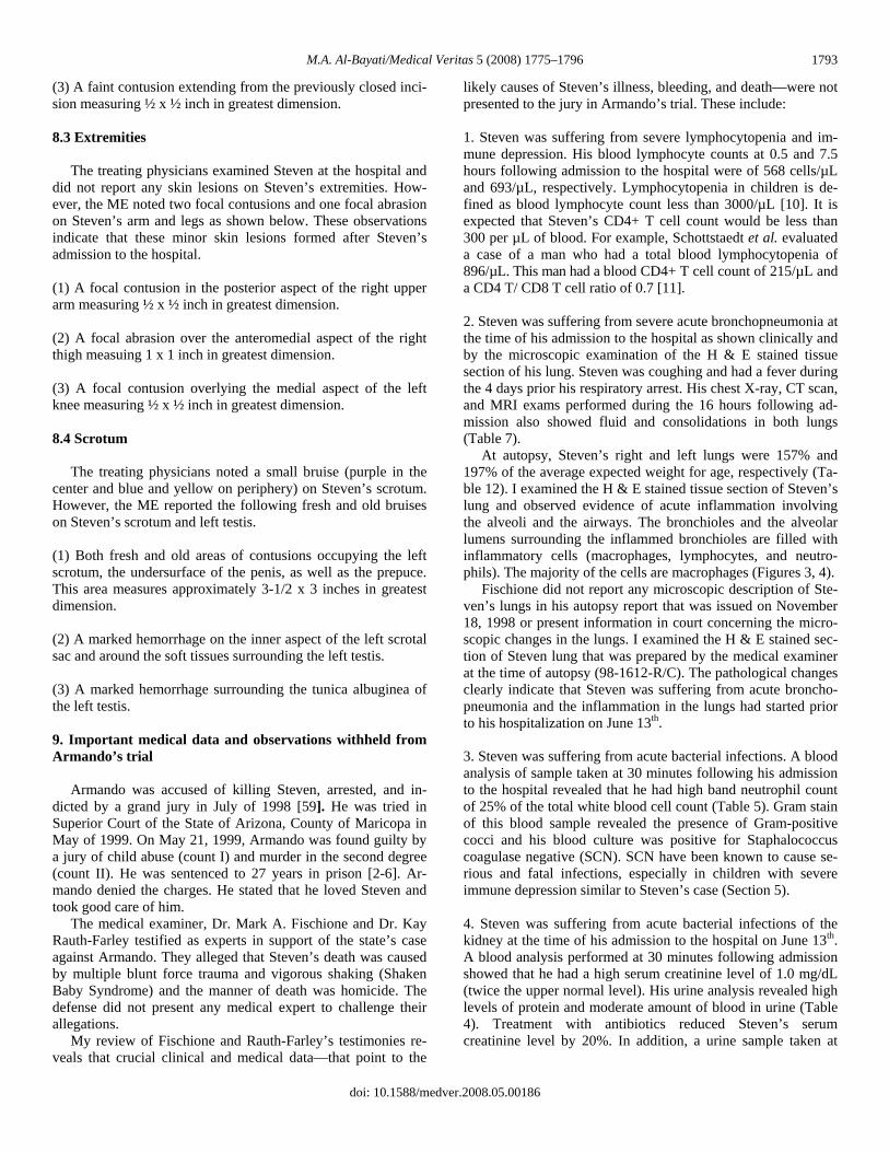
M.A. Al-Bayati/Medical Veritas 5 (2008) 1775–1796 1793
(3) A faint contusion extending from the previously closed inci-sion measuring ½ x ½ inch in greatest dimension. 8.3 Extremities The treating physicians examined Steven at the hospital and did not report any skin lesions on Steven’s extremities. How-ever, the ME noted two focal contusions and one focal abrasion on Steven’s arm and legs as shown below. These observations indicate that these minor skin lesions formed after Steven’s admission to the hospital. (1) A focal contusion in the posterior aspect of the right upper arm measuring ½ x ½ inch in greatest dimension. (2) A focal abrasion over the anteromedial aspect of the right thigh measuing 1 x 1 inch in greatest dimension. (3) A focal contusion overlying the medial aspect of the left knee measuring ½ x ½ inch in greatest dimension. 8.4 Scrotum The treating physicians noted a small bruise (purple in the center and blue and yellow on periphery) on Steven’s scrotum. However, the ME reported the following fresh and old bruises on Steven’s scrotum and left testis. (1) Both fresh and old areas of contusions occupying the left scrotum, the undersurface of the penis, as well as the prepuce. This area measures approximately 3-1/2 x 3 inches in greatest dimension. (2) A marked hemorrhage on the inner aspect of the left scrotal sac and around the soft tissues surrounding the left testis. (3) A marked hemorrhage surrounding the tunica albuginea of the left testis. 9. Important medical data and observations withheld from Armando’s trial Armando was accused of killing Steven, arrested, and in-dicted by a grand jury in July of 1998 [59]. He was tried in Superior Court of the State of Arizona, County of Maricopa in May of 1999. On May 21, 1999, Armando was found guilty by a jury of child abuse (count I) and murder in the second degree (count II). He was sentenced to 27 years in prison [2-6]. Ar-mando denied the charges. He stated that he loved Steven and took good care of him. The medical examiner, Dr. Mark A. Fischione and Dr. Kay Rauth-Farley testified as experts in support of the state’s case against Armando. They alleged that Steven’s death was caused by multiple blunt force trauma and vigorous shaking (Shaken Baby Syndrome) and the manner of death was homicide. The defense did not present any medical expert to challenge their allegations. My review of Fischione and Rauth-Farley’s testimonies re-veals that crucial clinical and medical data—that point to the
likely causes of Steven’s illness, bleeding, and death—were not presented to the jury in Armando’s trial. These include: 1. Steven was suffering from severe lymphocytopenia and im-mune depression. His blood lymphocyte counts at 0.5 and 7.5 hours following admission to the hospital were of 568 cells/µL and 693/µL, respectively. Lymphocytopenia in children is de-fined as blood lymphocyte count less than 3000/µL [10]. It is expected that Steven’s CD4+ T cell count would be less than 300 per µL of blood. For example, Schottstaedt et al. evaluated a case of a man who had a total blood lymphocytopenia of 896/µL. This man had a blood CD4+ T cell count of 215/µL and a CD4 T/ CD8 T cell ratio of 0.7 [11]. 2. Steven was suffering from severe acute bronchopneumonia at the time of his admission to the hospital as shown clinically and by the microscopic examination of the H & E stained tissue section of his lung. Steven was coughing and had a fever during the 4 days prior his respiratory arrest. His chest X-ray, CT scan, and MRI exams performed during the 16 hours following ad-mission also showed fluid and consolidations in both lungs (Table 7). At autopsy, Steven’s right and left lungs were 157% and 197% of the average expected weight for age, respectively (Ta-ble 12). I examined the H & E stained tissue section of Steven’s lung and observed evidence of acute inflammation involving the alveoli and the airways. The bronchioles and the alveolar lumens surrounding the inflammed bronchioles are filled with inflammatory cells (macrophages, lymphocytes, and neutro-phils). The majority of the cells are macrophages (Figures 3, 4). Fischione did not report any microscopic description of Ste-ven’s lungs in his autopsy report that was issued on November 18, 1998 or present information in court concerning the micro-scopic changes in the lungs. I examined the H & E stained sec-tion of Steven lung that was prepared by the medical examiner at the time of autopsy (98-1612-R/C). The pathological changes clearly indicate that Steven was suffering from acute broncho-pneumonia and the inflammation in the lungs had started prior to his hospitalization on June 13th. 3. Steven was suffering from acute bacterial infections. A blood analysis of sample taken at 30 minutes following his admission to the hospital revealed that he had high band neutrophil count of 25% of the total white blood cell count (Table 5). Gram stain of this blood sample revealed the presence of Gram-positive cocci and his blood culture was positive for Staphalococcus coagulase negative (SCN). SCN have been known to cause se-rious and fatal infections, especially in children with severe immune depression similar to Steven’s case (Section 5). 4. Steven was suffering from acute bacterial infections of the kidney at the time of his admission to the hospital on June 13th. A blood analysis performed at 30 minutes following admission showed that he had a high serum creatinine level of 1.0 mg/dL (twice the upper normal level). His urine analysis revealed high levels of protein and moderate amount of blood in urine (Table 4). Treatment with antibiotics reduced Steven’s serum creatinine level by 20%. In addition, a urine sample taken at
doi: 10.1588/medver.2008.05.00186

M.A. Al-Bayati/Medical Veritas 5 (2008) 1775–1796 1794
about 2 hours following treatment with antibiotics showed only a trace amount of protein and blood (Tables 3-4). 5. Fischione did not mention in court that Dr. Augusto per-formed a laparotomy on June 14th and found a large amount of dirty-looking fluid in Steven’s abdominal cavity (500-1000 mL). However, Fischione stated that he collected approximately 110 mL of cloudy tan-white liquid from Steven’s abdominal cavity on June 15th and he alleged that this fluid resulted from injury caused by trauma. The clinical evidence clearly shows that the 110 mL of fluid accumulated in Steven’s abdomen after the laparotomy of June 14th and resulted from inflamma-tion. On June 14th, Augusto irrigated the area thoroughly with saline solution and suctioned out all the fluid. In addition, Augusto examined the abdominal cavity and the bowel, and other organs carefully and he did not see bleeding, perforation of the bowel, and injuries of other organs that indi-cates trauma. However, Fischione observed focal acute hemor-rhage and contusion around the cecum and ascending colon and alleged that these minor lesions were caused by trauma prior to Steven’s admission to the hospitalization on June 13th. 6. Fischione did not state in his autopsy report or mention in court that Steven had bleeding in his lungs. I examined the H & E stained section of Steven’s lung microscopically and ob-served fresh bleeding involving a major portion of the section (Figures 1, 2). At autopsy, Steven’s right and left lungs were 157% and 197% of the average expected weight for age, respec-tively. These observations indicate that Steven had a systemic bleeding problem. Steven had several risk factors for internal and external bleeding. These include: (a) receiving high doses of epinephrine at the hospital which has been shown to cause intracranial bleeding in children and adults; (b) acute liver damage as indi-cated by the elevated serum liver enzyme; (c) acute bacterial infections and DIC (Sections 5 and 6). 7. Fischione alleged that Steven’s subcalpular hemorrhage over the left temporoparietal region was caused by trauma. This bleeding was not noted on Steven’s two CT scan head exams taken during the 5 hours following his admission to the hospital (Table 8). It occurred in the hospital after 2259 on June 13th. 8. Fischione and Rauth-Farley alleged that Steven’s bilateral subdural bleeding (SDB) and the subarachnoid hemorrhage (SAH) were caused by blunt trauma/vigorous shaking and the bleeding occurred prior Steven’s admission to the hospital. The jury was not aware that Steven’s two CT scans and the MRI head exams performed during the 16 hours following his ad-mission to the hospital showed SBD on the left side only and did not show Steven had SAH (Table 8). These observations indicate that the SAH and SDB on the right side developed after the MRI head exam of June 14th. 9. Fischione and Rauth-Farley alleged that Steven’s brain ischemia and axonal injuries were caused by blunt trauma/shaking. The jury was not aware that no area with ischemia was noted on Steven’s CT scan exams performed at 1 and 5 hours following admission to the hospital. Ischemia was
observed on his MRI head exam performed at 16 hours post admission. The ischemia in this case resulted from blockage of the arteries with blood clots (Table 8). Ischemia has been known to lead to nerve damage and axonal injuries as shown by medical studies described in Section 5 of this report. 10. Fischione and Rauth-Farley alleged that Steven’s retinal bleeding was caused by vigorous shaking. The members of the jury were not aware that Steven had several risk factors for reti-nal bleeding. These include treatment with high doses of epi-nephrine, increased intracranial pressure, septicemia, liver dam-age, and anemia as explained in Section 7 of this report. 11. Fischione described 14 minor external bruises and abrasions on Steven’s body and alleged that these minor external lesions were caused by trauma. The jury was not aware that the treating physicians noted only three minor bruises and two minor areas of petechial hemorrhage on Steven’s body (Table 11). These observations indicate that at least 11 of the minor skin lesions occurred after Steven’s admission to the hospital as a result of the treatment with epinephrine, liver damage, and infections. The medical evidence described above clearly show that (a) Steven illness was caused by bacterial infection. He suffered from septic shock and vomited after he ate around 1500 on June 13th and the vomit blocked his airways and caused respiratory arrest; (b) his internal and external bleeding was caused by in-fections and medications; and (c) the factual causes of Steven’s illness, bleeding, and death were not revealed in court and the jury convicted Armando based on a false theory. 10. Conclusions My review of Steven’s medical records, autopsy report, tes-timonies of witnesses, and the pertinent medical studies to this case reveals the following: 1. Steven was suffering from severe lymphocytopenia and im-mune depression. Blood samples taken at 0.5 and 7.5 hours following his admission revealed that he had lymphocyte counts of 568 cells/µL and 693/µL, respectively. Lymphocytopenia in children is defined as a blood lymphocyte count of less than 3000/µL. 2. His severe immune depression predisposed him to infection with Staphalococcus coagulase negative (SCN). Gram-stain study performed on Steven’s blood sample taken at 30 minutes following admission revealed the presence of Gram-positive cocci. His blood culture was positive for Staphalococcus coagu-lase negative (SCN). SCN have been known to cause serious and fatal infections, especially in children with severe immune depression similar to Steven’s case. 3. He developed peritonitis, bronchopneumonia, kidney infec-tions, and liver damage. 4. He suffered from septic shock and vomited after he ate around 1500 on June 13th. The vomit blocked his airways and caused respiratory arrest.
doi: 10.1588/medver.2008.05.00186

M.A. Al-Bayati/Medical Veritas 5 (2008) 1775–1796 1795
5. His internal and external bleeding, brain edema and ischemia, and fresh bruises were caused by bacterial infections, liver damage, and medications. 6. The old bruises observed on his abdomen and scrotum were caused by blood clotting problems which resulted from bacte-rial infections. It made Steven more susceptible to bruising. Steven was 25 months old and he was very active prior to his illness. 7. The factual causes of Steven’s illness, bleeding, and death were not revealed in court by the state and the jury convicted Armando Castillo based on a false theory. References [1] Steven R. Young’s medical record on 11/16/1996. St. Joseph’s Hospital
and Medical Center. 350 West Thomas Road, Phoenix, Arizona. [2] State of Arizona (plaintiff) vs. Armando Castillo (defendant) in the Supe-
rior Court of the State of Arizona, County of Maricopa (CR98-09620). May 12, 1999.
[3] State of Arizona (plaintiff) vs. Armando Castillo (defendant) in the Supe-rior Court of the State of Arizona, County of Maricopa (CR98-09620). May 13, 1999.
[4] State of Arizona (plaintiff) vs. Armando Castillo (defendant) in the Supe-rior Court of the State of Arizona, County of Maricopa (CR98-09620). May 17, 1999.
[5] State of Arizona (plaintiff) vs. Armando Castillo (defendant) in the Supe-rior Court of the State of Arizona, County of Maricopa (CR98-09620). May 18, 1999.
[6] State of Arizona (plaintiff) vs. Armando Castillo (defendant) in the Supe-rior Court of the State of Arizona, County of Maricopa (CR98-09620). May 19, 1999.
[7] Report on Steven R. Young issued by the Emergency Transportation Services. City of Phoenix, Arizona. June 13, 1998.
[8] Steven Young’s medical record on June 13-14, 1998. St. Joseph’s Hospi-tal and Medical Center. 350 West Thomas Road, Phoenix, Arizona.
[9] Fischione MA, Autopsy report on Steven R. Young’s case (Case # 98-01612), November 18, 1998. Maricopa County Office of the Medical Ex-aminer. 120 S. 6th Avenue, Phoenix, Arizona.
[10] Fauci AS, Braunwald E, Isslbacher KJ, Wilson, JD, Martin JB, Kasper DL, Hauser SL, Longo DL. Harrison's Principles of Internal Medicine. McGraw-Hill Companies, Inc. New York USA, ed. 14, 1998.
[11] Schottstaedt MW, Hurd ER, Stone MJ. Kaposi’s sarcoma in rheumatoid arthritis. Am J Med 1987;82(5):1021–6.
[12] Al-Bayati MA. Histopathological features of Eliza Jane Scovill’s and Destiny Jacobo’s lungs with analysis of the causes of death in both cases. Medical Veritas 2006 Nov.;3(2):1041–8.
[13] Goodman & Gilman’s. The Pharmacological Basis of Therapeutics. Edi-tors. Hardman JG, Limbird LE, Molinoff, PB, Ruddon RW, Gilman AG. 9th ed., 1996. McGraw-Hill, New York.
[14] Horowitz BZ, Jadallah S, Derlet RW. Fatal intracranial bleeding associ-ated with prehospital use of epinephrine. Ann Emerg Med. 1996 Dec.;28(6):725–7.
[15] Know O, Chung S, Lee K, Kim S. Spontaneous subarachnoid hemorrhage after intravenous epinephrine use for multiple bee stings. Am J Emerg Med. 2007 Feb.;25(2):249–50.
[16] Al-Bayati MA. Analysis of causes that led to Toddler Alexa Shearer’s cardiac arrest and death in November 1999. Medical Veritas 2004 Jan.;1(1): 86–117.
[17] Al-Bayati MA. Analysis of Causes That Led to Baby Lucas Alejandro Mullenax-Mendez’s Cardiac Arrest and Death in August-September of 2002. Medical Veritas 2004 Jan.;1(1):45–63.
[18] Al-Bayati MA. Analysis of causes that led to subdural bleeding, skull and rib fractures, and death in the case of baby Averial Buie. Medical Veritas 2007 Nov.;4(2):1452–69.
[19] Wisplinghoff H, Seifert H, Tallent SM, Bischoff T, Wenzel RP, Edmond MB. Nosocomial bloodstream infections in pediatric patients in United
States hospitals: epidemiology, clinical features and susceptibilities. Pedi-atr Infect Dis J. 2003 Aug.;22(8):686–91.
[20] Wisplinghoff H, Bischoff T, Tallent SM, Seifert H, Wenzel RP, Edmond MB. Nosocomial bloodstream infections in US hospitals: analysis of 24,179 cases from a prospective nationwide surveillance study. Clin Infect Dis. 2004 Aug 1;39(3):309–17. Epub 2004 Jul 15.
[21] Biedenbach DJ, Moet GJ, Jones RN. Occurrence and antimicrobial resis-tance pattern comparisons among bloodstream infection isolates from the SENTRY Antimicrobial Surveillance Program (1997-2002). Diagn Mi-crobiol Infect Dis. 2004 Sep.;50(1):59–69.
[22] Tantracheewathorn T, Vititpatarapak N, Phumisantiphong U. Epidemi-ologic study of nosocomial bacterial infection of pediatric patients at BMA Medical College and Vajira Hospital. J Med Assoc Thai. 2007 Feb.;90(2):258–65.
[23] Pérez-González LF, Ruiz-González JM, Noyola DE. Nosocomial bac-teremia in children: a 15-year experience at a general hospital in Mexico. Infect Control Hosp Epidemiol. 2007 Apr.;28(4):418–22. Epub 2007 Mar 9.
[24] Nimri LF, Rawashdeh M, Meqdam MM. Bacteremia in children: etiologic agents, focal sites, and risk factors. J Trop Pediatr. 2001 Dec.;47(6):356–60.
[25] Babay HA, Twum-Danso K, Kambal AM, Al-Otaibi FE. Bloodstream infections in pediatric patients. Saudi Med J. 2005 Oct.;26(10):1555–61.
[26] Urrea M, Pons M, Serra M, Latorre C, Palomeque A. Prospective inci-dence study of nosocomial infections in a pediatric intensive care unit. Pediatr Infect Dis J. 2003 Jun.;22(6):490–4.
[27] Urrea M, Rives S, Cruz O, Navarro A, García JJ, Estella J. Nosocomial infections among pediatric hematology/oncology patients: results of a pro-spective incidence study. Am J Infect Control. 2004 Jun.;32(4):205–8.
[28] Gayvallet-Montredon N, Sauvestre C, Bergeret M, Gendrel D, Raymond J. Bacteriologic surveillance of nosocomial septicemia and bacteremia in a pediatric hospital. Arch Pediatr. 1998 Nov.;5(11):1216–20.
[29] Simpson H, Flenley DC. Arterial blood-gas tensions and pH in acute lower-respiratory-tract infections in infancy and childhood. Lancet. 1967 Jan. 7;1(7480):7–12.
[30] Bhushan V, Gupta S. Acid-base and blood gas equilibrium in acute lower respiratory tract infections of infancy & childhood. Indian Pediatr. 1977 Nov.; 14(11):917–21.
[31] Simpson H, Matthew DJ, Habel AH, George EL. Acute respiratory failure in bronchiolitis and pneumonia in infancy. Modes of presentation and treatment. Br Med J. 1974 Jun. 22;2(5920):632–6.
[32] Singhi S, Dhawan A. Frequency and significance of electrolyte abnormali-ties in pneumonia. Indian Pediatr. 1992 Jun;29(6):735–40.
[33] Poddar U, Singhi S, Ganguli NK, Sialy R. Water electrolyte homeostasis in acute bronchiolitis. Indian Pediatr. 1995 Jan;32(1):59–65.
[34] Müller-Berghaus G. Sepsis and blood coagulation. Behring Inst Mitt. 1986 Feb.;(79):131–41.
[35] Hudecek J, Paceková M, Chudej J, Kubisz P. Infection and hemostasis. Vnitr Lek. 2004 Jun.;50(6):453–61.
[36] Bone RC. Modulators of coagulation. A critical appraisal of their role in sepsis. Arch Intern Med. 1992 Jul.;152(7):1381–9.
[37] Dosquet C, Wautier JL. Contact factors in severe sepsis. Presse Med. 1992 Feb. 8;21(5):210–5.
[38] Levy G, Paulin M, Dubouloz F, Francois G. Hemostatic disorders in sep-ticemia. Apropos of 65 cases. Ann Anesthesiol Fr. 1976;17(1):45–9.
[39] Levi M, ten Cate H, van der Poll T, van Deventer SJ. Pathogenesis of disseminated intravascular coagulation in sepsis. JAMA. 1993 Aug. 25; 270(8):975–9.
[40] Amitrano L, Guardascione MA, Brancaccio V, Balzano A. Coagulation disorders in liver disease. Semin Liver Dis. 2002 Feb.;22(1):83–96.
[41] Denninger MH. Liver diseases and hemostasis. Pathol Biol (Paris). 1999 Nov.;47(9):1006–15.
[42] Mammen EF. Coagulation abnormalities in liver disease. Hematol Oncol Clin North Am. 1992 Dec.;6(6):1247–57.
[43] Papadopoulos V, Filippou D, Manolis E, Mimidis K. Haemostasis im-pairment in patients with obstructive jaundice. J Gastrointestin Liver Dis. 2007 Jun.;16(2):177–86.
[44] Peck-Radosavljevic M. Review article: coagulation disorders in chronic liver disease. Aliment Pharmacol Ther. 2007 Nov.;26(Suppl 1):21–8.
[45] Trotter JF. Coagulation abnormalities in patients who have liver disease. Clin Liver Dis. 2006 Aug.;10(3):665–78.
[46] Téllez-Avila FI, Chávez-Tapia NC, Torre-Delgadillo A. Coagulation disorders in cirrhosis. Rev Invest Clin. 2007 Mar.-Apr.;59(2):153–60.
doi: 10.1588/medver.2008.05.00186

M.A. Al-Bayati/Medical Veritas 5 (2008) 1775–1796
doi: 10.1588/medver.2008.05.00186
1796
[47] Medele RJ, Stummer W, Mueller AJ, Steiger HJ, Reulen HJ. Terson's syndrome in subarachnoid hemorrhage and severe brain injury accompa-nied by acutely raised intracranial pressure. J Neurosurg. 1998 May;88(5):851–4.
[48] Stiebel-Kalish H, Turtel LS, Kupersmith MJ. The natural history of non-traumatic subarachnoid hemorrhage-related intraocular hemorrhages. Ret-ina. 2004 Feb.;24(1):36–40.
[49] Aisen ML, Bacon BR, Goodman AM, Chester EM. Retinal abnormalities associated with anemia. Arch Ophthalmol. 1983;101(7):1049–52.
[50] Al-Bayati MA. Analysis of causes that led to bleeding, cardiac arrest, and death in the case of baby Nadine. Medical Veritas 2006 Nov.;3(2):997–1012.
[51] Mayer SA, Lignelli A, Fink ME, Kessler DB, Thomas CE, Swarup R,Van Heertum RL. Perilesional blood flow and edema formation in acute in-tracerebral hemorrhage: a SPECT study. Stroke. 1998 Sep.;29(9):1791–8.
[52] Mehdiratta M, Kumar S, Hackney D, Schlaug G, Selim M. Association between serum ferritin level and perihematoma edema volume in patients with spontaneous intracerebral hemorrhage. Stroke. 2008 Apr.;39(4):1165–70. Epub 2008 Feb 21.
[53] Gong C, Hoff JT, Keep RF. Acute inflammatory reaction following ex-perimental intracerebral hemorrhage in rat. Brain Res. 2000 Jul. 14;871(1):57-65.
[54] Petty MA, Wettstein JG. White matter ischaemia. Brain Res Brain Res Rev. 1999 Dec.;31(1):58–64.
[55] White BC, Sullivan JM, DeGracia DJ, O'Neil BJ, Neumar RW, Grossman LI, Rafols JA, Krause GS. Brain ischemia and reperfusion: molecular mechanisms of neuronal injury. J Neurol Sci. 2000 Oct. 1;179(S 1-2):1–33.
[56] Stys PK. Anoxic and ischemic injury of myelinated axons in CNS white matter: from mechanistic concepts to therapeutics. J Cereb Blood Flow Metab. 1998 Jan.;18(1):2–25.
[57] Garthwaite G, Brown G, Batchelor AM, Goodwin DA, Garthwaite J. Mechanisms of ischaemic damage to central white matter axons: a quanti-tative histological analysis using rat optic nerve. Neuroscience. 1999;94(4):1219–30.
[58] Stys PK, Waxman SG, Ransom BR. Ionic mechanisms of anoxic injury in mammalian CNS white matter: role of Na+ channels and Na(+)-Ca2+ ex-changer. J Neurosci. 1992 Feb.;12(2):430–9.
[59] Procedings before the 221St Maricopa County Grand Jury. State of Ari-zona (plaintiff) vs. Armando Castillo (defendant) in the Superior Court of the State of Arizona, County of Maricopa (CR98-09620). July 9, 1998.