Embed Size (px)
Citation preview

Abstract
Purpose: The aim of the study is to present our experience withfascial or fasciocutaneous pedicle and island flaps in the treat-ment of recurrences of CTS with and without median nerve le-sions. Material and Methods: From 1987 to 2006 we have oper-ated on 25 patients (17 women and 8 men, ages ranging from 38to 76 years with a mean age of 55 years) due to a recurrence ofCTS. All the patients required nerve coverage using a local or dis-tant flap. There were 19 hypothenar fat flaps; two forearm radialartery flaps, a forearm ulnar artery flap, an ulnar fascial-fat flapand a posterior interosseous flap. Patients were clinically and in-strumentally evaluated before the operation. Assessments of theevaluation parameters were classified in excellent, good, fair andpoor according to clinical and return to work criteria. Results:Patients were evaluated after a mean follow-up of 51 months(12 to 168 months). The pain evaluation showed an improve-ment passing from a mean value of 9 to 4. The best results werefor those patients in whom the median nerve was undamaged(mean value of 1). Eleven patients obtained excellent results;good results were obtained in twelve cases; two patients demon-strated fair results due to partial median nerve injury. In thesecases, a hypothenar fat flap and an ulnar fascial-fat flap wereused, respectively. Conclusion: Protective coverage of the me-dian nerve by using fascial or fasciocutaneous flaps after failureof CTR and/or unsuccessful re-operations is a good solution tofurnish to the median nerve a gliding tissue to avoid adherenceswith the surrounding tissue of previous surgery. The protection
Zusammenfassung
Ziel: In der vorliegenden Studie sollen unsere Erfahrungen undErgebnisse mit der Weichteildeckung des N. medianus nach Kar-paldachspaltung mit oder ohne Nervenverletzungen durch Fas-zien-, Fasziokutane- oder Insel-Lappenplastiken zusammenge-fasst werden. Patienten und Methoden: Von 1987 bis 2006 wur-den 25 Patienten (17 Frauen und 8 Männer mit einem mittlerenAlter von 55 [38 bis 76] Jahren) wegen eines rezidivierenden Kar-paltunnelsyndroms operativ behandelt. Alle Patienten wurdenmit dem Ziel der Weichteildeckung des Nervs durch eine lokaleoder regionale Lappenplastik behandelt. Zur Anwendung kamendie Hypothenar-Fett-Faszien-Lappenplastik (n = 19), die distalgestielte A. radialis-Lappenplastik (n = 2), die A. ulnaris-Lappen-plastik (n = 1), die ulnarseitige Fett-Faszien-Lappenplastik (n = 1)sowie die A. interossea posterior-Lappenplastik (n = 1). Die Pa-tienten wurden prä- und postoperativ untersucht. Die Ergebnissewurden entsprechend der Evaluationskriterien in „sehr gut“,„gut“, „befriedigend“ und „schlecht“ eingestuft. Ergebnisse: DiePatienten wurden durchschnittlich 51 (12 bis 168) Monate nachder Operation untersucht. Die durchschnittliche Schmerzangabe(VAS) reduzierte sich von präoperativ 9 auf postoperativ 4. Diebesten Ergebnisse wurden bei Patienten ohne zusätzliche Schä-digung des N. medianus erzielt (durchschnittlicher Wert 1). ElfPatienten erreichten entsprechend der Kriterien ein sehr gutesErgebnis und n = 12 ein gutes Ergebnis. In den zwei Fällen mit be-friedigendem Ergebnis lag eine partielle Schädigung des N. me-dianus vor. In diesen Fällen war eine Hypothenar-Fett-Faszien-
Orig
ina
larb
eit
317
Institutsangaben1 Rimini Hand and Upper Extremities Centre, Rimini, Italy
2 Department of Hand Surgery, Clinic of Plastic and Reconstruction Surgery, University of Ancona,Torrette Hospital, Ancona, Italy
3 Rehabilitation Hand Centre, Private Centre, Bologna, Italy
KorrespondenzadresseRiccardo Luchetti, M.D. · Rimini Hand and Upper Extremities Centre · Via Pietro da Rimini 4 ·
47900 Rimini · Italy · E-mail: [email protected]
Eingang des Manuskriptes: 7. 8. 2006 · Angenommen: 10.8. 2006
BibliografieHandchir Mikrochir Plast Chir 2006; 38: 317 – 330 © Georg Thieme Verlag KG Stuttgart · New York ·
ISSN 0722-1819 · DOI 10.1055/s-2006-924551
Weichteildeckung des N. medianus durch Faszien-, Fasziokutane-und Insel-Lappenplastiken
R. Luchetti1
M. Riccio2
I. Papini Zorli1
T. Fairplay3
Protective Coverage of the Median Nerve UsingFascial, Fasciocutaneous or Island Flaps

Introduction
Recurrent carpal tunnel syndrome is a clinical problem that con-tinues to be an on-going challenge for hand surgeons. It is a dif-ferent entity with less favourable chances of success, both diag-nostically and in its final treatment outcome when compared toprimary carpal tunnel syndrome.
Results after secondary surgery depend on several factors rang-ing from the type of median nerve injury to the specific strategychosen to address the problem [2,14]. Frequently, intra-operativefindings consist of extensive fibrosis where the median nerve isscarred down to the flexor tendons or the roof of the tunnel[2,14]. This implies that its pathophysiology may be a conse-quence of poor nerve gliding and nerve compression, thus de-creasing its vascularisation [14].
To create a barrier against adhesions, we have several so-calledreconstructive ladder options that can be used, for example: lo-cal tissue, distant and pedicled tissue (e.g., posterior interosse-ous artery flap, reversed radial forearm flap) and free vascular-ised tissue [8]. Local tissue treatment examples of recurrent car-pal tunnel syndrome include the palmaris brevis turn-over flap[16], the pedicled hypothenar fat flap [9,19], the synovial flap[22], and the abductor digiti minimi flap [17]. In addition, wemay also make use of autologous tissue and allogenic absorbablebarriers which also have been used to reduce the recurrence rate[11].
A recent study compared the coverage of the median nerve withfree and pedicled flaps [6] and the authors of that study con-cluded that local vascularised tissue, preferably the ulnar-basedfat flap, it quite worthwhile, rendering good results [6].
The goal of this study is to present our experience in the treat-ment of CTS recurrence, with and without median nerve lesions,using fascial or fasciocutaneous pedicle and island flaps.
Material and Methods
From 1987 to 2006 we have operated on 75 patients due to re-currence of CTS after open (CTR) or endoscopic (ECTR) treatment(ten cases). Patient inclusion criteria were based on those whohad persistent, recurrent and new symptom onset according tothe Mackinnon classification [1]. Patients who had undergonemultiple CTS revision surgeries were also included in this study.
The majority of the 75 patients who where included in thisstudy had undergone a simple flexor retinaculum release for in-complete CTR, but 25 of these patients required nerve coverageby using a local or distant flap. This study included 17 womenand 8 men, ages ranging from 38 to 76 years with a mean age of55 years.
Patients were clinically and instrumentally evaluated before theoperation. Sensory and motor deficits were recorded using ahand-held dynamometer, the Semmes-Weinstein monofilmenttest and a two-point discrimination test, respectively. Pain wasevaluated by using the visual analogue scale (VAS) from 0 (nopain) to 10 (maximum pain). Paraesthesia at the fingers, Tinelsign and Phalen test were all documented. Neurophysiologicalstudies (VCS, VCM, EMG) were always performed and comparedwith the preoperative studies when available.
Assessments of the evaluation parameters were classified in ex-cellent, good, fair and poor according to clinical and return towork criteria (Table 1).
Median nerve coverage indications were decided during the sur-gical course according to the presence and amount of perineurialfibrosis and nerve damage [15]. Indications for performing onlythe hypothenar fat flaps were decided if there was a limitedamount of median nerve perineurial fibrosis without the pres-ence of nerve damage.
of the nerve can reduce painful symptoms even if it does not per-mit a return to a painless condition. However, the clinical resultsin terms of median nerve functional recovery cannot be pre-dicted: if the median nerve is damaged, protective coverage of itby flaps cannot give a favourable result in terms of recovery ofboth sensory and motor deficits.
Key wordsRecurrent carpal tunnel syndrome · recalcitrant CTS · fascial flap ·fasciocutaneous flap · island flap
Lappenplastik beziehungsweise eine ulnarseitige Fett-Faszien-Lappenplastik durchgeführt worden. Schlussfolgerungen: DieWeichteildeckung des N. medianus durch Faszien- oder Faszio-kutane-Lappenplastiken nach einer erfolglosen Karpaldachspal-tung und/oder einer erfolglosen Revisionsoperation ermöglichteine erfolgreiche Behandlung durch Bedeckung des N. medianusmit neuem Gleitgewebe. Dadurch können neue Vernarbungenmit dem umgebenden Narbengewebe vermieden werden. Die„Abpolsterung“ des N. medianus durch eine Lappenplastik er-laubt insgesamt eine deutliche Schmerzreduktion, auch wenneine gewisse Schmerzsymptomatik zurückbleibt. Ein günstige-res Ergebnis für die funktionelle Erholung des Nervs hinsichtlichsensibler und motorischer Defizite bei direkten Schäden desN. medianus wird durch die Operation jedoch nicht erreicht.
SchlüsselwörterKarpaltunnelsyndrom · Rezidiv-Faszien-Lappenplastiken · Fas-ziokutane-Lappenplastiken · Insel-Lappenplastiken
Luchetti R et al. Protective Coverage of … Handchir Mikrochir Plast Chir 2006; 38: 317 – 330
Orig
ina
larb
eit
318

The fasciocutaneous flaps were used based on several indica-tions: replacement of tenacious cutaneous scarring in the inter-thenar zone; the zone in which the median nerve had been dam-aged did not permit sufficient coverage by a fat-flap; broadamount of median nerve adherences, concomitant partial me-dian nerve injury or presence of a cutaneous palmar branch neu-roma. The above-mentioned cases were associated with previousmultiple surgeries.
Median nerve coverage procedures of choice are described in thefollowing paragraphs.
TechniqueSkin incisionThe preferred approach to the median nerve is made 1 –2 cm ul-narly in respect to the existing cutaneous scar incision [5, 7]. Theskin is incised in the direction of the ring finger from the proxi-mal palmar wrist flexion crease, continues along the ulnar side ofthe forearm. The median nerve neurolysis should begin at a pointin which the nerve is not scarred and move towards the directionof the scarred down nerve (i.e., proximal to distal or vice versa)(Fig. 1 a to c). After the nerve, together with its thenar branch,has been completely freed from the carpal tunnel scar tissue,the appropriate flap that should amply cover the nerve, is har-vested from the wrist or the forearm.
A cutaneous incision can be made on the pre-existing scar, espe-cially if the scar should be excised and replaced with a fascio-cutaneous flap.
Hypothenar fat-flap [9,19]The subcutaneous fatty tissue that is located immediately belowthe epidermis is dissected and moved in an ulnar direction up tothe abductor digiti minimi fascia. The fat flap, including the pal-maris brevis, is dissected in a radial direction up to Guyon’s ca-nal. The palmar branch of the ulnar nerve and its palmar digitalnerves must be carefully preserved since the ulnar nerve and ar-tery lie within Guyon’s canal. It is of utmost importance to pre-serve the fine terminal branches of ulnar artery which ensurethe blood supply to the flap. The flap dimensions are 4 × 3 cmand it is pedicled by the ulnar artery branches on its radial side,where it is bound like the page of a book, and can be used to cov-er the scarred region of the median nerve (Fig. 1 d). The flap’s ul-nar side is then stitched with absorbable sutures to the radialwall of the carpal canal and anchored just below, but within, the
carpal tunnel. In this way the median nerve is protected from fur-ther palmar displacement and cushioned by a sufficiently thickfatty layer against the palm’s hollowed arc (Fig. 1 e). The nerve isnow in a position to slide freely in a longitude direction.
Forearm radial artery flapsThe reverse island forearm radial artery flap is used the most inthe treatment of hand pathologies that require tissue reintegra-tion. The radial flaps can be classified on the basis of their com-position as fascial, adipofascial, and fasciocutaneous [4,15]. Thesurgery consists of harvesting the radial fascial or fasciocutane-ous island reverse flap by sacrificing the radial artery in the prox-imal segment of the forearm. A preoperative Allen test must beperformed to confirm the ulnar artery’s patency and its capacityto supply blood to the hand [15]. The incision should widely ex-pose the median nerve on the pre-existing scar. The incision isthen directed radially, towards the flexor crease of the wrist andmoves in a diagonal direction towards the radial aspect of theforearm. If intraoperative findings demonstrate severe mediannerve scar entrapment, the surgeon should proceed with har-vesting a reverse island forearm radial artery flap. To proceedwith the reverse island fascial radial artery flap, a linear incisionis made in a distal-proximal direction, extending proximally towithin a few centimetres of the elbow flexion crease. The intervalbetween the fat and the fascia must not be disrupted and care-fully preserved since it contains the epifascial vascular plexusnetwork and an adipose tissue layer. The fascia is incised at itsperiphery and dissected in a plane that lies immediately abovethe forearm muscle’s epimysium and extends ulnarly, as well asradially, towards the radial artery septum. The fascial flap is fedby the radial artery and its dimensions are 5 cm in length and4 cm in width. Once the flap has been raised, the radial arteryand the venae comitantes are sectioned to the flap’s proximal ex-tremity (Fig. 2). The fascial flap is turned down distally andburied under the flexor carpi radialis muscle, in such a way thatits gliding surfaces wrap around the median nerve in a dorso-palmar direction, enveloping its entire circumferential lengthwithin the carpal tunnel [20] (Fig. 3). Its pivot point is about4 cm proximal to the radial styloid. The flap margins are suturedwith 6/0 absorbable non-continuous sutures. The surgeonshould suture the flap so that its borders slide towards the tun-nel’s ulnar side in order to exclude the possibility of adhesionsforming between the three layers: epineurium, fascia, and skin.The tourniquet is then removed and the vitality of the flap canbe determined once accurate haemostasis has been established.
This surgical technique needs to be modified in specific cases inwhich multiple neurolyses have been performed and the pre-operative clinical exam indicates that the thenar and hypothenareminence have become too close together due to a tenacious scarretraction. In such cases, the surgeon needs to harvest a reverseisland fasciocutaneous radial artery flap. This method is compa-rable to the previously described technique, with the only differ-ence being in the harvesting of an elliptical shaped antebrachialisland skin flap that measures about 5 × 3 cm. It is slightly longerthan the carpal tunnel’s cutaneous area, therefore, it can be posi-tioned between the thenar and hypothenar eminences, in orderto avoid skin retraction recurrence in the palm thus resulting inmedian nerve compression. In addition, it can be used as a con-venient indicator as to the flap’s vitality [15]. The skin flap’s ellip-
Tab. 1 Scoring system of the result
Excellent Complete relief of symptoms and a normal postoperativetwo-point discrimination value. The patients returnedto their prior work level
Good Symptomatic patient with long periods of well-being fol-lowed by a period of pain, but not severe enough to impedethe full use of the hand. These patients suffered mild inter-mittent pain, numbness and/or scar tenderness
Fair Pain and persistent paraesthesia but of a reduced intensitywith respect to their preoperative symptoms
Poor Unmodified or aggravated symptoms from the surgery
Luchetti R et al. Protective Coverage of … Handchir Mikrochir Plast Chir 2006; 38: 317 – 330
Orig
ina
larb
eit
319

tical axis will be situated exactly along the flap’s long axis. It isuseful for the surgeon to dissect a fascial flap that extends in aradial direction by a few centimetres, thus maintaining the cuta-neous island slightly off centre on the fascial flap’s ulnar side. Inthis way, the fascia can be evenly wrapped around the mediannerve’s entire circumference and at the same time guaranteethat the suture margins fall on the tunnel’s ulnar side, maintain-ing the skin island precisely in the interthenar zone (Fig. 4 a to h).
Retrograde radial forearm flapThe retrograde radial forearm flap is a distally based adipofascialflap that is distinguished from the reverse radial artery forearmflap since it can be harvested without having to sacrifice the ra-dial artery [3, 21].
Fig. 1 a to e Clinical case. Female, 45 years old, with persistence ofsymptoms of CTS on the right wrist after OCTR. a Palm wrist skin inci-sion according to Dellon and Chang [7], ulnarly to the scar of the pre-vious. b Flexor retinaculum. c The scarred median nerve is adherentto the deep part of the flexor retinaculum. d The hypothenar fat-padflap has been harvested from the ulnar side. e The hypothenar fat-padflap is rotated to cover the median nerve.
Luchetti R et al. Protective Coverage of … Handchir Mikrochir Plast Chir 2006; 38: 317 – 330
Orig
ina
larb
eit
320

The surgery begins by exposing the median nerve, respecting thesame standard techniques that have already been described forharvesting the reverse fascial radial artery flap. The incision ex-tends proximal to the wrist flexors, slightly deviating itself radi-ally at the intersection with the palmaris brevis. It then contin-ues in a straight line along the forearm’s medial portion until itreaches the flap’s pivot point (which is located about 4 cm fromthe radial styloid) and up until the proximal third of the forearmwhich is located a few centimetres from the elbow crease. Thetwo flaps are cut out, respectively, ulnar and then radial, so thatthe antebrachial fascia is amply exposed and the epifascial sur-faces are carefully preserved by maintaining a thin layer of adi-pose tissue. Since the dissected tissue is of minimal thicknessit is compatible, on one hand, in facilitating flap gliding and onthe other, for avoiding the presence of an excessive depressionat the donor site. Therefore, a rectangular adipofascial flap canbe harvested, the length of which is traced by two longitudinalulnar and radial parallel incisions. It differs from the standardradial island fascial flap, since its diameters cover the entirely ex-posed antebrachial fascial surface and it contains all of the epi-fascial vascular network, which perfuses the flap both collater-ally and distally. The width of the flap is compatible with theamount of surface coverage that is needed for it to be wrappedaround the median nerve at the wrist and palm. The thicknessof the dissection plane must always include adipofascial tissuein the incision in order to preserve the integrity of the adipofas-cial vessels. They collect together in the virtual space situated be-tween the adipose tissue and fascia and assure that the flap isvascularised in a three-dimensional manner, passing over theforearm flexor muscles’ anterior surface. As the flap is elevated,the radial artery is not transected; it is protected and remains in-tact below the flap. A few small perforating vessels coming offthe radial artery to the fascia at the proximal and mid forearmlevel may be transected safely during elevation of the flap. It isimperative that particular attention is paid to respect and pre-serve the portion of the radial nerve that emerges from the ulnarborder of the brachioradialis muscle border and the forearm’slateral cutaneous branch. The nerve must be identified whenperforming a proximal third fascial radial border incision, so thatit is protected during the transverse incision of fascia, as well asto isolate it from the adipose tissue when cutting out the flap in aproximal distal direction. The retrograde radial flap’s pivot pointwill be located slightly proximal to that of the standard one. Theflap remains viable, being nourished by the radial artery’s distalperforating vessels and its distal collateral circulation which islocated just proximal to the wrist and flows in a retrograde fash-ion within the raised adipofascial flap. These vessels supply theadipofascial flap at about 5 – 8 cm from the wrist flexor crease. Itis necessary to check the presence and integrity of the distal ves-sels once the flap has been harvested, in order to verify if they aresufficient in number for providing proper flap perfusion. The flapis then elevated and turned distally towards the carpal tunnel,assuring 90– 180� of rotation, so that the gliding surface will beample enough to wrap around the median nerve. The flap will behoused and sutured in the tunnel, analogous to that which hasbeen previously described in the standard technique. The tourni-quet is then removed and the vitality of the flap can be deter-mined once accurate haemostasis has been established.
Forearm ulnar artery flapThe ulnar artery flap, described by Lovie et al. [12], is a fascio-cutaneous flap overlying the proximal and central one third ofthe forearm along the course of the ulnar artery. It can be raisedin a similar fashion to the radial artery flap. The ulnar artery flaphas important advantages: the skin paddle is thinner than theradial equivalent, pliable, without adipose tissue, and virtuallyhairless. It is much more compatible with the thickness and skintype quality of the palm. Nevertheless, it should be stressed thatthe flap’s mesenteric attachment to the ulnar artery is muchmore tenuous than the radial artery flap and consists of one ortwo small branches. The donor site is more acceptable becauseit offers better aesthetic results than the radial equivalent sinceit allows for an easier and less taut closure. The scar, which ispositioned laterally, is less apparent and has a lower incidenceof complications, such as hypertrophy and pain, whereas a skin
Fig. 2 The radial artery fascial flap raised from the forearm with thedistal radial artery and venae comitantes intact distally and showingthe fascia consistency and amplitude.
Fig. 3 Schematic drawing showing the technique of median nervewrapping (permission granted for use by Verduci Publisher, Roma,Italy). The radial artery island fascial flap is rotated towards the car-pal tunnel. The epifascial surface is circumferentially wrapped aroundthe median nerve. (a) Radial artery, (b) radial forearm fascial flap,(c) scarred median nerve.
Luchetti R et al. Protective Coverage of … Handchir Mikrochir Plast Chir 2006; 38: 317 – 330
Orig
ina
larb
eit
321

graft easily roots itself to a well-padded muscular bed. The ulnarflap’s rotation point is located more distally than that of the radi-al flap. It is located up by the palm and can totally compensatefor its minor rotation arc, which is under anatomical obligationdue to the position of the septal perforators. The sacrifice of theulnar artery does not cause ulnar nerve devascularisation anddoes not cause symptomatic functional disturbances. Notwith-standing such advantages, many authors are reluctant to use thistype of retrograde flap on the basis that there is a presumeddominance of the ulnar artery, with respect to the radial artery,in the blood supply of the hand.
The surgery consists of harvesting the reverse fascial or fasciocu-taneous ulnar flap by sacrificing the ulnar artery in the forearm’sproximal segment. A preoperative Allen test must be performedto confirm the patency of the radial artery and its capacity tosupply blood to the hand [15]. The flap’s longitudinal axis is cen-tred over the course of the ulnar artery in the cleft between theflexor carpi ulnaris and the palmaris longus. The course of thislongitudinal axial line is from the pisiform bone inferiorly, tothe medial epicondyle superiorly. The skin and subcutaneous tis-sue are incised linearly along this axis until it reaches the fore-arm’s proximal third in proximity to the elbow crease, and theantebrachial fascia is exposed on the forearm’s ulnar side. The el-
liptically shaped fascial flap is elevated from the forearm’s mid-dle and proximal third junctions, where the septal perforatorsare usually found. The radial side of the fascial flap is first incisedand the flap’s radial half should be lifted up along the subfascialplane, passing into the intermuscular cleft between the flexorcarpi ulnaris on the ulnar side and palmaris longus and flexorsuperficialis on the radial side. Once the ascending branches ofthe ulnar artery have been located, the flap’s ulnar side is elevat-ed and dissected from the subfascial plane up until the intermus-cular septum, where it passes over the ulnar nerve. The dissec-tion continues underneath the flap’s mesenteric layer, progress-ing downwards to the ulnar vessels. After having verified that thevessel has been sectioned distal to the bifurcation of the com-mon interosseous artery, a transversal incision is made on thefascia thus attaching and sectioning the ulnar artery to the flap’sproximal extremity. The flap is then lifted in a distal direction bydissecting it out until it reaches the wrist’s ulnar artery, whichmust be separated by the adjacent ulnar nerve, taking care tolimit it as much as possible from being devascularised. Once theflap is rotated, it is used for reconstructing the median nerve’sgliding system and for inter-thenar skin island coverage. Thispart of the surgical procedure is analogous to that which hasbeen described for the radial flap (Fig. 5 a to g). The rotation arcof the ulnar artery flap is shorter than that of the radial artery
Fig. 4 a to d
Luchetti R et al. Protective Coverage of … Handchir Mikrochir Plast Chir 2006; 38: 317 – 330
Orig
ina
larb
eit
322

flap. It is determined by the location of the common interosseousartery bifurcation. In order to completely cover the mediannerve, at the carpal tunnel level, the flap’s pivot point can be dis-tally displaced by extending dissection of the ulnar artery untilthe palm.
Ulnar fasciocutaneous and fascial fat flapThe ulnar artery flap, described by Becker and Gilbert [1], is afasciocutaneous or fascial fat flap vascularised by the medianbranch of the cubital-dorsal artery, which arises constantly(99% of cases) at the forearm from the ulnar artery between 2and 5 cm proximally to the pisiform bone, then reaches the deepface of the flexor carpi ulnaris tendon, where it divides into twosubcutaneous branches defined as the descending and ascendingbranches. The flap is vascularised by the ascending branch that,lying under the antebrachial fascia, goes towards the medial epi-condyle and vascularises the ulnar side of the antebrachial fasciaand the corresponding skin for a maximum extension of 20 cm inlength and 9 cm in width, allowing easy and safe dissection of asubcutaneous retrograde fascial fat flap 13 – 14 cm long and 3 cmwide. The distal point of rotation, between 2 and 5 cm proximally
to the pisiform, permits the flap to reach the distal side of the car-pal tunnel covering completely the median nerve with a soft fattissue. The arc of rotation of this reverse pedicle flap is complete.The fascial fat flap dissection will be described but the fascio-cutaneous flap can be also elevated in the same way includingthe skin over the fascial fat tissue.
The incision is outlined longitudinally on the skin overlying theulnar artery and extends proximally up to the mid third of theforearm. The pisiform bone is marked as the pivot point and theincision length is 15 cm. A sharp dissection is done just under-neath the subdermal plexus between the skin and the under-lying adipose surface, thus exposing an adipose plane which hasthe dimensions of 13 – 14 cm in length and 4– 5 cm in width(Fig. 6 a to f). Care must be taken to avoid harvesting too thin acutaneous flap, so that the skin does not become devascularised.Flap dissection is started proximally at the forearm’s distal bor-der: the adipose tissue and the underlying antebrachial fasciaare incised transversally in a plane just above the forearmmuscles’ epimysium. The incision continues in both a radial andulnar distal direction, elevating a flap that is 3 cm in width. The
Fig. 4 a to h a Male, 55 years old, with residual scarring (three arrows) after three OCTR operations. b Preoperative MRI image showing themedian nerve scar adhesions. c Preoperative procedure planning. d The median nerve after the external neurolysis: it is evident the intrafascic-ular scar tissue with interruption and dislocation of the nerve fasciculi. e The radial island fasciocutaneous flap is harvested and (f) transferred tocover the median nerve. g, h Functional results at long term follow-up.
Luchetti R et al. Protective Coverage of … Handchir Mikrochir Plast Chir 2006; 38: 317 – 330
Orig
ina
larb
eit
323

flap’s distal deep plane dissection identifies the ulnar neurovas-cular bundle and numerous small vascular branches extendingfrom the ulnar vessels to the fascio-adipose flap. These vesselsmust be cauterised. Division and elevation of the flap are care-fully performed up until the flap’s pivot point is reached (2 –5 cm proximal to the pisiform bone where the vascular pediclecan be identified). The fascial fat flap that has been obtainedmeasures 3 cm in width and 13– 14 cm in length. At about 2.5 cmproximal to the flap’s rotational point, it may be turned in a distaldirection on top of the median nerve up until it reaches the distalpart of the carpal tunnel. It is then passed subcutaneously undera small skin bridge that separates the ulnar from the carpal inci-sion. This manoeuvre is done so that the median nerve is coveredfor its entire length within the carpal tunnel. Mattress suturesare placed through the radial and ulnar walls of the tunnel andthrough the flap’s distal edge, thereby placing it over the nerve.The cutaneous tissue over the donor flap is closed primarily. Pri-mary skin closure in the carpal region is possible despite theflap’s mild bulging. The skin of the donor site can be closed pri-marily also when the fasciocutaneous flap version is used. Theskin of the fasciocutaneous flap is modelled over the axis of theinterthenar zone to replace the removed scar skin of previoussurgery. A light transverse compression bandage is placed overthe flap.
Posterior interosseous flapThis flap has been described by Zancolli and Angrigiani [23, 24]and, furthermore, specifically elaborated on by numerous au-thors [15] regarding its vascular anatomy and its multiple appli-cations. This flap has been accepted with enthusiasm for its usein traumatic hand treatments, since it holds the same applicativeadvantages as that of the other island flaps harvested from theforearm’s anterior side. It differs in that it has the enormous ad-vantage of not sacrificing a major artery to the hand. The flap iscentred on a line between the lateral epicondyle of the humerusand the distal radial ulnar joint with the forearm in a position offull pronation. The flap’s base is located 4 – 6 cm inferior to thelateral epicondyle and a point approximately 9 cm distal to thelateral epicondyle marks the centre of the skin island. At the in-ferior edge of the skin island margin, a vertical incision is madeand extended to the level of the distal radial ulnar joint in orderto permit the elevation of the vascular pedicle to its pivot point.At the wrist, the extensor carpi ulnaris and the extensor digitiminimi muscles are identified and separated in order to isolatethe posterior interosseous artery and the associated venae comi-tantes in its most distal tract on the dorsal surface of the inter-osseous membrane. Care is required to show the distal anasto-mosis with the anterior interosseous artery. The incision is ex-tended proximally through the flap’s radial margin and deep fas-
Fig. 5 a to d
Luchetti R et al. Protective Coverage of … Handchir Mikrochir Plast Chir 2006; 38: 317 – 330
Orig
ina
larb
eit
324

cia to the superficial dorsal forearm extensor musculature. Theflap is dissected out until it includes the fascia that covers the ex-tensor digiti communis. The fascia is sutured to the skin so thatthe flap does not come apart. By proceeding in this manner it ispossible to adapt the flap dissection based on the location of theunderlying perforators and by modifying the flap’s elevation in adistal and proximal direction so that its dissection is extendedand includes at least one or two large perforators. Once the posi-tion of the perforators has been identified, the skin island is in-cised on the ulnar side through the deep fascia to the underlyingextensor carpi ulnaris and extensor digiti minimi muscle bellies,until the muscle septum is completely exposed. At the distal flapedge, which is located in the proximal forearm, these muscle bel-lies are separated in order to inspect the underlying supinatormuscle. Immediately distal and deep to the supinator musclethe posterior interosseous artery and associated venae comi-tantes are identified, tied, and sectioned, immediately after theorigin of the large proximal perforator so it is included withinthe flap. The adjacent posterior interosseous nerve is identifiedtoo, and preserved. If the motor branch to the extensor carpi ul-naris from the deep radial nerve crosses superficially to the pos-terior interosseous artery, it is necessary to tie and divide thevascular pedicle distal to this motor branch. If an exclusively fas-cial flap has been dissected out, the cutaneous incision should be
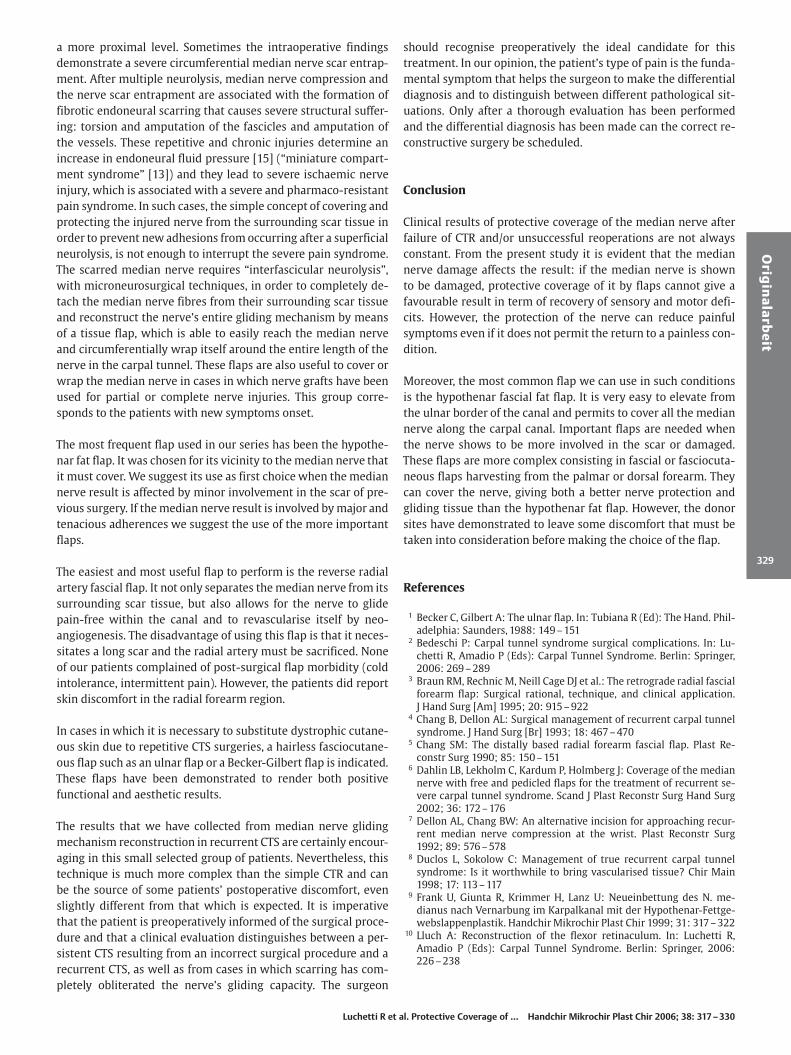
made along the flap axis exposing the fascia, by adequately ele-vating the two radial and ulnar cutaneous flaps, then the trans-fascial incision and the pedicle dissection follow the previouslydescribed techniques. The flap dissection is completed in a prox-imal-distal direction elevating the septum extending betweenthe extensor carpi ulnaris and extensor digiti minimi muscleand the overlying deep fascia so that it includes the posterior in-terosseous artery and its septal ascending branches. At the fore-arm’s distal third, the pedicle is usually wrapped in an osteo-fibrous sheath which is adherent to the ulna’s periosteum. Careis required to complete the dissection, preserving the vascularpedicle, and to avoid the interruption of the T-shaped distal anas-tomosis with the anterior interosseous artery. It is an importantanatomical landmark of the flap’s pivot point because of its im-portant role in perfusing the flap’s vascular pedicle. The flap isready for transposition to the carpal region, or through a sub-cutaneous tunnel which has an adequate width and accuratehaemostasis, or in an open surgical field by incising the skin atthe ulnar bridge in a dorso-palmar and also a proximo-distal di-rection (Fig. 7 a to f). The exposure of the median nerve at the car-pal tunnel and its entire circumferential wrapping is done usinga standard technique. However, this technique must always takeinto account the dimensional limitations of the posterior flapwith respect to the radial artery or ulnar artery flap, since it has
Fig. 5 a to g a, b Preoperative clinical aspect of the right hand of pa-tient (41 years old) with recurrence of CTS after thermal burn to thepalm and wrist. c Cross incision of the palm with extension incisionof the wrist and forearm. The median nerve was detached from thesurrounding scar tissue and the flexor retinaculum proved to be ab-sent. d Reconstruction of the flexor retinaculum according to Lluch’stechnique [10]. e Postoperative result of the distally based forearmulnar artery flap rotated onto the palm: the fascia covers the mediannerve and the island skin has reconstructed the palm. f, g Functionalresult at long-term follow-up.
Luchetti R et al. Protective Coverage of … Handchir Mikrochir Plast Chir 2006; 38: 317 – 330
Orig
ina
larb
eit
325

an inferior amount of fascia surface that is available for mediannerve wrapping and an inferior distal extension for the length ofthe nerve.
Postoperative treatment and rehabilitationDrainage is always used in order to prevent the formation of ahaematoma and is removed during the second postoperativeday. The wrist is splinted in slight extension with the fingers freefor immediate postoperative motion. The splint is removed after7 to 15 days depending upon the type of flap that has been used.
Fig. 6 a to f a Left hand of patient affected by median nerve paralysis after CTR and surgery for ganglia removal from the palmar ulnar DRUJ.b The Becker-Gilbert fascial flap was harvested and (c) turned distally to cover the median nerve. d Immediate postoperative result. e, f Clinicalresult at the long-term follow-up.
Luchetti R et al. Protective Coverage of … Handchir Mikrochir Plast Chir 2006; 38: 317 – 330
Orig
ina
larb
eit
326

Once the splint is removed, a rehabilitation programme is initi-ated which consists of active and passive wrist mobilisation,gentle scar massage, and a scar desensitisation programme.
Results
Twenty-five patients were evaluated after a mean follow-up of51 months (12 to 168 months).
The pain evaluation showed an improvement passing from amean value of 9 to 4. However, the best results were for thepatients in which the median nerve was undamaged (meanVAS = 1).
Postoperative sensory and motor evaluation demonstrated thatthe less the nerve was damaged, the more were the symptoms im-proved. Postoperative neurophysiological examinations (EMG)showed the same tendency.
Fig. 7 a to f a, b Patient, 66 years old, with her right hand affected by median nerve lesion after OCTR. Patient was operated twice for CTS.c The median nerve was damaged proximally to the carpal tunnel. d The posterior interosseous fasciocutaneous island flap is isolated fromthe dorsum of the forearm. e Only the fascial island flap was rotated on the palmar wrist to cover the median nerve because the skin remainedavascular. f Clinical result at the follow-up showing the little bulging over the median nerve at the palmar wrist and the donor site.
Luchetti R et al. Protective Coverage of … Handchir Mikrochir Plast Chir 2006; 38: 317 – 330
Orig
ina
larb
eit
327
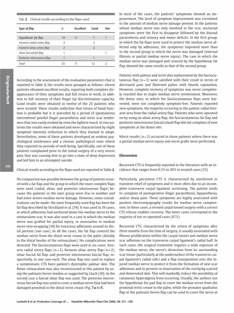
According to the assessment of the evaluation parameters that isreported in Table 1, the results were grouped as follows: elevenpatients obtained excellent results, reporting both complete dis-appearance of their symptoms and full return to work, in addi-tion to full recovery of their finger tip discrimination capacity.Good results were obtained in twelve of the 25 patients whowere treated. These results underline that return of hand func-tion is probable but it is preceded by a period of spontaneousintermittent painful finger paraesthesia and wrist scar tender-ness that was easily evoked by even the lightest touch. In two pa-tients fair results were obtained and were characterised by slightsymptom intensity reduction to which they learned to adapt.Nevertheless, some of these patients developed an evident psy-chological intolerance and a chronic pathological state wherethey reported no periods of well-being. Specifically, one of thesepatients complained prior to the initial surgery of a very severepain that was causing him to go into a state of deep depressionand led him to an attempted suicide.
Clinical results according to the flaps used are reported in Table 2.
No comparison was possible between the group of patients treat-ed with a fat flap and the group in which the more complex flapswere used (radial, ulnar, and posterior interosseous flaps) be-cause the patients in the later group were few in number andhad more severe median nerve damage. However, some consid-erations can be made: the most frequently used flap has been thefat flap described by Strickland et al. [19]. It was used in 19 cases,in which adhesions had anchored down the median nerve to theretinaculum scar. It was also used in a case in which the mediannerve was grafted for partial injury, in association to mediannerve vein wrapping [18] for tenacious adhesions around its dis-tal portion (one case). In all the cases, the fat flap covered themedian nerve from the distal wrist crease to the palm (distallyto the distal border of the retinaculum). No complications weredetected. The fasciocutaneous flaps were used in six cases: fore-arm radial artery flaps (n = 2), forearm ulnar artery flap (n = 2),ulnar fascial fat flap, and posterior interosseous fascial flap, re-spectively, in one case each. The ulnar flap was used to replacea symptomatic CTS burn patient’s dystrophic palmar skin. Theflexor retinaculum was also reconstructed in this patient by us-ing the palmaris brevis tendon as suggested by Lluch [10]. In thesecond case a fascial ulnar flap was used. The posterior interos-seous fascial flap was used to cover a median nerve that had beendamaged proximal to the distal wrist crease (Fig. 7 a to f).
In most of the cases, the patients’ symptoms showed an im-provement. The level of symptom improvement was correlatedto the amount of median nerve damage present. In the patientswhose median nerve was only involved in the scar, nocturnalsymptoms were the first to disappear followed by the diurnalparaesthesia and sensory and motor deficits. In the first group,in which the fat flaps were used to protect the median nerve af-fected only by adhesions, the symptoms improved more thanin the second group in which the nerve was damaged (internalfibrosis or partial median nerve injury). The case in which themedian nerve was damaged and covered by the hypothenar fatflap showed the same results as that of the second group.
Patients with palmar and wrist skin replacement by the fasciocu-taneous flap (n = 3) were satisfied with their result in terms ofdecreased pain and flattened palms with thumb supination.However, complete recovery of symptoms was never complete-ly reached due to major median nerve involvement. Moreover,the donor sites, in which the fasciocutaneous flaps were har-vested, were not completely symptom-free. Patients reportednew symptoms, the majority occurring in the palmar radial fore-arm area from the radial artery flap. Patients who were operatedon by using an ulnar artery flap, the fasciocutaneous fat flap andposterior interosseous fascial island flap did not complain of newsymptoms at the donor site.
Worst results (n = 2) occurred in those patients where there wasa partial median nerve injury and nerve grafts were performed.
Discussion
Recurrent CTS is frequently reported in the literature with an in-cidence that ranges from 0.3% to 20% in treated cases [15].
Particularly, persistent CTS is characterised by unrelieved ortransient relief of symptoms and is most often due to an incom-plete transverse carpal ligament sectioning. The patient tendsto complain of postoperative finger paraesthesia, hypaesthesia,and/or sharp pain. These symptoms are highly associated withpositive electromyography results for median nerve compres-sion. In these cases, a revised carpal tunnel surgery with a correctCTS release enables recovery. The latter cases correspond to themajority of our re-operated cases (67%).
Recurrent CTS, characterised by the return of symptoms afterthree months from the time of surgery, is usually associated withfibrous proliferation within the carpal tunnel and median nervescar adhesion on the transverse carpal ligament’s radial half. Insuch cases, the surgical treatment requires a wide exposure ofthe median nerve, the nerve’s dissection from its surroundingscar tissue (particularly at the undersurface of the transverse car-pal ligament’s radial side) and a flap transposition over the in-jured median nerve to protect it from the formation of new scaradhesions and to prevent re-innervation of the overlying scarredand denervated skin. This will markedly reduce the possibility ofcutaneous hyperalgesia from occurring. Usually, the authors usethe hypothenar fat-pad flap to cover the median nerve from theproximal wrist crease to the palm, while the pronator quadratusflap or the palmaris brevis flap can be used to cover the nerve at
Tab. 2 Clinical results according to the flaps used
Type of flap n Excellent Good Fair
Hypothenar fat flap 19 11 7 1
Forearm radial artery flap 2 2
Forearm ulnar artery flap 2 2
Ulnar fascial-fat flap 1 1
Posterior interosseous flap 1 1
Total 25 11 12 2
Luchetti R et al. Protective Coverage of … Handchir Mikrochir Plast Chir 2006; 38: 317 – 330
Orig
ina
larb
eit
328
a more proximal level. Sometimes the intraoperative findingsdemonstrate a severe circumferential median nerve scar entrap-ment. After multiple neurolysis, median nerve compression andthe nerve scar entrapment are associated with the formation offibrotic endoneural scarring that causes severe structural suffer-ing: torsion and amputation of the fascicles and amputation ofthe vessels. These repetitive and chronic injuries determine anincrease in endoneural fluid pressure [15] (“miniature compart-ment syndrome” [13]) and they lead to severe ischaemic nerveinjury, which is associated with a severe and pharmaco-resistantpain syndrome. In such cases, the simple concept of covering andprotecting the injured nerve from the surrounding scar tissue inorder to prevent new adhesions from occurring after a superficialneurolysis, is not enough to interrupt the severe pain syndrome.The scarred median nerve requires “interfascicular neurolysis”,with microneurosurgical techniques, in order to completely de-tach the median nerve fibres from their surrounding scar tissueand reconstruct the nerve’s entire gliding mechanism by meansof a tissue flap, which is able to easily reach the median nerveand circumferentially wrap itself around the entire length of thenerve in the carpal tunnel. These flaps are also useful to cover orwrap the median nerve in cases in which nerve grafts have beenused for partial or complete nerve injuries. This group corre-sponds to the patients with new symptoms onset.
The most frequent flap used in our series has been the hypothe-nar fat flap. It was chosen for its vicinity to the median nerve thatit must cover. We suggest its use as first choice when the mediannerve result is affected by minor involvement in the scar of pre-vious surgery. If the median nerve result is involved by major andtenacious adherences we suggest the use of the more importantflaps.
The easiest and most useful flap to perform is the reverse radialartery fascial flap. It not only separates the median nerve from itssurrounding scar tissue, but also allows for the nerve to glidepain-free within the canal and to revascularise itself by neo-angiogenesis. The disadvantage of using this flap is that it neces-sitates a long scar and the radial artery must be sacrificed. Noneof our patients complained of post-surgical flap morbidity (coldintolerance, intermittent pain). However, the patients did reportskin discomfort in the radial forearm region.
In cases in which it is necessary to substitute dystrophic cutane-ous skin due to repetitive CTS surgeries, a hairless fasciocutane-ous flap such as an ulnar flap or a Becker-Gilbert flap is indicated.These flaps have been demonstrated to render both positivefunctional and aesthetic results.
The results that we have collected from median nerve glidingmechanism reconstruction in recurrent CTS are certainly encour-aging in this small selected group of patients. Nevertheless, thistechnique is much more complex than the simple CTR and canbe the source of some patients’ postoperative discomfort, evenslightly different from that which is expected. It is imperativethat the patient is preoperatively informed of the surgical proce-dure and that a clinical evaluation distinguishes between a per-sistent CTS resulting from an incorrect surgical procedure and arecurrent CTS, as well as from cases in which scarring has com-pletely obliterated the nerve’s gliding capacity. The surgeon
should recognise preoperatively the ideal candidate for thistreatment. In our opinion, the patient’s type of pain is the funda-mental symptom that helps the surgeon to make the differentialdiagnosis and to distinguish between different pathological sit-uations. Only after a thorough evaluation has been performedand the differential diagnosis has been made can the correct re-constructive surgery be scheduled.
Conclusion
Clinical results of protective coverage of the median nerve afterfailure of CTR and/or unsuccessful reoperations are not alwaysconstant. From the present study it is evident that the mediannerve damage affects the result: if the median nerve is shownto be damaged, protective coverage of it by flaps cannot give afavourable result in term of recovery of sensory and motor defi-cits. However, the protection of the nerve can reduce painfulsymptoms even if it does not permit the return to a painless con-dition.
Moreover, the most common flap we can use in such conditionsis the hypothenar fascial fat flap. It is very easy to elevate fromthe ulnar border of the canal and permits to cover all the mediannerve along the carpal canal. Important flaps are needed whenthe nerve shows to be more involved in the scar or damaged.These flaps are more complex consisting in fascial or fasciocuta-neous flaps harvesting from the palmar or dorsal forearm. Theycan cover the nerve, giving both a better nerve protection andgliding tissue than the hypothenar fat flap. However, the donorsites have demonstrated to leave some discomfort that must betaken into consideration before making the choice of the flap.
References
1 Becker C, Gilbert A: The ulnar flap. In: Tubiana R (Ed): The Hand. Phil-adelphia: Saunders, 1988: 149 –151
2 Bedeschi P: Carpal tunnel syndrome surgical complications. In: Lu-chetti R, Amadio P (Eds): Carpal Tunnel Syndrome. Berlin: Springer,2006: 269 – 289
3 Braun RM, Rechnic M, Neill Cage DJ et al.: The retrograde radial fascialforearm flap: Surgical rational, technique, and clinical application.J Hand Surg [Am] 1995; 20: 915 – 922
4 Chang B, Dellon AL: Surgical management of recurrent carpal tunnelsyndrome. J Hand Surg [Br] 1993; 18: 467 –470
5 Chang SM: The distally based radial forearm fascial flap. Plast Re-constr Surg 1990; 85: 150 –151
6 Dahlin LB, Lekholm C, Kardum P, Holmberg J: Coverage of the mediannerve with free and pedicled flaps for the treatment of recurrent se-vere carpal tunnel syndrome. Scand J Plast Reconstr Surg Hand Surg2002; 36: 172 – 176
7 Dellon AL, Chang BW: An alternative incision for approaching recur-rent median nerve compression at the wrist. Plast Reconstr Surg1992; 89: 576 – 578
8 Duclos L, Sokolow C: Management of true recurrent carpal tunnelsyndrome: Is it worthwhile to bring vascularised tissue? Chir Main1998; 17: 113– 117
9 Frank U, Giunta R, Krimmer H, Lanz U: Neueinbettung des N. me-dianus nach Vernarbung im Karpalkanal mit der Hypothenar-Fettge-webslappenplastik. Handchir Mikrochir Plast Chir 1999; 31: 317 – 322
10 Lluch A: Reconstruction of the flexor retinaculum. In: Luchetti R,Amadio P (Eds): Carpal Tunnel Syndrome. Berlin: Springer, 2006:226 – 238
Luchetti R et al. Protective Coverage of … Handchir Mikrochir Plast Chir 2006; 38: 317 – 330
Orig
ina
larb
eit
329

11 Loick J, Joosten U, Lucke R: Einsatz von oxidierter, regenerierter Zellu-lose zur Rezidivprophylaxe bei der chirurgischen Therapie des Kar-paltunnelsndroms. Handchir Mikrochir Plast Chir 1997; 29: 209 – 213
12 Lovie MG, Juncan GM, Glasson JW: Ulnar artery forearm free flap. Br JPlast Surg 1984; 37: 486 – 492
13 Lundborg G, Myers R, Powell H: Nerve compression injury and in-creased endoneurial fluid pressure: A “miniature compartment syn-drome.” J Neurol Neurosurg Psychiatry 1983; 46: 1119 – 1124
14 Mackinnon SE: Secondary carpal tunnel surgery. Neurosurg Clin NAmer 1991; 2: 75 – 91
15 Riccio M, Bertani A, Morrison WA: Reverse island forearm flaps forthe coverage of the median nerve in recurrent carpal tunnel syn-drome. In: Luchetti R, Amadio P (Eds): Carpal Tunnel Syndrome. Ber-lin: Springer, 2006: 343– 360
16 Rose EH, Norris MS, Kowalski TA et al.: Palmaris brevis turnover flapas an adjunct to internal neurolysis of the chronically scarred mediannerve in recurrent carpal tunnel syndrome. J Hand Surg [Am] 1991;16: 191 – 201
17 Spokevicius S, Kleinert HE: The abductor digiti minimi flap: Its use inrevision carpal tunnel surgery. Hand Clin 1996; 12: 351 – 355
18 Sotereanos DG, Darlis NA: Vein wrapping of the median nerve. In: Lu-chetti R, Amadio P (Eds): Carpal Tunnel Syndrome. Berlin: Springer,2006: 333 – 337
19 Strickland JW, Idler RS, Lourie GM, Plancher KD: The hypothenarfat pad flap for management of recalcitrant carpal tunnel syndrome.J Hand Surg [Am] 1996; 21: 840 –848
20 Tham SKY, Ireland DCR, Riccio M et al.: Reverse radial artery fascialflap: A treatment for the chronically scarred median nerve in recur-rent carpal tunnel syndrome. J Hand Surg [Am] 1996; 21: 849 – 854
21 Weinzweig N, Chen L, Chen ZW: The distally based radial forearm fas-ciocutaneous flap with preservation of the radial artery: An anatomicand clinical approach. Plast Reconstr Surg 1994; 94: 675 – 683
22 Wulle C: The synovial flap as treatment of the recurrent carpal tunnelsyndrome. Hand Clin 1996; 12: 379 –388
23 Zancolli EA, Angrigiani C: Colgajo dorsal de antebrazo (“on isola”)(Pediculo de vases interosseos posteriores). Rev Assoc Arg OrthopTraumatol 1986; 51: 161– 168
24 Zancolli EA, Angrigiani C: Posterior interosseous island forearm flap.J Hand Surg [Br] 1988; 13: 130 – 135
Luchetti R et al. Protective Coverage of … Handchir Mikrochir Plast Chir 2006; 38: 317 – 330
Orig
ina
larb
eit
330