Embed Size (px)
Citation preview

Lyn Facilitates Glioblastoma Cell Survival underConditions of Nutrient Deprivation by PromotingAutophagyWeiMichael Liu1, Ping Huang1, Niladri Kar1, Monica Burgett1,4, GaelleMuller-Greven1,4, Amy S. Nowacki2,
Clark W. Distelhorst5, Justin D. Lathia3, Jeremy N. Rich3, John C. Kappes6, Candece L. Gladson1*
1Department of Cancer Biology, The Lerner Research Institute, Cleveland Clinic, Cleveland, Ohio, United States of America, 2Department of Quantitative Health Sciences,
Cleveland Clinic, Cleveland, Ohio, United States of America, 3Department of Stem Cell Biology and Regenerative Medicine, The Lerner Research Institute, Cleveland Clinic,
Cleveland, Ohio, United States of America, 4 School of Biomedical Sciences, Kent State University, Kent, Ohio, United States of America, 5Department of Medicine, Case
Western Reserve University, Cleveland, Ohio, United States of America, 6Department of Medicine, University of Alabama at Birmingham, Birmingham, Alabama, United
States of America
Abstract
Members of the Src family kinases (SFK) can modulate diverse cellular processes, including division, death and survival, buttheir role in autophagy has been minimally explored. Here, we investigated the roles of Lyn, a SFK, in promoting the survivalof human glioblastoma tumor (GBM) cells in vitro and in vivo using lentiviral vector-mediated expression of constitutively-active Lyn (CA-Lyn) or dominant-negative Lyn (DN-Lyn). Expression of either CA-Lyn or DN-Lyn had no effect on the survivalof U87 GBM cells grown under nutrient-rich conditions. In contrast, under nutrient-deprived conditions (absence ofsupplementation with L-glutamine, which is essential for growth of GBM cells, and FBS) CA-Lyn expression enhancedsurvival and promoted autophagy as well as inhibiting cell death and promoting proliferation. Expression of DN-Lynpromoted cell death. In the nutrient-deprived GBM cells, CA-Lyn expression enhanced AMPK activity and reduced the levelsof pS6 kinase whereas DN-Lyn enhanced the levels of pS6 kinase. Similar results were obtained in vitro using anothercultured GBM cell line and primary glioma stem cells. On propagation of the transduced GBM cells in the brains of nudemice, the CA-Lyn xenografts formed larger tumors than control cells and autophagosomes were detectable in the tumorcells. The DN-Lyn xenografts formed smaller tumors and contained more apoptotic cells. Our findings suggest that onnutrient deprivation in vitro Lyn acts to enhance the survival of GBM cells by promoting autophagy and proliferation as wellas inhibiting cell death, and Lyn promotes the same effects in vivo in xenograft tumors. As the levels of Lyn protein or itsactivity are elevated in several cancers these findings may be of broad relevance to cancer biology.
Citation: Liu WM, Huang P, Kar N, Burgett M, Muller-Greven G, et al. (2013) Lyn Facilitates Glioblastoma Cell Survival under Conditions of Nutrient Deprivation byPromoting Autophagy. PLoS ONE 8(8): e70804. doi:10.1371/journal.pone.0070804
Editor: Maciej S. Lesniak, The University of Chicago, United States of America
Received April 22, 2013; Accepted May 23, 2013; Published August 2, 2013
Copyright: � 2013 Liu et al. This is an open-access article distributed under the terms of the Creative Commons Attribution License, which permits unrestricteduse, distribution, and reproduction in any medium, provided the original author and source are credited.
Funding: This work was supported by NIH NCI grants CA043703 (CWD), CA109748, CA127620 and CA152883(CLG),the University of Alabama at Birmingham(UAB) Center for AIDS Research Virology and Sequencing Cores (P30-AI-27767) and the Genetically Defined Microbe and Expression Core of the UAB Mucosal HIVand Immunobiology Center (R24 DK-64400) (JCK). The funders had no role in study design, data collection and analysis, decision to publish, or preparation of themanuscript.
Competing Interests: The authors have declared that no competing interests exist.
* E-mail: [email protected]
Introduction
Lyn is one of eight members of the Src family of kinases (SFK)
expressed in human cells [1]. The SFKs are highly homologous
non-receptor cytoplasmic tyrosine kinases that modulate diverse
cellular processes including adhesion, migration, division, death
and survival [1–5]. Dysregulation of individual SFKs, including
Lyn, occurs in several different types of tumor [4,6–9]. Although
the functions of SFKs appear to be influenced by the microen-
vironment as well as cell type and post-translational modifications
[4,6–9], little attention has been paid to the role of SFKs in
promoting cell survival through regulation of autophagy. A
potential role for SFKs in autophagy is suggested by the reports
that Dasatinib, which inhibits multiple SFKs as well as Bcr-Abl,
induces autophagy in multiple types of cancer cells, including
GBM, under nutrient-rich conditions [10,11]. In addition, c-Src
has been shown to localize to autophagosomes in focal adhesion
kinase (FAK)-deficient cells under nutrient-rich conditions [12].
Lyn activity is elevated in GBM, the highest grade of glioma
tumors, as well as in breast cancer, acute myelocytic leukemia
(AML), B-cell chronic lymphocytic leukemia (CLL) and Ewing’s
sarcoma [13–19]. We found previously that Lyn activity and
protein levels are elevated significantly in human biopsies of GBM
[17], consistent with the earlier report that 15% of GFAP-v-Src
transgenic mice spontaneously develop low-grade gliomas that
progress to GBM tumors [20]. Neither gene amplification nor
mutation of SFK genes appears to play a role in the elevated SFK
activity in GBM or breast cancer cells (reviewed in [2,21]).
Here, we investigated the role of Lyn in promoting survival of
GBM cells under nutrient-rich conditions and conditions of
nutrient deprivation focusing on its role in autophagy. During
autophagy, cytoplasmic proteins and organelles are sequestered in
autophagasomes, which allows the cell to generate energy and
PLOS ONE | www.plosone.org 1 August 2013 | Volume 8 | Issue 8 | e70804

nutrients [22,23], and autophagy is thought to play a role in tumor
progression when the supply of nutrients is limited [22]. GBM
cells, like many other cancers cells are addicted to glutamine and
there is a growing body of evidence that glutamine plays a critical
role in the metabolic reprogramming utilized by cancer cells to
meet the demands of rapid proliferation and hypoxic conditions
[24,25]. Although glioblastoma tumors (GBM) are highly vascu-
larized, the neovasculature is abnormal and tumor cell starvation
and hypoxia can occur due to vascular thromboses and tumor
necrosis [2]. To our knowledge no prior study has investigated the
pro-survival function(s) of any SFKs in nutrient-deprived cells.
We found that the survival of GBM cells transduced with either
a lentiviral vector carrying constitutively-active (CA) Lyn or a
dominant-negative (DN) Lyn construct grown under nutrient-rich
conditions did not differ from control cells. In contrast, the results
of similar analyses carried out under conditions of nutrient
deprivation indicated that Lyn promotes survival of nutrient-
deprived GBM cells through both promotion of autophagy and
inhibition of apoptosis. When grown as xenografts, the CA-Lyn
tumor cells formed larger tumors that contained autophagosomes
and DN-Lyn cells formed smaller tumors with increased evidence
of apoptosis. These studies suggest that Lyn can play a role in
promoting survival of GBM cells by facilitating autophagy and
underscores the importance of gaining an improved understanding
of SFK-associated mechanisms in stressed cells.
Materials and Methods
Ethics StatementThe animal experiments were performed in accordance with an
approved protocol from the Cleveland Clinic Institutional Animal
Care and Use Committee (IACUC) (#2011–0554).
Cell Lines and Culture ConditionsU87 human GBM cells were obtained recently from the
American Type Culture Collection and maintained in L-
glutamine-free DMEM (Sigma Aldrich, D5030) supplemented
with 10% FBS and 1 mM L-glutamine. SNB19 human GBM cells
[26], a kind gift from Dr. Jasti Rao at the University of Illinois,
Peoria, were maintained in Hams F12 medium (Sigma Aldrich,
D6421) supplemented with 10% FBS and 1 mM L-glutamine
[27]. The GBM cells were plated in this nutrient-rich medium and
after 12 h, washed and then cultured in nutrient-deficient
medium, i.e., L-glutamine-free DMEM supplemented with 1%
BSA (Sigma Chemical Co.) and without FBS. GBM cells have an
increased demand for L-glutamine [24], which suppresses
autophagy in other cell types [28].
The primary human glioma stem cell (GSC) line (3832) [29] was
cultured as neurospheres in nutrient-rich neural basal medium
(NBM) containing EGF and bFGF [30].
In some experiments, inhibitors were added to the culture
medium, i.e., 4-amino-5-(4-chlorophenyl)-7-(dimethylethyl)pyra-
zolo[3,4-d]pyrimidine (PP2) (EMD Millipore), perifosine and
rapamycin (Selleck Chemicals), 3-methylamine (3-MA), and
chloroquine (Invitrogen).
Generation of Lyn Construct-Expressing LentiviralVectorsCA-Lyn (Y508F) and DN-Lyn (Y397F) cDNAs were kind gifts
from Dr. Evan Ingley at the Western Australian Institute for
Medical Research [31]. They were subcloned into the multiple
cloning site of the lentiviral vector (designated K072) that contains
the puromycin selectable marker and green fluorescent protein
(GFP) genes downstream of an internal ribosome entry site (IRES)
[32]. The lentiviral vector, the packaging construct, and the
vesicular stomatits virus G envelope (VSV-G) were transfected into
293T human embryo kidney cells to create infectious lentiviral
vector-containing particles. Cells were transduced (<56106
infectious particles/ml) with vectors expressing CA-Lyn, DN-
Lyn, or empty vector. Stable populations of U87 and SNB19 cells
were selected with 5 mg/ml puromycin (2 weeks), monitored for
GFP fluorescence on a regular basis, and sorted for GFP-
expressing cells as necessary. After transduction, GSCs were
sorted by FACS based on GFP positivity.
AntibodiesAntibodies were purchased as follows: rabbit anti-pY397FAK
and anti-total FAK (Upstate Biotechnology, Inc.), rabbit anti-Fyn,
anti-Lyn and anti-cSrc (Santa Cruz International); monoclonal
antibody (mAb) anti-glyceraldehyde 3-phosphate dehydrogenase
(GAPDH), and mAb anti-actin (Sigma Chemical Co.); rabbit anti-
LC3B, anti-phospho-Akt and anti-total Akt, and antibodies to pS6
kinase and total S6 kinase (Cell Signaling); rabbit anti-LC3B
(Nanotools); rabbit polyclonal IgG (sc-25575) (Santa Cruz
Biotechnology); anti-phospho-AMPK (Thr172) and anti-total
AMPK (Cell Signaling Technology) and anti-phospho-a1-AMPK
(Thr172) (Millipore).
Immunoprecipitation and Western Blot AnalysesCells were lysed using lysis buffer containing NP-40 and
protease inhibitors and electrophoresed or immunoprecipitated
followed by western blotting and densitometry [27]. Activation of
Lyn was assessed by immunoblotting to detect autophosphoryla-
tion of Y418 [1,17].
Viability, Cell Death, and Cell Cycle AnalysesViability was assessed by trypan blue exclusion and apoptosis
determined by FACS analysis of Annexin V-APC-stained cells,
blotting for cleaved caspase-7, or TUNEL assays [6,27,33]. Cell
cycle analysis was carried out by analysis of DNA content using a
FACScan (BD Biosciences) [33–35]. Proliferation was assessed by
EdU incorporation and FACS analysis [33].
Detection and Quantification of AutophagosomesAutophagosomes were detected by analysis of microtubule-
associated protein light chain 3 (LC3) [23]. Lentiviral vector-
transduced U87 cells expressing GFP were infected with red
fluorescent protein (RFP)-LC3 lentivirus, fixed using buffered 4%
paraformaldehyde and fluorescent puncta counted in at least 25
cells [23]. Primary human GSCs plated on laminin were fixed,
permeabilized and reacted with anti-LC3 antibody followed by
Alexa 594-conjugated secondary antibody [27]. Cells were viewed
using a Leica DMRB 4000X microscope. In some experiments,
the presence of autophagosomes or late autophagic compartments
(autophagic vacuoles) were detected by transmission electron
microscopy (EM), which was performed as described [36] using a
FIT Tecnai G2 EM scope in the Core Facility at the Cleveland
Clinic. Vesicles were counted as autophagosomes and late
autophagic vacuoles only if they were limited by a double
membrane and contained undegraded cytoplasm [36]. A random
sampling of tumor cells was performed and examined by EM from
5 tumors expressing LV, 5 expressing DN-Lyn, and 4 expressing
CA-Lyn.
Animals and TreatmentsXenografting was performed as described previously [33]. Cells
were injected with stereotactic assistance into the basal ganglia.
Lyn Promotes Autophagy in Nutrient Deprivation
PLOS ONE | www.plosone.org 2 August 2013 | Volume 8 | Issue 8 | e70804
Figure 1. Expression of CA-Lyn promotes the survival of nutrient-deprived U87 human GBM cells. U87 human GBM cells expressing CA-Lyn, DN-Lyn or LV control were plated in DMEM with L-glutamine and 10% FBS. After 12 h, nutrient deprivation was induced by replacing the
Lyn Promotes Autophagy in Nutrient Deprivation
PLOS ONE | www.plosone.org 3 August 2013 | Volume 8 | Issue 8 | e70804
On day 35 the mice were euthanized and the brains fixed in
buffered-formalin or electron microscopy fixative followed by
embedment in paraffin. All animal care, housing and procedures
were in accordance with the guidelines and regulations established
by the Animal Welfare Act (PL99–158) and the Guide of the Care
and Use of Laboratory Animals as part of the fully accredited
(American Association for the Accreditation of Laboratory Animal
Care International) Animal Resources Program of the Cleveland
Clinic (ARC08826). Method of Euthanasia: At euthanasia, mice
were anesthetized with ketamine (100 mg/kg mouse weight i.p.)
and xylazine (15 mg/kg mouse weight i.p.), and once a surgical
plane of anesthesia was reached based on absence of palpebral and
toe reflexes, the mice were euthanized by cervical dislocation.
Immediately after euthanasia, the chest was opened and 20 ml of
saline was infused with a pump into the heart, followed by 30 ml
of 10% formalin or by 30 ml of cold 4% paraformaldehyde (for
EM analysis).
ImmunohistochemistryThis was performed as described [27]. Intensity of staining was
compared to that of the IgG negative control.
Statistical AnalysisStatistical analyses were performed by the biostatistician and
author (ASN). The statistical test utilized is stated in each figure
legend.
Results
Expression of CA-Lyn Promotes Survival of Nutrient-Deprived GBM CellsThe levels of Lyn activity were manipulated by transducing U87
GBM cells with lentiviral vectors expressing a CA-Lyn construct or
a DN-Lyn construct. The SFKs, including Lyn, are maintained in
an inactive state by the C-terminal Src kinase (Csk), which
phosphorylates the C-terminal negative regulatory peptide (Y508
in Lyn). The phosphorylated tyrosine binds to the SH2 domain
folding SFK into an inactive configuration [1,4]. Dephosphory-
lation of the negative regulatory peptide by tyrosine phosphatase-aor direct binding of the SH2 and SH3 domain to intracellular
proteins such as FAK or activated tyrosine kinase growth factor
receptors, allows the SFK structure to assume its active
configuration [1,4]. Constitutively active Lyn is generated by a
Y508F mutation [1,31]. We also utilized a DN-Lyn construct in
which the autophosphorylation site is mutated (Y397F) [31] as we
were unable to downregulate Lyn by .50% with a single shRNA
lentiviral construct (WM Liu and CL Gladson, unpublished data).
After expressing the constructs in U87 GBM cells using lentiviral
vector, stable populations of cells were selected with puromycin,
then sorted for GFP expression to maintain homogeneous,
expression-positive populations. The expression of the constructs
was confirmed by western blotting using anti-Lyn antibody, which
demonstrated enhanced intensity of the band migrating at 53-kDa
(5th panel, Fig. 1A). Western blotting using an anti-Src(pY418)
antibody further confirmed enhanced activity of Lyn in the U87-
CA-Lyn cells as indicated by detection of a broad band migrating
at 53–56-kDa and absence of Lyn activity in the U87-DN-Lyn
cells (top panel, Fig. 1A). Only one other SFK, Lck, migrates on
SDS PAGE at 53–56-kDa, but it is not expressed in human brain
or GBM tumor tissue [17]. The SFK activity in control U87-LV
cells was similar to that in parent U87 cells (data not shown) and
no significant change in the expression of c-Src was detected in the
U87-CA-Lyn or U87-DN-Lyn cells (Fig. 1A). Fyn protein was
decreased by approximately 30% in U87 cells expressing CA-Lyn
or DN-Lyn (Fig. 1A). No differences in the morphology of the
control cells and those expressing the constructs were apparent.
Analysis of cell viability indicated that neither the expression of
CA-Lyn nor DN-Lyn affected viability under nutrient-rich
conditions (Fig. 1B). In contrast, under nutrient-deprivation
conditions the viability of the U87-CA-Lyn cells was significantly
greater and the viability of DN-Lyn cells significantly lower than
that of U87-LV cells (Fig. 1C and D). Assessment of apoptosis by
Annexin-V-staining (Fig. 1E), blotting for cleaved capsase-7
(Fig. 1F), or labeling for Annexin-V and propidium-iodide
(Fig. 1G) showed that under nutrient-deprivation conditions the
levels of apoptosis were significantly lower in the U87-CA-Lyn
cells and significantly higher in the U87-DN-Lyn cells than the
U87-LV cells (Fig. 1E-G). Annexin-V-labeling was not significant-
ly different in the parent U87 and LV cells (data not shown).
FACS analysis revealed a small increase in the percentage of U87
cells in S phase with expression of CA-Lyn as compared to the
control LV cells (Fig. 2A). EdU incorporation studies also showed
an increase in the percent of labeled CA-Lyn cells (4%) as
compared to the control LV cells (1%) (Fig. 2B), which is consistent
with CA-Lyn promoting proliferation. A small increase in the
percentage of U87-DN-Lyn cells in sub-G1 (Fig. 2A) was detected
on propidium-iodide labeling consistent with the increased
apoptosis observed on Annexin-V-labeling as the percentage of
apoptotic cells detected by cell cycle analysis (sub-G1) is typically
smaller than that detected by Annexin-V-labeling [34,35]. Taken
together, these data suggested that Lyn plays an important role in
regulating the survival of GBM cells propagated in the absence of
L-glutamine and FBS.
To confirm that the kinase activity of CA-Lyn is required for
Lyn promotion of GBM cell survival under nutrient deprivation,
we utilized PP2 (a broad SFK inhibitor). We first selected the
dosage of PP2 that inhibited SFK activity of the U87 cells when
added to the media 24 h after initiation of nutrient deprivation.
Blotting for pSrcY418 on day 5 confirmed inhibition of Lyn
activity in the U87-CA-Lyn cells (Fig. S1A). In the presence of
PP2, the survival of the nutrient-deprived U87-CA-Lyn cells was
no different from that of U87-LV cells at days 3 and 5 (Fig. 2C),
suggesting that SFK activity is necessary for the pro-survival effect
of CA-Lyn.
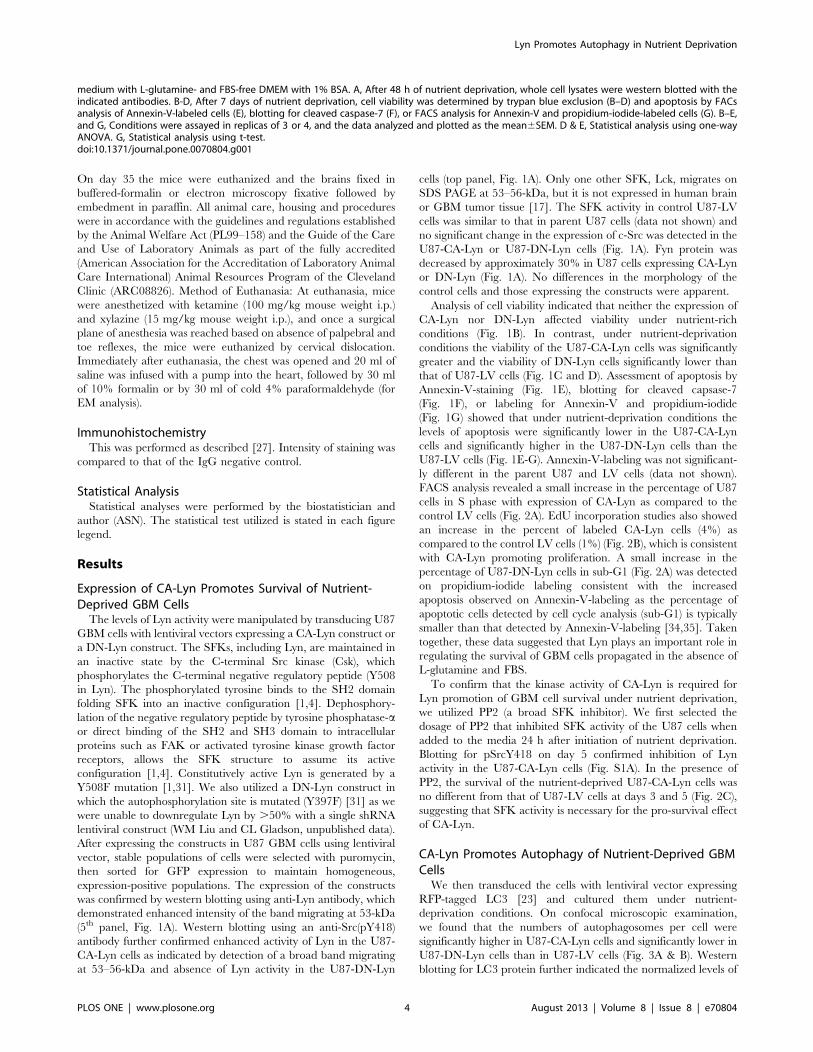
CA-Lyn Promotes Autophagy of Nutrient-Deprived GBMCellsWe then transduced the cells with lentiviral vector expressing
RFP-tagged LC3 [23] and cultured them under nutrient-
deprivation conditions. On confocal microscopic examination,
we found that the numbers of autophagosomes per cell were
significantly higher in U87-CA-Lyn cells and significantly lower in
U87-DN-Lyn cells than in U87-LV cells (Fig. 3A & B). Western
blotting for LC3 protein further indicated the normalized levels of
medium with L-glutamine- and FBS-free DMEM with 1% BSA. A, After 48 h of nutrient deprivation, whole cell lysates were western blotted with theindicated antibodies. B-D, After 7 days of nutrient deprivation, cell viability was determined by trypan blue exclusion (B–D) and apoptosis by FACsanalysis of Annexin-V-labeled cells (E), blotting for cleaved caspase-7 (F), or FACS analysis for Annexin-V and propidium-iodide-labeled cells (G). B–E,and G, Conditions were assayed in replicas of 3 or 4, and the data analyzed and plotted as the mean6SEM. D & E, Statistical analysis using one-wayANOVA. G, Statistical analysis using t-test.doi:10.1371/journal.pone.0070804.g001
Lyn Promotes Autophagy in Nutrient Deprivation
PLOS ONE | www.plosone.org 4 August 2013 | Volume 8 | Issue 8 | e70804

the lipid-modified LC3B-II protein, which is generated during
autophagy, were higher in the U87-CA-Lyn cells than in the U87-
LV cells at days 5 and 7 (Fig. 3C & D). At days 2, 3 and 5, lower
levels of LC3B-II were present in the cells expressing DN-Lyn than
in the U87-LV cells (Fig. 3C & D). The number of days required
to detect autophagy under the experimental conditions used in
Figure 2. Expression of CA-Lyn increases EdU incorporation in nutrient-deprived U87 GBM cells. A–C, U87 cells expressing CA-Lyn, DN-Lyn or the LV control were plated and then starved of L-glutamine and FBS as described in Figure 1A. A, After 7 days of starvation cells were acetonefixed, stained with propidium iodide and analyzed for DNA content by FACS analysis. B, EdU was added after 41/2 days of nutrient deprivation and 18hours later propidium-iodide was added and EdU incorporation analyzed by FACS. A & B, Conditions were assayed in replicas of 3 and the dataanalyzed as the mean6SD and plotted as a bar graph. C. 20 h after initiation of nutrient deprivation PP2 (200 nM) or DMSO (vehicle control) wasadded to the media. On day 5, viable adherent cells were counted by trypan blue exclusion. Conditions were performed in replicas of 3 or 4, and thedata analyzed and presented as the mean6SEM.doi:10.1371/journal.pone.0070804.g002
Lyn Promotes Autophagy in Nutrient Deprivation
PLOS ONE | www.plosone.org 5 August 2013 | Volume 8 | Issue 8 | e70804
Figure 3. Expression of CA-Lyn increases the numbers of autophagosomes per cell and the levels of LC3B-II protein in nutrient-deprived U87 GBM cells. U87 GBM cells expressing CA-Lyn, DN-Lyn or the LV control and GFP downstream of the IRES were transduced with RFP-LC3 lentiviral vector then plated and subjected to nutrient deprivation as in Figure 1A. A-B, After 5 days, cells were fixed and stained with DAPI
Lyn Promotes Autophagy in Nutrient Deprivation
PLOS ONE | www.plosone.org 6 August 2013 | Volume 8 | Issue 8 | e70804
these studies is likely due to the initial plating of the cells in
nutrient-rich medium, but it also is possible that the GBM cells can
synthesize L-glutamine as has been described for MCF7 cells [37].
Similar results were obtained using another human GBM cell
line, SNB19. We found that lentiviral-mediated expression of CA-
Lyn resulted in a broad band migrating at 53–56-kDa on blotting
for active Src(pY418) (Fig. S1B) that was positive for Lyn on
reprobing, and the levels of c-Src protein were not affected
significantly (Fig. S1B). Fyn protein was decreased by approx-
imately 30% in SNB19 cells expressing CA-Lyn or DN-Lyn (Fig.
S1B). In addition, the level of normalized LC3B-II protein was
higher in nutrient-deprived SNB19 cells expressing CA-Lyn at day
7 than in the SNB19 cells expressing the lentivirus vector alone
(Fig. S1C).
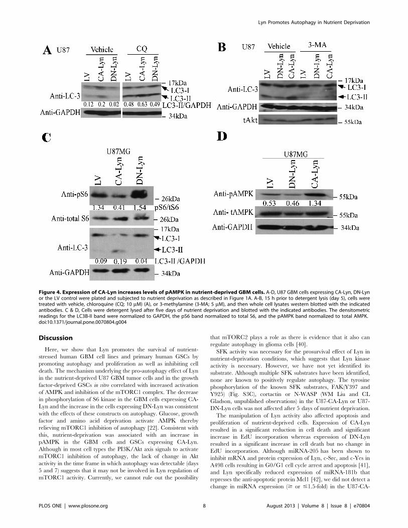
To confirm the effects of Lyn on autophagy in the nutrient-
deprived cells we used several different approaches. Overnight
treatment of nutrient-deprived cells with chloroquine, an inhibitor
of lysosomal fusion, resulted in an increase in the levels of LC3B-II
protein in the U87-CA-Lyn, U87-LV and U87-DN-Lyn cells
(Fig. 4A), consistent with accumulation of lipid-modified LC3B-II
protein on blockade of fusion of the autophagosome with the
lysosome. Treatment with 3-MA, an upstream inhibitor of
autophagy that blocks type II PI3K [22], caused a significant
reduction in the levels of LC3B-II protein in U87-CA-Lyn, U87-
LV and U87-DN-Lyn cells (Fig. 4B). Furthermore, treatment of
the U87 cells expressing LV, CA-Lyn or DN-Lyn with chloro-
quine or 3-MA for 5 or 7 days under the same nutrient
deprivation conditions resulted in a dramatic reduction in cell
viability in all cell populations (Fig. S2), consistent with autophagy
playing an important role in the CA-Lyn-associated increase in cell
survival.
Expression of CA-Lyn in Nutrient-Deprived GBM Cells isAssociated with Enhanced AMPK Activity and aReduction in the Levels of pS6 KinaseAs autophagy is negatively regulated by the mTORC1 complex,
we estimated mTORC1 activity by assessing phosphorylation of
S6 kinase, a downstream effector of mTORC1 [22,23]. After
5 days of nutrient-deprivation, the levels of pS6 kinase were lower
in U87-CA-Lyn and higher in U87-DN-Lyn cells than in U87-LV
cells (Fig. 4C), consistent with the observed differences in
autophagosome number and LC3B-II protein levels at this time
point (Fig. 3A-D). Moreover, treatment with rapamycin, an
inhibitor of mTORC1, resulted in higher levels of LC3B-II protein
in nutrient-deprived U87-CA-Lyn cells than in vehicle-treated
cells (Fig. S3A).
Akt activation of mTORC1 can inhibit autophagy [22]. The
levels of pAkt(S308) in nutrient-deprived U87-CA-Lyn cells were
unchanged at days 5 and 7 when the cells were undergoing
autophagy (Fig. S3B); however, there was a transient increase in
pAkt at day 2, prior to signs of autophagy (Fig. S3B). The levels of
pAkt in the nutrient-deprived U87-DN-Lyn cells remained
constant (Fig. S3B). Perifosine, an Akt inhibitor, enhanced the
levels of LC3B-II protein in all cell populations (Fig. S3A). Others
have reported that generalized SFK inhibition in osteosarcoma
cells results in apoptosis, which was attributed to inhibition of the
FAK/p130CAS signaling axis [38]; however, we did not find any
changes in FAK activity (pY397 and pY925) with expression of
CA-Lyn or DN-Lyn in the nutrient-deprived U87 or SNB19 GBM
cells (Fig. S3C and data not shown). Nutrient deprivation is known
to activate AMPK [22], and we found higher levels of pAMPK in
nutrient-deprived U87-CA-Lyn cells than in nutrient-deprived
U87-LV cells (Fig. 4D). There was no significant difference in the
level of pAMPK in nutrient-deprived U87-DN-Lyn cells as
compared to U87-LV cells.
Expression of CA-Lyn in Nutrient-Deprived PrimaryHuman Glioma Stem Cells Increases AutophagyGSCs are thought to be the tumor-initiating cell in GBM [39].
We therefore transduced primary human GSCs with GFP-CA-
Lyn, GFP-DN-Lyn or GFP-LV, sorted for GFP-expressing cells,
and plated the cells on laminin in NBM. Higher Src activity was
confirmed in the GSCs expressing CA-Lyn than those expressing
LV (data not shown). Immunofluorescent staining for LC3 protein
in the GSCs starved of EGF and bFGF for 6 h indicated that the
numbers of autophagosomes per cell was significantly higher in the
GSCs expressing CA-Lyn as compared to those expressing LV,
and significantly lower in the GSCs expressing DN-Lyn as
compared to those expressing LV (Fig. S4A & B). We also found
higher pAMPK levels in the GSCs expressing CA-Lyn as
compared to LV (Fig. S4C), suggesting that increased AMPK
activity relieves the inhibition of mTORC1 on autophagy.
Expression of CA-Lyn Promotes the Survival of GBM CellsPropagated as Intracerebral Xenografts and PromotesAutophagy in the Tumor CellsTo determine whether Lyn promotes the survival of GBM cells
in vivo, we injected U87-CA-Lyn, U87-DN-Lyn and U87-LV cells
into the brains of nude mice. After 35 days, the U87-CA-Lyn cells
had formed significantly larger tumors than the U87-LV cells
whereas the U87-DN-Lyn cells had formed significantly smaller
tumors (Fig. 5A & B). GFP expression was confirmed in tumor
cells in all tumors (Fig. S4D). As compared to the U87-LV tumors,
there were significantly fewer TUNEL-positive cells in the U87-
CA-Lyn tumors and a significantly higher number of TUNEL-
positive cells in the U87-DN-Lyn tumors (Fig. 5C & D), which was
consistent with our in vitro findings when cells were propagated as a
monolayer under nutrient-deprivation conditions (Fig. 1E & F).
Detection of proliferating cells by Ki67 mAb labeling showed a
significant increase in the CA-Lyn versus the LV tumors,
indicating that the larger tumor size with expression of CA-Lyn
was due in part to increased proliferation (Fig. 5E & F). Ki67
labeling of the DN-Lyn tumors showed a significant decrease as
compared to the LV tumors (Fig. 5E & F).
To evaluate autophagy in the xenograft tumors we utilized EM
analysis. Representative photomicrographs of autophagic vacuoles
(autophagosomes and late autophagic compartments) in tumor
cells and the results of quantitation are shown in Figure 6A-K.
These experiments revealed that while the occurrence of
autophagic vacuoles in the U87-LV tumors (Fig. 6F & G) and
U87-DN-CA tumors (Fig. 6H & I) was uncommon, autophagic
vacuoles were present in most U87-CA-Lyn tumor cells examined
(Fig. 6A-E, J & K). Collectively, these data indicate that Lyn
promotes the survival of GBM cells in vivo through promotion of
autophagy and proliferation, as well as inhibition of cell death.
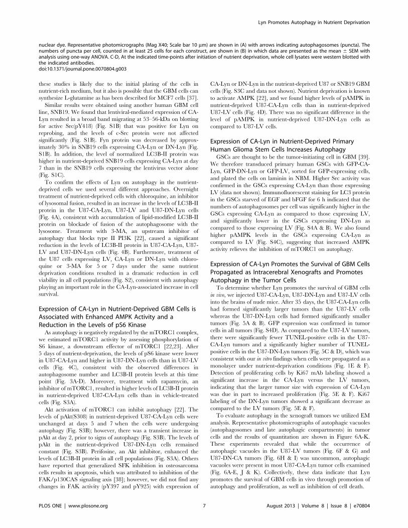
nuclear dye. Representative photomicrographs (Mag X40; Scale bar 10 mm) are shown in (A) with arrows indicating autophagosomes (puncta). Thenumbers of puncta per cell, counted in at least 25 cells for each construct, are shown in (B) in which data are presented as the mean 6 SEM withanalysis using one-way ANOVA. C-D, At the indicated time-points after initiation of nutrient deprivation, whole cell lysates were western blotted withthe indicated antibodies.doi:10.1371/journal.pone.0070804.g003
Lyn Promotes Autophagy in Nutrient Deprivation
PLOS ONE | www.plosone.org 7 August 2013 | Volume 8 | Issue 8 | e70804
Discussion
Here, we show that Lyn promotes the survival of nutrient-
stressed human GBM cell lines and primary human GSCs by
promoting autophagy and proliferation as well as inhibiting cell
death. The mechanism underlying the pro-autophagy effect of Lyn
in the nutrient-deprived U87 GBM tumor cells and in the growth
factor-deprived GSCs in vitro correlated with increased activation
of AMPK and inhibition of the mTORC1 complex. The decrease
in phosphorylation of S6 kinase in the GBM cells expressing CA-
Lyn and the increase in the cells expressing DN-Lyn was consistent
with the effects of these constructs on autophagy. Glucose, growth
factor and amino acid deprivation activate AMPK thereby
relieving mTORC1 inhibition of autophagy [22]. Consistent with
this, nutrient-deprivation was associated with an increase in
pAMPK in the GBM cells and GSCs expressing CA-Lyn.
Although in most cell types the PI3K/Akt axis signals to activate
mTORC1 inhibition of autophagy, the lack of change in Akt
activity in the time frame in which autophagy was detectable (days
5 and 7) suggests that it may not be involved in Lyn regulation of
mTORC1 activity. Currently, we cannot rule out the possibility
that mTORC2 plays a role as there is evidence that it also can
regulate autophagy in glioma cells [40].
SFK activity was necessary for the prosurvival effect of Lyn in
nutrient-deprivation conditions, which suggests that Lyn kinase
activity is necessary. However, we have not yet identified its
substrate. Although multiple SFK substrates have been identified,
none are known to positively regulate autophagy. The tyrosine
phosphorylation of the known SFK substrates, FAK(Y397 and
Y925) (Fig. S3C), cortactin or N-WASP (WM Liu and CL
Gladson, unpublished observations) in the U87-CA-Lyn or U87-
DN-Lyn cells was not affected after 5 days of nutrient deprivation.
The manipulation of Lyn activity also affected apoptosis and
proliferation of nutrient-deprived cells. Expression of CA-Lyn
resulted in a significant reduction in cell death and significant
increase in EdU incorporation whereas expression of DN-Lyn
resulted in a significant increase in cell death but no change in
EdU incorporation. Although miRNA-205 has been shown to
inhibit mRNA and protein expression of Lyn, c-Src, and c-Yes in
A498 cells resulting in G0/G1 cell cycle arrest and apoptosis [41],
and Lyn specifically reduced expression of miRNA-181b that
represses the anti-apoptotic protein Mcl1 [42], we did not detect a
change in miRNA expression ($ or #1.5-fold) in the U87-CA-
Figure 4. Expression of CA-Lyn increases levels of pAMPK in nutrient-deprived GBM cells. A-D, U87 GBM cells expressing CA-Lyn, DN-Lynor the LV control were plated and subjected to nutrient deprivation as described in Figure 1A. A-B, 15 h prior to detergent lysis (day 5), cells weretreated with vehicle, chloroquine (CQ; 10 mM) (A), or 3-methylamine (3-MA; 5 mM), and then whole cell lysates western blotted with the indicatedantibodies. C & D, Cells were detergent lysed after five days of nutrient deprivation and blotted with the indicated antibodies. The densitometricreadings for the LC3B-II band were normalized to GAPDH, the pS6 band normalized to total S6, and the pAMPK band normalized to total AMPK.doi:10.1371/journal.pone.0070804.g004
Lyn Promotes Autophagy in Nutrient Deprivation
PLOS ONE | www.plosone.org 8 August 2013 | Volume 8 | Issue 8 | e70804
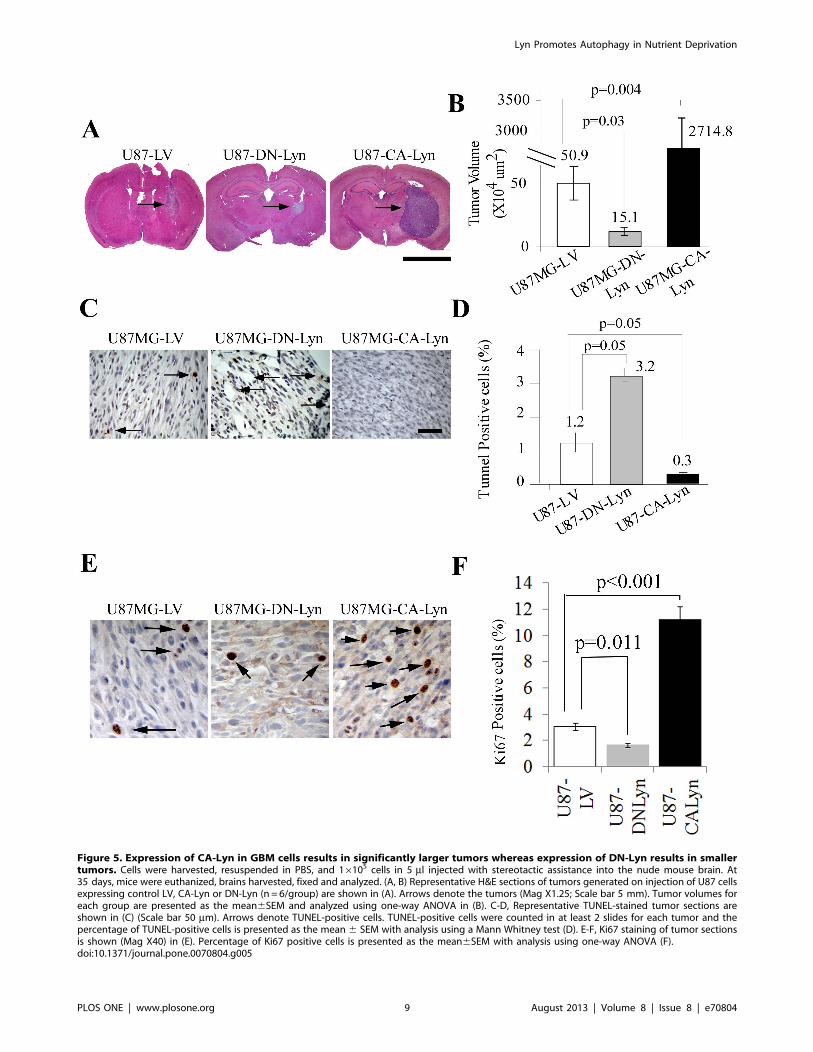
Figure 5. Expression of CA-Lyn in GBM cells results in significantly larger tumors whereas expression of DN-Lyn results in smallertumors. Cells were harvested, resuspended in PBS, and 16105 cells in 5 ml injected with stereotactic assistance into the nude mouse brain. At35 days, mice were euthanized, brains harvested, fixed and analyzed. (A, B) Representative H&E sections of tumors generated on injection of U87 cellsexpressing control LV, CA-Lyn or DN-Lyn (n = 6/group) are shown in (A). Arrows denote the tumors (Mag X1.25; Scale bar 5 mm). Tumor volumes foreach group are presented as the mean6SEM and analyzed using one-way ANOVA in (B). C-D, Representative TUNEL-stained tumor sections areshown in (C) (Scale bar 50 mm). Arrows denote TUNEL-positive cells. TUNEL-positive cells were counted in at least 2 slides for each tumor and thepercentage of TUNEL-positive cells is presented as the mean 6 SEM with analysis using a Mann Whitney test (D). E-F, Ki67 staining of tumor sectionsis shown (Mag X40) in (E). Percentage of Ki67 positive cells is presented as the mean6SEM with analysis using one-way ANOVA (F).doi:10.1371/journal.pone.0070804.g005
Lyn Promotes Autophagy in Nutrient Deprivation
PLOS ONE | www.plosone.org 9 August 2013 | Volume 8 | Issue 8 | e70804

Lyn or U87-DN-Lyn cells as compared to U87-LV cells using the
Human Cancer miRNA PCR Array (MAH-102A, Qiagen) (WM
Liu and CL Gladson, unpublished data). Similarly, although SFKs
can promote survival through the FAK/p130CAS or PI3K/Akt
pro-survival pathways [38,43], neither FAK nor Akt activity was
altered in the nutrient-deprived GBM cells expressing CA-Lyn or
DN-Lyn. It is possible that the cellular localization of CA-Lyn
[44,45], the cell surface binding partner of Lyn [46], or Lyn
phosphorylation of the growth arrest and DNA damage protein 34
(GADD34) [47] contribute to the anti-death effect in GBM but
this remains to be elucidated.
Our in vivo studies in which GBM cells expressing DN-Lyn were
propagated in the nude mouse brain showed a significant
reduction in tumor size that was associated with increased tumor
cell death, suggesting Lyn is important for tumor survival in vivo.
This is consistent with a prior report that dasatinib inhibits
malignant glioma growth in a mouse model [8]. However,
dasatinib used as a single agent in GBM patients who had failed
therapy with AvastinH did not result in either a partial or complete
response [48]. The reason for this is unclear but may be related to
the diverse effects of dasatinib, e.g., in addition to blocking the
function of all SFKs it also targets the cytoplasmic non-receptor
tyrosine kinase c-Abl, which is necessary for apoptosis of brain
microvessel endothelial cells induced by an integrin avb3/avb5inhibitor and by latrunculin [49]. Currently, it is not possible to
conclude that nutrient deprivation is the trigger of the effects of
Lyn in vivo, but a focal lack of nutrients does occur in GBM [2].
Figure 6. Expression of CA-Lyn in GBM cells results in larger tumors containing autophagic vacuoles. Xenograft tumors expressing LV,CA-Lyn or DN-Lyn were generated as described in the legend for Figure 5 and after euthanasia on day 35 tumors were fixed in EM fixative, followedby transmission EM as described in the Materials and Methods. A-E, Three different representative cells from CA-Lyn tumors; panel B-highermagnification of the box in Panel A, and panel D-higher magnification of the box in panel C. F & G, Representative tumor cell from LV tumor; panel G-higher magnification of the box in panel F. H & I, Representative tumor cell from DN-Lyn tumor; panel I-higher magnification of the box in panel H.Scale bars in each panel denote 1 mm. Arrows denote examples of autophagic vacuoles. J, Quantitation of autophagic vacuoles/tumor cell shows asignificant increase in the CA-Lyn tumors as compared to the LV tumors (p value,0.0001; t-test). K, Scattergram denoting the number of autophagicvacuoles/tumor cell.doi:10.1371/journal.pone.0070804.g006
Lyn Promotes Autophagy in Nutrient Deprivation
PLOS ONE | www.plosone.org 10 August 2013 | Volume 8 | Issue 8 | e70804
Elevation of Lyn is associated with resistance to therapy. In
CML, it is associated with the development of resistance to
imatinib mesylate, a Bcr-Abl tyrosine kinase inhibitor [13,14]. It
also is found in AML refractory to therapy [15,16], and in non-
small cell lung cancer cells resistant to cetuximab (an anti-EGFR
antibody) where Lyn promotes EGFR nuclear translocation [5]. In
addition, Lyn cooperates with a CD44-variant receptor in
promoting chemoresistance in colon cancer cells [43]. In cancers
refractory to therapy, autophagy can be a pro-survival response to
the stress of chemotherapeutic agents [22,40]; however, autophagy
can lead to cell death under certain circumstances (reviewed in
[50]). There is an evolving concept that cross-talk between
autophagy and apoptosis signaling pathways occurs in cells, and
not infrequently these two processes appear to be regulated in an
inverse manner [51]. Hydroxychloroquine, an inhibitor of
autophagy, has been entered into a phase II clinical trial involving
patients with multiple types of cancer including GBM (reviewed in
[22]), and blockade of autophagy has been shown to result in the
sensitization of prostate cancer cells, multi-drug resistant v-Ha-ras
transformed NIH-3T3 cells and other cells to a SFK inhibitor in
nutrient-rich conditions [11,52].
In summary, we demonstrate the novel findings that Lyn
promotes autophagy and proliferation, as well as inhibits cell
death, in nutrient-deprived GBM cells in culture and in vivo using a
mouse model. Lyn promotes the malignant phenotype of GBM
and multiple other cancers, including promotion of the epithelial-
mesenchymal transition in breast cancer [18], as well as
chemoresistance in certain cancers. Moreover, a Lyn-specific
peptide that inhibits Lyn-dependent phosphorylation has been
shown to decrease prostate cancer growth and induce apoptosis
in vivo [53]. Thus, Lyn could be an important new therapeutic
target for multiple cancers.
Supporting Information
Figure S1 Inhibition of SFKs with PP2 inhibits theactivity of CA-Lyn, and CA-Lyn promotion of autophagyin nutrient deprived SNB19 GBM cells. A, U87 GBM cells
stably expressing CA-Lyn, DN-Lyn or the LV control were plated
and then starved of L-glutamine and FBS as indicated in
Figure 1A. A, PP2 (200 nM) or vehicle DMSO was added to
the media after overnight starvation. On day 5 of starvation, cells
were detergent lysed and immunoblotted with the indicated
antibodies. B & C, SNB19 cells were plated in Hams F12 media
with L-glutamine and 10% FBS, at 16 h washed and the media
replaced with L-glutamine and FBS-free media with 1% BSA. B,
After 48 hours of starvation, cells were lysed in NP40 lysis buffer
with protease inhibitors; equivalent amount of lysate (100 mg)subjected to SDS-PAGE, and immunoblotted with the indicated
antibodies, or C, after 7 days of starvation cells were detergent
lysed and immunoblotted with the indicated antibodies.
(TIF)
Figure S2 Inhibitors of autophagy block the survival ofGBM cells in nutrient deprivation conditions. U87 GBM
cells stably expressing CA-Lyn, DN-Lyn or the LV control were
plated and then starved of L-glutamine and FBS as indicated in
Figure 1A. After 20 hours of starvation, 3-MA or chloroquine were
added and viable adherent cells counted by trypan blue exclusion
on days 3 and 5. Representative fields were photographed on day
5 (A), and the data analyzed and presented as the mean6SEM (B).
(TIF)
Figure S3 Effect of Rapamycin on LC3 protein andanalysis of Akt and FAK activity in GBM cells expressingCA-Lyn or DN-Lyn. U87 GBM cells stably expressing CA-Lyn,
DN-Lyn or the LV control were plated and then starved of L-
glutamine and FBS as indicated in Figure 1A, or SNB19 GBM
cells were plated in Hams F12 media with L-glutamine and 10%
FBS, at 16 h washed, and the media replaced with L-glutamine
and FBS-free media with 1% BSA. A, After 4K days of starvation
cells were treated with vehicle, 100 nM Rapamycin or 5 mMperfosine (overnight), followed by detergent lysis and immuno-
blotting with the indicated antibodies. All samples were electro-
phoresed on the same gel. B & C, On the indicated days of
starvation, cells were detergent lysed and immunoblotted with the
indicated antibodies. The normalized pAkt was determined based
on the densitometric ratio of pAkt to normalized total Akt (Akt/
GAPDH), and the normalized pFAK was determined based on the
densitometric ratio of pFAK to normalized total FAK (FAK/
GAPDH).
(TIF)
Figure S4 Expression of CA-Lyn increased the autopha-gosome number/cell and the levels of pAMPK innutrient-deprived glioma stem cells, and GFP expres-sion in xenograft tumors indicates expression of thelentiviral construct. A-B, Human GSCs expressing CA-Lyn,
DN-Lyn or LV were plated onto laminin-coated wells in NBM.
After 24 h, the media was replaced with NBM lacking EGF and
bFGF and 6 h later the cells were fixed, and reacted with anti-LC3
antibody followed by Alexa-594-conjugated secondary antibody
and DAPI and visualized and photographed. Representative
photomicrographs (scale bar 10 mm) are shown in (A). The
number of red autophagosomes were counted in at least 25 cells
with each construct. Data are presented as the mean 6 SEM and
analyzed using one-way ANOVA (B). It should be noted that the
absolute number of autophagosomes per cell in the GSCs cannot
be compared to those in the U87 GBM cells (Figure 3A & B) as the
time of nutrient deprivation and the method used to detect the
autophagosomes were different. C, Human GSCs expressing CA-
Lyn, DN-Lyn or LV were plated and starved of EGF and bFGF as
in panels A-B, whole cell lysates were then western blotted with the
indicated antibodies. D, U87-LV, U87-CA-Lyn and U87-DN-Lyn
expressing cells were harvested, resuspended in PBS, and 16105
cells in 5 ml injected with stereotactic assistance into the nude
mouse brain. At 35 days, mice were euthanized, and the brains
harvested, fixed and analyzed. Expression of the IRES-driven GFP
gene in the lentiviral vector of LV, CA-Lyn and DN-Lyn is
demonstrated by GFP immunohistochemistry. T, denotes tumor;
and AMB, denotes adjacent mouse brain.
(TIF)
Acknowledgments
We thank Dr. Fiona Hunter for editorial assistance, Dr. James T.
McMahon for reviewing the EM photographs, Mei Yin for technical
assistance at the EM Core, and Jerry Stewart (University of Alabama at
Birmingham), Russell Tipps, Dr. Chun Fan and Robert Galvin (Cleveland
Clinic) for sharing reagents and experimental strategy.
Author Contributions
Conceived and designed the experiments: WML PH NK CLG. Performed
the experiments: WML PH MB GMG NK. Analyzed the data: WML PH
NK MB GMG ASN CLG. Contributed reagents/materials/analysis tools:
CWD JDL JNR JCK. Wrote the paper: WML CLG.
Lyn Promotes Autophagy in Nutrient Deprivation
PLOS ONE | www.plosone.org 11 August 2013 | Volume 8 | Issue 8 | e70804
References
1. Thomas SM, Brugge JS (1997) Cellular functions regulated by Src family
kinases. Annu Rev Cell Dev Biol 13: 513–609.
2. Ahluwalia MS, de Groot J, Liu WM, Gladson CL (2010) Targeting SRC in
glioblastoma tumors and brain metastases: rationale and preclinical studies.Cancer Lett 298: 139–149.
3. Kharas MG, Daley GQ (2010) From Hen House to Bedside: TracingHanafusa’s Legacy from Avian Leukemia Viruses to SRC to ABL and Beyond.
Genes Cancer 1: 1164–1169.
4. Summy JM, Gallick GE (2003) Src family kinases in tumor progression and
metastasis. Cancer Metastasis Rev 22: 337–358.
5. Lida M, Brand TM, Campbell DA, Li C, Wheeler DL (2013) Yes and Lyn play
a role in nuclear translocation of the epidermal growth factor receptor.Oncogene 32: 759–767.
6. Ding Q, Stewart J Jr., Olman MA, Klobe MR, Gladson CL (2003) The patternof enhancement of Src kinase activity on platelet-derived growth factor
stimulation of glioblastoma cells is affected by the integrin engaged. J Biol
Chem 278: 39882–39891.
7. Cai H, Smith DA, Memarzadeh S, Lowell CA, Cooper JA, et al. (2011)
Differential transformation capacity of Src family kinases during the initiation ofprostate cancer. Proc Natl Acad Sci U S A 108: 6579–6584.
8. Lu KV, Zhu S, Cvrljevic A, Huang TT, Sarkaria S, et al. (2009) Fyn and SRCare effectors of oncogenic epidermal growth factor receptor signaling in
glioblastoma patients. Cancer Res 69: 6889–6898.
9. Summy JM, Qian Y, Jiang BH, Guappone-Koay A, Gatesman A, et al. (2003)
The SH4-Unique-SH3-SH2 domains dictate specificity in signaling thatdifferentiate c-Yes from c-Src. J Cell Sci 116: 2585–2598.
10. Milano V, Piao Y, LaFortune T, de Groot J (2009) Dasatinib-induced autophagyis enhanced in combination with temozolomide in glioma. Mol Cancer Ther 8:
394–406.
11. Wu Z, Chang PC, Yang JC, Chu CY, Wang LY, et al. (2010) Autophagy
Blockade Sensitizes Prostate Cancer Cells towards Src Family Kinase Inhibitors.
Genes Cancer 1: 40–49.
12. Sandilands E, Serrels B, McEwan DG, Morton JP, Macagno JP, et al. (2012)
Autophagic targeting of Src promotes cancer cell survival following reducedFAK signalling. Nat Cell Biol 14: 51–60.
13. Samanta AK, Chakraborty SN, Wang Y, Kantarjian H, Sun X, et al. (2009)Jak2 inhibition deactivates Lyn kinase through the SET-PP2A-SHP1 pathway,
causing apoptosis in drug-resistant cells from chronic myelogenous leukemiapatients. Oncogene 28: 1669–1681.
14. Donato NJ, Wu JY, Stapley J, Gallick G, Lin H, et al. (2003) BCR-ABLindependence and LYN kinase overexpression in chronic myelogenous leukemia
cells selected for resistance to STI571. Blood 101: 690–698.
15. Dos Santos C, Demur C, Bardet V, Prade-Houdellier N, Payrastre B, et al.
(2008) A critical role for Lyn in acute myeloid leukemia. Blood 111: 2269–2279.
16. Mahon FX, Hayette S, Lagarde V, Belloc F, Turcq B, et al. (2008) Evidence that
resistance to nilotinib may be due to BCR-ABL, Pgp, or Src kinase
overexpression. Cancer Res 68: 9809–9816.
17. Stettner MR, Wang W, Nabors LB, Bharara S, Flynn DC, et al. (2005) Lyn
kinase activity is the predominant cellular SRC kinase activity in glioblastomatumor cells. Cancer Res 65: 5535–5543.
18. Choi YL, Bocanegra M, Kwon MJ, Shin YK, Nam SJ, et al. (2010) LYN is amediator of epithelial-mesenchymal transition and a target of dasatinib in breast
cancer. Cancer Res 70: 2296–2306.
19. Guan H, Zhou Z, Gallick GE, Jia SF, Morales J, et al. (2008) Targeting Lyn
inhibits tumor growth and metastasis in Ewing’s sarcoma. Mol Cancer Ther 7:1807–1816.
20. Weissenberger J, Steinbach JP, Malin G, Spada S, Rulicke T, et al. (1997)Development and malignant progression of astrocytomas in GFAP-v-src
transgenic mice. Oncogene 14: 2005–2013.
21. Verhaak RG, Hoadley KA, Purdom E, Wang V, Qi Y, et al. (2010) Integratedgenomic analysis identifies clinically relevant subtypes of glioblastoma charac-
terized by abnormalities in PDGFRA, IDH1, EGFR, and NF1. Cancer Cell 17:98–110.
22. Amaravadi RK, Lippincott-Schwartz J, Yin XM, Weiss WA, Takebe N, et al.(2011) Principles and current strategies for targeting autophagy for cancer
treatment. Clin Cancer Res 17: 654–666.
23. Klionsky DJ, Abeliovich H, Agostinis P, Agrawal DK, Aliev G, et al. (2008)
Guidelines for the use and interpretation of assays for monitoring autophagy inhigher eukaryotes. Autophagy 4: 151–175.
24. DeBerardinis RJ, Mancuso A, Daikhin E, Nissim I, Yudkoff M, et al. (2007)
Beyond aerobic glycolysis: transformed cells can engage in glutaminemetabolism that exceeds the requirement for protein and nucleotide synthesis.
Proc Natl Acad Sci U S A 104: 19345–19350.
25. Anastasiou D, Cantley LC (2012) Breathless cancer cells get fat on glutamine.
Cell Res 22: 443–446.
26. Gondi CS, Lakka SS, Dinh DH, Olivero WC, Gujrati M, et al. (2007)
Intraperitoneal injection of a hairpin RNA-expressing plasmid targetingurokinase-type plasminogen activator (uPA) receptor and uPA retards
angiogenesis and inhibits intracranial tumor growth in nude mice. Clin CancerRes 13: 4051–60.
27. Wang D, Olman MA, Stewart J, Jr., Tipps R, Huang P, et al. (2011)
Downregulation of FIP200 induces apoptosis of glioblastoma cells andmicrovascular endothelial cells by enhancing Pyk2 activity. PLoS One 6:
e19629.
28. Nicklin P, Bergman P, Zhang B, Triantafellow E, Wang H, et al. (2009)Bidirectional transport of amino acids regulates mTOR and autophagy. Cell
136: 521–534.
29. Guryanova OA, Wu Q, Cheng L, Lathia JD, Huang Z, et al. (2011)
Nonreceptor tyrosine kinase BMX maintains self-renewal and tumorigenic
potential of glioblastoma stem cells by activating STAT3. Cancer Cell 19: 498–511.
30. Eyler CE, Foo WC, LaFiura KM, McLendon RE, Hjelmeland AB, et al. (2008)Brain cancer stem cells display preferential sensitivity to Akt inhibition. Stem
Cells 26: 3027–3036.
31. Tilbrook PA, Ingley E, Williams JH, Hibbs ML, Klinken SP (1997) Lyn tyrosinekinase is essential for erythropoietin-induced differentiation of J2E erythroid
cells. EMBO J 16: 1610–1619.
32. Mulky A, Sarafianos SG, Jia Y, Arnold E, Kappes JC (2005) Identification ofamino acid residues in the human immunodeficiency virus type-1 reverse
transcriptase tryptophan-repeat motif that are required for subunit interactionusing infectious virions. J Mol Biol 349: 673–684.
33. Ding Q, Grammer JR, Nelson MA, Guan JL, Stewart JE, Jr., et al. (2005)
p27Kip1 and cyclin D1 are necessary for focal adhesion kinase regulation of cellcycle progression in glioblastoma cells propagated in vitro and in vivo in the scid
mouse brain. J Biol Chem 280: 6802–6815.
34. Mazumdar T, Devecchio J, Agyeman A, Shi T, Houghton JA (2011) Blocking
Hedgehog survival signaling at the level of the GLI genes induces DNA damage
and extensive cell death in human colon carcinoma cells. Cancer Res 71: 5904–5914.
35. Mazumdar T, DeVecchio J, Shi T, Jones J, Agyeman A, et al. (2011) Hedgehogsignaling drives cellular survival in human colon carcinoma cells. Cancer Res 71:
1092–1102.
36. Yla-Anttila P, Vihinen H, Jokitalo E, Eskelinen EL (2009) Monitoring autophagyby electron microscopy in Mammalian cells. Methods Enzymol 452: 143–164.
37. Kung HN, Marks JR, Chi JT (2011) Glutamine synthetase is a geneticdeterminant of cell type-specific glutamine independence in breast epithelia.
PLoS Genet 7: e1002229.
38. Shor AC, Keschman EA, Lee FY, Muro-Cacho C, Letson GD, et al. (2007)Dasatinib inhibits migration and invasion in diverse human sarcoma cell lines
and induces apoptosis in bone sarcoma cells dependent on SRC kinase for
survival. Cancer Res 67: 2800–2808.
39. Lathia JD, Gallagher J, Myers JT, Li M, Vasanji A, et al. (2011) Direct in vivo
evidence for tumor propagation by glioblastoma cancer stem cells. PLoS One 6:e24807.
40. Fan QW, Cheng C, Hackett C, Feldman M, Houseman BT, et al. (2010) Akt
and autophagy cooperate to promote survival of drug-resistant glioma. SciSignal 3: ra81.
41. Majid S, Saini S, Dar AA, Hirata H, Shahryari V, et al. (2011) MicroRNA-205inhibits Src-mediated oncogenic pathways in renal cancer. Cancer Res 71:
2611–2621.
42. Zimmerman EI, Dollins CM, Crawford M, Grant S, Nana-Sinkam SP, et al.(2010) Lyn kinase-dependent regulation of miR181 and myeloid cell leukemia-1
expression: implications for drug resistance in myelogenous leukemia. MolPharmacol 78: 811–817.
43. Bates RC, Edwards NS, Burns GF, Fisher DE (2001) A CD44 survival pathway
triggers chemoresistance via lyn kinase and phosphoinositide 3-kinase/Akt incolon carcinoma cells. Cancer Res 61: 5275–5283.
44. Trentin L, Frasson M, Donella-Deana A, Frezzato F, Pagano MA, et al. (2008)
Geldanamycin-induced Lyn dissociation from aberrant Hsp90-stabilized cyto-solic complex is an early event in apoptotic mechanisms in B-chronic
lymphocytic leukemia. Blood 112: 4665–4674.
45. Tibaldi E, Brunati AM, Zonta F, Frezzato F, Gattazzo C, et al. (2011) Lyn-
mediated SHP-1 recruitment to CD5 contributes to resistance to apoptosis of B-
cell chronic lymphocytic leukemia cells. Leukemia 25: 1768–1781.
46. Chudakova DA, Zeidan YH, Wheeler BW, Yu J, Novgorodov SA, et al. (2008)
Integrin-associated Lyn kinase promotes cell survival by suppressing acidsphingomyelinase activity. J Biol Chem 283: 28806–28816.
47. Grishin AV, Azhipa O, Semenov I, Corey SJ (2001) Interaction between growth
arrest-DNA damage protein 34 and Src kinase Lyn negatively regulatesgenotoxic apoptosis. Proc Natl Acad Sci U S A 98: 10172–10177.
48. Lu-Emerson C, Norden AD, Drappatz J, Quant EC, Beroukhim R, et al. (2011)
Retrospective study of dasatinib for recurrent glioblastoma after bevacizumabfailure. J Neurooncol 104: 287–291.
49. Xu J, Millard M, Ren X, Cox OT, Erdreich-Epstein A (2010) c-Abl mediatesendothelial apoptosis induced by inhibition of integrins alphavbeta3 and
alphavbeta5 and by disruption of actin. Blood 115: 2709–2718.
50. Kondo Y, Kanzawa T, Sawaya R, Kondo S (2005) The role of autophagy incancer development and response to therapy. Nat Rev Cancer 5: 726–734.
51. Marquez RT, Xu L (2012) Bcl-2:Beclin 1 complex: multiple, mechanismsregulating autophagy/apoptosis toggle switch. Am J Cancer Res 2: 214–221.
Lyn Promotes Autophagy in Nutrient Deprivation
PLOS ONE | www.plosone.org 12 August 2013 | Volume 8 | Issue 8 | e70804
52. Ahn JH, Lee M (2011) Suppression of autophagy sensitizes multidrug resistant
cells towards Src tyrosine kinase specific inhibitor PP2. Cancer Lett 310: 188–197.
53. Goldenberg-Furmanov M, Stein I, Pikarsky E, Rubin H, Kasem S, et al. (2004)
Lyn is a target gene for prostate cancer: sequence-based inhibition inducesregression of human tumor xenografts. Cancer Res 64: 1058–1066.
Lyn Promotes Autophagy in Nutrient Deprivation
PLOS ONE | www.plosone.org 13 August 2013 | Volume 8 | Issue 8 | e70804

![The Roles of Glutamine in the Intestine and Its ...€¦ · utilize large amounts of glutamine, exceeding the endogenous glutamine production [12,13], and that plasma and muscle glutamine](https://img.dokumen.tips/doc/110x75/5fd64d48c22ac35b4b7b6b55/the-roles-of-glutamine-in-the-intestine-and-its-utilize-large-amounts-of-glutamine.jpg)