Embed Size (px)
Citation preview
LOUISIANA STATE UNIVERSITYMEDICAL CENTER
School of Medicine in New Orleans
LOUISIANA STATE UNIVERSITYMEDICAL CENTER
School of Medicine in New Orleans
Michael Maristany MDContributions from Carlos R. Giménez, MD
Michael Maristany MDContributions from Carlos R. Giménez, MD
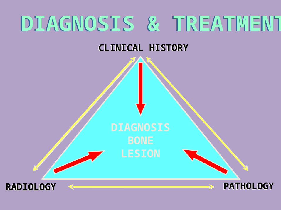
DIAGNOSIS & TREATMENTDIAGNOSIS & TREATMENTCLINICAL HISTORYCLINICAL HISTORY
RADIOLOGYRADIOLOGY PATHOLOGYPATHOLOGY
DIAGNOSISBONE
LESION
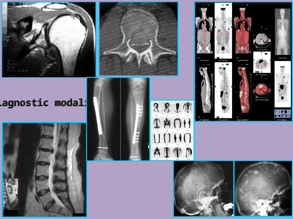
Diagnostic modalitiesDiagnostic modalitiesDiagnostic modalitiesDiagnostic modalities
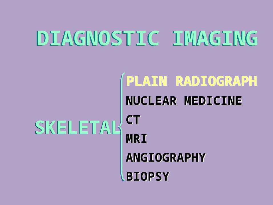
DIAGNOSTIC IMAGINGDIAGNOSTIC IMAGING
SKELETALSKELETAL
PLAIN RADIOGRAPHNUCLEAR MEDICINE
CT
MRI
ANGIOGRAPHY
BIOPSY
PLAIN RADIOGRAPHNUCLEAR MEDICINE
CT
MRI
ANGIOGRAPHY
BIOPSY
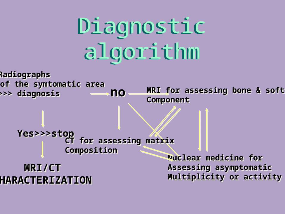
Diagnostic algorithmDiagnostic algorithm
Yes>>>stopYes>>>stop
nono
CT for assessing matrixCompositionCT for assessing matrixComposition
MRI for assessing bone & soft tissueComponentMRI for assessing bone & soft tissueComponent
Nuclear medicine forAssessing asymptomaticMultiplicity or activity
Nuclear medicine forAssessing asymptomaticMultiplicity or activity
MRI/CTCHARACTERIZATION
MRI/CTCHARACTERIZATION
• Radiographs of the symtomatic area• >>> diagnosis
• Radiographs of the symtomatic area• >>> diagnosis
CONVENTIONAL Rx CONVENTIONAL Rx IT REMAINS AS THE MOST RELIABLE IN
THE
HISTOLOGIC NATURE OF A SPECIFIC
LESION 4 DETECTION4 LOCALIZATION4 CHARACTERIZATION
IT REMAINS AS THE MOST RELIABLE IN
THE
HISTOLOGIC NATURE OF A SPECIFIC
LESION 4 DETECTION4 LOCALIZATION4 CHARACTERIZATION
Tid bitsTid bits3 It is always a good idea to start with a radiograph
of the area in question.
Proceed with MRI if you are concern with ligaments or
soft tissue problems, occult fracture or characterization
A CT if you are more concern with bony problemsSometimes you need both.
3 It is always a good idea to start with a radiograph of the area in question.
Proceed with MRI if you are concern with ligaments or
soft tissue problems, occult fracture or characterization
A CT if you are more concern with bony problemsSometimes you need both.
Ligament injuriesLigament injuries
3 CT is more optimal than
MRI
3 True or False
3 CT is more optimal than
MRI
3 True or False
3 For the evaluation of Disc disease, ligamentous or spinal cord injury in trauma MRI is preferred
3 For the evaluation of vertebral fractures in spine trauma CT is preferred.
3 Point: Both are use in evaluation of the spine in trauma.!
3 For the evaluation of Disc disease, ligamentous or spinal cord injury in trauma MRI is preferred
3 For the evaluation of vertebral fractures in spine trauma CT is preferred.
3 Point: Both are use in evaluation of the spine in trauma.!
DIAGNOSTIC RADIOLOGYDIAGNOSTIC RADIOLOGYANATOMY- MORPHOLOGY
PHYSIOLOGY/FUNCTION 3 X- ray3 CT Nuclear MedicineD Ultrasound D MRI
ANATOMY- MORPHOLOGY PHYSIOLOGY/FUNCTION
3 X- ray3 CT Nuclear MedicineD Ultrasound D MRI
TRANSMISSION IMAGING(X-RAY)
TRANSMISSION IMAGING(X-RAY)
3 X-Ray tube outside the body
3 Patient is positioned in front of the source
3 Image is recovered on X-Ray film or Matrix
which
is positioned behind the patient.
3 X-Ray tube outside the body
3 Patient is positioned in front of the source
3 Image is recovered on X-Ray film or Matrix
which
is positioned behind the patient.

3 An advantage of radionuclide bone scanning is
that the entire osseous system is demonstrated.
3 It relatively nonspecific and the history and
correlation with other imaging modalities is
necessity.
3 An advantage of radionuclide bone scanning is
that the entire osseous system is demonstrated.
3 It relatively nonspecific and the history and
correlation with other imaging modalities is
necessity.
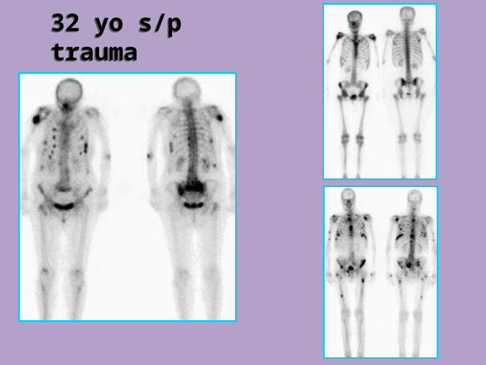
32 yo s/p trauma32 yo s/p trauma
TRAUMA
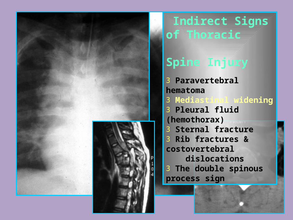
Indirect Signs of Thoracic Spine Injury
3 Paravertebral hematoma3 Mediastinal widening3 Pleural fluid (hemothorax)3 Sternal fracture3 Rib fractures & costovertebral dislocations3 The double spinous process sign
DEGENERATIVE CHANGES / ARTHRITIS
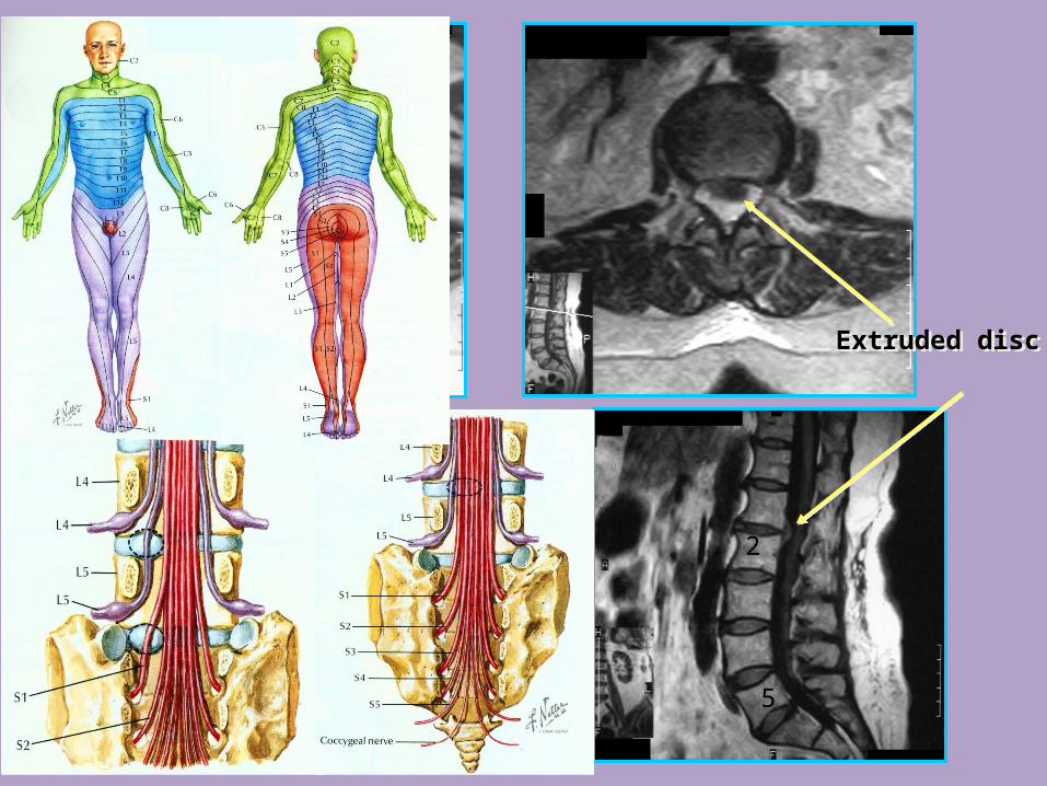
5
2
Extruded discExtruded disc
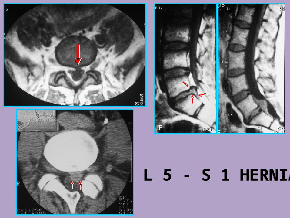
L 5 - S 1 HERNIAL 5 - S 1 HERNIA
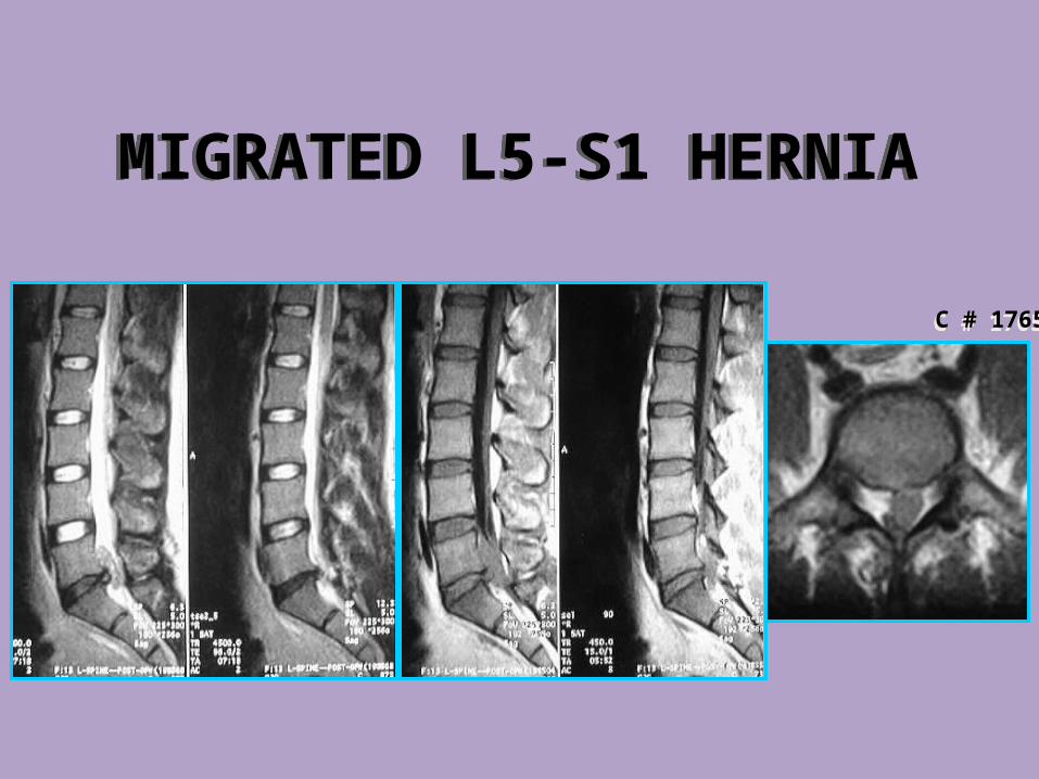
MIGRATED L5-S1 HERNIAMIGRATED L5-S1 HERNIA
C # 1765C # 1765
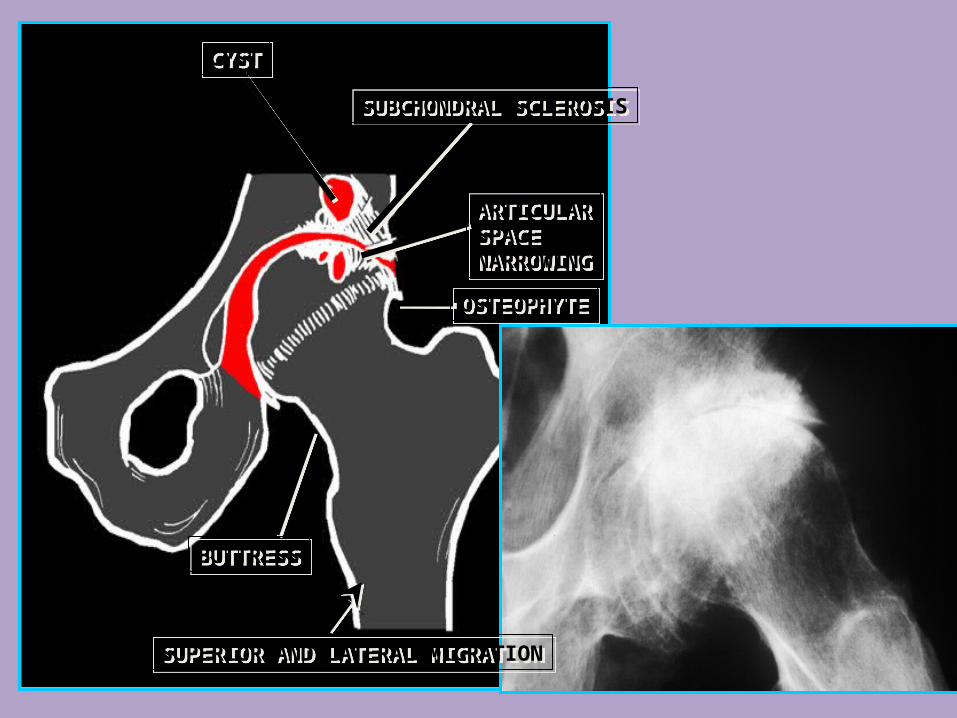
CYSTCYST
SUBCHONDRAL SCLEROSISSUBCHONDRAL SCLEROSIS
ARTICULARSPACENARROWING
ARTICULARSPACENARROWING
OSTEOPHYTEOSTEOPHYTE
BUTTRESSBUTTRESS
SUPERIOR AND LATERAL MIGRATIONSUPERIOR AND LATERAL MIGRATION
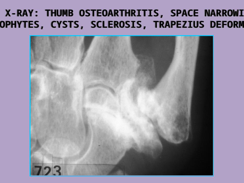
HAND X-RAY: THUMB OSTEOARTHRITIS, SPACE NARROWING, OSTEOPHYTES, CYSTS, SCLEROSIS, TRAPEZIUS DEFORMITY.HAND X-RAY: THUMB OSTEOARTHRITIS, SPACE NARROWING, OSTEOPHYTES, CYSTS, SCLEROSIS, TRAPEZIUS DEFORMITY.
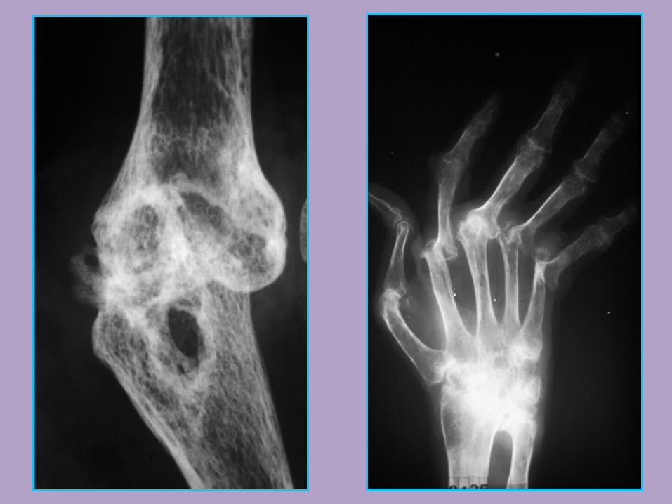
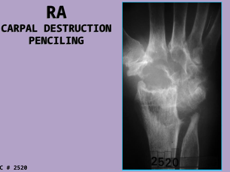
RACARPAL DESTRUCTION
PENCILING
RACARPAL DESTRUCTION
PENCILING
C # 2520
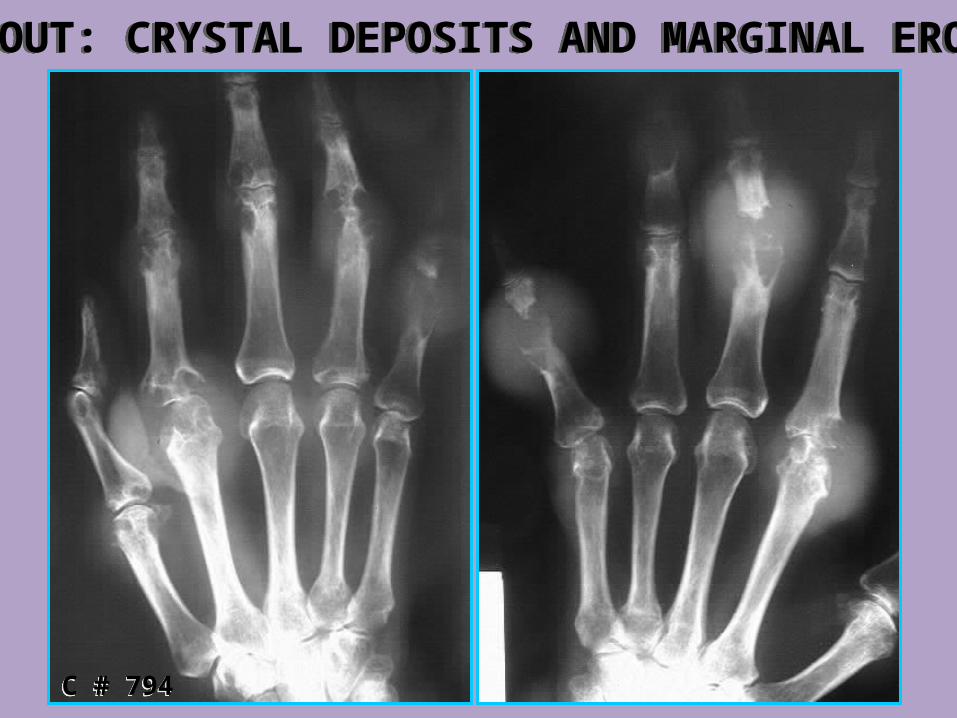
GOUT: CRYSTAL DEPOSITS AND MARGINAL EROSIONSGOUT: CRYSTAL DEPOSITS AND MARGINAL EROSIONS
C # 794C # 794
METABOLIC DISEASE/ OSTEOMALACIA
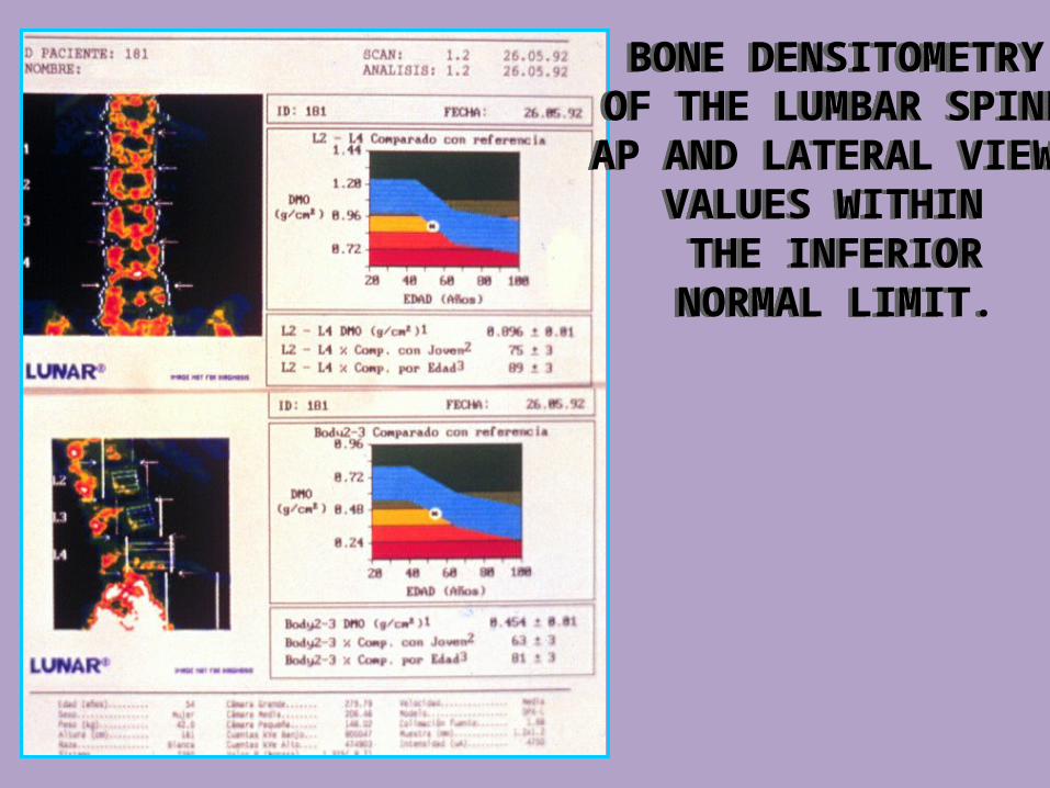
BONE DENSITOMETRYOF THE LUMBAR SPINE
AP AND LATERAL VIEWSVALUES WITHIN THE INFERIORNORMAL LIMIT.
BONE DENSITOMETRYOF THE LUMBAR SPINE
AP AND LATERAL VIEWSVALUES WITHIN THE INFERIORNORMAL LIMIT.
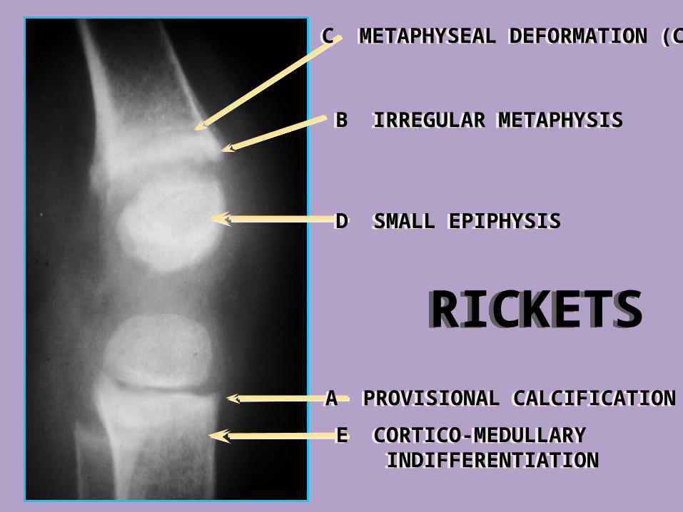
D SMALL EPIPHYSISD SMALL EPIPHYSIS
C METAPHYSEAL DEFORMATION (CUP) C METAPHYSEAL DEFORMATION (CUP)
B IRREGULAR METAPHYSISB IRREGULAR METAPHYSIS
A PROVISIONAL CALCIFICATIONA PROVISIONAL CALCIFICATION
E CORTICO-MEDULLARY INDIFFERENTIATIONE CORTICO-MEDULLARY INDIFFERENTIATION
RICKETSRICKETS
TUMORS
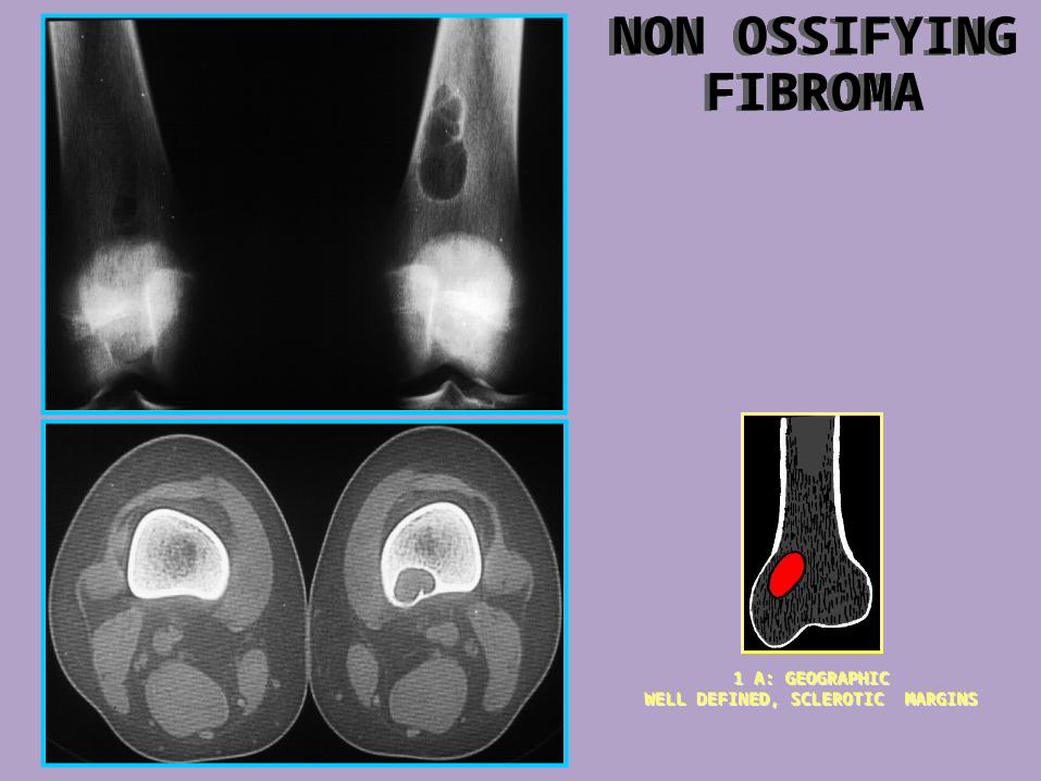
NON OSSIFYINGFIBROMA
NON OSSIFYINGFIBROMA
1 A: GEOGRAPHICWELL DEFINED, SCLEROTIC MARGINS
1 A: GEOGRAPHICWELL DEFINED, SCLEROTIC MARGINS

NON OSSIFYINGFIBROMA
NON OSSIFYINGFIBROMA
1 A: GEOGRAPHICWELL DEFINED, SCLEROTIC MARGINS
1 A: GEOGRAPHICWELL DEFINED, SCLEROTIC MARGINS
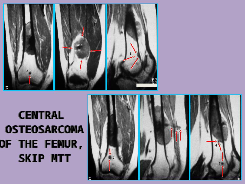
CENTRAL OSTEOSARCOMAOF THE FEMUR,
SKIP MTT
CENTRAL OSTEOSARCOMAOF THE FEMUR,
SKIP MTT
1
22
33
22
22
44
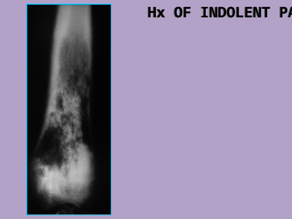
Hx OF INDOLENT PAIN Hx OF INDOLENT PAIN
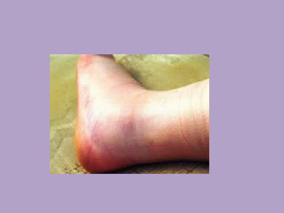
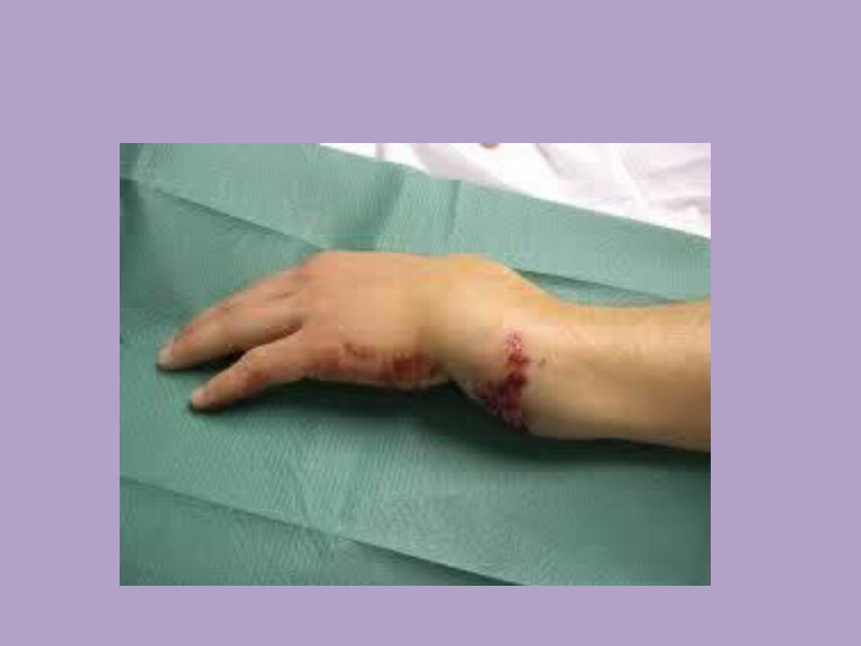
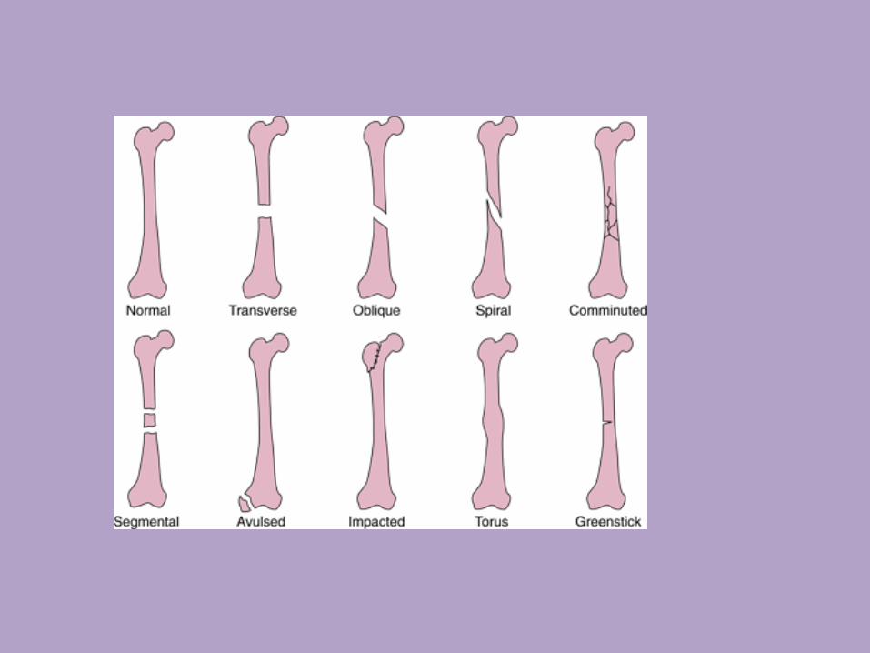
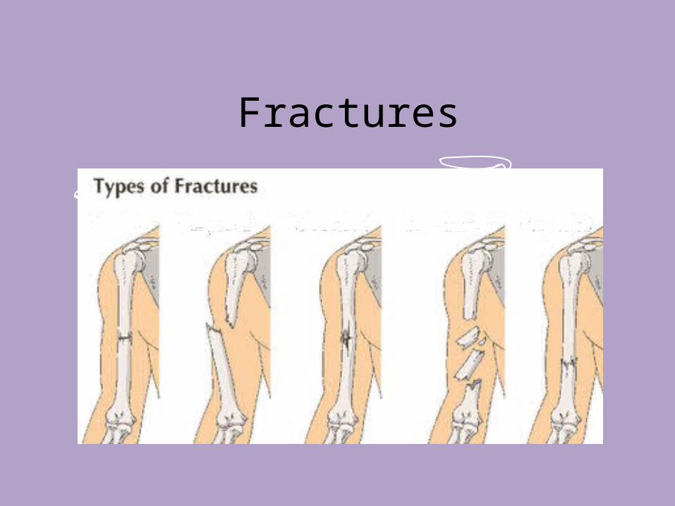
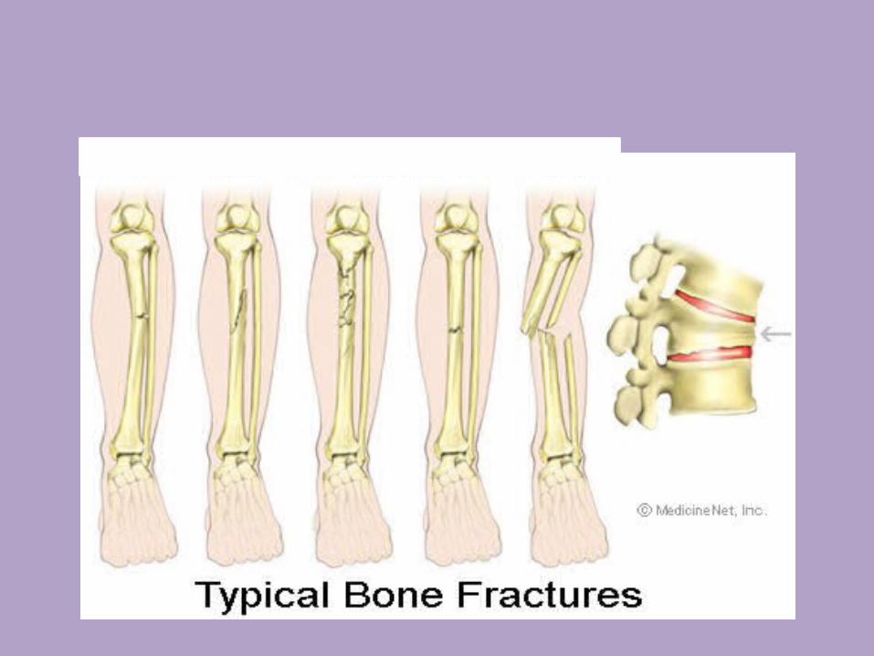
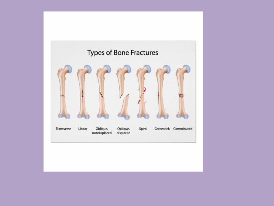
Fractures