Embed Size (px)
Citation preview

Loss of T Regulatory Cell Suppression following Signalingthrough Glucocorticoid-induced Tumor Necrosis Receptor(GITR) Is Dependent on c-Jun N-terminal Kinase Activation*
Received for publication, October 25, 2011, and in revised form, February 23, 2012 Published, JBC Papers in Press, March 29, 2012, DOI 10.1074/jbc.M111.316943
Anthony Joetham, Hiroshi Ohnishi, Masakazu Okamoto, Katsuyuki Takeda, Michaela Schedel, Joanne Domenico,Azzeddine Dakhama, and Erwin W. Gelfand1
From the Division of Cell Biology, Department of Pediatrics, National Jewish Health, Denver, Colorado 80206
Background: Ligation of the glucocorticoid-induced tumor necrosis (TNF) receptor (GITR) regulates T suppressor cellactivity.Results: Regulation of JNK phosphorylation following GITR ligation plays a central role in the suppressive activity of Tregulatory cells.Conclusion: Inhibition of JNK phosphorylation modulates T regulatory effector cell function in vitro and in vivo.Significance: Identification of JNK phosphorylation as a regulator of T suppressor cell function.
Naturally occurring Foxp3�CD4�CD25� T regulatory cell(nTreg)-mediated suppression of lung allergic responses isabrogated following ligation of glucocorticoid-induced tumornecrosis receptor (GITR) family-related protein. In vitro stimu-lation of nTregs with GITR ligand increased phosphorylation ofc-Jun N-terminal kinase (JNK) but not extracellular signal-reg-ulated protein kinase (ERK) or p38 MAPK. SP600125, a knownJNK inhibitor, prevented GITR-mediated phosphorylation ofJNK. Activation of JNK was associated with increases in theupstream mitogen-activated protein kinase kinase 7 (MKK7)and the downstream transcription factor NF-��. Phosphory-lated c-Jun (p-c-Jun), indicative of the activation of JNK, wasdetected in the immunoprecipitates of nTregs from wild-typebut not JNK- or GITR-deficient mice. Treatment with an inhib-itor of JNK phosphorylation resulted in complete reversal of allGITR-induced changes in nTreg phenotype and function, withfull restoration of suppression of in vivo lung allergic responsesand in vitro proliferation of activated CD4�CD25� T cells.Thus, regulation of JNK phosphorylation plays a central role inT regulatory cell function with therapeutic implications for thetreatment of asthma and autoimmune diseases.
Glucocorticoid-induced tumor necrosis factor receptor(GITR)2 family-related protein, also know as TNFRSF18, wasfirst described in mice as a dexamethasone-inducible moleculein T cells (1), and subsequently the human equivalent wasdefined (2). GITR is a type 1 transmembrane protein with anextracellular N terminus and cytoplasmic C terminus, and itshares significant homology in the C-terminal domain with
other members of the TNF receptor family, including 4-1BB,CD27, CD40, and OX40 (1, 2). Signaling cascades triggeredthrough members of the TNF receptor superfamily influencemany physiologic and pathologic immune responses by differ-entially regulating proliferation, differentiation, survival, andfunctions of cells in both the innate and adaptive immune sys-tems (3). Specifically in the lung, GITRwas shown to play a rolein acute inflammation (4) and airway hyper-responsiveness(AHR) (5). Constitutive expression and function of GITR inCD4�CD25� T regulatory cells (Tregs) have been described (6,7). Stimulation of GITR on T regulatory cells (nTregs) by eitherGITR ligand (GITRL) or an agonistic antibody (DTA-1) abol-ished suppression of allergen-induced lung allergic responses(8), in contrast to rendering effector cells resistant to the sup-pressive activities of Tregs (9–11).In both humans and experimental animal models, allergic
asthma is an inflammatory disease of the airways characterizedby an increase in AHR, airway inflammation, Th2 cytokineskewing, goblet cell metaplasia, excessive mucus production,elevated antigen-specific IgE, and structural remodeling of theairways. Increasingly, nTregs have been shown to be importantin modifying the development and outcome of lung allergicdiseases (12, 13) through both antigen-specific (14) and anti-gen-nonspecific mechanisms (15). However, the phenotypicand functional stability of nTregs has been shown to depend ona number of factors, including the expression of the key tran-scription factor, Foxp3 (16). Mutations of Foxp3 are associatedwith multiorgan autoimmune disease in Scurfy mice (17) andimmune dysregulation, polyendocrinopathy, enteropathy, andX-linked syndrome in humans (18). Cytokines such as IL-6 (19)and the expression of CD8 have also been shown to alter nTregfunction (13, 15). Importantly, in the absence and/or interfer-ence with MHC-I/CD8 interactions, regulatory functions weresubverted, resulting in the loss of suppression. Indeed, underthese conditions nTregs were converted into pathogenic effec-tor cells, enhancing lung allergic responses in sensitized andchallengedmice (8). Furthermore, themaintenance of suppres-sive activities in peripheral tissues and the regulation of endog-
* This work was supported, in whole or in part, by National Institutes of HealthGrants AI-77609, HL-36577, and HL-61005.
1 To whom correspondence should be addressed: Dept. of Pediatrics,National Jewish Health, 1400 Jackson St., Denver, CO 80206. Tel.: 303-398-1196; Fax: 303-270-2105; E-mail: [email protected].
2 The abbreviations used are: GITR, glucocorticoid-induced tumor necrosisreceptor; AHR, airway hyper-responsiveness; Treg, T regulatory cell; nTreg,naturally occurring Treg; GITRL, GITR ligand; OVA, ovalbumin; PE, phyco-erythrin; BAL, bronchoalveolar lavage.
THE JOURNAL OF BIOLOGICAL CHEMISTRY VOL. 287, NO. 21, pp. 17100 –17108, May 18, 2012© 2012 by The American Society for Biochemistry and Molecular Biology, Inc. Published in the U.S.A.
17100 JOURNAL OF BIOLOGICAL CHEMISTRY VOLUME 287 • NUMBER 21 • MAY 18, 2012
by guest on August 20, 2019
http://ww
w.jbc.org/
Dow
nloaded from

enous IL-6 production by nTregs were shown to be dependenton the presence of CD8� T cells (19). Conversion of Tregs topathogenic effector cells has now been described in other ani-mal models (20, 21).In contrast to earlier studies (6, 7), host GITRL was demon-
strated to effectively abrogate suppression in vivo, associatedwith the pathogenic conversion of nTregs (8). Moreover,enhancement of lung allergic responses was prevented by invivo administration of anti-GITRL antibody prior to the trans-fer of nTregs, identifying the importance of GITRL-GITR sig-naling in controlling the phenotype of these cells (8). Althoughvarious signaling cascades through GITR have been describedin T cells (22–24) and macrophages (25), there are little or nodata linking GITR signal transduction pathways to the loss ofsuppression and phenotypic conversion of nTregs.In this study, we defined the signaling pathways activated
throughGITR in nTregs in vitro and in vivo and determined theimpact of interfering with activation on the expression of sur-face receptors, production of immunosuppressive cytokines,suppression of proliferation of activated T cells, and the conse-quences in the airways of allergen-sensitized and challengedmice.
EXPERIMENTAL PROCEDURES
Animals—Pathogen-free, 6–8-week-old female C57BL/6and JNK�/� mice were obtained from The Jackson Laboratory(Bar Harbor, ME). GITR�/� mice were kindly provided by Dr.Carlo Riccardi (Perugia, Italy). All mice were maintained on anOVA-free diet, and all protocols were approved by the Institu-tional Animal Care and Use Committee of National JewishHealth.Sensitization and Challenge—Sensitization was carried out
by intraperitoneal injection of 20�g ofOVA (Sigma) emulsifiedin 2.25 mg of alum hydroxide (AlumImject; Pierce) in a totalvolume of 100 �l on days 1 and 14. Sensitized and challengedmice, denoted OVA/OVA, and nonsensitized but challengedlittermates (PBS/OVA) received aerosol challenges for 20 mineach day on 3 consecutive days (days 26, 27, and 28) with 1%OVA in PBS using an ultrasonic nebulizer (Omron, VernonHills, IL) (10).Measurement of AirwayResponsiveness—Airway responsive-
ness was assessed 48 h following the last challenge as a changein airway function to increasing concentrations of aerosolizedmethacholine administered for 10 s (60 breaths/min, 500 �l oftidal volume). Lung resistance was continuously computed(Labview, National Instruments, TX) by fitting flow, volume,and pressure to an equation of motion. Maximum values oflung resistance were taken and expressed as the percent changefrom base line following PBS aerosol.Bronchoalveolar Lavage—Immediately following measure-
ment of AHR, lungs were lavaged. Total leukocyte numberswere counted (Coulter Counter, Coulter Corp., Hialeah, FL),and differential cell countswere performed in a blindedmannerunder light microscopy by counting at least 200 cells on cyto-centrifuged preparations (Cytospin 2; Cytospin, Shandon Ltd.,Runcorn, Cheshire, UK) stained with Leukostat (Fisher Diag-nostics,Middletown, VA) and differentiated by standard hema-tological procedures.
Cell Preparation and Culture—CD4�CD25� and CD4�
CD25� T cells from naive C57BL/6, JNK�/�, and GITR�/�
donors were isolated by collagenase digestion from lungs andenriched using nylon wool columns as described previously (10).Lymphocytes were further purified by CD4�CD25� regulatory TcellMACSbeads (MiltenyiBiotec, Bergisch-Gladbach,Germany),resulting in a purity of �95% CD4�CD25� cells and by sortingon MoFlo (DakoCytomation, Fort Collins, CO) �99% ofCD4�CD25� T cells.
Cells were washed, counted, and resuspended to a final con-centration of 4 � 106 cells per ml in RPMI 1640 (MediatechCelgro,Manassas, VA) tissue culturemedium, containing heat-inactivated fetal calf serum (FCS 10%; Sigma), L-glutamine (5mM), �-mercaptoethanol (2 mM), Hepes buffer (15 mM), peni-cillin (100 units/ml), and streptomycin (100 �g/ml) (all fromInvitrogen).In Vitro Treatment and Adoptive Transfer—Isolated CD4�
CD25� T cells were pretreated with JNKi (SP600125) or inac-tive JNKi alone (50 �M, all fromCalbiochem) and prior to stim-ulationwithGITRL (1�g/ml, R&DSystems,Minneapolis,MN)in complete medium for assessing the in vitro production ofcytokines in the supernatants, protein extraction for quantify-ing kinase phosphorylation by Western blotting, and intratra-cheal adoptive transfer of 5 � 105 nTregs in 50 �l of PBS intosensitized recipients prior to allergen challenge.Measurement of Cytokine Levels—Cytokine levels in the BAL
fluid and supernatants of in vitro cultured lung cells weremeas-ured byELISA (IL-4, IL-5, IL-10, IL-13, and IFN-� (eBioscience,San Diego)), and TGF-� (Pharmingen). ELISAs were per-formed according to the manufacturers’ directions. The limitsof detection were 4 pg/ml for IL-4 and IL-5, 10 pg/ml for IL-10and IFN-�, 8 pg/ml for IL-13, and 6 pg/ml for TGF-�.FACS Analysis—Isolated cells, following preincubation with
naive mouse serum in staining buffer (PBS, 2% FCS, 0.2%sodium azide), were labeled with the following conjugated anti-bodies purchased from Pharmingen: anti-CD3 FITC, PE,PerCP, and allophycocyanin (17A2); anti-CD4 FITC, PE,PerCP, andAPC (L3T4); anti-CD25 FITC (7D4) and PE (PC61);anti-GITR FITC, PE, and APC (DTA-1); CD152 FITC and PEAPC (9H10). For intracellular staining, cells were stimulatedwith phorbol 12-myristate 13-acetate (100 ng/ml) and ionomy-cin (2�g/ml, Sigma) in completemedium overnight and for 6 hin the presence of brefeldin A (10 �g/ml, Sigma). Cells werefixed with 4% formaldehyde in PBS, permeabilized in 0.5% sap-onin, and stained with anti-IL-10 PE and APC (JES5-16E3);IFN-� PE and APC (XMG1.2); and Foxp3 PE and TGF-� (A75-3.1) (eBioscience). Fluorochrome (FITC, PE, PerCPAPC)-la-beled isotype-matched control antibodies were used for back-ground fluorescence staining. Staining was analyzed on aFACSCalibur flow cytometer (Pharmingen) using CellQuestPro software. Fluorescence intensity was compared with cellsstained with the corresponding labeled isotype-matchedcontrols.Real Time PCR—Total RNA was isolated using the Qiagen
RNeasy mini kit (Valencia, CA) according to the manufactur-er’s instructions. Total RNAwas reverse-transcribed using Bio-Rad iScript cDNA synthesis kit. TaqMan primers and probesformouse Foxp3 andGAPDHwere obtained fromApplied Bio-
GITR Activates JNK and Loss of T Cell Suppression
MAY 18, 2012 • VOLUME 287 • NUMBER 21 JOURNAL OF BIOLOGICAL CHEMISTRY 17101
by guest on August 20, 2019
http://ww
w.jbc.org/
Dow
nloaded from

systems (Carlsbad, CA). Real time PCR was performed usingABI Prism 7000 sequence detection system. Relative amountsof cDNA were normalized by subtracting the threshold cycle(Ct) of internal GAPDH reference gene to target gene generat-ing a�Ct value. RelativemRNA expression of specific gene wasobtained by using the formula 2ˆ (��Ct).Western Blotting—Cells (5 � 106) were lysed with RIPA
buffer containing protease inhibitors and phosphatase inhibi-tors (Sigma) on ice. Lysates were resolved by SDS-PAGE andtransferred to nitrocellulose membranes. Blotted membraneswere blocked in TBST buffer (25 mM Tris, pH 8.0, 125 mM
NaCl, 0.025% Tween 20) containing 2% BSA (Promega, Madi-son,WI) and then incubated overnight at 4 °CwithTBSTbuffercontaining 2% BSA with anti-phospho-ERK1/2, anti-phospho-p38MAPK, anti-phospho-JNK (Cell Signaling), anti-ERK, anti-p38 MAPK, anti-JNK, anti-MAPK kinases 4 and 7, anti-c-Jun,anti-p-c-Jun, anti-p50, anti-p65, anti-c-Rel mAbs, or anti-I�B-� (Santa Cruz Biotechnology, Santa Cruz, CA or Cell Sig-naling). Specific proteins were detected by a chemilumines-cence method using a horseradish peroxidase-conjugatedanti-rabbit, anti-goat, or anti mouse IgG Ab (Amersham Bio-sciences). Immunoreactive bands were quantified by densito-metric quantification of autoradiographs using ImageJ(National Institutes of Health, Bethesda) and expressed as rel-ative phosphorylated kinase to total kinase or �-actin normal-ized to the unstimulated, untreated cells.JNK Activation—The assay for JNK activity was performed
according to the manufacturer’s directions (SAPK/JNK kinaseassay, Cell Signaling, Danvers, MA). Briefly, similar amounts ofcell lysate from the different cell groups were incubated withthe recommended amount of immobilized c-Jun fusion proteinovernight. Mixtures were washed extensively with 1� cell lysisbuffer and the kinase buffer. SAPK phosphorylation of c-Junsubstratewas carried out for 30min at 30 °C by adding the samevolume of 1� kinase buffer supplemented with 200 �M ATP.The reaction was terminated with 25 �l of 3� SDS. The super-natantswere then prepared for standardWestern immunoblot-ting probing for p-c-Jun.Proliferation Assays—IsolatedWT CD4�CD25� (2.5 � 104)
cells were first activated with bound anti-CD3 (2 �g/ml, 17A2)and soluble anti-CD28 (1 �g/ml, 37.51; both from eBioscience)cultured with or without CD4�CD25� T cells treated withactive (SP600125) or inactive JNKi (25 �M, both from Calbi-ochem) and then stimulated with GITRL (1 �g/ml, R&D Sys-tems) at a 1:1 ratio in complete medium. GITR-stimulatedCD4�CD25� T cells were carboxyfluorescein diacetate succin-imidyl ester-labeled for dilution assay according to the manu-facturer’s instructions (eBioscience). Cultures were pulsedduring the final 6 h with 1 �Ci/well thymidine, and cell prolif-eration was determined as counts/min.Histochemistry—Lungs were fixed by inflation (1 ml) and
immersion in 10% formalin. Cells containing eosinophilicmajor basic protein were identified by immunohistochemicalstaining as described previously using rabbit anti-mouse majorbasic protein (kindly provided by Dr. J. J. Lee, Mayo Clinic,Scottsdale, AZ). The slides were examined in a blinded fashionwith a Nikon light microscope. Numbers of eosinophils in thetissues were evaluated with ImageJ counting 6–8 different
fields per animal. For detection of mucus-containing cells informalin-fixed airway tissue, sections were stained with peri-odic acid-Schiff.Statistical Analysis—Analysis of variance was used to deter-
mine statistical significance. Comparisons for all pairs wereperformed by the Tukey-Kramer highest significant differencetest. The p values for significance were set to 0.05. Values for allmeasurements were expressed as the means � S.E.
RESULTS
GITR Stimulation Activates c-Jun N-terminal Kinase—Inmammalian cells, three main families of the MAPKs, includingextracellular signal-regulated kinase (ERK), c-Jun N-terminalkinase (JNK), and p38, have been implicated in developingimmune responses, and individualMAPKs are activated by dif-ferent stimuli (26). Although p38 (27), ERK (28), andTAK1 (29)have been implicated in the development and function ofTregs,the involvement of MAPK in GITR-mediated loss of suppres-sion and phenotypic conversion has not been described. Weinvestigated the activation of the MAPK pathways in nTregsfollowing GITR stimulation with different concentrations ofGITRL (Fig. 1, A–C). Compared with nonstimulated cells,GITRL significantly increased the phosphorylation of JNKwithout evidence for activation of ERKor p38, as determined bydensitometric quantification of immunoreactive bands onautoradiographs. Total JNK, ERK, and p38 were unchangedunder all conditions. In contrast to CD4�CD25� T cells,CD4�CD25� T cells, which express lower levels of GITR (8),demonstrated no increases in GITRL-stimulated increases inp-JNK (Fig. 1D).Similar to other MAPKs, full activation of JNK requires dual
phosphorylation on Thr-183 and Tyr-184 of the tripeptidemotif (Thr-Xaa-Tyr) (30), and specific activity of JNK can beblocked by SP600125, an anthrapyrazolone inhibitor (31). In ananimal model, treatment with SP600125 was shown to reduceairway inflammation and levels of OVA-IgE in the serum ofsensitized and challenged mice (32). The effect of treatmentwith SP600125 on the phosphorylation of JNK following GITRstimulation was investigated. Pretreatment of nTregs with theactive but not inactive JNK inhibitor (JNKi) decreased GITRL-induced phosphorylation of JNK (Fig. 2A).The specificity of activation of JNK following GITR ligation
was confirmed by comparing nTregs fromwild-type (WT)withnTregs from JNK�/� or GITR�/� mice. JNK activity detectedas phosphorylation of c-Jun (p-c-Jun) was only detected in theimmunoprecipitates fromWTbut not the deficient (JNK�/� orGITR�/�) or JNKi-treated nTregs (Fig. 2B).We next investigated which of the upstream MAPK kinases
(MKK) were involved in GITR-mediated activation of JNK atthe time point associated with changes in levels of cytokines inthe supernatants of in vitro cultured cells. To date, only MKK4(SEK1) and MKK7 have been implicated in the activation ofJNK (26, 33, 34). Following the in vitro addition of GITRL,increases in protein levels of MKK7 but not MKK4 weredetected (Fig. 3).Upon activation, JNK phosphorylates a number of down-
stream targets, including c-Jun, which heterodimerizes withFos to form the transcription factor activator protein 1 (35). To
GITR Activates JNK and Loss of T Cell Suppression
17102 JOURNAL OF BIOLOGICAL CHEMISTRY VOLUME 287 • NUMBER 21 • MAY 18, 2012
by guest on August 20, 2019
http://ww
w.jbc.org/
Dow
nloaded from

further define the role of JNK in the signaling cascade throughGITR, total protein and phosphorylation of c-Jun were deter-mined. Both total protein and phosphorylation of c-Junincreased following GITR stimulation, consistent with theresults detected in immunoprecipitates. Correspondingly,increased c-Jun was detected in the nuclear extracts of stimu-lated compared with nonstimulated nTregs.Consistent with a previous report (9), increases in activation
of subunits of NF-�B, including c-Rel, p65, and to a lesserdegree p50, were detected following GITR stimulation, indi-cated by the increases in nuclear translocation of these subunitproteins (Fig. 3). Activation ofNF-�B can also be determined byassessing the levels of the protein inhibitor of NF-�B (I��) (22).
In contrast to nonstimulated cells, GITR-stimulated cells hadless I�� protein in the cytoplasm.GITRL Stimulation Induces Changes in the Phenotype, Func-
tion, and Cytokine Production of nTregs and Are Prevented byInhibition of JNK Activation—Naturally occurring CD4�
CD25� nTregs have been characterized by the expression ofCD25, CTLA-4, and GITR (6, 7, 12, 36), and under certainexperimental conditions, decreased expression of these recep-tors was shown to associate with loss or attenuation of suppres-sive activity (19–21, 36). The effects of GITR ligation on theexpression of surface markers were assessed by flow cytometryin the presence or absence of the JNKi. Compared with non-stimulated cells, nTregs treated with GITRL expressed lower
FIGURE 1. Effect of GITR ligation by GITRL on MAPK phosphorylation in nTregs. nTregs were isolated from the lungs of C57BL/6 mice and incubated withdifferent concentrations of GITRL for varying lengths of time. Data illustrated are derived from densitometric quantification of autoradiographs using ImageJ.Levels of relative phosphorylation are derived from base-line values (nonstimulated, time 0) of the ratio of p-ERK to total ERK. A–C, dose and time course ofGITR-mediated phosphorylation of JNK but not ERK or p38. D, GITR-mediated phosphorylation of JNK in CD4�CD25� T cells. PI, phorbol/ionomycin. Med,medium. Results are from three independent experiments carried out in duplicate. **, p � 0.01; *, p � 0.05.
FIGURE 2. Effect of JNKi (SP600125) on phosphorylation of JNK and JNK activity in immunoprecipitates. JNKi was added 1–2 h prior to GITRL. A, doseeffect of JNKi on GITR-induced phosphorylation of JNK. B, JNK activity is detected in immunoprecipitates of GITR-stimulated, untreated but not JNKi-treated,nTregs from WT, and GITR�/�, or JNK�/� mice. Results are from three independent experiments carried out in duplicate. **, p � 0.01; *, p � 0.05. PI,phorbol/ionomycin.
GITR Activates JNK and Loss of T Cell Suppression
MAY 18, 2012 • VOLUME 287 • NUMBER 21 JOURNAL OF BIOLOGICAL CHEMISTRY 17103
by guest on August 20, 2019
http://ww
w.jbc.org/
Dow
nloaded from

mean fluorescence intensities of CD25, GITR, and CTLA-4(Fig. 4A). In contrast, treatment of nTregs with the active JNKiprevented the down-regulation of expression of these recep-tors, retaining the regulatory phenotype.Previously, nTregs isolated from naive BALB/c and C57BL/6
micewere shown to produce the immunosuppressive cytokinesIL-10 and TGF-� (12, 13, 15). We determined the conse-quences of sustained GITR ligation over 72 h on cytokine pro-duction and the impact of adding the JNKi. Despite the pheno-typic changes induced following the shorter incubation,intracellular staining of Foxp3 and immunocytokines (Fig. 4A),similar to levels detected in the supernatants of in vitro cultures(Fig. 4B), did not change to any appreciable degree. In contrast,significant changes in cytokine profile were detected followingthe longer stimulation. Compared with nonstimulated nTregs,GITRL-stimulated nTregs released lower amounts of IL-10 andTGF-� in the culture supernatants (Fig. 4C), similar to GITRL-treated cells in the presence of the inactive inhibitor (data notshown). In contrast, treatment with the active JNKi preventedthese decreases, maintaining the levels seen in untreated cells.In parallel, similar to previous results showing increased IL-13production when nTregs were shown to have reduced IL-10and TGF-� production (8), levels of IL-13 were increased incultures of GITRL-treated nTregs but not those that includedthe active JNKi. Furthermore, decreases in Foxp3 protein (Fig.4D) and mRNA (Fig. 4E) expression were associated with thechanges in cytokine production (Fig. 4C).Consistent with others (6, 7), we reported that the prolifera-
tion of CD4�CD25� T cells activated with anti-CD3 and anti-CD28 was inhibited by nTregs (19) and that treatment ofnTregs with either the GITRL or agonistic anti-GITR antibody(DTA-1) abolished their suppressive activity. Here, we con-firmed that in vitro stimulation of nTregs with GITRL abol-ished the suppression of CD4�CD25� T cell proliferationtriggered by anti-CD3/anti-CD28 (Fig. 5A). The loss of sup-pression, in contrast to a previous report (40), was not associ-atedwithGITR-induced proliferation of nTregs (Fig. 5B).How-ever, suppression was maintained in the presence of the active,
but not inactive, JNKi. This was not due to any toxic effects ofthe inhibitor, as washing out of the inhibitor after a similarincubation period was associated with full proliferative activityof the cells (data not shown).Taken together, these in vitro studies demonstrated that liga-
tion of GITR had profound effects on kinase phosphorylation,surface receptor expression, suppressive activity, and cytokineproduction, and all of these effects were reversed in the pres-ence of the active JNKi.Prevention of JNKActivation in Vivo Is Associated withMainte-
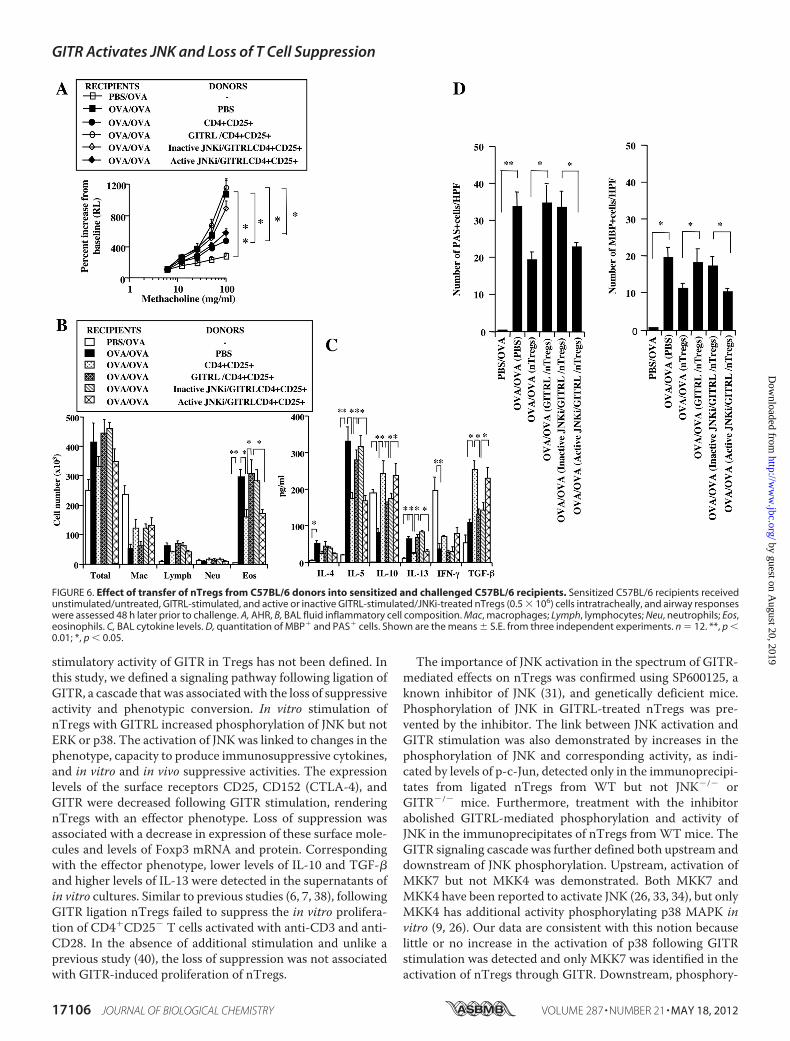
nance of Suppression in an Experimental Model of Asthma—Following adoptive transfer, nTregs suppressed the develop-ment of allergen-induced lung allergic responses in sensitizedand challenged mice (8, 12, 13, 15, 19). This suppression medi-ated by nTregs was abolished by the in vitro treatment ofnTregs with GITRL or agonistic antibody when transferredafter sensitization but prior to challenge (8). We determinedwhether prevention of JNK activation affected the activity ofnTregs in sensitized and challenged mice. Prior to transfer, cellviability of GITRL- and JNK-treated cells exceeded 85–90%.Transfer of nTregs treated with GITRL, as compared with non-treated cells, failed to suppressAHR (Fig. 6A). In contrast, treat-ment of nTregs with the active but not inactive JNKi restoredthe suppression of AHR. Associated with the increases in AHR,the numbers of airway eosinophils were significantly increasedin sensitized and challenged mice as shown in Fig. 6B. Airwayeosinophilia was significantly reduced in recipients of non-stimulated nTregs and GITRL-stimulated nTregs treated withthe active inhibitor but was maintained in the GITRL-stimu-lated nTregs treated with the inactive inhibitor. Similar effectsof treatment with the JNKi were also observed in BALB/c mice(data not shown).Following sensitization and challenge with allergen, Th2
cytokine levels were increased in BAL fluid. Significantincreases in levels of IL-4, IL-5, and IL-13 were detected, andlevels of IL-10 and IFN-� were decreased; levels of TGF-� weremodestly increased (Fig. 6C). Intratracheal transfer of nonli-gated nTregs decreased the levels of IL-5 and IL-13, whereasIL-10 and TGF-� levels were increased. Little change was seenin levels of IL-4 and IFN-�. Transfer of GITRL-stimulatednTregs, untreated or treated with the inactive JNKi, failed tosignificantly reduce the levels of IL-5 and IL-13, and the levels ofIL-4, IL-10, TGF-�, and IFN-� were similar to the levels insensitized and challenged mice that did not receive transferredcells (Fig. 6C). In contrast, treatment of GITRL-stimulatednTregs with the active JNKi reversed the effects of GITR stim-ulation, with restoration of levels of IL-10 and TGF-� anddecreased levels of IL-5 and IL-13.Lung histopathology paralleled the changes in inflammatory
cell composition and numbers and levels of cytokines in BALfluid. The lung tissue from sensitized and challenged mice thatreceived GITRL-stimulated nTregs, untreated or treated withinactive JNKi, similar to mice given PBS, showed a significantaccumulation of eosinophils, mucus hyperproduction, andgoblet cell metaplasia (Fig. 6D). In contrast, mice that receivednon-GITRL-stimulated or nTregs that were GITRL-stimulatedbut treated with active JNKi exhibited significantly less lunghistopathology.
FIGURE 3. MAPK cascade in JNK activation. GITR ligation (1 �g) for 72 hincreases levels of MKK7, c-Jun, p-c-Jun, and subunits of NF-�B in cytoplasmicand nuclear cell extracts, and decreases levels of NF-�B inhibitor (I�B) in cyto-plasmic extracts. Data are representative of three independent and similarexperiments. PI, phorbol/ionomycin; Med, medium.
GITR Activates JNK and Loss of T Cell Suppression
17104 JOURNAL OF BIOLOGICAL CHEMISTRY VOLUME 287 • NUMBER 21 • MAY 18, 2012
by guest on August 20, 2019
http://ww
w.jbc.org/
Dow
nloaded from

DISCUSSION
Naturally occurring CD4�CD25�Foxp3� T cells (nTregs),although comprising only a small subset of T cells derived in thethymus (37), appear essential to the maintenance of immuno-logical tolerance. In the absence of functional Foxp3, the keytranscription regulator for Treg development and function(38), severe and life-threatening autoimmune and allergic dis-eases develop (17, 18). These regulatory T cells have also been
implicated in the maintenance of immune homeostasis in theairways as they have been shown to regulate allergen-inducedAHR and inflammation, at least in part, through the actions ofIL-10 and TGF-� (12), and in an antigen-nonspecific manner(15). In the lungs of both BALB/c and C57BL/6 mice, the acti-vation and expression of this suppressive phenotype appeareddependent on the interaction of MHC class I on nTregs withCD8; inhibition or interference with this interaction led to theloss of nTreg suppressive activity (13, 15). A role for CD8 inmaintaining suppressive activity in the periphery and regulat-ing the production of endogenous IL-6 in Tregs was recentlydemonstrated (19). Similar to other reports (6, 7), in theabsence of CD8-expressing cells, activation of nTregs throughGITR in the lungs of recipient mice was shown to be sufficientto abrogate suppression and convert these cells to pathogeniceffector cells resulting in the enhancement of lung allergicresponses (8). Significantly, GITRL expression in recipientsappeared to provide the necessary signals to attenuate suppres-sion, as administration of anti-GITRL antibody preventedrather than enhanced lung allergic responses in associationwith little increase in levels of IL-13 and maintenance of basallevels of IL-10 andTGF-� known to be essential for suppressiveactivity (12, 39).In this study, we investigated the molecular events following
stimulation of nTregs throughGITR. Although the roles of p38(27), ERK (28), and TAK1 (29) have been implicated in thedevelopment and function of Tregs, their involvement in the
FIGURE 4. Effect of GITR ligation on expression of surface markers, production of cytokines at different time course in the presence and absence ofJNKi (50 �M), and expression of Foxp3. A, FACS analysis of nTregs following overnight GITR ligation; *, p � 0.05 compared with expression in medium control.B, cytokine levels in supernatants (overnight); C, cytokine levels in supernatants (72 h). D, expression of Foxp3 protein following GITRL stimulation (72 h).E, expression of Foxp3 mRNA following GITRL stimulation. Results are from three independent experiments carried out in duplicate. Med, medium. **, p � 0.01;*, p � 0.05.
FIGURE 5. Effect of GITR ligation on suppression of in vitro proliferation ofactivated CD4CD25� T cell in the presence and absence of JNKi (50 �M).A, GITRL stimulation abrogates suppression of CD4�CD25� T cell prolifera-tion, and treatment of nTregs with JNKi restores suppression. B, absence ofcarboxyfluorescein diacetate succinimidyl ester dilution in GITR-stimulatedCD4�CD25� T cells. The means � S.E. from three independent experimentswere carried out in triplicate. *, p � 0.05.
GITR Activates JNK and Loss of T Cell Suppression
MAY 18, 2012 • VOLUME 287 • NUMBER 21 JOURNAL OF BIOLOGICAL CHEMISTRY 17105
by guest on August 20, 2019
http://ww
w.jbc.org/
Dow
nloaded from
stimulatory activity of GITR in Tregs has not been defined. Inthis study, we defined a signaling pathway following ligation ofGITR, a cascade that was associatedwith the loss of suppressiveactivity and phenotypic conversion. In vitro stimulation ofnTregs with GITRL increased phosphorylation of JNK but notERK or p38. The activation of JNK was linked to changes in thephenotype, capacity to produce immunosuppressive cytokines,and in vitro and in vivo suppressive activities. The expressionlevels of the surface receptors CD25, CD152 (CTLA-4), andGITR were decreased following GITR stimulation, renderingnTregs with an effector phenotype. Loss of suppression wasassociated with a decrease in expression of these surface mole-cules and levels of Foxp3 mRNA and protein. Correspondingwith the effector phenotype, lower levels of IL-10 and TGF-�and higher levels of IL-13 were detected in the supernatants ofin vitro cultures. Similar to previous studies (6, 7, 38), followingGITR ligation nTregs failed to suppress the in vitro prolifera-tion of CD4�CD25� T cells activated with anti-CD3 and anti-CD28. In the absence of additional stimulation and unlike aprevious study (40), the loss of suppression was not associatedwith GITR-induced proliferation of nTregs.
The importance of JNK activation in the spectrum of GITR-mediated effects on nTregs was confirmed using SP600125, aknown inhibitor of JNK (31), and genetically deficient mice.Phosphorylation of JNK in GITRL-treated nTregs was pre-vented by the inhibitor. The link between JNK activation andGITR stimulation was also demonstrated by increases in thephosphorylation of JNK and corresponding activity, as indi-cated by levels of p-c-Jun, detected only in the immunoprecipi-tates from ligated nTregs from WT but not JNK�/� orGITR�/� mice. Furthermore, treatment with the inhibitorabolished GITRL-mediated phosphorylation and activity ofJNK in the immunoprecipitates of nTregs fromWTmice. TheGITR signaling cascade was further defined both upstream anddownstream of JNK phosphorylation. Upstream, activation ofMKK7 but not MKK4 was demonstrated. Both MKK7 andMKK4 have been reported to activate JNK (26, 33, 34), but onlyMKK4 has additional activity phosphorylating p38 MAPK invitro (9, 26). Our data are consistent with this notion becauselittle or no increase in the activation of p38 following GITRstimulation was detected and only MKK7 was identified in theactivation of nTregs through GITR. Downstream, phosphory-
FIGURE 6. Effect of transfer of nTregs from C57BL/6 donors into sensitized and challenged C57BL/6 recipients. Sensitized C57BL/6 recipients receivedunstimulated/untreated, GITRL-stimulated, and active or inactive GITRL-stimulated/JNKi-treated nTregs (0.5 � 106) cells intratracheally, and airway responseswere assessed 48 h later prior to challenge. A, AHR, B, BAL fluid inflammatory cell composition. Mac, macrophages; Lymph, lymphocytes; Neu, neutrophils; Eos,eosinophils. C, BAL cytokine levels. D, quantitation of MBP� and PAS� cells. Shown are the means � S.E. from three independent experiments. n � 12. **, p �0.01; *, p � 0.05.
GITR Activates JNK and Loss of T Cell Suppression
17106 JOURNAL OF BIOLOGICAL CHEMISTRY VOLUME 287 • NUMBER 21 • MAY 18, 2012
by guest on August 20, 2019
http://ww
w.jbc.org/
Dow
nloaded from
lation of JNK was associated with increases in total c-Jun andp-c-Jun, a known downstream substrate of JNK (30, 31). Cor-responding with the increase in p-c-Jun, greater amounts ofc-Jun were also detected in the nucleus.The loss of nTreg-mediated suppression following GITR
ligation has been shown to be dependent on the activation ofNF-�B (9).We confirmed thatGITR ligation increased the acti-vation of subunits of NF-�B, including c-rel, p65, and to a lesserextent p50 in cytoplasmic and nuclear cell extracts. Interest-ingly, the activation of NF-�B coincided with divergent effectson the levels of proinflammatory and immunosuppressive cyto-kines. Multiple factors may be involved in these transcriptionalresponses, including selective phosphorylation and subsequentdegradation of NF-�B inhibitors or the result of differentialsubunit distribution, affinity, or number of binding sites en-gaged in these reactions.The importance of JNK activation in mediating the abroga-
tion of nTreg suppressive activity through GITR was also dem-onstrated in vivo in sensitized and challenged mice. As in pre-vious studies (12, 13, 15, 19), following sensitization andallergen challenge, mice developed a significant increase in air-way responsiveness to inhaled methacholine, eosinophilicinflammation, and Th2 skewing in cytokine production. Thesefindings were accompanied by changes in lung pathology, espe-cially eosinophil accumulation in the lung parenchyma andgoblet cell metaplasia and mucus hyperproduction. Consistentwith our previous report (8), GITR ligation of nTregs prior totransfer into sensitized and challenged mice eliminated thesuppression of lung allergic responses. However, the full sup-pression of all lung allergic responses and lung pathology couldbe restored when the GITR-ligated nTregs were treated withthe JNKi prior to transfer into sensitized recipients prior toairway allergen challenge.Taken together, the results demonstrate that stimulation of
nTregs throughGITR initiates a signaling cascade that involvesMKK7, JNK phosphorylation, c-Jun activation, and the activa-tion of NF-�B. Associated with the phosphorylation of JNKwere changes in nTreg surface marker expression, conversionof the cytokine profile from IL-10/TGF-� to IL-13 production,attenuation of in vitro suppressive activities, and in an experi-mental model of allergic asthma, elimination of the nTreg reg-ulatory activities on development of altered airway responsive-ness, eosinophilic airway inflammation, and Th2 cytokineproduction. Incubation of the GITRL-stimulated nTregs withan inhibitor of JNK activation resulted in the restoration ofnTreg activities and phenotype in vitro and in vivo. These find-ings demonstrate that targeting JNK phosphorylation can havea major influence on nTreg suppressive function.
Acknowledgments—We are indebted to Drs. Joseph Lucas, RobertaPelanda, Raul Torres, and Laurent Gapin for critical review of themanuscript; to Dr. Carlo Riccardi for providing the GITR-deficientmice, and Diana Nabighian for assistance in preparation of thismanuscript.
REFERENCES1. Nocentini, G., Giunchi, L., Ronchetti, S., Krausz, L. T., Bartoli, A.,Moraca,
R., Migliorati, G., and Riccardi, C. (1997) A new member of the tumor
necrosis factor/nerve growth factor receptor family inhibits T cell-recep-tor-induced apoptosis. Proc. Natl. Acad. Sci. U.S.A. 94, 6216–6221
2. Kwon, B., Yu, K. Y., Ni, J., Yu, G. L., Jang, I. K., Kim, Y. J., Xing, L., Liu, D.,Wang, S. X., and Kwon, B. S. (1999) Identification of a novel activation-inducible protein of the tumor necrosis factor receptor superfamily and itsligand. J. Biol. Chem. 274, 6056–6061
3. Watts, T. H. (2005) TNF/TNFR familymembers in costimulation of T cellresponses. Annu. Rev. Immunol. 23, 23–68
4. Cuzzocrea, S., Nocentini, G., Di Paola, R., Agostini, M., Mazzon, E.,Ronchetti, S., Crisafulli, C., Esposito, E., Caputi, A. P., and Riccardi, C.(2006) Proinflammatory role of glucocorticoid-induced TNF receptor-related gene in acute lung inflammation. J. Immunol. 177, 631–641
5. Motta, A. C., Vissers, J. L., Gras, R., Van Esch, B. C., Van Oosterhout, A. J.,and Nawijn, M. C. (2009) GITR signaling potentiates airway hyper-re-sponsiveness by enhancing Th2 cell activity in a mouse model of asthma.Respir. Res. 10, 93
6. Shimizu, J., Yamazaki, S., Takahashi, T., Ishida, Y., and Sakaguchi, S.(2002) Stimulation of CD25�CD4� regulatory T cells through GITRbreaks immunological self-tolerance. Nat. Immunol. 3, 135–142
7. McHugh, R. S., Whitters, M. J., Piccirillo, C. A., Young, D. A., Shevach,E. M., Collins, M., and Byrne, M. C. (2002) CD4�CD25� immunoregula-tory T cells. Gene expression analysis reveals a functional role for theglucocorticoid-induced TNF receptor. Immunity 16, 311–323
8. Joetham, A., Matsubara, S., Okamoto, M., Takeda, K., Miyahara, N.,Dakhama, A., and Gelfand, E. W. (2008) Plasticity of regulatory T cells.Subversion of suppressive function and conversion to enhancement oflung allergic responses. J. Immunol. 180, 7117–7124
9. Ji, H. B., Liao, G., Faubion,W.A., Abadía-Molina, A. C., Cozzo, C., Laroux,F. S., Caton, A., and Terhorst, C. (2004) Cutting Edge. The natural ligandfor glucocorticoid-induced TNF receptor-related protein abrogates regu-latory T cell suppression. J. Immunol. 172, 5823–5827
10. Stephens, G. L., McHugh, R. S., Whitters, M. J., Young, D. A., Luxenberg,D., Carreno, B. M., Collins, M., and Shevach, E. M. (2004) Engagement ofglucocorticoid-induced TNFR family-related receptor on effector T cellsby its ligand mediates resistance to suppression by CD4�CD25� T cells.J. Immunol. 173, 5008–5020
11. Cardona, I. D., Goleva, E.,Ou, L. S., and Leung,D. Y. (2006) Staphylococcalenterotoxin B inhibits regulatory T cells by inducing glucocorticoid-in-duced TNF receptor-related protein ligand on monocytes. J. Allergy Clin.Immunol. 117, 688–695
12. Joetham, A., Takeda, K., Takada, K., Taube, C., Miyahara, N., Matsubara,S., Matsubara, S., Koya, T., Rha, Y. H., Dakhama, A., and Gelfand, E. W.(2007) Naturally occurring lung CD4�CD25� T-cell regulation of airwayallergic responses depends on IL-10 induction of TGF-�. J. Immunol. 178,1433–1442
13. Joetham, A., Takeda, K., Miyahara, N., Matsubara, S., Ohnishi, H., Koya,T., Dakhama, A., and Gelfand, E. W. (2007) Functional activation of nat-urally occurring lung CD4�CD25� regulatory cells on lung allergic re-sponses requires CD8 and MHC I interaction. Proc. Natl. Acad. Sci. 104,15057–15062
14. Akbari, O., Freeman, G. J., Meyer, E. H., Greenfield, E. A., Chang, T. T.,Sharpe, A. H., Berry, G., DeKruyff, R. H., and Umetsu, D. T. (2002) Anti-gen-specific regulatory T cells develop via the ICOS-ICOSSL pathway andinhibit allergen-induced airway hyper-reactivity.Nat. Med. 8, 1024–1032
15. Joetham, A., Takeda, K., Okamoto, M., Taube, C., Matsuda, H., Dakhama,A., and Gelfand, E. W. (2009) Antigen specificity is not required for mod-ulation of lung allergic responses by naturally occurring regulatory T cells.J. Immunol. 183, 1821–1827
16. Fontenot, J. D., Rasmussen, J. P., Williams, L. M., Dooley, J. L., Farr, A. G.,and Rudensky, A. Y. (2005) Regulatory T cell lineage specification by theforkhead transcription factor Foxp3. Immunity 22, 329–341
17. Clark, L. B., Appleby,M.W., Brunkow,M. E.,Wilkinson, J. E., Ziegler, S. F.,and Ramsdell, F. (1999) Cellular and molecular characterization of thescurfy mouse mutant. J. Immunol. 162, 2546–2554
18. Bennett, C. L., Christie, J., Ramsdell, F., Brunkow, M. E., Ferguson, P. J.,Whitesell, L., Kelly, T. E., Saulsbury, F. T., Chance, P. F., and Ochs, H. D.(2001) The immune dysregulation, polyendocrinopathy, enteropathy, andX-linked syndrome are caused by mutations of Foxp3. Nat. Genet. 27,
GITR Activates JNK and Loss of T Cell Suppression
MAY 18, 2012 • VOLUME 287 • NUMBER 21 JOURNAL OF BIOLOGICAL CHEMISTRY 17107
by guest on August 20, 2019
http://ww
w.jbc.org/
Dow
nloaded from
20–2119. Joetham, A., Okamoto, M., Takeda, K., Schedel, M., Ohnishi, H.,
Dakhama, A., and Gelfand, E. W. (2011) CD8 regulates the endogenousproduction of IL-6 in naturally occurring T regulatory cells andmaintainstheir suppressive phenotype in allergic lung disease. J. Immunol. 186,113–120
20. Wan, Y. Y., and Flavell, R. A. (2007) Regulatory T-cell functions are sub-verted and converted owing to attenuated Foxp3 expression.Nature 445,766–770
21. Zhou, X., Bailey-Bucktrout, S. L., Jeker, L. T., Penaranda, C., Martínez-Llordella, M., Ashby, M., Nakayama, M., Rosenthal, W., and Bluestone,J. A. (2009) Instability of the transcription factor Foxp3 leads to generationof pathogenic memory T cells in vivo. Nat. Immunol. 10, 1000–1007
22. Esparza, E. M., and Arch, R. H. (2005) Glucocorticoid-induced TNF re-ceptor, a costimulatory receptor on naive and activated T cells, uses TNFreceptor-associated factor 2 in a novel fashion as an inhibitor of NF-�Bactivation. J. Immunol. 174, 7875–7882
23. Kanamaru, F., Youngnak, P., Hashiguchi, M., Nishioka, T., Takahashi, T.,Sakaguchi, S., Ishikawa, I., and Azuma, M. (2004) Costimulation via glu-cocorticoid-induced TNF receptor in both conventional and CD25� reg-ulatory CD4� T cells. J. Immunol. 172, 7306–7314
24. Ronchetti, S., Nocentini, G., Bianchini, R., Krausz, L. T.,Migliorati, G., andRiccardi, C. (2007) Glucocorticoid-induced TNFR-related protein lowersthe threshold of CD28 costimulation in CD8� T cells. J. Immunol. 179,5916–5926
25. Bae, E. M., Kim, W. J., Suk, K., Kang, Y. M., Park, J. E., Kim, W. Y., Choi,E. M., Choi, B. K., Kwon, B. S., and Lee, W. H. (2008) Reverse signalinginitiated from GITRL induces NF-�B activation through ERK in the in-flammatory activation of macrophages.Mol. Immunol. 45, 523–533
26. Dong, C., Davis, R. J., and Flavell, R. A. (2002) MAPKs in the immuneresponse. Annu. Rev. Immunol. 20, 55–72
27. Adler,H. S., Kubsch, S., Graulich, E., Ludwig S., Knop, J., and Steinbrink, K.(2007)Activation ofMAPkinase p38 is critical for the cell cycle-controlledsuppressor function of regulatory T cells. Blood 109, 4351–4359
28. Luo, X., Zhang, Q., Liu, V., Xia, Z., Pothoven, K. L., and Lee, C. (2008)Cutting Edge. TGF-b-induced expression of Foxp3 in T cells is mediatedthrough inactivation of ERK. J. Immunol. 180, 2757–2761
29. Sato, S., Sanjo, H., Tsujimura, T., Ninomiya-Tsuji, J., Yamamoto, M.,Kawai, T., Takeuchi, O., and Akira, S. (2006) TAK1 is indispensable fordevelopment of T cells and prevention of colitis by the generation ofregulatory T cells. Int. Immunol. 18, 1405–1411
30. Dérijard, B., Hibi,M.,Wu, I. H., Barrett, T., Su, B., Deng, T., Karin,M., andDavis, R. J. (1994) JNK1. A protein kinase stimulated by UV light andHa-ras that binds and phosphorylates the c-Jun activation domain. Cell76, 1025–1037
31. Bennett, B. L., Sasaki, D. T.,Murray, B.W., O’Leary, E. C., Sakata, S. T., Xu,W., Leisten, J. C., Motiwala, A., Pierce, S., Satoh, Y., Bhagwat, S. S., Man-ning, A. M., and Anderson, D. W. (2001) SP600125, an anthrapyrazoloneinhibitor of Jun N-terminal kinase. Proc. Natl. Acad. Sci. U.S.A. 98,13681–13686
32. Chialda, L., Zhang, M., Brune, K., and Pahl, A. (2005) Inhibitors of mito-gen-activated protein kinases differentially regulate costimulated T cellscytokine production and mouse airway eosinophilia. Respir. Res. 6, 36
33. Tournier, C., Whitmarsh, A. J., Cavanagh, J., Barrett, T., and Davis, R. J.(1997) Mitogen-activated protein kinase kinase 7 is an activator of thec-Jun N-terminal kinase. Proc. Natl. Acad. Sci. U.S.A. 94, 7337–7342
34. Sánchez, I., Hughes, R. T., Mayer, B. J., Yee, K., Woodgett, J. R., Avruch, J.,Kyriakis, J. M., and Zon, L. I. (1994) Role of SAPK/ERK kinase-1 in thestress-activated pathway regulating transcription factor c-Jun. Nature372, 794–798
35. Whitmarsh, A. J., and Davis, R. J. (1996) Transcription factor AP-1 regu-lation by mitogen-activated protein kinase signal transduction pathways.J. Mol. Med. 17, 2360–2371
36. Wing, K., Onishi, Y., Prieto-Martin, P., Yamaguchi, T., Miyara, M., Feher-vari, Z., Nomura, T., and Sakaguchi, S. (2008) CTLA-4 control overFoxp3� regulatory T cell function. Science 322, 271–275
37. Sakaguchi, S., Yamaguchi, T., Nomura, T., andOno,M. (2008) RegulatoryT cells and immune tolerance. Cell 133, 775–787
38. Lin, A., Minden, A., Martinetto, H., Claret, F. X., Lange-Carter, C., Mer-curio, F., Johnson, G. L., and Karin, M. (1995) Identification of a dualspecificity kinase that activates the Jun kinases and p38-Mpk2. Science268, 286–290
39. Presser, K., Schwinge, D.,Wegmann,M., Huber, S., Schmitt, S., Quaas, A.,Maxeiner, J. H., Finotto, S., Lohse, A. W., Blessing, M., and Schramm, C.(2008) Coexpression of TGF-�1 and IL-10 enables regulatory T cells tocompletely suppress airway hyper-reactivity. J. Immunol. 181, 7751–7758
40. Liao, G., Nayak, S., Regueiro, J. R., Berger, S. B., Detre, C., Romero, X., deWaal Malefyt, R., Chatila, T. A., Herzog, R. W., and Terhorst, C. (2010)GITR engagement preferentially enhances proliferation of functionallycompetent CD4�CD25� Foxp3 regulatory cells. Int. Immunol. 22,259–270
GITR Activates JNK and Loss of T Cell Suppression
17108 JOURNAL OF BIOLOGICAL CHEMISTRY VOLUME 287 • NUMBER 21 • MAY 18, 2012
by guest on August 20, 2019
http://ww
w.jbc.org/
Dow
nloaded from

Schedel, Joanne Domenico, Azzeddine Dakhama and Erwin W. GelfandAnthony Joetham, Hiroshi Ohnishi, Masakazu Okamoto, Katsuyuki Takeda, Michaela
N-terminal Kinase ActivationGlucocorticoid-induced Tumor Necrosis Receptor (GITR) Is Dependent on c-Jun
Loss of T Regulatory Cell Suppression following Signaling through
doi: 10.1074/jbc.M111.316943 originally published online March 29, 20122012, 287:17100-17108.J. Biol. Chem.
10.1074/jbc.M111.316943Access the most updated version of this article at doi:
Alerts:
When a correction for this article is posted•
When this article is cited•
to choose from all of JBC's e-mail alertsClick here
http://www.jbc.org/content/287/21/17100.full.html#ref-list-1
This article cites 40 references, 21 of which can be accessed free at
by guest on August 20, 2019
http://ww
w.jbc.org/
Dow
nloaded from


![Agonist anti-GITR monoclonal antibody and stereotactic ...GITR is upregulated 24–72 h following an antigenic stimulus and remains expressed at high levels for several days [8, 9]](https://img.dokumen.tips/doc/110x75/60c4c1cdc6f8e819d163abf6/agonist-anti-gitr-monoclonal-antibody-and-stereotactic-gitr-is-upregulated-24a72.jpg)


