Embed Size (px)
Citation preview
Long-term potentiation and long-term depression: aclinical perspectiveTimothy V.P. Bliss,I Sam F. CookeII
I National Institute for Medical Research, Ridgeway, Mill Hill, London, U.K. II Picower Institute for Learning and Memory, Massachusetts Institute of
Technology, Cambridge, Massachusetts, U.S.
Long-term potentiation and long-term depression are enduring changes in synaptic strength, induced by specificpatterns of synaptic activity, that have received much attention as cellular models of information storage in thecentral nervous system. Work in a number of brain regions, from the spinal cord to the cerebral cortex, and in manyanimal species, ranging from invertebrates to humans, has demonstrated a reliable capacity for chemical synapses toundergo lasting changes in efficacy in response to a variety of induction protocols. In addition to their physiologicalrelevance, long-term potentiation and depression may have important clinical applications. A growing insight intothe molecular mechanisms underlying these processes, and technological advances in non-invasive manipulation ofbrain activity, now puts us at the threshold of harnessing long-term potentiation and depression and other forms ofsynaptic, cellular and circuit plasticity to manipulate synaptic strength in the human nervous system. Drugs may beused to erase or treat pathological synaptic states and non-invasive stimulation devices may be used to artificiallyinduce synaptic plasticity to ameliorate conditions arising from disrupted synaptic drive. These approaches holdpromise for the treatment of a variety of neurological conditions, including neuropathic pain, epilepsy, depression,amblyopia, tinnitus and stroke.
KEYWORDS: LTP; LTD; synaptic plasticity; STDP; TMS; IPAS; TENS; DCS; VNS.
Bliss TVP, Cooke SF. Long-term potentiation and long-term depression: a clinical perspective. Clinics. 2011;66(S1):3-17.
Received for publication on March 31, 2011; Accepted for publication on April 1, 2011
E-mail: [email protected]
INTRODUCTION
Long-term potentiation (LTP) is a form of activity-dependent plasticity which results in a persistent enhance-ment of synaptic transmission. LTP has been a source ofgreat fascination to neuroscientists since its discovery in theearly 19709s1 because it satisfies criteria proposed by DonaldHebb for a synaptic memory mechanism in his influentialbook ‘The Organization of Behavior’.2 Notably, LTP is long-lasting and input-specific (changes can be induced at one setof synapses on a cell without affecting other synapses). Thecomplementary process of long-term depression (LTD), inwhich the efficacy of synaptic transmission is reduced,shares these characteristics and has also received muchattention as a candidate mnemonic process.3,4 Whether LTPand LTD are the actual synaptic processes underlyinglearning and memory, as most neuroscientists believe, hasnot yet been definitively resolved.5,6 However, at themolecular level, it is very clear that LTP/LTD and manyforms of memory rely upon similar molecular mechanisms.In addition, it has been demonstrated that LTP- and LTD-like changes in synaptic strength occur as a memory isformed at various sets of synapses in the brain, and thatthese changes can occlude the artificial induction of LTP andcan be occluded by the prior induction of LTP.7-13 The
debate on the relevance of LTP and LTD to human memorywill in all likelihood continue until we can harness theseprocesses to mimic the formation of a memory without priorexperience.14
LTP and LTD have another potentially important role inmodern neuroscience, and that is the possibility that theymay be exploited to treat disorder and disease in the humancentral nervous system (CNS). A variety of neurologicalconditions arise from lost or excessive synaptic drive due tosensory deprivation during childhood, brain damage ordisease. Manipulation of synaptic strength using variousdeveloping technologies may provide a means of normal-izing synaptic strength and thereby ameliorating plasticity-related disorders of the CNS. In this review we will discussclinical applications of LTP, LTD and related forms ofsynaptic plasticity and the technologies that may allow theerasure and induction of changes in synaptic strength in thehuman CNS. As a framework for this discussion we willfirst provide some background on LTP and LTD.
Features of LTP/LTD: animal studiesLTP was originally observed in vivo in the hippocampus
of anaesthetized rabbits at synapses between the medialperforant path and granule cells of the dentate gyrus.1 Inthis study, LTP was induced using a stimulating electrodeto induce a brief high-frequency train of action potentials inthe afferent pathway, thereby ensuring coincident pre- andpost-synaptic depolarization. Recordings of the synapticresponse (the population EPSP) evoked in the population ofactivated granule cells revealed a lasting enhancementof synaptic strength following tetanic (high frequency)
Copyright � 2011 CLINICS – This is an Open Access article distributed underthe terms of the Creative Commons Attribution Non-Commercial License (http://creativecommons.org/licenses/by-nc/3.0/) which permits unrestricted non-commercial use, distribution, and reproduction in any medium, provided theoriginal work is properly cited.
CLINICS 2011;66(S1):3-17
3

stimulation. Subsequent studies have been almost exclu-sively conducted on rats and mice. Later it was found thatlow frequency trains of electrical stimulation (1 Hz) caninduce LTD in hippocampal and cortical pathways.15,16
Experiments in intact animals allow for assessment of thelongevity of LTP in the hippocampus using chronicallyimplanted recording and stimulating electrodes.17 Underthese conditions, and using multiple induction tetani, LTPhas been observed to last for a year in rats.18 In vitropreparations, however, have provided most of the insightsrelating to the cellular mechanisms of synaptic plasticity.LTP and LTD have been studied throughout the CNS but,most commonly, at Schaffer collateral-pyramidal cellsynapses in the CA1 region of the rodent transversehippocampal slice. 19 This preparation has proved advanta-geous in several ways, not least because it allows for patch-clamp recordings to be conducted with relative ease,thereby enabling experimental control over membranepotential. This approach has revealed that repeated pairingof single presynaptic stimuli (causing transmitter release)with post-synaptic depolarization is sufficient to induceLTP, bypassing the requirement for high frequency stimula-tion.20 Furthermore, the concept of spike timing-dependentplasticity (STDP) has been developed following the impor-tant observation in other in vitro preparations that thetiming of pre- and post-synaptic action potentials (spikes)determines the polarity of synaptic change. Repeatedactivation of a presynaptic spike followed by post-synapticspike, within a brief time window of approximately 50 ms,leads to LTP, while the reverse order leads to LTD.21,22 Thetransverse slice allows for easy placement of stimulatingelectrodes in clearly defined afferent fibre populationsbecause the dendritic and cell body subfields can bevisualized. Independent stimulation of two afferent path-ways has revealed that neighbouring synapses can beindependently potentiated or depressed. This property of‘input specificity’ is an important characteristic of HebbianLTP and LTD4,23 (see figure 1). The same two-pathwayapproach led to the discovery of another key characteristicof LTP, associativity. LTP is associative because weaklystimulated synapses, which would not ordinarily undergopotentiation because insufficient postsynaptic depolariza-tion is achieved, do so when the weak stimulation is pairedwith strong, LTP-inducing, stimulation of other synapses onthe same cell.24 As initially implied by Hebb,2 associativityof synaptic storage mechanisms might reflect the associativenature of human memory. These three characteristics oflongevity, input-specificity and associativity are important,not just because they fulfill criteria predicted of an efficientmemory mechanism, but because they provide clues as tothe molecular mechanisms underlying LTP and LTD,mechanisms that could potentially be addressed to rectifysynaptic malfunction.
Molecular mechanisms of inductionThe transverse slice has the further advantage of allowing
the rapid application and removal of pharmacological agentsin order to examine their impact on the various phases of LTPand LTD. In this way, the involvement of a wide range ofreceptors, enzymes and other intracellular signalingmolecules in the induction, expression and maintenance ofLTP and LTD has been tested. This approach, combined withgenetic manipulations in mice to remove or express keyproteins, has permitted an extensive characterization of the
molecular underpinnings of synaptic plasticity. That LTPrequires near simultaneous pre- and post-synaptic depolar-ization indicates the involvement of a coincidence detectionmechanism. It is now known that coincidence detection isaccomplished through the NMDA receptor (NMDAR), avoltage-dependent subtype of glutamate receptor that allowspermeation of calcium and other cations only when twocriteria are satisfied: neurotransmitter is bound followingrelease of glutamate from the presynaptic terminal, and thepost-synaptic membrane is sufficiently depolarized to allowthe ejection of Mg2+ ions which, at near-resting membranepotentials, block the ion pore of NMDARs.25 Blockade ofNMDAR with selective antagonists, such as AP5, CPP orMK801, prevent the induction of LTP but has no effect on itsmaintenance.26 Genetic manipulation to prevent NMDARexpression in the CA1 pyramidal cell population alsoprevents the induction of LTP, as well as the expressionof several forms of hippocampus-dependent memory.27
Interestingly, this antagonism also blocks the induction ofhippocampal LTD,4,28 demonstrating that the two forms ofplasticity share a common NMDAR-dependent inductionmechanism. Calcium influx through the NMDAR is central tothe induction of both LTP and LTD because intracellularapplication of calcium chelators, such as BAPTA or EGTA,prevents induction of plasticity.28,29 Moreover, uncaging ofcalcium itself can induce a form of LTP or LTD,30 dependingon the concentration of calcium, and both effects are occludedby electrically induced plasticity.31 These are the canonicalinduction mechanisms for hippocampal LTP/LTD but it isclear that there exist at these synapses and throughoutthe CNS a wide range of other forms of LTP/LTD that donot rely upon the NMDAR, notably LTD that is inducedthrough activation of the metabotropic glutamate receptor(mGluR).32,33 In addition, the induction protocols that lead toLTP or LTD can vary in different parts of the CNS; forinstance, pairing of pre- and post-synaptic depolarizationthat would lead to LTP in the hippocampus results in LTD atparallel fibre-Purkinje cell synapses in the cerebellar cortex.Purkinje cells are GABAergic and therefore have aninhibitory action on their targets; thus, LTD in this case hasa similar net effect to LTP at excitatory principal cells.3
ExpressionA wide range of calcium-detection mechanisms has been
implicated in the interface between LTP/LTD induction andexpression. Activation of enzymes, such as cyclic AMP(cAMP)-dependent kinase (PKA) and calcium calmodulin-dependent kinase 2 (CaMKII), is essential for induction ofthe canonical NMDAR-dependent form of LTP in area CA1of the hippocampus. Both kinases detect elevations in thelevel of calcium, either directly or indirectly, and are knownto phosphorylate proteins that are involved in the expres-sion of LTP, notably AMPA receptors (AMPAR),34 alteringtheir function in ways that enhance synaptic efficacy – forexample, by increasing channel conductance.35 Conversely,phosphatases such as calcineurin and PP1, which are alsosensitive to calcium but at lower concentrations, candephosphorylate the same or different protein residueseither to reverse LTP, through a process of depotentiation,or to induce de novo LTD, by reducing AMPAR efficacy.36
Thus a complex interplay between the activity of kinasesand phosphatases, enzymes which can also either directly orindirectly cross-modulate each others’ activity, determinesthe polarity of synaptic plasticity.37 Persistent activation of
LTP and LTD: a clinical perspectiveBliss TVP and Cooke SF
CLINICS 2011;66(S1):3-17
4
these mechanisms initiates a cascade of signaling events thatculminate in gene expression and the production of newproteins,38 eventually resulting in much more robust, long-lasting changes in synaptic strength. These protein synth-esis-dependent mechanisms underlie the sustained expres-sion of LTP/LTD beyond the first few hours, when the earlyphase of plasticity (E-LTP and E-LTD) gives way to the latephase (L-LTP and L-LTD). Much of the signaling from thesynapse to the nucleus that initiates novel gene transcriptionis accomplished by a cAMP-dependent signaling cascadeinvolving PKA, mitogen activated protein kinases (MAPK)and the transcription factor cAMP-responsive elementbinding protein (CREB).39,40 Modulatory neurotransmitterssuch as dopamine (DA), noradrenaline (NA), serotonin (5-HT) and acetylcholine (ACh), which act on their respec-tive receptors to activate cAMP-dependent signaling in
neurons,41-44 also play a role in regulating the longevity ofsynaptic plasticity. Broadly speaking these neurotransmit-ters serve as physiological effectors of reward, punishment,arousal and attention, all brain-states that modulate thelongevity of memory. Once transcription has occurred therelevant plasticity-related proteins are incorporated, by aprocess that is not well understood, into just the synapsesthat are undergoing change and not their neighbours. Aninteresting theory of synaptic tagging,45 which now hasconsiderable experimental backing,46,47 proposes that amolecular tag is placed at synapses undergoing input-specific plasticity. Newly translated effector proteins areglobally expressed and the molecular tag ensures that therelevant effectors are captured only by recently activesynapses expressing the tag. The seeming metabolicprofligacy of this system may be partially overcome by
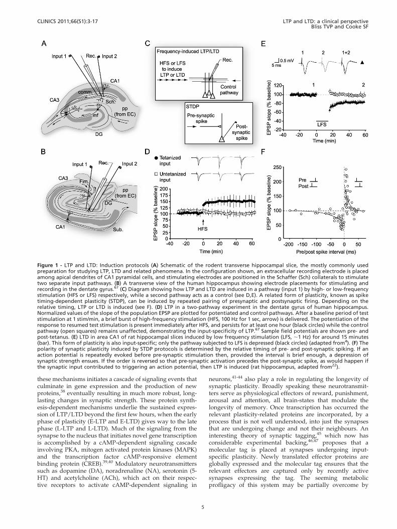
Figure 1 - LTP and LTD: Induction protocols (A) Schematic of the rodent transverse hippocampal slice, the mostly commonly usedpreparation for studying LTP, LTD and related phenomena. In the configuration shown, an extracellular recording electrode is placedamong apical dendrites of CA1 pyramidal cells, and stimulating electrodes are positioned in the Schaffer (Sch) collaterals to stimulatetwo separate input pathways. (B) A transverse view of the human hippocampus showing electrode placements for stimulating andrecording in the dentate gyrus.67 (C) Diagram showing how LTP and LTD are induced in a pathway (input 1) by high- or low-frequencystimulation (HFS or LFS) respectively, while a second pathway acts as a control (see D,E). A related form of plasticity, known as spiketiming-dependent plasticity (STDP), can be induced by repeated pairing of presynaptic and postsynaptic firing. Depending on therelative timing, LTP or LTD is induced (see F). (D) LTP in a two-pathway experiment in the dentate gyrus of human hippocampus.Normalized values of the slope of the population EPSP are plotted for potentiated and control pathways. After a baseline period of teststimulation at 1 stim/min, a brief burst of high-frequency stimulation (HFS, 100 Hz for 1 sec, arrow) is delivered. The potentiation of theresponse to resumed test stimulation is present immediately after HFS, and persists for at least one hour (black circles) while the controlpathway (open squares) remains unaffected, demonstrating the input-specificity of LTP.67 Sample field potentials are shown pre- andpost-tetanus. (E) LTD in area CA1 of rat hippocampal slices induced by low frequency stimulation (LFS, ,1 Hz) for around 15 minutes(bar). This form of plasticity is also input-specific; only the pathway subjected to LFS is depressed (black circles) (adapted from4). (F) Thepolarity of synaptic plasticity induced by STDP protocols is determined by the relative timing of pre- and post-synaptic spiking. If anaction potential is repeatedly evoked before pre-synaptic stimulation then, provided the interval is brief enough, a depression ofsynaptic strength ensues. If the order is reversed so that pre-synaptic activation precedes the post-synaptic spike, as would happen ifthe synaptic input contributed to triggering an action potential, then LTP is induced (rat hippocampus, adapted from22).
CLINICS 2011;66(S1):3-17 LTP and LTD: a clinical perspectiveBliss TVP and Cooke SF
5
local translation of existing or newly transcribed mRNAs atribosomes positioned in the dendrites; local protein synth-esis, triggered by a molecular tag, would drastically reducethe number of protein molecules required to potentiate ordepress individual synapses.48,49 The molecules that act astags are not yet determined but there have been someinteresting recent proposals, and there is also now a betterunderstanding of what the effector proteins themselves are,as we now discuss.
MaintenanceCaMKII has also been championed as a long-term main-
tenance mechanism for LTP37 because it can phosphorylateitself and in this way remain autonomously active for a periodof time after the dissipation of elevated calcium. This attribute,in principle, would allow it to act as a ‘memorase’.50 Whetheror not CaMKII does actually maintain LTP expression for longperiods of time has been questioned.51 Inhibitors of this kinaseappear to have little effect on already established LTP.52
However, recent data suggests that it may remain active forseveral minutes after LTP induction. This could enable thekinase to act as a ‘tag’ for recently potentiated synapses andallow for the synapse-specific recruitment of newly synthe-sized proteins that participate in the maintenance of long-lasting synaptic potentiation.46 Another kinase, PKMf, hasnow been recognized to act in a localized fashion to maintainsynaptic potentiation for long periods of time. This kinase, anatypical isoform of the calcium-dependent kinase PKC, isremarkable in that it is newly expressed after LTP inductionand remains persistently active, in part because it lacks aregulatory domain that would put a brake on its activity in theabsence of calcium. Thus, the kinase is capable of maintainingLTP expression at least for its own lifetime and, probablythrough persistent expression and some as yet not fullyunderstood autoregulation, for much longer periods.53 Indeed,L-LTP can be reversed at least a day after induction throughspecific inhibition of this kinase in vivo.54 It is now known thatPKMf maintains LTP by increasing the number of AMPARs inthe synapse, thereby keeping synaptic transmission poten-tiated.55 There is abundant evidence that expression of LTPdepends on persistent increases in AMPAR number in thesynapse,56 although pre-synaptic changes in the probability ofneurotransmitter release also play a role, and in somecircumstances a dominant role, in supporting at least the earlyphase of LTP.57-59 The evidence for presynaptic involvement inthe expression of LTP in the hippocampus is strong, and hasbeen hard to reconcile with the compelling evidence forchanges in glutamate receptor number and/or modification;we are still some way from a unified model of LTPexpression.60 LTD also relies on both pre and post-synapticexpression mechanisms61 although here too the maintenancemechanism is not fully understood.
Finally, it is important to mention that structural changesin the size and shape of pre- and post-synaptic specializa-tions may mediate permanent or near-permanent changes insynaptic efficacy. Growth may allow for an increase in thesize or number of active zones on both sides of the synapse.Spines can increase in volume after L-LTP induction anddecrease after L-LTD induction.62 The degree to whichstructural re-organisation of synapses occurs in adultanimals is not yet clear. An intriguing participant in thislater phase of synaptic plasticity is brain-derived neuro-trophic factor (BDNF), a substance that is newly synthesised
as a result of MAPK and CREB signaling and which mayinitiate structural change at tagged synapses.63 The role ofBDNF in synaptic plasticity is multimodal and it partici-pates in the early phases of both LTP and LTD through co-release with presynaptic glutamate. BDNF, initially in theform pro-BDNF, binds to two postsynaptic receptors: thetyrosine kinase B (TrkB) receptor, whose activation facil-itates the induction of LTP,64 and the P75 receptor, whoseactivation results in an alteration of the subunit compositionof the NMDA receptor that promotes the subsequentinduction of LTD.65 Thus, BDNF is a major player insynaptic plasticity, although its action is complex (seefigure 2).
LTP and LTD in human hippocampal slicesMany of the mechanisms described above have since been
demonstrated to be crucial for LTP in hippocampal tissueexcised from humans undergoing temporal lobe surgery toalleviate otherwise intractable epilepsy. In these slicestudies, LTP was readily induced in the temporal lobe andat perforant path-granule cell synapses in the dentate gyrus(see figure 1B) using induction tetani comparable to thoseused in slices from mouse brain.66,67 LTP in human tissuecould be blocked by application of the NMDAR antagonistAP5, just as in animal models. Furthermore, artificialelevation of cAMP through application of forskolin, whichincreases adenylate cyclase activity, results in chemically-induced LTP, a phenomenon that is well documented inrodent hippocampal slices68 and a principal piece ofevidence for the involvement of the cAMP-dependentcascade in L-LTP. These studies are important for theirverification that the human brain supports an LTP-likephenomenon, but it should be borne in mind that the tissuemay have been in a pathological state as it was taken fromindividuals with an epileptic focus in the temporal lobe.Indeed, slices taken from individuals in whom the focuswas present in the hippocampus itself produced far less LTPthan those in whom the focus was elsewhere in the temporallobe. It is possible that LTP was occluded in those slicestaken directly from the vicinity of an epileptic focus byprevious synaptic potentiation occurring through excessiveneural activity. In keeping with this notion, levels ofCaMKII expression were found to be significantly higherand levels of the phosphatase PP2B significantly lower indentate granule cells of individuals with hippocampalepileptic foci.69 Thus, unsurprisingly, it is clear that LTPcan be readily induced in the human CNS and that severalmolecular mechanisms are shared with rodent models. Withthis in mind we can now consider whether LTP and LTDcan be harnessed for therapeutic purposes in humans (seeTable 1).
Inducing LTP and LTD-like changes in the humanCNS non-invasively
Broadly speaking there are two categories of non-invasiveapproach that can be taken to induce lasting change inneural activity in the human CNS. The first category mimicsthe high- or low-frequency electrical stimulation used toinduce LTP or LTD, respectively. The second removes therequirement for frequency-based stimulation and insteadattempts to mimic pairing or STDP-like protocols ofinduction (see figure 1F and figure 3).
LTP and LTD: a clinical perspectiveBliss TVP and Cooke SF
CLINICS 2011;66(S1):3-17
6
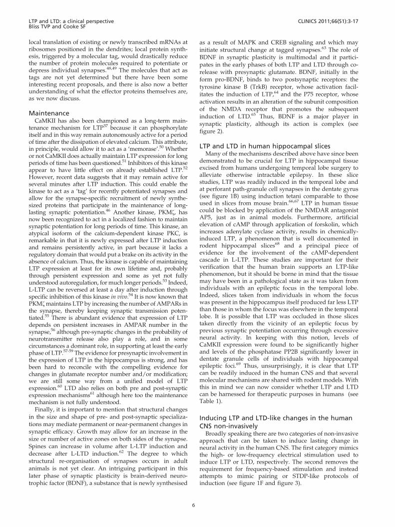
Figure 2 - LTP and LTD: Schematic of molecular mechanisms. (A) The induction of canonical forms of both LTP and LTD is triggered byactivation of the NMDA class of glutamate receptor. This ionotropic receptor detects the coincidence of presynaptic and strongpostsynaptic activity by a mechanism that involves both the binding of transmitter and depolarization-induced repulsion of the Mg2+
ions that block its ionophore at near-resting membrane potentials. In its unblocked state Ca2+ ions are able to permeate the channel,gaining access to Ca2+-dependent processes in the spine and triggering synaptic plasticity. (B) Ca2+ binds to Ca2+/calmodulin which, inturn activates numerous kinases and phosphatases, including CaMKII, PKC and Calcineurin (PP2B) directly and PKA and PP1 indirectly.The balance of kinase and phosphatase activity depends on the concentration and temporal profile of the postsynaptic Ca2+ transient(including Ca2+ released from intracellular stores). The Ca2+ transient determines the polarity of the induced plasticity, with low andprolonged Ca2+ transients inducing LTD and brief, steeper transients inducing LTP. (C) One means by which LTP is expressed is throughphosphorylation of the AMPA receptor, an ionotropic glutamate receptor that mediates baseline chemical transmission at excitatorysynapses in the CNS. Phosphorylation by CaMKII enhances the conductance of these channels. LTD, by contrast, results, in part, from thedephosphorylation of the AMPA receptor by phosphatases. (D) Trafficking of AMPA receptors plays a major role in the expression ofLTP and LTD by increasing or decreasing the number of receptors in the postsynaptic membrane. (E) Presynaptic mechanisms leading toa sustained increase in the probability of transmitter release also contribute to the expression of LTP. The relative contributions of pre-and post-synaptic mechanisms may vary at different times after induction and also across different classes of synapse. Since induction ofLTP and LTD is controlled by the post-synaptic NMDA receptor, any presynapic component of expression requires a retrogrademessenger that can signal to the pre-synaptic terminal that coincidence has occurred. Two candidates are nitric oxide (NO) andendocannabinoids (EC). (F) A second form of LTD that has been much studied is dependent on group 1 metabotropic glutamatereceptors (mGluR). Glutamate binding to this receptor initiates a signal cascade, involving the breakdown of the membrane lipid PIP2
by phospholipase C (PLC) to the important signaling molecules IP3, which releases Ca2+ from Ca2+ stores (not shown) and diacylglycerol(DAG), which leads to the activation of the calcium sensitive kinase PKC. This enzyme then phosphorylates the AMPA receptor but insuch a manner that the conductance is reduced. An offshoot is the production of NO. (G) Brain-derived neurotrophic factor (BDNF)plays a complicated role in both LTP and LTD and contributes in different ways to short-term and long-term plasticity. (H) Longer-lasting ‘late’ forms of LTP and LTD, persisting for more than a few hours, require the synthesis of new proteins, either through novelgene transcription or through initiation of local translation of existing transcripts. Novel gene expression requires signaling to thenucleus from newly potentiated or depressed synapses. A major player in this process is the cAMP-dependent signaling cascadeinitiated by calcium influx and involving adenylyl cyclase (AC) and cAMP-dependent kinase (PKA), which also acts directly on the AMPAreceptor in LTP expression. Catecholaminergic modulatory input plays a major role in determining the longevity of LTP and LTD,through interaction with AC which increases levels of cAMP and thereby activates PKA. PKA then sets in action a chain of signals thatleads to the expression of new transcripts which, in turn are translated into proteins contributing to the long-term expression ofsynaptic plasticity. This signaling pathway has been a major recent target of attempts to find nootropic substances. (I) There are parallelsignaling pathways, involving mitogen activated protein kinases (MAPK), that also result in the synthesis of new proteins. However, inthis case existing transcripts are locally translated into proteins, without further requirement for nuclear signaling. The MAPK pathwayis strongly implicated in mGluR-dependent LTD. (J) One newly synthesized protein that acts as a maintenance mechanism for late LTP isPKMf. This remarkable kinase comprises the active subunit of PKM, an isoform of PKC, that is now known to maintain the presence ofAMPA receptors inserted during LTP induction, and thereby maintain LTP. Inhibition of this kinase can erase LTP and memory manydays after induction. (K) Finally, BDNF can also play a second role in synaptic plasticity, as a newly synthesized product that alters thestructure of the synapse to enforce long-term changes in synaptic strength.
CLINICS 2011;66(S1):3-17 LTP and LTD: a clinical perspectiveBliss TVP and Cooke SF
7

Repetitive Transcranial Magnetic Stimulation(rTMS)
rTMS delivers relatively small electrical currents gener-ated by fluctuating magnetic fields administered over theskull using a figure-of-eight magnetic coil. In most cases itonly allows for the excitation of neural circuitry instructures relatively close to the brain surface. For thisreason the technique has primarily been used in theneocortex and is not a suitable substitute for the kind ofinvasive deep brain stimulation (DBS) currently used totreat Parkinson’s disease.70 Single TMS pulses evoke event-related potentials (ERPs) that can be recorded using scalprecording electrodes. Importantly, delivery of a highfrequency train of rTMS pulses can induce lasting potentia-tion of ERP amplitude and low frequency rTMS can havethe opposite effect. In primary sensory cortex, ERPs can begenerated by discrete, transient sensory stimuli within themodality of interest. These ERPs also undergo amplitudepotentiation as a result of tetanic stimulation with rTMS.71
rTMS is being assessed as a potential treatment for anumber of neurological disorders. Parkinson’s disease is acommon neurodegenerative disorder that results from thespecific loss of dopaminergic cells in the substantia nigra(s.n.) leading to tremor, and impairment of movementinitiation and termination. Deep brain stimulation ofsubthalamic nuclei is a well established treatment inadvanced cases of Parkinson’s disease, and can be veryeffective.72 rTMS has been suggested as an alternative, non-invasive treatment. Although the primary locus of dysfunc-tion in Parkinson’s is too deep for standard rTMS there aresecondary effects that can be addressed with rTMS. Whencells in the s.n. degenerate, a conspicuous beta-frequencysynchronized activity arises in primary motor cortex (M1)that is believed to contribute to limb rigidity and akinesia.73
In an established animal model of Parkinson’s disease, inwhich primates are treated with MPTP, a substance whichselectively kills dopaminergic cells in the s.n., thissynchronization of activity in M1 occurs in addition to allthe classic motor symptoms of Parkinson’s disease. Deliveryof high-frequency rTMS (130 Hz) to M1 in these monkeysinduces a lasting amelioration of rigidity and akinesia.74
Promisingly, rTMS has also been applied to motor cortex inhuman Parkinson’s sufferers to produce significant butshort-lasting improvements in motor performance.75 Thisexample illustrates the important point that, even if theprimary area of dysfunction in the CNS is not accessible tonon-invasive stimulation, secondary dysfunction in moresuperficial regions can be targeted in order to ameliorate theresulting behavioral abnormality.76 It is worth noting,though, that a very recently developed technology, ‘deep’rTMS77 may soon permit non-invasive stimulation of deeptargets such as the thalamus, basal ganglia and brainstemnuclei without delivering dangerously large currents tothe overlying cortex. This technique, which uses a three-dimensional magnetic coil system that surrounds thecranium in order to generate activity at much greaterdepths than the traditional figure-of-8 system, may come toreplace DBS as a non-invasive treatment for Parkinson’sdisease. It may also be of benefit in the treatment of obesity,Alzheimer’s disease and depression.
A long standing treatment for major depression has beenelectroconvulsive therapy (ECT). Although still one offew effective treatments for this troubling disorder, it isonly used when antidepressants fail because it requires
anaesthesia and carries the risk of neural damage. One effectof ECT may be to reduce cortical excitability and, thereby,reverse increases in the excitability of cerebral cortex,notably within prefrontal cortex, during treatment-resistantdepression.78 Two explanations have been offered forpathological increases in excitability - a persistent decreasein inhibitory tone, or a persistent potentiation of excitatorysynapses as a result of an LTP-like process. ECT results inthe expression of several biochemical markers of LTD inanimals79 suggesting that ECT causes induction of de novoLTD – or depotentiation of already potentiated synapses - inthe cortex. Low frequency rTMS, thought to induce LTD-like plasticity in humans, has been applied to the prefrontalcortex to achieve results similar to ECT in depressivepatients.80 rTMS may, therefore, eventually serve as a morecontrolled and focused alternative to ECT, one which doesnot require anaesthesia and can be used over multiplesessions without fear of major brain damage.
The fact that low frequency rTMS can be used to reducecortical excitability, perhaps through LTD-like plasticityin humans71,81 suggests that it may also be a potentialtreatment for epilepsy. Epilepsy results from hyperexcit-ability, perhaps through excessive LTP at glutamatergicsynapses, or through other mechanisms such as increasedintrinsic excitability or reduced inhibition. Thus, LTD-likeplasticity, induced with low frequency rTMS, may provide ameans to reverse LTP at saturated synapses in order toreduce circuit excitability. Low frequency (0.3 Hz) rTMSwas first used as a treatment for epilepsy in an open-casestudy performed over a decade ago, resulting in asignificant reduction in the incidence of seizures for up toa month after five days of treatment.82 Subsequent double-blind randomized trials have produced mixed results,83,84
and it is clear that further work is required to identify moreprecisely the type of epilepsies that respond to rTMS and tooptimize the parameters of stimulation in different cases.Generalized seizures may be more responsive than focalseizures, particularly where the focus is in deep brainstructures.
The potential of rTMS to address epileptic foci hasbrought to the fore the importance of navigation. Theefficacy of rTMS can be greatly increased if it is coupledwith imaging techniques such as functional MRI (fMRI) tomap the affected area, or by systematically recording theeffects of stimulation on ERP amplitude or motor output.85
This approach enables the tailoring of treatment toindividual cases. For disorders such as Parkinson’s diseaseit is very clear which structure in the brain is the primarysite of pathology and, although there are slight individualdifferences in brain shape, it is relatively straightforward totarget the region to be stimulated with DBS. In the case ofstroke, a condition in which rTMS application to primarymotor cortex holds promise as a treatment, the region ofmajor dysfunction must be tracked in the individual beforeTMS is applied because each stroke victim has a uniquepattern of damage. Neuro-navigated TMS maps motorevoked potentials (MEPs) and the motor threshold foreliciting responses from the muscles of interest to optimiseTMS delivery points in individual subjects.86,87 Highfrequency rTMS can then be used to enhance motor outputin affected regions. A systematic study of the efficacy ofneuro-navigated TMS as compared to conventional stereo-taxically-guided TMS, targeting the representation of thedorsal interosseous muscle of the hand and evaluating the
LTP and LTD: a clinical perspectiveBliss TVP and Cooke SF
CLINICS 2011;66(S1):3-17
8
impact of rTMS on reaction time and pinch force, revealedsignificantly greater efficacy for navigated TMS in boththese behavioral outputs as well as in potentiating MEPsand reducing motor threshold.88
In summary, rTMS holds promise in the treatment of avariety of neurological disorders, perhaps through theinduction of LTP and LTD-like plasticity. Advances intechnology, allowing reliable, safe stimulation of deepstructures and careful individual tailoring of stimulationsites and protocols using neuro-navigated TMS, will onlyexpand this potential (Figure 3A).
Transcutaneous electrical nerve stimulation (TENS)Electrical stimulation can also be delivered directly to
peripheral nerves through the skin to induce LTP-like andLTD-like changes in central neural responsiveness. Certainclinical conditions result from pathology just a singlesynapse from the periphery and may be addressed withTENS. Chief amongst these conditions is hyperalgesia.Plasticity at two locations contributes to this chronic paincondition which arises from repeated peripheral applicationof a noxious stimulus to the periphery. The first occurs atthe point of nociception itself (peripheral sensitization); andthe second at the first central synapses in the pathwaywithin the dorsal horn of the spinal cord (central sensitiza-tion).89 High frequency electrical stimulation can bedelivered to nociceptive C fibres in animal preparations90,91
to induce a lasting form of LTP within the dorsal horn.Alternatively, LTP can be induced at these same synapsesthrough natural stimulation of peripheral nociceptors withheat, the noxious chemical formalin or mechanical pinchingof the skin.92 Some of this potentiation is homosynaptic andconfined to the stimulated nociceptive (C) fibres but thereis also a component of heterosynaptic potentiation thatenhances transmission at other, surrounding C fibres, andmay contribute to secondary hyperalgesia. In addition,sensory (A) fibres that do not mediate nociception may alsobe potentiated heterosynaptically by C fibre stimulation,perhaps through changes in the intrinsic excitability ofcommon target cells in the dorsal horn. As a result there canbe a lasting sensitization of sensory nerves around the site ofinjury leading to hypersensitivity to non-noxious stimuli(allodynia).89
Central sensitization has several molecular parallels withLTP in the hippocampus.93 LTP in the C fibre pathway isboth NMDAR-dependent and calcium-dependent,94,95 andalso relies upon other molecular players such as Substance Pand the NK-1 receptor.96 Blockade of the NMDA receptorwith the non-competitive antagonist ketamine duringsurgery in humans helps to prevent central sensitizationand reduce subsequent hyperalgesia on recovery,97 andother NMDA receptor antagonists have similar effects,98
Given the evidence for the involvement of LTP inhyperalgesia, as well as other forms of peripheral andcentral plasticity that increase responsiveness to peripheralstimulation, one clinical approach is to attempt to reversecentral sensitization in the dorsal horn with low frequencystimulation of C fibres to induce LTD, or to ameliorateperipheral sensitization by reducing dorsal horn respon-siveness with LTD of all afferent sensory fibres. TENSdelivered to peripheral nerves containing both A and Cfibres can be used to induce LTP or LTD in the dorsal horn.Significantly, these forms of plasticity respectively increaseor decrease the perception of pain in human subjects,99,100 so
that, for example, pinpricks become less painful after thedelivery of low frequency TENS. The effect of inducing LTDis, however, complicated by the fact that, while lowfrequency stimulation reduces pain sensitivity directly atthe conditioning site, it has the reverse effect of secondaryhyperalgesia at adjacent sites.99 It is also important to notethat low frequency stimulation of C fibres, which mimics acharacteristic activity pattern after injury, can induce LTPwith similar biochemical hallmarks to that induced withhigh frequency stimulation.96 In fact, the most effective wayto induce LTD of C fibre-evoked responses in the dorsalhorn is to stimulate at a low intensity that will activate Afibres specifically and not the C fibres themselves.91 Thus, itis clear that while the use of TENS holds promise foraddressing hyperalgesia, the condition is complex and islikely to require additional treatments to ameliorate all itsvarious components. Nonetheless, we regard the use ofTENS to treat neuropathic pain as an exemplar for thedevelopment of non-invasive clinical strategies based on theinduction or suppression of LTP or LTD. The fact thatperipheral stimulation can be delivered immediately affer-ent to the targeted synapses, as well as the availability ofgood animal models of hyperalgesia, emphasises thepotential for a non-invasive strategy based on TENS.Rational drug design based on manipulating the potentiatedand/or depressed state of synapses at the first stages oftransmission in peripheral nociceptive pathways offersanother promising approach.
The application of TENS has largely been limited tosituations where stimulation of monosynaptic pathways ispossible. Potentiation or depression may occur di or tri-synaptically but this is difficult to monitor, or to predict, asthe frequency of stimulation will change due to filtering byafferent circuitry. Nevertheless, it is known that hyperalge-sia is associated with changes in the brain as well as thosedocumented in the spinal cord. The cortex, notablysomatosensory cortex and anterior cingulate cortex (ACC),has been a site of investigation because peripheral injury, inthe form of removal of a digit in rats, induces NMDAR-dependent plasticity in these regions of cortex.101,102 Thereare at least three synapses between nociceptive inputs andcortical synapses undergoing LTP-like plasticity but it maybe that chronic pain arises from a concatenation ofpathological synaptic plasticity at multiple synapsesthroughout the CNS. Indeed, the delivery of high frequencyTENS, producing hypersensitivity to tactile stimulation,does lead to a potentiation of ERPs in the cortex100 anddelivery of low frequency stimulation with rTMS in thecortex can lead to a temporary relief of chronic pain.103 In afascinating recent study of hyperalgesia induced by periph-eral nerve injury in mice, local inhibition in the ACC ofPKMf a key maintenance molecule for NMDAR-dependentLTP, resulted in reversal both of LTP-like plasticity inducedin the ACC by nerve damage and reduction of hypersensi-tivity to tactile stimuli.104 This latter study opens up anotherintriguing avenue of treatment for neurological disordersarising from pathological LTP-like plasticity, namely thelocal and selective erasure of potentiation by suppressingthe activity of PKMf. From a clinical perspective the majorproblem with this approach is the fact that it would behighly invasive if used in humans and potentially danger-ous given the indiscriminate nature of the PKMf inhibitor inerasing synaptic plasticity and memory. However, theremay be other means of selectively erasing LTP such
CLINICS 2011;66(S1):3-17 LTP and LTD: a clinical perspectiveBliss TVP and Cooke SF
9
as depotentiation,36,105,106 a phenomenon related to butmechanistically different from LTD. For a limited time afterinduction, LTP can be reversed, or depotentiated, by low-frequency stimulation, suggesting a potential role for TENSor rTMS in the treatment of pathological plasticity,particularly in cases where the patient can be brought totreatment soon after the causative event.
Photic and auditory tetanizationA series of animal and human studies has demonstrated
that plasticity can be induced in primary sensory corticeswithout electrical stimulation. Here, the electrical stimulusis replaced by rapid sensory stimulation within themodality of interest.107 For example, LTP-like changes, asmeasured using changes in the amplitude of one compo-nent of the visual evoked potential (VEP), can be induced
in visual cortex using a photic tetanus (9-13 Hz) thatconsists of flashing or phase-reversing visual stimuli(for example, checkerboards or sinusoidal gratings) on acomputer monitor.108-110 Three important criteria must bemet if the location of plasticity underlying this phenom-enon is to be securely identified. First, potentiation shouldbe specific to the particular stimulus, so that the potentia-tion is no longer expressed if the spatial frequency109
or the orientation108 of the stimulus is altered, consistentwith plasticity occurring in primary visual cortex, whererepresentations of sensory primitives such as orientationand spatial frequency have not yet been integrated intomore complex object representations.111 This latter obser-vation is reminiscent of the well-documented phenomenonof perceptual learning, in which over time subjects becomegradually more and more proficient at responding to
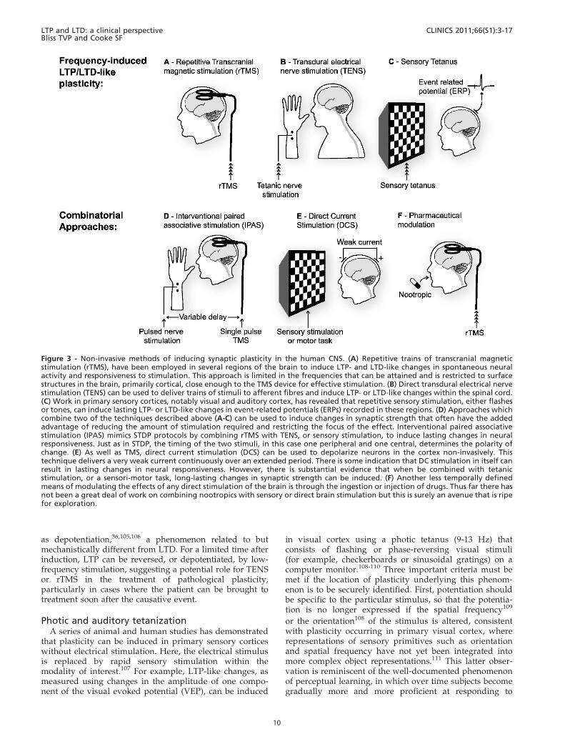
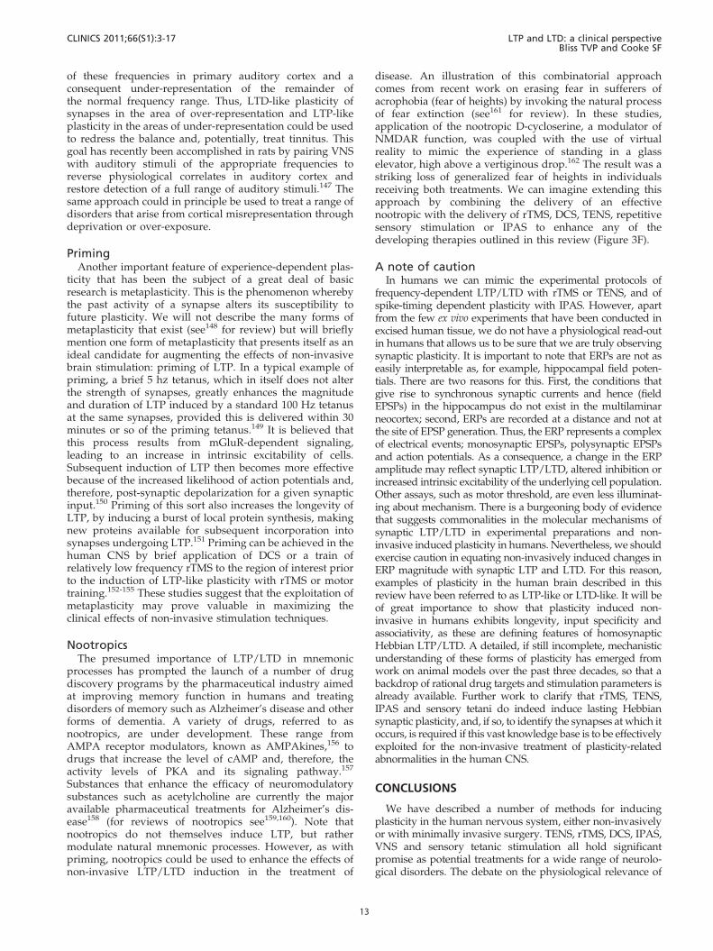
Figure 3 - Non-invasive methods of inducing synaptic plasticity in the human CNS. (A) Repetitive trains of transcranial magneticstimulation (rTMS), have been employed in several regions of the brain to induce LTP- and LTD-like changes in spontaneous neuralactivity and responsiveness to stimulation. This approach is limited in the frequencies that can be attained and is restricted to surfacestructures in the brain, primarily cortical, close enough to the TMS device for effective stimulation. (B) Direct transdural electrical nervestimulation (TENS) can be used to deliver trains of stimuli to afferent fibres and induce LTP- or LTD-like changes within the spinal cord.(C) Work in primary sensory cortices, notably visual and auditory cortex, has revealed that repetitive sensory stimulation, either flashesor tones, can induce lasting LTP- or LTD-like changes in event-related potentials (ERPs) recorded in these regions. (D) Approaches whichcombine two of the techniques described above (A-C) can be used to induce changes in synaptic strength that often have the addedadvantage of reducing the amount of stimulation required and restricting the focus of the effect. Interventional paired associativestimulation (IPAS) mimics STDP protocols by combining rTMS with TENS, or sensory stimulation, to induce lasting changes in neuralresponsiveness. Just as in STDP, the timing of the two stimuli, in this case one peripheral and one central, determines the polarity ofchange. (E) As well as TMS, direct current stimulation (DCS) can be used to depolarize neurons in the cortex non-invasively. Thistechnique delivers a very weak current continuously over an extended period. There is some indication that DC stimulation in itself canresult in lasting changes in neural responsiveness. However, there is substantial evidence that when be combined with tetanicstimulation, or a sensori-motor task, long-lasting changes in synaptic strength can be induced. (F) Another less temporally definedmeans of modulating the effects of any direct stimulation of the brain is through the ingestion or injection of drugs. Thus far there hasnot been a great deal of work on combining nootropics with sensory or direct brain stimulation but this is surely an avenue that is ripefor exploration.
LTP and LTD: a clinical perspectiveBliss TVP and Cooke SF
CLINICS 2011;66(S1):3-17
10

highly specific visual stimuli,112 and is also consistent withobservations made in animals using photic stimulation, inwhich potentiation of VEPs is both eye-specific andorientation-specific.113 Second, in these animal models itis possible to intervene in the visual pathway andsubstitute electrical thalamic stimulation for baseline photicstimulation and observe potentiation of the electricallyevoked test responses after inducing potentiation usingphotic stimulation.13,114 The converse is also observed. LTPinduced in primary visual cortex of the rat using tetanicelectrical stimulation of the thalamus is also reflected in apotentiation of the VEP.13,115 Therefore, the plasticityunderlying VEP potentiation does not occur in the retinaor in retinothalamic pathways, but in the thalamocorticalprojection. Finally, it is worth noting that induction ofsimilar plasticity can be prevented through local blockadein visual cortex of AMPA receptor insertion113 or systemicblockade of NMDA receptors,113,114 and expression of LTPcan be reversed with local inhibition of PKMf13 in rodents.Thus, mechanisms that support LTP also support potentia-tion of VEPs with photic stimulation.
From a clinical perspective, this work may prove useful intreating monocular amblyopia, a prevalent disorder thataffects around 1% of individuals worldwide. Amblyopiaresults from a mismatch in ocular function early in life,leading to preferential allocation of visual cortex to inputthrough the fully functional eye. Many of the oculardysfunctions that cause this disorder, such as cataracts(lens opacity), anisometropia (mismatched lens refraction)and strabismus (misalignment of the eyes) can be fullyrepaired. However, if treatment is delayed beyond around 8years of age in humans, as often occurs for either diagnosticor economic reasons, the cortex will not recover normalresponsiveness to input through the previously dysfunc-tional eye.116 Extensive work in animals has demonstratedthat monocular deprivation through eyelid suture, whichmodels the causes of amblyopia in humans, results in LTDof excitatory transmission within visual cortex.117-119
Therefore, an obvious strategy to rescue the effects ofamblyopia is to artificially induce LTP at the same synapses.Two treatments that have been shown to have some efficacyin adult amblyopes are perceptual learning120,121 andrTMS,122 although in general functional recovery has beenboth modest and short-lived. Future studies may make useof high frequency photic stimulation as a means ofrecovering function through the amblyopic eye (Figure 3C).It is also worth considering combinatorial approaches toinducing LTP; peripheral sensory stimulation, combinedwith central activation by rTMS, may be more fruitful ininducing lasting plasticity.
Interventional Paired-Associative Stimulation (IPAS)One non-invasive way to achieve an effect akin to spike
timing-dependent plasticity (STDP) in the human cortexwould be to activate afferent sensory pathways using, forexample TENS or visual stimulation, in appropriately timedconjunction with single pulses of TMS to activate post-synaptic neurons at the target site. This approach, known asinterventional paired associative stimulation (IPAS), usesthe amplitude of the event-related potential (ERP) todetermine the point at which peripheral stimulation initiatessynaptic release. Central stimulation can then be deliveredeither before or after this delay in order to induce LTD orLTP-like effects, respectively (Figure 3D). Note that while
IPAS, like STDP, depends on the relative timing of pre- andpost-synaptic events, the parallel is not exact, since in thecase of IPAS polysynaptic pathways are involved. One clearadvantage of IPAS over frequency-based methods is that itavoids potential risks associated with the delivery of high-frequency stimulation - there are reported instances ofseizure as a result of using high-frequency rTMS.123
IPAS has been applied in both somatosensory cortex andmotor cortex, using ERPs in response to stimulation of themedian and ulnar nerves as a metric of plasticity in thecortex. Responses of the abductor pollicis brevis (APB) andabductor digiti minimi (ADM) muscles in the hand, to bothperipheral stimulation and single cortical TMS pulses, alsoserved as an assay of enhanced or decreased functionaloutput.124 One beautiful aspect of the results from theseexperiments is the regional specificity of the LTP/LTD-likeeffects. Appropriate timing of nerve stimulation withsomatosensory cortical stimulation can lead to a specificenhancement of ERPs driven in a small partial hand-representation area in cortex without effect on neighbouringrepresentations,125 which contrasts with a broader effect ofdirect rTMS application. This specificity also holds true formotor output as the muscle response to probe stimulation,either peripherally or cortically, can be potentiated ordepressed by appropriate IPAS delays without effect on theneighbouring control muscle.126 The immediate import ofthis work, from a clinical perspective, may be in addressingfocal dystonia, such as writer’s cramp, in which chronicspasm or cramping develops in hand muscles, preventing theperformance of fine motor skills such as writing. It is unclearwhether this disorder arises primarily from cortical or basalganglia dysfunction but the responses in sufferers of writer’scramp to IPAS were markedly different from normal controlsubjects in that the specificity of the effect was lost. In contrastto the effects in control subjects, targeting one muscle withIPAS had a comparable potentiating or depressing effect onthe other, control muscle, which had not undergone IPAS.127
Thus, a loss of specificity in cortical connectivity maycontribute to the dystonia. This example provides anillustration of how non-invasive stimulation techniques inhumans may provide information about the aetiology ofdisorders - almost as important an outcome as the develop-ment of a treatment itself.
Direct Current Stimulation (DCS)Another approach uses an older and more basic method
to induce plasticity. Direct current stimulation (DCS)applies continuous weak current to a superficial region ofthe brain using two electrodes fitted on the scalp surface atcarefully selected sites. Delivery of this current can havedramatic and lasting effects on ERP amplitude. The polarity
of change is determined by the polarity of stimulation.128
From a theoretical perspective, given the careful researchthat has been conducted in animal models to determine theoptimal frequency ranges of trains of discrete electricalpulses for inducing LTP and LTD, and the precise timing ofpulses required to induce STDP, the effects of DCS, which isnon-pulsatile, are puzzling. It has previously been demon-strated, however, that in rats DCS produces changes in the
spontaneous firing rate of cortical neurons.129 Thus,plasticity induced by DCS may be the product of STDP-like interactions between DCS-induced neural activity anddiscrete stimulation, either in the form of electrical pulses in
CLINICS 2011;66(S1):3-17 LTP and LTD: a clinical perspectiveBliss TVP and Cooke SF
11

ex vivo slices,130 or, in humans, rTMS131 or sensorystimulation resulting from a subject’s participation in
particular tasks.132,133 It is known that DCS causes therelease of BDNF and that this substance modulates theinduction of NMDAR-dependent LTP through the TrkBreceptor.64 Moreover, a combination of DCS and low-frequency stimulation leads to LTP in ex vivo corticalslices.130 Slices taken from mice that do not express BDNF,or in which the TrkB receptor is blocked, fail to show DCS-induced LTP. The fact that low frequencies, which wouldnormally induce LTD, cause LTP when paired with DCSsuggests that post-synaptic cells are in a permissive state forLTP as a result of DCS, just as clamping cells in adepolarized state results in low frequency-induced LTP.134
A major application of DCS has been in enhancing motorlearning through stimulation of motor cortex, an approachthat has great promise in facilitating recovery from stroke bypromoting compensatory plasticity in spared motor corticaltissue.133 Interestingly, mice which do not express BDNFhave significant deficits in motor learning.130 A commonpolymorphism (Val66Met) found at a frequency of .30% inthe human population is, in animal models, associated witha reduced BDNF concentrations in the synaptic cleft andwith impaired motor learning,130 which suggests that asignificant minority of the population may suffer from amild impediment in motor skill acquisition. Remarkably,humans possessing this polymorphism show a reduction inmotor cortical plasticity, measured using ERPs, induced byrTMS, IPAS or DCS/rTMS applied to motor cortex.131,135
Thus, there is some mechanistic understanding of the effectsof DCS and why it may impact LTP/LTD. DCS is apotentially important tool for clinicians attempting tomodulate synaptic plasticity to ameliorate neurologicaldisorders, particularly in the motor system (Figure 3E).
Vagal nerve stimulationVagal nerve stimulation (VNS) is a related technique to
TENS, but is more invasive. Direct electrical stimulationrequires a minimal surgical procedure in which electrodesare wrapped around the left vagal nerve in the neck.136
Stimulation of the vagus causes, indirectly, widespreadrelease of neuromodulatory substances such as acetylcho-line, noradrenaline and dopamine through activation of thenucleus of the solitary tract.137 Release of acetylcholine fromneurons originating in the nucleus basalis promotes theinduction of plasticity in the cortex.138-141 Several studieshave shown that coincident occupation of cholinergicreceptors with stimulation of glutamatergic pathwaysenhances the induction of LTP/LTD.44,142-144 VNS servesas a minimally invasive, and therefore clinically viable,means of stimulating the nucleus basalis and otherbrainstem structures to induce release of acetylcholine andother neuromodulators. Thus it can be paired with sensorystimulation, task performance or rTMS in a combinatorialapproach to maximize recuperative cortical plasticity andenhance its longevity. A recent study used this strategy toaddress the prevalent neurological condition of tinnitus. Themanifestation of this disorder is a distracting, continuouslydetected tone in the absence of an external auditorystimulus. There are a multitude of causes for tinnitus,including dysfunction of the sensory apparatus itself.However, it is hypothesized to result, in part, from excessivecortical representation of a small range of sound frequenciesto which the individual has previously been heavilyover-exposed,145 and application of rTMS to this regionhas had short-lived beneficial effects.146 Induction oftinnitus in rats by repeated exposure to a loud auditorystimulus in a fixed range of frequencies leads to an over-representation
Table 1 - The potential applications of LTP/LTD-like plasticity to the treatment of neurological disorders.
Disease Underlying plasticity Potential Method References
Depression Hyper-excitability of prefrontal
cortex leading to suppression of
activity in target sites.
Low frequency rTMS in prefrontal cortex to induce
LTD-like plasticity and ameliorate hyper-excitability of
prefrontal cortex.
163
Parkinson’s disease Reduced nigral drive and resultant
loss of function in basal ganglia
and motor cortex.
High frequency rTMS in motor cortex to induce LTP-like
plasticity and ameliorate lost basal ganglia drive;
75
Schizophrenia Hyper-excitability of sensory cortex. rTMS in auditory cortex to suppress auditory hallucinations
with LTD-like plasticity.
164
Epilepsy Over-excitability of neural tissue in
epileptic focus.
Low frequency rTMS to induce LTD-like plasticity and
ameliorate hyper-excitability.
82,165
Stroke Lost motor cortical tissue. DCS in combination with motor task or rTMS to induce
BDNF release and promote LTP-like plasticity in surviving
cortical circuitry.
131,135
Chronic pain LTP-like plasticity in dorsal
horn and ACC.
TENS to induce LTD in dorsal horn; rTMS to induce LTD in
ACC; Inhibition of PKM to erase LTP in ACC.
99,103,104
Amblyopia LTD-like plasticity in primary
visual cortex.
Photic tetanus to induce LTP-like plasticity in primary visual
cortex; rTMS to primary visual cortex.
108,121,122
Tinnitus Over-representation of selected
frequencies in auditory cortex
due to LTP-like plasticity
Vagal nerve stimulation to cause neuromodulator release
coupled with auditory stimulation at lost frequencies to
win back incorrectly allocated cortical tissue.
147
LTP and LTD: a clinical perspectiveBliss TVP and Cooke SF
CLINICS 2011;66(S1):3-17
12
of these frequencies in primary auditory cortex and aconsequent under-representation of the remainder ofthe normal frequency range. Thus, LTD-like plasticity ofsynapses in the area of over-representation and LTP-likeplasticity in the areas of under-representation could be usedto redress the balance and, potentially, treat tinnitus. Thisgoal has recently been accomplished in rats by pairing VNSwith auditory stimuli of the appropriate frequencies toreverse physiological correlates in auditory cortex andrestore detection of a full range of auditory stimuli.147 Thesame approach could in principle be used to treat a range ofdisorders that arise from cortical misrepresentation throughdeprivation or over-exposure.
PrimingAnother important feature of experience-dependent plas-
ticity that has been the subject of a great deal of basicresearch is metaplasticity. This is the phenomenon wherebythe past activity of a synapse alters its susceptibility tofuture plasticity. We will not describe the many forms ofmetaplasticity that exist (see148 for review) but will brieflymention one form of metaplasticity that presents itself as anideal candidate for augmenting the effects of non-invasivebrain stimulation: priming of LTP. In a typical example ofpriming, a brief 5 hz tetanus, which in itself does not alterthe strength of synapses, greatly enhances the magnitudeand duration of LTP induced by a standard 100 Hz tetanusat the same synapses, provided this is delivered within 30minutes or so of the priming tetanus.149 It is believed thatthis process results from mGluR-dependent signaling,leading to an increase in intrinsic excitability of cells.Subsequent induction of LTP then becomes more effectivebecause of the increased likelihood of action potentials and,therefore, post-synaptic depolarization for a given synapticinput.150 Priming of this sort also increases the longevity ofLTP, by inducing a burst of local protein synthesis, makingnew proteins available for subsequent incorporation intosynapses undergoing LTP.151 Priming can be achieved in thehuman CNS by brief application of DCS or a train ofrelatively low frequency rTMS to the region of interest priorto the induction of LTP-like plasticity with rTMS or motortraining.152-155 These studies suggest that the exploitation ofmetaplasticity may prove valuable in maximizing theclinical effects of non-invasive stimulation techniques.
NootropicsThe presumed importance of LTP/LTD in mnemonic
processes has prompted the launch of a number of drugdiscovery programs by the pharmaceutical industry aimedat improving memory function in humans and treatingdisorders of memory such as Alzheimer’s disease and otherforms of dementia. A variety of drugs, referred to asnootropics, are under development. These range fromAMPA receptor modulators, known as AMPAkines,156 todrugs that increase the level of cAMP and, therefore, theactivity levels of PKA and its signaling pathway.157
Substances that enhance the efficacy of neuromodulatorysubstances such as acetylcholine are currently the majoravailable pharmaceutical treatments for Alzheimer’s dis-ease158 (for reviews of nootropics see159,160). Note thatnootropics do not themselves induce LTP, but rathermodulate natural mnemonic processes. However, as withpriming, nootropics could be used to enhance the effects ofnon-invasive LTP/LTD induction in the treatment of
disease. An illustration of this combinatorial approachcomes from recent work on erasing fear in sufferers ofacrophobia (fear of heights) by invoking the natural processof fear extinction (see161 for review). In these studies,application of the nootropic D-cycloserine, a modulator ofNMDAR function, was coupled with the use of virtualreality to mimic the experience of standing in a glasselevator, high above a vertiginous drop.162 The result was astriking loss of generalized fear of heights in individualsreceiving both treatments. We can imagine extending thisapproach by combining the delivery of an effectivenootropic with the delivery of rTMS, DCS, TENS, repetitivesensory stimulation or IPAS to enhance any of thedeveloping therapies outlined in this review (Figure 3F).
A note of cautionIn humans we can mimic the experimental protocols of
frequency-dependent LTP/LTD with rTMS or TENS, and ofspike-timing dependent plasticity with IPAS. However, apartfrom the few ex vivo experiments that have been conducted inexcised human tissue, we do not have a physiological read-outin humans that allows us to be sure that we are truly observingsynaptic plasticity. It is important to note that ERPs are not aseasily interpretable as, for example, hippocampal field poten-tials. There are two reasons for this. First, the conditions thatgive rise to synchronous synaptic currents and hence (fieldEPSPs) in the hippocampus do not exist in the multilaminarneocortex; second, ERPs are recorded at a distance and not atthe site of EPSP generation. Thus, the ERP represents a complexof electrical events; monosynaptic EPSPs, polysynaptic EPSPsand action potentials. As a consequence, a change in the ERPamplitude may reflect synaptic LTP/LTD, altered inhibition orincreased intrinsic excitability of the underlying cell population.Other assays, such as motor threshold, are even less illuminat-ing about mechanism. There is a burgeoning body of evidencethat suggests commonalities in the molecular mechanisms ofsynaptic LTP/LTD in experimental preparations and non-invasive induced plasticity in humans. Nevertheless, we shouldexercise caution in equating non-invasively induced changes inERP magnitude with synaptic LTP and LTD. For this reason,examples of plasticity in the human brain described in thisreview have been referred to as LTP-like or LTD-like. It will beof great importance to show that plasticity induced non-invasive in humans exhibits longevity, input specificity andassociativity, as these are defining features of homosynapticHebbian LTP/LTD. A detailed, if still incomplete, mechanisticunderstanding of these forms of plasticity has emerged fromwork on animal models over the past three decades, so that abackdrop of rational drug targets and stimulation parameters isalready available. Further work to clarify that rTMS, TENS,IPAS and sensory tetani do indeed induce lasting Hebbiansynaptic plasticity, and, if so, to identify the synapses at which itoccurs, is required if this vast knowledge base is to be effectivelyexploited for the non-invasive treatment of plasticity-relatedabnormalities in the human CNS.
CONCLUSIONS
We have described a number of methods for inducingplasticity in the human nervous system, either non-invasivelyor with minimally invasive surgery. TENS, rTMS, DCS, IPAS,VNS and sensory tetanic stimulation all hold significantpromise as potential treatments for a wide range of neurolo-gical disorders. The debate on the physiological relevance of
CLINICS 2011;66(S1):3-17 LTP and LTD: a clinical perspectiveBliss TVP and Cooke SF
13
LTP and LTD to human learning will no doubt continue but, inthe meantime, analogous processes may be harnessed as toolsto recover lost function in the nervous system and toameliorate the effects of pathological plasticity. These methodscould, in the future, be used to rescue deficits by directlyaddressing the site of pathology or by inducing compensatoryplasticity elsewhere. The development of stimulation strate-gies for the non-invasive induction of plasticity in the humanCNS has been guided by basic research into LTP, LTD andSTDP and, as we have seen, common molecular mechanismsare beginning to emerge in the human and animal literatures.In the future, a combination of nootropic and amnestic drugswith non-invasive stimulation techniques may emerge as theoptimal therapy for plasticity-related disorders of the humanCNS.
ACKNOWLEDGEMENTS
We would like to thank Dr. Steve Gomperts for reading this manuscript
and providing critical comments.
REFERENCES
1. Bliss TV, Lomo T. Long-lasting potentiation of synaptic transmission inthe dentate area of the anaesthetized rabbit following stimulation of theperforant path. J Physiol. 1973;232(2):331-356.
2. Hebb DO. The Organization of Behavior. New York, Wiley. 1949.3. Ito M, Kano M. Long-lasting depression of parallel fiber-Purkinje cell
transmission induced by conjunctive stimulation of parallel fibers andclimbing fibers in the cerebellar cortex. Neurosci Lett. 1982;33(3):253-258, doi: 10.1016/0304-3940(82)90380-9.
4. Dudek SM, Bear MF. Homosynaptic long-term depression in area CA1of hippocampus and effects of N-methyl-D-aspartate receptor blockade.Proc Natl Acad Sci U S A. 1992;89(10):4363-4367, doi: 10.1073/pnas.89.10.4363.
5. Martin SJ, Grimwood PD, Morris RG. Synaptic plasticity and memory:an evaluation of the hypothesis. Annu Rev Neurosci. 2000;23:649-711,doi: 10.1146/annurev.neuro.23.1.649.
6. Sanes JR, Lichtman JW. Can molecules explain long-term potentiation?Nat Neurosci. 1999;2(7):597-604, doi: 10.1038/10154.
7. Doyere V, Debiec J, Monfils MH, Schafe GE, LeDoux JE. Synapse-specific reconsolidation of distinct fear memories in the lateralamygdala. Nat Neurosci. 2007;10(4):414-416.
8. Tye KM, Stuber GD, de Ridder B, Bonci A, Janak PH. Rapidstrengthening of thalamo-amygdala synapses mediates cue-rewardlearning. Nature. 2008;453(7199):1253-1257, doi: 10.1038/nature06963.
9. Rioult-Pedotti MS, Friedman D, Donoghue JP. Learning-induced LTP inneocortex. Science. 2000;290(5491):533-536, doi: 10.1126/science.290.5491.533.
10. Whitlock JR, Heynen AJ, Shuler MG, Bear MF. Learning induces long-term potentiation in the hippocampus. Science. 2006;313(5790):1093-1097, doi: 10.1126/science.1128134.
11. Gruart A, Munoz MD, Delgado-Garcia JM. Involvement of the CA3-CA1 synapse in the acquisition of associative learning in behaving mice.J Neurosci. 2006;26(4):41077-1087, doi: 10.1523/JNEUROSCI.2834-05.2006.
12. Sacchetti B, Lorenzini CA, Baldi E, Bucherelli C, Roberto M, Tassoni G,et al. Long-lasting hippocampal potentiation and contextual memoryconsolidation. Eur J Neurosci. 2001;13(12):2291-2298, doi: 10.1046/j.0953-816x.2001.01609.x.
13. Cooke SF, Bear MF. Visual experience induces long-term potentiation inthe primary visual cortex. J Neurosci. 2010;30(48):16304-16313, doi: 10.1523/JNEUROSCI.4333-10.2010.
14. Neves G, Cooke SF, Bliss TV. Synaptic plasticity, memory and thehippocampus: a neural network approach to causality. Nat RevNeurosci. 2008;9(1):65-75, doi: 10.1038/nrn2303.
15. Kemp A, Manahan-Vaughan D. Hippocampal long-term depressionand long-term potentiation encode different aspects of noveltyacquisition. Proc Natl Acad Sci U S A. 2004;101(21):8192-8197, doi: 10.1073/pnas.0402650101.
16. Heynen A, Bear MF, Abraham WC. Low-frequency stimulation of theSchaffer collaterals produces homosynaptic LTD in area CA1 of theadult rat hippocampus in vivo. Soc Neurosci Abstr. 1995.
17. Bliss TV, Gardner-Medwin AR. Long-lasting potentiation of synaptictransmission in the dentate area of the unanaestetized rabbit followingstimulation of the perforant path. J Physiol. 1973;232(2):357-374.
18. Abraham WC, Logan B, Greenwood JM, Dragunow M. Induction andexperience-dependent consolidation of stable long-term potentiationlasting months in the hippocampus. J Neurosci. 2002;22(21):9626-9634.
19. Skrede KK, Westgaard RH. The transverse hippocampal slice: a well-defined cortical structure maintained in vitro. Brain Res. 1971;35(2):589-593, doi: 10.1016/0006-8993(71)90508-7.
20. Gustafsson B, Wigstrom H, Abraham WC, Huang YY. Long-termpotentiation in the hippocampus using depolarizing current pulses asthe conditioning stimulus to single volley synaptic potentials.J Neurosci. 1987;7(3):774-780.
21. Markram H, Lubke J, Frotscher M, Sakmann B. Regulation of synapticefficacy by coincidence of postsynaptic APs and EPSPs. Science.1997;275(5297):213-215, doi: 10.1126/science.275.5297.213.
22. Bi GQ, Poo MM. Synaptic modifications in cultured hippocampalneurons: dependence on spike timing, synaptic strength, and post-synaptic cell type. J Neurosci. 1998;18(24):10464-10472.
23. Andersen P, Sundberg SH, Sveen O, Swann JW, Wigstrom H. Possiblemechanisms for long-lasting potentiation of synaptic transmission inhippocampal slices from guinea-pigs. J Physiol. 1980;302:463-482.
24. Barrionuevo G, Brown TH. Associative long-term potentiation inhippocampal slices. Proc Natl Acad Sci U S A. 1983;80(23):7347-7351,doi: 10.1073/pnas.80.23.7347.
25. Nowak L, Bregestovski P, Ascher P, Herbet A, Prochiantz A.Magnesium gates glutamate-activated channels in mouse centralneurones. Nature. 1984;307(5950):462-465, doi: 10.1038/307462a0.
26. Collingridge GL, Kehl SJ, McLennan H. Excitatory amino acids insynaptic transmission in the Schaffer collateral-commissural pathway ofthe rat hippocampus. J Physiol. 1983;334:33-46.
27. Tsien JZ, Huerta PT, Tonegawa S. The essential role of hippocampalCA1 NMDA receptor-dependent synaptic plasticity in spatial memory.Cell. 1996;87(7):1327-1338, doi: 10.1016/S0092-8674(00)81827-9.
28. Mulkey RM, Malenka RC. Mechanisms underlying induction ofhomosynaptic long-term depression in area CA1 of the hippocampus.Neuron. 1992;9(5):967-975, doi: 10.1016/0896-6273(92)90248-C.
29. Lynch G, Larson J, Kelso S, Barrionuevo G, Schottler F. Intracellularinjections of EGTA block induction of hippocampal long-termpotentiation. Nature. 1983;305(5936):719-721, doi: 10.1038/305719a0.
30. Malenka RC, Kauer JA, Zucker RS, Nicoll RA. Postsynaptic calcium issufficient for potentiation of hippocampal synaptic transmission.Science. 1988;242(4875):81-84, doi: 10.1126/science.2845577.
31. Cummings JA, Mulkey RM, Nicoll RA, Malenka RC. Ca2+ signalingrequirements for long-term depression in the hippocampus. Neuron.1996;16(4):825-833, doi: 10.1016/S0896-6273(00)80102-6.
32. Kemp N, Bashir ZI. Induction of LTD in the adult hippocampus by thesynaptic activation of AMPA/kainate and metabotropic glutamatereceptors. Neuropharmacology. 1999;38(4):495-504, doi: 10.1016/S0028-3908(98)00222-6.
33. Huber KM, Kayser MS, Bear MF. Role for rapid dendritic proteinsynthesis in hippocampal mGluR-dependent long-term depression.Science. 2000;288(5469):1254-1257, doi: 10.1126/science.288.5469.1254.
34. Lee HK, Takamiya K, Han JS, Man H, Kim CH, Rumbaugh G, et al.Phosphorylation of the AMPA receptor GluR1 subunit is required forsynaptic plasticity and retention of spatial memory. Cell.2003;112(5):631-643, doi: 10.1016/S0092-8674(03)00122-3.
35. Palmer MJ, Isaac JT, Collingridge GL. Multiple, developmentallyregulated expression mechanisms of long-term potentiation at CA1synapses. J Neurosci. 2004;24(21):4903-4911, doi: 10.1523/JNEUROSCI.0170-04.2004.
36. Lee HK, Barbarosie M, Kameyama K, Bear MF, Huganir RL. Regulationof distinct AMPA receptor phosphorylation sites during bidirectionalsynaptic plasticity. Nature. 2000;405(6789):955-959, doi: 10.1038/35016089.
37. Lisman JE. A mechanism for memory storage insensitive to molecularturnover: a bistable autophosphorylating kinase. Proc Natl AcadSci U S A. 1985;82(9):3055-3057, doi: 10.1073/pnas.82.9.3055.
38. Dash PK, Karl KA, Colicos MA, Prywes R, Kandel ER. cAMP responseelement-binding protein is activated by Ca2+/calmodulin- as well ascAMP-dependent protein kinase. Proc Natl Acad Sci U S A.1991;88(11):5061-5065, doi: 10.1073/pnas.88.11.5061.
39. Frey U, Huang YY, Kandel ER. Effects of cAMP simulate a late stage ofLTP in hippocampal CA1 neurons. Science. 1993;260(5114):1661-1664,doi: 10.1126/science.8389057.
40. Huang YY, Martin KC, Kandel ER. Both protein kinase A and mitogen-activated protein kinase are required in the amygdala for themacromolecular synthesis-dependent late phase of long-term potentia-tion. J Neurosci. 2000;20(17):6317-6325.
41. Huang YY, Kandel ER. D1/D5 receptor agonists induce a proteinsynthesis-dependent late potentiation in the CA1 region of thehippocampus. Proc Natl Acad Sci U S A. 1995;92(7):2446-2450, doi: 10.1073/pnas.92.7.2446.
42. Johnston D, Hopkins WF, Gray R. Cellular mechanisms of noradrener-gic enhancement of long-term synaptic potentiation in hippocampus.NIDA Res Monogr. 1987;78:95-107.
LTP and LTD: a clinical perspectiveBliss TVP and Cooke SF
CLINICS 2011;66(S1):3-17
14
43. Huang YY, Kandel ER. 5-Hydroxytryptamine induces a protein kinaseA/mitogen-activated protein kinase-mediated and macromolecularsynthesis-dependent late phase of long-term potentiation in theamygdala. J Neurosci. 2007;27(12):3111-3119, doi: 10.1523/JNEUROSCI.3908-06.2007.
44. Welsby PJ, Rowan MJ, Anwyl R. Intracellular mechanisms underlyingthe nicotinic enhancement of LTP in the rat dentate gyrus.Eur J Neurosci. 2009;29(1):65-75, doi: 10.1111/j.1460-9568.2008.06562.x.
45. Frey U, Morris RG. Synaptic tagging and long-term potentiation.Nature. 1997;385(6616):533-536, doi: 10.1038/385533a0.
46. Redondo RL, Morris RG. Making memories last: the synaptic taggingand capture hypothesis. Nat Rev Neurosci. 2010;12(1):17-30, doi: 10.1038/nrn2963.
47. Ballarini F, Moncada D, Martinez MC, Alen N, Viola H. Behavioraltagging is a general mechanism of long-term memory formation. ProcNatl Acad Sci U S A. 2009;106(34):14599-14604, doi: 10.1073/pnas.0907078106.
48. Sutton MA, Schuman EM. Dendritic protein synthesis, synapticplasticity, and memory. Cell. 2006;127(1):49-58, doi: 10.1016/j.cell.2006.09.014.
49. Bradshaw KD, Emptage NJ, Bliss TV. A role for dendritic proteinsynthesis in hippocampal late LTP. Eur J Neurosci. 2003;18(11):3150-3152, doi: 10.1111/j.1460-9568.2003.03054.x.
50. Crick F. Memory and molecular turnover. Nature. 1984;312(5990):101,doi: 10.1038/312101a0.
51. Lengyel I, Voss K, Cammarota M, Bradshaw K, Brent V, Murphy KP,et al. Autonomous activity of CaMKII is only transiently increasedfollowing the induction of long-term potentiation in the rat hippocam-pus. Eur J Neurosci. 2004;20(11):3063-3072, doi: 10.1111/j.1460-9568.2004.03748.x.
52. Buard I, Coultrap SJ, Freund RK, Lee YS, Dell’Acqua ML, Silva AJ, et al.CaMKII ‘‘autonomy’’ is required for initiating but not for maintainingneuronal long-term information storage. J Neurosci. 2010;30(24):8214-8220.
53. Sacktor TC. How does PKMzeta maintain long-term memory? Nat RevNeurosci. 2010;12(1):9-15, doi: 10.1038/nrn2949.
54. Pastalkova E, Serrano P, Pinkhasova D, Wallace E, Fenton AA, SacktorTC. Storage of spatial information by the maintenance mechanism ofLTP. Science. 2006;313(5790):1141-1144, doi: 10.1126/science.1128657.
55. Migues PV, Hardt O, Wu DC, Gamache K, Sacktor TC, Wang YT, et al.PKMzeta maintains memories by regulating GluR2-dependent AMPAreceptor trafficking. Nat Neurosci. 2010;13(5):630-634, doi: 10.1038/nn.2531.
56. Shepherd JD, Huganir RL. The cell biology of synaptic plasticity: AMPAreceptor trafficking. Annu Rev Cell Dev Biol. 2007;23:613-643, doi: 10.1146/annurev.cellbio.23.090506.123516.
57. Enoki R, Hu YL, Hamilton D, Fine A. Expression of long-term plasticityat individual synapses in hippocampus is graded, bidirectional, andmainly presynaptic: optical quantal analysis. Neuron. 2009;62(2):242-253, doi: 10.1016/j.neuron.2009.02.026.
58. McGuinness L, Taylor C, Taylor RD, Yau C, Langenhan T, Hart ML,et al. Presynaptic NMDARs in the hippocampus facilitate transmitterrelease at theta frequency. Neuron. 2010;68(6):1109-1127, doi: 10.1016/j.neuron.2010.11.023.
59. Errington ML, Galley PT, Bliss TV. Long-term potentiation in thedentate gyrus of the anaesthetized rat is accompanied by an increase inextracellular glutamate: real-time measurements using a novel dialysiselectrode. Philos Trans R Soc Lond B Biol Sci. 2003;358(1432):675-687,doi: 10.1098/rstb.2002.1251.
60. Lisman J, Raghavachari S. A unified model of the presynaptic andpostsynaptic changes during LTP at CA1 synapses. Sci STKE.2006;2006(356):re11, doi: 10.1126/stke.3562006re11.
61. Nosyreva ED, Huber KM. Developmental switch in synaptic mechan-isms of hippocampal metabotropic glutamate receptor-dependent long-term depression. J Neurosci. 2005;25(11):2992-3001, doi: 10.1523/JNEUROSCI.3652-04.2005.
62. Harris KM, Fiala JC, Ostroff L. Structural changes at dendritic spinesynapses during long-term potentiation. Philos Trans R Soc Lond B BiolSci. 2003;358(1432):745-748, doi: 10.1098/rstb.2002.1254.
63. Lu B, Pang PT, Woo NH. The yin and yang of neurotrophin action. NatRev Neurosci. 2005;6(8):603-614, doi: 10.1038/nrn1726.
64. Figurov A, Pozzo-Miller LD, Olafsson P, Wang T, Lu B. Regulation ofsynaptic responses to high-frequency stimulation and LTP by neuro-trophins in the hippocampus. Nature. 1996;381(6584):706-709, doi: 10.1038/381706a0.
65. Woo NH, Teng HK, Siao CJ, Chiaruttini C, Pang PT, Milner TA, et al.Activation of p75NTR by proBDNF facilitates hippocampal long-termdepression. Nat Neurosci. 2005;8(8):1069-1077, doi: 10.1038/nn1510.
66. Chen WR, Lee S, Kato K, Spencer DD, Shepherd GM, Williamson A.Long-term modifications of synaptic efficacy in the human inferior andmiddle temporal cortex. Proc Natl Acad Sci U S A. 1996;93(15):8011-8015, doi: 10.1073/pnas.93.15.8011.
67. Beck H, Goussakov IV, Lie A, Helmstaedter C, Elger CE. Synapticplasticity in the human dentate gyrus. J Neurosci. 2000;20(18):7080-7086.
68. Weisskopf MG, Castillo PE, Zalutsky RA, Nicoll RA. Mediation ofhippocampal mossy fiber long-term potentiation by cyclic AMP.Science. 1994;265(5180):1878-1882, doi: 10.1126/science.7916482.
69. Lie AA, Blumcke I, Beck H, Schramm J, Wiestler OD, Elger CE. Alteredpatterns of Ca2+/calmodulin-dependent protein kinase II and calci-neurin immunoreactivity in the hippocampus of patients with temporallobe epilepsy. J Neuropathol Exp Neurol. 1998;57(11):1078-1088, doi: 10.1097/00005072-199811000-00011.
70. Limousin P, Pollak P, Benazzouz A, Hoffmann D, Broussolle E, PerretJE, et al. Bilateral subthalamic nucleus stimulation for severeParkinson’s disease. Mov Disord. 1995;10(5):672-674, doi: 10.1002/mds.870100523.
71. Ziemann U. TMS induced plasticity in human cortex. Rev Neurosci.2004;15(4):253-266.
72. Anderson WS, Lenz FA. Surgery insight: Deep brain stimulation formovement disorders. Nature clinical practice. 2006;2(6):310-320, doi: 10.1038/ncpneuro0193.
73. Goldberg JA, Boraud T, Maraton S, Haber SN, Vaadia E, Bergman H.Enhanced synchrony among primary motor cortex neurons in the 1-methyl-4-phenyl-1,2,3,6-tetrahydropyridine primate model ofParkinson’s disease. J Neurosci. 2002;22(11):4639-4653.
74. Drouot X, Oshino S, Jarraya B, Besret L, Kishima H, Remy P, et al.Functional recovery in a primate model of Parkinson’s diseasefollowing motor cortex stimulation. Neuron. 2004;44(5):769-778, doi:10.1016/j.neuron.2004.11.023.
75. Lefaucheur JP, Drouot X, Von Raison F, Menard-Lefaucheur I, Cesaro P,Nguyen JP. Improvement of motor performance and modulation ofcortical excitability by repetitive transcranial magnetic stimulation ofthe motor cortex in Parkinson’s disease. Clin Neurophysiol.2004;115(11):2530-2541, doi: 10.1016/j.clinph.2004.05.025.
76. Gradinaru V, Mogri M, Thompson KR, Henderson JM, Deisseroth K.Optical deconstruction of parkinsonian neural circuitry. Science.2009;324(5925):354-359, doi: 10.1126/science.1167093.
77. Rosenberg O, Shoenfeld N, Kotler M, Dannon PN. Deep TMS in aresistant major depressive disorder: a brief report. Depress Anxiety.2010;27(5):465-469, doi: 10.1002/da.20689.
78. Sackeim HA, Decina P, Prohovnik I, Malitz S, Resor SR. Anticonvulsantand antidepressant properties of electroconvulsive therapy: a proposedmechanism of action. Biol Psychiatry. 1983;18(11):1301-1310.
79. Kato N. Neurophysiological mechanisms of electroconvulsive therapyfor depression. Neurosci Res. 2009;64(1):3-11, doi: 10.1016/j.neures.2009.01.014.
80. Fitzgerald PB, Daskalakis ZJ. The effects of repetitive transcranialmagnetic stimulation in the treatment of depression. Expert Rev MedDevices. 2011;8(1):85-95, doi: 10.1586/erd.10.57.
81. Chen R, Classen J, Gerloff C, Celnik P, Wassermann EM, Hallett M, et al.Depression of motor cortex excitability by low-frequency transcranialmagnetic stimulation. Neurology. 1997;48(5):1398-1403.
82. Tergau F, Naumann U, Paulus W, Steinhoff BJ. Low-frequency repetitivetranscranial magnetic stimulation improves intractable epilepsy. Lancet.1999;353(9171):2209, doi: 10.1016/S0140-6736(99)01301-X.
83. Fregni F, Pascual-Leone A. Technology insight: noninvasive brainstimulation in neurology-perspectives on the therapeutic potential ofrTMS and tDCS. Nature clinical practice. 2007;3(7):383-393, doi: 10.1038/ncpneuro0530.
84. Kimiskidis VK. Transcranial magnetic stimulation for drug-resistantepilepsies: rationale and clinical experience. European neurology.2010;63(4):205-210, doi: 10.1159/000282735.
85. Ahdab R, Ayache SS, Brugieres P, Goujon C, Lefaucheur JP.Comparison of ‘‘standard’’ and ‘‘navigated’’ procedures of TMS coilpositioning over motor, premotor and prefrontal targets in patientswith chronic pain and depression. Neurophysiol Clin. 2010;40(1):27-36.
86. Julkunen P, Saisanen L, Danner N, Niskanen E, Hukkanen T, MervaalaE, et al. Comparison of navigated and non-navigated transcranialmagnetic stimulation for motor cortex mapping, motor threshold andmotor evoked potentials. Neuroimage. 2009;44(3):790-795, doi: 10.1016/j.neuroimage.2008.09.040.
87. Saisanen L, Julkunen P, Niskanen E, Danner N, Hukkanen T, Lohioja T,et al. Motor potentials evoked by navigated transcranial magneticstimulation in healthy subjects. J Clin Neurophysiol. 2008;25(6):367-372,doi: 10.1097/WNP.0b013e31818e7944.
88. Bashir S, Edwards D, Pascual-Leone A. Neuronavigation Increases thePhysiologic and Behavioral Effects of Low-Frequency rTMS of PrimaryMotor Cortex in Healthy Subjects. Brain Topogr. 2010.
89. Woolf CJ. Evidence for a central component of post-injury painhypersensitivity. Nature. 1983;306(5944):686-688, doi: 10.1038/306686a0.
90. Randic M, Jiang MC, Cerne R. Long-term potentiation and long-termdepression of primary afferent neurotransmission in the rat spinal cord.J Neurosci. 1993;13(12):5228-5241.
91. Liu XG, Morton CR, Azkue JJ, Zimmermann M, Sandkuhler J. Long-term depression of C-fibre-evoked spinal field potentials by stimulationof primary afferent A delta-fibres in the adult rat. Eur J Neurosci.1998;10(10):3069-3075, doi: 10.1046/j.1460-9568.1998.00310.x.
CLINICS 2011;66(S1):3-17 LTP and LTD: a clinical perspectiveBliss TVP and Cooke SF
15
92. Sandkuhler J, Liu X. Induction of long-term potentiation at spinalsynapses by noxious stimulation or nerve injury. Eur J Neurosci.1998;10(7):2476-2480, doi: 10.1046/j.1460-9568.1998.00278.x.
93. Ji RR, Kohno T, Moore KA, Woolf CJ. Central sensitization and LTP: dopain and memory share similar mechanisms? Trends Neurosci.2003;26(12):696-705, doi: 10.1016/j.tins.2003.09.017.
94. Liu XG, Sandkuhler J. Long-term potentiation of C-fiber-evokedpotentials in the rat spinal dorsal horn is prevented by spinal N-methyl-D-aspartic acid receptor blockage. Neurosci Lett. 1995;191(1-2):43-46, doi: 10.1016/0304-3940(95)11553-0.
95. Ikeda H, Heinke B, Ruscheweyh R, Sandkuhler J. Synaptic plasticity inspinal lamina I projection neurons that mediate hyperalgesia. Science.2003;299(5610):1237-1240, doi: 10.1126/science.1080659.
96. Drdla R, Sandkuhler J. Long-term potentiation at C-fibre synapses bylow-level presynaptic activity in vivo. Mol Pain. 2008;4:18, doi: 10.1186/1744-8069-4-18.
97. Stubhaug A, Breivik H, Eide PK, Kreunen M, Foss A. Mapping ofpunctuate hyperalgesia around a surgical incision demonstrates thatketamine is a powerful suppressor of central sensitization to painfollowing surgery. Acta Anaesthesiol Scand. 1997;41(9):1124-1132, doi:10.1111/j.1399-6576.1997.tb04854.x.
98. Klein T, Magerl W, Hanschmann A, Althaus M, Treede RD.Antihyperalgesic and analgesic properties of the N-methyl-D-aspartate(NMDA) receptor antagonist neramexane in a human surrogate modelof neurogenic hyperalgesia. Eur J Pain. 2008;12(1):17-29, doi: 10.1016/j.ejpain.2007.02.002.
99. Klein T, Magerl W, Hopf HC, Sandkuhler J, Treede RD. Perceptualcorrelates of nociceptive long-term potentiation and long-term depres-sion in humans. J Neurosci. 2004;24(4):964-971, doi: 10.1523/JNEUROSCI.1222-03.2004.
100. van den Broeke EN, van Rijn CM, Biurrun Manresa JA, Andersen OK,Arendt-Nielsen L, Wilder-Smith OH. Neurophysiological correlates ofnociceptive heterosynaptic long-term potentiation in humans.J Neurophysiol. 2010;103(4):2107-2113, doi: 10.1152/jn.00979.2009.
101. Wei F, Zhuo M. Potentiation of sensory responses in the anteriorcingulate cortex following digit amputation in the anaesthetised rat.J Physiol. 2001;532(Pt 3):823-833, doi: 10.1111/j.1469-7793.2001.0823e.x.
102. Wu MF, Pang ZP, Zhuo M, Xu ZC. Prolonged membrane potentialdepolarization in cingulate pyramidal cells after digit amputation inadult rats. Mol Pain. 2005;1:23, doi: 10.1186/1744-8069-1-23.
103. De Ridder D, De Mulder G, Verstraeten E, Sunaert S, Moller A.Somatosensory cortex stimulation for deafferentation pain. ActaNeurochir Suppl. 2007;97(Pt 2):67-74, doi: 10.1007/978-3-211-33081-4_8.
104. Li XY, Ko HG, Chen T, Descalzi G, Koga K, Wang H, et al. Alleviatingneuropathic pain hypersensitivity by inhibiting PKMzeta in the anteriorcingulate cortex. Science. 2010;330(6009):1400-1404, doi: 10.1126/science.1191792.
105. Staubli U, Scafidi J. Time-dependent reversal of long-term potentiationin area CA1 of the freely moving rat induced by theta pulse stimulation.J Neurosci. 1999;19(19):8712-8719.
106. Bliss TVP, Collingridge GL, Morris RGM. Synaptic Plasticity in theHippocampus The Hippocampus Book 2007;eds. Andersen P,MorrisRGM, AmaralDG, O’KeefeJ, Bliss TVP (OUP (New York)): 343-474.
107. Clapp WC, Zaehle T, Lutz K, Marcar VL, Kirk IJ, Hamm JP, et al. Effectsof long-term potentiation in the human visual cortex: a functionalmagnetic resonance imaging study. Neuroreport. 2005;16(18):1977-1980,doi: 10.1097/00001756-200512190-00001.
108. Ross RM, McNair NA, Fairhall SL, Clapp WC, Hamm JP, Teyler TJ, et al.Induction of orientation-specific LTP-like changes in human visualevoked potentials by rapid sensory stimulation. Brain Res Bull.2008;76(1-2):97-101, doi: 10.1016/j.brainresbull.2008.01.021.
109. McNair NA, Clapp WC, Hamm JP, Teyler TJ, Corballis MC, Kirk IJ.Spatial frequency-specific potentiation of human visual-evoked poten-tials. Neuroreport. 2006;17(7):739-741, doi: 10.1097/01.wnr.0000215775.53732.9f.
110. Teyler TJ, Hamm JP, Clapp WC, Johnson BW, Corballis MC, Kirk IJ.Long-term potentiation of human visual evoked responses.Eur J Neurosci. 2005;21(7):2045-2050, doi: 10.1111/j.1460-9568.2005.04007.x.
111. Hubel DH, Wiesel TN. Ferrier lecture. Functional architecture ofmacaque monkey visual cortex. Proc R Soc Lond B Biol Sci.1977;198(1130):1-59.
112. Gilbert CD, Li W, Piech V. Perceptual learning and adult corticalplasticity. J Physiol. 2009;587(Pt 12):2743-2751, doi: 10.1113/jphysiol.2009.171488.
113. Frenkel MY, Sawtell NB, Diogo AC, Yoon B, Neve RL, Bear MF.Instructive effect of visual experience in mouse visual cortex. Neuron.2006;51(3):339-349, doi: 10.1016/j.neuron.2006.06.026.
114. Clapp WC, Eckert MJ, Teyler TJ, Abraham WC. Rapid visualstimulation induces N-methyl-D-aspartate receptor-dependent sensorylong-term potentiation in the rat cortex. Neuroreport. 2006;17(5):511-515, doi: 10.1097/01.wnr.0000209004.63352.10.
115. Heynen AJ, Bear MF. Long-term potentiation of thalamocorticaltransmission in the adult visual cortex in vivo. J Neurosci.2001;21(24):9801-9813.
116. Webber AL, Wood J. Amblyopia: prevalence, natural history, functionaleffects and treatment. Clin Exp Optom. 2005;88(6):365-375, doi: 10.1111/j.1444-0938.2005.tb05102.x.
117. Hubel DH, Wiesel TN. The period of susceptibility to the physiologicaleffects of unilateral eye closure in kittens. J Physiol. 1970;206(2):419-436.
118. Antonini A, Stryker MP. Rapid remodeling of axonal arbors in thevisual cortex. Science. 1993;260(5115):1819-1821, doi: 10.1126/science.8511592.
119. Khibnik LA, Cho KK, Bear MF. Relative contribution of feedforwardexcitatory connections to expression of ocular dominance plasticity inlayer 4 of visual cortex. Neuron. 2010;66(4):493-500, doi: 10.1016/j.neuron.2010.04.012.
120. Huang CB, Zhou Y, Lu ZL. Broad bandwidth of perceptual learning inthe visual system of adults with anisometropic amblyopia. Proc NatlAcad Sci U S A. 2008;105(10):4068-4073, doi: 10.1073/pnas.0800824105.
121. Li RW, Klein SA, Levi DM. Prolonged perceptual learning of positionalacuity in adult amblyopia: perceptual template retuning dynamics.J Neurosci. 2008;28(52):14223-14229, doi: 10.1523/JNEUROSCI.4271-08.2008.
122. Thompson B, Mansouri B, Koski L, Hess RF. Brain plasticity in theadult: modulation of function in amblyopia with rTMS. Curr Biol.2008;18(14):1067-1071, doi: 10.1016/j.cub.2008.06.052.
123. Oberman L, Edwards D, Eldaief M, Pascual-Leone A. Safety of ThetaBurst Transcranial Magnetic Stimulation: A Systematic Review of theLiterature. J Clin Neurophysiol. 2011.
124. Classen J, Wolters A, Stefan K, Wycislo M, Sandbrink F, Schmidt A, et al.Paired associative stimulation. Suppl Clin Neurophysiol. 2004;57:563-569, doi: 10.1016/S1567-424X(09)70395-2.
125. Wolters A, Schmidt A, Schramm A, Zeller D, Naumann M, Kunesch E,et al. Timing-dependent plasticity in human primary somatosensorycortex. J Physiol. 2005;565(Pt 3):1039-1052, doi: 10.1113/jphysiol.2005.084954.
126. Wolters A, Sandbrink F, Schlottmann A, Kunesch E, Stefan K, CohenLG, et al. A temporally asymmetric Hebbian rule governing plasticity inthe human motor cortex. J Neurophysiol. 2003;89(5):2339-2345, doi: 10.1152/jn.00900.2002.
127. Weise D, Schramm A, Stefan K, Wolters A, Reiners K, Naumann M,et al. The two sides of associative plasticity in writer’s cramp. Brain.2006;129(Pt 10):2709-2721, doi: 10.1093/brain/awl221.
128. Paulus W. Outlasting excitability shifts induced by direct currentstimulation of the human brain. Suppl Clin Neurophysiol. 2004;57:708-714, doi: 10.1016/S1567-424X(09)70411-8.
129. Bindman LJ, Lippold OC, Redfearn JW. Long-lasting changes in thelevel of the electrical activity of the cerebral cortex producedbypolarizing currents. Nature. 1962;196:584-585, doi: 10.1038/196584a0.
130. Fritsch B, Reis J, Martinowich K, Schambra HM, Ji Y, Cohen LG, et al.Direct current stimulation promotes BDNF-dependent synaptic plasti-city: potential implications for motor learning. Neuron. 2010;66(2):198-204, doi: 10.1016/j.neuron.2010.03.035.
131. Cheeran B, Talelli P, Mori F, Koch G, Suppa A, Edwards M, et al. Acommon polymorphism in the brain-derived neurotrophic factor gene(BDNF) modulates human cortical plasticity and the response to rTMS.J Physiol. 2008;586(Pt 23):5717-5725, doi: 10.1113/jphysiol.2008.159905.
132. Kleim JA, Chan S, Pringle E, Schallert K, Procaccio V, Jimenez R, et al.BDNF val66met polymorphism is associated with modified experience-dependent plasticity in human motor cortex. Nat Neurosci.2006;9(6):735-737, doi: 10.1038/nn1699.
133. Reis J, Schambra HM, Cohen LG, Buch ER, Fritsch B, Zarahn E, et al.Noninvasive cortical stimulation enhances motor skill acquisition overmultiple days through an effect on consolidation. Proc Natl AcadSci U S A. 2009;106(5):1590-1595, doi: 10.1073/pnas.0805413106.
134. Kirkwood A, Bear MF. Hebbian synapses in visual cortex. J Neurosci.1994;14(3 Pt 2):1634-1645.
135. Antal A, Chaieb L, Moliadze V, Monte-Silva K, Poreisz C,Thirugnanasambandam N, et al. Brain-derived neurotrophic factor(BDNF) gene polymorphisms shape cortical plasticity in humans. BrainStimul. 2010;3(4):230-237, doi: 10.1016/j.brs.2009.12.003.
136. Panescu D. Vagus nerve stimulation for the treatment of depression.IEEE Eng Med Biol Mag. 2005;24(6):68-72, doi: 10.1109/MEMB.2005.1549737.
137. Ghanem T, Early SV. Vagal nerve stimulator implantation: anotolaryngologist’s perspective. Otolaryngol Head Neck Surg.2006;135(1):46-51, doi: 10.1016/j.otohns.2006.02.037.
138. Kang JI, Vaucher E. Cholinergic pairing with visual activation results inlong-term enhancement of visual evoked potentials. PLoS One.2009;4(6):e5995, doi: 10.1371/journal.pone.0005995.
139. Goard M, Dan Y. Basal forebrain activation enhances cortical coding ofnatural scenes. Nat Neurosci. 2009;12(11):1444-1449, doi: 10.1038/nn.2402.
LTP and LTD: a clinical perspectiveBliss TVP and Cooke SF
CLINICS 2011;66(S1):3-17
16
140. Froemke RC, Merzenich MM, Schreiner CE. A synaptic memory tracefor cortical receptive field plasticity. Nature. 2007;450(7168):425-429,doi: 10.1038/nature06289.
141. Kilgard MP, Merzenich MM. Cortical map reorganization enabled bynucleus basalis activity. Science. 1998;279(5357):1714-1718, doi: 10.1126/science.279.5357.1714.
142. Shinoe T, Matsui M, Taketo MM, Manabe T. Modulation of synapticplasticity by physiological activation of M1 muscarinic acetylcholinereceptors in the mouse hippocampus. J Neurosci. 2005;25(48):11194-11200, doi: 10.1523/JNEUROSCI.2338-05.2005.
143. Hasselmo ME, Barkai E. Cholinergic modulation of activity-dependentsynaptic plasticity in the piriform cortex and associative memory functionin a network biophysical simulation. J Neurosci. 1995;15(10):6592-6604.
144. Brocher S, Artola A, Singer W. Agonists of cholinergic and noradre-nergic receptors facilitate synergistically the induction of long-termpotentiation in slices of rat visual cortex. Brain Res. 1992;573(1):27-36,doi: 10.1016/0006-8993(92)90110-U.
145. Muhlnickel W, Elbert T, Taub E, Flor H. Reorganization of auditorycortex in tinnitus. Proc Natl Acad Sci U S A. 1998;95(17):10340-10343,doi: 10.1073/pnas.95.17.10340.
146. De Ridder D, De Mulder G, Menovsky T, Sunaert S, Kovacs S. Electricalstimulation of auditory and somatosensory cortices for treatment oftinnitus and pain. Prog Brain Res. 2007;166:377-388, doi: 10.1016/S0079-6123(07)66036-1.
147. Engineer ND, Riley JR, Seale JD, Vrana WA, Shetake JA, SudanaguntaSP, et al. Reversing pathological neural activity using targetedplasticity. Nature. 2011.
148. Abraham WC. Metaplasticity: tuning synapses and networks forplasticity. Nat Rev Neurosci. 2008;9(5):387, doi: 10.1038/nrn2356.
149. Christie BR, Abraham WC. Priming of associative long-term depressionin the dentate gyrus by theta frequency synaptic activity. Neuron.1992;9(1):79-84, doi: 10.1016/0896-6273(92)90222-Y.
150. Cohen AS, Coussens CM, Raymond CR, Abraham WC. Long-lastingincrease in cellular excitability associated with the priming of LTPinduction in rat hippocampus. J Neurophysiol. 1999;82(6):3139-3148.
151. Raymond CR, Thompson VL, Tate WP, Abraham WC. Metabotropicglutamate receptors trigger homosynaptic protein synthesis to prolonglong-term potentiation. J Neurosci. 2000;20(3):969-976.
152. Nitsche MA, Roth A, Kuo MF, Fischer AK, Liebetanz D, Lang N, et al.Timing-dependent modulation of associative plasticity by generalnetwork excitability in the human motor cortex. J Neurosci.2007;27(14):3807-3812, doi: 10.1523/JNEUROSCI.5348-06.2007.
153. Carey JR, Fregni F, Pascual-Leone A. rTMS combined with motorlearning training in healthy subjects. Restor Neurol Neurosci.2006;24(3):191-199.
154. Lang N, Siebner HR, Ernst D, Nitsche MA, Paulus W, Lemon RN, et al.Preconditioning with transcranial direct current stimulation sensitizes
the motor cortex to rapid-rate transcranial magnetic stimulation andcontrols the direction of after-effects. Biol Psychiatry. 2004;56(9):634-639,doi: 10.1016/j.biopsych.2004.07.017.
155. Butefisch CM, Khurana V, Kopylev L, Cohen LG. Enhancing encodingof a motor memory in the primary motor cortex by corticalstimulation. J Neurophysiol. 2004;91(5):2110-2116, doi: 10.1152/jn.01038.2003.
156. Simmons DA, Rex CS, Palmer L, Pandyarajan V, Fedulov V, Gall CM,et al. Up-regulating BDNF with an ampakine rescues synapticplasticity and memory in Huntington’s disease knockin mice. ProcNatl Acad Sci U S A. 2009;106(12):4906-4911, doi: 10.1073/pnas.0811228106.
157. Barad M, Bourtchouladze R, Winder DG, Golan H, Kandel E. Rolipram,a type IV-specific phosphodiesterase inhibitor, facilitates the establish-ment of long-lasting long-term potentiation and improves memory.Proc Natl Acad Sci U S A. 1998;95(25):15020-15025, doi: 10.1073/pnas.95.25.15020.
158. Birks J, Harvey RJ. Donepezil for dementia due to Alzheimer’s disease.Cochrane Database Syst Rev. 2006;1:CD001190.
159. Cooke SF, Bliss TV. Long-term potentiation and cognitive drugdiscovery. Curr Opin Investig Drugs. 2005;6(1):25-34.
160. McGaugh JL, Roozendaal B. Drug enhancement of memory consolida-tion: historical perspective and neurobiological implications.Psychopharmacology. 2009;202(1-3):3-14, doi: 10.1007/s00213-008-1285-6.
161. Quirk GJ, Pare D, Richardson R, Herry C, Monfils MH, Schiller D, et al.Erasing fear memories with extinction training. J Neurosci.2010;30(45):14993-14997, doi: 10.1523/JNEUROSCI.4268-10.2010.
162. Ressler KJ, Rothbaum BO, Tannenbaum L, Anderson P, Graap K,Zimand E, et al. Cognitive enhancers as adjuncts to psychotherapy: useof D-cycloserine in phobic individuals to facilitate extinction of fear.Arch Gen Psychiatry. 2004;61(11):1136-1144, doi: 10.1001/archpsyc.61.11.1136.
163. Fitzgerald PB, Benitez J, de Castella A, Daskalakis ZJ, Brown TL,Kulkarni J. A randomized, controlled trial of sequential bilateralrepetitive transcranial magnetic stimulation for treatment-resistantdepression. Am J Psychiatry. 2006;163(1):88-94, doi: 10.1176/appi.ajp.163.1.88.
164. Aleman A, Sommer IE, Kahn RS. Efficacy of slow repetitive transcranialmagnetic stimulation in the treatment of resistant auditory hallucina-tions in schizophrenia: a meta-analysis. J Clin Psychiatry.2007;68(3):416-421, doi: 10.4088/JCP.v68n0310.
165. Rotenberg A, Bae EH, Takeoka M, Tormos JM, Schachter SC, Pascual-Leone A. Repetitive transcranial magnetic stimulation in the treatmentof epilepsia partialis continua. Epilepsy Behav. 2009;14(1):253-257, doi:10.1016/j.yebeh.2008.09.007.
CLINICS 2011;66(S1):3-17 LTP and LTD: a clinical perspectiveBliss TVP and Cooke SF
17