Embed Size (px)
Citation preview
Washington University School of MedicineDigital Commons@Becker
Open Access Publications
2016
Long-term calorie restriction enhances cellularquality-control processes in human skeletal muscleLing YangHarvard University
Danilo LicastroCBM Scrl
Edda CavaWashington University School of Medicine in St. Louis
Nicola VeroneseWashington University School of Medicine in St. Louis
Francesco SpeltaWashington University School of Medicine in St. Louis
See next page for additional authors
Follow this and additional works at: http://digitalcommons.wustl.edu/open_access_pubs
This Open Access Publication is brought to you for free and open access by Digital Commons@Becker. It has been accepted for inclusion in OpenAccess Publications by an authorized administrator of Digital Commons@Becker. For more information, please contact [email protected].
Recommended CitationYang, Ling; Licastro, Danilo; Cava, Edda; Veronese, Nicola; Spelta, Francesco; Rizza, Wanda; Bertozzi, Beatrice; Villareal, Dennis T.;Hotamisligil, Gokhan S.; Holloszy, John O.; and Fontana, Luigi, ,"Long-term calorie restriction enhances cellular quality-controlprocesses in human skeletal muscle." Cell Reports.14,3. 422-428. (2016).http://digitalcommons.wustl.edu/open_access_pubs/4534

AuthorsLing Yang, Danilo Licastro, Edda Cava, Nicola Veronese, Francesco Spelta, Wanda Rizza, Beatrice Bertozzi,Dennis T. Villareal, Gokhan S. Hotamisligil, John O. Holloszy, and Luigi Fontana
This open access publication is available at Digital Commons@Becker: http://digitalcommons.wustl.edu/open_access_pubs/4534
Report
Long-Term Calorie Restriction Enhances CellularQuality-Control Processes in HumanSkeletalMuscle
Graphical Abstract
Highlights
d Calorie restriction increases health-span and lifespan in
model organisms
d Little is known about the metabolic and molecular effects of
CR in humans
d CR inhibits inflammation in part by increasing serum cortisol
concentration
d CR elevates expression of genes and proteins that enhance
protein quality control
Authors
Ling Yang, Danilo Licastro, Edda Cava, ...,
Gokhan S. Hotamisligil, John O. Holloszy,
Luigi Fontana
In Brief
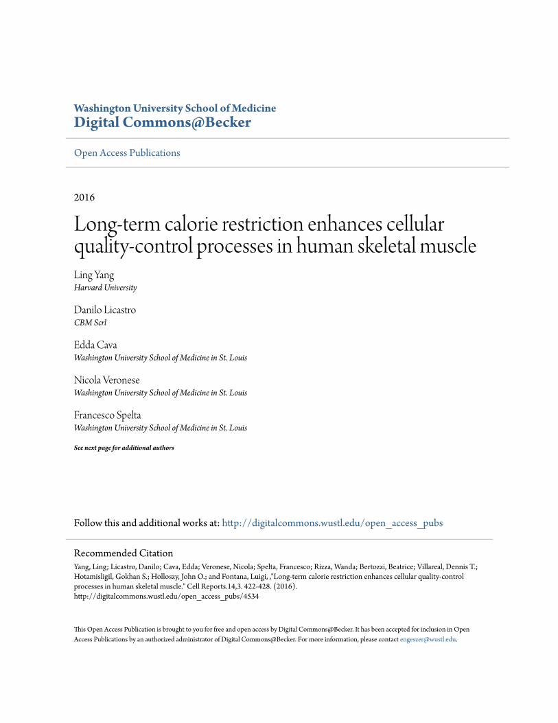
Yang et al. show that calorie restriction
without malnutrition in humans inhibits
inflammation, at least in part by elevating
serum cortisol concentration, and
increases chaperone and autophagy
genes and proteins involved in protein
quality control and organelle
homeostasis in the removal of
dysfunctional proteins and organelles
from cell.
Yang et al., 2016, Cell Reports 14, 422–428January 26, 2016 ª2016 The Authorshttp://dx.doi.org/10.1016/j.celrep.2015.12.042
Cell Reports
Report
Long-Term Calorie Restriction Enhances CellularQuality-Control Processes in Human Skeletal MuscleLing Yang,1,11 Danilo Licastro,2,11 Edda Cava,3,4,11 Nicola Veronese,3,5 Francesco Spelta,3,6 Wanda Rizza,3,7
Beatrice Bertozzi,3 Dennis T. Villareal,3,8 Gokhan S. Hotamisligil,1 John O. Holloszy,3 and Luigi Fontana3,9,10,*1Department of Genetics and Complex Diseases and Sabri Ulker Center, Harvard T.H. Chan School of Public Health, Boston, MA 02115, USA2CBM Scrl—Genomics, Area Science Park, Basovizza, 34149 Trieste, Italy3Division of Geriatrics and Nutritional Sciences and Center for Human Nutrition, Washington University School of Medicine, St. Louis,
MO 63110, USA4Department of Experimental Medicine, University of Rome ‘‘La Sapienza,’’ 00161 Rome, Italy5Division of Geriatrics, Department of Medicine, University of Padova, 35128 Padova, Italy6Department of Medicine, University of Verona, 37129 Verona, Italy7Department of Food and Human Nutrition Science, University Campus Bio-Medico, 00128 Rome, Italy8Baylor College of Medicine and Michael E. DeBakey VA Medical Center, Houston, TX 77030, USA9Department of Clinical and Experimental Sciences, Brescia University, 25121 Brescia, Italy10CEINGE Biotecnologie Avanzate, 80122 Napoli, Italy11Co-first author
*Correspondence: [email protected]
http://dx.doi.org/10.1016/j.celrep.2015.12.042
This is an open access article under the CC BY-NC-ND license (http://creativecommons.org/licenses/by-nc-nd/4.0/).
SUMMARY
Calorie restriction (CR) retards aging, acts as a hor-metic intervention, and increases serum corticoste-rone and HSP70 expression in rodents. However,less is known regarding the effects of CR on thesefactors in humans. Serum cortisol and molecularchaperones and autophagic proteins weremeasuredin the skeletalmuscle of subjects onCRdiets for 3–15years and in control volunteers. Serum cortisol washigher in the CR group than in age-matched seden-tary and endurance athlete groups (15.6 ± 4.6 ng/dlversus 12.3 ± 3.9 ng/dl and 11.2 ± 2.7 ng/dl, respec-tively; p % 0.001). HSP70, Grp78, beclin-1, and LC3mRNA and/or protein levels were higher in the skel-etal muscle of the CR group compared to controls.Our data indicate that CR in humans is associatedwith sustained rises in serum cortisol, reducedinflammation, and increases in keymolecular chaper-ones and autophagic mediators involved in cellularprotein quality control and removal of dysfunctionalproteins and organelles.
INTRODUCTION
Calorie restriction (CR) without malnutrition increases average
and maximal lifespan and prevents a range of chronic disease
in model organisms (Fontana et al., 2010). The mechanisms by
which CR delays aging and prevents or delays chronic diseases
are still unclear. Many interrelated and overlapping neuroendo-
crine adaptations have been proposed to play a role, including
reduction of several growth factors (e.g., insulin growth factor-1
[IGF-1] and insulin) that control the insulin/IGF-1/forkhead
box O (FOXO)/mammalian target of rapamycin (mTOR) pathway
and an increase in serum concentrations of glucocorticoids
(stress-induced hormones secreted by the adrenal cortex) (An-
derson et al., 2009; Mercken et al., 2012; de Cabo et al., 2003;
Omodei et al., 2013; Csiszar et al., 2013). Cortisol, the most
important human glucocorticoid, regulates important metabolic
functions and activates anti-stress and anti-inflammatory path-
ways (Sapolsky et al., 2000; Busillo and Cidlowski, 2013).
It has been hypothesized that CR works as a mild stressor
to trigger a hormetic response, resulting in reduced inflamma-
tion and increased expression of stress resistance proteins,
including the heat shock protein (HSP) molecular chaperones
(Mattson, 2008). In particular, CR in rodents has been shown
to increase the highly conserved HSP70 family, which serves
crucial roles in protein homeostasis and quality control (Heydari
et al., 1996; Selsby et al., 2005). HSP70 is amolecular chaperone
that coordinates several key cellular functions, including the un-
folding of misfolded or denatured proteins and the maintenance
of these proteins in an unfolded, folding-competent state. They
also protect nascently translated proteins, promote intracellular
transport of proteins, and reduce proteotoxicity by stabilizing
existing proteins against aggregation (Mayer and Bukau, 2005;
Stricher et al., 2013).
The purpose of the present study was to evaluate some of the
metabolic and molecular effects of long-term CR on stress-
induced hormones and molecular pathways in healthy lean and
weight-stablemen andwomen. Serumconcentrations of cortisol
and aldosterone in individuals consuming a CR diet were
compared with values obtained in two comparison groups: (1)
age- and sex-matched sedentary individuals consuming aWest-
ern diet (WD) and (2) age-, sex-, and body fat-matched endur-
ance runners consuming a WD. In this study, we also examined
the stress-related and anti-inflammatory molecular adaptations
induced by long-term CR in the skeletal muscle of healthy lean
men and women.
422 Cell Reports 14, 422–428, January 26, 2016 ª2016 The Authors
RESULTS
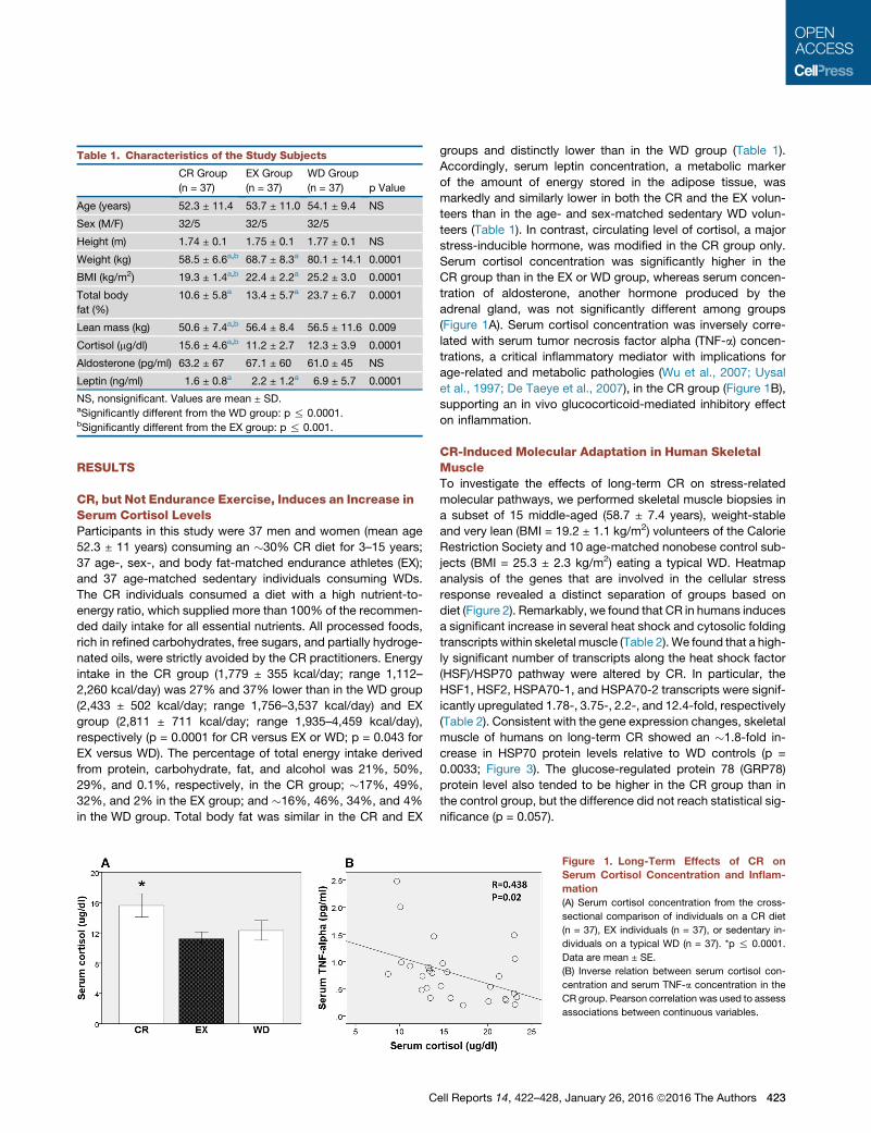
CR, but Not Endurance Exercise, Induces an Increase inSerum Cortisol LevelsParticipants in this study were 37 men and women (mean age
52.3 ± 11 years) consuming an �30% CR diet for 3–15 years;
37 age-, sex-, and body fat-matched endurance athletes (EX);
and 37 age-matched sedentary individuals consuming WDs.
The CR individuals consumed a diet with a high nutrient-to-
energy ratio, which supplied more than 100% of the recommen-
ded daily intake for all essential nutrients. All processed foods,
rich in refined carbohydrates, free sugars, and partially hydroge-
nated oils, were strictly avoided by the CR practitioners. Energy
intake in the CR group (1,779 ± 355 kcal/day; range 1,112–
2,260 kcal/day) was 27% and 37% lower than in the WD group
(2,433 ± 502 kcal/day; range 1,756–3,537 kcal/day) and EX
group (2,811 ± 711 kcal/day; range 1,935–4,459 kcal/day),
respectively (p = 0.0001 for CR versus EX or WD; p = 0.043 for
EX versus WD). The percentage of total energy intake derived
from protein, carbohydrate, fat, and alcohol was 21%, 50%,
29%, and 0.1%, respectively, in the CR group; �17%, 49%,
32%, and 2% in the EX group; and �16%, 46%, 34%, and 4%
in the WD group. Total body fat was similar in the CR and EX
groups and distinctly lower than in the WD group (Table 1).
Accordingly, serum leptin concentration, a metabolic marker
of the amount of energy stored in the adipose tissue, was
markedly and similarly lower in both the CR and the EX volun-
teers than in the age- and sex-matched sedentary WD volun-
teers (Table 1). In contrast, circulating level of cortisol, a major
stress-inducible hormone, was modified in the CR group only.
Serum cortisol concentration was significantly higher in the
CR group than in the EX or WD group, whereas serum concen-
tration of aldosterone, another hormone produced by the
adrenal gland, was not significantly different among groups
(Figure 1A). Serum cortisol concentration was inversely corre-
lated with serum tumor necrosis factor alpha (TNF-a) concen-
trations, a critical inflammatory mediator with implications for
age-related and metabolic pathologies (Wu et al., 2007; Uysal
et al., 1997; De Taeye et al., 2007), in the CR group (Figure 1B),
supporting an in vivo glucocorticoid-mediated inhibitory effect
on inflammation.
CR-Induced Molecular Adaptation in Human SkeletalMuscleTo investigate the effects of long-term CR on stress-related
molecular pathways, we performed skeletal muscle biopsies in
a subset of 15 middle-aged (58.7 ± 7.4 years), weight-stable
and very lean (BMI = 19.2 ± 1.1 kg/m2) volunteers of the Calorie
Restriction Society and 10 age-matched nonobese control sub-
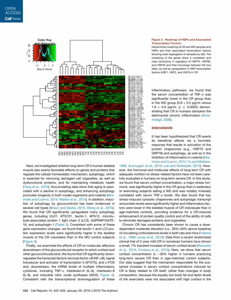
jects (BMI = 25.3 ± 2.3 kg/m2) eating a typical WD. Heatmap
analysis of the genes that are involved in the cellular stress
response revealed a distinct separation of groups based on
diet (Figure 2). Remarkably, we found that CR in humans induces
a significant increase in several heat shock and cytosolic folding
transcripts within skeletal muscle (Table 2).We found that a high-
ly significant number of transcripts along the heat shock factor
(HSF)/HSP70 pathway were altered by CR. In particular, the
HSF1, HSF2, HSPA70-1, and HSPA70-2 transcripts were signif-
icantly upregulated 1.78-, 3.75-, 2.2-, and 12.4-fold, respectively
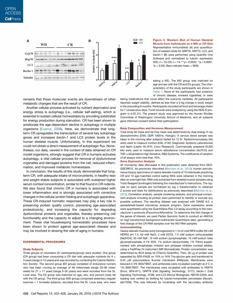
(Table 2). Consistent with the gene expression changes, skeletal
muscle of humans on long-term CR showed an �1.8-fold in-
crease in HSP70 protein levels relative to WD controls (p =
0.0033; Figure 3). The glucose-regulated protein 78 (GRP78)
protein level also tended to be higher in the CR group than in
the control group, but the difference did not reach statistical sig-
nificance (p = 0.057).
Figure 1. Long-Term Effects of CR on
Serum Cortisol Concentration and Inflam-
mation
(A) Serum cortisol concentration from the cross-
sectional comparison of individuals on a CR diet
(n = 37), EX individuals (n = 37), or sedentary in-
dividuals on a typical WD (n = 37). *p % 0.0001.
Data are mean ± SE.
(B) Inverse relation between serum cortisol con-
centration and serum TNF-a concentration in the
CR group. Pearson correlation was used to assess
associations between continuous variables.
Table 1. Characteristics of the Study Subjects
CR Group
(n = 37)
EX Group
(n = 37)
WD Group
(n = 37) p Value
Age (years) 52.3 ± 11.4 53.7 ± 11.0 54.1 ± 9.4 NS
Sex (M/F) 32/5 32/5 32/5
Height (m) 1.74 ± 0.1 1.75 ± 0.1 1.77 ± 0.1 NS
Weight (kg) 58.5 ± 6.6a,b 68.7 ± 8.3a 80.1 ± 14.1 0.0001
BMI (kg/m2) 19.3 ± 1.4a,b 22.4 ± 2.2a 25.2 ± 3.0 0.0001
Total body
fat (%)
10.6 ± 5.8a 13.4 ± 5.7a 23.7 ± 6.7 0.0001
Lean mass (kg) 50.6 ± 7.4a,b 56.4 ± 8.4 56.5 ± 11.6 0.009
Cortisol (mg/dl) 15.6 ± 4.6a,b 11.2 ± 2.7 12.3 ± 3.9 0.0001
Aldosterone (pg/ml) 63.2 ± 67 67.1 ± 60 61.0 ± 45 NS
Leptin (ng/ml) 1.6 ± 0.8a 2.2 ± 1.2a 6.9 ± 5.7 0.0001
NS, nonsignificant. Values are mean ± SD.aSignificantly different from the WD group: p % 0.0001.bSignificantly different from the EX group: p % 0.001.
Cell Reports 14, 422–428, January 26, 2016 ª2016 The Authors 423
Next, we investigated whether long-term CR in human skeletal
muscle also exerts favorable effects on genes and proteins that
regulate the cellular homeostatic mechanism, autophagy, which
is essential for removing damaged cell organelles, as well as
dysfunctional proteins, and for maintaining metabolic health
(Yang et al., 2010). Accumulating data show that aging is asso-
ciated with a decline in autophagy, and enhancing autophagy
promotes longevity in both model organisms and rodents (Mor-
imoto and Cuervo, 2014; Madeo et al., 2015). In addition, induc-
tion of autophagy by glucocorticoid has been evidenced in
several cell types (Braun and Marks, 2015; Wang et al., 2015).
We found that CR significantly upregulated many autophagy
genes, including ULK1, ATG101, beclin-1, APG12, microtu-
bule-associated protein 1 light chain 3 (LC3), GAPRAP/GATE-
16, and autophagin-1 (Table 2). Consistent with some of these
gene expression changes, we found that beclin-1 and LC3 pro-
tein expression levels were significantly higher in the skeletal
muscle of the CR volunteers than in the WD control subjects
(Figure 3).
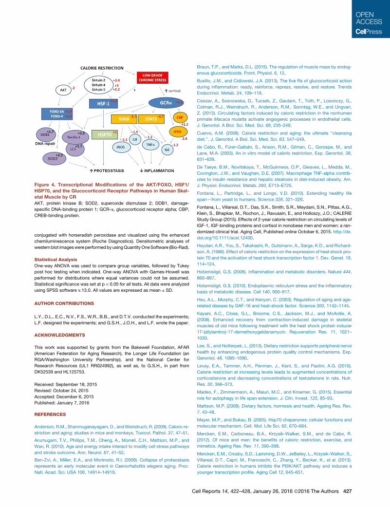
Finally, we examined the effects of CR on molecular effectors
downstream of the glucocorticoid receptor to which cortisol and
other glucocorticoids bind.We found that CR significantly down-
regulates the transcript factors necrosis factor kB (NF-kB), signal
transducer and activator of transcription 5 (STAT5), and c-FOS
and, downstream, the mRNA levels of multiple inflammatory
cytokines, including TNF-a, interleukin-6 (IL-6), interleukin-8
(IL-8), and inducible nitric oxide synthases (iNOS; Figure 4).
Consistent with the transcriptional downregulation of these
Figure 2. Heatmap of HSPs and Associated
Transcription Factors
Hierarchical clustering of CR andWD samples and
HSPs and their associated transcription factors
showing clear segregation of samples by diet. The
clustering of the genes show a consistent and
clear dichotomy in regulation of HSP70, HSP90,
and HSP40 and their homologs between the two
diets, as well as upregulation in HSP transcription
factors (HSF1, HSF2, and HSF3) in CR.
inflammatory pathways, we found that
the serum concentration of TNF-a was
significantly lower in the CR group than
in the WD group (0.8 ± 0.5 pg/ml versus
1.6 ± 0.9 pg/ml, p % 0.0002) demon-
strating that CR in humans dampens the
detrimental chronic inflammation (Hota-
misligil, 2006).
DISCUSSION
It has been hypothesized that CR exerts
its beneficial effects via a hormetic
response that results in activation of the
protein chaperones (e.g., HSP70 and
GRP78) and autophagy, as well as in the
inhibition of inflammation in rodents (Mor-
imoto and Cuervo, 2014; Yu andMattson,
1999; Arumugam et al., 2010; Lee and Notterpek, 2013). How-
ever, the hormonal and molecular effects of long-term CR with
adequate nutrition on stress-related factors have not been care-
fully evaluated in humans on long-term severe CR. In this study,
we found that serum cortisol concentration, a major stress hor-
mone, was significantly higher in the CR group than in sedentary
or exercising subjects eating a WD and was notably inversely
correlated with serum TNF-a levels. We also found that key
stress-induced cytosolic chaperones and autophagic transcript
and protein levels were significantly higher and inflammatory fac-
tors were lower in the skeletal muscle of CR individuals than in
age-matched controls, providing evidence for a CR-induced
enhancement of protein quality control and of the ability of cells
to eliminate damaged proteins and organelles.
Chronic CR has consistently been shown to cause a dose-
dependent moderate elevation (i.e., 30%–50% above baseline)
of circulating corticosterone levels in both rats andmice (Yaktine
et al., 1998; Levay et al., 2010). Data from a recent randomized
clinical trial of 2-year mild CR in nonobese humans have shown
a small, 7% transient increase of serum cortisol levels (Ravussin
et al., 2015; Fontana et al., 2015). Here, we show that serum
cortisol concentration is �30% higher in humans practicing
long-term severe CR than in age-matched control subjects.
Our data suggest that the mechanism responsible for the sus-
tained increase in serum cortisol concentrations induced by
CR is likely related to CR itself, rather than changes in body
composition, because the equally low body fat and leptin levels
of the exercisers were not associated with high cortisol in the
424 Cell Reports 14, 422–428, January 26, 2016 ª2016 The Authors
exercisers. Elevation of glucocorticoid levels is an essential
adaptation required to cope with a variety of stressors (Munck
et al., 1984), and in CR animals, high corticosterone level has
been shown to play a role in inhibiting inflammation and cancer
progression. Adrenalectomy abrogates the CR-induced cancer
inhibition, and glucocorticoid supplementation partially restores
cancer inhibition in CR adrenalectomized rodents (Pashko and
Schwartz, 1996; Stewart et al., 2005).
Whether the increased level of cortisol plays a direct role in
upregulating HSPs is unclear. However, it is well known that
CR increases HSF1 and HSP70 levels in rodents (Heydari
et al., 1996; Selsby et al., 2005). Here, we show that long-term
CR significantly upregulates transcripts along the HSF/HSP70
pathway and increases HSP70 and GRP78 protein levels in
the human skeletal muscle. Because aging is associated with
reduced protein folding capacity and ability to maintain homeo-
stasis in response to stress (Ben-Zvi et al., 2009; Kayani et al.,
2008), these data suggest that CR in humans prevents this
decrease and may be involved in the slowing of age-dependent
accumulation of damaged and dysfunctional proteins. These
changes would also contribute to overall functional capacity
and health of organelles, such as endoplasmic reticulum, that
are integral to inflammatory and metabolic regulation (Hotamisli-
gil, 2010). Overexpression of HSF and HSP70 has been shown to
extend lifespan by 50%–100% in C. elegans (Hsu et al., 2003;
Yokoyama et al., 2002). Finally, suppression of inflammation
may contribute to proteostasis, because endoplasmic reticulum
function is also compromised in the presence of inflammation
through nitrosylation and inhibition of key adaptive molecules
such as IRE1 (Yang et al., 2015). However, the possibility
Table 2. Effects of CR on Stress-Inducible Chaperones and Autophagy Genes in Muscle
Symbol Function LogFC p Value
HSP Family of Molecular Chaperones
HSF1 +1.78 6.6e-05
HSF2 DNA-binding protein that specifically binds heat shock promoter elements and activates
transcription
+3.75 2.6e-12
HSPA70-1A and B cytosolic major stress-inducible chaperones required for refolding of damaged and
unfolded proteins
+2.16 0.017
HSPA70-1 like signalosome, mitochondrial matrix; response to unfolded protein +2.88 2e-6
HSPA70-2 cytosol, constitutive +12.45 0.000001
HSPA70-4 +2.54 1e-6
HSPA70-5 endoplasmic reticulum; folding or assembly of proteins in endoplasmic reticulum and
stress-induced autophagy
�2.86 0.000001
HSPA70-6 cytosol, inducible by severe stress insults �3.39 9.3e-5
HSPA70-8 cytosol, constitutive, homeostatic function +1.7 0.12
HSPA70-9 mitochondria, constitutive +2.19 1e-6
HSPA70-13 +1.95 8.0e-6
HSPA70-14 +2.35 0.00001
HSP90-AA1 (a) stress inducible �4.35 0.00001
HSP90-AB1 (b) constitutive +1.45 0.001
Autophagy
ULK1 serine/threonine protein kinase is involved in autophagy in response to starvation +1.53 3.8e-05
ATG101 cytosol protects ATG13 from proteasomal degradation, therefore stabilizing levels of
ATG13 found in cells and regulating levels of macroautophagy
+1.45 8.3e-05
Beclin-1 Beclin-1 plays a central role in autophagy and, with its binding partner class III
phosphoinositide 3-kinase, is required for the initiation of the formation of the
autophagosome in autophagy
+1.51 1.7e-7
APG12 Apg12 conjugation of Apg5 is required for elongation of the isolation membrane to form a
complete spherical autophagosome
�2.07 1e-10
APG16L1 the protein encoded by this gene is part of a large protein complex that is necessary for
autophagy
+1.59 4.5e-10
LC3 LC3 is involved in elongation of the phagophore membrane and is an important marker and
effector of starvation-induced autophagy
+1.68 4.1e-7
GAPRAP GAPRAP is essential for a later stage in autophagosome maturation +1.47 3.5e-06
GATE-16 GATE-16 is essential for a later stage in autophagosome maturation +1.27 0.00013
Autophagin-1 Autophagin-1 cleaves the carboxyl termini of the LC3, GABARAP, and GATE-16, a reaction
essential for its lipidation during autophagy
+1.44 0.05
LogFC, log fold change.
Cell Reports 14, 422–428, January 26, 2016 ª2016 The Authors 425
remains that these molecular events are downstream of other
metabolic changes that are the result of CR.
Another cellular process activated by nutrient deprivation and
energy stress is autophagy (i.e., cellular self-eating), which is
essential to sustain cellular homeostasis by providing substrates
for energy production during starvation. CR has been shown to
ameliorate the age-dependent decline in autophagy in multiple
organisms (Cuervo, 2008). Here, we demonstrate that long-
term CR upregulates the transcription of several key autophagy
genes and increases beclin-1 and LC3 protein levels in the
human skeletal muscle. Unfortunately, in this experiment we
could not obtain a direct measurement of autophagic flux. None-
theless, our data, viewed in the context of data obtained on CR
model organisms, strongly suggest that CR in humans activates
autophagy, a vital cellular process for removal of dysfunctional
organelles and damaged proteins from the cell, reduced inflam-
mation, and improved metabolic homeostasis.
In conclusion, the results of this study demonstrate that long-
term CR, with adequate intake of micronutrients, in healthy lean
and weight-stable subjects is associated with sustained higher
serum cortisol concentration, similar to that found in CR rodents.
We also found that chronic CR in humans is associated with
lower inflammation and is strongly associated with corrective
changes in the cellular protein folding and autophagic apparatus.
These CR-induced hormetic responses may play a key role in
preserving protein quality control, preventing age-associated
proteotoxicity, and increasing the capacity for degrading
dysfunctional proteins and organelles, thereby preserving cell
functionality and the capacity to adjust to a changing environ-
ment. These vital housekeeping homeostatic processes have
been shown to protect against age-associated disease and
may be involved in slowing the rate of aging in humans.
EXPERIMENTAL PROCEDURES
Study Subjects
Three groups of volunteers (37 participants/group) were studied. One group
(CR group) had been consuming a CR diet with adequate nutrients for 6 ±
3 years (range 3–15 years) andwas recruited by contacting the Calorie Restric-
tion Society. The second group (EX group) consisted of endurance runners
who had been running an average of 48 miles/week (range 20–90 miles/
week) for 21 ± 11 years (range 5–35 years) and were recruited from the St.
Louis area. The EX group was matched on age, sex, and percent body fat
with the CR group. The third group (WD group) comprised sedentary (regular
exercise < 1 hr/week) subjects, recruited from the St. Louis area, who were
A B Figure 3. Western Blot of Human Skeletal
Muscle from Individuals on a WD or CR Diet
Representative immunoblots (A) and quantifica-
tion of western blots for GRP78, HSP70, LC3, and
beclin-1 (B) were performed using Quantity One
Software and normalized to tubulin expression
(WD, n = 10; CR, n = 14; ***p = 0.0033, **p < 0.0007,
*p < 0.03). Bars indicate mean ± SEM.
eating a WD. The WD group was matched on
age and sex with the CR and EX groups. The char-
acteristics of the study participants are shown in
Table 1. None of the participants had evidence
of chronic disease, smoked cigarettes, or were
taking medications that could affect the outcome variables. All participants
reported weight stability, defined as less than a 2-kg change in body weight
in the preceding 6 months. Participants recorded all food and beverage intake
for 7 consecutive days. Food records were analyzed by using the NDS-R pro-
gram (v.4.03_31). The present study was approved by the Human Studies
Committee of Washington University School of Medicine, and all subjects
gave informed consent before their participation.
Body Composition and Hormone Measurement
Total body fat mass and fat-free mass was determined by dual-energy X-ray
absorptiometry (DXA; QDR 1000/w; Hologic). A venous blood sample was
taken in the morning after subjects fasted for 12 hr. Radioimmunoassay kits
were used to measure cortisol (DSL-2100; Diagnostic Systems Laboratories)
and leptin (Leptin HL-81K; Linco Research). Commercially prepared ELISA
kits were used to measure serum aldosterone concentration (ALPCO) and
TNF-a (Quantakine High Sensitive, R&D Systems). The coefficients of variation
of all assays were less than 10%.
Gene Expression Analysis
All microarray data discussed in this publication were obtained from GEO:
GSE38012. As previously described (Mercken et al., 2013), human percuta-
neous biopsy specimens of vastus lateralis muscle of 15 individuals practicing
CR and 10 age-matched control eating WDs were obtained in the morning
after an overnight fast. RNAwas extracted from skeletal muscle samples using
Trizol Reagent (Invitrogen) following the manufacturer’s instructions. The sig-
nals on each sample are normalized by log z transformation to obtained
Z scores and tests for distributions as previously described (Mercken et al.,
2013). Correlation analysis, sample clustering analysis, and principal-compo-
nent analysis including all probes were performed to identify or exclude any
possible outliners. The resulting dataset was analyzed with DIANE 6.0, a
spreadsheet-based microarray analysis program. Gene expression levels
were quantitated using the QuantiGene Plex 2.0 assay according to the man-
ufacturer’s protocols (Panomics/Affymetrix). To determine the fold change of
the genes of interest, we used Partek Genomic Suite to conduct an ANOVA
on log2 transformed background-subtracted QuantiGene data, generating a
fold change of the CR RNA samples over the WD RNA samples.
Immunoblotting
Vastus lateralis muscles were homogenized in 1 ml of cold RIPA buffer (50 mM
HEPES pH 7.4, 40 mM NaCl, 2 mM EDTA, 1.5 mM sodium orthovanadate
(Na3VO4), 50 mM NaF, 10 mM sodium pyrophosphate, 10 mM sodium beta
glycerophosphate, 0.1% SDS, 1% sodium deoxycholate, 1% Triton) supple-
mented with phosphatase inhibitor and protease inhibitor cocktail tablets,
using a FastPrep 24 instrument (MP Biomedicals). Protein concentration was
determined by BCA assay kit (Thermo Scientific). Then, 30 mg of protein was
separated by SDS-PAGE on 10% or 16% Tris-glycine gels and transferred to
0.45 mM polyvinylidene fluoride membrane (Millipore). Membranes were
blocked in 3% BSA/TBST, and antibodies were incubated overnight at 4�C in
1% BSA/TBST. The membranes were incubated with antibodies to HSP70
(Enzo, SPA-811), GRP78 (Cell Signaling Technology, 3177), beclin-1 (Cell
Signaling Technology, 3738), and LC3 (Novus Biologicals, NB100-2220), and
loading was verified by blotting for tubulin-horseradish peroxidase (Abcam,
ab21058). This was followed by incubating with the secondary antibody
426 Cell Reports 14, 422–428, January 26, 2016 ª2016 The Authors
conjugated with horseradish peroxidase and visualized using the enhanced
chemiluminescence system (Roche Diagnostics). Densitometric analyses of
westernblot imageswere performedbyusingQuantityOneSoftware (Bio-Rad).
Statistical Analysis
One-way ANOVA was used to compare group variables, followed by Tukey
post hoc testing when indicated. One-way ANOVA with Games-Howell was
performed for distributions where equal variances could not be assumed.
Statistical significance was set at p < 0.05 for all tests. All data were analyzed
using SPSS software v.13.0. All values are expressed as mean ± SD.
AUTHOR CONTRIBUTIONS
L.Y., D.L., E.C., N.V., F.S., W.R., B.B., and D.T.V. conducted the experiments;
L.F. designed the experiments; and G.S.H., J.O.H., and L.F. wrote the paper.
ACKNOWLEDGMENTS
This work was supported by grants from the Bakewell Foundation, AFAR
(American Federation for Aging Research), the Longer Life Foundation (an
RGA/Washington University Partnership), and the National Center for
Research Resources (UL1 RR024992), as well as, to G.S.H., in part from
DK52539 and HL125753.
Received: September 18, 2015
Revised: October 24, 2015
Accepted: December 6, 2015
Published: January 7, 2016
REFERENCES
Anderson, R.M., Shanmuganayagam, D., andWeindruch, R. (2009). Caloric re-
striction and aging: studies in mice and monkeys. Toxicol. Pathol. 37, 47–51.
Arumugam, T.V., Phillips, T.M., Cheng, A., Morrell, C.H., Mattson, M.P., and
Wan, R. (2010). Age and energy intake interact to modify cell stress pathways
and stroke outcome. Ann. Neurol. 67, 41–52.
Ben-Zvi, A., Miller, E.A., and Morimoto, R.I. (2009). Collapse of proteostasis
represents an early molecular event in Caenorhabditis elegans aging. Proc.
Natl. Acad. Sci. USA 106, 14914–14919.
Braun, T.P., and Marks, D.L. (2015). The regulation of muscle mass by endog-
enous glucocorticoids. Front. Physiol. 6, 12.
Busillo, J.M., and Cidlowski, J.A. (2013). The five Rs of glucocorticoid action
during inflammation: ready, reinforce, repress, resolve, and restore. Trends
Endocrinol. Metab. 24, 109–119.
Csiszar, A., Sosnowska, D., Tucsek, Z., Gautam, T., Toth, P., Losonczy, G.,
Colman, R.J., Weindruch, R., Anderson, R.M., Sonntag, W.E., and Ungvari,
Z. (2013). Circulating factors induced by caloric restriction in the nonhuman
primate Macaca mulatta activate angiogenic processes in endothelial cells.
J. Gerontol. A Biol. Sci. Med. Sci. 68, 235–249.
Cuervo, A.M. (2008). Calorie restriction and aging: the ultimate ‘‘cleansing
diet.’’. J. Gerontol. A Biol. Sci. Med. Sci. 63, 547–549.
de Cabo, R., F€urer-Galban, S., Anson, R.M., Gilman, C., Gorospe, M., and
Lane, M.A. (2003). An in vitro model of caloric restriction. Exp. Gerontol. 38,
631–639.
De Taeye, B.M., Novitskaya, T., McGuinness, O.P., Gleaves, L., Medda, M.,
Covington, J.W., and Vaughan, D.E. (2007). Macrophage TNF-alpha contrib-
utes to insulin resistance and hepatic steatosis in diet-induced obesity. Am.
J. Physiol. Endocrinol. Metab. 293, E713–E725.
Fontana, L., Partridge, L., and Longo, V.D. (2010). Extending healthy life
span—from yeast to humans. Science 328, 321–326.
Fontana, L., Villareal, D.T., Das, S.K., Smith, S.R., Meydani, S.N., Pittas, A.G.,
Klein, S., Bhapkar, M., Rochon, J., Ravussin, E., and Holloszy, J.O.; CALERIE
Study Group (2015). Effects of 2-year calorie restriction on circulating levels of
IGF-1, IGF-binding proteins and cortisol in nonobese men and women: a ran-
domized clinical trial. Aging Cell, Published online October 6, 2015. http://dx.
doi.org/10.1111/acel.12400.
Heydari, A.R., You, S., Takahashi, R., Gutsmann, A., Sarge, K.D., and Richard-
son, A. (1996). Effect of caloric restriction on the expression of heat shock pro-
tein 70 and the activation of heat shock transcription factor 1. Dev. Genet. 18,
114–124.
Hotamisligil, G.S. (2006). Inflammation and metabolic disorders. Nature 444,
860–867.
Hotamisligil, G.S. (2010). Endoplasmic reticulum stress and the inflammatory
basis of metabolic disease. Cell 140, 900–917.
Hsu, A.L., Murphy, C.T., and Kenyon, C. (2003). Regulation of aging and age-
related disease by DAF-16 and heat-shock factor. Science 300, 1142–1145.
Kayani, A.C., Close, G.L., Broome, C.S., Jackson, M.J., and McArdle, A.
(2008). Enhanced recovery from contraction-induced damage in skeletal
muscles of old mice following treatment with the heat shock protein inducer
17-(allylamino)-17-demethoxygeldanamycin. Rejuvenation Res. 11, 1021–
1030.
Lee, S., and Notterpek, L. (2013). Dietary restriction supports peripheral nerve
health by enhancing endogenous protein quality control mechanisms. Exp.
Gerontol. 48, 1085–1090.
Levay, E.A., Tammer, A.H., Penman, J., Kent, S., and Paolini, A.G. (2010).
Calorie restriction at increasing levels leads to augmented concentrations of
corticosterone and decreasing concentrations of testosterone in rats. Nutr.
Res. 30, 366–373.
Madeo, F., Zimmermann, A., Maiuri, M.C., and Kroemer, G. (2015). Essential
role for autophagy in life span extension. J. Clin. Invest. 125, 85–93.
Mattson, M.P. (2008). Dietary factors, hormesis and health. Ageing Res. Rev.
7, 43–48.
Mayer, M.P., and Bukau, B. (2005). Hsp70 chaperones: cellular functions and
molecular mechanism. Cell. Mol. Life Sci. 62, 670–684.
Mercken, E.M., Carboneau, B.A., Krzysik-Walker, S.M., and de Cabo, R.
(2012). Of mice and men: the benefits of caloric restriction, exercise, and
mimetics. Ageing Res. Rev. 11, 390–398.
Mercken, E.M., Crosby, S.D., Lamming, D.W., JeBailey, L., Krzysik-Walker, S.,
Villareal, D.T., Capri, M., Franceschi, C., Zhang, Y., Becker, K., et al. (2013).
Calorie restriction in humans inhibits the PI3K/AKT pathway and induces a
younger transcription profile. Aging Cell 12, 645–651.
Figure 4. Transcriptional Modifications of the AKT/FOXO, HSF1/
HSP70, and the Glucocorticoid Receptor Pathways in Human Skel-
etal Muscle by CR
AKT, protein kinase B; SOD2, superoxide dismutase 2; DDB1, damage-
specific DNA-binding protein 1; GCR-a, glucocorticoid receptor alpha; CBP,
CREB-binding protein.
Cell Reports 14, 422–428, January 26, 2016 ª2016 The Authors 427
Morimoto, R.I., and Cuervo, A.M. (2014). Proteostasis and the aging proteome
in health and disease. J. Gerontol. A Biol. Sci. Med. Sci. 69 (Suppl 1), S33–S38.
Munck, A., Guyre, P.M., and Holbrook, N.J. (1984). Physiological functions of
glucocorticoids in stress and their relation to pharmacological actions. Endocr.
Rev. 5, 25–44.
Omodei, D., Licastro, D., Salvatore, F., Crosby, S.D., and Fontana, L. (2013).
Serum from humans on long-term calorie restriction enhances stress resis-
tance in cell culture. Aging (Albany, N.Y.) 5, 599–606.
Pashko, L.L., and Schwartz, A.G. (1996). Inhibition of 7,12-dimethylbenz[a]
anthracene-induced lung tumorigenesis in A/J mice by food restriction is
reversed by adrenalectomy. Carcinogenesis 17, 209–212.
Ravussin, E., Redman, L.M., Rochon, J., Das, S.K., Fontana, L., Kraus, W.E.,
Romashkan, S., Williamson, D.A., Meydani, S.N., Villareal, D.T., et al.;
CALERIE Study Group (2015). A 2-year randomized controlled trial of human
caloric restriction: feasibility and effects on predictors of health span and
longevity. J. Gerontol. A Biol. Sci. Med. Sci. 70, 1097–1104.
Sapolsky, R.M., Romero, L.M., and Munck, A.U. (2000). How do glucocorti-
coids influence stress responses? Integrating permissive, suppressive, stimu-
latory, and preparative actions. Endocr. Rev. 21, 55–89.
Selsby, J.T., Judge, A.R., Yimlamai, T., Leeuwenburgh, C., and Dodd, S.L.
(2005). Life long calorie restriction increases heat shock proteins and protea-
some activity in soleus muscles of Fisher 344 rats. Exp. Gerontol. 40, 37–42.
Stewart, J.W., Koehler, K., Jackson, W., Hawley, J., Wang, W., Au, A., Myers,
R., and Birt, D.F. (2005). Prevention of mouse skin tumor promotion by dietary
energy restriction requires an intact adrenal gland and glucocorticoid supple-
mentation restores inhibition. Carcinogenesis 26, 1077–1084.
Stricher, F., Macri, C., Ruff, M., and Muller, S. (2013). HSPA8/HSC70
chaperone protein: structure, function, and chemical targeting. Autophagy 9,
1937–1954.
Uysal, K.T., Wiesbrock, S.M., Marino, M.W., and Hotamisligil, G.S. (1997). Pro-
tection from obesity-induced insulin resistance in mice lacking TNF-alpha
function. Nature 389, 610–614.
Wang, L., Fan, J., Lin, Y.S., Guo, Y.S., Gao, B., Shi, Q.Y., Wei, B.Y., Chen, L.,
Yang, L., Liu, J., and Luo, Z.J. (2015). Glucocorticoids induce autophagy in rat
bone marrow mesenchymal stem cells. Mol. Med. Rep. 11, 2711–2716.
Wu, D., Ren, Z., Pae, M., Guo, W., Cui, X., Merrill, A.H., and Meydani, S.N.
(2007). Aging up-regulates expression of inflammatory mediators in mouse
adipose tissue. J. Immunol. 179, 4829–4839.
Yaktine, A.L., Vaughn, R., Blackwood, D., Duysen, E., and Birt, D.F. (1998).
Dietary energy restriction in the SENCAR mouse: elevation of glucocorticoid
hormone levels but no change in distribution of glucocorticoid receptor in
epidermal cells. Mol. Carcinog. 21, 62–69.
Yang, L., Li, P., Fu, S., Calay, E.S., and Hotamisligil, G.S. (2010). Defective he-
patic autophagy in obesity promotes ER stress and causes insulin resistance.
Cell Metab. 11, 467–478.
Yang, L., Calay, E.S., Fan, J., Arduini, A., Kunz, R.C., Gygi, S.P., Yalcin, A., Fu,
S., and Hotamisligil, G.S. (2015). Metabolism. S-nitrosylation links obesity-
associated inflammation to endoplasmic reticulum dysfunction. Science
349, 500–506.
Yokoyama, K., Fukumoto, K., Murakami, T., Harada, S., Hosono, R., Wadhwa,
R., Mitsui, Y., and Ohkuma, S. (2002). Extended longevity of Caenorhabditis
elegans by knocking in extra copies of hsp70F, a homolog of mot-2 (mor-
talin)/mthsp70/Grp75. FEBS Lett. 516, 53–57.
Yu, Z.F., and Mattson, M.P. (1999). Dietary restriction and 2-deoxyglucose
administration reduce focal ischemic brain damage and improve behavioral
outcome: evidence for a preconditioning mechanism. J. Neurosci. Res. 57,
830–839.
428 Cell Reports 14, 422–428, January 26, 2016 ª2016 The Authors


![Effects of Popular Diets without Specific Calorie Targets on ......weight loss induced by calorie-restricted diets [1], alternative dietary approaches to achieve short-and long-term](https://img.dokumen.tips/doc/110x75/6039e66269d7655eb0436a25/effects-of-popular-diets-without-specific-calorie-targets-on-weight-loss.jpg)