Embed Size (px)
Citation preview
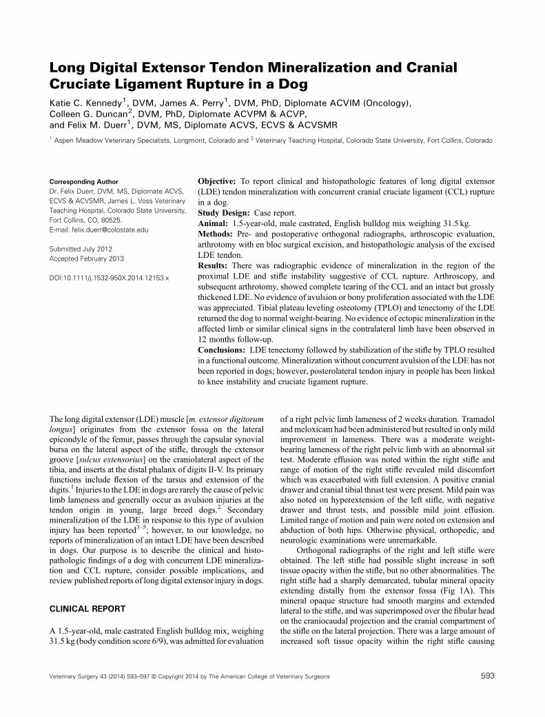
Long Digital Extensor Tendon Mineralization and CranialCruciate Ligament Rupture in a DogKatie C. Kennedy1, DVM, James A. Perry1, DVM, PhD, Diplomate ACVIM (Oncology),Colleen G. Duncan2, DVM, PhD, Diplomate ACVPM & ACVP,and Felix M. Duerr1, DVM, MS, Diplomate ACVS, ECVS & ACVSMR1 Aspen Meadow Veterinary Specialists, Longmont, Colorado and 2 Veterinary Teaching Hospital, Colorado State University, Fort Collins, Colorado
Corresponding AuthorDr. Felix Duerr, DVM, MS, Diplomate ACVS,ECVS & ACVSMR, James L. Voss VeterinaryTeaching Hospital, Colorado State University,Fort Collins, CO, 80525.E‐mail: [email protected]
Submitted July 2012Accepted February 2013
DOI:10.1111/j.1532-950X.2014.12153.x
Objective: To report clinical and histopathologic features of long digital extensor(LDE) tendon mineralization with concurrent cranial cruciate ligament (CCL) rupturein a dog.Study Design: Case report.Animal: 1.5‐year‐old, male castrated, English bulldog mix weighing 31.5 kg.Methods: Pre‐ and postoperative orthogonal radiographs, arthroscopic evaluation,arthrotomy with en bloc surgical excision, and histopathologic analysis of the excisedLDE tendon.Results: There was radiographic evidence of mineralization in the region of theproximal LDE and stifle instability suggestive of CCL rupture. Arthroscopy, andsubsequent arthrotomy, showed complete tearing of the CCL and an intact but grosslythickened LDE. No evidence of avulsion or bony proliferation associated with the LDEwas appreciated. Tibial plateau leveling osteotomy (TPLO) and tenectomy of the LDEreturned the dog to normal weight‐bearing. No evidence of ectopic mineralization in theaffected limb or similar clinical signs in the contralateral limb have been observed in12 months follow‐up.Conclusions: LDE tenectomy followed by stabilization of the stifle by TPLO resultedin a functional outcome. Mineralization without concurrent avulsion of the LDE has notbeen reported in dogs; however, posterolateral tendon injury in people has been linkedto knee instability and cruciate ligament rupture.
The long digital extensor (LDE) muscle [m. extensor digitorumlongus] originates from the extensor fossa on the lateralepicondyle of the femur, passes through the capsular synovialbursa on the lateral aspect of the stifle, through the extensorgroove [sulcus extensorius] on the craniolateral aspect of thetibia, and inserts at the distal phalanx of digits II‐V. Its primaryfunctions include flexion of the tarsus and extension of thedigits.1 Injuries to the LDE in dogs are rarely the cause of pelviclimb lameness and generally occur as avulsion injuries at thetendon origin in young, large breed dogs.2 Secondarymineralization of the LDE in response to this type of avulsioninjury has been reported3–5; however, to our knowledge, noreports of mineralization of an intact LDE have been describedin dogs. Our purpose is to describe the clinical and histo-pathologic findings of a dog with concurrent LDE mineraliza-tion and CCL rupture, consider possible implications, andreview published reports of long digital extensor injury in dogs.
CLINICAL REPORT
A 1.5‐year‐old, male castrated English bulldog mix, weighing31.5 kg (body condition score 6/9), was admitted for evaluation
of a right pelvic limb lameness of 2 weeks duration. Tramadolandmeloxicam had been administered but resulted in onlymildimprovement in lameness. There was a moderate weight‐bearing lameness of the right pelvic limb with an abnormal sittest. Moderate effusion was noted within the right stifle andrange of motion of the right stifle revealed mild discomfortwhich was exacerbated with full extension. A positive cranialdrawer and cranial tibial thrust test were present. Mild pain wasalso noted on hyperextension of the left stifle, with negativedrawer and thrust tests, and possible mild joint effusion.Limited range of motion and pain were noted on extension andabduction of both hips. Otherwise physical, orthopedic, andneurologic examinations were unremarkable.
Orthogonal radiographs of the right and left stifle wereobtained. The left stifle had possible slight increase in softtissue opacity within the stifle, but no other abnormalities. Theright stifle had a sharply demarcated, tubular mineral opacityextending distally from the extensor fossa (Fig 1A). Thismineral opaque structure had smooth margins and extendedlateral to the stifle, and was superimposed over the fibular headon the craniocaudal projection and the cranial compartment ofthe stifle on the lateral projection. There was a large amount ofincreased soft tissue opacity within the right stifle causing
Veterinary Surgery 43 (2014) 593–597 © Copyright 2014 by The American College of Veterinary Surgeons 593

effacement of the infrapatellar fat pad and distention of thecaudal joint pouch. There were irregular mineral opacitiessuperimposed over the distal aspect of the tibia on the lateralprojections. Degenerative changes of the right coxofemoraljoint were also noted on the craniocaudal projection. The rightand left tibial plateau angles were measured according tostandard technique and both were determined to be 26°.
Arthroscopy (2.7mm 30° arthroscope; Karl Storz,Tuttlingen, Germany) of the right stifle using medial andlateral parapatellar portals revealed a complete tear of the CCLwith no visible or palpable evidence of damage to the medial orlateral menisci, moderate synovitis, minimal degenerativechange, and no evidence of osteochondrosis. The LDE tendonappeared stretched and moderately inflamed/thickened but nogross evidence of mineralization was observed (Fig 2). Thelateral parapatellar portal was extended to a 3 cm arthrotomy toallow further evaluation of the LDE. The tendon was intact, butgrossly thickened and mineralization was palpable. Release ofthe tendon at its origin was performed using a #11 blade andthe segment of tendon with palpable changes (�1 cm) wasremoved en bloc. Rongeurs were used to further debride theorigin of the LDE. The free portion of the remaining tendonwas sutured to proximolateral tibial fascia associated with theextensor groove of the tibia using 2–0 polypropylene in ahorizontal mattress pattern. TPLO was performed using a21mm blade (New Generation Devices, Glen Rock, NJ) with7.25mm rotation and a mini‐3.5mm Synthes (West Chester,PA) locking TPLO plate. Three locking screws (VeterinaryOrthopedic Implants, Saint Augustine, FL) were placed for theproximal fragment and conventional self‐tapping corticalscrews (Veterinary Orthopedic Implants) were placed for thedistal 3 screws. The procedure resulted in a stable stiflepostoperatively (no cranial tibial thrust). The lateral arthrotomywas closed and the pes anserinus apposed using 2–0
poliglecaprone in a simple continuous pattern. The medialskin incision was closed with 2–0 poliglecaprone in asubcuticular pattern followed by skin apposition with staples.
On postoperative radiographs of the right stifle, there wascomplete removal of the abnormal area of mineral opacity(Fig 1B) confirming that the radiographic lesion indeed was theLDE. Tibial plateau angle after surgery was 5°. The excisedportion of LDE tendon was radiographed ex vivo confirming itsradiodensity and was then fixed in formalin for microscopicevaluation. Histologically, the radiodense mass consisted of aregionally extensive area of mineral (Fig 3). At the periphery of
Figure 1 Lateral radiograph of the right stifle. (A) A sharply demarcated, tubular mineral opacity can be seen extending distally from the extensor fossain the region of the LDE. (B) After TPLO and en bloc resection of the grossly abnormal portion of LDE.
Figure 2 Arthroscopic evaluation of the LDE tendon within the stifle.
594 Veterinary Surgery 43 (2014) 593–597 © Copyright 2014 by The American College of Veterinary Surgeons
Long Digital Extensor Tendon Mineralization and Cranial Cruciate Ligament Rupture Kennedy et al.
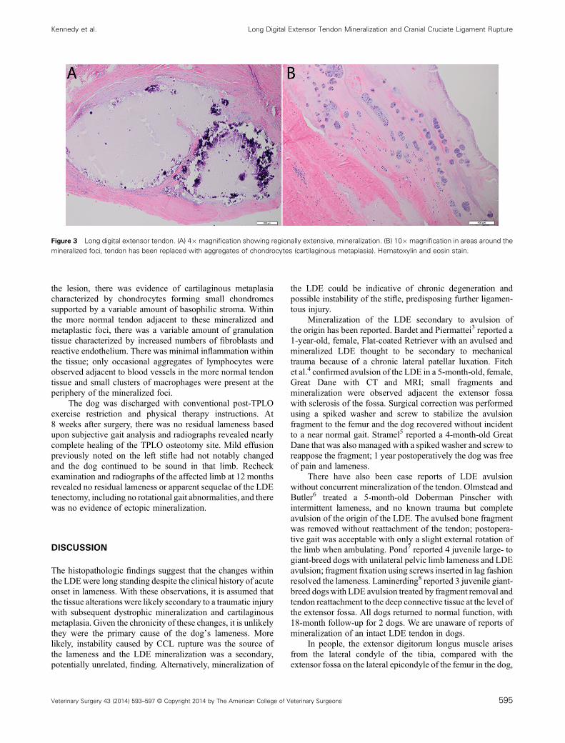
the lesion, there was evidence of cartilaginous metaplasiacharacterized by chondrocytes forming small chondromessupported by a variable amount of basophilic stroma. Withinthe more normal tendon adjacent to these mineralized andmetaplastic foci, there was a variable amount of granulationtissue characterized by increased numbers of fibroblasts andreactive endothelium. There was minimal inflammation withinthe tissue; only occasional aggregates of lymphocytes wereobserved adjacent to blood vessels in the more normal tendontissue and small clusters of macrophages were present at theperiphery of the mineralized foci.
The dog was discharged with conventional post‐TPLOexercise restriction and physical therapy instructions. At8 weeks after surgery, there was no residual lameness basedupon subjective gait analysis and radiographs revealed nearlycomplete healing of the TPLO osteotomy site. Mild effusionpreviously noted on the left stifle had not notably changedand the dog continued to be sound in that limb. Recheckexamination and radiographs of the affected limb at 12 monthsrevealed no residual lameness or apparent sequelae of the LDEtenectomy, including no rotational gait abnormalities, and therewas no evidence of ectopic mineralization.
DISCUSSION
The histopathologic findings suggest that the changes withinthe LDEwere long standing despite the clinical history of acuteonset in lameness. With these observations, it is assumed thatthe tissue alterations were likely secondary to a traumatic injurywith subsequent dystrophic mineralization and cartilaginousmetaplasia. Given the chronicity of these changes, it is unlikelythey were the primary cause of the dog’s lameness. Morelikely, instability caused by CCL rupture was the source ofthe lameness and the LDE mineralization was a secondary,potentially unrelated, finding. Alternatively, mineralization of
the LDE could be indicative of chronic degeneration andpossible instability of the stifle, predisposing further ligamen-tous injury.
Mineralization of the LDE secondary to avulsion ofthe origin has been reported. Bardet and Piermattei3 reported a1‐year‐old, female, Flat‐coated Retriever with an avulsed andmineralized LDE thought to be secondary to mechanicaltrauma because of a chronic lateral patellar luxation. Fitchet al.4 confirmed avulsion of the LDE in a 5‐month‐old, female,Great Dane with CT and MRI; small fragments andmineralization were observed adjacent the extensor fossawith sclerosis of the fossa. Surgical correction was performedusing a spiked washer and screw to stabilize the avulsionfragment to the femur and the dog recovered without incidentto a near normal gait. Stramel5 reported a 4‐month‐old GreatDane that was also managed with a spiked washer and screw toreappose the fragment; 1 year postoperatively the dog was freeof pain and lameness.
There have also been case reports of LDE avulsionwithout concurrent mineralization of the tendon. Olmstead andButler6 treated a 5‐month‐old Doberman Pinscher withintermittent lameness, and no known trauma but completeavulsion of the origin of the LDE. The avulsed bone fragmentwas removed without reattachment of the tendon; postopera-tive gait was acceptable with only a slight external rotation ofthe limb when ambulating. Pond7 reported 4 juvenile large‐ togiant‐breed dogs with unilateral pelvic limb lameness and LDEavulsion; fragment fixation using screws inserted in lag fashionresolved the lameness. Laminerding8 reported 3 juvenile giant‐breed dogs with LDE avulsion treated by fragment removal andtendon reattachment to the deep connective tissue at the level ofthe extensor fossa. All dogs returned to normal function, with18‐month follow‐up for 2 dogs. We are unaware of reports ofmineralization of an intact LDE tendon in dogs.
In people, the extensor digitorum longus muscle arisesfrom the lateral condyle of the tibia, compared with theextensor fossa on the lateral epicondyle of the femur in the dog,
Figure 3 Long digital extensor tendon. (A) 4� magnification showing regionally extensive, mineralization. (B) 10� magnification in areas around themineralized foci, tendon has been replaced with aggregates of chondrocytes (cartilaginous metaplasia). Hematoxylin and eosin stain.
Veterinary Surgery 43 (2014) 593–597 © Copyright 2014 by The American College of Veterinary Surgeons 595
Kennedy et al. Long Digital Extensor Tendon Mineralization and Cranial Cruciate Ligament Rupture

and so does not cross the joint to participate in knee function.9
However, the adjacent structures of the posterolateral aspect ofthe knee, termed the posterolateral ligamentous complex, havebeen shown to be primary stabilizers of the human knee.10 In acanine model, a similar course and function of the caudolateralstifle structures was demonstrated, along with proximity ofthe LDE origin to these structures, specifically the popliteustendon.11 Whereas the popliteus muscles of people and dogsare largely analogous, the proximity of the LDE origin to theorigin of the popliteus muscle in the dog stifle suggests that itwould be subject to similar forces whereas in people these areborne solely by the popliteus tendon. Therefore, pathology andforces distributed within the popliteus tendon in people arelikely experienced to some extent by the LDE as well as thepopliteus tendon in dogs.
Commonly, injuries to this posterolateral ligamentouscomplex of the knee, which is made up primarily of the fibular(lateral) collateral ligament, popliteus muscle, and thepopliteofibular ligament, are associated with injuries to thepopliteus tendon.12 Isolated injuries to the popliteus musclehave been reported in less than 10% of human cases.13–15
Typically, injuries in people are associated with concurrentcruciate ligament injuries, in which case surgical stabilizationand anatomic reconstruction of the posterolateral corner injuryas well as cruciate ligament reconstruction is recommended forimproved postoperative outcome.16 Treatment of isolatedinjuries are often dependent on the severity and location of theinjury and range from conservative management to surgicalcorrection, with improved outcomes in acute versus chronicinjuries.16 When left undiagnosed, posterolateral injuries leadto poor outcomes in cruciate reconstruction, chronic instability,and early‐onset arthritis.16
In people, mineralization of the popliteus tendon has beentermed “calcific tendonitis” and is thought to be caused bygenetic and metabolic factors; however, no specific mechanismhas been elucidated.17–20 Calcific tendinitis occurs mostcommonly in the shoulder and typically affects patients 40–70 years of age.20 Treatment of popliteal tendon calcificationincluded surgical resection of the calcified portion in 2individuals and conservative management in the remainingcases; all had resolution of clinical signs, with most havingresolution of the mineralization radiographically withinmonths of initial presentation. In the 2 cases of surgicalresection, histopathology showed hydroxyapatite crystalswithin degenerated tendon.17,20
In dogs, trauma or the instability seen secondary to CCLrupture could result in injury to the LDE tendon, leading tochronic inflammation and mineralization. In the dog wedescribe, histologic evidence of chronic metaplasia arguesagainst that possibility with a history of recent and acute onsetlameness. It is also possible that chronic injury of theposterolateral complex resulted in joint instability that mayhave predisposed cruciate ligament injury and dystrophicmineralization of the LDE. Loss of the stability to the stifleafforded by the posterolateral complex because of injury ordegeneration could then exacerbate forces on the cruciateligaments, LDE, and encourage development of osteoarthritis,as seen in people.20 However, the lack of reported of LDE
calcification in dogs considering abundant cruciate injuriessuggest this is less likely. More likely, the 2 lesions areunrelated.
Primary trauma to the lateral aspect of the stifle couldinduce injury directly to the LDE tendon, resulting ininflammation and dystrophic mineralization of the tendonwithout concurrent boney or ligamentous injury. This dog hadno history of obvious trauma or lameness; however, directminor trauma could feasibly cause these effects without noticeby the owners. In the reported cases of human calcifictendonitis, frequently there is no known associated trauma.Previously, calcification has only been reported with avulsionsof the LDE. In this dog, the LDE was intact at the time ofsurgery and no evidence of avulsion was identified. Partialavulsion of the LDE tendon may provide impetus fordystrophic mineralization without overt clinical signs; howev-er, no evidence of previous fissuring or bone remodeling wasnoted radiographically or during surgery to support this.Additionally, LDE avulsion has only been reported in juvenilelarge to giant breed dogs, neither of which applies to thisdog.
Metaplasia is a tissue change after physiologic orpathologic stress resulting in the replacement of onedifferentiated cell type with another, usually more robust,cell type. In this dog, the inciting change was not determined onhistorical, gross, or histopathologic evaluation. Regardless ofcause, mineralization of the tendon is unlikely to be of clinicalconsequence because the mineralization had likely beenpresent for longer than the 2 weeks of lameness. Given theconcurrent CCL rupture, the clinical signs were most likelysecondary to that instability rather than LDE pathology.Further, mineralization secondary to LDE avulsion inpreviously reported cases has not caused lameness or otherclinical signs. Release or resection of a mineralized LDEtendon in a dog with concurrent cruciate disease may not benecessary. The clinician should be aware of the radiographicappearance of LDE calcification and possible relationships toLDE avulsion or stifle instability. If there is question regardingcontribution of LDE pathology to a clinical syndrome, thereappears to be no detriment to resection of the mineralizedportion of the tendon, as all reported canine cases have returnedto a sound gait.
REFERENCES
1. Evans HE: Miller’s anatomy of the dog (ed 3). Philadelphia, PA,Saunders, 1993, pp 367–368
2. Verhoeven G, Ryssen BV, Gielen I, et al: Unusual presentation ofan avulsion of the long digital extensor tendon in a dog:radiographic, computed tomographic, arthroscopic, surgical andhistological findings. Vlaams Diergeneeskd Tijdschr2007;76:438–442
3. Bardet J, Piermattei D: Long digital extensor and popliteal tendonavulsion associated with lateral patellar luxation in a dog. J Am VetMed Assoc 1983;183:465–466
4. Fitch R, Wilson E, Hathcock JT, et al: Radiographic, computedtomographic and magnetic resonance imaging evaluation of a
596 Veterinary Surgery 43 (2014) 593–597 © Copyright 2014 by The American College of Veterinary Surgeons
Long Digital Extensor Tendon Mineralization and Cranial Cruciate Ligament Rupture Kennedy et al.
chronic long digital extensor tendon avulsion in a dog. Vet RadiolUltrasound 1997;38:177–181
5. Stramel D: Avulsion of the long digital extensor tendon. CaninePract 1997;22:16–17
6. Olmstead ML, Butler HC: Surgical correction of avulsion of theorigin of the long distal extensor muscle in the dog (a case report).Vet Med Small Anim Clin 1976;71:608–610
7. PondMJ: Avulsion of the extensor digitorum longus muscle in thedog: a report of four cases. J Small Anim Pract 1973;14:785–796
8. Laminerding J, Noser G, Brinker W, et al: Avulsion fracture of theorigin of the extensor digitorum longus muscle in 3 dogs. J AmAnim Hosp Assoc 1976;12:764–767
9. Lewis WH: Gray’s anatomy of the human body (ed 20).Philadelphia, PA, Lea & Febiger, 1918, p 481
10. Seebacher J, Inglis A, Marshall J, et al: The structure of theposterolateral aspect of the knee. J Bone Joint Surg 1982;64‐A:536–541
11. Griffith CJ, Laprade RF, Coobs BR, et al: Anatomy andbiomechanics of the posterolateral aspect of the canine knee.J Orthop Res 2007;25,1231–1242
12. LaPrade RF, Ly TV, Wentorf FA, et al: The posterolateralattachments of the knee: a qualitative and quantitativemorphologic analysis of the fibular collateral ligament, popliteus
tendon, popliteofibular ligament, and lateral gastrocnemiustendon. Am J Sports Med 2003;31:854–860
13. Brown TR, Quinn SF, Wensel JP, et al: Diagnosis of popliteusinjuries with MR imaging. Skeletal Radiol 1995;24:511–514
14. Recondo JA, Salvador E, Villanúa JA, et al: Lateral stabilizingstructures of the knee: functional anatomy and injuries assessedwith MR imaging. Radiographics 2000;20:S91–S102
15. Guha AR, Gorgees KA, Walker DI: Popliteus tendon rupture:a case report and review of the literature. Brit J Sports Med2003;37:358–360
16. Lunden JB, Bzdusek PJ, Monson JK, et al: Current concepts in therecognition and treatment of posterolateral corner injuries of theknee. J Orthop Sports Physical Ther 2010;40:502–516
17. Holden N: Deposition of calcium salts in the popliteus tendon.J Bone Joint Surg 1955;37B:446–447
18. Werlich T: An interesting case–calcific tendinitis of the poplitealtendon. Zeitschrift Orthop Grenz 1999;137:54–56
19. Tibrewal SB: Acute calcific tendinitis of the popliteus tendon—anunusual site and clinical syndrome. Ann Royal Coll Surg Engl2002;84:338–341
20. Shenoy PM, Kim DH, Wang KH, et al: Calcific tendinitis ofpopliteus tendon: arthroscopic excision and biopsy. Orthopedics2009;32:127–129
Veterinary Surgery 43 (2014) 593–597 © Copyright 2014 by The American College of Veterinary Surgeons 597
Kennedy et al. Long Digital Extensor Tendon Mineralization and Cranial Cruciate Ligament Rupture