Embed Size (px)
Citation preview
Cell Injury, Repair, Aging and Apoptosis
Lipoxin A4 Modifies Platelet-Derived Growth Factor-Induced Profibrotic Gene Expression inHuman Renal Mesangial Cells
Karen Rodgers, Blaithin McMahon,Derick Mitchell, Denise Sadlier, andCatherine GodsonFrom the Department of Medicine and Therapeutics, The
Conway Institute for Biomolecular and Biomedical Research,
University College Dublin, Dublin; and The Dublin Molecular
Medicine Centre, Dublin, Ireland
Lipoxins (LXs), endogenously produced eicosanoids,possess potent anti-inflammatory, proresolution bio-activities. We investigated the potential of LXA4 (1 to10 nmol/L) to modify the effects of platelet-derivedgrowth factor (PDGF)-induced gene expression in hu-man renal mesangial cells (hMCs). Using oligonucle-otide microarray analysis we profiled profibrotic cy-tokines and matrix-associated genes induced inresponse to PDGF. LXA4 modulated the expression ofmany PDGF-induced genes, including transforminggrowth factor-�1, fibronectin, thrombospondin, ma-trix metalloproteinase 1, and several collagens. Anal-ysis of both transcript and protein levels confirmedthese findings. Because the activated glomerulus isfrequently a source of injurious mediators that con-tribute to tubulointerstitial damage, we investigatedthe effect of hMC-secreted products on the integrity ofrenal proximal tubular epithelial cells using an invitro model of progressive renal disease. Cell super-natant from PDGF-stimulated hMCs caused morpho-logical and genetic changes in proximal tubular epi-thelial cells, consistent with a profibrotic phenotype.Interestingly, supernatant from cells pre-exposed toLXA4 and PDGF did not induce these effects. Theseresults suggest a novel role for LXA4 as a potent mod-ulator of matrix accumulation and profibrotic changeand suggest a potential protective role in progressiverenal disease. (Am J Pathol 2005, 167:683–694)
Cellular proliferation, migration, and extracellular matrixexpansion are fundamental responses of mesangial cellsto glomerular injury. The affected glomerulus initiates a
host of reactions involving the release of proinflammatorymediators including proteases, extracellular matrix prod-ucts, eicosanoids, and growth factors. Resolution of suchappropriate host defense is a dynamically regulated pro-cess that may be subverted in chronic inflammation.1
Sustained inflammatory and growth factor responsesresult in the development of progressive glomerulardiseases characterized by tubulointerstitial fibrosis. Al-though the mechanisms underlying these changes areincompletely understood, many studies have implicateda major role for cytokines and growth factors includingplatelet-derived growth factor (PDGF), basic fibroblastgrowth factor, and transforming growth factor(TGF)-�1.
2,3
PDGF is implicated in the progression of renal disease,being synthesized by infiltrating macrophages and plate-lets, as well as resident mesangial cells in response tomultiple stimuli including endothelin, thrombin, and an-giotensin II.3 PDGF bioactivity encompasses increasedcellular proliferation,4 mesangial matrix expansion,5 andincreased expression of the profibrotic cytokine TGF-�1.
6,7 The roles of both PDGF and TGF-�1 in renal dis-ease have been well characterized. However, currenttherapeutic interventions to regulate chronic renal inflam-mation are limited and PDGF has been proposed as apotential therapeutic target.8,9
There is growing evidence that lipoxins (LXs), endog-enously produced eicosanoids, may have significant an-ti-inflammatory and proresolution bioactions. Biphasiclipid mediator production has been demonstrated in thecontext of an effective host defense. Initial proinflamma-
Supported by The Health Research Board Ireland, The Mater Foundation,The Wellcome Trust, The Government of Ireland Programe for Research inThird Level Institutes administered through the Higher Education Author-ity, and an Amgen Renal Research Bursary (to K.R.).
K.R. and B.M. contributed equally towards this manuscript.
Accepted for publication May 26, 2005.
Supplemental data (all Affymetrix data) are provided at www.ebi.ac.uk.
Address reprint requests to Prof. Catherine Godson, Department ofMedicine and Therapeutics, The Conway Institute for Biomolecular andBiomedical Research, University College Dublin, Belfield, Dublin 4, Ire-land. E-mail: [email protected].
American Journal of Pathology, Vol. 167, No. 3, September 2005
Copyright © American Society for Investigative Pathology
683

tory mediator production is superseded by the produc-tion of anti-inflammatory, proresolution mediators includ-ing LXs, resolvins, and docosatrienes.10,11 LXs are welldocumented to inhibit neutrophil chemotaxis, adhesion,and transmigration12 and to stimulate monocyte chemo-taxis.13 More recent evidence of their anti-inflammatoryroles includes modulation of eosinophil activation.14 Arole for LX promoting the resolution of inflammation hasbeen demonstrated by stimulating phagocytic clearanceof apoptotic neutrophils in vivo15 and in vitro.12 In thecontext of renal inflammation we have previously re-ported that human mesangial cells (hMC) express a G-protein-coupled receptor that binds LXA4, known as theALXR.16 We have reported LXA4 inhibition of PDGF andepidermal growth factor (EGF)-induced hMC mitogene-sis.4,6 These effects are mediated by modulation of re-ceptor activation and inhibition of specific downstreamsignaling pathways, including the Akt/PKB pathway.6
Given the ability of LXA4 to inhibit the effects of PDGFreceptor activation in hMCs, it was of further interest toelucidate the effects of LXA4 on the induction of PDGF-mediated gene expression changes. Here we report thatPDGF stimulates the expression of multiple genes asso-ciated with matrix expansion and fibrosis, and that LXA4
modulates PDGF-induced gene expression. We reportthat soluble factors released by PDGF-stimulated hMCscan induce a profibrotic response in renal epithelia char-acterized by epithelial-to-mesenchymal transformation(EMT). The equivalent profibrotic response was pre-vented in supernatants from hMCs treated with LXA4 andPDGF. These data suggest that LXA4 may have distinctanti-fibrotic actions in human renal disease.
Materials and Methods
Materials
LXA4 was obtained from Biomol (Plymouth Meeting, PA).Human recombinant PDGF-BB was acquired from Up-state Biotechnology, Milton Keynes, UK. Anti-throm-bospondin and anti-fibronectin monoclonal antibodieswere from Calbiochem (Nottingham, UK). Enzyme-linkedimmunosorbent assay (ELISA) kits for TGF-�1 and matrixmetalloproteinase (MMP)-1 were from R&D (Abingdon,Oxon, UK) and Amersham (Buckinghamshire, UK) re-spectively. Anti-�-smooth muscle actin monoclonal anti-body was from Sigma-Aldrich (Tallaght, Dublin, Ireland)and anti-E-cadherin monoclonal antibody was obtainedfrom BD Biosciences (Oxford, UK), alternatively, forimmunoblotting anti-E-cadherin (U3254) from Sigma-Aldrich was used. Fluorescein isothiocyanate-conju-gated phalloidin was obtained from Molecular Probes(Eugene, OR).
Mesangial Cell Culture
hMCs were isolated from a nephrectomy sample ob-tained from the Mater Misercordiae University Hospital inaccordance with institutional ethical guidelines. As pre-viously described by Mitchell and colleagues,6 a sample
of cortex was isolated and differentially sieved to extractthe glomeruli, which were subsequently grown on colla-gen-coated plates. Cells were cultured in RPMI 1640supplemented with 10% fetal calf serum, penicillin (100U/ml), and streptomycin (100 �g/ml), which was selectivefor mesangial cell growth. These cells retained the phe-notypic characteristics of hMCs, including stellate mor-phology, positive staining for vimentin and �-smoothmuscle actin, and negative staining for ZO-1 andoccludin.17
In Vitro Model of EMT
Murine cortical tubular (MCT) cells were grown in Dul-becco’s modified Eagle’s medium-F12 Hams supple-mented with 10% fetal calf serum, penicillin (100 U/ml),and streptomycin (100 �g/ml) and were stimulated inDulbecco’s modified Eagle’s medium-F12 Hams supple-mented with insulin-transferrin-selenium supplement(Sigma), L-glutamine, penicillin (100 U/ml), streptomycin(100 �g/ml), and hydrocortisone (K1 media). Supernatantfrom hMCs treated with vehicle, LXA4 (1 nmol/L), PDGF(10 ng/ml), or LXA4 (15 minutes) pretreatment followed byPDGF were removed after 24 hours. MCT cells werestimulated with supernatants from pre-exposed hMCs,diluted in K1 media (Figure 1). To control for variability inmesangial cell number in PDGF versus vehicle-treatedcells, supernatants were diluted accordingly. PDGF treat-ment was associated with a twofold increase in cell num-ber as compared to vehicle, therefore conditioned mediawas diluted 1:3 with K1 media. Vehicle-treated superna-tant was alternatively diluted 1:1 with K1 media.
Oligonucleotide Microarray Analysis
hMCs were serum restricted (RPMI 1640 supplementedwith 0.2% fetal calf serum) for 48 hours before serumstarving (RPMI 1640 supplemented with 0% fetal calfserum) for a further 1 hour before stimulating. Cells weresubsequently treated with PDGF (10 ng/ml) � LXA4 (1nmol/L) in 0% fetal calf serum-RPMI 1640 for 24 hours.RNA from three independent experiments was pooledafter isolation using lysis buffer, in accordance with theQiagen minicolumn preparation (Qiagen, Valencia, CA).Complementary DNA (cDNA) synthesis, in vitro transcrip-tion, and microarray analysis were performed as we havepreviously reported.18 Briefly, cDNA was synthesizedfrom total RNA using the Superscript Choice kit (Invitro-gen, Carlsbad, CA). Biotin-labeled cRNA prepared fromtemplate cDNA was fragmented and hybridized to Af-fymetrix HGU133A arrays according to the Affymetrixprotocol (Affymetrix, Santa Clara, CA). Arrays were thenfluorescently labeled before scanning with a confocalscanner (Affymetrix). This process was repeated to ob-tain a duplicate chip from which data were compiled andanalyzed.
Image files were obtained through Affymetrix Gene-Chip software (MAS5). Robust multichip analysis (RMA)was subsequently performed. RMA is a technique thatanalyzes directly from the Affymetrix microarray and is
684 Rodgers et alAJP September 2005, Vol. 167, No. 3

comprised of three steps; background adjustment, quan-tile normalization, and summarization. RMAexpress wasused to make the data accessible to a Microsoft Windowsoperating system for further analysis, as per Sadlier andcolleagues.19
The data from each microarray were collected andexpression data for each condition were compared tocontrol. Genes altered by PDGF, causing a 0.5 signal logratio (SLR) or greater change, with respect to control onboth microarrays (equivalent to a fold change in expres-sion of 1.4 or greater) were termed significant and furtheranalyzed for differential expression. Using unsupervisedhierarchical cluster analysis as described in Eisen andcolleagues20 a visual representation of genomic differen-tial expression was attained. Furthermore, genes couldbe categorized using a web-based ontology program(Onto-Express).21 This program assigns genes a cate-gory based on current known biological function, how-ever redundancy of genes between several categoriesmay exist.
Promoter and Transcription Factor Analysis
Genes significantly altered by PDGF were analyzed usingthe web-based software Genomatix (Genomatix SoftwareGmbH, Munich, Germany). Genes mapped to loci withexperimentally verified promoter regions were further an-alyzed for common transcription factor binding sites.22 Arandom expectation value (re-value) was assigned toeach transcription factor (the program assigns an expec-tation value for the number of transcription factor bindingsite matches per 1000 bp of random DNA sequence).The actual occurrence and random expectation of agiven transcription factor were compared to confirm thepresence of a binding site. Furthermore, we examinedthe binding of stimulating protein 1 (SP-1) to consensus
SP-1 binding sites using the TransAM SP-1 kit (ActiveMotif, Rixensart, Belgium). hMCs were treated with PDGF(10 ng/ml) � LXA4 (1 nmol/L) for 24 hours before extrac-tion of nuclear lysate. Ten �g of nuclear lysate proteinwas added to each well and the assay was conductedaccording to the manufacturer’s protocol.
Quantitative Reverse Transcriptase-PolymeraseChain Reaction (RT-PCR)
Quantitative real-time PCR was performed using TaqManuniversal PCR master mix (P/N 4304437; Applied Biosys-tems, Weiterstadt, Germany) as per the manufacturer’sprotocol. Samples were run in duplicate and were ana-lyzed using the ABI Prism 7700 sequence detection sys-tem (Applied Biosystems). TaqMan PCR reactions weremultiplexed for the gene of interest and VIC-labeled 18SrRNA, as an endogenous control (P/N 4310893E, AppliedBiosystems). FAM-labeled forward primers, reverse prim-ers, and TaqMan probes are listed below (Table 1).
Western Blot Analysis
hMCs were treated as follows; cells were serum-starvedfor 24 hours before the addition of stimuli, they were thenpretreated with LXA4 (1 nmol/L) or vehicle for 15 minutesbefore addition of PDGF (10 ng/ml). The treatment wasfor the indicated time periods (24 to 72 hours). Superna-tants were retained and lysates were harvested in RIPAbuffer (150 mmol/L NaCl, 50 mmol/L Tris, 5 mmol/L eth-ylenediamine tetraacetic acid, 10 mmol/L NaF, 1% Non-idet P-40, 0.5% deoxycholate, 0.1% sodium dodecyl sul-fate). The lysates were clarified by centrifugation at10,621 � g for 10 minutes, the supernatant fraction wasretained, and the protein concentration quantified using a
Figure 1. Experimental model of hMC-induced changes in MCT cells.
LXA4 in Human Renal Mesangial Cells 685AJP September 2005, Vol. 167, No. 3

Bradford protein assay. MCT cells were conditioned in K1media, TGF-�1 (10 ng/ml) and EGF (10 ng/ml) wereadded as a positive control for EMT. MCT cells weretreated with pretreated hMC supernatant, the volume ofsupernatant from each condition was determined by hMCnumber. Cells were subsequently harvested as aboveand protein levels were determined by the Bradford as-say. Specific protein levels were detected by immuno-blotting lysates from hMC or MCT cell culture.
For Western blot analysis, 20 to 40 �g of hMC proteinor 30 �g of MCT protein were loaded into each laneunder reducing conditions on a sodium dodecyl sulfate-polyacrylamide gel electrophoresis gel and subsequentlytransferred to polyvinylidene difluoride membranes (Mil-lipore, Bedford, MA) by electroblotting. Nonspecific anti-body binding was reduced by blocking the membrane in3% bovine serum albumin (Sigma, Dublin, Ireland) for 1hour at room temperature. The antibody was then addedat a concentration according to the manufacturer’s in-structions and incubated on a rocking platform at 4°C,overnight. Membranes were subsequently incubated witha horseradish peroxidase-conjugated secondary anti-body for 1 hour at room temperature and visualized usingenhanced chemiluminescence (Santa Cruz, Heidelberg,Germany) and X-ray film. To check for equal loading themembranes were stripped using sodium dodecyl sulfate,blocked and reprobed with anti-�-actin monoclonal anti-body (Sigma).
Quantitaion of TGF-�1 Production
Quiescent hMCs were treated as indicated and at 72hours the supernatant was retained and assayed forTGF-�1 release by ELISA (R&D Systems) as per manu-facturer’s protocol.
Quantitaion of MMP-1 Production
Quiescent hMCs were treated as above, at 6, 18, 24, 48and 72 hours time-points the supernatant was retainedand assayed for MMP-1 release by ELISA (AmershamBiosciences, Bucks, UK) as per the manufacturer’sprotocol.
Immunofluorescent Microscopy
MCT cells were cultured on chamber slides (Nalge Nunc,Naperville, IL), and subsequently stimulated with pre-treated hMC supernatant, as detailed above. Cells werethen washed and fixed with 4% paraformaldehyde added
directly to the wells. After washing in phosphate-bufferedsaline (PBS), cells were permeabilized in 0.5% Triton-Xfor 10 minutes, blocking agent (PBS containing 1% goatserum and 3% bovine serum albumin) was then added.Cells were kept on a rocking platform for 1 hour at roomtemperature. The primary antibody was subsequentlyadded at concentrations according to the manufacturer’sinstructions and incubated overnight on a rocking plat-form at 4°C. After washing, a fluorescein isothiocyanate-conjugated secondary antibody, Goat anti-mouse (Mo-lecular Probes) was added at room temperature for 1hour, after this, cells were washed in PBS containing4,6-diamidino-2-phenylindole (DAPI) stain for nuclei visu-alization. Slides were then mounted with coverslips andanalyzed using phase contrast (�40 magnification) andalso using Axiovert 200 fluorescent microscopy DAPIand fluorescein isothiocyanate filters (Carl Zeiss, Jena,Germany).
Results
PDGF Induces Expression of a Distinct Cohortof Genes in hMCs
Differential gene expression was examined using oli-gonucleotide microarrays (HGU133A), cDNA was ex-tracted from hMCs exposed to 10 ng/ml PDGF for 24hours with or without LXA4 pretreatment (15 minutes, 1nmol/L). This procedure allowed whole genome analy-sis of hMCs in normal and growth factor-treated cells,identifying signatures of the conditions.20 Data ob-tained from each condition were normalized with re-spect to control (ie, vehicle-stimulated cells). An over-view of the information acquired is represented inFigure 2A, PDGF significantly altered (0.5 SLRs orgreater) 4.16% of genes represented on the array (926of 22,283 genes). Consistent with the modulation ofPDGF signaling, pretreatment with LXA4 diminishedthe effects of PDGF. LXA4 pretreatment of the PDGF-stimulated response prevented significant PDGF re-sponses in almost 40% of the 926 genes. Of the 582genes increased significantly by PDGF, pretreatmentof hMCs with LXA4 diminished this response to 353genes stimulated by PDGF. Genes decreased byPDGF consisted of 344 transcripts, again LXA4 pre-treatment diminished this to 205 genes. To gain moreinsight into the significance of these changes, genesare given functionality based on current known biolog-ical process using Onto-Express,21 a web-based onto-logical program (Figure 2B). Categories include
Table 1. Primer Sequences for Quantitative RT-PCR
Primer Forward Reverse Probe
Human fibronectin CCGCCGAATGTAGGACAAGA TGCCAACAGGATGACATGAAA CAACCATCTCATGGGCCCCATTCCHuman collagen I � I TGTTGGCCCAAGAGGTCCT CACCGGGCTCTCCCTTATC TGGCCCACAAGGCATTCGTGGRat fibronectin AAACAGGTCTGGACTCCCCA CAGAATGCTCGGCGTGATG TCTTCTGATGTCACCGCCAACTCATTCAMouse VEGF AACGATGAAGCCCTGGAGTG TTGATCCGCATGATCTGCAT TGCCCACGTCAGAGAGCAACATCACMouse CTGF CCCACACAAGGGCCTCTTC CCATCTTTGGCAGTGCACAC CCCCCGCCAACCGCAAGATT
686 Rodgers et alAJP September 2005, Vol. 167, No. 3

embryogenesis, cell proliferation, fibrosis-related, im-mune response, and lipid metabolism. LXA4 de-creased the number of PDGF-driven genes, with par-ticular attention to cell proliferation (42% reduction)and fibrosis-related (49% reduction).
Data obtained from RMA analysis of the oligonucleo-tide microarray were organized using pairwise averagelinkage clustering that arranges data into unsupervisedhierarchical clusters, whereby each gene represented isequally weighted. This was achieved using Eisen-Lab20
web-based cluster software (Figure 2C). Genes signifi-cantly altered by PDGF (less than or equal to 0.5 SLR andgreater than and equal to 0.5 SLR) were represented.Genome-wide data were clustered using statistical algo-rithms to arrange genes according to similarity in patternsof expression with co-expressed genes believed to havefunctional similarity.20 The primary cluster represented allgenes significantly altered by PDGF, normalized withrespect to control. Additional to this, each ontologicalcategory was clustered. The cell proliferation cluster dis-played 65 transcripts, of these, 36 were significantly al-tered in the presence of LXA4, including various cyclinsand p53-induced proteins. Of the 35 fibrosis transcripts,18 were significantly altered by LXA4 pretreatment. Fi-bronectin, thrombospondin, and TGF-�1 were amongthese. Immune response, embryogenesis, and lipid me-tabolism clusters also displayed altered transcript ex-pression with LXA4 pretreatment.
Of particular interest to us was whether we could definespecific transcription factors (TFs) activated in hMCs whenexposed to PDGF and whether these might further be sub-divided into LXA4-regulated and LXA4-independent genes.Analysis of gene promoter regions was conducted usingGenomatix22 software. Genes significantly affected byPDGF were mapped to loci containing experimentally veri-fied promoter regions. Subsequently, common TF bindingregions were extracted using MatInspector software. Sev-eral ubiquitously expressed transcription factor-bindingsites were found including cAMP response element bindingsite (CREB), early growth response factor (EGRF), and stim-ulating protein 1 (SP-1) (Figure 2D). SP-1 was found to bepresent on almost half of all PDGF-stimulated genes. EachTF is assigned a random-expectation (re) value based onthe estimated number of binding sites for that TF per 1000bp of sequence. For the SP-1 TF, the re-value was pre-dicted at 1.7, however actual occurrence was averaged at2.8. Subsequently, we analyzed SP-1 TF binding using aTransAM assay that measures SP-1 and posttranslationallymodified SP-1. We found that PDGF significantly elevatedlevels of SP-1 (1.6-fold) and that this response was attenu-ated with LXA4 pretreatment (1.18-fold) (Figure 2E).
Validation of Oligonucleotide MicroarrayFindings
A cohort of genes was selected for further analysis tovalidate gene expression changes identified by oligo-nucleotide microarray. In Figure 3a quantitative RT-PCR data displayed PDGF-induced (10 ng/ml) in-creases in the expression of fibronectin and collagen
type I �1 transcripts at 24 hours normalized with re-spect to 18S-ribosomal RNA (1.2- and 1.7-fold induc-tion, respectively). Pretreatment with LXA4 (1 nmol/L)prevented PDGF-induced gene expression increases.PDGF also induced alterations in the protein levels offibronectin, decorin, and thrombospondin (Figure 3b).Stimulation of mesangial cells with PDGF for 24 hoursresulted in elevated levels of fibronectin and throm-bospondin, a TGF-�1 activator.23 Corresponding de-creases were observed of the small proteoglycandecorin, a known inhibitor of the profibrotic cytokineTGF-�1.
24 These changes in protein expression wereconsistent with gene expression array findings andwere altered with the pretreatment of hMCs with LXA4
(1 nmol/L).Transforming growth factor (TGF)-�1 and MMP-1 se-
cretion from hMCs was also measured in response toPDGF (10 ng/ml) with or without LXA4 (1 nmol/L) (Figure3c). Supernatant from stimulated cells was collected atthe indicated time points and assayed using ELISA.TGF-�1 levels were increased significantly by PDGF at 72hours (2.1-fold change) with LXA4 pretreatment ablatingthis effect (1.1-fold change). Consistent with this, se-creted MMP-1 levels were significantly enhanced in re-sponse to PDGF at 6, 18, and 24 hours, (1.2-, 1.5-, and1.6-fold change, respectively) whereas LXA4 achievedsignificant diminution of the PDGF effect at 6 and 18hours only (0.9- and 0.7-fold change).
Epithelial-to-Mesenchymal Transformation ofProximal Tubular Epithelial Cells
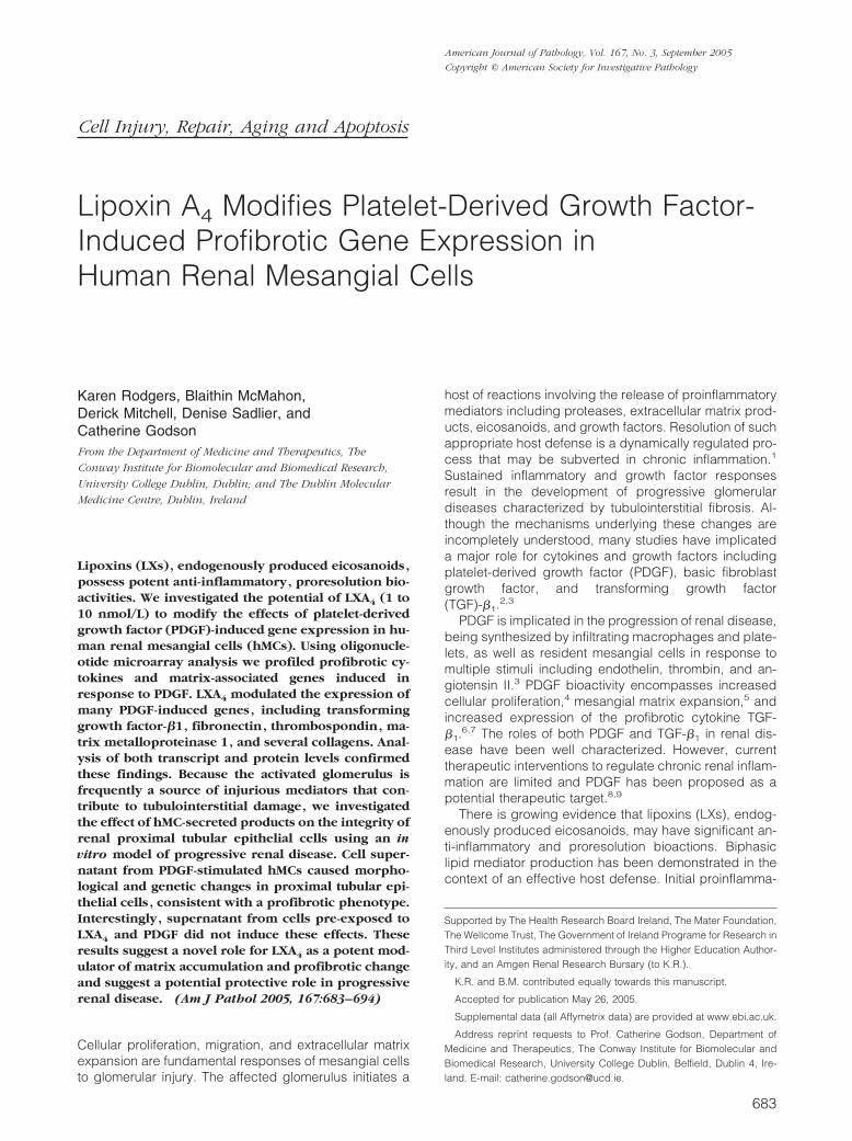
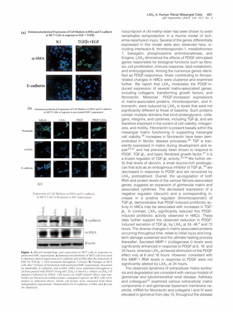
It has been proposed that pleiotropic mediators releasedfrom the inflamed glomerulus play an important role inprogressive renal disease by tubulointerstitial fibro-sis.25,26 Tubulointerstitial fibrosis is frequently character-ized by EMT. Consistent with this we observed that su-pernatants from hMCs treated with PDGF (10 ng/ml, 24hours) induced a loss of E-cadherin expression concom-itant with increased levels of �-smooth muscle actin inMCT cells (Figure 4b). These phenotypic changes mir-rored those seen with the prototypic inducers of EMT,TGF-�1, and EGF (Figure 4a).27 MCT cells treated withcell supernatant from vehicle or LXA4-treated hMCs re-tained much of their epithelial phenotype. Remarkably,this observed change in MCT phenotype was less sub-stantial after treatment with supernatant from PDGF-stim-ulated and LXA4-pretreated hMCs. Pretreatment withLXA4 diminished the effects of PDGF thereby impedingthe indicated transformation. These changes were veri-fied using Western blot analysis of MCT lysate (Figure4c). PDGF-stimulated hMC supernatant caused de-creases in E-cadherin and corresponding increases in�-SMA levels. Again, supernatant from hMCs pretreatedwith LXA4 diminished this effect.
Changes in MCT gene expression of fibronectin, vas-cular endothelial growth factor (VEGF), and connectivetissue growth factor (CTGF) were measured using quan-titative real-time RT-PCR. The positive control (TGF-�1
and EGF) displayed significant increased expression of
LXA4 in Human Renal Mesangial Cells 687AJP September 2005, Vol. 167, No. 3

each gene, as compared to K1 media alone (Figure 5A).Consistent with this, supernatant from PDGF-stimulated(10 ng/ml, 24 hours) hMCs caused an increase in theexpression of VEGF, CTGF, and fibronectin in MCT cells.Supernatant from hMCs pretreated with LXA4 (1 nmol/L,15 minutes) and subsequently PDGF-stimulated, dimin-ished these observed changes in gene expression (Fig-ure 5; B, C, and D), consistent with LXA4 modulation ofPDGFR signals. Expression of all three genes was con-tinuously decreased with LXA4-pretreated supernatant.Changes in epithelial cell gene expression induced byhMC supernatants were not due to residual PDGF in themedia. This was verified by the addition of the tyrophostinPDGFR kinase inhibitor AG1296 (10 nmol/L) to hMC su-
pernatant. Addition of the inhibitor did not significantlyeffect observed gene expression changes (data notshown). Furthermore, direct stimulation of the MCT cellswith PDGF-BB did not induce significant expression offibronectin, VEGF, or CTGF (Figure 5A).
Discussion
The pathology of progressive renal disease in glomeru-lonephritis (GN) involves mesangial cell proliferation andmigration, matrix expansion, and cytokine production,which left unchecked may result in end-stage renal dis-ease characterized by tubulointerstitial fibrosis. Initial in-
Figure 2. Global changes in gene expression in hMCs, representative ontological data examining biological functionality, and transcription factor analysis ofgenes. Gene expression changes in hMCs treated with PDGF (10 ng/ml) alone or PDGF with LXA4 (1 nmol/L) pretreatment were analyzed. Three independentexperiments were pooled for hybridization to each microarray. Duplicate experiments using HGU133A Affymetrix microarrays are illustrated, data arerepresentative of genes differentially expressed by 0.5 SLRs or greater (�0.5 SLR and �0.5 SLR) from the average of duplicate arrays. A: An overview of thenumbers of differentially expressed genes. B: Changes in gene expression within major functional families using OntoExpress.
688 Rodgers et alAJP September 2005, Vol. 167, No. 3

Figure 2. Continued. C: Unsupervised hierarchical cluster analysis of the average of duplicate arrays and ontological clustering. D: Transcription factor consensusbinding sequences on PDGF-altered genes, random-expectation values, and actual binding site occurrence are shown. E: SP-1 expression in hMC nuclear lysateafter treatment with PDGF (10 ng/ml), LXA4 (1 nmol/L), vehicle, or LXA4 (15 minutes) followed by PDGF. Data are mean � SEM of duplicate measurements fromthree independent experiments (*P � 0.05 compared to vehicle, #P � 0.05 compared to PDGF).
LXA4 in Human Renal Mesangial Cells 689AJP September 2005, Vol. 167, No. 3

sult to the glomerulus causes a host of inflammatoryreactions resulting in the progressive accumulation ofextracellular matrix components, thus decreasing filtra-tion surface area.28 The release of inflammatory media-tors and cytokines/growth factors into the surroundinginterstitium accompanies the development of fibrosis andtubular atrophy. Previous reports have suggested theinvolvement of PDGF in mediating these effects.7,29
LXs have been shown to exert potent anti-inflammatoryactions in the early stages of GN mediated through inhi-bition of leukocyte infiltration and or adhesion30 and areproposed to act as endogenously produced proresolu-tion agents in host defense and inflammation.31 As ob-served in intestinal enterocytes, expression of the LXA4
receptor was up-regulated in response to cytokines, fur-
thermore addition of LXA4 or its stable, synthetic ana-logues inhibited interleukin-8 chemokine release, therebymodulating the initiation of inflammation.32 Previous datasuggest that the anti-proliferative effects of LXA4 aremediated via intracellular mechanisms associated withmodulation of growth factor receptor (PDGF and EGF)activation,4,6 and recruitment of specific SH-2 containingsignaling molecules (Mitchell D, Gaffney A, Crean JK,Kinsella BT, Godson C, submitted). Here we demonstratethat LXA4 can counteract PDGF-induced gene expres-sion. In agreement with other models in-vestigating LXA4-induced differential gene expression,18,33,34 we reportthat LXA4 modulated the expression of the NAB1 co-repressor, up-regulating expression by 0.3 SLRs. Thestable synthetic analogue 15-epi-16-(para-fluorophe-
Figure 3. Altered expression levels of hMC DNA, protein, and secreted protein in response to PDGF � LXA4, validating changes demonstrated by Affymetrixmicroarray. hMCs were treated with PDGF (10 ng/ml), LXA4 (1 nmol/L), vehicle, or LXA4 (15 minutes) followed by PDGF. a: Changes in the expression of collagenI �I and fibronectin cDNA after 24 hours, as measured by real-time PCR. The average of two independent experiments performed in duplicate is shown. b: Alteredlevels of fibronectin, thrombospondin 1, and decorin protein measured by immunoblotting after 24 hours, corresponding �-actin blots demonstrate equal loading.Data are representative of three independent experiments. c: TGF-�1 and MMP-1 secretion from hMCs as measured by ELISA at the indicated time points. Datashown are mean � SEM of duplicate measurements from three independent experiments (*P � 0.05, **P � 0.005).
690 Rodgers et alAJP September 2005, Vol. 167, No. 3
noxy)-lipoxin A (4)-methyl ester has been shown to exertremarkable renoprotection in a murine model of isch-emia-reperfusion injury. Several of the genes differentiallyexpressed in this model were also observed here, in-cluding interleukin-6, thrombospondin-1, metallothionein1, transgelin, phosphoserine aminotransferase, andEnigma. LXA4 diminished the effects of PDGF-stimulatedgenes responsible for biological functions such as fibro-sis, cell proliferation, immune response, lipid metabolism,and embryogenesis. Among the numerous genes identi-fied as PDGF-responsive, those contributing to fibrosis-related changes in hMCs were clustered and examinedfurther. We report that LXA4 modulates the PDGF-in-duced expression of several matrix-associated genes,including collagens, transforming growth factors, andfibronectin. Moreover, PDGF-increased expressionsof matrix-associated proteins, thrombospondin, and fi-bronectin, were reduced by LXA4 to levels that were notsignificantly different to those of baseline. Such proteinscontain multiple domains that bind proteoglycans, colla-gens, integrins, and cytokines, including TGF-�1 and aretherefore important in the control of cell viability, mitogen-esis, and motility. Fibronectin is present basally within themesangial matrix functioning in supporting mesangialcell viability,35 increases in fibronectin have been dem-onstrated in fibrotic disease processes.36 TSP is tran-siently expressed in matrix during development and re-pair23,37 and has previously been shown to respond toPDGF, TGF-�1, and basic fibroblast growth factor,37 it isa known regulator of TGF-�1 activity.23,38 We further ver-ify that levels of decorin, a small leucine-rich proteogly-can that acts as an endogenous inhibitor of TGF-�1,
39 aredecreased in response to PDGF and are recovered byLXA4 pretreatment. Overall, the up-regulation of bothRNA and protein levels of the various fibrosis-associatedgenes, suggests an expansion of glomerular matrix andassociated cytokines. The decreased expression of anegative regulator (decorin) and a corresponding in-crease in a positive regulator (thrombospondin) ofTGF-�1 demonstrates that PDGF-induced profibrotic ac-tivity in hMCs may be associated with increases in TGF-�1. In contrast, LXA4 significantly reduced this PDGF-induced profibrotic activity observed in hMCs. Thesedata further support the observed reduction in PDGF-induced secretion of TGF-�1 by LXA4 at 24, 48,6 and 72hours. The diverse changes in matrix-associated proteinsoccurring throughout time, relate to initial injury and long-term damage sustained and the ultimate healing processthereafter. Secreted MMP-1 (collagenase I) levels weresignificantly enhanced in response to PDGF at 6, 18, and24 hours, whereas LXA4 achieved diminution of the PDGFeffect only at 6 and 18 hours. However, consistent withthis MMP-1 RNA levels in response to PDGF were notsignificantly altered by LXA4 at 24 hours.
The observed dynamics of extracellular matrix synthe-sis and degradation are consistent with various models ofglomerular and tubulointerstitial renal disease. Adhikaryand colleagues40 examined various extracellular matrixcomponents in anti-glomerular basement membrane ne-phritis, mRNA for fibronectin and collagens I and IV wereelevated in glomeruli from day 15, throughout the disease
Figure 4. Altered morphology and expression in MCT cells in response topretreated hMC supernatant. a: Immunocytochemistry of MCT cells was usedto illustrate altered expression in E-cadherin and �-SMA after the induction ofEMT by TGF-�1 � EGF treatment throughout 72 hours. b: Changes in MCTcells after 72 hours of incubation with pretreated hMC supernatant, measuredby phalloidin, E-cadherin, and �-SMA. hMCs were stimulated throughout a24-hour period with PDGF (10 ng/ml), LXA4 (1 nmol/L), vehicle or LXA4 (15minutes) followed by PDGF. Cell nuclei are DAPI stained (blue) and anti-bodies are fluorescein isothiocyanate conjugated (green). c: MCT cells weretreated as indicated above, whole cell lysates were extracted from threeindependent experiments. Immunoblots for E-cadherin, �-SMA, and �-actinare displayed.
LXA4 in Human Renal Mesangial Cells 691AJP September 2005, Vol. 167, No. 3

course. TIMP-1 and TGF-�1 mRNA were also enhanced.A similar profile of extracellular matrix components andTGF-�1 was observed in the cortex, increasing moregradually from day 15 to day 29 as tubular damageprogressed.40
The reduction in secreted levels of both MMP-1 andTGF-�1 in LXA4 pretreated cells may reflect reducedautokinase activity of the receptor tyrosine kinases(RTKs) and/or enhanced tyrosine phosphatase activity(Mitchell D, Gaffney A, Crean JK, Kinsella BT, Godson C,submitted). Significant evidence for the cross-talk be-tween the LX G-protein-coupled receptor activated byLXA4 (ALXR) and the RTKs for both PDGF and EGF hasbeen shown.4,6 In the context of LXA4 modulation ofprofibrotic changes, it is noteworthy that a role for theALXR has recently been proposed in the anti-fibrotictreatment of a lung fibrosis model in mice.41 In theseexperiments LXA4 also significantly prevented enhancedproliferation of NIH3T3 fibroblasts and collagen expres-sion by TGF-�1. Furthermore Sodin-Semrl and col-leagues42 described a role for LXA4 in regulating humansynovial fibroblast activation, levels of MMP-1 and MMP-3were diminished in response to LXA4 treatment of stimu-lated fibroblasts.
The effects of LXA4 on PDGFR activation are relativelyspecific, being restricted to recruitment to precise phos-photyrosine residues (Mitchell D, Gaffney A, Crean JK,Kinsella BT, Godson C, submitted). In this regard it was ofinterest to investigate whether LXA4 might modulate tran-scription factors downstream of specific signal transduc-tion processes. It has been reported that LXA4 can me-diate NF-�B-induced gene expression in intestinalepithelial cells.43 Using MatInspector, many ubiquitousTF binding sequences were observed in the promoters ofPDGF-induced genes implicated in both matrix-associ-ated and proliferative gene regulation. Verrecchia andcolleagues44 showed that inhibition of the SP-1 TF pre-vented the expression of extracellular matrix genes indermal fibroblasts. Although SP-1 is a ubiquitous TF, ithas also been shown to respond to several growth fac-tors, MAPK and glucose.45 Posttranslational modificationof SP-1 may have a significant role in TF activation. In ourstudy we measured the binding of nuclear SP-1 fromhMCs treated with PDGF and PDGF pretreated with LXA4,
we found that SP-1 was significantly increased in re-sponse to PDGF. LXA4 consistently diminished thiseffect.
Figure 5. Altered fibronectin, VEGF, and CTGF gene expression levels in MCT cells, as measured by quantitative real-time PCR. A: Differences in fibronectin,VEGF, and CTGF mRNA in MCT cells treated with a combination of TGF-�1 and EGF or PDGF-BB for 72 hours. B–D: Altered mRNA levels of fibronectin, VEGF,and CTGF, respectively. RNA was extracted from MCT cells treated with pretreated hMC supernatant for 72 hours as previously indicated. Results are displayedas normalized relative quantity, data are mean � SEM of duplicate measurements from three independent experiments (*P � 0.05 compared to vehicle, #P � 0.05compared to PDGF). All values have been normalized to respective 18s rRNA.
692 Rodgers et alAJP September 2005, Vol. 167, No. 3

The translation of a relatively modest glomerular injuryto devastating tubulointerstitial fibrosis is increasingly ap-preciated. Such fibrosis can reflect on a combination ofEMT of resident epithelia or progenitor cells, infiltration ofcirculating or proliferation of resident fibroblasts.46,47 Al-terations in the mediators present in the interstitium and inthe glomerular filtrate contribute to the development oftubulointerstitial fibrosis.26 Tubulointerstitial fibrosis en-compasses loss of epithelium polarity, adherens junc-tions, tight junctions, desmosomes, and cytokeratin inter-mediate filaments to rearrange their F-actin stress fibersand express filopodia and lamellopodia.48 Epithelial cellsgain plasticity during the remodeling process, promotinghealing or scarring as a response to injury.49 Induction ofEMT is associated with the expression of cytokines andthe proteolytic degradation of epithelial basement mem-brane.50 Matrix metalloproteinases or membrane assem-bly inhibitors dismantle the membrane, whereas localexpression of TGF-�, EGF, IGF-II, or FGF-2 facilitatesEMT.48,51,52 These cytokines contribute to elevated levelsof matrix metalloproteinases and alter the cell proteome.Higgins and colleagues53 observed increases in the ex-pression of collagen I and PAI-1, among others, in EMTusing the in vivo model of unilateral ureteral obstructionand the in vitro model of stimulated murine proximal tu-bular (MCT) cultured cells. Signature markers for thedisease were identified using oligonucleotide microarray.
We report here that PDGF-treated hMC supernatantcaused a morphological change in renal tubular MCTcells, similar to that of TGF-�1 and EGF. Loss of epithelialtight junction marker E-cadherin and gain of mesenchy-mal actin cytoskeleton marker �-SMA in MCT cellstreated with PDGF-stimulated hMC supernatant was ob-served. Interestingly, these effects were diminished byLXA4 pretreatment. LXA4 pretreatment could alter theseeffects perhaps by altering PDGF-induced receptor acti-vation in the hMCs, changing the spectrum of solublefactors present in the ultrafiltrate. Furthermore, immuno-blotting for E-cadherin and �-SMA under the same con-ditions confirmed these findings. Previously, we havereported the inhibition of PDGF and EGF mediated PKB/Akt phosphorylation by LXA4.
6 This pathway may be in-volved in the release of cytokines or mediators fromhMCs, which we observe to cause morphologicalchanges in MCT cells, a role for this pathway in fibrosis iswell established. Stimulation of fibroblasts with endothe-lin-1 promotes enhanced contractile phenotype or acti-vation, this is prevented by blockade of the PI3K/Aktpathway.54 In agreement with this Vittal and colleagues55
observed that inhibition of PKB/Akt in bleomycin-inducedlung fibrosis markedly reduced accumulation of�-smooth muscle actin-expressing myofibroblasts.
Supernatant from PDGF-stimulated hMCs stimulatedan increase in RNA expression of VEGF, CTGF, andfibronectin. This effect was modulated by LXA4 pretreat-ment. To control for the possibility that residual PDGF inthe media from pretreated hMCs might have inducedprofibrotic changes in the epithelial cells, these cellswere treated with the PDGF-specific phosphotyrosine in-hibitor, AG1296. However, in these cells changes in pro-fibrotic gene expression persisted. Similarly, treatment of
MCT cells with PDGF ligand did not induce profibroticgene expression. Soluble factors released from hMCs inresponse to PDGF in our in vitro model may indicate theprogression of renal disease seen in vivo.
Collectively these data demonstrate LXA4 as a poten-tial anti-fibrotic agent, preventing growth factor-inducedmesangial matrix production and the progression of renaldisease, by alleviating the effect of hMC products ontubular cells. Further investigation into these bioactivitiesof LXA4 and its stable synthetic analogues will includeexamining effects on hMCs and tubular cells in an in vivomodel of progressive renal disease.
Acknowledgment
We thank Dr. Hugh Brady for interest and advice.
References
1. Anders HJ, Vielhauer V, Schlondorff D: Chemokines and chemokinereceptors are involved in the resolution or progression of renal dis-ease. Kidney Int 2003, 63:401–415
2. Floege J, Eng E, Young BA, Johnson RJ: Factors involved in theregulation of mesangial cell proliferation in vitro and in vivo. Kidney IntSuppl 1993, 39:S47–S54
3. Abboud HE, Grandaliano G, Pinzani M, Knauss T, Pierce GF, Jaffer F:Actions of platelet-derived growth factor isoforms in mesangial cells.J Cell Physiol 1994, 158:140–150
4. McMahon B, Mitchell D, Shattock R, Martin F, Brady HR, Godson C:Lipoxin, leukotriene, and PDGF receptors cross-talk to regulate mes-angial cell proliferation. FASEB J 2002, 16:1817–1819
5. Johnson RRE, Floege J, Yoshimura A, Pritzl P, Alpers C, Ross R:Inhibition of mesangial cell proliferation and matrix expansion in glo-merulonephritis in the rat by antibody to platelet-derived growth fac-tor. J Exp Med 1992, 175:1413–1416
6. Mitchell D, Rodgers K, Hanly J, McMahon B, Brady HR, Martin F,Godson C: Lipoxins inhibit Akt/PKB activation and cell cycle progres-sion in human mesangial cells. Am J Pathol 2004, 164:937–946
7. Abboud HE: Role of platelet-derived growth factor in renal injury.Annu Rev Physiol 1995, 57:297–309
8. Gilbert RE, Kelly DJ, McKay T, Chadban S, Hill PA, Cooper ME, AtkinsRC, Nikolic-Paterson DJ: PDGF signal transduction inhibition amelio-rates experimental mesangial proliferative glomerulonephritis. KidneyInt 2001, 59:1324–1332
9. Ostendorf T, Kunter U, van Roeyen C, Dooley S, Janjic N, Ruckman J,Eitner F, Floege J: The effects of platelet-derived growth factor an-tagonism in experimental glomerulonephritis are independent of thetransforming growth factor-beta system. J Am Soc Nephrol 2002,13:658–667
10. Serhan CN, Chiang N: Lipid-derived mediators in endogenous anti-inflammation and resolution: lipoxins and aspirin-triggered 15-epi-lipoxins. Scientific World J 2002, 2:169–204
11. Serhan CN, Gotlinger K, Hong S, Arita M: Resolvins, docosatrienes,and neuroprotectins, novel omega-3-derived mediators, and theiraspirin-triggered endogenous epimers: an overview of their protec-tive roles in catabasis. Prostaglandins Other Lipid Mediat 2004,73:155–172
12. Godson C, Mitchell S, Harvey K, Petasis NA, Hogg N, Brady HR:Cutting edge: lipoxins rapidly stimulate nonphlogistic phagocytosis ofapoptotic neutrophils by monocyte-derived macrophages. J Immunol2000, 164:1663–1667
13. Maddox JF, Serhan CN: Lipoxin A4 and B4 are potent stimuli forhuman monocyte migration and adhesion: selective inactivation bydehydrogenation and reduction. J Exp Med 1996, 183:137–146
14. Bandeira-Melo C, Bozza PT, Diaz BL, Cordeiro RS, Jose PJ, MartinsMA, Serhan CN: Cutting edge: lipoxin (LX) A4 and aspirin-triggered15-epi-LXA4 block allergen-induced eosinophil trafficking. J Immunol2000, 164:2267–2271
LXA4 in Human Renal Mesangial Cells 693AJP September 2005, Vol. 167, No. 3

15. Mitchell S, Thomas G, Harvey K, Cottell D, Reville K, Berlasconi G,Petasis NA, Erwig L, Rees AJ, Savill J, Brady HR, Godson C: Lipoxins,aspirin-triggered epi-lipoxins, lipoxin stable analogues, and the res-olution of inflammation: stimulation of macrophage phagocytosis ofapoptotic neutrophils in vivo. J Am Soc Nephrol 2002, 13:2497–2507
16. McMahon B, Stenson C, McPhillips F, Fanning A, Brady HR, GodsonC: Lipoxin A4 antagonizes the mitogenic effects of leukotriene D4 inhuman renal mesangial cells. Differential activation of MAP kinasesthrough distinct receptors. J Biol Chem 2000, 275:27566–27575
17. Mene P: Mesangial cell cultures. J Nephrol 2001, 14:198–20318. Kieran NE, Doran PP, Connolly SB, Greenan MC, Higgins DF, Leo-
nard M, Godson C, Taylor CT, Henger A, Kretzler M, Burne MJ, RabbH, Brady HR: Modification of the transcriptomic response to renalischemia/reperfusion injury by lipoxin analog. Kidney Int 2003,64:480–492
19. Sadlier DM, Connolly SB, Kieran NE, Roxburgh S, Brazil DP, KairaitisL, Wang Y, Harris DC, Doran P, Brady HR: Sequential extracellularmatrix-focused and baited-global cluster analysis of serial transcrip-tomic profiles identifies candidate modulators of renal tubulointersti-tial fibrosis in murine adriamycin induced nephropathy. J Biol Chem2004, 279:29670–29680
20. Eisen MB, Spellman PT, Brown PO, Botstein D: Cluster analysis anddisplay of genome-wide expression patterns. Proc Natl Acad Sci USA1998, 95:14863–14868
21. Draghici S, Khatri P, Martins RP, Ostermeier GC, Krawetz SA: Globalfunctional profiling of gene expression. Genomics 2003, 81:98–104
22. Werner T: Target gene identification from expression array data bypromoter analysis. Biomol Eng 2001, 17:87–94
23. Daniel C, Wiede J, Krutzsch HC, Ribeiro SM, Roberts DD, Murphy-Ullrich JE, Hugo C: Thrombospondin-1 is a major activator of TGF-beta in fibrotic renal disease in the rat in vivo. Kidney Int 2004,65:459–468
24. Harper JR, Spiro RC, Gaarde WA, Tamura RN, Pierschbacher MD,Noble NA, Stecker KK, Border WA: Role of transforming growth factorbeta and decorin in controlling fibrosis. Methods Enzymol 1994,245:241–254
25. Yamamoto T, Noble NA, Miller DE, Border WA: Sustained expressionof TGF-beta 1 underlies development of progressive kidney fibrosis.Kidney Int 1994, 45:916–927
26. Wang SN, LaPage J, Hirschberg R: Role of glomerular ultrafiltration ofgrowth factors in progressive interstitial fibrosis in diabetic nephrop-athy. Kidney Int 2000, 57:1002–1014
27. Strutz F, Zeisberg M, Ziyadeh FN, Yang CQ, Kalluri R, Muller GA,Neilson EG: Role of basic fibroblast growth factor-2 in epithelial-mesenchymal transformation. Kidney Int 2002, 61:1714–1728
28. Couser WG, Johnson RJ: Mechanisms of progressive renal disease inglomerulonephritis. Am J Kidney Dis 1994, 23:193–198
29. Ludewig D, Kosmehl H, Sommer M, Bohmer FD, Stein G: PDGFreceptor kinase blocker AG1295 attenuates interstitial fibrosis in ratkidney after unilateral obstruction. Cell Tissue Res 2000, 299:97–103
30. Levy BD, Clish CB, Schmidt B, Gronert K, Serhan CN: Lipid mediatorclass switching during acute inflammation: signals in resolution. NatImmunol 2001, 2:612–619
31. McMahon B, Godson C: Lipoxins: endogenous regulators of inflam-mation. Am J Physiol 2004, 286:F189–F201
32. Gronert K, Gewirtz A, Madara JL, Serhan CN: Identification of ahuman enterocyte lipoxin A4 receptor that is regulated by interleukin(IL)-13 and interferon gamma and inhibits tumor necrosis factor al-pha-induced IL-8 release. J Exp Med 1998, 187:1285–1294
33. Qiu FH, Devchand PR, Wada K, Serhan CN: Aspirin-triggered lipoxinA4 and lipoxin A4 up-regulate transcriptional corepressor NAB1 inhuman neutrophils. FASEB J 2001, 15:2736–2738
34. Leonard MO, Hannan K, Burne MJ, Lappin DW, Doran P, Coleman P,Stenson C, Taylor CT, Daniels F, Godson C, Petasis NA, Rabb H,Brady HR: 15-Epi-16-(para-fluorophenoxy)-lipoxin A(4)-methyl ester,a synthetic analogue of 15-epi-lipoxin A(4), is protective in experi-mental ischemic acute renal failure. J Am Soc Nephrol 2002,13:1657–1662
35. Sugiyama H, Kashihara N, Maeshima Y, Okamoto K, Kanao K,Sekikawa T, Makino H: Regulation of survival and death of mesangialcells by extracellular matrix. Kidney Int 1998, 54:1188–1196
36. Barnes JL, Mitchell RJ, Kanalas JJ, Barnes VL: Differential expressionof thrombospondin and cellular fibronectin during remodeling in
proliferative glomerulonephritis. J Histochem Cytochem 1999,47:533–544
37. Hugo C, Pichler R, Meek R, Gordon K, Kyriakides T, Floege J,Bornstein P, Couser WG, Johnson RJ: Thrombospondin 1 is ex-pressed by proliferating mesangial cells and is up-regulated byPDGF and bFGF in vivo. Kidney Int 1995, 48:1846–1856
38. Daniel C, Takabatake Y, Mizui M, Isaka Y, Kawashi H, Rupprecht H,Imai E, Hugo C: Antisense oligonucleotides against throm-bospondin-1 inhibit activation of TGF-beta in fibrotic renal disease inthe rat in vivo. Am J Pathol 2003, 163:1185–1192
39. Huijun W, Long C, Zhigang Z, Feng J, Muyi G: Ex vivo transfer of thedecorin gene into rat glomerulus via a mesangial cell vector sup-pressed extracellular matrix accumulation in experimental glomeru-lonephritis. Exp Mol Pathol 2005, 78:17–24
40. Adhikary LP, Yamamoto T, Isome M, Nakano Y, Kawasaki K, Yaoita E,Kihara I: Expression profile of extracellular matrix and its regulatoryproteins during the process of interstitial fibrosis after anti-glomerularbasement membrane antibody-induced glomerular sclerosis inSprague-Dawley rats. Pathol Int 1999, 49:716–725
41. Sato Y, Kitasato H, Murakami Y, Hashimoto A, Endo H, Kondo H,Inoue M, Hayashi I: Down-regulation of lipoxin A4 receptor by throm-boxane A2 signaling in RAW246.7 cells in vitro and bleomycin-in-duced lung fibrosis in vivo. Biomed Pharmacother 2004, 58:381–387
42. Sodin-Semrl S, Spagnolo A, Barbaro B, Varga J, Fiore S: Lipoxin A4counteracts synergistic activation of human fibroblast-like synovio-cytes. Int J Immunopathol Pharmacol 2004, 17:15–25
43. Gewirtz AT, Collier-Hyams LS, Young AN, Kucharzik T, Guilford WJ,Parkinson JF, Williams IR, Neish AS, Madara JL: Lipoxin A4 analogsattenuate induction of intestinal epithelial proinflammatory gene ex-pression and reduce the severity of dextran sodium sulfate-inducedcolitis. J Immunol 2002, 168:5260–5267
44. Verrecchia F, Rossert J, Mauviel A: Blocking sp-1 transcription factorbroadly inhibits extracellular matrix gene expression in vitro and invivo: implications for the treatment of tissue fibrosis. J Invest Dermatol2001, 116:755–763
45. Goldberg HJ, Whiteside CI, Fantus IG: The hexosamine pathwayregulates the plasminogen activator inhibitor-1 gene promoter andSp1 transcriptional activation through protein kinase C-beta I and-delta. J Biol Chem 2002, 277:33833–33841
46. Iwano M, Plieth D, Danoff TM, Xue C, Okada H, Neilson EG: Evidencethat fibroblasts derive from epithelium during tissue fibrosis. J ClinInvest 2002, 110:341–350
47. Hirschberg R, Wang S: Proteinuria and growth factors in the devel-opment of tubulointerstitial injury and scarring in kidney disease. CurrOpin Nephrol Hypertens 2005, 14:43–52
48. Kalluri R, Neilson EG: Epithelial-mesenchymal transition and its im-plications for fibrosis. J Clin Invest 2003, 112:1776–1784
49. El-Nahas AM: Plasticity of kidney cells: role in kidney remodeling andscarring. Kidney Int 2003, 64:1553–1563
50. Liu Y: Epithelial to mesenchymal transition in renal fibrogenesis:pathologic significance, molecular mechanism, and therapeutic in-tervention. J Am Soc Nephrol 2004, 15:1–12
51. Zeisberg M, Kalluri R: The role of epithelial-to-mesenchymal transitionin renal fibrosis. J Mol Med 2004, 82:175–181
52. Okada H, Inoue T, Suzuki H, Strutz F, Neilson EG: Epithelial-mesen-chymal transformation of renal tubular epithelial cells in vitro and invivo. Nephrol Dial Transplant 2000, 15(Suppl 6):44–46
53. Higgins DF, Lappin DW, Kieran NE, Anders HJ, Watson RW, Strutz F,Schlondorff D, Haase VH, Fitzpatrick JM, Godson C, Brady HR: DNAoligonucleotide microarray technology identifies fisp-12 among otherpotential fibrogenic genes following murine unilateral ureteral ob-struction (UUO): modulation during epithelial-mesenchymal transi-tion. Kidney Int 2003, 64:2079–2091
54. Shi-Wen X, Chen Y, Denton CP, Eastwood M, Renzoni EA, Bou-Gharios G, Pearson JD, Dashwood M, du Bois RM, Black CM, LeaskA, Abraham DJ: Endothelin-1 promotes myofibroblast inductionthrough the ETA receptor via a rac/phosphoinositide 3-kinase/Akt-dependent pathway and is essential for the enhanced contractilephenotype of fibrotic fibroblasts. Mol Biol Cell 2004, 15:2707–2719
55. Vittal R, Horowitz JC, Moore BB, Zhang H, Martinez FJ, Toews GB,Standiford TJ, Thannickal VJ: Modulation of prosurvival signaling infibroblasts by a protein kinase inhibitor protects against fibrotic tissueinjury. Am J Pathol 2005, 166:367–375
694 Rodgers et alAJP September 2005, Vol. 167, No. 3


![Мінрегіонdfrr.minregion.gov.ua/foto/projt_addition/2016/02/... · 150 aBTOM06iJ1iB ga n06 ] [10;aaBaTH a60 Ha lipoxin 3MiHi KiJ1bKOCTi Flpox0ÄiB HOPMH 2-4-33 [MPH BHKOHaHHi](https://img.dokumen.tips/doc/110x75/60d1547388e155243e230173/oedfrr-150-abtom06ij1ib-ga-n06-10aabath-a60-ha-lipoxin-3mihi.jpg)