Embed Size (px)
Citation preview

LIPID NANOPARTICLES FOR DELIVERY OF NUCLEIC ACID THERAPEUTICS
by
Sam Chen
B.Sc., The University of British Columbia, 2009
A THESIS SUBMITTED IN PARTIAL FULFILLMENT OF
THE REQUIREMENTS FOR THE DEGREE OF
DOCTOR OF PHILOSPHY
in
THE FACULTY OF GRADUATE AND POSTDOCTORAL STUDIES
(Biochemistry and Molecular Biology)
THE UNIVERSITY OF BRITISH COLUMBIA
(Vancouver)
July 2016
© Sam Chen, 2016

ii
Abstract
Nucleic acid therapies have the potential to enable the treatment of disorders previously
untreatable. However, significant barriers prevent the rapid development of nucleic acid
therapeutics and necessitate the use of sophisticated delivery systems. The overall objective of this
dissertation is to develop more effective and tolerable lipid nanoparticles (LNPs) for nucleic acid
delivery. Specifically, LNP that vary in size, stability and composition were tested for activity after
subcutaneous and intravenous injection in order to optimize LNP properties. Furthermore, the
incorporation of novel lipophilic pro-drugs co-delivered with nucleic acids was explored as a
means to improve tolerability.
The first part of this dissertation explores the use of subcutaneous administration for LNP-
siRNA. There are compelling reasons to develop LNP-siRNA that can be administered
subcutaneously. These include the potential for self-administration, a prolonged therapeutic
window due to a depot effect and access to cell types that are in contact with the lymphatic system,
in addition to tissues available through the circulation. We found that particle size and PEG steric
barrier are both important for drainage from the injection site and subsequent accumulation in the
liver.
Although small LNP exhibited improved drainage and access to the systemic circulation,
activity was impaired. The second part of this dissertation addresses this issue. The decrease in
LNP activity can be attributed to the pronounced size dependent instability of LNP. By altering
the amino-lipid content and the PEG-lipid used, the activity and stability of these systems can be
greatly improved.
The previous two parts of this dissertation identified limitations to existing LNP systems.
First, administration of LNP containing nucleic acids could result in immune stimulation. Second,

iii
efforts to improve LNP activity must go beyond existing components. The last part of this
dissertation proposes a general pro-drug strategy for enabling direct incorporation of additional
compounds into the LNP. As an example dexamethasone, a corticosteroid commonly used to
minimize infusion-related reactions, was used. Direct incorporation greatly improved the ability
of dexamethasone to ameliorate immune stimulation by LNP containing nucleic acids. This work
shows that when appropriately designed, LNP systems can have improved activity and tolerability,
potentially expanding its clinical utility.

iv
Preface
The preparation and analysis of all LNP-siRNA formulations and all subsequent in vitro
experiments were performed by myself. In vivo experiments involving mice were performed by
myself with the assistance of Ms. Yan Liu, Dr. Ying K. Tam, Dr. Paulo J.C. Lin and Dr. Yuen Yi
C. Tam. Cryogenic transmission electron microscopy of LNP-siRNA in Chapter 3 was performed
by myself with the assistance of Dr. Alex K. K. Leung and members of the BioImaging Facility
(Bradford Ross and Garnet Martens) at The University of British Columbia. Synthesis of lipophilic
dexamethasone pro-drugs in Chapter 5 was carried out by Dr. Josh Zaifman in collaboration with
the laboratory of Dr. Marco A. Ciufolini in the department of Chemistry at The University of
British Columbia.
Experimental designs and data analyses were performed by myself with important
contributions, input and feedback from Dr. Yuen Yi C. Tam, Dr. Ying K. Tam, Dr. Paulo J. C.
Lin, Dr. Alex K. K. Leung and Mr. Jayesh Kulkarni. I was responsible for the writing and
preparation of the entire dissertation with the exception of the chemical synthesis of
dexamethasone pro-drugs used in Chapter 5, which was written by Dr. Josh Zaifman and can be
found in Appendix A. Drs. Pieter R. Cullis, Yuen Yi C. Tam and Karen Lam were responsible for
editing this dissertation.
A portion of the introduction in Chapter 1 regarding the development of lipid nanoparticles
for siRNA delivery (section 1.2) has been published as a mini-review article: Tam, Y.Y., Chen,
S., and Cullis, P.R. (2013). Advances in Lipid Nanoparticles for siRNA Delivery. Pharmaceutics
5(3):498-507.
Chapter 3 of this dissertation regarding the subcutaneous administration of LNP-siRNA
has been published in the Journal of Controlled Release: Chen, S., Tam, Y.Y., Lin, P.J., Leung,

v
A.K., Tam, Y.K., and Cullis, P.R. (2014). Development of lipid nanoparticle formulations of
siRNA for hepatocyte gene silencing following subcutaneous administration. J Control Release.
2014 Dec 28;196:106-12.
Chapter 4 of this dissertation regarding the impact of particle size and polyethylene glycol
content on LNP-siRNA activity has also been published in the Journal of Controlled Release:
Chen, S., Tam, Y.Y., Lin, P.J., Sung M.M.H., Tam, Y.K., and Cullis, P.R. (2016). Influence of
particle size on the in vivo potency of lipid nanoparticle formulations of siRNA. J Control Release.
2016 Aug 10;235:236-44.
Some of the data presented in Chapter 5 has been included in an invention disclosure.
All work and procedures involving animals presented in this dissertation have been
approved by the Animal Care Committee at The University of British Columbia and were
performed in accordance with guidelines established by the Canadian Council on Animal Care.
Animal Care and Ethics Protocol: A08-0668, A11-0359, A13-0022, A15-0026
Animal Care and Ethics Training Certificate: 6682-14, RBH-932-10, RA-133-11, RSx-71-12

vi
Table of Contents
Abstract .......................................................................................................................................... ii
Preface ........................................................................................................................................... iv
Table of Contents ......................................................................................................................... vi
List of Tables ................................................................................................................................ xi
List of Figures .............................................................................................................................. xii
List of Abbreviations ................................................................................................................. xiv
Acknowledgements .................................................................................................................... xix
Chapter 1: Introduction ................................................................................................................1
1.1 Lipid-based Vehicles for Drug Delivery ......................................................................... 1
1.1.1 Lipid Nanoparticles in Nature ..................................................................................... 1
1.1.2 Traditional Liposomes: Model Membranes to Drug Delivery ................................... 5
1.1.3 Drug Loading and Controlled Release ........................................................................ 6
1.1.4 Improving Circulation Parameters and Target Site Accumulation ............................. 9
1.2 Lipid Nanoparticles for Nucleic Acid Delivery ............................................................ 10
1.2.1 Challenges of Nucleic Acids Therapeutics ............................................................... 10
1.2.2 Early Attempts to Encapsulate Nucleic Acids in Lipid Nanoparticles ..................... 12
1.2.3 Manufacturing of Lipid Nanoparticles for Nucleic Acid Delivery ........................... 13
1.2.3.1 Pre-formed Vesicle and Extrusion-based Methods........................................... 13
1.2.3.2 Spontaneous Formation by Rapid Mixing Methods ......................................... 15
1.2.4 General Composition, Structure and Size ................................................................. 21
1.2.5 Cationic and Ionizable Amino-lipids ........................................................................ 25
1.2.6 Steric Barrier for Stability and Prolonged Circulation ............................................. 26

vii
1.2.7 Promoting Cellular Uptake with Endogenous and Exogenous Ligands ................... 28
1.3 Clinical Evaluation of siRNA Delivered by Lipid Nanoparticles ................................ 31
1.3.1 LNP-siRNA in the Clinic .......................................................................................... 31
1.3.2 Observations, Challenges and Limitations of Current LNP Systems for Nucleic Acid
Delivery................................................................................................................................. 34
1.4 Thesis Objectives .......................................................................................................... 35
Chapter 2: Material and Methods ..............................................................................................38
2.1 Materials ....................................................................................................................... 38
2.2 Preparation of Lipid Nanoparticle-siRNA by Rapid Mixing Techniques .................... 39
2.3 Analysis of Lipid Nanoparticles ................................................................................... 40
2.4 In Situ Determination of the Apparent Acid Dissociation Constant of LNP-siRNA ... 41
2.5 In Vitro Lipid and siRNA Dissociation ........................................................................ 41
2.6 Pharmacokinetics and Biodistribution .......................................................................... 42
2.7 In Vivo LNP-siRNA Activity in Mouse Factor FVII Model ........................................ 43
2.8 Synthesis of Dexamethasone Pro-drugs........................................................................ 44
2.9 In Vitro Degradation of Dexamethasone Pro-drugs ...................................................... 44
2.10 In Vitro LNP Tolerability by MTT and Hemolysis ...................................................... 45
2.11 In Vivo Immune Suppression by LNP with Dexamethasone Pro-drugs. ...................... 45
Chapter 3: Development of Lipid Nanoparticle Formulations of siRNA for Hepatocyte
Gene Silencing Following Subcutaneous Administration ........................................................47
3.1 Synopsis ........................................................................................................................ 47
3.2 Results ........................................................................................................................... 48

viii
3.2.1 LNP-siRNA Systems Containing PEG-DSG Exhibit Maximum Delivery to Liver
Following Subcutaneous Administration .............................................................................. 48
3.2.2 Intermediate Sized (45 nm) LNP–siRNA Systems Exhibit Maximal Hepatic Gene
Silencing Following Subcutaneous Administration.............................................................. 57
3.3 Discussion ..................................................................................................................... 64
Chapter 4: Influence of Particle Size on the In Vivo Potency of Lipid Nanoparticle
Formulations of siRNA ................................................................................................................70
4.1 Synopsis ........................................................................................................................ 70
4.2 Results ........................................................................................................................... 71
4.2.1 LNP-siRNA Systems with Diameter ~80 nm Exhibit Maximum Hepatic Gene
Silencing Following i.v. Injection ......................................................................................... 71
4.2.2 LNP of Different Sizes All Distribute Rapidly to the Liver Following i.v.
Administration ...................................................................................................................... 73
4.2.3 The Stability of LNP-siRNA Systems is Size Dependent ........................................ 75
4.2.4 Increasing Ionizable Amino-lipid Content Improves Small LNP-siRNA Activity .. 80
4.2.5 A PEG Coating that Dissociates Slowly Dramatically Improves Particle Stability . 85
4.2.6 The Presence of PEG-DSG Extends LNP-siRNA Circulation Lifetimes and Reduces
Hepatic Localization ............................................................................................................. 87
4.3 Discussion ..................................................................................................................... 90
Chapter 5: Lipophilic Dexamethasone Pro-drugs as Potent Suppressors of the
Immunostimulatory Effects of Lipid Nanoparticle Formulations of Nucleic Acid
Polymers........................................................................................................................................96
5.1 Synopsis ........................................................................................................................ 96

ix
5.2 Results ........................................................................................................................... 97
5.2.1 Acylated Dexamethasone Pro-drugs Can Be Incorporated into LNP with Nucleic
Acid ................................................................................................................................... 97
5.2.2 Formulated Pro-drugs are Degradable by Esterases ............................................... 100
5.2.3 Formulated Pro-drugs do not Impact the Tolerability of LNP ............................... 103
5.2.4 Dexamethasone Pro-drugs Ameliorate Immune Stimulation in CpG Mouse Model ...
................................................................................................................................. 105
5.2.5 LD003 Ameliorates LNP-mRNA Mediated Immune Stimulation in Mice ............ 112
5.2.6 LD003 Reduces Injection Site Reactions to LNP-siRNA ...................................... 114
5.3 Discussion ................................................................................................................... 116
Chapter 6: Conclusion and Future Directions ........................................................................121
6.1 Extra Hepatic Applications and Considerations ......................................................... 121
6.2 Methods for Improving LNP Design .......................................................................... 123
6.2.1 Optimizing Existing Lipid Components ................................................................. 124
6.2.2 Adding Lipid Components for Synergistic Improvements ..................................... 125
Bibliography ...............................................................................................................................128
Appendices ..................................................................................................................................144
Appendix A Detailed Synthesis of Lipophilic Dexamethasone Prodrugs (Chapter 5) ........... 144
A.1 General Procedures ................................................................................................. 144
A.2 Synthesis of LD001................................................................................................. 144
A.3 Synthesis of LD002................................................................................................. 147
A.4 Synthesis of LD003................................................................................................. 149
A.5 Synthesis of LD004................................................................................................. 150

x
A.6 Synthesis of LD005................................................................................................. 156

xi
List of Tables
Table 1.1 Lipoprotein Composition and Characteristics* ............................................................. 4
Table 1.2 FDA Approved Lipid/Liposomal Drugs ......................................................................... 6
Table 3.1 Area-under-curve (AUC) and Maximum Blood Concentration (Cmax) of LNP with
Varying Sizes. ............................................................................................................................... 56
Table 5.1 Pro-drug and Lipid Nanoparticle Parameters ............................................................ 100

xii
List of Figures
Figure 1.1 Diagrammatic Representation of the Spontaneous Formation of LNP by Rapid
Mixing Methods. ........................................................................................................................... 17
Figure 1.2 Spontaneous Formation of LNP by Rapid Mixing Through a Staggered Herringbone
Microfluidic Mixer........................................................................................................................ 20
Figure 1.3 Typical Lipid Components Found in LNP. ................................................................ 23
Figure 1.4 Schematic Representation of Lipid Nanoparticles Containing Nucleic Acids. .......... 24
Figure 1.5 Current Model of LNP-siRNA Delivery to Hepatocytes ........................................... 29
Figure 3.1 LNP Size is Correlated with Amount of PEG-lipid. .................................................. 50
Figure 3.2 A Greater Proportion of Small LNP Drain from the Site of Injection and Accumulate
at Brachial Lymph Nodes Following s.c. Administration. ........................................................... 52
Figure 3.3 LNP with Non-dissociating PEG Coating Drain More Quickly into the Blood and
Exhibit Enhanced Accumulation in the Liver Following s.c. Administration. ............................. 54
Figure 3.4 LNPs are not hemolytic at physiological pH. ............................................................. 56
Figure 3.5 ~45 nm LNP Exhibit Maximum Gene Silencing Post s.c. Injection. ......................... 58
Figure 3.6 Persistent Gene Silencing (< 50% protein) is Observed Over 11 Days Following s.c.
Administration of ~45 nm Diameter LNP. ................................................................................... 61
Figure 3.7 Hepatic Gene Silencing Can be Enhanced Using s.c. Administered LNP-siRNA
Containing GalNAc-PEG lipid. .................................................................................................... 64
Figure 4.1 Hepatocyte Gene Silencing Following i.v. Injection of LNP-siRNA Systems is
Dependent on LNP Size. ............................................................................................................... 72
Figure 4.2 Circulation Lifetime and Biodistribution of Small, Medium and Large LNP. .......... 75

xiii
Figure 4.3 The Rate at which Component Lipids Dissociate from LNP is a Sensitive Function of
LNP size. ....................................................................................................................................... 79
Figure 4.4 Chemical Structures of DMAP-BLP and DLin-MC3-DMA...................................... 80
Figure 4.5 The Gene Silencing Potency of Small LNP-siRNA Systems Can be Improved by
Increasing the Amount of Amino-lipid in the LNP. ..................................................................... 83
Figure 4.6 pKa of LNP are Not Affected by Particle Size, Quantity of Amino-lipid or Amine-to-
phosphate Charge Ratios............................................................................................................... 84
Figure 4.7 Substitution of PEG-DSG for PEG-DMG Greatly Decreases the Dissociation Rates
of Component Lipids. ................................................................................................................... 87
Figure 4.8 Substitution of PEG-DMG for PEG-DSG Results in Increased LNP-siRNA
Circulation Lifetimes. ................................................................................................................... 90
Figure 5.1 Structures of Lipophilic Dexamethasone Prodrugs. ................................................... 98
Figure 5.2 Formulated Prodrugs are Degradable by Esterase-rich Plasma. .............................. 101
Figure 5.3 LD003 is Degraded by Esterases. ............................................................................. 102
Figure 5.4 Formulated Prodrugs Do Not Impact the Tolerability of LNP................................. 104
Figure 5.5 Lipophilic Pro-drug Derivatives of Dexamethasone Can Dramatically Ameliorate
Immunostimulatory Effects of LNP-CpG in Mice. .................................................................... 107
Figure 5.6 LD003 Continues to Dramatically Ameliorate Immunostimulatory Effects of LNP-
CpG After 4 hr. ........................................................................................................................... 109
Figure 5.7 LD005 is Unable to Mitigate LNP-CpG Mediated Immune Stimulation. ............... 111
Figure 5.8 LD003 Ameliorates LNP-mRNA Mediated Immune Stimulation. .......................... 113
Figure 5.9 LD003 Reduces Immune Cell Infiltration of 80 nm LNP-siRNA at the Subcutaneous
Injection Site ............................................................................................................................... 115

xiv
List of Abbreviations
ApoA apolipoprotein A
ApoB apolipoprotein B
ApoC apolipoprotein C
ApoE apolipoprotein E
AUC Area-under-curve
ASGPR asialoglycoprotein receptor
ASO Antisense oligonucleotide
CHE cholesteryl hexadecylether
COC Cyclin olefin copolymer
COP Cyclin olefin polymer
CpG Phosphorothioated unmethylated cytosine-guanine dinucleotide motif
containing oligodeoxynucleotide
Cmax Maximum blood concentration
cryoTEM cryogenic electron Microscopy
Dex Dexamethasone
Dex-21-P Dexamethasone-21-phosphate
DLinDAP 1,2-dilinoleoyl-3-dimethylaminopropane
DLinDMA 1,2-dilinoleyloxy-N,N-dimethyl-3-aminopropane
DLin-KC2-DMA 2,2-dilinoleyl-4-(2-dimethylaminoethyl)-[1,3]-dioxolane
DLin-MC3-DMA (6Z,9Z,28Z,31Z)-Heptatriaconta-6,9,28,31-tetraen-19-yl4-
(dimethylamino)butanoate (or dilinoleylmethyl-4-dimethylaminobutyrate)

xv
DLS Dynamic light scattering
DMAP-BLP 3-(dimethylamino)propyl(12Z,15Z)-3-[(9Z,12Z)-octadeca-9,12-dien-1-
yl]henicosa-12,15-dienoate
DNA deoxyribonucleic acid
DODAC N,N-dioleoyl-N,N-dimethylammonium chloride
DODAP 1,2-dioleoyl-3-dimethylammonium propane
DOPE 1, 2 dioleoyl-sn-glycero-3-phosphoethanolamine
DOTMA N-[1-(2,3-dioleyloxy) propyl]-N,N,N-trimethylammonium chloride
DSPC 1,2-distearoyl-sn-glycero-3-phosphocholine
DSPE 1,2-distearoyl-sn-glycero-3- phosphoethanolamine
ED50 Median effective dose
EPR enhanced permeability and retention
FDA Food and drug administration
FRET Forster (fluorescent) resonance energy transfer
FVII Factor VII
GalNAc N-acetylgalactosamine
GalNAc-PEG N-acetylgalactosamine cluster conjugated to PEG-DSG
HBV Hepatitis B virus
HDL High-density lipoprotein
HEPES 4-(2-Hydroxyethyl) piperazine-1-ethanesulfonic acid
i.p. intraperitoneal
i.v. intravenous

xvi
IDL Intermediate-density lipoprotein
KSP kinesin spindle protein
LDL Low-density lipoprotein
LNP Lipid nanoparticle
LRB-DOPE 1,2-dioleoyl-sn-glycero-3-phosphoethanolamine-N-(lissamine rhodamine
B sulfonyl)
MES 2-(N-Morpholino) ethanesulfonic acid
MPS Mononuclear phagocyte system
mRNA Messenger RNA
MTT 3-[4,5-Dimethylthiazol-2-yl]-2,5-diphenyl-tetrazolium bromide
MWCO Molecular weight cut-off
NBD-DOPE 1,2-dioleoyl-sn-glycero-3-phosphoethanolamine-N-(7-nitro-2-1,3-
benzoxadiazol-4-yl)
NMR Nuclear magnetic resonance
ODN oligodeoxynucleotide
PBS Phosphate buffered saline
PC Phosphatidylcholine
PCSK9 proprotein convertase subtilisin/kexin type 9
PDMS polydimethylsiloxane
PdI polydispersity indexes
PEG Polyethylene glycol
PEG-Cer Polyethylene glycol conjugated ceramide lipid

xvii
PEG-DMG (R)-2,3-bis(octadecyloxy)propyl-1-(methoxy polyethylene glycol 2000)
carbamate
PEG-DSG (R)-2,3-bis(stearyloxy)propyl-1-(methoxy poly(ethylene glycol)2000
carbamate
PEG-s-DAG PEG-succinoyl-diacylglycerols
PK/BD pharmacokinetics and biodistribution
pKa acid dissociation constant
PLK1 Polo-like kinase 1
POPC 1-palmitoyl-2-oleoyl-sn-glycero-3-phosphocholine
RBC Red blood cells (erythrocytes)
RISC RNA-Induced Silencing Complex
RNA Ribonucleic acid
s.c. subcutaneous
s.d. standard deviation
SALP Stabilized antisense lipid particle
siRNA Short-interfering RNA
SNALP stable nucleic acid lipid particle
SPLP Stabilized plasmid lipid particle
TNS 2-(p-toluidino)-6-napthalene sulfonic acid
TTR Transthyretin
TX-100 Triton X-100
UHPLC ultra high pressure liquid chromatography

xviii
v volume
VEGF vascular endothelial growth factor
VLDL Very-low density lipoprotein
wt weight

xix
Acknowledgements
I would like to thank my supervisor, Dr. Pieter Cullis for giving me the opportunity to carry
out my research in his laboratory. It is very fitting that the first time I worked with liposomes and
lipid nanoparticles was at a company he started (one of many) as an undergraduate research
student. Since then, Pieter took me into his lab and introduced to me the many facets of science
which included things far beyond bench work. Pieter’s resilience and passion is inspirational and
contagious! I would also like to thank my supervisory committee members Drs. Marcel Bally and
Christian Kastrup for their support and tolerance of my bad behaviour. They have always pulled
through for me despite my ridiculous requests and I would not be here if they hadn’t.
Next, I would like to thank my lab family (both past and present). My thesis was built on
the shoulders of everyone in the lab and this would not have been possible if it weren’t for them.
I would like to especially thank Dr. Chris Tam for being a great mentor and friend. It was through
her encouragement I pursued my PhD. I have been extremely lucky to have someone I can always
depend on. I could go on about all that she has done for me and how she’s not lazy but it would
get horribly long and sappy. I would also like to thank Dr. Karen Lam for happily editing all my
applications, manuscripts and even this dissertation. Many thanks to Drs. Mick Hope, Barb Mui
and Terry Allen for always welcoming my silly questions and being my permanent go-to’s. Along
similar lines but less welcoming, caring and helpful are Drs. Ying Tam and Paulo Lin. Ying and
Paulo frequently reminded me of the importance of never showing your weakness. I mustn’t forget
the research associates and post-doctoral fellows that I have had the pleasure to work with: Drs.
Genc Basha, Souvik Biswas, Ismail Hafez, Roy van der Meel, Chen Wan, Lizzie Wang, Josh
Zaifman and Igor Zhigaltsev. Thanks to Cayetana Schluter and Yan Liu for making my lab life
easier. Nobody handles fires, pots, lids and packrats quite like Tania. Thanks to the current and

xx
past graduate students, Dr. Justin Lee, Dr. Alex Leung, Mina Ordobadi, Joslyn Quick, Nisha
Chander, Andrew Cottle and especially Jayesh Kulkarni for always being difficult and a complete
pain. Thanks to my super talented and helpful minions Molly Sung, Sophie Sigurdson and Caitlin
Wong for doing everything I did not want to!
I would also like to thank all the mentors and collaborators I’ve had that is/was not
associated with liposomes and lipid nanoparticles. Many thanks to Dr. Masayuki Numata who was
my first research supervisor. His enthusiasm made me fall in love with research. I would like to
thank Ali McAfee (Foster Lab), Heather Denroche (Verchere Lab), Morgan Roberts (Harder Lab),
Soroush Nasseri (Cheung Lab), Ursula Neumann and Michelle Kwon (Kieffer Lab) for being
incredibly passionate and enthusiastic collaborators. These collaborations give purpose to the lipid
nanoparticles we develop and I truly thank them for making my degree more interesting. I would
also like to thank Julian and his team at the ARU for always being cheery and helpful.
To my friends, thank you for being patient and understanding over the years. Winnie Lam
and Victoria Ng, thank you for tolerating my constant cold. Finally, I would like to thank my
family for giving me the safe haven to return to. It hasn’t been easy but as a family, we’ve endured
and overcome numerous obstacles. I thank my parents for always emphasizing the importance of
hard-work, compassion, dedication, integrity and responsibility. These core values will always be
a part of me. To my brother Ernest, thank you for being as stubborn as you are even in the face of
hardship! I’ve watched you overcome every obstacle in your way, big or small, since I can
remember. Your stubbornness, although often unwelcomed, is a constant reminder to not give up.
Nothing easy is ever worth your time.

1
Chapter 1: Introduction
1.1 Lipid-based Vehicles for Drug Delivery
1.1.1 Lipid Nanoparticles in Nature
Lipids are a diverse class of hydrocarbon-based biomolecules characterized by their
solubility in organic solvents. In biological systems, lipids play numerous roles ranging from
chemical signaling to energy storage. Amphipathic lipids such as glycerophospholipids are often
structural components that allow cellular and sub-cellular compartmentalization. Due to the
chemical complexity and biological importance of lipids, humans have evolved the ability to
transport these biomolecules throughout the body, either from dietary sources or from the liver
where most lipid biosynthesis occurs (Fielding and Fielding, 2008; Jonas and Phillips, 2008; Vance
and Adeli, 2008). As all lipids are water-insoluble, their transport through circulation to various
destinations in the body is enabled by assembly into nanoparticles as plasma lipoproteins.
Lipoproteins consist of protein components, or apolipoproteins, which serve various functions
such as emulsifying lipids, activating enzymes involved in lipid metabolism, targeting lipid cargo
to its desired destination, and facilitating endocytosis into the cell. Several classes of
apolipoproteins (ApoA, ApoB, ApoC, ApoD, ApoE, ApoH), along with the variants therein, exist
to confer such functional diversity. Both apolipoprotein and lipid compositions of lipoproteins are
dynamic, with a given lipoprotein constantly exchanging its content with the surrounding
environment. Lipoproteins are generally classified based on their densities, protein component
and/or particle diameter. A summary of the various lipoproteins and their properties are found in
Table 1.1.
Lipoproteins are critical for transporting lipids, primarily cholesterol, phospholipids and
triacylglycerols between tissues. In the context of energy and energy storage, such tissues include

2
muscles and adipose tissue. Chylomicrons, the least dense of lipoproteins due to its high
triglyceride content, have a single copy of ApoB48 and are made up of lipids generally derived
from dietary sources of fatty acids. Chylomicrons are formed and secreted from the small intestine
and must transit through the lymphatic system before reaching the peripheral circulation. There,
they deliver its lipid cargo to the liver, muscles or adipose tissue for further processing. When
chylomicrons are depleted of their triglyceride content, the remnants are returned to the liver via
an ApoE-mediated pathway.
Very low-density lipoproteins (VLDLs) are formed and secreted by the liver when excess
fatty acids are available. VLDLs contain a single full length ApoB (or ApoB100) and transport
triglycerides to peripheral tissues for energy storage. When depleted of some of its triglyceride
content, VLDLs either return to the liver via an ApoB-mediated pathway or continue to circulate
in the peripheral circulation as intermediate density lipoproteins (IDLs). As further loss of
triglyceride occurs, these IDLs are converted to low-density lipoproteins (LDLs), which are
cleared from the circulation by the liver (Brown and Goldstein, 1986).
Lipoproteins are also involved with the reverse process in which lipids, primarily
cholesterol, are transported from peripheral tissues to the liver. This is often referred to as reverse
cholesterol transport and involves high density lipoproteins (HDLs) (Miller, 1990). HDLs are first
secreted as disk-like nanoparticles by the liver and intestines. These nascent HDLs primarily
contain phospholipids and apolipoproteins with very little cholesterol or cholesteryl-ester.
Transiting through peripheral circulation, the disk-like HDLs become spherical as they scavenge
peripheral tissues and immune cells for cholesterol and cholesteryl-ester. Ultimately, HDLs return
their lipid payload to the liver for further processing in either a lipid-transfer protein dependent
mechanism or an ApoE dependent mechanism where the entire particle is endocytosed by

3
hepatocytes. It is important to note that lipoproteins are diverse and differ in apolipoprotein and
lipid composition but invariably, these lipid-rich particles are coated with a surface monolayer of
phospholipids surrounding a hydrophobic core of lipids such as triglycerides and cholesterol
esterified to fatty acids. In essence, lipoproteins are nature’s lipid nanoparticles (LNPs).
Lipoprotein transport within the body has provided important insights on how therapeutic LNPs
may behave in vivo (Section 1.2). However, the use of LNPs for drug delivery did not arise from
the realization of how nature transports lipids throughout the body, but began with research on cell
membranes and its ability to form permeability barriers. Current LNPs used for drug delivery have
their roots traced back to the early work of pioneers in liposome research.

4
Table 1.1 Lipoprotein Composition and Characteristics*
Chylomicrons Very low
density
lipoproteins
Low density
lipoproteins
High density
lipoproteins
Density (g/mL) < 0.94 0.94-1.006 1.006-1.063 1.063-1.210
Approximate
diameter (nm)
>80 30-80 18-25 5-12
Total lipid
(% wt)
98-99 90-92 75-80 40-48
Glycerolipids
(% wt lipid)
81-89 50-58 7-11 6-7
Cholesteryl
esters
(% wt lipid)
2-4 15-23 47-51 24-45
Cholesterol
(% wt lipid)
1-3 4-9 10-12 6-8
Phospholipids
(% wt lipid)
7-9 19-21 28-30 42-51
Apolipoproteins A1, A4, B48, C1,
C2, C3, E
B100, C1, C2,
C3, E
B100, C3, E A1, A2, C1, C2,
C3, D, E
Transport
Function
Dietary
triglyceride and
cholesterol
Triglyceride to
peripheral
tissues
Cholesterol to
peripheral
tissues
Reverse
cholesterol
transport to liver
* Adapted from (Jonas and Phillips, 2008), (Fielding and Fielding, 2008), (Vance and Adeli,
2008) and (Mahley et al., 1984)

5
1.1.2 Traditional Liposomes: Model Membranes to Drug Delivery
A search of the term “liposome” on PubMed results in over 50,000 publications, all
originating from the first description of multilamellar lipid vesicles entrapping an aqueous core in
1964 (Bangham and Horne, 1964). By negative staining electron microscopy, lecithin extracted
from egg yolk formed lamellar structures when hydrated. These swollen phospholipid systems
were capable of maintaining ion concentration gradients that were disrupted by detergents
(Bangham et al., 1965). These enclosed bilayers quickly became model membrane systems to
study the behaviours of lipids in nature. Liposomes were initially called “Banghasomes” in honour
of Alec Bangham who first described these vesicles. Among the first applications of liposomes
was a study of how anesthetics affected the permeability of model membranes, which have
implications for the mechanism of action within cells. It was found that liposomes exposed to
anesthetics became significantly more permeable to ions (Johnson et al., 1973).
It was soon recognized that biologically active compounds could also be entrapped within
the lipid bilayer or the aqueous compartment of liposomes. In the 1990s, AmBisome, a liposomal
formulation of amphotericin B, was approved in Europe and DOXIL, a liposomal formulation of
doxorubicin, was approved in the United States. These were the first lipid nanoparticle
formulations approved for human use. Some of these drugs and their indications are summarized
in Table 1.2. Interestingly, the manner by which these two compounds incorporate into liposomes
are distinct and highlight the two classes of molecules that can benefit from liposomal
formulations: hydrophobic compounds that can associate with the lipids and hydrophilic weak base
compounds that can be remotely loaded based on transmembrane gradients.
Fundamentally, drug carriers are used to improve disease site accumulation while reducing
the exposure of healthy tissues to the deleterious effects of the entrapped compounds. A number

6
of critical nanoparticle properties and design parameters, including such factors as drug loading
and circulation lifetime, have been identified by studying traditional liposomes.
Table 1.2 FDA Approved Lipid/Liposomal Drugs
Trade Name Active Compound Indications*
Abelcet Amphotericin B Invasive fungal infections
AmBisome Amphotericin B Invasive fungal infections
Aspergillus, Candida, Cryptococcus infections
Visceral leishmaniasis
Amphotec Amphotericin B Aspergillosis infection
DaunoXome Daunorubicin HIV-Associated Kaposi’s Sarcoma
DepoCyt Cytosine Arabinoside (Cytarabine) Lymphomatous meningitis
DepoDur Morphine Sulfate Pain pre/post surgery
Diprivan Propofol Anesthesia
Doxil Doxorubicin Ovarian Cancer
HIV-Associated Kaposi’s Sarcoma
Multiple Myeloma
Exparel Bupivacaine Postsurgical analgesia
Marqibo Vincristine sulfate Philadelphia chromosome-negative acute
lymphoblastic leukemia
Onivyde Irinotecan Pancreatic adenocarcinoma
Visudyne Verteporphin Age-related macular degeneration, myopia or
ocular histoplasmosis
*Indications from the FDA and https://dailymed.nlm.nih.gov/
1.1.3 Drug Loading and Controlled Release
Not all compounds can be entrapped and stably retained within liposomes. Hydrophobic
compounds which embed within the lipid bilayer are prone to dissociate from the liposome upon
interacting with serum proteins or lipophilic reservoirs such as circulating lipoproteins. Taxanes,

7
a family of potent antimicrotubule chemotherapeutics, are prime examples of such compounds
where, despite favourable drug loading, the poor retention of the drug within liposomes greatly
reduces the benefits of a liposomal formulation (Sharma et al., 1997; Soepenberg et al., 2004).
Many researchers have addressed this and similar challenges by chemically modifying the active
compound into a pro-drug form that enables efficient loading and entrapments.
Liposomes are permeability barriers for most hydrophilic compounds that are entrapped
within the aqueous lumen. Lipid composition plays a critical role in the retention of water-soluble
compounds. The use of more saturated phospholipids with higher phase transition temperatures
(e.g., distearoylphosphatidylcholine versus dioleoylphosphatidylcholine or
palmitoyloleylphosphatidylcholine) reduces permeability (Charrois and Allen, 2004; Gabizon et
al., 1993). Cholesterol abolishes phase transition behaviours of phospholipid bilayers, and can
tighten the bilayer and reduce permeability (Papahadjopoulos et al., 1973; Papahadjopoulos et al.,
1972). Inclusion of sphingomyelin in the presence of cholesterol also stabilizes bilayer structures
(Cullis and Hope, 1980) and reduces permeability (Webb et al., 1995). In addition to establishing
stable and persistent permeability barriers, the retention of compounds can be further controlled
by choosing compounds with appropriate physical properties. Anions, for example, exhibit higher
rates of diffusion across the bilayer when compared to cations, while water remains freely
permeable (Bangham et al., 1965). Furthermore, larger compounds diffuse across the bilayer more
slowly than smaller compounds.
Loading technique is another factor that has significant impact on therapeutic potential.
Some of the first applications of liposomes as drug carriers utilized passive loading techniques
whereby the bilayer is formed in a bulk solution containing the compound of interest. This
generally results in poor drug entrapment efficiencies, a serious limitation to realizing the
8
therapeutic potential of liposomes. The discovery that a stable transmembrane pH gradient can
concentrate catecholamines within the lumen of the liposome was a major milestone in LNP drug
delivery (Nichols and Deamer, 1976). When weak base compounds are exterior to the liposome,
where the pH of the solution is greater than the compounds’ acid dissociation constant (pKa), they
exist in both protonated cationic and deprotonated neutral forms. The neutral form freely diffuses
across the liposome membrane down its concentration gradient. Upon reaching the acidic interior
of the liposome, the compound becomes protonated and is trapped within the liposome. As the
concentration of the compound within the liposome increases, crystallization may occur, which
further promotes the diffusion of the compound into the liposome down the concentration gradient.
This established mechanism prompted researchers to attempt loading other weak base compounds
using similar remote loading strategies involving pH gradients (Fenske et al., 1998; Haran et al.,
1993; Madden et al., 1990; Mayer et al., 1986). Drugs like doxorubicin can accumulate at high
concentrations and crystalize within the acidic interior of liposomes, resulting in the characteristic
“coffee-bean” structures observed under electron microscopy (Abraham et al., 2005; Barenholz,
2012). In contrast to passive loading, active or remote loading can result in very efficient (> 80%)
drug entrapment.
The therapeutic activity of a liposomal formulation depends on both drug retention and
release. Retention is often dependent on a drug’s chemical properties. For example, compounds
that can precipitate or crystalize within the liposome tend to be retained well (eg: doxorubicin) in
contrast to other compounds that do not readily precipitate (eg: ciprofloxacin (Maurer et al.,
1998)). Furthermore, drugs that remain within the liposome carriers are not bioavailable and do
not act therapeutically. Therefore, it is critical that liposomes accumulate at the disease site which

9
increases the local concentration, prior to releasing its payload. The drug must reach therapeutic
local concentrations for an appropriate duration in order to maximize therapeutic effects.
1.1.4 Improving Circulation Parameters and Target Site Accumulation
In order for liposomes to reach target disease sites while avoiding healthy tissues, it must
remain sufficiently long in the circulation. This allows it to capitalize on a natural targeting
phenomena that occurs in regions with permeable vasculatures such as sites of inflammation or
tumorigenesis. For tumors, the architecture of neovasculature is often defective and leaky, thereby
allowing large macromolecules (> 40 KDa) or nanoparticles (< 200 nm) to permeate. Along with
impaired lymphatic drainage, this results in the accumulation of particulates at the solid tumor.
This phenomenon is termed the enhanced permeability and retention (EPR) effect (Fang et al.,
2011; Maeda et al., 2000; Matsumura and Maeda, 1986).
Early liposomes were rapidly cleared from the circulation by the mononuclear phagocyte
system (MPS) found in the liver and spleen, which limits the amount of liposomes that can
accumulate at the site of disease. To overcome this, researchers “saturated” the MPS by using high
or repeat dosing regimens or pre-dosing with large quantities of liposomes not carrying a
therapeutic payload (Abra et al., 1980; Kao and Juliano, 1981). Consequently, the clearance of
liposomes is dose dependent, with circulation lifetime increasing concomitantly with dosage
(Allen and Hansen, 1991). An important breakthrough in this area came from the recognition that
a likely mechanism for clearance by the MPS was opsonization of liposomes (Chonn et al., 1992;
Hoekstra and Scherphof, 1979). This finding led to surface modifications of liposomes.
The first demonstration that surface modifications of liposomes resulted in improved
circulation properties was the addition of the natural lipid monosialoglycoprotein GM1. It was

10
found that GM1 prolonged the circulation of liposomes and enhanced tumor uptake without the
need to saturate the MPS. However, the difficulty associated with purifying or synthesizing GM1
at the time significantly limited its usefulness (Allen and Chonn, 1987; Gabizon and
Papahadjopoulos, 1988). In the early 1990s, polyethylene glycol conjugated lipids (PEG-lipids)
were described to extend circulation lifetimes of liposomes by several research groups (Allen et
al., 1991; Blume and Cevc, 1990; Klibanov et al., 1990; Papahadjopoulos et al., 1991; Senior et
al., 1991). Unlike natural lipids like GM1, these polymer-lipid conjugates can be easily and cost
effectively synthesized and purified, enabling rapid adoption in the field. Liposomes containing
PEG-lipids did not have dose-dependent blood clearance and also showed greater tumor
accumulation. These PEGylated liposomes are referred to as “Stealth” or sterically stabilized
liposomes. The extended circulation lifetimes was demonstrated in mice, dogs and humans
(Gabizon et al., 1994; Gabizon et al., 1993), and ultimately led to the approval of DOXIL, a stealth
liposomal formulation of doxorubicin.
1.2 Lipid Nanoparticles for Nucleic Acid Delivery
1.2.1 Challenges of Nucleic Acids Therapeutics
The use of lipid delivery vehicles such as conventional liposomes has dramatically
improved the therapeutic potential of a variety of drugs. This is owing to the ability of the delivery
vehicle to modify the pharmaocokinetics and biodistribution of the compounds entrapped, which
has direct impacts on the toxicology of the compound. Small molecule drugs such as doxorubicin
(Doxil, Caelyx, Myocet), vincristine (Marqibo), daunorubicin (DaunoXome) and amphotericin B
(AmBisome, Amphotec, Abelcet) have all been clinically improved by being entrapped in or

11
associated with lipid delivery vehicles (Allen and Cullis, 2012). Similar improvements could apply
to genetic drugs or nucleic acid based therapeutics.
Multiple barriers prevent the widespread therapeutic use of nucleic acids. To start, nucleic
acid polymers tend to be susceptible to nucleases present in biological fluids. RNA (Tsui et al.,
2002) and single stranded DNA (Eder et al., 1991) are more susceptible to degradation than double
stranded DNA. Nevertheless, it has been shown that plasmid DNA exposed to whole blood can be
completely degraded within minutes (Kawabata et al., 1995). Even if nuclease degradation is
avoided, molecules less than 10 nm in diameter would be susceptible to rapid renal filtration or
clearance given the glomerular pore size is approximately 8 nm. Shorter nucleic acid polymers
like antisense oligonucleotides (ASO) or short-interfering RNA (siRNA) are only several
nanometers in diameter and thus are subject to rapid clearance (Geary et al., 2001; Sands et al.,
1994; van de Water et al., 2006). Therefore, free nucleic acid has very poor bioavailability.
Notwithstanding the issue of clearance, there is no means for nucleic acids to extravasate the
endothelium and reach disease causing tissues or preferentially accumulate at the site of disease.
The polyanionic nature of nucleic acids prevent efficient cellular uptake at disease sites, since large
and negatively charged molecules diffuse very slowly across the cell membrane (Whitehead et al.,
2009). If internalization by endocytosis does occur, nucleic acids lack the means to disrupt the
endosomal membrane and reach the cytoplasm, where nucleic acids such as siRNA and messenger
RNA (mRNA) must reach in order to inhibit or induce gene expression. In the case of plasmid
DNA, the nuclear membrane is an extra barrier that must be crossed in order to access
transcriptional machinery (Roth and Sundaram, 2004). An additional overarching complication is
that nucleic acids are potent activators of the innate immune response. Systemic administration of
unmodified RNA can activate Toll-like receptors, leading to rapid increases in serum cytokine

12
levels (Barbalat et al., 2011; Judge et al., 2005; Sahin et al., 2014). In order to overcome these
barriers, sophisticated delivery technologies for nucleic acids are necessary.
1.2.2 Early Attempts to Encapsulate Nucleic Acids in Lipid Nanoparticles
Early attempts to encapsulate nucleic acids in conventional liposomes without the use of
cationic lipids or polymers generally resulted in poor encapsulation efficiencies (Wheeler et al.,
1999) since entrapment within the aqueous lumen of the liposome is an entirely passive process.
Thus, in order to get reasonable nucleic acid-to-lipid ratios, high concentrations of nucleic acids
must be used. Because the majority of the nucleic acid remains exterior to the vesicles, most of the
nucleic acid is removed and wasted. The use of cationic lipids to improve encapsulation efficiency
was an important breakthrough, most notably enabling the transfection of plasmid DNA into cells.
This was first described by Felgner and colleagues in 1987 using the cationic lipid N-[1-(2, 3-
dioleyloxy)propyl]-N,N,N-trimethylammonium chloride (DOTMA) and membrane destabilizing
lipid 1,2-dioleoyl-sn-glycero-3-phosphoethanolamine (DOPE) (Felgner et al., 1987). Through
ionic interactions, the positively charged DOTMA would interact with negatively charged
phosphates on the nucleic acid, resulting the formation of lipid-DNA complexes or lipoplexes with
near complete encapsulation of the DNA (Felgner et al., 1993). To further improve on cationic
lipid design, a series of novel lipids were synthesized and tested for transfection efficiency (Felgner
et al., 1994). These lipids form the basis of lipid-based transfection reagents that are now
commercially available and routinely used in the laboratory. Transfection efficiency as measured
by plasmid gene expression is often higher than more traditional methods that use calcium
phosphate or DEAE-dextran (Felgner et al., 1987). Although cationic lipoplexes have been very
successful in transfecting cultured cells with nucleic acids, it has been less successful for in vivo

13
applications. This is largely due to its uncontrolled particle parameters (lipoplexes often have
diameters in the micron range) and permanent cationic charge, which together result in rapid
plasma clearance and hemolytic, coagulation, liver and complement related toxicities (Audouy et
al., 2002; Litzinger et al., 1996; Tam et al., 2000; Zhang et al., 2005). More robust manufacturing
methods were later employed to control LNP parameters (Section 1.2.3), and ionizable rather than
cationic amino-lipids were developed to reduce systemic toxicities (Section 1.2.5).
1.2.3 Manufacturing of Lipid Nanoparticles for Nucleic Acid Delivery
1.2.3.1 Pre-formed Vesicle and Extrusion-based Methods
Early development of LNP for nucleic acid delivery was based on plasmid DNA and
antisense oligonucleotides. Stabilized plasmid-lipid particles (SPLPs) were LNP composed of
DOPE, dioleoyldimethylammonium chloride (DODAC), and PEG-ceramide lipid conjugate (Tam
et al., 2000; Wheeler et al., 1999; Zhang et al., 1999). Plasmid DNA and lipids are solubilized and
mixed in an aqueous solution of the detergent octylglucopyranoside. Removal of the detergent by
dialysis results in the formation of SPLPs, which appear to be unilamellar liposomes of
approximately 70 nm in diameter. DNA encapsulation efficiencies reached 50-70%. SPLPs
exhibited a prolonged circulation half-life (6-7 hours in mice) and also accumulated at distal
tumors resulting in gene expression (Fenske et al., 2001; Tam et al., 2000). Luciferase gene
expression was observed primarily in tumor tissue with very little expression in the liver, spleen,
lungs and kidneys. Despite these advantages, however, this detergent dialysis method is difficult
to scale and manufacture.
Stabilized antisense lipid particles (SALP) LNP containing ASO and were composed of
the ionizable lipid 1,2-dioleoyl-3-dimethyaminopropane (DODAP), cholesterol, distearoyl-

14
phosphatidylcholine (DSPC) and PEG-lipid. The lipid components were first dissolved in ethanol
and slowly added to an acidic aqueous solution of ASO while under constant vortex mixing
(Semple et al., 2001). Rather than using a detergent to solubilize all the components as in the case
of SPLP production, ethanol was used instead. The acidic pH ensured that the ionizable amino-
lipid, DODAP (pKa ~6.6) is protonated, thus promoting its interaction with the nucleic acid. This
mixture was then extruded 10 times through three stacked polycarbonate membranes to ensure
uniform size. The residual ethanol was removed and pH was raised to pH 7.0 by dialysis to ensure
that SALP is electrostatically neutral in circulation. This method resulted in high nucleic acid
encapsulation efficiencies (up to 70%) and particle sizes of approximately 100 nm.
A variation to this method was developed where large unilamellar liposomes containing
the same lipids (DODAP, cholesterol, DSPC and PEG-lipid) were first formed by either lipid film
hydration with an acidic aqueous buffer followed by extrusion and addition of ethanol, or slow
addition of lipids dissolved in ethanol to the acidic buffer with constant vigorous mixing followed
by extrusion (Maurer et al., 2001). Both procedures resulted in large unilamellar liposome
dispersions in an acidic ethanol-aqueous solution. ASO was then slowly added under mixing, then
incubated for one hour and finally dialyzed, removing the residual ethanol and raising the pH to
deprotonate DODAP. The systems generated using these extrusion based methods exhibited
multilamellar structures and had high nucleic acid encapsulation efficiencies (up to 80%).
Mechanistically, it is proposed that the nucleic acids interact with DODAP on the surface of the
unilamellar liposome, forming adhesion points between adjacent particles. Because of the high
ethanol content, remodeling of the vesicles can occur, which facilitates the formation of
multilamellar vesicles and the entrapment of the nucleic acid between concentric bilayers. These
methods have been successfully adapted and employed for plasmid DNA and siRNA, but are,

15
unfortunately, not scalable and difficult to reproduce. Furthermore, the bulk mixing of solutions
may result in batch-to-batch and user variability. This has led to the development of simpler and
more robust rapid mixing methods of LNP formation.
1.2.3.2 Spontaneous Formation by Rapid Mixing Methods
Two forms of rapid mixing are commonly employed for the spontaneous formation of
LNP. The first was developed as a more robust method to produce SPLP that does not require the
use of detergents and, similar to the production of SALP, uses ethanol for lipid solubilization (Jeffs
et al., 2005). Plasmid DNA is first prepared in an acidic buffer, while lipids are dissolved in
ethanol. These two mixtures are then rapidly injected together with peristaltic pumps into a T-
shaped mixing chamber resulting in the spontaneous self-assembly of LNP. The remaining ethanol
is then removed by dialysis or tangential flow filtration (Figure 1.1). Because this method requires
the injection of the two streams into a T-junction for mixing to occur, it is often referred to as T-
tube or in-line mixing. In-line mixing was later adapted for use with siRNA and the resulting LNP-
siRNA were termed stable nucleic acid lipid particle (SNALP). The high flowrates required for
efficient mixing makes laboratory scale formulations difficult, but because of the scalability of in-
line mixing, this is currently the method of choice for manufacturing LNP for clinical development
(Section 1.3.1).
Numerous reports have demonstrated the efficacy of SNALP in rodents, non-human
primates and humans. One of the earliest demonstrations of SNALP efficacy was in a mouse model
of hepatitis B virus (HBV) infection. SNALPs containing siRNA against the HBV genome
inhibited replication and greatly reduced serum HBV levels after three daily injections (Morrissey
et al., 2005). Because chemically modified siRNA were used, the silencing effects were observed
16
for over a week with no immune stimulation detected. Shortly after, SNALP efficacy was again
demonstrated in cynomolgus monkeys where circulating levels of apolipoprotein B (ApoB) was
substantially reduced after a single intravenous (i.v.) administration of SNALP containing siRNA
against ApoB (Zimmermann et al., 2006). At an siRNA dose of 2.5 mg/kg body weight, persistent
gene silencing in the liver (> 90% mRNA reduction) was observed over 11 days, with concomitant
reduction in total plasma cholesterol and LDL. No significant effects on HDL were observed.
SNALP formulations were very well tolerated in this non-human primate model with no toxicities
observed. Similar successes have been observed in clinical trials (Section 1.3.1).

17
Figure 1.1 Diagrammatic Representation of the Spontaneous Formation of LNP by Rapid
Mixing Methods.
It is currently believed that LNP spontaneously formed by rapid mixing methods are a result of
lipids condensing nucleic acid based on ionic interactions and the increase in solvent polarity. As
lipids dissolve in solvent, typically ethanol, rapidly mixes with nucleic acids dissolved in a low
pH aqueous buffer, the amino-lipids become cationic and form ionic interactions with the anionic
phosphates on the nucleic acids. These lipid-nucleic acid condensates form the nucleating core of
the LNP. As the solvent polarity rapidly rises, the various lipid components precipitate according
to their solubilities. The most polar PEG-lipid component falls out of solution last and coats the
surface of the LNP.

18
More recently, as it became recognized that rapid and homogenous mixing is critical to the
spontaneous formation or self-assembly of LNP, a microfluidic-based mixing method was
developed. Whereas the variable local mixing of slower mixing methods may result in inconsistent
compositions, heterogeneity and high particle polydispersities, rapid local mixing enables the lipid
compositions of LNP to dictate the final physical properties. Similar to in-line mixing, nucleic acid
is dissolved in a low pH buffer and lipids are dissolved in ethanol or other water-miscible solvents.
These two solutions are injected into the micromixer or microfluidic chamber though a Y-shaped
junction. The current microfluidic mixing chambers employed to make LNP-siRNA is a staggered
herringbone micromixer (Figure 1.2). This herringbone architecture results in the two fluid streams
folding and wrapping on itself which increases the total surface area of contact of the two streams
and results in mixing by chaotic advection (Walsh et al., 2014). This promotes diffusion and allows
for mixing on a millisecond timescale. Using the staggered herringbone micromixers at a total
combined flowrate of 2 mL/min, complete mixing is achieved in 3 milliseconds (Belliveau et al.,
2012). These micromixers were initially composed of polydimethylsiloxane (PDMS), a polymer
that enables simple fabrication but has limited chemical compatibility. Current micromixers are
composed of cyclic olefin polymers or copolymers (COP or COC) rather than PDMS, which allow
for higher total flowrates (> 30 mL/min) and importantly, greatly improve chemical compatibility.
One benefit of using microfluidic mixing to generate LNP is that very small volumes can be made,
which drastically reduces the cost of research. Scale up of this procedure is possible by
parallelization of micromixers.
Because of the efficient mixing enabled by the microfluidics-based formulation process,
scalable production of LNP systems over the size range of 20-100 nm in diameter has been
demonstrated (Belliveau et al., 2012; Zhigaltsev et al., 2012). As depicted in Figure 1.1, the rapid

19
mixing and consequent rapid rise in polarity cause the most hydrophobic components to fall out
of solution first, creating nucleation sites where inverted micelles are formed by association of
cationic lipids with the negatively charged macromolecule. This is followed by deposition of the
more polar lipids and finally the most polar PEG-lipid component (Leung et al., 2012). This
formulation concept was further extended to manufacturing of LNP that contain mRNA, plasmid
DNA and anionic colloidal gold (Leung et al., 2015). In addition, “limit-size” particles can be
generated by adjusting the core-to-surface lipid ratio. For example, in the case of triolein-1-
palmitoyl-2-oleoyl-sn-glycero-3-phosphocholine (POPC) mixtures, the hydrophobic triolein falls
out of solution first to be subsequently coated by the more polar POPC. By changing the ratio of
surface lipid (POPC) to core lipid (triolein), LNP with sizes over the range of 20-100 nm can be
readily generated (Zhigaltsev et al., 2012). These systems are “limit size” in the sense that they are
the smallest possible systems that are compatible with the molecular makeup of the particle.
20
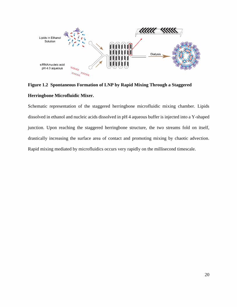
Figure 1.2 Spontaneous Formation of LNP by Rapid Mixing Through a Staggered
Herringbone Microfluidic Mixer.
Schematic representation of the staggered herringbone microfluidic mixing chamber. Lipids
dissolved in ethanol and nucleic acids dissolved in pH 4 aqueous buffer is injected into a Y-shaped
junction. Upon reaching the staggered herringbone structure, the two streams fold on itself,
drastically increasing the surface area of contact and promoting mixing by chaotic advection.
Rapid mixing mediated by microfluidics occurs very rapidly on the millisecond timescale.

21
1.2.4 General Composition, Structure and Size
Currently, the most advanced LNP formulations contain siRNA and have diameters less
than 100 nm as measured by dynamic light scattering and cryo-transmission electron microscopy
(cryoTEM). They are generally composed of ionizable amino-lipids, a phospholipid such as DSPC,
cholesterol and a surface PEG-lipid at a molar ratio of 50:10:38.5:1.5. Figure 1.3 depicts the most
commonly used LNP components. The ionizable amino-lipid heptatriaconta-6,9,28,31-tetraen-19-
yl 4-(dimethylamino)butanoate (DLin-MC3-DMA) is the most active among the amino-lipids
depicted in Figure 1.3B. CryoTEM revealed that LNP systems formed by rapidly mixing an
ethanol stream containing the lipid mixture with an aqueous stream containing the siRNA, by
either a T-junction (Crawford et al., 2011) or microfluidic-based mixing (Belliveau et al., 2012;
Leung et al., 2012), have an electron-dense core, which is in contrast to the less dense aqueous
cores that are characteristic of conventional liposomes or vesicular structures (Leung et al., 2012;
Zhigaltsev et al., 2006). Indeed, these LNP-siRNA systems have an interior lipid core containing
siRNA complexed with ionizable cationic lipid, as shown by the absence of 31P NMR signal from
free phosphorothioate in the siRNA and the complete protection of siRNA degradation by external
RNases (Leung et al., 2012). Computer simulation of the self-assembly of lipids suggests a
nanostructured core in which siRNA is located in internal inverted micelles complexed with
ionizable amino-lipid (Figure 1.4). Furthermore, DSPC interacts with siRNA phosphate through
its choline-headgroup, cholesterol is dispersed evenly between the core and surface, and the PEG-
lipid is distributed predominantly on the surface. It is postulated that the rapid mixing of lipid
components with the siRNA allows for the formation of nucleating structures composed of siRNA
and ionizable amino-lipid. Other LNP systems that contain ASO and a larger proportion of DSPC,
which promote bilayer formation, exhibit multilamellar structures. In this case, the nucleic acid is

22
thought to be sandwiched or entrapped between the concentric bilayers (refer to Section 1.2.3.1).
This further supports the hypothesis that, with appropriate mixing techniques, LNP structure and
physical properties can be controlled by its lipid constituents. Indeed, a continuum of structures
were observed based on varying lipid compositions and siRNA content (Leung et al., 2015). For
example, reducing the amount of ionizable amino-lipid from 40 mol% to 20 mol% results in LNP-
siRNA that contain lamellar structures. Increasing the saturation of the amino-lipid from linoleyl
to oleyl promotes the formation of multilamellar structures within the LNP, while increasing
nucleic acid content reduces multilamellar structures. These observations were further extended to
other macromolecules such as mRNA, plasmid DNA and negatively charged colloidal gold.
23
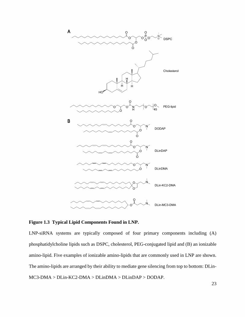
Figure 1.3 Typical Lipid Components Found in LNP.
LNP-siRNA systems are typically composed of four primary components including (A)
phosphatidylcholine lipids such as DSPC, cholesterol, PEG-conjugated lipid and (B) an ionizable
amino-lipid. Five examples of ionizable amino-lipids that are commonly used in LNP are shown.
The amino-lipids are arranged by their ability to mediate gene silencing from top to bottom: DLin-
MC3-DMA > DLin-KC2-DMA > DLinDMA > DLinDAP > DODAP.

24
Figure 1.4 Schematic Representation of Lipid Nanoparticles Containing Nucleic Acids.
LNP are composed of four primary lipid components: polyethyleneglycol conjugated lipids (PEG-
lipid, purple), structural or helper lipids such as distearoylphosphatidylcholine (DSPC, gray),
cationic or ionizable amino-lipids (blue) and cholesterol (orange). During spontaneous formation
by rapid mixing methods, the ionizable amino-lipid is cationic, which allows it to interact with the
polyanionic nucleic acids. This forms inverted micelle structures which are further coated by other
more polar lipid components such as PEG-lipid. Nucleic acid is completely entrapped within the
core of the LNP, preventing nuclease access and immune detection.

25
1.2.5 Cationic and Ionizable Amino-lipids
A major advancement in the design of ionizable amino-lipids was the modulation of their
pKa. pKa values of 7 and lower have been shown to be critical for encapsulation of nucleic acids
and in vivo activity (Maurer et al., 2001; Semple et al., 2001). In environments where the pH is
below the pKa of the ionizable lipid (e.g., pH 4.0), the tertiary amino group is protonated and
interacts with the negatively charged nucleic acids, thereby promoting the self-assembly of the
formulation components. In physiological environments where the pH is above the pKa of the
ionizable lipid (e.g., pH 7.4), the surface of the LNP has an almost neutral charge, resulting in
improved circulation and reduced toxicity. Subsequently, in the acidic environment of endosomes,
the amino group of the ionizable lipid becomes protonated and associates with the anionic
endosomal lipids. This interaction enables the destabilization of the endosomal membranes and
promotes the release of siRNA into the cytosol (Hafez et al., 2001; Xu and Szoka, 1996).
The first ionizable amino-lipid that was used for nucleic acid encapsulation was DODAP,
which has a pKa of 6.6-7 and one double bond in each of its acyl chains (Figure 1.3B) (Bailey and
Cullis, 1994; Maurer et al., 2001; Semple et al., 2001). Subsequent work focused on the impact of
the number of double bonds or the degree of unsaturation in the alkyl chain demonstrated that
ionizable amino-lipids containing fully saturated alkyl chains showed no silencing of luciferase
activity in vitro, whereas ionizable amino-lipids containing two or three double bonds per alkyl
chain showed enhanced silencing activity (Heyes et al., 2005). Encapsulation efficiencies of
siRNA seemed to be compromised using amino-lipids that contain three double bonds per alkyl
chain. Therefore, the linoleyl moiety has become the alkyl chain configuration of choice in
ionizable lipid development.

26
Structure-activity relationship studies have guided the synthesis and screening of a large
number of ionizable amino-lipids with various types of linkers connecting the amino group and
alkyl chains. These studies have identified DLin-KC2-DMA (2,2-dilinoleyl-4-(2-
dimethylaminoethyl)-[1,3]-dioxolane) (Semple et al., 2010), and DLin-MC3-DMA (Jayaraman et
al., 2012) to be 100-fold and 1000-fold more potent, respectively, in silencing of a hepatic gene
(Factor VII) in comparison to the previous generation lipid DLinDMA (1,2-dilinoleyloxy-N,N-
dimethyl-3-aminopropane) (Heyes et al., 2005). The ED50 (median effective dose) for LNP
containing DLin-MC3-DMA to silence Factor VII in mice and TTR in non-human primates was
0.005 mg/kg and 0.03 mg/kg, respectively (Jayaraman et al., 2012). A key finding from these
studies was that an optimal lipid pKa value of 6.2-6.5 was a determining factor of hepatic gene-
silencing activity in vivo. DLin-MC3-DMA, having a pKa of 6.44, is currently the most active
ionizable lipid being used in clinical trials (Section 1.3.1).
In hopes of further promoting biocompatibility, a novel generation of ionizable amino-
lipids was synthesized with biodegradable functionalities in the alkyl chains (Maier et al., 2013).
These novel lipids were well tolerated and rapidly eliminated from plasma and tissues in animal
studies. Importantly, they exhibited excellent potencies (ED50 < 0.01 mg/kg in mice, similar to that
of DLin-MC3-DMA) in both rodent and non-human primate models. Although their relevance to
human treatment remains to be proven, these preclinical results for biodegradable LNP-siRNA
formulations show great promise for clinical applications.
1.2.6 Steric Barrier for Stability and Prolonged Circulation
Unprotected lipid-based delivery systems are rapidly cleared by the mononuclear
phagocyte system (Allen, 1994; Ishida et al., 2002). To overcome this, hydrophilic polyethylene

27
glycol has been widely used to coat lipid-based delivery systems. The PEG coating prevents
aggregation during the formulation process and provides “stealth” like characteristics post i.v.
administration (refer to Section 1.1.4). The optimized PEG-lipid characteristics in current LNP-
siRNA systems were largely derived from earlier work on PEGylated liposomes (Klibanov et al.,
1990; Webb et al., 1998; Woodle and Lasic, 1992) and stabilized plasmid or antisense lipid
particles (SPLP or SALP). It was found that the length of the ceramide lipid anchor dictated how
long the PEG-lipid remained associated with the LNP. For instance, SPLP or SALP containing
PEG-ceramide with C20 anchors (PEG-CerC20) exhibited poor transfection efficiency in cultured
cells compared to shorter anchor counterparts such as PEG-CerC14 or PEG-CerC8 (Mok et al.,
1999; Song et al., 2002; Wheeler et al., 1999). PEG-lipids with longer acyl anchors remain
associated with the LNP longer, thus preventing LNP interaction with cells and subsequently with
the endosomal membranes. They also provide the LNP with a more durable coat, which extends
circulation lifetime (Monck et al., 2000; Tam et al., 2000). Consistent findings were observed
when PEG-ceramides were replaced with PEG-succinoyl-diacylglycerols (PEG-s-DAG) that have
various acyl anchor lengths (Ambegia et al., 2005). Due to the ease of synthesis and purification,
PEG-s-DAG replaced PEG-Cer in later SPLP formulations, and long acyl chain versions of PEG-
s-DAG such as PEG-s-distearoylglycerol (PEG-s-DSG) were used to increase circulation lifetime
in hopes of exploiting the EPR effect at tumor sites.
Unfortunately, despite being efficacious and non-toxic after a single bolus injection,
PEGylated LNP with long acyl anchors (PEG-DSPE (1,2-distearoyl-sn-glycero-3-
phosphoethanolamine), PEG-s-DSG, or PEG-CerC20) were rapidly cleared upon repeated
administration as a result of a robust immune response to the PEG component (Judge et al., 2006;
Semple et al., 2005). The use of rapidly dissociating, shorter anchor PEG-lipids such as PEG-s-

28
dimyristoyl (DMG) or PEG-CerC14, however, mitigated this immunogenic response.
Coincidentally, it was also observed that LNP containing PEG-s-DAG progressively lost the PEG
moiety due to hydrolysis of its succinate linker, leading to particle aggregation and a reduced shelf-
life. In response to this, the succinate linker was replaced with a carbamate linker to confer
improved chemical stability without affecting efficacy (Heyes et al., 2006). PEG-lipids containing
carbamate linkages are now used in the most advanced LNP-siRNA systems in the clinic.
1.2.7 Promoting Cellular Uptake with Endogenous and Exogenous Ligands
Neutral liposomes have been shown to bind to proteins in serum, exchange components
with lipoproteins and acquire factors that can potentially target them to specific cell types (Chonn
et al., 1992; Cullis et al., 1998). In particular, they interact with ApoE and ApoA1 (Mendez et al.,
1988; Rensen et al., 1997). ApoE, but not ApoA1 or ApoA4, was further found to enhance uptake
of neutral liposomes in HepG2 cells and primary hepatocytes (Bisgaier et al., 1989). The role of
ApoE in LNP uptake into hepatocytes was confirmed in vivo using ApoE-deficient mice (Yan et
al., 2005), which cleared LNP more slowly from the circulation and showed at least 20-fold less
LNP take up by hepatocytes when compared to wild-type animals. Similarly, LNP-siRNA systems
containing ionizable amino-lipids require ApoE for activity (Akinc et al., 2010). Silencing of
Factor VII was compromised in mice lacking ApoE or LDL receptor, suggesting that ApoE acts
as an endogenous ligand for LNP-siRNA systems that facilitates uptake into hepatocytes via the
LDL receptor. The ApoE and LDL receptor dependence is reminiscent of how natural lipoprotein
transport occurs within the body (Section 1.1.1). Figure 1.5 depicts the current model for LNP
uptake into hepatocytes.

29
Figure 1.5 Current Model of LNP-siRNA Delivery to Hepatocytes
Following i.v. administration, LNPs rapidly lose its PEG-lipid coat and are opsonized by
apolipoproteins such as ApoE (1). LNP are able to extravasate from the circulation and enter the
liver sinusoids where there is a significant reduction in blood flow. Here, LNP are able to cross
the fenestrated vasculature and enter the Space of Disse where it can directly interact with
hepatocytes (2). The ApoE bound to the LNP interact with LDL-receptors found on the surface of
hepatocytes, triggering endocytosis (3). As the endosome acidifies, the ionizable amino-lipids
become protonated, facilitating the interaction of the now positively charged LNP with the
negatively charged endosomal lipids (4). The now cationic amino-lipids disrupt the endosomal
membrane (5), resulting in the release of siRNA into the cytoplasm of the hepatocyte (6) where it

30
is free to interact with the RNA-Induced Silencing Complex (RISC) and cause sequence specific
degradation of mRNA and suppression of protein production.
When endogenous ligands are not available, exogenous ligands can be used to enhance
uptake of LNP-siRNA systems into target cells. Exogenous ligands such as antibodies, antibody
fragments and peptides have been widely used in the field of liposome technology (Allen et al.,
2002b; Cressman et al., 2009; Di Paolo et al., 2011; Sapra and Allen, 2003). However, they are
expensive and difficult to manufacture, as well as potentially immunogenic. In contrast, small
molecule ligands conjugated to the distal end of PEG-lipids can be synthesized and formulated
into LNP in a straightforward manner (Akinc et al., 2010; Li and Huang, 2006; Tam et al., 2013).
N-acetylgalactosamine (GalNAc), which binds with high affinity to the asialoglycoprotein
receptor (ASGPR) found on hepatocytes, have been shown to rescue gene-silencing activity of
LNP-siRNA systems in ApoE deficient mice (Akinc et al., 2010). Other exogenous small molecule
ligands have shown utility in non-hepatic cells. Anisamide, which interacts with sigma receptors,
increases delivery and activity of siRNA containing nanoparticles in lung tumors and metastases
(Chen et al., 2009; Li et al., 2008; Li and Huang, 2006). Furthermore, strophanthidin, a cardiac
glycoside that binds to the ubiquitously expressed cell surface receptor Na+/K+ ATPase, has been
shown to enhance delivery of LNP-siRNA to various cell types originating from the ovary, breast,
pancreas, lung and prostate (Tam et al., 2013). While the delivery of LNP using a promiscuous
ligand may be non-specific, target specificity can be conferred using siRNA against tissue specific
disease causing genes.

31
1.3 Clinical Evaluation of siRNA Delivered by Lipid Nanoparticles
1.3.1 LNP-siRNA in the Clinic
Because LNP have preferential affinity for the liver, there are several LNP-siRNA in
development for disorders such as hepatocellular carcinoma, hypercholesterolemia and
transthyretin-mediated amyloidosis. LNP formulations with similar composition and structures
(refer to Section 1.2) under clinical evaluation utilize either DLinDMA (first generation LNP) or
DLin-MC3-DMA (second generation LNP).
One of the first demonstrations of LNP-siRNA activity in humans is by Alnylam
Pharmaceuticals (Cambridge, MA). Alnylam’s ALN-VSP is an LNP that targets kinesin spindle
protein (KSP) and vascular endothelial growth factor (VEGF); it utilizes DLinDMA and contains
two siRNAs at a 1:1 molar ratio (Tabernero et al., 2013). In preclinical murine orthotopic liver
tumor models, a single i.v. administration of ALN-VSP into mice with established hepatocellular
carcinoma resulted in dramatic reductions (up to 50%) of both KSP and VEGF mRNA levels.
Within 48 hours of the injection, phenotypic changes as a result of KSP knockdown were observed
and tumor cells were arrested in mitosis. Reductions in microvascular density and intratumoral
hemorrhaging were also observed and is thought to be a result of VEGF knockdown. After 3 weeks
of repeated administration (6 injections at siRNA dose of 4 mg/kg body weight), an approximate
improvement of 50% in mean survival was observed in tumor bearing mice. These results in
addition to non-human primate data warranted a Phase I clinical study in 2009, with an enrollment
of 41 cancer patients. This study successfully demonstrated siRNA-mediated mRNA degradation
in the liver and that biweekly administration was generally safe and well tolerated with dose-
limiting toxicities (e.g.: thrombocytopenia, hypokalemia, hepatic failure) observed at 1.25 and 1.5
mg/kg. Due to the disease progression in the enrolled patients and the heterogeneity of the tumor

32
biopsies, it was difficult to determine if KSP and VEGF were downregulated in tumors, but both
KSP and VEGF siRNA were present. Prolonged disease stabilization of up to 1.5 years was also
observed in one patient with extensive metastasis who showed complete response to treatment.
In 2011, another LNP-siRNA formulation developed by Alnylam Pharmaceuticals entered
Phase I clinical trials (Fitzgerald et al., 2014). ALN-PCS targets the proprotein convertase
subtilisin/kexin type 9 (PCSK9) for the treatment of familial hypercholesterolemia. PCSK9 is
primarily expressed in the liver and intestine, and loss-of-function mutations result in reduced
plasma LDL levels and consequently reduced risks for heart disease. Other than the lower LDL
levels, humans and mice with reduced PCSK9 are generally healthy (perhaps arguably healthier),
making this an attractive target for siRNA therapeutics. With a single i.v. administration of ALN-
PCS at an siRNA dose of 0.4 mg/kg body weight, serum PCSK9 levels were reduced by
approximately 70% by day 3 post-injection and LDL-cholesterol was reduced by 40%. The
duration of PCSK9 and LDL-cholesterol reduction was also dose-dependent with higher doses
resulting in more persistent reductions. Not surprisingly, the Phase I results agree with non-human
primate preclinical studies. In favour of the novel and propriety Enhanced Stabilization Chemistry
platform, chemically modified siRNA against PCSK9 conjugated to a GalNAc-targeting moiety
has been developed to largely replace the LNP-based ALN-PCS.
One of the most advanced LNP formulations is a therapeutic that utilizes DLin-MC3-DMA
and carries siRNA against transthyretin (TTR). TTR is a serum transport protein that is primarily
synthesized in the liver and carries thyroxine and retinol binding protein. Mutations in TTR causes
misfolding, which promotes the formation of fibrils that can eventually deposit in various tissues
as amyloid plaques (Sekijima et al., 2008; Ueda and Ando, 2014). These amyloid deposits prevent
tissues and organs from functioning properly and often manifest as cardiomyopathies or

33
neuropathies. There is evidence suggesting that a reduction of 50% in disease-causing or fibril
forming protein can result in positive clinical outcomes (Lachmann et al., 2003). Currently,
Alnylam Pharmaceuticals is developing ALN-TTR02, also known as Patisiran, which targets both
wild-type and mutant TTR to reduce serum TTR levels and prevent the progression of amyloid
formation. Preclinical studies in non-human primates show that > 80% reduction in serum TTR
can be achieved with a single i.v. dose of LNP-siRNA (with an siRNA dose of 0.3 mg/kg body
weight) (Coelho et al., 2013). At an siRNA dose of 0.1 mg/kg body weight, ALN-TTR02 is able
to reduce serum TTR by approximately 70% whereas ALN-TTR01 (which utilizes DLinDMA) is
only able to reduce serum TTR by 45% at a much higher siRNA dose of 10 mg/kg. This dramatic
improvement in LNP-siRNA potency again demonstrates the importance of the ionizable amino-
lipid. Furthermore, a dose dependent reduction in TTR was observed, indicating that ALN-TTR02
is indeed responsible for mediating gene silencing. At the siRNA dose of 0.3 mg/kg, persistent
gene silencing can be observed for over a month, and repeated i.v. injections every 4 weeks results
in repeated reduction in serum TTR. It is important to note that at 0.3 mg/kg, Phase I clinical trial
data matches that of the non-human primate pre-clinical data, providing confidence for this LNP-
siRNA strategy. Phase II multi-dose studies show that administration of ALN-TTR02 at 0.3 mg/kg
every three weeks was generally well tolerated and led to a rapid, dose dependent reduction of
both wildtype and mutant (Val30Met) TTR (Suhr et al., 2015). The maximum knockdown
achieved was impressive at 96%. Preliminary Phase II open label extension studies show that
sustained TTR knockdown of ~80% is achieved after 168 days. Neurological symptoms due to
amyloid buildup stabilized and improvements were seen over 6 months. Currently, Phase III
development of ALN-TTR02 (Patisiran) is underway.

34
Other LNP-based therapeutics currently under Phase I/II clinical development include
TKM-PLK1 (TKM-080301) and ARB-1467, both of which are developed by Arbutus Biopharma,
formerly Tekmira Pharmaceuticals. TKM-PLK1 contains siRNA against polo-like kinase 1, a
protein involved in tumor cell proliferation. TKM-PLK1 is currently being evaluated for three
indications: gastrointestinal neuroendocrine tumors, adrenocortical carcinoma and hepatocellular
carcinoma. ARB-1467 contains multiple siRNA against the hepatitis B virus (HBV) to ensure that
all variants of HBV are targeted.
1.3.2 Observations, Challenges and Limitations of Current LNP Systems for Nucleic Acid
Delivery
Mild infusion related reaction (IRRs) have been observed following administration of
LNP-siRNA (Barros and Gollob, 2012; Coelho et al., 2013; Fitzgerald et al., 2014; Suhr et al.,
2015; Tabernero et al., 2013). These mild reactions have been fairly well controlled by co- or pre-
administration with glucocorticoids such as dexamethasone. Because siRNA are short 19-21mer
nucleic acids, it is relatively easy to modify the bases to mask it from immune detection. Significant
progress has been made in the chemical modification of RNA such that it is immune silent. These
improvements, however, are limited to shorter nucleic acid species like ASO or siRNA. Longer
nucleic acid polymers such as plasmid DNA or mRNA are both difficult and costly to modify, and
require delivery vehicles such as LNP (refer to Section 1.2.1). In addition, given that IRRs for LNP
containing chemically modified, immune silent siRNA are still observed, more severe reactions
are suspected to occur for LNP containing longer nucleic acid polymers. This continues to be a
significant clinical obstacle for the development of nucleic acid therapeutics.

35
Another challenge is the ease of administration of LNP-based drugs. LNP systems were
designed for systemic delivery and require slow i.v. infusions to reduce the IRRs. Although i.v.
injection is the most direct route of administration and results in the greatest bioavailability, it also
requires the help of trained professionals. This is a burden to existing health care systems and may
result in compliance issues. This route of administration may be tolerable for more life threatening
and debilitating conditions such as cancer; however, it is less appealing for manageable metabolic
disorders such as moderate high cholesterol (not to be confused with more sever
hypercholesterolemia). Indeed, in the development of siRNA therapeutics for liver applications,
there is a preference for novel chemical modifications of nucleic acids that allow for direct
subcutaneous (s.c.) administration of the siRNA (Nair et al., 2014; Sehgal et al., 2015). These
modifications not only prevent rapid degradation but also integrates a liver targeting aspect.
Although much higher siRNA doses are required to achieve similar degrees of gene silencing when
compared to the i.v. administered LNP-siRNA, s.c. injections enable patient self-administration
and reduces the costs of manufacturing. Advances in chemical modifications to enhance stability,
evade immune detection and target the liver have drastically increased the therapeutic index of
siRNA despite the need for much higher doses. At the moment, this technology is only available
for siRNA and short nucleic acid species.
1.4 Thesis Objectives
Recent advances in LNP systems for in vivo delivery of siRNA have dramatically improved
the potency of siRNA-mediated gene silencing and are undergoing clinical development for
various diseases, as discussed in Section 1.3.1. The overall objective of this thesis is to build on

36
the existing LNP technologies and develop more effective and safer delivery vehicles of nucleic
acid therapeutics.
As noted, current LNP-siRNA formulations are optimized for activity in the liver after i.v.
administration. There are, however, compelling reasons to develop LNP-siRNA that can be s.c.
administered. These include the potential for self-administration, a prolonged therapeutic window
(due to a depot effect and access to cell types that are in contact with the lymphatic system, such
as immune cells), and increased tissue availability through the circulation. In order to reach distal
tissues beyond the local lymph nodes, LNP must drain from the site of injection to the lymphatic
system and subsequently enter the arterial circulation. In Chapter 3, two key properties were
explored to develop LNP for s.c. administration: 1) Particle size and effect on tissue penetration
and 2) Degree and robustness of PEG-coating. We hypothesized that having a persisting PEG-coat
prevents opsonization and particle destabilization and therefore immune-related clearance, while
small particle sizes allow the LNP to better traverse the loose connective tissue and promote
lymphatic drainage and systemic delivery. The ability of existing LNP-siRNA to mediate gene
silencing at the liver was explored. The effects of size and PEG-coating on the rate of drainage
from the injection site, pharmacokinetics and biodistribution (PK/BD), and the duration and
reproducibility of gene silencing was determined for LNP formulations administered
subcutaneously.
In the work presented in Chapter 3, we found that small LNP (< 45 nm in diameter) are
able to reach the circulation after s.c administration and indeed others have shown that small
particles better penetrate into tumor tissue (Cabral et al., 2011; Dreher et al., 2006; Huo et al.,
2013; Popovic et al., 2010). Interestingly, it was shown that small LNP-siRNA were less
efficacious at hepatic gene silencing post s.c. administration. Continuing the efforts to develop

37
highly potent small LNP-siRNA, in Chapter 4 we sought to understand this size-related
impairment. In order to remove confounding effects due to s.c. administration such as the need to
drain from the site of injection, hepatic gene silencing of LNP with sizes ranging from 30-100 nm
was characterized following i.v administration. It was shown that the decreased potency of small
30 nm LNP-siRNA systems can be attributed, at least in part, to a pronounced instability of LNP-
siRNA systems as their size is reduced below 45 nm diameter. Here, we demonstrated that
improvements in activity and stability can be achieved with proper LNP design.
Finally, motivated by the observations that LNP-siRNA administered subcutaneously
results in skin irritations likely as a result of local immune stimulation (Chapter 3) and that the
potency of small LNP-siRNA cannot be further improved with existing components of the LNP
(Chapter 4), lipophilic pro-drugs that can efficiently incorporate into LNP were designed.
Recognizing that most LNP-siRNA systems used in the clinic require co- or pre-dosing of
corticosteroids to ameliorate infusion-related reactions and that the skin irritations post s.c.
administration of LNP are likely caused by immune stimulation, the first series of compounds
synthesized is a pro-drug of a potent corticosteroid, dexamethasone, that is currently used to reduce
IRRs for LNP drugs. In Chapter 5, we demonstrate that incorporation of dexamethasone directly
into LNP can greatly reduce the level of proinflammatory cytokines induced compared to co-
administration of free dexamethasone with LNP. Various versions of these pro-drugs were
synthesized and tested to better understand the requirements for effective incorporation, the ability
for esterases to access the pro-drug, the tolerability, and the ability of these pro-drugs to mitigate
cytokine induction by using LNP containing immune stimulatory DNA or in vitro transcribed
mRNA.

38
Chapter 2: Material and Methods
2.1 Materials
(R)-2,3-bis(octadecyloxy)propyl-1-(methoxy polyethylene glycol 2000) carbamate (PEG-
DMG) and (R)-2,3-bis(stearyloxy)propyl-1-(methoxy poly(ethylene glycol)2000 carbamate
(PEG-DSG) were provided by Alnylam Pharmaceuticals or synthesized as previously described
(Akinc et al., 2008). N-acetylgalactosamine cluster conjugated PEG-DSG (GalNAc-PEG) were
provided by Alnylam Pharmaceuticals. 3-(dimethylamino)propyl(12Z,15Z)-3-[(9Z,12Z)-
octadeca-9,12-dien-1-yl]henicosa-12,15-dienoate (DMAP-BLP) and (6Z,9Z,28Z,31Z)-
Heptatriaconta-6,9,28,31-tetraen-19-yl4-(dimethylamino)butanoate (DLin-MC3-DMA) were
provided by Alnylam Pharmaceuticals or synthesized by BioFine International (Vancouver, BC)
(Jayaraman et al., 2012; Rungta et al., 2013). 1,2-distearoyl-sn-glycero-3-phosphocholine (DSPC)
was purchased from Avanti Polar Lipids (Alabaster, AL) and cholesterol was purchased from
Sigma-Aldrich (St. Louis, MO). The sense and antisense strand sequences of siRNA against Factor
VII (siFVII) are 5′-GGAucAucucAAGucuuAcT*T-3′ and 5′-GuAAGAcuuGAGAuGAuccT*T-3,
respectively (Akinc et al., 2009; Akinc et al., 2008; Semple et al., 2010). 2’-Fluoro-modified
nucleotides are represented in lower case and phosphorothioate linkages are represented by
asterisks. All siRNA were either provided by Alnylam Pharmaceuticals or purchased from
Integrated DNA Technologies (San Diego, CA). Phosphorothioated unmethylated cytosine-
guanine containing oligonucleotides (CpG) complementary to the initiation codon region of the
human/mouse c-myc proto-oncogene (5’-AACGTTGAGGGGCAT-3’) was purchased from
TriLink Biotechnologies (San Diego, CA). This sequence has been previously identified as highly
immunostimulatory when administered to mice due to the presence of the CpG dinucleotide motif

39
(de Jong et al., 2007; Mui et al., 2001; Semple et al., 2005; Wilson et al., 2007). In vitro
transcription of luciferase mRNA was carried out as described previously (Wang et al., 2013).
2.2 Preparation of Lipid Nanoparticle-siRNA by Rapid Mixing Techniques
Ionizable amino-lipid (either DMAP-BLP or DLin-MC3-DMA), DSPC, cholesterol and
PEG-DMG were dissolved in ethanol at the mole ratio of 50:10:(39.75-x):(0.25+x), respectively
where the quantity of cholesterol and PEG-DMG are altered accordingly to ± x. The lipids in
ethanol and siRNA prepared in 25 mM acetate buffer (pH 4.0) was then injected into microfluidic
mixers (Precision Nanosystems, Vancouver, BC) at a flow rate ratio of 1:3 respectively with a
combined final flow rate of 4 mL/min. The LNP-siRNA mixtures were immediately dialyzed for
4 hr against a pH 6.7 buffer containing 50 mM MES and 50 mM citrate followed by phosphate
buffered saline overnight (12-14 kD MWCO dialysis tubing, Spectrum Labs, Rancho Dominguez,
CA). LNP-siRNA were then filtered through a 0.2 µm filter (Pall, Ville St. Laurent, Quebec) and
concentrated to approximately 1 mg/mL siRNA using Amicon Ultra centrifugal filters (Millipore,
Billerica, MA). The theoretical siRNA-to-lipid ratios for all formulations were maintained at 0.056
mg siRNA per µmole lipid which results in N/P charge ratios (ratios of the charge on the cationic
lipid, assuming it is in the positively charged protonated form, to the negative charge on the siRNA
oligonucleotide) of 3. Formulations that deviated more than 10% from the theoretical target were
not used. In experiments requiring radiolabelled LNP, 3H-cholesteryl hexadecylether (3H-CHE,
Perkin Elmer; Boston, Massachusetts) was incorporated at the ratio of 3.9 µCi/µmol total lipid.
All LNP preparation work was carried out at room temperature.
Similarly, LNP containing CpG were prepared by rapid mixing methods as described
previously (Belliveau et al., 2012; Jeffs et al., 2005; Semple et al., 2010). Briefly, DMAP-BLP,

40
DSPC, cholesterol and PEG-DMG were dissolved in ethanol at the mole ratio of 50, 10, 38.5 and
1.5 respectively. Dexamethasone pro-drugs were incorporated at 0, 1, 4 or 10 mol% at the expense
of DMAP-BLP. CpG were dissolved in 25 mM sodium acetate buffer at pH 4.0 such that the final
mixture would have a CpG-to-lipid weight ratio of 0.1. Ethanolic and aqueous mixtures were
mixed together at a 1:3 volume and flow rate ratio with final flow rates > 2 mL/min at room
temperature with a t-junction. Resulting mixtures were dialyzed against phosphate buffered saline
(PBS) overnight.
For LNP containing in vitro transcribed mRNA, DLin-MC3-DMA was used instead of
DMAP-BLP and the mRNA-to-lipid ratio was maintained at 0.028 mg mRNA per µmole lipid.
CpG concentration and entrapment efficiency were determined by measuring the absorbance at
260 nm after lipids were extracted using the Bligh and Dyer method (Bligh and Dyer, 1959). Lipid
concentrations were determined by measuring the cholesterol content (Cholesterol E Assay, Wako
Chemicals, Richmond, VA).
2.3 Analysis of Lipid Nanoparticles
LNP size and morphology were determined using cryogenic-transmission electron
microscopy (cryoTEM) as described previously (Belliveau et al., 2012; Leung et al., 2012). LNP
size (number weighting) was further confirmed by dynamic light scattering (DLS) using the
Malvern Zetasizer NanoZS (Worcestershire, UK). LNP formulations generally had polydispersity
indexes (PdI) below 0.05. Formulations with PdI > 0.1 were not used for further studies. siRNA
encapsulation efficiency was determined using the Quant-iT Ribogreen RNA assay (Life
Technologies, Burlington, ON). Briefly, LNP-siRNA was incubated at 37°C for 10 min in the
presence or absence of 1% Triton X-100 (Sigma-Aldrich, St. Louis, MO) followed by the addition

41
of the ribogreen reagent. The fluorescence intensity (Ex/Em: 480/520 nm) was determined and
samples treated with Triton X-100 represent total siRNA while untreated samples represent
unencapsulated siRNA. Total lipid was determined by measuring the cholesterol content using the
Cholesterol E assay (Wako Chemicals, Richmond, VA) and the siRNA concentration was
determined by measuring the absorbance at 260 nm. mRNA encapsulation efficiency was
determined similarly with Quant-iT Ribogreen RNA assay. CpG concentration and entrapment
efficiency were determined by measuring the absorbance at 260 nm after lipids were extracted
using the Bligh and Dyer method (Bligh and Dyer, 1959).
2.4 In Situ Determination of the Apparent Acid Dissociation Constant of LNP-siRNA
Apparent acid dissociation constant (pKa) of LNP-siRNA was determined as previously
described (Jayaraman et al., 2012). Briefly, 2-(p-toluidino)-6-napthalene sulfonic acid (TNS,
Sigma-Aldrich, St. Louis, MO) was dissolved in water to a working concentration of 0.6 mM and
LNP-siRNA was diluted with PBS to 0.05-0.1 mM total lipid. 0.12 mL of diluted LNP-siRNA was
mixed with 20 µL of diluted TNS to a final volume of 2 mL with pH 2.5-11 buffer containing 10
mM HEPES, 10 mM MES and 10 mM ammonium acetate. Samples were mixed thoroughly and
the fluorescence intensity was measured (Ex/Em: 321/445 nm) using a Perkin Elmer LS55. A
sigmoidal best fit analysis was applied using Sigma Plot and the pKa was measured as the pH at
half-maximal fluorescence intensity.
2.5 In Vitro Lipid and siRNA Dissociation
LNP were prepared with 0.5 mol% of 14C labelled DSPC (American Radiolabeled
Chemicals, Saint Louis, MO) and 0.25 mol% of 3H labelled PEG-DMG (Alnylam

42
Pharmaceuticals, Cambridge, MA) or 0.5 mol% of 14C labelled dilinoleylmethyl-4-
dimethylaminobutyrate (DLin-MC3-DMA, Alnylam Pharmaceuticals) and 0.002 mol% of 3H-
CHE. The 3H:14C ratios were maintained at approximately 3:1. For siRNA dissociation studies,
siRNA conjugated to Quasar 570 fluorophore (Alnylam Pharmaceuticals) was used. LNP were
incubated for 0, 0.25, 1, 2, 4 and 8 hr at 37°C in 200 µL of sterile mouse plasma (Cedarlane,
Burlington, Ontario) at a final lipid concentration of 0.3 mM. At the various time-points, samples
were loaded onto a 1.5 × 27 cm Sepharose CL-4B column (Sigma-Aldrich, St. Louis, MO). 30 ×
2 mL fractions were collected and analyzed by liquid scintillation counting. To determine the
amount of siRNA remaining in the LNP fractions, 135 µL of each fraction was incubated with 0.5
mM hexaethylene glycol monodecyl ether (Sigma-Aldrich, St. Louis, MO) and the fluorescence
at 590 nm was measured. All dissociation rates were estimated from a linear fit of the first four
timepoints. For experiments monitoring Forster Resonance Energy Transfer (FRET) as a measure
of particle stability, LNP were prepared as described previously with the addition of 1 mol% each
of 1,2-dioleoyl-sn-glycero-3-phosphoethanolamine-N-(7-nitro-2-1,3-benzoxadiazol-4-yl) (NBD-
DOPE) and 1,2-dioleoyl-sn-glycero-3-phosphoethanolamine-N-(lissamine rhodamine B sulfonyl)
(LRB-DOPE). LNP were incubated at 37°C in mouse plasma for up to 24 hr at a final lipid
concentration of 0.3 mM. Fluorescence of NBD-DOPE was measured (Ex/Em: 485/530 nm) in
the presence or absence of 1.5% Triton X-100.
2.6 Pharmacokinetics and Biodistribution
3H-CHE-labelled LNP (based on siRNA concentration) were injected by i.v. or s.c. under
the loose interscapular skin at the back of the neck of female CD1 mice (Charles River
Laboratories, Wilmington, MA). Mice were euthanized 15 min to 48 hr post-injection and tissues

43
were collected. To determine LNP concentration, approximately 600 mg of tissue was
homogenized using the FastPrep homogenizer from MP Biomedicals (Santa Ana, CA) and
incubated with 0.5 mL of Solvable (Perkin Elmer) at 50°C overnight. Samples were cooled then
decolourized with the addition of 200 uL of 30% (v/v) hydrogen peroxide followed by the addition
of 5 mL of Pico-Fluor Plus scintillation fluid (Perkin Elmer). To determine LNP concentration in
blood, 100 µL of whole blood was added directly to solvable and hydrogen peroxide and allowed
to decolourize overnight before the addition of Pico-Fluor Plus. All samples were read on a
Beckman LS 6500 liquid scintillation counter. All procedures were approved by the Animal Care
Committee at the University of British Columbia and were performed in accordance with
guidelines established by the Canadian Council on Animal Care.
2.7 In Vivo LNP-siRNA Activity in Mouse Factor FVII Model
LNP-siRNA were diluted with PBS such that injection volumes were maintained at 10
mL/kg body weight and administered (based on siRNA concentration) either i.v. or s.c. under the
loose interscapular skin in 6-8 weeks old female C57Bl/6 mice (Charles River Laboratories,
Wilmington, MA). Animals were euthanized after 24 hr and blood was collected via intracardiac
sampling. Serum was separated from whole blood using Microtainer Serum Seperator tubes
(Becton Dickinson, Franklin Lakes, NJ). To study FVII knockdown over time, at 1, 2, 4, 7, 11, 15,
21 and 28 days post-injection, 20-30 µL of blood was collected from the lateral saphenous vein.
Blood samples were allowed to coagulate at 4°C overnight and the serum was separated followed
by centrifugation for 15 min at 12,000 rpm. The residual serum FVII levels were determined using
the Biophen VII chromogenic assay (Aniara, Mason, OH) according to manufacturer’s protocol
and normalized to that of control mice injected with PBS.

44
2.8 Synthesis of Dexamethasone Pro-drugs
Log P (or Log D) values were predicted using MarvinSketch v6.1.2 following modified
parameters previously described (Viswanadhan et al., 1989). Detailed descriptions of the synthesis
of LD001-005 used in Chapter 5 can be found in Appendix A.
2.9 In Vitro Degradation of Dexamethasone Pro-drugs
To determine the biodegradability of the LD001-005, 1 mg/mL LNP was incubated in
mouse plasma (Cedarlane, Burlington, Ontario) or PBS supplemented with 10U of purified porcine
esterase (Sigma-Aldrich, St. Louis, MO) for up to 4 hours at 37°C. Post incubation, four volumes
of chloroform/methanol (2:1) were added and the mixture was vortex mixed. Samples were
centrifuged at 13,000g for 5 minutes and the upper phase was discarded. The remaining organic
phase was dried down under vacuum and the resulting lipid extract was dissolved in
methanol/acetonitrile (1:1). Quantity of parent dexamethasone pro-drug was determined by ultra
high pressure liquid chromatography (UHPLC) on a Waters Acquity H-Class UHPLC System
equipped with a BEH C18 column (1.7 µm, 2.1×100 mm) and a photodiode array detector.
Separation was achieved at a flow rate of 0.5 mL/min, with a mobile phase consisting of a linear
methanol-water gradient (85:15 to 100:0) over 5 minutes at a column temperature of 55oC. The
absorbance at 239 nm was measured and the analyte concentration was determined using
calibration curves.
45
2.10 In Vitro LNP Tolerability by MTT and Hemolysis
In vitro LNP tolerability was determined by performing an MTT cell proliferation assay
on cultured HeLa cells. Briefly, 10,000 cells/well were seeded in 96-well plates, grown overnight
and treated with various concentrations of LNP (5×10⁻⁷ to 5 mg/mL, with or without pro-drug) for
24 h. Cells were then incubated with 1 mg/mL of 3-[4,5-Dimethylthiazol-2-yl]-2,5-diphenyl-
tetrazolium bromide (MTT, Sigma-Aldrich, St. Louis, MO) in 100 µL of complete media for 2 h
followed by an overnight incubation with 100 µL of 20% sodium dodecyl sulfate dissolved in
dimethylformamide/water (1:1) at pH 4.7 at 37°C. Absorbance at 570 nm was measured after the
overnight incubation and % viability was determined by normalizing the absorbance of LNP
treatment with PBS controls.
The ability of LNP to cause hemolysis was. Briefly, human erythrocytes (RBC) (Innovative
Research, Novi, MI) were washed three times with cold isotonic saline (0.9% w/v NaCl in water)
and resuspended in pH 7.4 PBS to a final concentration of 4% vol/vol. LNP (made with varying
amounts of PEG-lipid or with dexamethasone pro-drug) were then incubated with the RBC
solution at a final lipid concentration of 0.15 mg/mL for 1 hour at 37°C. RBC solutions incubated
with saline or 0.2% vol/vol Triton X-100 were used as controls. Samples were then cooled and
centrifuged for 5 minutes at 1000×g at 4℃ and the supernatant was removed and the absorbance
at 562 nm was determined. % hemolysis was determined by normalizing the absorbance at 562
nm to Triton X-100 treated samples.
2.11 In Vivo Immune Suppression by LNP with Dexamethasone Pro-drugs.
6-8 Weeks old female C57Bl/6 mice (Charles River Laboratories, Wilmington, MA) were
injected intravenously with Dex-LNP containing CpG (10 mg/kg) or mRNA (3 mg/kg). Animals
46
were euthanized 2 and 4 hours post-injection and blood was collected via intracardiac sampling.
Plasma was separated from whole blood by centrifugation and analyzed for proinflammatory
cytokines using the Mesoscale Proinflammatory Multiplex kit (Rockville, MD). All procedures
were approved by the Animal Care Committee at the University of British Columbia and were
performed in accordance with guidelines established by the Canadian Council on Animal Care.

47
Chapter 3: Development of Lipid Nanoparticle Formulations of siRNA for
Hepatocyte Gene Silencing Following Subcutaneous Administration
3.1 Synopsis
Recently developed lipid nanoparticle (LNP) formulations of siRNA have proven to be
effective agents for hepatocyte gene silencing following intravenous administration with at least
three LNP-siRNA formulations in clinical trials. The aim of this work was to develop LNP-siRNA
systems for hepatocyte gene silencing that can be administered subcutaneously (s.c.). Three
parameters were investigated, namely LNP size, residence time of the polyethylene glycol (PEG)-
lipid coating and the influence of hepatocyte-specific targeting ligands. LNP sizes were varied
over the range of 30 to 115 nm in diameter and PEG-lipid that dissociates rapidly (PEG-DMG)
and slowly (PEG-DSG) were employed. In mice, results show that large (~80 nm) LNP exhibited
limited accumulation in the liver and poor Factor VII (FVII) gene silencing at 1 mg siRNA/kg
body weight. Conversely, small (~30 nm) LNP systems showed maximal liver accumulation yet
still had minimal activity. Interestingly, intermediate size (~45 nm) LNP containing PEG-DSG
exhibited nearly equivalent liver accumulation as the smaller systems following s.c. administration
but reduced FVII levels by 80% at 1 mg siRNA/kg body weight. Smaller systems (~35 nm
diameter) containing either PEG-DMG or PEG-DSG were less active, however addition of 0.5
mol% of a GalNAc-PEG lipid to these smaller systems improved activity to levels similar to that
observed for the ~45 nm diameter systems. In summary, this work shows that appropriately
designed LNP-siRNA systems can result in effective hepatocyte gene silencing following s.c
administration and has been published (Chen et al., 2014).

48
3.2 Results
3.2.1 LNP-siRNA Systems Containing PEG-DSG Exhibit Maximum Delivery to Liver
Following Subcutaneous Administration
Previous investigations have shown that the diameter of LNP-siRNA systems can be
modulated over the range of 25-100 nm by varying the proportion of PEG-lipid in the formulation
using a microfluidic mixing process (Belliveau et al., 2012). As the lipid composition employed
here is different than used previously, we examined the size distribution achieved for LNP-siRNA
systems composed of DMAP-BLP, DSPC, cholesterol and PEG-DMG in the molar ratios
50:10:(39.75-x):(0.25+x), where x was varied from 0 to 4.75, encapsulating siRNA at a N/P (+/-)
charge ratio of 3. The total amount of PEG-lipid was therefore varied from 0.25 to 5.0 mol%. The
size and structure of these LNP-siRNA systems were determined by cryoTEM and DLS (Figure
3.1). LNP-siRNA systems of sizes ranging from 30 nm to 115 nm in diameter were generated that
exhibited electron dense cores as previously described (Belliveau et al., 2012; Leung et al., 2012)
(Figure 3.1 A-D). Particle sizes determined using DLS were consistent to those determined using
cryoTEM (Figure 3.1E). Size differences compared to previous reports (Belliveau et al., 2012) can
be attributed to the different lipid compositions employed. Hereafter for simplicity, reported
particle sizes are rounded to the nearest multiple of five and based on DLS measurements.

49
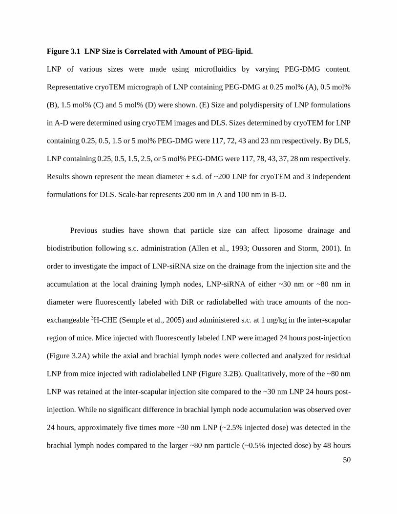
50
Figure 3.1 LNP Size is Correlated with Amount of PEG-lipid.
LNP of various sizes were made using microfluidics by varying PEG-DMG content.
Representative cryoTEM micrograph of LNP containing PEG-DMG at 0.25 mol% (A), 0.5 mol%
(B), 1.5 mol% (C) and 5 mol% (D) were shown. (E) Size and polydispersity of LNP formulations
in A-D were determined using cryoTEM images and DLS. Sizes determined by cryoTEM for LNP
containing 0.25, 0.5, 1.5 or 5 mol% PEG-DMG were 117, 72, 43 and 23 nm respectively. By DLS,
LNP containing 0.25, 0.5, 1.5, 2.5, or 5 mol% PEG-DMG were 117, 78, 43, 37, 28 nm respectively.
Results shown represent the mean diameter ± s.d. of ~200 LNP for cryoTEM and 3 independent
formulations for DLS. Scale-bar represents 200 nm in A and 100 nm in B-D.
Previous studies have shown that particle size can affect liposome drainage and
biodistribution following s.c. administration (Allen et al., 1993; Oussoren and Storm, 2001). In
order to investigate the impact of LNP-siRNA size on the drainage from the injection site and the
accumulation at the local draining lymph nodes, LNP-siRNA of either ~30 nm or ~80 nm in
diameter were fluorescently labeled with DiR or radiolabelled with trace amounts of the non-
exchangeable 3H-CHE (Semple et al., 2005) and administered s.c. at 1 mg/kg in the inter-scapular
region of mice. Mice injected with fluorescently labeled LNP were imaged 24 hours post-injection
(Figure 3.2A) while the axial and brachial lymph nodes were collected and analyzed for residual
LNP from mice injected with radiolabelled LNP (Figure 3.2B). Qualitatively, more of the ~80 nm
LNP was retained at the inter-scapular injection site compared to the ~30 nm LNP 24 hours post-
injection. While no significant difference in brachial lymph node accumulation was observed over
24 hours, approximately five times more ~30 nm LNP (~2.5% injected dose) was detected in the
brachial lymph nodes compared to the larger ~80 nm particle (~0.5% injected dose) by 48 hours

51
post administration. Not surprisingly, very little (less than 0.1% injected dose) of either
formulation was detected in the axial lymph nodes (data not shown) since the inter-scapular site
of injection drains almost exclusively to the brachial lymph nodes (Tilney, 1971).

52
Figure 3.2 A Greater Proportion of Small LNP Drain from the Site of Injection and
Accumulate at Brachial Lymph Nodes Following s.c. Administration.
(A) Mice injected with DiR-labelled LNP that were either ~80 nm (left), ~30 nm (center) or PBS
(right) were imaged 24 post-injection. (B) 3H-labelled LNP containing PEG-DSG that were ~80
nm (black) and ~30 nm (gray) in diameter were administered s.c. at 1 mg/kg body weight. Brachial
lymph nodes were harvested at 2, 6, 24 and 48 hours post injection. Results shown represent the
mean ± s.d. of four animals.

53
In order to investigate the impact of particle size and PEG coating on the distribution of
s.c. administered LNP-siRNA to blood and subsequently to the liver, radiolabelled LNP-siRNA
were tracked for 48 hours post s.c. injection. PEG-lipids containing either the rapidly dissociating
C14 (PEG-DMG) or the non-dissociating C18 (PEG-DSG) acyl anchors were tested. As shown in
Figure 3.3A, the small systems containing 5% PEG-DSG exhibited by far the highest circulating
concentration (Cmax) of LNP (17.1% of the injected dose) following s.c. administration (Table 3.1).
However, this did not result in higher levels of accumulation in the liver. Instead, equivalent levels
of accumulation corresponding to approximately 15% of the total dose were observed 48 hours
post injection for small (~30 nm) LNP containing either 5% PEG-DMG or PEG-DSG (Figure
3.3B). Furthermore, these levels were equivalent to those observed for large (~80 nm) and
intermediately sized (~45 nm) LNP with PEG-DSG. The fact that the small LNP containing 5%
of the non-exchangeable PEG-DSG do not exhibit greater liver accumulation despite achieving a
higher maximum concentration in the blood can be attributed to their long circulation lifetimes
following penetration to the blood compartment which facilitates distribution to tissues other than
the liver (Allen et al., 1991; Cullis et al., 1998; Woodle and Lasic, 1992). At the maximum
concentration of LNP appearing in the blood, no hemolysis-induced toxicity is expected since
LNP-siRNA showed no hemolytic activity at pH 7.4 (Figure 3.4). Less than 5% of the ~45 nm and
~80 nm PEG-DMG LNP was found in the liver.
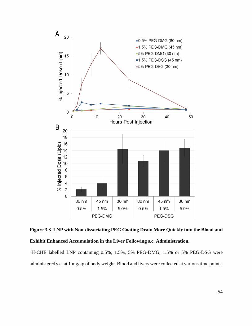
54
Figure 3.3 LNP with Non-dissociating PEG Coating Drain More Quickly into the Blood and
Exhibit Enhanced Accumulation in the Liver Following s.c. Administration.
3H-CHE labelled LNP containing 0.5%, 1.5%, 5% PEG-DMG, 1.5% or 5% PEG-DSG were
administered s.c. at 1 mg/kg of body weight. Blood and livers were collected at various time points.

55
(A) Clearance of LNP from blood over a period of 48 hr. (B) Amount of LNP found in liver at 48
h. Results shown represent the mean ± s.d. of four animals.

56
Table 3.1 Area-under-curve (AUC) and Maximum Blood Concentration (Cmax) of LNP
with Varying Sizes.
LNP Size
(diameter, nm)
AUC
(% inj. dose·hour)
Cmax
(% injected dose)
0.5% PEG-DMG 80 39.0 1.0±0.2
1.5% PEG-DMG 45 33.6 0.9±0.1
5% PEG-DMG 30 51.6 1.6±0.6
1.5% PEG-DSG 45 75.3 2.7±0.3
5% PEG-DSG 30 376.1 17.1±1.6
Note: AUC and Cmax determined from blood clearance data presented in Figure 3.3
Figure 3.4 LNPs are not hemolytic at physiological pH.
LNPs containing 0.5, 1.5 or 5 mol% of PEG-DMG or PEG-DSG were incubated with human RBC
for 1 hour at 37℃. Samples were centrifuged at 1000×g and the absorbance of the supernatant was
measured at 562 nm. Data is expressed as % hemolysis relative to the Triton X-100 control (TX-
100).
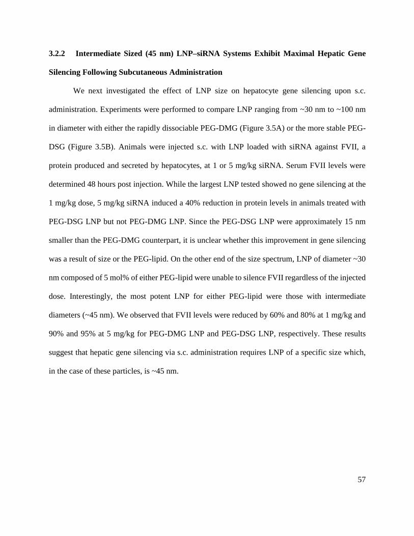
57
3.2.2 Intermediate Sized (45 nm) LNP–siRNA Systems Exhibit Maximal Hepatic Gene
Silencing Following Subcutaneous Administration
We next investigated the effect of LNP size on hepatocyte gene silencing upon s.c.
administration. Experiments were performed to compare LNP ranging from ~30 nm to ~100 nm
in diameter with either the rapidly dissociable PEG-DMG (Figure 3.5A) or the more stable PEG-
DSG (Figure 3.5B). Animals were injected s.c. with LNP loaded with siRNA against FVII, a
protein produced and secreted by hepatocytes, at 1 or 5 mg/kg siRNA. Serum FVII levels were
determined 48 hours post injection. While the largest LNP tested showed no gene silencing at the
1 mg/kg dose, 5 mg/kg siRNA induced a 40% reduction in protein levels in animals treated with
PEG-DSG LNP but not PEG-DMG LNP. Since the PEG-DSG LNP were approximately 15 nm
smaller than the PEG-DMG counterpart, it is unclear whether this improvement in gene silencing
was a result of size or the PEG-lipid. On the other end of the size spectrum, LNP of diameter ~30
nm composed of 5 mol% of either PEG-lipid were unable to silence FVII regardless of the injected
dose. Interestingly, the most potent LNP for either PEG-lipid were those with intermediate
diameters (~45 nm). We observed that FVII levels were reduced by 60% and 80% at 1 mg/kg and
90% and 95% at 5 mg/kg for PEG-DMG LNP and PEG-DSG LNP, respectively. These results
suggest that hepatic gene silencing via s.c. administration requires LNP of a specific size which,
in the case of these particles, is ~45 nm.

58
Figure 3.5 ~45 nm LNP Exhibit Maximum Gene Silencing Post s.c. Injection.
(A) PEG-DMG or (B) PEG-DSG LNP were s.c. administered at 1.0 (black) or 5.0 mg/kg (gray)
of body weight and the serum FVII was determined 48 hours post-injection. Results shown
represent the mean ± s.d. of four animals.

59
To explore the duration of gene silencing following s.c. administration, blood was collected
from animals injected with the most active LNP-siRNA formulation (~45 nm diameter) over 28
days and serum FVII levels were determined (Figure 3.6). A single injection of 1 mg/kg led to
greater than 50% FVII protein reduction which persisted for at least 4 days for PEG-DMG LNP
and 7 days for PEG-DSG LNP (Figure 3.6A). This result implicates the importance of a stable
PEG coat for gene silencing using LNP administered s.c. The higher hepatic gene silencing ability
of PEG-DSG LNP can be attributed, at least in part, to the fact that they accumulated at least 3-
fold more in the liver than PEG-DMG LNP of a similar size (Figure 3.3B). When the mice were
dosed at 5 mg/kg, a reduction in FVII of approximately 90% was observed for a minimum of 4
days and protein levels were reduced below 50% for at least 11 days (Figure 3.6B). There was no
significant difference in the degree or duration of silencing between LNP containing PEG-DMG
or PEG-DSG likely due to fact that the dosage was high enough to mask any potency differences.

60

61
Figure 3.6 Persistent Gene Silencing (< 50% protein) is Observed Over 11 Days Following
s.c. Administration of ~45 nm Diameter LNP.
LNP of similar sizes (~45 nm) but different PEG-lipids were administered s.c. in C57Bl/6 mice at
1 mg/kg or 5 mg/kg of body weight. Blood was collected over 28 days. Serum factor VII silencing
was determined and normalized to that of control mice treated with PBS. Residual FVII levels
were plotted against time for (A) 1 mg/kg and (B) 5 mg/kg siRNA. Results shown represent the
mean ± s.d. of four animals.
It is surprising that small ~30 nm LNP with the stable PEG-DSG coat were less active in
gene silencing given that they showed favourable blood exposure and liver accumulation. We
speculated that the reduced gene silencing was likely the result of inadequate uptake of LNP into
hepatocytes since the stable PEG-DSG coat has been shown to inhibit opsonization in circulation
and delay LNP clearance to the liver. In order to explore this possibility, GalNAc conjugated PEG-
lipid (GalNAc-PEG) that targets asialoglycoprotein receptor expressed on the surface of
hepatocytes was used as a targeting lipid (Akinc et al., 2010). Two PEG-DSG LNP compositions
resulting in approximately 45 nm and 35 nm in diameter were tested with or without GalNAc-PEG
lipid. Interestingly, no significant improvement on activity was observed at 48 h post
administration when 0.5 mol% GalNAc-PEG was incorporated into 45 nm LNP (Figure 3.7A).
This is not surprising since these formulations were the most active as shown in Figure 3.5 and
were likely able to utilize ApoE-mediated endocytosis. However, when 0.5 mol% of GalNAc-PEG
was incorporated into the smaller ~35 nm PEG-DSG LNP, a dramatic improvement in FVII
silencing was observed (Figure 3.7B). Targeted LNP (containing GalNAc-PEG) showed almost
90% FVII knockdown for at least 7 days whereas only ~50% protein reduction was observed using

62
non-targeted LNP. Furthermore, the duration and degree of gene silencing of GalNAc-PEG LNP
was similar to PEG-DMG LNP.

63

64
Figure 3.7 Hepatic Gene Silencing Can be Enhanced Using s.c. Administered LNP-siRNA
Containing GalNAc-PEG lipid.
(A) Mice were s.c. injected with ~45 nm PEG-DSG LNP with or without 0.5% GalNAc-PEG at 1
or 5 mg/kg siRNA. Residual FVII was monitored for 28 days. (B) Residual FVII was monitored
over 28 days for animals injected with ~35 nm PEG-DSG or PEG-DSG with 0.5% GalNAc-PEG
LNP. Results shown represent the mean ± s.d. of four animals.
3.3 Discussion
To our knowledge, this is the first report on s.c. administration of LNP-siRNA for in vivo
gene silencing. It was hypothesized that particle size and PEG steric barrier are crucial factors in
determining the potency of s.c. administered LNP-siRNA since the particles must traverse through
the meshwork of loose connective tissue to reach the local lymph nodes and drain into the
lymphatic system and subsequently into the peripheral circulation. Consistent with this hypothesis,
it was found that small LNP-siRNA with a stable PEG coat had a much greater AUC in blood
(Table 3.1) than other formulations tested indicating that they were made available to access tissues
such as the liver. We also demonstrated the importance of the stability of the PEG coat and particle
size on the potency of s.c. administered LNP-siRNA. We discuss the implications of this study for
the development of next generation LNP-siRNA systems with improved potency following s.c.
administration.
The utility of liposomes as small as 40 nm in diameter for s.c. administration have been
previously explored; however, these were bilayer systems manufactured by sonication and are
poorly suited for carrying a therapeutic payload such as siRNA (Hope et al., 1986; Oussoren et al.,
1997). For the non-bilayer “solid core" LNP-siRNA systems described here (Belliveau et al., 2012;

65
Leung et al., 2012), an inverse relationship between LNP size and the ability to drain from the site
of injection and enter the blood following s.c. administration was observed. This is consistent with
previous literature indicating that smaller LNP are able to drain faster into the circulation than their
larger counterparts (Oussoren and Storm, 2001; Oussoren et al., 1997). At the brachial lymph
nodes where the s.c. injected LNP are expected to drain to, ~2.5% of the small (~30 nm) PEG-
DSG LNP was detected 48 hours following s.c. administration, compared to only < 0.5% of the
larger (~80 nm) LNP (Figure 3.2). This suggests that the larger PEG-DSG LNP remain at the site
of injection and slowly permeate into the circulation, whereas the small PEG-DSG LNP reach the
lymph node at a faster rate resulting in a greater accumulation at the lymph node and subsequent
release into the circulation. This is also supported by our observation that large and small LNP
exhibit low and high Cmax in blood, respectively (Figure 3.3A and Table 3.1).
For all LNP containing the rapidly dissociable PEG-DMG, the maximum dose appearing
in the blood was less than 2% of the total injected dose (Figure 3.3A and Table 3.1). This low Cmax
may be the result of the PEG-DMG lipid being rapidly dissociated from the LNP leading to
insufficient steric barrier necessary for the LNP to avoid immune clearance or particle aggregation,
both of which could result in a build-up of LNP at the injection site and cause local toxicities. Any
quantity that reaches the peripheral circulation will be rapidly cleared by the liver because PEG-
DMG LNP administered i.v. have a circulation lifetime of less than 1 hour (Mui et al., 2013). It is
estimated that less than 5% of the ~45 and ~80 nm PEG-DMG LNP administered s.c. reached the
circulation and virtually all of this material is subsequently accumulated in the liver, resulting in
the levels observed 48 hours post injection (Figure 3.3B). It is noteworthy that although the ~30
nm PEG-DMG LNP had a low Cmax and AUC in the blood, they showed relatively high
accumulation in the liver further supporting a rapid hepatic clearance of PEG-DMG LNP that were

66
drained into the blood. It is likely that at least 14% of the injected dose of the 30 nm PEG-DMG
LNP reached the peripheral circulation. The rapid dissociation of PEG-DMG favours hepatocyte
uptake while hindering drainage from the injection site. This may prove to be a useful aspect of
LNP-siRNA in vivo behaviour as others have used particle aggregation as a strategy to promote
liposome retention in lymph nodes (Phillips et al., 2000). It may be possible to modulate the rate
of PEG-lipid dissociation by using different acyl anchor lengths such that an adequate quantity of
LNP drains from the injection site while facilitating aggregation on reaching the lymph node.
When PEG-DMG was substituted with a more stable PEG-DSG, liver accumulation of
LNP-siRNA was greatly enhanced. In the case of the ~45 nm diameter LNP, PEG-DSG LNP
accumulated to levels three times higher than PEG-DMG LNP. This indicates that a stable PEG
coating is an important factor, in addition to size, in allowing extravasation from the s.c. injection
site to reach the liver, at least for the 30-80 nm size range tested here (Figure 3.3B). Furthermore,
all LNP made with PEG-DSG accumulate to similar levels (~15% of the injected dose) in the liver
48 hours post injection while having drastically different maximum blood concentrations (2.7%
for ~45 nm and 17.1% for ~30 nm). Finally, despite similar liver accumulation for PEG-DSG LNP
systems (Figure 3.3B), differences in gene silencing were observed (Figure 3.5B) suggesting that
other factors in addition to tissue localization such as cellular uptake and endosome destabilization
are involved in dictating LNP activity.
While a stable PEG-coat may prevent unwanted protein interactions and promote
lymphatic drainage and circulation lifetimes, it is likely that this coat will impair LNP uptake into
hepatocytes since this process requires the association of LNP with ApoE (Akinc et al., 2010; Yan
et al., 2005). In this case, an exogenous targeting ligand such as GalNAc used in this study, can be
tethered to the distal end of the PEG-lipid to enhance LNP uptake into target cells (Akinc et al.,

67
2010). The incorporation of 0.5 mol% of GalNAc-PEG greatly improved the activity of ~35 nm
PEG-DSG LNP suggesting that inefficient cellular uptake was, in part, responsible for the
reduction in activity of LNP containing PEG-DSG (Figure 3.7B). Interestingly, the addition of
GalNAc-PEG to larger ~45 nm systems did not improve the activity to the same extent as for
smaller systems (Figure 3.7A). This is presumably because LNP with 1.5 mol% PEG-lipid are still
able to associate with ApoE and the incorporation of an additional targeting ligand confers little
additional benefit.
It is important to note that, regardless of the type of PEG-lipid coating, small LNP of size
~30 nm showed a reduction in activity compared to their larger counterparts (Figure 3.5) despite
showing favourable blood and liver accumulation following s.c. administration (Figure 3.3). This
presumably is due, in part, to inefficient release of the siRNA cargo from the endosome or a
reduced siRNA and/or ionisable lipid payload per particle. Work is underway to investigate these
factors and methods to improve efficacy of these small LNP-siRNA systems.
In addition to having rapid access to the circulation, small LNP, or LNP that have a stable
PEG coating, also have the advantage of minimizing local immune responses at the site of
injection. Indeed, signs of immune stimulation at the site of injection was observed for ~80 nm
LNP containing the rapidly dissociable PEG-DMG. Mice injected with these relatively large
particles showed lesions on the skin around the injection site (data not shown). Although the
lesions resolved after one week, these possible immune-related responses raise concerns especially
if high or repeat doses are required. It is possible that immunosuppressants could be co-
administered to reduce the immune responses to LNP (Abrams et al., 2010).
The work described in this study has significant implications for the development of LNP-
siRNA to silence disease-causing genes in vivo following s.c. administration. An important reason

68
for developing s.c. administrable drugs is the possibility of self-administration by patients.
Previous work has shown that LNP-siRNA systems are well tolerated with therapeutic indices
close to 1000 in rodents following i.v. injection (Jayaraman et al., 2012; Maier et al., 2013).
However, the potency of LNP-siRNA systems delivered i.v. are significantly greater (~0.01 mg
siRNA/kg body weight for 50% protein reduction) than achieved here for LNP-siRNA delivered
s.c. (~1 mg siRNA/kg body weight for 50% protein reduction). As shown here, a major reason for
this difference in potency is that a majority of s.c. administered material does not access the
circulation. Smaller systems with stable PEG coatings that more readily extravasate to the
circulation are less potent because of the presence of the stable PEG coat and an inherent lack of
potency of smaller LNP-siRNA systems. To some extent this can be rectified by the inclusion of
a targeting ligand such as GalNAc, however further gains in potency, in the range of a factor of
10, are necessary before s.c. administration of LNP-siRNA systems has clinical potential. This is
because an injection volume of 10 mL/kg in rodents would translate to 800 mL for an 80 kg person.
Assuming the maximum s.c. injection volume is ~2 mL, this would require 400 separate injections.
To achieve an siRNA dose of 1 mg/kg for 2 mL injection volumes of the formulations employed
here (0.1 mg siRNA/mL), the formulation would have to be concentrated to a lipid concentration
of over 400 mg/mL, which is impractical.
As shown in this study, an important aspect of s.c. administration concerns enhanced
delivery to the regional lymph nodes, particularly by small (~30 nm) systems. Methods of
enhancing the gene silencing potency of these small LNP-siRNA systems are of interest and
successful silencing of genes in immune cells in regional lymph could lead to treatments for
inflammatory and autoimmune diseases as well as organ rejection (Hill et al., 2003; Ichim et al.,
2003; Zheng et al., 2009). Also, tumour therapy would be expected to benefit from small LNP-

69
siRNA systems of ~30 nm. Others have shown that small ~30 nm particles are able to penetrate
into poorly permeable tumor tissue (Cabral et al., 2011; Huo et al., 2013).
In summary, the work presented here is the first demonstration of LNP-siRNA mediated
gene silencing in the liver following s.c. administration of LNP-siRNA systems over the size range
of 30-80 nm with dissociable or stable PEG coatings. LNP-siRNA that are intermediate in size and
~45 nm in diameter exhibit maximum efficacy and with a dose as low as 1 mg/kg, greater than
50% hepatic gene silencing is observed and maintained over one week. Since small LNP size and
a stable PEG coat appear to be essential for s.c. administration, future work will focus on
improving the activity of small stably shielded LNP-siRNA. Other potential issues remain to be
addressed to enable pre-clinical and clinical development of s.c. administered LNPs. These include
potential injection site reactions caused by the LNPs or their components and the high injection
volumes required.

70
Chapter 4: Influence of Particle Size on the In Vivo Potency of Lipid
Nanoparticle Formulations of siRNA
4.1 Synopsis
Lipid nanoparticles (LNP) can provide a clinically effective method for delivering small
interfering RNA (siRNA) to silence pathological genes in hepatocytes. The gene silencing potency
of these LNP-siRNA systems has been shown to depend on a variety of factors including
association with serum factors such as ApoE and the pKa of component ionizable lipids. Here we
investigate the influence of LNP size, an important parameter affecting tissue penetration of LNP
systems, on the pharmacokinetics, biodistribution, and hepatic gene silencing potency of LNP-
siRNA systems following intravenous administration. For LNP systems stabilized by a
polyethylene glycol (PEG)-lipid that can dissociate from the LNP following injection it is shown
that small (diameter ≤ 30 nm) systems are considerably less potent than their larger counterparts.
This is attributed in part to the ability of other lipid components, particularly the ionizable amino-
lipid, to dissociate from the LNP following dissociation of the PEG-lipid. Small LNP stabilized
by PEG-lipids with slow dissociation rates exhibited much reduced amino-lipid dissociation rates,
however such systems are relatively impotent due to the continued presence of the PEG coating.
These results demonstrate the delicate balance between the in vivo potency of LNP-siRNA systems
and the residence times of component lipids in the LNP particle itself and suggest new directions
to optimize the in vivo gene silencing potency of small LNP-siRNA systems. The work
summarized in this Chapter has been accepted for publication (Chen et al., 2016).

71
4.2 Results
4.2.1 LNP-siRNA Systems with Diameter ~80 nm Exhibit Maximum Hepatic Gene
Silencing Following i.v. Injection
We previously reported that LNP-siRNA with a diameter of ~45-50 nm exhibited most
potent hepatic gene silencing following s.c. administration (refer to Chapter 3) (Chen et al., 2014).
However, the influence of size on LNP potency in the s.c. model is complicated by the need to
drain from the site of injection into the circulation, which favours smaller systems. We therefore
examined the influence of LNP size on hepatic gene silencing following i.v. injection, the most
direct route of administration. LNP-siRNA systems were produced with different diameters (27 to
117 nm) by varying the PEG-DMG content from 0.25 to 5 mol% lipid. The siRNA was designed
to silence coagulation factor VII (siFVII), which is expressed in and secreted by hepatocytes
(Akinc et al., 2008; Semple et al., 2010).
Mice were injected i.v. over the dose range of 0.01 to 3 mg siRNA/kg and FVII levels in
blood were determined 24 hr post-injection. As shown in Figure 4.1A the 78 nm LNP systems
exhibited the maximum gene silencing potency. A plot of the siRNA dose required to achieve 50%
gene silencing (ED50) versus particle size shows that both the smallest (27 nm) and the largest (117
nm) systems were relatively impotent (Figure 4.1B). The poor activity of the 117 nm LNP is likely
due to an inability to penetrate the fenestrations in the liver vasculature, which are approximately
100-140 nm in diameter in mice (Braet and Wisse, 2002; Snoeys et al., 2007). However, the lack
of potency of the 27 nm diameter LNP (ED50 of 0.8 mg/kg), which are more than 20 times less
potent than the 78 nm diameter LNP (ED50 of 0.04 mg/kg), cannot be so readily rationalized. For
clarity, particle diameters are rounded to the nearest multiple of five herein.

72
Figure 4.1 Hepatocyte Gene Silencing Following i.v. Injection of LNP-siRNA Systems is
Dependent on LNP Size.
LNP-siFVII of various sizes were made by titrating the amount of PEG-DMG lipid present during
the formulation process. (A) FVII gene silencing of LNP containing 0.25%, 0.5%, 1.5%, 2.5% or

73
5% PEG-DMG lipid which resulted in LNP with diameters ranging from 117 to 27 nm. Blood was
collected at 24 hr post-injection of LNP and FVII levels were determined and normalized to that
of control mice injected with PBS. Results shown represent the mean ± s.d. of three animals. (B)
The dose required to achieve 50% reduction (ED50) of factor VII protein expression for LNP with
different diameters. ED50 values are extrapolated from the dose titration curve presented in Figure
4.1A. ED50 ± s.e. are calculated from the dose titration curve presented in Figure 4.1A.
4.2.2 LNP of Different Sizes All Distribute Rapidly to the Liver Following i.v.
Administration
The size-dependent differences in LNP-siRNA activity could result from changes in in vivo
characteristics such as pharmacokinetics (PK) and biodistribution (BD). To investigate the PK/BD
of small (~30 nm diameter), medium (~45 nm diameter) and large (~80 nm diameter) LNPs, trace
amounts of 3H-CHE were incorporated into the LNP formulations and concentrations in various
tissues were tracked over 24 hr in mice post i.v. administration. 3H-CHE was used as a marker for
LNP as it has been shown to be non-exchangeable and non-metabolizable (Semple et al., 2005;
Stein et al., 1980). As shown in Figure 4.2A, the 30 nm and 45 nm LNP were very rapidly cleared
from the blood compartment with a circulation half-life of < 15 min, whereas the larger 80 nm
LNP were longer lived with a half-life of ~1.2 hr. At 24 hr post-injection, the vast majority of the
LNP were found in the liver, regardless of size (Figure 4.2 B and C). Accumulation of LNP in the
spleen, kidney, pancreas, lung, femur and heart was also examined. Less than 1% of any
formulation was found in these tissues, with the exception of the spleen in which 10% of the 80
nm LNP was found at 24 hr (Figure 4.2C).

74

75
Figure 4.2 Circulation Lifetime and Biodistribution of Small, Medium and Large LNP.
Small (30 nm diameter), medium (45 nm diameter) or large (80 nm diameter) LNP labelled with
trace amounts of 3H-CHE were injected into mice at 0.3 mg siRNA/kg body weight. Blood and
tissues were collected at indicated time points and 3H-CHE levels were determined using liquid
scintillation counting. (A) Clearance of LNP from blood over 24 hr post i.v. injection. (B)
Accumulation of LNP in liver over 24 hr post injection. (C) Accumulation of LNP in various
tissues at 24 hr post injection. Results shown represent the mean ± s.d. of four animals.
4.2.3 The Stability of LNP-siRNA Systems is Size Dependent
Given the fact that liver accumulation is not dramatically affected by LNP size and
therefore is not likely to account for the difference in activity between 30 nm and larger systems,
the question is what other effects could be reducing the potency of the smaller systems. Smaller
nanoparticles exhibit larger surface to volume ratios with the attendant possibility of increased
dissociation rates of LNP components (Hope et al., 1986; Scherphof et al., 1984). In order to
examine the dissociation rates of the PEG-DMG, DSPC, amino-lipid and siRNA, radiolabelled
versions of these compounds were obtained and studies were performed to ascertain the ability of
these components to exchange out of LNP-siRNA systems when incubated with mouse plasma. It
was found that larger LNP-siRNA systems (45 and 80 nm diameter) could be separated from serum
components using a Sepharose CL-4B size exclusion column (data not shown). Unfortunately this
technique could not be applied to smaller (30 nm diameter) systems because the LNP eluted in the
same fraction as lipoproteins in the mouse plasma.
LNP-siRNA systems of 45 and 80 nm diameter were formulated with radiolabelled lipids
and were incubated with mouse plasma at 37°C for up to 8 hr and then separated from plasma

76
using size exclusion chromatography as described in Methods. As shown in Figure 4.3A, the 3H-
CHE remained in the LNP fractions for at least 8 hr for both LNP systems. This is expected for
this non-exchangeable marker (Semple et al., 2005). PEG-DMG is designed to dissociate from
LNP systems (Akinc et al., 2010; Semple et al., 2010) to render them transfection competent,
however, as shown in Figure 4.3B, the 3H-PEG-DMG in the 45 nm diameter LNP dissociated at a
significantly faster rate than for the 80 nm diameter LNP. The halftime (t1/2) of dissociation for
PEG-DMG from the 45 nm diameter LNP can be estimated to be 1.1 hr, as compared to 1.7 hr for
the 80 nm LNP. Furthermore, even the DSPC dissociates from the 45 nm diameter LNP at an
appreciable rate (t1/2 = 6.2 hr) as compared to 80 nm LNP (t1/2 = 43 hr) (Figure 4.3C).
In order to monitor the dissociation of the ionizable amino-lipid, 14C-labelled DLin-MC3-
DMA was used as a surrogate for DMAP-BLP. DLin-MC3-DMA is structurally very similar to
DMAP-BLP (Figure 4.4) and results in similar particle parameters and in vivo FVII silencing
activity when incorporated into LNP-siRNA systems (Jayaraman et al., 2012; Rungta et al., 2013).
As shown in Figure 4.3D, DLin-MC3-DMA dissociated over two times faster from the 45 nm LNP
(t1/2 = 4.9 hr) than from the larger 80 nm system (t1/2 = 12 hr) (Figure 4.3D). It is interesting to note
the relatively little loss of the siRNA cargo on plasma incubation for either the 45 or 80 nm
diameter LNP systems. There is an initial loss of approximately 20%, possibly corresponding to
siRNA adsorbed to the LNP surface, however the remainder of the siRNA cargo remained
associated with the LNP for up to 8 hr regardless of the initial particle size (Figure 4.3E).
The ability of the DSPC and particularly the amino-lipid to dissociate more rapidly from
the 45 nm diameter LNP than from the 80 nm diameter system suggests that the relatively poor
transfection potency of the 30 nm LNP system could arise from rapid dissociation of the amino-
lipid from these systems following in vivo administration. In order to corroborate the observation

77
that smaller LNP sizes lead to increased desorption rates of component lipids, a fluorescence
resonance energy transfer (FRET)-based assay using NBD-DOPE and LRB-DOPE was developed
(Figure 4.3F). When these two lipid species are in close proximity, the fluorescence of NBD is
quenched by LRB. Thus when NBD or LRB dissociate from the LNP, the NBD fluorescence will
recover and can be used to assay for the dissociation of (fluorescent) lipids from the LNP. The
relative dissociation rates of these fluorescent lipids should be influenced by particle size in a
similar way as other lipid components. LNP-siRNA systems of 30, 45 and 80 nm diameter
containing NBD and LRB were incubated with mouse plasma and the NBD fluorescence recovery
was measured at 530 nm over 24 hr. It was found (Figure 4.3F) that NBD and/or LRB dissociated
more slowly from the 80 nm LNP (NBD fluorescence recovery t1/2 = 4.9 hr) than from the 45 nm
LNP (NBD fluorescence t1/2 = 3.4 hr) which was in turn slower than that observed for the 30 nm
LNP (fluorescent recovery t1/2 = 2.9 hr). This observation is consistent with the chromatography-
based lipid exchange studies showing that lipids dissociated more slowly from the 80 nm LNP
than the 45 nm diameter LNP and suggests that lipids dissociate from the 30 nm LNP even more
rapidly.

78

79
Figure 4.3 The Rate at which Component Lipids Dissociate from LNP is a Sensitive
Function of LNP size.
3H-PEG-DMG, 14C-DSPC LNP, 14C-DLin-MC3-DMA or 3H-CHE were formulated with either
0.5 mol% or 1.5 mol% PEG-DMG to produce 80 nm or 45 nm diameter LNP and were incubated
for 0, 0.25, 1, 2, 4 and 8 hr at 37°C in mouse plasma and separated by size exclusion
chromatography. The amounts of each lipid species that remained associated with the LNP
fractions were determined over time and the rates of dissociation were determined by fitting to the
first four time-points. (A) The amount of CHE remaining in the LNP fractions over 8 hr. As
expected, CHE is non-exchangeable and remains with the LNP over the 8 hr time course for both
particle sizes. (B) PEG-DMG dissociates at a rate of 26%/hr from 80 nm diameter LNP and at a
rate of 36%/hr from 45 nm diameter LNP. (C) DSPC dissociates at a rate of 8%/hr from 45 nm
diameter LNP and 1%/hr for 80 nm diameter systems. (D) DLin-MC3-DMA dissociates from 80
and 45 nm diameter LNP at 3%/hr and 6%/hr respectively. (E) 45 nm and 80 nm diameter LNP
were formulated with siRNA conjugated to the Quasar 570 fluorophore, incubated with mouse
plasma and separated by size exclusion. The amount that remained associated with LNP fractions
was monitored for up to 16 hr. Over 80% of the siRNA remained associated with the LNP fractions
for at least 8 hr. (F) LNP (30, 45 and 80 nm diameter) formulated with NBD and LRB –DOPE
were incubated in plasma for up to 24 hr. The NBD fluorescence intensity was measured as a
readout of dissociation of DOPE from the LNP and normalized to the fluorescence intensity of
samples incubated with 1.5% Triton X-100. The rates of dissociation were found to be correlated
with particle size (dissociation rates for 80 nm diameter < 45 nm diameter< 30 nm diameter).

80
Figure 4.4 Chemical Structures of DMAP-BLP and DLin-MC3-DMA.
Top depicts 3-(dimethylamino)propyl(12Z,15Z)-3-[(9Z,12Z)-octadeca-9,12-dien-1-yl]henicosa-
12,15-dienoate (DMAP-BLP) and the bottom depicts (6Z,9Z,28Z,31Z)-Heptatriaconta-6,9,28,31-
tetraen-19-yl4-(dimethylamino)butanoate (DLin-MC3-DMA) which is also known as
dilinoleylmethyl-4-dimethylaminobutyrate.
4.2.4 Increasing Ionizable Amino-lipid Content Improves Small LNP-siRNA Activity
The lipid dissociation studies suggest that a possible reason for the low potency of the small
30 nm LNP-siRNA systems is that a significant amount of the amino-lipid dissociates from the
particle before it reaches or enters the target hepatocytes. The amino-lipid is critical for endosomal
destabilization and release of siRNA into the cytoplasm (Hafez et al., 2001; Jayaraman et al., 2012;
Semple et al., 2010). If reduced levels of amino-lipid are causing the low potency of the small (30
nm diameter) systems, then higher initial proportions of amino-lipid in the LNP should improve
potency. The amine-to-phosphate charge ratio (positive charge on the ionized amino-lipid to the
negative charge on the phosphates found on the siRNA), was therefore increased by decreasing
the amount of siRNA encapsulated in the LNP and the in vivo potency was determined using the
FVII assay. When the amine-to-phosphate ratio was increased from 1 to 12 in the 30 nm PEG-
DMG LNP-siRNA, a progressive improvement in potency as indicated by the ED50 value was
observed up to an amine-to-phosphate ratio of 6, with little improvement in LNP activity beyond

81
this point (Figure 4.5A). This suggests that a critical amount of excess amino-lipid is necessary for
maximum endosome destabilization and that beyond this amount there is little additional benefit.
The amount of amino-lipid was also increased by increasing the molar fraction in the LNP
formulation. Previous work has shown that 50 mol% amino-lipid provides optimum activity for
systems of 50 nm or greater in diameter (Jayaraman et al., 2012). When 60 mol% of DMAP-BLP
was incorporated into 30 nm LNP-siRNA systems at an amine-to-phosphate ratio of 6, FVII gene
silencing potency improved by approximately a factor of two (at 0.3 mg siRNA/kg) over LNP-
siRNA that contained 50 mol% DMAP-BLP (Figure 4.5B). By increasing the amine-to-phosphate
ratio from 3 to 6 and increasing the mole fraction of DMAP-BLP in the LNP from 50 to 60 mol%,
the activity of 30 nm LNP-siRNA systems was increased at least three times from an ED50 of 0.8
mg/kg to < 0.3 mg/kg. It is important to note that changes in particle size, amount of amino-lipid
or amine-to-phosphate charge ratios do not result in changes in pKa (Figure 4.6) which is a critical
parameter that dictates LNP-siRNA activity (Jayaraman et al., 2012). This strongly supports the
proposal that the activity of smaller systems is compromised by the rapid dissociation of the amino-
lipid component.

82

83
Figure 4.5 The Gene Silencing Potency of Small LNP-siRNA Systems Can be Improved by
Increasing the Amount of Amino-lipid in the LNP.
(A) Small 30 nm diameter LNP-siFVII containing 50 mol% DMAP-BLP were formulated at
amino-lipid-to-siRNA phosphate charge ratios of 1-12 and injected into mice at doses of 0.3, 1 and
3 mg siRNA/kg body weight. The ED50 for FVII gene silencing is plotted against the amine-to-
phosphate charge ratios. ED50 ± s.e. are calculated from dose titration curves. (B) Small 30 nm
diameter LNP-siRNA containing 50 mol% DMAP-BLP at an amine-to-phosphate ratio of 3 or 6
and 60 mol% DMAP-BLP at an amine-to-phosphate ratio of 6 were injected into mice at 0.3 and
1 mg/kg body weight. The residual FVII (%) is normalized to control mice. Results shown
represent the mean ± s.d. of three animals.

84
Figure 4.6 pKa of LNP are Not Affected by Particle Size, Quantity of Amino-lipid or Amine-
to-phosphate Charge Ratios.
pKa of LNP-siRNA were determined by measuring the fluorescence intensity (Ex/Em: 321/445
nm) after mixing with TNS at pH 2-11.5 and applying a sigmoidal fit. (A) The pKa of 30-80 nm

85
LNP were determined to be between 6.2 and 6.4. (B) The pKa of 30 nm LNP with 50 or 60 mol%
DMAP-BLP and an amine-to-phosphate charge ratio of 3, 6 or 12 were determined to also be
between 6.2 and 6.4. Representative plots from three independent measurements are shown.
4.2.5 A PEG Coating that Dissociates Slowly Dramatically Improves Particle Stability
PEG-lipids with longer carbon chains dissociate from LNP more slowly than those with
shorter acyl chains (Mui et al., 2013; Wheeler et al., 1999). Since the PEG-lipid comprises the coat
lipid of the LNP (Leung et al., 2012) its loss would likely enhance the dissociation of other lipid
components with the LNP. In order to improve particle stability, PEG-DSG, which has alkyl chains
of 18 carbons was used in place of PEG-DMG. Figure 4.7 shows the dissociation rates of all lipid
components in PEG-DSG LNP following incubation in mouse plasma. As expected, no
measurable dissociation of the non-exchangeable 3H-CHE was detected (Figure 4.7A). PEG-DSG
did dissociate from the LNP over time but at a significantly lower rate (24 and 47 hr t1/2 for 45 and
80 nm LNP) in comparison to PEG-DMG (1.1 and 1.7 hr) (Figure 4.7B). Similarly, the dissociation
rate for DSPC was reduced with t1/2 of 55 and 200 hr for 45 and 80 nm LNP (Figure 4.7C).
Furthermore, as seen in Figure 4.7D, DLin-MC3-DMA dissociation was also reduced (dissociation
t1/2 of 15 and 19 hr). The siRNA cargo remained stably associated with the LNP fractions (> 90%)
for up to 16 hr regardless of the initial particle size (Figure 4.7E).

86

87
Figure 4.7 Substitution of PEG-DSG for PEG-DMG Greatly Decreases the Dissociation
Rates of Component Lipids.
3H-PEG-DSG, 14C-DSPC, 14C-DLin-MC3-DMA or 3H-CHE formulated with either 0.5 mol% or
1.5 mol% PEG-DSG were incubated for 0, 1, 2, 4, 8 and 16 hr at 37°C in mouse plasma and
isolated by size exclusion chromatography. The rate of lipid dissociation from the LNP fractions
was determined by using a linear fit for the first four time-points. (A) CHE remains associated
with LNP. (B) PEG-DSG dissociates from 80 nm LNP at 1.2 %/hr and 2.2 %/hr for 45 nm LNP.
(C) DSPC dissociates at less than 1 %/hr from both 80 and 45 nm diameter LNP. (D) DLin-MC3-
DMA dissociates from 80 and 45 nm LNP at 2.6 %/hr and 3.3 %/hr respectively. (E) Over 90% of
the siRNA remained associated with 45 and 80 nm diameter LNP for up to 16 hr as assayed for
siRNA conjugated to Quasar 570 fluorophore, incubated with mouse plasma and resolved by size
exclusion.
4.2.6 The Presence of PEG-DSG Extends LNP-siRNA Circulation Lifetimes and Reduces
Hepatic Localization
A stable PEG coating on LNP systems results in long circulation lifetimes following i.v.
administration and enhanced distribution to disease sites such as tumour sites (Allen et al., 2002a;
Allen, 1994) and thus the LNP-siRNA systems containing PEG-DSG would be expected to exhibit
different pharmacokinetics and biodistribution compared to systems containing PEG-DMG.
Small, medium and large (~ 30, 45, 80 nm diameter) PEG-DSG LNP containing trace amounts of
3H-CHE were formulated and blood levels assayed over 24 hr in mice following i.v.
administration. LNP containing PEG-DSG exhibited dramatically increased circulation lifetimes
(Figure 4.8A) compared to their counterparts containing PEG-DMG (Figure 4.2A). The greatest

88
difference is observed for 30 nm diameter PEG-DSG LNP, which has a circulation half-life of
~10.9 hr compared to < 0.25 hr for PEG-DMG LNP of similar size. The 45 and 80 nm diameter
PEG-DSG LNP have approximate circulation half-lives of 0.5 and 3 hr, respectively. The
relatively long circulation lifetime of 30 nm LNP can be attributed to the high PEG-lipid density
on the surface which inhibits ApoE adsorption and cellular accumulation (Akinc et al., 2010; Mui
et al., 2013).
Liver localization of LNP-siRNA was also affected by PEG-DSG. Not surprisingly, the
rate of liver accumulation mirrors that of blood clearance (Figure 4.8B). At 24 hr post-injection,
70% of the total injected 45 nm LNP was found at the liver, whereas only 30% and 50% of the 30
nm and 80 nm LNP were found, respectively. Accumulation of LNP in the spleen, kidney,
pancreas, lung, femurs and heart was also examined. Similar to the biodistribution profile of PEG-
DMG LNP, less than 1% of the injected dose of any formulation was found in the pancreas, lung,
femurs or heart (Figure 4.8C). Interestingly, only 5% of the injected 80 nm PEG-DSG LNP
accumulated in the spleen, compared to 10% for PEG-DMG LNP (Figure 4.2C). Furthermore,
there is an overall increase in the injected dose found in the kidney.

89

90
Figure 4.8 Substitution of PEG-DMG for PEG-DSG Results in Increased LNP-siRNA
Circulation Lifetimes.
Small (30nm), medium (45nm) or large (80nm) LNP formulated with PEG-DSG and labelled with
trace amounts of 3H-CHE were injected into mice at 0.3 mg siRNA/kg of body weight. Blood and
tissues were collected at various time points and the amount of 3H-CHE was determined. (A)
Clearance of LNP from blood over a period of 24 hr. (B) Accumulation of LNP in liver over 24
hr. (C) Accumulation of LNP in various tissues at 24 hr. Results shown represent the mean ± s.d.
of four animals.
4.3 Discussion
Small LNP with diameters of 30 nm or less exhibit enhanced penetration into diseased
tissues such as tumours following i.v. administration (Cabral et al., 2011; Huo et al., 2013; Jain
and Stylianopoulos, 2010). Similarly, we have recently shown that small LNP-siRNA are better
able to reach regional lymph nodes and enter the circulation after being administered
subcutaneously (Chapter 3)(Chen et al., 2014). However, we show here that small LNP-siRNA
systems that contain rapidly dissociating PEG-lipids are less potent than their larger counterparts,
possibly because the amino-lipid component also rapidly dissociates from the smaller particles.
This reduced potency can be partially rescued by increasing the mole fraction of amino-lipid in the
LNP or by increasing the amine-to-phosphate charge ratio. Although these results relate to the
liver, the overall activity of small LNP-siRNA likely translates to tissues that require deeper
penetration such as in tumors. The mouse FVII gene silencing model used here represents a
measure for LNP-siRNA activity (Akinc et al., 2008; Jayaraman et al., 2012; Love et al., 2010;
Semple et al., 2010). The ability of LNP-siRNA to penetrate into tissues require other models such

91
as 3D cell cultures or in vivo solid tumors which are currently being investigated in our laboratory.
There are three aspects of this work that require discussion. The first concerns whether the time
taken for the LNP-siRNA systems to reach the liver is long enough for appreciable loss of amino-
lipid and second, whether factors other than amino-lipid content could be contributing to the poor
potency of the smaller systems. The third topic concerns the delicate interplay between LNP size,
amino-lipid content and the type of PEG-lipid employed and the biodistribution and gene silencing
potency of LNP-siRNA systems, leading to questions regarding methods for improving the
potency of LNP systems with diameters less than 50 nm. We discuss these areas in turn.
In this work we have demonstrated a correlation between the reduced potency of small
(~30 nm diameter) LNP-siRNA systems and the increased rate of dissociation of lipid components,
particularly the amino-lipid, for the smaller systems (Figure 4.3). The first question to be asked is
whether there is adequate time following i.v. administration for the amino-lipid to dissociate to the
extent that activity is compromised. It may be noted that the large majority of 30 and 45 nm
diameter PEG-DMG LNP accumulate in the liver within 30 min post-injection (Figure 4.2B),
however lipid dissociation likely continues after extravasation into the space of Disse in the liver
and during the early stages of endocytosis into hepatocytes. It was recently reported that siRNA-
gold conjugates delivered by LNP systems were found in the hepatocyte cytosol 1.5-2 hr post-
administration (Gilleron et al., 2013). As noted, the t1/2 for amino-lipid in the 30 nm diameter LNP
could not be measured directly however it can be extrapolated from the t1/2 measured for the 45
and 80 nm diameter systems assuming that the proportion of lipid dissociating per unit time is
proportional to the surface area/particle volume, or 1/radius. Under this assumption, and using the
measured dissociation t1/2 of DLin-MC3-DMA from 45 nm (4.9 hr) and 80 nm diameter LNP (12
hr) (Figure 4.3D), the dissociation t1/2 of DLin-MC3-DMA from 30 nm diameter LNP should be

92
in the range of 3.9 hr. Thus if it takes 2 hr for the LNP to effectively deliver its cargo to the cell
interior it can be estimated that the proportion of amino-lipid in the LNP would be reduced from
50 mol% to 46 mol% for 80 nm LNP, 40 mol% for 45 nm diameter LNP and 37 mol% for 30 nm
LNP. As noted elsewhere pronounced reductions in LNP-siRNA gene silencing activity have been
noted for >50 nm diameter LNP containing less than 50 mol% amino-lipid (Belliveau et al., 2012;
Jayaraman et al., 2012; Semple et al., 2010), consistent with the possibility that the limited activity
of the 30 nm diameter systems arises at least in part from reduced levels of amino-lipid. It is
important to note that despite being the most active LNP-siRNA, the 80 nm system had the lowest
liver accumulation (~70% injected dose). Because the liver fenestrae are ~100 nm in diameter and
80 nm is the mean diameter of the size distribution, a small population of 80 nm LNP beyond 100
nm in diameter is likely excluded.
The second question is whether the reduction in potency of the smaller LNP-siRNA
systems can be entirely attributed to enhanced lipid dissociation rates for smaller LNP. As shown
in this work, a modest improvement in gene silencing potency was observed on increasing the
LNP amino-lipid-to-siRNA ratio and a 3-fold improvement in potency was observed when the
proportion of amino-lipid was increased to 60 mol% (Figure 4.5B). Both these observations are
consistent with loss of amino-lipid contributing to the reduced potency of the 30 nm systems.
However even with increased proportions of amino-lipid and reduced siRNA content, gene
silencing activity cannot be raised to levels observed for the 80 nm diameter systems (ED50 0.04
mg siRNA/kg vs ED50 of 0.3 mg siRNA/kg), suggesting that inefficient endosomal release
resulting from diminished amino-lipid content cannot fully explain the overall reduction in potency
of small LNP-siRNA systems. The difference in activity between large and small systems could
arise due to differences in liver accumulation. However small and medium-sized (~30 and 45 nm

93
diameter) PEG-DMG LNP exhibit similar clearance and liver accumulation kinetics (Figure 4.2)
yet have markedly different gene silencing potencies (Figure 4.1). It is also conceivable that the
higher initial PEG-lipid content for smaller LNP could have some influence on LNP-siRNA
activity. Because particle size is directly related to the amount of PEG-lipid used, these variables
are difficult to evaluate independently. Differential protein adsorption could also explain the
difference in activity since it has been shown that the efficacy of LNP-siRNA requires the
adsorption of ApoE (Akinc et al., 2010). LNP that differ in size also differs in surface curvature
which could directly affect the type and amount of protein adsorbed. This is currently being
investigated. Another possibility is that the reduced activity of smaller systems is related to the
reduced amino-lipid or siRNA payload for smaller LNP systems. For example, a 30 nm diameter
LNP would only deliver ~1/20 as many siRNA copies as compared to an 80 nm diameter system.
The third topic for discussion concerns alternative ways of improving the gene silencing
potency of small LNP-siRNA systems. We show here that the dissociation of lipid components
from LNP can be dramatically reduced by utilizing the PEG-DSG coat lipid during formulation
rather than the rapidly dissociating PEG-DMG lipid. However these systems are markedly less
potent than systems containing PEG-DMG due to the long-lived PEG coat, which inhibits ApoE
adsorption and prolongs circulation (Akinc et al., 2010; Mui et al., 2013), illustrating the complex
interplay between particle size, amino-lipid content and the type of PEG-lipid employed to achieve
LNP-siRNA systems with high potencies in vivo. It is also interesting to note that in contrast to 30
nm LNP that contain PEG-DMG, those that contain PEG-DSG exhibit the longest circulation
lifetimes. The reason for the long circulation lifetime for the smaller LNP can be attributed to the
fact that this system contained 5mol% PEG-DSG (which does not dissociate from the LNP)
leading to long residence times in the circulation. The reduced circulation lifetimes of the 45 nm

94
diameter system can be attributed to the much lower PEG-DSG levels (1.5 mol%). The surprising
observation that the 80 nm diameter system (which contained 0.5 mol% PEG-DSG) exhibits longer
circulation lifetimes may arise due to reduced rates of liver accumulation as such systems will
penetrate less readily through the fenestrated liver vasculature.
It is important to note that these studies elucidating the delicate interplay between LNP
size, activity and stability is made possible by the use of rapid mixing techniques. This has enabled
the production of LNP with properties largely dictated by its lipid composition. Although this also
enables the study of parameters previously unexplored, this results in an important criticism in the
work presented in Chapters 3 and 4. PEG-lipid is used to produce LNP with diameters ranging
from approximately 30-120 nm and because of this, the amount of PEG-lipid and particle diameter
will always be confounded variables. Therefore, the contribution of each parameter to the ultimate
change in LNP properties in vivo remain difficult to separate.
It is possible that further improvements in the potency of small (30 nm diameter) systems
will only be achievable by new approaches. This is particularly true if the activity of smaller
systems is fundamentally limited by the siRNA payload. It is possible to increase the amount of
siRNA per particle by increasing the siRNA-to-lipid ratio to values approaching charge ratios of
one, however >50 nm LNP-siRNA exhibit reduced gene silencing activity (Belliveau et al., 2012)
which is consistent with our current findings for 30 nm systems (Figure 4.5A), indicating excess
cationic amino-lipid is necessary for endosomal release. It is possible that gains could be made in
improving the efficiency of siRNA release from endosomes following endocytosis, as it has been
shown that only ~1-2% of endocytosed siRNA reaches the cytoplasm (Gilleron et al., 2013).
However methods for achieving this are not obvious. Replacement of DSPC with lipids such as
DOPE may facilitate endosomal fusion somewhat (Farhood et al., 1995; Hafez et al., 2001). If the

95
limiting factor is the amount of amino-lipid per particle, it should be noted that incorporation of
more than 60 mol% amino-lipid leads to progressively lower encapsulation efficiencies (Leung et
al., 2015) and larger sizes and polydispersity (data not shown). Also, raising the amino-lipid
content may compromise the therapeutic index of LNP-siRNA formulations as the dose limiting
toxicity is related to the nanoparticle carrier (Barros and Gollob, 2012; Chonn et al., 1991).
In summary, we have shown that although LNP-siRNA systems with diameters < 45 nm
can be readily manufactured employing microfluidic mixing techniques, the gene silencing
potency of these systems in hepatocytes in vivo is significantly reduced compared to their larger
counterparts. At least part of this reduction in activity can be attributed to the reduced stability of
smaller systems which leads to rapid dissociation of lipid components such as the amino-lipid. The
activity of small LNP-siRNA can be improved by increasing the amino-lipid content which
improves the potency by approximately a factor of three, however these LNP systems are still
approximately 10 times less potent than 80 nm diameter systems. It is possible that gene silencing
potency of smaller systems is fundamentally limited by the small siRNA payload and that new
approaches that enhance the efficiency of endosomal escape may be required for further gains in
potency.

96
Chapter 5: Lipophilic Dexamethasone Pro-drugs as Potent Suppressors of the
Immunostimulatory Effects of Lipid Nanoparticle Formulations of Nucleic
Acid Polymers
5.1 Synopsis
Lipid nanoparticle (LNP) systems are playing a leading role to enable clinical applications
of gene therapies based on DNA or RNA polymers (Yin et al., 2014). A factor impeding more
rapid clinical acceptance of LNP therapeutics is that LNP formulations of nucleic acid polymers
can be immunostimulatory, necessitating co-administration of immunosuppressive agents such as
dexamethasone. Here we develop hydrophobic pro-drug derivatives of dexamethasone that can be
readily incorporated into LNP systems. We show that the presence of dexamethasone pro-drug
LD003 effectively suppresses production of cytokines such as KC/GRO, TNFα, IL-1β and IL-6
following i.v. administration of LNP loaded with immune stimulatory DNA oligonucleotides
containing cytosine-guanine dinucleotide (CpG) motifs. Superior immunosuppressive effects are
observed at LD003 dose levels corresponding to 0.5 mg/kg dexamethasone than can be achieved
from doses of 20 mg/kg of free dexamethasone. Further, the presence of LD003 in LNP containing
unmodified mRNA significantly reduced pro-inflammatory cytokine levels following i.v.
administration. These results suggest that incorporation of low levels of hydrophobic pro-drugs
such as LD003 into LNP systems could provide a convenient method for avoiding the
immunostimulatory consequences of systemic administration of LNP formulations of genetic
drugs. In general, hydrophobic pro-drugs enable LNP to contain multiple therapeutic agents in
addition to nucleic acids enabling it to perform multiple functions simultaneously.

97
5.2 Results
5.2.1 Acylated Dexamethasone Pro-drugs Can Be Incorporated into LNP with Nucleic
Acid
Dexamethasone has been used prophylactically to reduce inflammatory responses to
antibiotics and chemotherapeutics (Szebeni et al., 2011) as well as LNP formulations of siRNA
(Abrams et al., 2010; Coelho et al., 2013). LNP systems are accumulated by cells of the immune
system (macrophages and dendritic cells) following systemic administration (Basha et al., 2011;
Litzinger et al., 1996), offering the possibility of delivering dexamethasone more specifically to
immune cells if associated with the LNP. To achieve this association we synthesized five
hydrophobic derivatives of dexamethasone (LD001-005) by attaching linoleoyl acyl chains (using
a succinate linker containing biodegradable ester bonds) to the hydroxyl located on carbon 21 of
dexamethasone as shown in Figure 5.1. The formulation characteristics of hydrophobic materials
into LNP systems can be a sensitive function of their hydrophobicity, thus compounds were
synthesized with one or two linoleoyl acyl chains and with or without ionizable amino functions.
The compounds with ionizable amino functions mimic the amino-lipids that are routinely used to
encapsulate nucleic acids in LNP (Jayaraman et al., 2012; Semple et al., 2010). Compounds LD001
and LD003 contain a single linoleoyl chain whereas compounds LD002 and LD004 contain two,
and LD001 and LD004 also contain a single ionizable dimethylamino group. Compound LD005,
which resembles LD003 as it contains a single alkyl chain and no amino function, does not have a
succinate linker and is not biodegradable and thus was used as a negative control.

98
Figure 5.1 Structures of Lipophilic Dexamethasone Prodrugs.
Dexamethasone containing a single linoleyl moiety with a tertiary amine (LD001), two linoleyl
moieties (LD002), single linoleyl moiety (LD003), two linoleyl moieties with a tertiary amine
(LD004) and a single linoleyl moiety but no ester bonds (LD005) were synthesized to increase
its relative hydrophobicity.
The physical properties and formulation characteristics of compounds LD001-005 are
summarized in Table 5.1. The hydrophobicity of the compounds is reflected by their logP or logD
values (for non-ionizable or ionizable compounds respectively) calculated as the predicted octanol-
water partitioning coefficient. The log P (or log D) values ranged from 5 for the mono-acylated
compounds to 14 for the diacylated compounds. The dexamethasone derivatives (up to 10 mol%
of total lipid) were dissolved in ethanol together with DMAP-BLP (a potent ionizable cationic
lipid (Chen et al., 2014; Rungta et al., 2013)), distearoylphosphatidylcholine (DSPC), cholesterol

99
and a polyethylene glycol-containing lipid with C14 acyl chains (PEG-DMG (Chen et al., 2014))
at mole ratios of 10, 40, 10, 38.5 and 1.5 respectively. LNP containing the dexamethasone
derivative and an immunostimulatory CpG-containing oligonucleotide (CpG ODN) were
formulated by mixing the lipid mixture in ethanol with the CpG ODN in aqueous media using
rapid mixing techniques as indicated in Methods. As shown in Table 5.1 all formulations had
particle diameters of approximately 50 nm with polydispersity indices (PdI) < 0.1 indicating that
incorporation of the dexamethasone derivatives into the LNP did not significantly affect particle
size or homogeneity. Complete entrapment of LD002, LD003 and LD005 was observed, whereas
somewhat lower trapping efficiencies were observed for the dexamethasone derivatives containing
amino functions (LD001 and LD004). Therefore, hydrophobicity alone cannot be used to predict
the likelihood of complete entrapment of the molecule and the ionizable moiety plays a major
contributing factor. This is not surprising since LNP are formed by rapid mixing of lipids dissolved
in ethanol with a low pH buffer in order to entrap nucleic acid. The amino-moiety will be charged
and without having anchors of sufficient hydrophobicity (such as dilinoleyl for amino-lipids), the
compound may be partially soluble in the aqueous phase, reducing the incorporation into the
primarily hydrophobic core of LNP.

100
Table 5.1 Pro-drug and Lipid Nanoparticle Parameters
Pro-drug Structural
Features
Predicted
LogP
(or LogD)*
Particle
Diameter
(nm)
PdI Target
mol%
% Pro-drug
Entrapment
LD001 Monolinoleoyl,
ionizable 5.07/7.57 46 0.08 10 ~40
LD002 Dilinoleoyl 14.95 46 0.09 10 100
LD003 Monolinoleoyl 8.92 49 0.06 10 100
LD004 Dilinoleoyl,
ionizable 11.58/14.01 48 0.04 10 ~60
LD005 Monolinoleoyl,
non-degradable 10.36 50 0.06 10 100
*LogD predicted at two pH values (pH 4 / pH 7.4).
5.2.2 Formulated Pro-drugs are Degradable by Esterases
Biological activity of LNP-associated dexamethasone derivatives requires the release of
the active dexamethasone compound. In order to ascertain potential bioavailability, the LNP
containing dexamethasone derivatives were incubated with mouse plasma and the amount of intact
dexamethasone derivative was determined at various times. As shown in Figure 5.2, incubation
for 1 hr resulted in degradation of approximately 90% and 70% respectively of LNP-associated
LD001 and LD003. Little degradation was observed for LNP containing LD002, LD004 or LD005.
Degradation can be attributed to the presence of esterases in mouse plasma (Berry et al., 2009;
Rudakova et al., 2011). As LD005 did not contain any biodegradable ester linkages no degradation
was expected, however the lack of degradation of LD002 and LD004 suggests that these
derivatives were less accessible to plasma esterases. This could be attributed to the more
hydrophobic nature of these compounds which may cause the derivative to reside deep in the

101
hydrophobic LNP core, where it would be less available to esterases. It is possible that the rate of
dexamethasone release could be modulated by altering the hydrophobicity of the pro-drug
molecule. In order to demonstrate that esterases can in fact degrade LNP-associated
dexamethasone derivatives, LNP containing LD003 were incubated with purified esterases (Figure
5.3), resulting in appreciable degradation after 4 hr. This degradation was abolished by heat
denaturation of the enzyme.
Figure 5.2 Formulated Prodrugs are Degradable by Esterase-rich Plasma.
Lipophilic dexamethasone pro-drugs were formulated into LNP containing nucleic acid and
incubated in for 1 hr at 37°C in PBS or mouse plasma which is enriched in carboxylesterases. The
amount of residual parent compound in the mixture was determined by UPLC analysis and the
data was normalized to amounts in pre-incubation mixtures (t=0). Error bars represent ± s.d. of at
least three replicates.
102
Figure 5.3 LD003 is Degraded by Esterases.
LD003 was formulated into LNP and incubated for 1 hr (A) or 4 hr (B) at 37°C with either 10U of
purified porcine esterase (PBS+E) or heat inactivated esterase (H.I.) in PBS. Amount of residual
parent LD003 was determined by UPLC analysis and data is normalized to pre-incubation mixtures
(t=0). Error bars represent ± s.d. of at least three replicates.
103
5.2.3 Formulated Pro-drugs do not Impact the Tolerability of LNP
LNP has been shown to be very well tolerated in both cultured cells and in vivo. In order
to assess whether the incorporation of pro-drugs impact LNP tolerability, the effect on HeLa cell
viability and the ability of LNP to lyse red blood cells (RBC) were determined. It was found that
LNP were very well tolerated over the concentration range of 0.5 ng/mL to 5 mg/mL total lipid
(Figure 5.4A). When 10 mol% of LD003 was incorporated into LNP, no change in cell viability
was observed indicating that LNP containing hydrophobic pro-drugs are just as well tolerated as
LNP. Typically, less than 50 ug/mL of lipid is necessary for LNP-siRNA mediated gene silencing
(Tam et al., 2013). Furthermore, very little hemolysis was observed for LNP with or without
LD003 and up to 2 mg/mL lipid (Figure 5.4B) suggesting that pro-drug containing LNP can be
used in vivo. It is important to note that a concentration of 2 mg/mL lipid is equivalent to a lipid
dose of 160 mg/kg assuming a 25 g mouse has 80 mL/kg blood.

104
Figure 5.4 Formulated Prodrugs Do Not Impact the Tolerability of LNP.
LD003 was formulated into LNP and tolerability was assessed by measuring cell viability and
hemolysis. (A) HeLa cells were incubated with LNP or LD003-LNP over the lipid concentration
range of 5×10⁻⁷ to 5 mg/mL for 24 hr and cell viability was measured by MTT assay. (B) The
ability for LD003-LNP to cause hemolysis was assessed over the concentration range of 0.5-2.0
mg/mL. Control LNP contains no LD003. Data was normalized to samples incubated with Triton
X-100 (TX-100).

105
5.2.4 Dexamethasone Pro-drugs Ameliorate Immune Stimulation in CpG Mouse Model
LNP-mediated immune stimulation is exacerbated when the cargo is not designed to evade
immune detection (Dow et al., 1999; Mui et al., 2001; Whitmore et al., 1999). It has been shown
previously that co-administration of dexamethasone with LNP greatly reduces immune stimulation
(Abrams et al., 2010; Coelho et al., 2013). We suspect that incorporation of dexamethasone into
the LNP will greatly improve its ability to abrogate immune stimulation since it will
simultaneously enter the cells and release with the immune stimulatory payload. Previous work
has shown that the CpG-ODN 5’-AACGTTGAGGGGCAT-3’, which is complementary to the
initiation codon region of the human/mouse c-myc proto-oncogene, is highly immunostimulatory
(Mui et al., 2001; Semple et al., 2005), and can serve as an effective vaccine immune adjuvant (de
Jong et al., 2007). LNP formulations containing CpG-ODN (LNP-CpG) and various amounts and
types of dexamethasone pro-drugs were prepared (CpG-ODN-to-total lipid ratio 0.1 wt/wt). The
lipid composition was dexamethasone pro-drug, DMAP-BLP, DSPC, cholesterol and PEG-DMG
at the mole ratios of X, 50-X, 10, 38.5 and 1.5 respectively. The dexamethasone pro-drugs were
incorporated at X=0, 1, 4 mol% corresponding to 0, 0.5 and 2 mg/kg dexamethasone equivalents.
Mice were injected with LNP-CpG or LNP-CpG with dexamethasone pro-drug at a dose level of
10 mg CpG-ODN/kg body weight and blood was collected after 2 and 4 hr. Free dexamethasone-
21-phosphate (Dex-21-P) was also co-administered with LNP-CpG at doses of 5 and 20 mg/kg for
comparison.
The ability of the dexamethasone derivatives and free dexamethasone to ameliorate the
immunostimulatory effects of LNP-CpG are shown in Figure 5.5. Animals injected with LNP-
CpG alone had elevated plasma cytokine (IL-10, KC/GRO, TNFα, IL-1β, IL-6) levels 2 hr post
administration. As shown in Figure 5.5 A-D, incorporation of dexamethasone pro-drugs at 0.5

106
mg/kg and 2 mg/kg levels resulted in substantially reduced plasma cytokine levels, particularly for
LD001 and LD003 where as little as 0.5 mg/kg dexamethasone pro-drug resulted in more than
90% inhibition of KC/GRO, TNFα, IL-1β and IL-6 production. These levels are close to
background levels (Figure 5.5F). Equally impressive immunosuppressive effects were observed at
4 hr (Figure 5.6). The immunosuppressive potencies of LD001 and LD003 are particularly notable
when contrasted with the effects of free dexamethasone. As shown in Figure 5.5E, a dose of Dex-
21-P at 20 mg/kg resulted in markedly less cytokine inhibition as compared to 0.5 mg/kg LD001
or LD003 incorporated into the LNP. As expected, incorporation of the non-hydrolizable
dexamethasone derivative LD005 into LNP-CpG formulations did not result in any
immunosuppressive effects (Figure 5.7).

107
Figure 5.5 Lipophilic Pro-drug Derivatives of Dexamethasone Can Dramatically Ameliorate
Immunostimulatory Effects of LNP-CpG in Mice.
Mice were injected with LNP-CpG at doses corresponding to 10 mg/kg CpG. (A-D)
Dexamethasone pro-drugs (LD001-004) were formulated into LNP CpG-oligo at 0, 1 and 4 mol%
dexamethasone derivative (corresponding to 0.5 and 2 mg/kg dexamethasone equivalents), blood

108
was collected after 2 hr and the levels of IL-10, KC/GRO, TNFα, IL-1β, and IL-6 were measured
in the plasma. (E) Cytokine levels observed for mice receiving free dexamethasone
(dexamethasone-21-phosphate; Dex-21-P) co-injected with LNP-CpG (10 mg/kg) at 0, 5 and 20
mg/kg Dex-21-P dose levels. (F) Cytokine levels observed for mice injected with PBS. Data is
normalized to the cytokine levels observed for mice treated with LNP-CpG that did not contain a
dexamethasone derivative. Error bars represent ± s.d. using four animals.

109
Figure 5.6 LD003 Continues to Dramatically Ameliorate Immunostimulatory Effects of
LNP-CpG After 4 hr.
Mice were injected with LNP-CpG at doses corresponding to 10 mg/kg CpG. (A-D)
Dexamethasone pro-drugs (LD001-004) were formulated into LNP-CpG at 0, 1 and 4 mol%
dexamethasone derivative (corresponding to 0.5 and 2 mg/kg dexamethasone equivalents), blood
was collected after 4 hr and the levels of IL-10, KC/GRO, TNFα, IL-1β, and IL-6 were measured

110
in the plasma. (E) Cytokine levels observed for mice receiving Dex-21-P co-injected with LNP-
CpG (10 mg/kg) at 0, 5 and 20 mg/kg Dex-21-P dose levels. Data is normalized to the cytokine
levels observed for mice treated with LNP CpG-oligo that did not contain a dexamethasone
derivative. Error bars represent ± s.d. using four animals.

111
Figure 5.7 LD005 is Unable to Mitigate LNP-CpG Mediated Immune Stimulation.
Dex-21-P was co-injected at 0 and 20 mg/kg with LNP containing CpG oligonucleotide at 10
mg/kg CpG. Dexamethasone pro-drug LD005 was formulated into LNP-CpG and injected at the
dexamethasone equivalence of 0.5 and 2 mg/kg. Plasma was collected after 2 and 4 hr and the
amount of IL-10, IL-6, IL-12p70, KC/GRO, IL-1β and TNFα in the plasma was measured. Results
shown represent the mean ± s.d. of four animals

112
5.2.5 LD003 Ameliorates LNP-mRNA Mediated Immune Stimulation in Mice
The immunosuppressive effects of LD001 and LD003 for LNP containing CpG-ODN
suggest potential utility as a minor component in LNP formulations of antisense oligonucleotides
and siRNA to avoid potential immunostimulation. However from a wider gene therapy point of
view it is of interest to determine whether these immunosuppressive qualities extend to longer
RNA polymers such as plasmid DNA or mRNA. LNP-mRNA formulations are showing
considerable therapeutic promise (Thess et al., 2015). The potential for obviating the need to
include modified nucleosides to reduce immunostimulatory potential (Anderson et al., 2011;
Anderson et al., 2010; Kariko et al., 2005; Kariko et al., 2008; Kormann et al., 2011) is of particular
interest as it would significantly simplify mRNA synthesis. In order to test the effectiveness of a
dexamethasone derivative for preventing LNP-mRNA mediated immune activation, an
unmodified 1.7 kb mRNA coding for firefly luciferase was formulated into LNP containing 0 or
10 mol% LD003 and injected into mice at a dose level of 3 mg mRNA /kg. For the LNP containing
10 mol% LD003 this corresponded to a dose of ~5 mg dexamethasone equivalents/kg body weight.
Blood was collected 2 hr post-injection and IL-6, TNFα, KC/GRO, IL-12p70, IL-1β, and IL-10
levels in the plasma were determined. As shown in Figure 5.8, significantly elevated cytokine
levels were observed in animals treated with LNP-mRNA as compared to the PBS control group:
IL-6, TNFα and KC/GRO were highly elevated (> 10-fold compared to PBS) while IL-12p70, IL-
1β, and IL-10 were mildly elevated. Animals that were treated with LNP-mRNA containing 10
mol% LD003 showed much lower cytokine levels than animals treated with LNP-mRNA without
pro-drug. Production of IL-12p70, IL-1β, and IL-10 was completely prevented while IL-6 and
TNFα were reduced by nearly 20-fold. KC/GRO levels were reduced four-fold.

113
Figure 5.8 LD003 Ameliorates LNP-mRNA Mediated Immune Stimulation.
LNP-mRNA with or without 10 mol% dexamethasone pro-drug LD003 was formulated and
injected at an mRNA dose of 3 mg/kg (approximately 5 mg/kg dexamethasone). Plasma was
collected after 2 hr and the amount of (A) IL-6, (B) TNFα, (C) KC-GRO, (D) IL-12p70, (E) IL-
1β and (F) IL-10 was measured. Data is normalized to the non-stimulated PBS control group and
error bars represent the mean ± s.d. of four animals.

114
5.2.6 LD003 Reduces Injection Site Reactions to LNP-siRNA
It was observed previously that injection of large (80 nm) LNP-siRNA containing PEG-
DMG in the interscapular subcutaneous space resulted in skin lesions at the injection site (refer
to Chapter 3 (Chen et al., 2014)). This local reaction was postulated to be a local immune
reaction to the LNP-siRNA deposit and these lesions are a result of irritation and discomfort. We
proposed that the use of immunosuppressants could possibly ameliorate this response. Here we
show that administration of soluble Dex-21-P intraperitoneally 30 minutes prior to the injection
of 80 nm LNP-siRNA in the interscapular space was unable to reduce the visible irritation
caused by LNP buildup the immune cell infiltration at the injection site (Figure 5.9 A and B left).
In contrast, drastic improvements were observed when the 80 nm LNP-siRNA contained LD003
(Figure 5.9C left). Furthermore, the loose interscapular space for animals injected with LNP with
or without Dex-21-P became very rigid while the skin remained loose for mice injected with
LD003-LNP. Isolation, sectioning and staining of the injection site reveal that the cell density
around the injection site was visibly higher for animals injected with LNP with or without Dex-
21-P (Figure 5.9 A and B right). The cell density is visibly reduced for animals injected with
LD003-LNP (Figure 5.9C right), although still greater than compared to control animals (Figure
5.9D right).

115
Figure 5.9 LD003 Reduces Immune Cell Infiltration of 80 nm LNP-siRNA at the
Subcutaneous Injection Site

116
C57Bl/6 mice were injected subcutaneously with 80 nm LNP-siRNA with or without
dexamethasone. (A) LNP-siRNA was injected at an siRNA dose of 5 mg/kg. (B) 30 minutes prior
to the injection of LNP-siRNA, animals were injected I.P. with 5 mg/kg Dex-21-P. (C) LNP-
siRNA containing 10 mol% LD003 pro-drug (approximately equivalent to 5 mg/kg Dex-21-P) was
administered subcutaneously. (D) Control animals injected with PBS. LNP-siRNA and or
dexamethasone was re-administered 7 days after the initial injection. Mice were euthanized 10
days after the initial injection and the interscapular space was collected, sectioned and stained.
Representative images from four mice per treatment group are shown.
5.3 Discussion
There is growing interest in using nucleic acid based therapeutics for vaccines, protein
expression, gene regulation and gene silencing (Sahin et al., 2014; Sehgal et al., 2013; Yin et al.,
2014). Because nucleic acids are potent activators of the innate immune response (Barbalat et al.,
2011), the use of chemically modified nucleoside bases is necessary to evade immune detection.
Unfortunately, this has largely been limited to smaller oligonucleotides such as siRNA and ASO
despite growing interest in delivering larger constructs such as plasmids or mRNA. This is further
exacerbated by the need for complex delivery vehicles such as LNP, which are the most clinically
advanced delivery system for therapeutic nucleic acids. In this work, we show that co-
administration of dexamethasone, a potent broad spectrum corticosteroid, can ameliorate the
immune stimulation induced by LNP containing nucleic acids. Specifically, co-delivering
dexamethasone with the immune stimulatory payload greatly reduces the induction of
immunomodulating cytokines. In order to achieve co-encapsulation, dexamethasone was acylated
to increase its hydrophobicity and promote incorporation into LNP. Here, we discuss the

117
formulation of these pro-drugs, the ability for pro-drugs to degrade in esterase rich environments,
the ability of pro-drugs to ameliorate immune stimulation by both LNP-CpG and LNP-mRNA and
finally the implications and possible applications of this technology.
In order for compounds to be incorporated into LNP that contain nucleic acid, it needs to
be at least weakly hydrophobic. The hydrophobic properties of these pro-drugs should allow for
simple and straightforward incorporation without further modification of the formulation process.
To achieve this, lipid moieties were conjugated to dexamethasone via biodegradable ester linkers
(Figure 5.1). Because these compounds are expected to reside within the hydrophobic core of the
LNP, bilayer promoting or cylindrical properties were avoided and therefore, linoleyl moieties
were used. By changing the number of linoleyl moieties, the logD of the pro-drug can be modified
(Table 5.1). For example, with a single acyl chain added, LD003 has the predicted logD of 8.92
while LD002 has a predicted logD of 14.95 compared to 1.68 for dexamethasone and -0.69 for
Dex-21-P at pH 4.0 (pH of the aqueous formulation buffer). Complete incorporation of LD002
and LD003 was observed. Another parameter explored is the addition of an ionizable tertiary
amine in addition to the linoleyl moiety. Interestingly, despite the overall increase in the predicted
logD values for LD001 and LD004 (5.07 and 11.58 at pH 4, respectively), these pro-drugs did not
fully incorporate into LNP suggesting that at pH 4.0 when the tertiary amine is protonated, these
compounds remain somewhat soluble in the aqueous phase and is unable to properly incorporate
into the core of the LNP. The weakly associated LD001 and LD004 is then lost during dialysis
when the pH is raised to 7.4. Therefore, the overall logD of the pro-drug cannot be used as the sole
predictor for LNP incorporation. Despite the inefficient entrapment, both LD001 and LD004
outperformed co-administered Dex-21-P in terms of cytokine suppression (Figure 5.5). In contrast,
LD002 which exhibited complete entrapment in LNP, failed to suppress the induction of cytokines

118
by LNP-CpG. LD003 exhibited both excellent entrapment and immune suppression. Together, this
suggests that there needs to be a degree of hydrophobicity that promotes pro-drug incorporation
into LNP while compounds that are too hydrophobic may be trapped or embedded too deeply in
the nanoparticle and do not efficiently breakdown to its active form. The biodegradable linker must
remain accessible such that the release of the active drug would occur in the presence of esterases.
Dexamethasone pro-drugs were designed to contain several ester moieties within the linker
to ensure that the active dexamethasone is released when the LNP is in an esterase rich
environment. When the simplest pro-drug, LD003 was formulated into LNP and incubated with
purified porcine esterase, it was confirmed that degradation occurs and this degradation is
abolished when heat-inactivated enzyme was used (Figure 5.3). Because mouse plasma is known
to be rich in carboxylesterases (unlike human plasma) (Berry et al., 2009; Rudakova et al., 2011),
prolonged incubation should result in pro-drug degradation. LD001-005 exhibited different
degrees of degradation after 1 hr incubation with mouse plasma at 37°C (Figure 5.2). In particular,
LD001 and LD003 exhibited the most pronounced degradation while the majority of LD002,
LD004 and LD005 parent compounds remain intact. This is in line with the ability of Dex to
mitigate immune stimulation of LNP-CpG in vivo (Figure 5.5 and 5.6). These results also bring
into question whether pro-drugs embedded in LNP would degrade in circulation prior to reaching
target cells. Although this cannot be excluded and is likely occurring while LNP remain in
circulation in mice, these formulations are rapidly cleared from the circulation (primarily by the
liver and immune cells) (Basha et al., 2011; Chen et al., 2016). Therefore there is little time for
degradation to occur in plasma. Furthermore, the ability of LD001 and LD003 to be far more
effective at suppressing immune stimulation than 10-fold higher doses of Dex-21-P suggest that it
is the pro-drug within the LNP that is responsible for the improved suppression.
119
With growing interest in the therapeutic use of nucleic acids, methods for safe and effective
delivery are required. In this work, we show that incorporation of a dexamethasone pro-drug, is
able to greatly reduce the immune stimulation by highly immunopotent LNP-CpG. LNP-CpG have
been shown to be far more potent activators of the immune response than free CpG oligonucleotide
(de Jong et al., 2007; Mui et al., 2001). However, rather than using the immunopotent properties
therapeutically, in the current study we use LNP-CpG as a model for immune stimulatory LNP.
With as little as 0.5 mg/kg of pro-drug incorporated into these systems, immunomodulators such
as KC/GRO, TNFα, IL-1β and IL-6 could be suppressed (Figure 5.5 and 5.6). The level of
suppression observed is far superior compared to co-administration of 20 mg/kg Dex-21-P.
Because such small quantities of dexamethasone is required in the form of the pro-drugs, it is
expected that the adverse side effects associated with high doses of corticosteroids can also be
avoided. This enhanced potency is likely the result of having the immunosuppressant enter the
identical cell as the immune stimulatory payload. Although LNP-CpG is only used as a model for
immune stimulatory LNP, our findings translate to LNP-mRNA (Figure 5.8) suggesting broad
utility for other nucleic acid based therapeutics or other immune stimulatory agents such as
proteins. The lipophilic dexamethasone pro-drugs presented in this work is ideal for nanoparticles
that have hydrophobic compartments and is not limited to the LNP systems described here. In fact,
efficient incorporation of LD003 was observed in traditional liposomal systems composed of
DSPC and cholesterol (data not shown). Using a similar strategy, a wide range of small molecules
can be incorporated to achieve combination therapy, enhanced endosome destabilization or
perhaps even triggered release. Ultimately, small molecules which are not amenable to traditional
loading methods can be incorporated into nanoparticle delivery systems.

120
In summary the results presented here indicate that incorporation of a hydrophobic pro-
drug of a potent immunosuppressant such as dexamethasone into LNP formulations of genetic
drugs such as antisense oligonucleotides, siRNA, mRNA and plasmids could obviate the need for
concomitant administration of immunosuppressive drugs such as dexamethasone. Further, the
immunosuppressive properties of hydrophobic dexamethasone derivatives such as LD003 are
considerably more potent than free drug, a finding that may be attributed to the rapid accumulation
of LNP systems following i.v. administration. Essentially all exogenous nanoparticles such as LNP
systems are eventually accumulated by macrophages and dendritic cells in the body, providing an
enormous potential for “natural” targeting of immunosuppressive agents to the immune system.
The dramatically improved potency that results may be expected to mitigate against toxic side
effects immunosuppressive agents may have on cells and tissues other than the immune system,
as effective doses can be much lower. Such dramatically improved potency may also be expected
to effectively reduce or prevent potentially dangerous immune reactions to macromolecules such
as RNA or DNA-based therapeutics used for gene therapies.

121
Chapter 6: Conclusion and Future Directions
This thesis demonstrated several novel aspects for the design of LNP formulations: First,
it was shown that small LNP size and stable PEG coating was necessary for traversing tissues
following subcutaneous injection (Chapter 3). Second, it was determined that the accelerated
dissociation rate of ionizable amino lipids is causative for the loss of activity of small LNP and
strategies were devised to improve activity of small LNP (Chapter 4). Lastly, the unwanted
immune responses associated with the use of nucleic acid LNP was characterized and successfully
ameliorated (Chapter 5). Here, the possible applications of these LNP systems and methods to
improve LNP design are discussed.
6.1 Extra Hepatic Applications and Considerations
At the time of writing, LNP formulations that enable the therapeutic use of nucleic acids
were being evaluated in a Phase III clinical trial for the treatment of transthyretin (TTR)-mediated
amyloidosis. The available clinical data reveal the incredible potency and tolerability of LNP for
silencing the hepatic gene TTR in humans. This success is the result of lessons learned from
decades of research on LNP systems for plasmid DNA, ASO and siRNA delivery, as well as the
nature of the target organ, the liver. Due to the size of the current LNP siRNA systems (< 100 nm
in diameter) and their ability to bind ApoE, they can efficiently cross the liver fenestrae and enter
hepatocytes via ApoE-mediated endocytosis. Although LNP delivery for liver applications is
extremely promising, their potency is limited to hepatic tissues. High doses are needed to silence
genes in immune cells, bone marrow cells or tumours, and such dose levels can be toxic and
expensive. There is an urgent need to further improve LNP design in order to establish clinical
utility for gene therapies of disorders involving extrahepatic tissues.

122
LNP can potentially enable the treatment of extrahepatic disorders given the proper design
and administration. An example is the application of LNP in solid tumors. Traditional liposomes
have been used very successfully for altering the pharmacokinetics and biodistribution of small
molecule anticancer drugs. Liposomes can exploit the naturally occurring passive targeting EPR
effect to prolong circulation lifetime and increase the amount of drug accumulation at the disease
site. LNP containing nucleic acids have similar potential. Recently, we have shown that LNP can
deliver siRNA to an orthotopic model of prostate cancer (Lee et al., 2012). In animals treated with
LNP siRNA, gene silencing was observed and serum PSA levels were reduced compared to control
animals. However, given these LNP were originally designed for liver applications, very high
doses (6 injections of 10 mg/kg) were necessary to achieve the observed effect. LNP application
can also be extended to immune disease or blood cancers. Our group and others have demonstrated
that efficient delivery of LNP siRNA systems to immune cells in the peripheral blood, bone
marrow and spleen. However, gene silencing in these sites require siRNA doses as high as 5 mg/kg
(Basha et al., 2011; Novobrantseva et al., 2012; Ruschmann et al., 2016). Several considerations
for successful extrahepatic delivery were identified in this study: particle size, administration
route, and circulation lifetime. These considerations are of note due to their effects on LNP activity,
stability and accumulation at disease sites. First, it was shown in this study and by others that, in
general, small nanoparticles can penetrate more effectively into solid tumors and deeper into
tissues (Cabral et al., 2011; Dreher et al., 2006; Huo et al., 2013; Popovic et al., 2010).
Furthermore, it was shown in Chapter 3 that small LNP containing high PEG-lipid content (5
mol%) were able to traverse the loose connective tissue after an s.c. injection, reaching the local
lymph nodes and beyond. S.c. administration has a clear advantage over i.v. in terms of patient
self-administration, but more importantly, this route would directly access immune cells in the s.c.

123
space as well as local lymph nodes. However, despite the increased levels of small LNP reaching
the liver after the s.c. injection, these LNP were far less active than their larger counterparts. This
impairment is a potential impediment for future therapeutic applications of small LNP. Thus, the
work in Chapter 4 subsequently identified that LNP containing PEG-DMG, a PEG-lipid that
rapidly dissociate from LNP, also lose other lipid components including the amino-lipid.
Importantly, the rates of dissociation are size dependent, with small LNP showing a dramatic
reduction in amino-lipid content. Rapid lipid dissociation is also indicative of particle instability.
To address this, PEG-DMG was replaced with PEG-DSG, a PEG-lipid that does not readily
dissociate from LNP. Incorporation of PEG-DSG greatly reduced lipid dissociation and improved
particle stability. Like the traditional liposome, LNP with persistent PEG-lipid also remained in
circulation longer, which is necessary for accumulation in distal tumours via the EPR effect. It is
likely that LNP for tumor applications will benefit from the improved activity, stability and
circulation lifetime.
6.2 Methods for Improving LNP Design
The work presented in this dissertation proposes two distinct methods for improving LNP
design. The first method involves the optimization of existing lipid components. Two key
avenues could be explored: 1) altering the type of helper/structural lipids, and 2) altering the
ionizable lipids. The second method involves the incorporation of small molecule compounds
that confer additional functions through a lipophilic pro-drug strategy.
124
6.2.1 Optimizing Existing Lipid Components
Structural and helper lipids are key targets for improving existing components of LNP. In
Chapter 4, it was shown that increasing the amount of amino-lipid (either by altering the
composition or increasing the amine-to-phosphate ratios) results in modest improvements in small
LNP activity. Increasing the amino-lipid content further would have been expected to improve the
ability of LNP to destabilize membranes; however, it appears to compromise LNP integrity. This
observation is in agreement with previous work from our laboratory showing that increasing
amino-lipid content can result in the reduction of nucleic acid entrapment, as well as affect particle
size and polydispersity concomitantly (Leung et al., 2015). Some of these issues may be overcome
by incorporating helper lipids that are more conical in shape or tend to form hexagonal phases.
Not only have these conical helper lipids been used to improve siRNA entrapment in similar LNP
systems with high amino-lipid content (Leung et al., 2015), but lipids such as DOPE have also
been shown to improve the transfection efficiency of plasmid DNA due to its membrane
destabilizing property (Farhood et al., 1995; Hafez et al., 2001). Of the four primary lipid
components, only amino- and PEG-lipids have been extensively studied (refer to Section 1.2.5 and
1.2.6, respectively). A future systematic or multiparametric screen of existing “structural” lipids
would provide additional insights into constructing LNP with improved activity.
Another way to improve the activity of LNP is to maximize the ability to disrupt the
endosome. This could be done through the modification of the ionizable amino-lipid. For example,
increasing the overall charge density may enable the LNP to better interact with the negatively
charged lipids found in the endosome, promoting lipid-mixing and membrane destabilization. In
order to increase the charge density, the amino-lipid could be modified to contain multiple
ionizable moieties. Indeed, others have shown that lipid-like materials form potent LNP systems

125
for the delivery of siRNA and mRNA (Akinc et al., 2009; Dong et al., 2014; Fenton et al., 2016;
Love et al., 2010). These lipid-like materials are also ionizable, but many are polycationic when
protonated. It would be interesting to test these lipid-like materials for extrahepatic delivery in the
future. It is important to note that the current gold-standard amino-lipid, DLin-MC3-DMA, was
identified from a screen of a library containing hundreds of novel ionizable amino-lipids for gene
silencing in the liver (Jayaraman et al., 2012; Semple et al., 2010). Using the existing library of
lipids, similar screens, but specifically focused on gene silencing in non-hepatic tissues such as the
immune cells, could be carried out. These screens could identify potent lipids that are more suited
for extrahepatic applications.
6.2.2 Adding Lipid Components for Synergistic Improvements
The second method to improve LNP design involves the incorporation of components that
confer additional functions. In Chapter 5, a general strategy (i.e., lipophilic pro-drug) for enabling
incorporation of additional compounds or drugs into LNP systems was proposed. As a first
example, a commonly used corticosteroid (dexamethasone) that minimizes IRRs was directly
incorporated as a lipophilic pro-drug. This greatly improved the ability of dexamethasone to
ameliorate immune stimulation by LNP containing nucleic acids.
Another example is to use a lipophilic pro-drug to manipulate intracellular trafficking
pathways. It was recently identified that the majority of LNP are recycled to the extracellular space
(Sahay et al., 2013) and that only 1-2% of the siRNA that is endocytosed is released into the
cytoplasm (Gilleron et al., 2013). These findings suggest that endosome destabilization mediated
by LNP continues to be inefficient despite the incredible progress made in the rational design of
amino-lipids (Jayaraman et al., 2012; Semple et al., 2010). Using the lipophilic pro-drug strategy,

126
small molecule compounds that disrupt endocytic or recycling processes could be incorporated
into LNP to promote accumulation and release of the nucleic acid. For example, one of the
regulators identified to be involved in the LNP recycling process is Niemman Pick Type C-1
protein (NPC1). Natively, NPC1 is involved with cholesterol trafficking within the cell and a loss
of function results in cholesterol buildup in lysosomes. Knockdown of NPC1 results in the
intracellular accumulation of LNP and concomitant improvement in gene silencing (Sahay et al.,
2013). Therefore, inhibition of key regulators such as NPC1 may improve LNP activity. Small
molecule inhibitors for NPC1 have been identified (Cote et al., 2011; Lee et al., 2013; Shoemaker
et al., 2013) and shown to improve LNP accumulation and gene silencing in cultured cells (Wang
et al., 2016). Other compounds such as UNC7938 have also been demonstrated to improve the
activity of antisense, siRNA and splice-switching oligonucleotides (Yang et al., 2015). Similarly,
others have screened chemical libraries for pharmacological enhancers of LNP-siRNA activity and
have identified a number of compounds that improve cellular uptake or endosome disruption
(Gilleron et al., 2015). When directly incorporated into the LNP using the lipophilic pro-drug
approach, these compounds could be more effective than their free forms due to their co-
localization within the cell and intracellular compartment. This was indeed the case for the
dexamethasone pro-drugs (Chapter 5).
Reducing immune reactions or manipulating intracellular trafficking could be thought of
as indirect approaches to improve LNP systems. The addition of the pro-drug either improves
tolerability (eg: dexamethasone) or improves endosome disruption (eg: NPC1 inhibitors) but the
pro-drugs themselves are not therapeutic agents. Another, more direct approach would be to
incorporate another class of therapeutic agent, thus enabling combination treatment that comprise
both nucleic acids and small molecules. Current LNP do not form strong permeability barriers and

127
thus cannot stably entrap small molecule compounds in the same way traditional liposomes do; on
the other hand, liposomes do not efficiently entrap nucleic acid macromolecules. By adding acyl
moieties via biodegradable linkers to small molecule drugs to increase their hydrophobicity, the
resulting pro-drugs are fully compatible with the hydrophobic interior of LNP. Dexamethasone
pro-drugs described in Chapter 5 are the first demonstration of this. Co-entrapment greatly
improves the ability to suppress immune reaction to immune stimulatory nucleic acid within the
LNP. Similarly, compounds that act on different pathways underlying the same disease can be
used in conjunction with either induction or knockdown of gene expression. Using prostate cancer
as an example, the combination of commonly used anticancer drugs (e.g., docetaxel) and siRNA
against a clinically validated target (e.g., androgen receptor, clusterin) could potentially improve
therapeutic outcomes by targeting two distinct mechanisms to prevent tumor progression.
Similarly, molecular inhibitors of androgen production such as abiraterone can be used in
conjunction with cytotoxic drugs and/or nucleic acids.
Overall, the improvements to LNP systems presented in this dissertation, ranging from
control of particle size and stability to circulation lifetime and tolerability, will enable the
development of next generation LNP systems for nucleic acid delivery. In particular, the ability to
incorporate small molecules that can impart additional functions to the LNP can be an effective
method for increasing activity and tolerability, while also enabling combination treatments.

128
Bibliography
Abe, M., Niibayashi, R., Koubori, S., Moriyama, I., and Miyoshi, H. (2011). Molecular
mechanisms for the induction of peroxidase activity of the cytochrome c-cardiolipin complex.
Biochemistry 50, 8383-8391.
Abra, R.M., Bosworth, M.E., and Hunt, C.A. (1980). Liposome disposition in vivo: effects of
pre-dosing with lipsomes. Research communications in chemical pathology and pharmacology
29, 349-360.
Abraham, S.A., Waterhouse, D.N., Mayer, L.D., Cullis, P.R., Madden, T.D., and Bally, M.B.
(2005). The liposomal formulation of doxorubicin. Methods Enzymol 391, 71-97.
Abrams, M.T., Koser, M.L., Seitzer, J., Williams, S.C., DiPietro, M.A., Wang, W., Shaw, A.W.,
Mao, X., Jadhav, V., Davide, J.P., et al. (2010). Evaluation of efficacy, biodistribution, and
inflammation for a potent siRNA nanoparticle: effect of dexamethasone co-treatment. Mol Ther
18, 171-180.
Akinc, A., Goldberg, M., Qin, J., Dorkin, J.R., Gamba-Vitalo, C., Maier, M., Jayaprakash, K.N.,
Jayaraman, M., Rajeev, K.G., Manoharan, M., et al. (2009). Development of lipidoid-siRNA
formulations for systemic delivery to the liver. Mol Ther 17, 872-879.
Akinc, A., Querbes, W., De, S., Qin, J., Frank-Kamenetsky, M., Jayaprakash, K.N., Jayaraman,
M., Rajeev, K.G., Cantley, W.L., Dorkin, J.R., et al. (2010). Targeted delivery of RNAi
therapeutics with endogenous and exogenous ligand-based mechanisms. Mol Ther 18, 1357-
1364.
Akinc, A., Zumbuehl, A., Goldberg, M., Leshchiner, E.S., Busini, V., Hossain, N., Bacallado,
S.A., Nguyen, D.N., Fuller, J., Alvarez, R., et al. (2008). A combinatorial library of lipid-like
materials for delivery of RNAi therapeutics. Nat Biotechnol 26, 561-569.
Allen, C., Dos Santos, N., Gallagher, R., Chiu, G.N., Shu, Y., Li, W.M., Johnstone, S.A., Janoff,
A.S., Mayer, L.D., Webb, M.S., et al. (2002a). Controlling the physical behavior and biological
performance of liposome formulations through use of surface grafted poly(ethylene glycol).
Bioscience reports 22, 225-250.
Allen, T.M. (1994). The use of glycolipids and hydrophilic polymers in avoiding rapid uptake of
liposomes by the mononuclear phagocyte system. Advanced Drug Delivery Reviews 13, 285-
309.
Allen, T.M., and Chonn, A. (1987). Large unilamellar liposomes with low uptake into the
reticuloendothelial system. FEBS letters 223, 42-46.
Allen, T.M., and Cullis, P.R. (2012). Liposomal drug delivery systems: From concept to clinical
applications. Adv Drug Deliv Rev.

129
Allen, T.M., and Hansen, C. (1991). Pharmacokinetics of stealth versus conventional liposomes:
effect of dose. Biochim Biophys Acta 1068, 133-141.
Allen, T.M., Hansen, C., Martin, F., Redemann, C., and Yau-Young, A. (1991). Liposomes
containing synthetic lipid derivatives of poly(ethylene glycol) show prolonged circulation half-
lives in vivo. Biochim Biophys Acta 1066, 29-36.
Allen, T.M., Hansen, C.B., and Guo, L.S. (1993). Subcutaneous administration of liposomes: a
comparison with the intravenous and intraperitoneal routes of injection. Biochim Biophys Acta
1150, 9-16.
Allen, T.M., Sapra, P., Moase, E., Moreira, J., and Iden, D. (2002b). Adventures in targeting.
Journal of liposome research 12, 5-12.
Ambegia, E., Ansell, S., Cullis, P., Heyes, J., Palmer, L., and MacLachlan, I. (2005). Stabilized
plasmid-lipid particles containing PEG-diacylglycerols exhibit extended circulation lifetimes and
tumor selective gene expression. Biochim Biophys Acta 1669, 155-163.
Anderson, B.R., Muramatsu, H., Jha, B.K., Silverman, R.H., Weissman, D., and Kariko, K.
(2011). Nucleoside modifications in RNA limit activation of 2'-5'-oligoadenylate synthetase and
increase resistance to cleavage by RNase L. Nucleic acids research 39, 9329-9338.
Anderson, B.R., Muramatsu, H., Nallagatla, S.R., Bevilacqua, P.C., Sansing, L.H., Weissman,
D., and Kariko, K. (2010). Incorporation of pseudouridine into mRNA enhances translation by
diminishing PKR activation. Nucleic acids research 38, 5884-5892.
Audouy, S.A., de Leij, L.F., Hoekstra, D., and Molema, G. (2002). In vivo characteristics of
cationic liposomes as delivery vectors for gene therapy. Pharm Res 19, 1599-1605.
Bailey, A.L., and Cullis, P.R. (1994). Modulation of membrane fusion by asymmetric
transbilayer distributions of amino lipids. Biochemistry 33, 12573-12580.
Bangham, A.D., and Horne, R.W. (1964). Negative Staining of Phospholipids and Their
Structural Modification by Surface-Active Agents as Observed in the Electron Microscope. J
Mol Biol 8, 660-668.
Bangham, A.D., Standish, M.M., and Watkins, J.C. (1965). Diffusion of univalent ions across
the lamellae of swollen phospholipids. J Mol Biol 13, 238-252.
Barbalat, R., Ewald, S.E., Mouchess, M.L., and Barton, G.M. (2011). Nucleic acid recognition
by the innate immune system. Annu Rev Immunol 29, 185-214.
Barenholz, Y. (2012). Doxil(R)--the first FDA-approved nano-drug: lessons learned. J Control
Release 160, 117-134.
Barros, S.A., and Gollob, J.A. (2012). Safety profile of RNAi nanomedicines. Adv Drug Deliv
Rev 64, 1730-1737.

130
Basha, G., Novobrantseva, T.I., Rosin, N., Tam, Y.Y., Hafez, I.M., Wong, M.K., Sugo, T., Ruda,
V.M., Qin, J., Klebanov, B., et al. (2011). Influence of Cationic Lipid Composition on Gene
Silencing Properties of Lipid Nanoparticle Formulations of siRNA in Antigen-Presenting Cells.
Mol Ther 19, 2186-2200.
Belliveau, N.M., Huft, J., Lin, P.J., Chen, S., Leung, A.K., Leaver, T.J., Wild, A.W., Lee, J.B.,
Taylor, R.J., Tam, Y.K., et al. (2012). Microfluidic Synthesis of Highly Potent Limit-size Lipid
Nanoparticles for In Vivo Delivery of siRNA. Molecular therapy Nucleic acids 1, e37.
Berry, L.M., Wollenberg, L., and Zhao, Z. (2009). Esterase activities in the blood, liver and
intestine of several preclinical species and humans. Drug metabolism letters 3, 70-77.
Bisgaier, C.L., Siebenkas, M.V., and Williams, K.J. (1989). Effects of apolipoproteins A-IV and
A-I on the uptake of phospholipid liposomes by hepatocytes. The Journal of biological chemistry
264, 862-866.
Bligh, E.G., and Dyer, W.J. (1959). A rapid method of total lipid extraction and purification.
Canadian journal of biochemistry and physiology 37, 911-917.
Blume, G., and Cevc, G. (1990). Liposomes for the sustained drug release in vivo. Biochim
Biophys Acta 1029, 91-97.
Braet, F., and Wisse, E. (2002). Structural and functional aspects of liver sinusoidal endothelial
cell fenestrae: a review. Comparative hepatology 1, 1.
Brown, M.S., and Goldstein, J.L. (1986). A receptor-mediated pathway for cholesterol
homeostasis. Science 232, 34-47.
Cabral, H., Matsumoto, Y., Mizuno, K., Chen, Q., Murakami, M., Kimura, M., Terada, Y., Kano,
M.R., Miyazono, K., Uesaka, M., et al. (2011). Accumulation of sub-100 nm polymeric micelles
in poorly permeable tumours depends on size. Nat Nanotechnol 6, 815-823.
Charrois, G.J., and Allen, T.M. (2004). Drug release rate influences the pharmacokinetics,
biodistribution, therapeutic activity, and toxicity of pegylated liposomal doxorubicin
formulations in murine breast cancer. Biochim Biophys Acta 1663, 167-177.
Chen, S., Tam, Y.Y., Lin, P.J., Leung, A.K., Tam, Y.K., and Cullis, P.R. (2014). Development of
lipid nanoparticle formulations of siRNA for hepatocyte gene silencing following subcutaneous
administration. J Control Release 196C, 106-112.
Chen, S., Tam, Y.Y., Lin, P.J., Sung, M.M., Tam, Y.K., and Cullis, P.R. (2016). Influence of
particle size on the in vivo potency of lipid nanoparticle formulations of siRNA. J Control
Release 235, 236-244.
Chen, Y., Sen, J., Bathula, S.R., Yang, Q., Fittipaldi, R., and Huang, L. (2009). Novel cationic
lipid that delivers siRNA and enhances therapeutic effect in lung cancer cells. Mol Pharm 6, 696-
705.

131
Chonn, A., Cullis, P.R., and Devine, D.V. (1991). The role of surface charge in the activation of
the classical and alternative pathways of complement by liposomes. J Immunol 146, 4234-4241.
Chonn, A., Semple, S.C., and Cullis, P.R. (1992). Association of blood proteins with large
unilamellar liposomes in vivo. Relation to circulation lifetimes. J Biol Chem 267, 18759-18765.
Coelho, T., Adams, D., Silva, A., Lozeron, P., Hawkins, P.N., Mant, T., Perez, J., Chiesa, J.,
Warrington, S., Tranter, E., et al. (2013). Safety and efficacy of RNAi therapy for transthyretin
amyloidosis. The New England journal of medicine 369, 819-829.
Cote, M., Misasi, J., Ren, T., Bruchez, A., Lee, K., Filone, C.M., Hensley, L., Li, Q., Ory, D.,
Chandran, K., et al. (2011). Small molecule inhibitors reveal Niemann-Pick C1 is essential for
Ebola virus infection. Nature 477, 344-348.
Crawford, R., Dogdas, B., Keough, E., Haas, R.M., Wepukhulu, W., Krotzer, S., Burke, P.A.,
Sepp-Lorenzino, L., Bagchi, A., and Howell, B.J. (2011). Analysis of lipid nanoparticles by
Cryo-EM for characterizing siRNA delivery vehicles. International journal of pharmaceutics
403, 237-244.
Cressman, S., Dobson, I., Lee, J.B., Tam, Y.Y., and Cullis, P.R. (2009). Synthesis of a labeled
RGD-lipid, its incorporation into liposomal nanoparticles, and their trafficking in cultured
endothelial cells. Bioconjug Chem 20, 1404-1411.
Cullis, P.R., Chonn, A., and Semple, S.C. (1998). Interactions of liposomes and lipid-based
carrier systems with blood proteins: Relation to clearance behaviour in vivo. Adv Drug Deliv
Rev 32, 3-17.
Cullis, P.R., and Hope, M.J. (1980). The bilayer stabilizing role of sphingomyelin in the presence
of cholesterol: a 31P NMR study. Biochim Biophys Acta 597, 533-542.
de Jong, S., Chikh, G., Sekirov, L., Raney, S., Semple, S., Klimuk, S., Yuan, N., Hope, M.,
Cullis, P., and Tam, Y. (2007). Encapsulation in liposomal nanoparticles enhances the
immunostimulatory, adjuvant and anti-tumor activity of subcutaneously administered CpG ODN.
Cancer immunology, immunotherapy : CII 56, 1251-1264.
Di Paolo, D., Ambrogio, C., Pastorino, F., Brignole, C., Martinengo, C., Carosio, R., Loi, M.,
Pagnan, G., Emionite, L., Cilli, M., et al. (2011). Selective therapeutic targeting of the anaplastic
lymphoma kinase with liposomal siRNA induces apoptosis and inhibits angiogenesis in
neuroblastoma. Mol Ther 19, 2201-2212.
Dong, Y., Love, K.T., Dorkin, J.R., Sirirungruang, S., Zhang, Y., Chen, D., Bogorad, R.L., Yin,
H., Chen, Y., Vegas, A.J., et al. (2014). Lipopeptide nanoparticles for potent and selective
siRNA delivery in rodents and nonhuman primates. Proc Natl Acad Sci U S A 111, 3955-3960.
Dow, S.W., Fradkin, L.G., Liggitt, D.H., Willson, A.P., Heath, T.D., and Potter, T.A. (1999).
Lipid-DNA complexes induce potent activation of innate immune responses and antitumor
activity when administered intravenously. J Immunol 163, 1552-1561.

132
Dreher, M.R., Liu, W., Michelich, C.R., Dewhirst, M.W., Yuan, F., and Chilkoti, A. (2006).
Tumor vascular permeability, accumulation, and penetration of macromolecular drug carriers.
Journal of the National Cancer Institute 98, 335-344.
Eder, P.S., DeVine, R.J., Dagle, J.M., and Walder, J.A. (1991). Substrate specificity and kinetics
of degradation of antisense oligonucleotides by a 3' exonuclease in plasma. Antisense Res Dev 1,
141-151.
Fang, J., Nakamura, H., and Maeda, H. (2011). The EPR effect: Unique features of tumor blood
vessels for drug delivery, factors involved, and limitations and augmentation of the effect.
Advanced Drug Delivery Reviews 63, 136-151.
Farhood, H., Serbina, N., and Huang, L. (1995). The role of dioleoyl phosphatidylethanolamine
in cationic liposome mediated gene transfer. Biochim Biophys Acta 1235, 289-295.
Felgner, J., Bennett, F., and Felgner, P.L. (1993). Cationic Lipid-Mediated Delivery of
Polynucleotides. Methods 5, 67-75.
Felgner, J.H., Kumar, R., Sridhar, C.N., Wheeler, C.J., Tsai, Y.J., Border, R., Ramsey, P.,
Martin, M., and Felgner, P.L. (1994). Enhanced gene delivery and mechanism studies with a
novel series of cationic lipid formulations. J Biol Chem 269, 2550-2561.
Felgner, P.L., Gadek, T.R., Holm, M., Roman, R., Chan, H.W., Wenz, M., Northrop, J.P.,
Ringold, G.M., and Danielsen, M. (1987). Lipofection: a highly efficient, lipid-mediated DNA-
transfection procedure. Proc Natl Acad Sci U S A 84, 7413-7417.
Fenske, D.B., MacLachlan, I., and Cullis, P.R. (2001). Long-circulating vectors for the systemic
delivery of genes. Current opinion in molecular therapeutics 3, 153-158.
Fenske, D.B., Wong, K.F., Maurer, E., Maurer, N., Leenhouts, J.M., Boman, N., Amankwa, L.,
and Cullis, P.R. (1998). Ionophore-mediated uptake of ciprofloxacin and vincristine into large
unilamellar vesicles exhibiting transmembrane ion gradients. Biochim Biophys Acta 1414, 188-
204.
Fenton, O.S., Kauffman, K.J., McClellan, R.L., Appel, E.A., Dorkin, J.R., Tibbitt, M.W.,
Heartlein, M.W., DeRosa, F., Langer, R., and Anderson, D.G. (2016). Bioinspired Alkenyl
Amino Alcohol Ionizable Lipid Materials for Highly Potent In Vivo mRNA Delivery. Advanced
materials.
Fielding, C.J., and Fielding, P.E. (2008). Dynamics of Lipoprotein Transport in the Circulatory
System. In Biochemistry of Lipids, Lipoproteins and Membranes, D.E. Vance, and J.E. Vance,
eds. (Elsevier B.V.), pp. 533-553.
Fitzgerald, K., Frank-Kamenetsky, M., Shulga-Morskaya, S., Liebow, A., Bettencourt, B.R.,
Sutherland, J.E., Hutabarat, R.M., Clausen, V.A., Karsten, V., Cehelsky, J., et al. (2014). Effect
of an RNA interference drug on the synthesis of proprotein convertase subtilisin/kexin type 9

133
(PCSK9) and the concentration of serum LDL cholesterol in healthy volunteers: a randomised,
single-blind, placebo-controlled, phase 1 trial. Lancet 383, 60-68.
Gabizon, A., Catane, R., Uziely, B., Kaufman, B., Safra, T., Cohen, R., Martin, F., Huang, A.,
and Barenholz, Y. (1994). Prolonged circulation time and enhanced accumulation in malignant
exudates of doxorubicin encapsulated in polyethylene-glycol coated liposomes. Cancer Res 54,
987-992.
Gabizon, A., and Papahadjopoulos, D. (1988). Liposome formulations with prolonged circulation
time in blood and enhanced uptake by tumors. Proc Natl Acad Sci U S A 85, 6949-6953.
Gabizon, A.A., Barenholz, Y., and Bialer, M. (1993). Prolongation of the circulation time of
doxorubicin encapsulated in liposomes containing a polyethylene glycol-derivatized
phospholipid: pharmacokinetic studies in rodents and dogs. Pharm Res 10, 703-708.
Geary, R.S., Watanabe, T.A., Truong, L., Freier, S., Lesnik, E.A., Sioufi, N.B., Sasmor, H.,
Manoharan, M., and Levin, A.A. (2001). Pharmacokinetic properties of 2'-O-(2-methoxyethyl)-
modified oligonucleotide analogs in rats. J Pharmacol Exp Ther 296, 890-897.
Gilleron, J., Paramasivam, P., Zeigerer, A., Querbes, W., Marsico, G., Andree, C., Seifert, S.,
Amaya, P., Stoter, M., Koteliansky, V., et al. (2015). Identification of siRNA delivery enhancers
by a chemical library screen. Nucleic acids research 43, 7984-8001.
Gilleron, J., Querbes, W., Zeigerer, A., Borodovsky, A., Marsico, G., Schubert, U., Manygoats,
K., Seifert, S., Andree, C., Stoter, M., et al. (2013). Image-based analysis of lipid nanoparticle-
mediated siRNA delivery, intracellular trafficking and endosomal escape. Nat Biotechnol 31,
638-646.
Hafez, I.M., Maurer, N., and Cullis, P.R. (2001). On the mechanism whereby cationic lipids
promote intracellular delivery of polynucleic acids. Gene Ther 8, 1188-1196.
Haran, G., Cohen, R., Bar, L.K., and Barenholz, Y. (1993). Transmembrane ammonium sulfate
gradients in liposomes produce efficient and stable entrapment of amphipathic weak bases.
Biochim Biophys Acta 1151, 201-215.
Heyes, J., Hall, K., Tailor, V., Lenz, R., and MacLachlan, I. (2006). Synthesis and
characterization of novel poly(ethylene glycol)-lipid conjugates suitable for use in drug delivery.
J Control Release 112, 280-290.
Heyes, J., Palmer, L., Bremner, K., and MacLachlan, I. (2005). Cationic lipid saturation
influences intracellular delivery of encapsulated nucleic acids. J Control Release 107, 276-287.
Hill, J.A., Ichim, T.E., Kusznieruk, K.P., Li, M., Huang, X., Yan, X., Zhong, R., Cairns, E., Bell,
D.A., and Min, W.P. (2003). Immune modulation by silencing IL-12 production in dendritic cells
using small interfering RNA. J Immunol 171, 691-696.

134
Hoekstra, D., and Scherphof, G. (1979). Effect of fetal calf serum and serum protein fractions on
the uptake of liposomal phosphatidylcholine by rat hepatocytes in primary monolayer culture.
Biochim Biophys Acta 551, 109-121.
Hope, M.J., Bally, M.B., Mayer, L.D., Janoff, A.S., and Cullis, P.R. (1986). Generation of
multilamellar and unilamellar phospholipid vesicles. Chemistry and Physics of Lipids 40, 89-
107.
Huo, S., Ma, H., Huang, K., Liu, J., Wei, T., Jin, S., Zhang, J., He, S., and Liang, X.J. (2013).
Superior Penetration and Retention Behavior of 50 nm Gold Nanoparticles in Tumors. Cancer
Res 73, 319-330.
Ichim, T.E., Zhong, R., and Min, W.P. (2003). Prevention of allograft rejection by in vitro
generated tolerogenic dendritic cells. Transplant immunology 11, 295-306.
Ishida, T., Harashima, H., and Kiwada, H. (2002). Liposome clearance. Bioscience reports 22,
197-224.
Jain, R.K., and Stylianopoulos, T. (2010). Delivering nanomedicine to solid tumors. Nature
reviews Clinical oncology 7, 653-664.
Jayaraman, M., Ansell, S.M., Mui, B.L., Tam, Y.K., Chen, J., Du, X., Butler, D., Eltepu, L.,
Matsuda, S., Narayanannair, J.K., et al. (2012). Maximizing the potency of siRNA lipid
nanoparticles for hepatic gene silencing in vivo. Angew Chem Int Ed Engl 51, 8529-8533.
Jeffs, L.B., Palmer, L.R., Ambegia, E.G., Giesbrecht, C., Ewanick, S., and MacLachlan, I.
(2005). A scalable, extrusion-free method for efficient liposomal encapsulation of plasmid DNA.
Pharm Res 22, 362-372.
Johnson, S.M., Miller, K.W., and Bangham, A.D. (1973). The opposing effects of pressure and
general anaesthetics on the cation permeability of liposomes of varying lipid composition.
Biochim Biophys Acta 307, 42-57.
Jonas, A., and Phillips, M.C. (2008). Lipoprotein Structure. In Biochemistry of Lipids,
Lipoproteins and Membranes, D.E. Vance, and J.E. Vance, eds. (Elsevier B.V.), pp. 485-506.
Judge, A., McClintock, K., Phelps, J.R., and Maclachlan, I. (2006). Hypersensitivity and loss of
disease site targeting caused by antibody responses to PEGylated liposomes. Mol Ther 13, 328-
337.
Judge, A.D., Sood, V., Shaw, J.R., Fang, D., McClintock, K., and MacLachlan, I. (2005).
Sequence-dependent stimulation of the mammalian innate immune response by synthetic siRNA.
Nat Biotechnol 23, 457-462.
Jung, H., Park, J.S., Yeom, J., Selvapalam, N., Park, K.M., Oh, K., Yang, J.A., Park, K.H., Hahn,
S.K., and Kim, K. (2014). 3D tissue engineered supramolecular hydrogels for controlled
chondrogenesis of human mesenchymal stem cells. Biomacromolecules 15, 707-714.

135
Kao, Y.J., and Juliano, R.L. (1981). Interactions of liposomes with the reticuloendothelial
system. Effects of reticuloendothelial blockade on the clearance of large unilamellar vesicles.
Biochim Biophys Acta 677, 453-461.
Kariko, K., Buckstein, M., Ni, H., and Weissman, D. (2005). Suppression of RNA recognition by
Toll-like receptors: the impact of nucleoside modification and the evolutionary origin of RNA.
Immunity 23, 165-175.
Kariko, K., Muramatsu, H., Welsh, F.A., Ludwig, J., Kato, H., Akira, S., and Weissman, D.
(2008). Incorporation of pseudouridine into mRNA yields superior nonimmunogenic vector with
increased translational capacity and biological stability. Mol Ther 16, 1833-1840.
Kawabata, K., Takakura, Y., and Hashida, M. (1995). The fate of plasmid DNA after intravenous
injection in mice: involvement of scavenger receptors in its hepatic uptake. Pharm Res 12, 825-
830.
Klibanov, A.L., Maruyama, K., Torchilin, V.P., and Huang, L. (1990). Amphipathic
polyethyleneglycols effectively prolong the circulation time of liposomes. FEBS letters 268,
235-237.
Kormann, M.S., Hasenpusch, G., Aneja, M.K., Nica, G., Flemmer, A.W., Herber-Jonat, S.,
Huppmann, M., Mays, L.E., Illenyi, M., Schams, A., et al. (2011). Expression of therapeutic
proteins after delivery of chemically modified mRNA in mice. Nat Biotechnol 29, 154-157.
Lachmann, H.J., Gallimore, R., Gillmore, J.D., Carr-Smith, H.D., Bradwell, A.R., Pepys, M.B.,
and Hawkins, P.N. (2003). Outcome in systemic AL amyloidosis in relation to changes in
concentration of circulating free immunoglobulin light chains following chemotherapy. British
journal of haematology 122, 78-84.
Lee, J.B., Zhang, K., Tam, Y.Y., Tam, Y.K., Belliveau, N.M., Sung, V.Y., Lin, P.J., LeBlanc, E.,
Ciufolini, M.A., Rennie, P.S., et al. (2012). Lipid nanoparticle siRNA systems for silencing the
androgen receptor in human prostate cancer in vivo. Int J Cancer 131, E781-790.
Lee, K., Ren, T., Cote, M., Gholamreza, B., Misasi, J., Bruchez, A., and Cunningham, J. (2013).
Inhibition of Ebola Virus Infection: Identification of Niemann-Pick C1 as the Target by
Optimization of a Chemical Probe. ACS medicinal chemistry letters 4, 239-243.
Leung, A.K., Hafez, I.M., Baoukina, S., Belliveau, N.M., Zhigaltsev, I.V., Afshinmanesh, E.,
Tieleman, D.P., Hansen, C.L., Hope, M.J., and Cullis, P.R. (2012). Lipid Nanoparticles
Containing siRNA Synthesized by Microfluidic Mixing Exhibit an Electron-Dense
Nanostructured Core. The journal of physical chemistry C, Nanomaterials and interfaces 116,
18440-18450.
Leung, A.K., Tam, Y.Y., Chen, S., Hafez, I.M., and Cullis, P.R. (2015). Microfluidic Mixing: A
General Method for Encapsulating Macromolecules in Lipid Nanoparticle Systems. The journal
of physical chemistry B 119, 8698-8706.

136
Li, S.D., Chono, S., and Huang, L. (2008). Efficient oncogene silencing and metastasis inhibition
via systemic delivery of siRNA. Mol Ther 16, 942-946.
Li, S.D., and Huang, L. (2006). Surface-modified LPD nanoparticles for tumor targeting. Ann N
Y Acad Sci 1082, 1-8.
Litzinger, D.C., Brown, J.M., Wala, I., Kaufman, S.A., Van, G.Y., Farrell, C.L., and Collins, D.
(1996). Fate of cationic liposomes and their complex with oligonucleotide in vivo. Biochim
Biophys Acta 1281, 139-149.
Lopez, S., and Simons, S.S., Jr. (1991). Dexamethasone 21-(beta-isothiocyanatoethyl) thioether:
a new affinity label for glucocorticoid receptors. J Med Chem 34, 1762-1767.
Love, K.T., Mahon, K.P., Levins, C.G., Whitehead, K.A., Querbes, W., Dorkin, J.R., Qin, J.,
Cantley, W., Qin, L.L., Racie, T., et al. (2010). Lipid-like materials for low-dose, in vivo gene
silencing. Proc Natl Acad Sci U S A 107, 1864-1869.
Madden, T.D., Harrigan, P.R., Tai, L.C., Bally, M.B., Mayer, L.D., Redelmeier, T.E., Loughrey,
H.C., Tilcock, C.P., Reinish, L.W., and Cullis, P.R. (1990). The accumulation of drugs within
large unilamellar vesicles exhibiting a proton gradient: a survey. Chem Phys Lipids 53, 37-46.
Maeda, H., Wu, J., Sawa, T., Matsumura, Y., and Hori, K. (2000). Tumor vascular permeability
and the EPR effect in macromolecular therapeutics: a review. Journal of Controlled Release 65,
271-284.
Mahley, R.W., Innerarity, T.L., Rall, S.C., Jr., and Weisgraber, K.H. (1984). Plasma
lipoproteins: apolipoprotein structure and function. Journal of lipid research 25, 1277-1294.
Maier, M.A., Jayaraman, M., Matsuda, S., Liu, J., Barros, S., Querbes, W., Tam, Y.K., Ansell,
S.M., Kumar, V., Qin, J., et al. (2013). Biodegradable Lipids Enabling Rapidly Eliminated Lipid
Nanoparticles for Systemic Delivery of RNAi Therapeutics. Mol Ther 21, 1570-1578.
Mao, L., Wang, H., Tan, M., Ou, L., Kong, D., and Yang, Z. (2012). Conjugation of two
complementary anti-cancer drugs confers molecular hydrogels as a co-delivery system. Chemical
communications 48, 395-397.
Matsumura, Y., and Maeda, H. (1986). A new concept for macromolecular therapeutics in cancer
chemotherapy: mechanism of tumoritropic accumulation of proteins and the antitumor agent
smancs. Cancer Res 46, 6387-6392.
Maurer, N., Wong, K.F., Hope, M.J., and Cullis, P.R. (1998). Anomalous solubility behavior of
the antibiotic ciprofloxacin encapsulated in liposomes: a 1H-NMR study. Biochim Biophys Acta
1374, 9-20.
Maurer, N., Wong, K.F., Stark, H., Louie, L., McIntosh, D., Wong, T., Scherrer, P., Semple,
S.C., and Cullis, P.R. (2001). Spontaneous entrapment of polynucleotides upon electrostatic
interaction with ethanol-destabilized cationic liposomes. Biophys J 80, 2310-2326.

137
Mayer, L.D., Bally, M.B., and Cullis, P.R. (1986). Uptake of adriamycin into large unilamellar
vesicles in response to a pH gradient. Biochim Biophys Acta 857, 123-126.
Mendez, A.J., He, J.L., Huang, H.S., Wen, S.R., and Hsia, S.L. (1988). Interaction of rabbit
lipoproteins and red blood cells with liposomes of egg yolk phospholipids. Lipids 23, 961-967.
Miller, N.E. (1990). HDL metabolism and its role in lipid transport. European heart journal 11
Suppl H, 1-3.
Mok, K.W., Lam, A.M., and Cullis, P.R. (1999). Stabilized plasmid-lipid particles: factors
influencing plasmid entrapment and transfection properties. Biochim Biophys Acta 1419, 137-
150.
Monck, M.A., Mori, A., Lee, D., Tam, P., Wheeler, J.J., Cullis, P.R., and Scherrer, P. (2000).
Stabilized plasmid-lipid particles: pharmacokinetics and plasmid delivery to distal tumors
following intravenous injection. Journal of drug targeting 7, 439-452.
Morrissey, D.V., Lockridge, J.A., Shaw, L., Blanchard, K., Jensen, K., Breen, W., Hartsough, K.,
Machemer, L., Radka, S., Jadhav, V., et al. (2005). Potent and persistent in vivo anti-HBV
activity of chemically modified siRNAs. Nat Biotechnol 23, 1002-1007.
Mui, B., Raney, S.G., Semple, S.C., and Hope, M.J. (2001). Immune stimulation by a CpG-
containing oligodeoxynucleotide is enhanced when encapsulated and delivered in lipid particles.
J Pharmacol Exp Ther 298, 1185-1192.
Mui, B.L., Tam, Y.K., Jayaraman, M., Ansell, S.M., Du, X., Tam, Y.Y., Lin, P.J., Chen, S.,
Narayanannair, J.K., Rajeev, K.G., et al. (2013). Influence of Polyethylene Glycol Lipid
Desorption Rates on Pharmacokinetics and Pharmacodynamics of siRNA Lipid Nanoparticles.
Molecular therapy Nucleic acids 2, e139.
Nair, J.K., Willoughby, J.L.S., Chan, A., Charisse, K., Alam, M.R., Wang, Q., Hoekstra, M.,
Kandasamy, P., Kel’in, A.V., Milstein, S., et al. (2014). Multivalent N-Acetylgalactosamine-
Conjugated siRNA Localizes in Hepatocytes and Elicits Robust RNAi-Mediated Gene Silencing.
Journal of the American Chemical Society 136, 16958-16961.
Nichols, J.W., and Deamer, D.W. (1976). Catecholamine uptake and concentration by liposomes
maintaining pH gradients. Biochimica et Biophysica Acta (BBA) - Biomembranes 455, 269-271.
Novobrantseva, T.I., Borodovsky, A., Wong, J., Klebanov, B., Zafari, M., Yucius, K., Querbes,
W., Ge, P., Ruda, V.M., Milstein, S., et al. (2012). Systemic RNAi-mediated Gene Silencing in
Nonhuman Primate and Rodent Myeloid Cells. Molecular therapy Nucleic acids 1, e4.
Ohwada, J., Inouye, Y., Kimura, M., and Kakisawa, H. (1990). Acyliminium Cyclization of
Several Chiral <I>N</I>-Alkenylsuccinimide Derivatives. Bulletin of the Chemical Society of
Japan 63, 287-289.
138
Oussoren, C., and Storm, G. (2001). Liposomes to target the lymphatics by subcutaneous
administration. Adv Drug Deliv Rev 50, 143-156.
Oussoren, C., Zuidema, J., Crommelin, D.J., and Storm, G. (1997). Lymphatic uptake and
biodistribution of liposomes after subcutaneous injection. II. Influence of liposomal size, lipid
compostion and lipid dose. Biochim Biophys Acta 1328, 261-272.
Papahadjopoulos, D., Allen, T.M., Gabizon, A., Mayhew, E., Matthay, K., Huang, S.K., Lee,
K.D., Woodle, M.C., Lasic, D.D., Redemann, C., et al. (1991). Sterically stabilized liposomes:
improvements in pharmacokinetics and antitumor therapeutic efficacy. Proc Natl Acad Sci U S A
88, 11460-11464.
Papahadjopoulos, D., Jacobson, K., Nir, S., and Isac, T. (1973). Phase transitions in phospholipid
vesicles. Fluorescence polarization and permeability measurements concerning the effect of
temperature and cholesterol. Biochim Biophys Acta 311, 330-348.
Papahadjopoulos, D., Nir, S., and Oki, S. (1972). Permeability properties of phospholipid
membranes: effect of cholesterol and temperature. Biochim Biophys Acta 266, 561-583.
Phillips, W.T., Klipper, R., and Goins, B. (2000). Novel method of greatly enhanced delivery of
liposomes to lymph nodes. J Pharmacol Exp Ther 295, 309-313.
Popovic, Z., Liu, W., Chauhan, V.P., Lee, J., Wong, C., Greytak, A.B., Insin, N., Nocera, D.G.,
Fukumura, D., Jain, R.K., et al. (2010). A nanoparticle size series for in vivo fluorescence
imaging. Angew Chem Int Ed Engl 49, 8649-8652.
Rensen, P.C., Schiffelers, R.M., Versluis, A.J., Bijsterbosch, M.K., Van Kuijk-Meuwissen, M.E.,
and Van Berkel, T.J. (1997). Human recombinant apolipoprotein E-enriched liposomes can
mimic low-density lipoproteins as carriers for the site-specific delivery of antitumor agents.
Molecular pharmacology 52, 445-455.
Roth, C.M., and Sundaram, S. (2004). Engineering synthetic vectors for improved DNA
delivery: insights from intracellular pathways. Annual review of biomedical engineering 6, 397-
426.
Rudakova, E.V., Boltneva, N.P., and Makhaeva, G.F. (2011). Comparative analysis of esterase
activities of human, mouse, and rat blood. Bulletin of experimental biology and medicine 152,
73-75.
Rungta, R.L., Choi, H.B., Lin, P.J., Ko, R.W., Ashby, D., Nair, J., Manoharan, M., Cullis, P.R.,
and Macvicar, B.A. (2013). Lipid Nanoparticle Delivery of siRNA to Silence Neuronal Gene
Expression in the Brain. Molecular therapy Nucleic acids 2, e136.
Ruschmann, J., Lin, P.J.C., Wan, C., Chen, S., Tam, Y.Y.C., Humphries, R.K., Cullis, P.R., and
Tam, Y.K. (2016). siRNA Lipid Nanoparticles as an Effective In Vivo Functional Genomics
Tool for the Study of In Situ Hematopoiesis. Manuscript submitted.

139
Sahay, G., Querbes, W., Alabi, C., Eltoukhy, A., Sarkar, S., Zurenko, C., Karagiannis, E., Love,
K., Chen, D., Zoncu, R., et al. (2013). Efficiency of siRNA delivery by lipid nanoparticles is
limited by endocytic recycling. Nat Biotechnol 31, 653-658.
Sahin, U., Kariko, K., and Tureci, O. (2014). mRNA-based therapeutics--developing a new class
of drugs. Nat Rev Drug Discov 13, 759-780.
Sands, H., Gorey-Feret, L.J., Cocuzza, A.J., Hobbs, F.W., Chidester, D., and Trainor, G.L.
(1994). Biodistribution and metabolism of internally 3H-labeled oligonucleotides. I. Comparison
of a phosphodiester and a phosphorothioate. Mol Pharmacol 45, 932-943.
Sapra, P., and Allen, T.M. (2003). Ligand-targeted liposomal anticancer drugs. Prog Lipid Res
42, 439-462.
Scherphof, G., Damen, J., Dijkstra, J., Roerdink, F., and Spanjer, H. (1984). Interactions of
Liposomes with Lipoproteins and Liver Cells In Vivo and In Vitro Studies. In Receptor-
Mediated Targeting of Drugs, G. Gregoriadis, G. Poste, J. Senior, and A. Trouet, eds. (Springer
US), pp. 267-295.
Sehgal, A., Barros, S., Ivanciu, L., Cooley, B., Qin, J., Racie, T., Hettinger, J., Carioto, M.,
Jiang, Y., Brodsky, J., et al. (2015). An RNAi therapeutic targeting antithrombin to rebalance the
coagulation system and promote hemostasis in hemophilia. Nat Med 21, 492-497.
Sehgal, A., Vaishnaw, A., and Fitzgerald, K. (2013). Liver as a target for oligonucleotide
therapeutics. Journal of hepatology 59, 1354-1359.
Sekijima, Y., Kelly, J.W., and Ikeda, S. (2008). Pathogenesis of and therapeutic strategies to
ameliorate the transthyretin amyloidoses. Current pharmaceutical design 14, 3219-3230.
Semple, S.C., Akinc, A., Chen, J., Sandhu, A.P., Mui, B.L., Cho, C.K., Sah, D.W., Stebbing, D.,
Crosley, E.J., Yaworski, E., et al. (2010). Rational design of cationic lipids for siRNA delivery.
Nat Biotechnol 28, 172-176.
Semple, S.C., Harasym, T.O., Clow, K.A., Ansell, S.M., Klimuk, S.K., and Hope, M.J. (2005).
Immunogenicity and rapid blood clearance of liposomes containing polyethylene glycol-lipid
conjugates and nucleic Acid. J Pharmacol Exp Ther 312, 1020-1026.
Semple, S.C., Klimuk, S.K., Harasym, T.O., Dos Santos, N., Ansell, S.M., Wong, K.F., Maurer,
N., Stark, H., Cullis, P.R., Hope, M.J., et al. (2001). Efficient encapsulation of antisense
oligonucleotides in lipid vesicles using ionizable aminolipids: formation of novel small
multilamellar vesicle structures. Biochim Biophys Acta 1510, 152-166.
Senior, J., Delgado, C., Fisher, D., Tilcock, C., and Gregoriadis, G. (1991). Influence of surface
hydrophilicity of liposomes on their interaction with plasma protein and clearance from the
circulation: studies with poly(ethylene glycol)-coated vesicles. Biochim Biophys Acta 1062, 77-
82.

140
Sharma, A., Mayhew, E., Bolcsak, L., Cavanaugh, C., Harmon, P., Janoff, A., and Bernacki, R.J.
(1997). Activity of paclitaxel liposome formulations against human ovarian tumor xenografts. Int
J Cancer 71, 103-107.
Shoemaker, C.J., Schornberg, K.L., Delos, S.E., Scully, C., Pajouhesh, H., Olinger, G.G.,
Johansen, L.M., and White, J.M. (2013). Multiple cationic amphiphiles induce a Niemann-Pick
C phenotype and inhibit Ebola virus entry and infection. PLoS One 8, e56265.
Simons, S.S., Pons, M., and Johnson, D.F. (1980). .alpha.-Keto mesylate: a reactive, thiol-
specific functional group. The Journal of Organic Chemistry 45, 3084-3088.
Smith, P.A.S. (1969). Reagents for organic synthesis. L. F. Fieser and M. Fieser. Wiley, New
York, 1967. ix + 1457 pp. $27.50. Journal of Polymer Science Part A-1: Polymer Chemistry 7,
795-796.
Snoeys, J., Lievens, J., Wisse, E., Jacobs, F., Duimel, H., Collen, D., Frederik, P., and De Geest,
B. (2007). Species differences in transgene DNA uptake in hepatocytes after adenoviral transfer
correlate with the size of endothelial fenestrae. Gene Ther 14, 604-612.
Soepenberg, O., Sparreboom, A., de Jonge, M.J., Planting, A.S., de Heus, G., Loos, W.J.,
Hartman, C.M., Bowden, C., and Verweij, J. (2004). Real-time pharmacokinetics guiding
clinical decisions; phase I study of a weekly schedule of liposome encapsulated paclitaxel in
patients with solid tumours. European journal of cancer 40, 681-688.
Song, L.Y., Ahkong, Q.F., Rong, Q., Wang, Z., Ansell, S., Hope, M.J., and Mui, B. (2002).
Characterization of the inhibitory effect of PEG-lipid conjugates on the intracellular delivery of
plasmid and antisense DNA mediated by cationic lipid liposomes. Biochim Biophys Acta 1558,
1-13.
Stein, Y., Halperin, G., and Stein, O. (1980). Biological stability of [3H]cholesteryl oleyl ether in
cultured fibroblasts and intact rat. FEBS letters 111, 104-106.
Suhr, O.B., Coelho, T., Buades, J., Pouget, J., Conceicao, I., Berk, J., Schmidt, H., Waddington-
Cruz, M., Campistol, J.M., Bettencourt, B.R., et al. (2015). Efficacy and safety of patisiran for
familial amyloidotic polyneuropathy: a phase II multi-dose study. Orphanet journal of rare
diseases 10, 109.
Szebeni, J., Muggia, F., Gabizon, A., and Barenholz, Y. (2011). Activation of complement by
therapeutic liposomes and other lipid excipient-based therapeutic products: prediction and
prevention. Adv Drug Deliv Rev 63, 1020-1030.
Tabernero, J., Shapiro, G.I., LoRusso, P.M., Cervantes, A., Schwartz, G.K., Weiss, G.J., Paz-
Ares, L., Cho, D.C., Infante, J.R., Alsina, M., et al. (2013). First-in-humans trial of an RNA
interference therapeutic targeting VEGF and KSP in cancer patients with liver involvement.
Cancer discovery 3, 406-417.

141
Tam, P., Monck, M., Lee, D., Ludkovski, O., Leng, E.C., Clow, K., Stark, H., Scherrer, P.,
Graham, R.W., and Cullis, P.R. (2000). Stabilized plasmid-lipid particles for systemic gene
therapy. Gene Ther 7, 1867-1874.
Tam, Y.Y., Chen, S., Zaifman, J., Tam, Y.K., Lin, P.J., Ansell, S., Roberge, M., Ciufolini, M.A.,
and Cullis, P.R. (2013). Small molecule ligands for enhanced intracellular delivery of lipid
nanoparticle formulations of siRNA. Nanomedicine : nanotechnology, biology, and medicine 9,
665-674.
Thess, A., Grund, S., Mui, B.L., Hope, M.J., Baumhof, P., Fotin-Mleczek, M., and Schlake, T.
(2015). Sequence-engineered mRNA Without Chemical Nucleoside Modifications Enables an
Effective Protein Therapy in Large Animals. Mol Ther 23, 1456-1464.
Tilney, N.L. (1971). Patterns of lymphatic drainage in the adult laboratory rat. Journal of
anatomy 109, 369-383.
Tsui, N.B., Ng, E.K., and Lo, Y.M. (2002). Stability of endogenous and added RNA in blood
specimens, serum, and plasma. Clinical chemistry 48, 1647-1653.
Ueda, M., and Ando, Y. (2014). Recent advances in transthyretin amyloidosis therapy.
Translational neurodegeneration 3, 19.
van de Water, F.M., Boerman, O.C., Wouterse, A.C., Peters, J.G., Russel, F.G., and Masereeuw,
R. (2006). Intravenously administered short interfering RNA accumulates in the kidney and
selectively suppresses gene function in renal proximal tubules. Drug metabolism and disposition:
the biological fate of chemicals 34, 1393-1397.
Vance, J.E., and Adeli, K. (2008). Assembly and Secretion of Triacylglycerol-rich Lipoproteins.
In Biochemistry of Lipids, Lipoproteins and Membranes, D.E. Vance, and J.E. Vance, eds.
(Elsevier B.V.), pp. 507-531.
Viswanadhan, V.N., Ghose, A.K., Revankar, G.R., and Robins, R.K. (1989). Atomic
physicochemical parameters for three dimensional structure directed quantitative structure-
activity relationships. 4. Additional parameters for hydrophobic and dispersive interactions and
their application for an automated superposition of certain naturally occurring nucleoside
antibiotics. Journal of Chemical Information and Computer Sciences 29, 163-172.
Volante, R.P. (1981). A new, highly efficient method for the conversion of alcohols to thiolesters
and thiols. Tetrahedron Letters 22, 3119-3122.
Walsh, C., Ou, K., Belliveau, N.M., Leaver, T.J., Wild, A.W., Huft, J., Lin, P.J., Chen, S.,
Leung, A.K., Lee, J.B., et al. (2014). Microfluidic-based manufacture of siRNA-lipid
nanoparticles for therapeutic applications. Methods in molecular biology 1141, 109-120.
Wang, H., Tam, Y.Y.C., Chen, S., Zaifman, J., van der Meel, R., and Cullis, P.R. (2016). The
Niemann-Pick C1 Inhibitor NP3.47 Enhances The Gene Silencing Potency of Lipid
Nanoparticles Containing siRNA. Manuscript submitted.

142
Wang, Q.S., Au, H.H., and Jan, E. (2013). Methods for studying IRES-mediated translation of
positive-strand RNA viruses. Methods 59, 167-179.
Webb, M.S., Harasym, T.O., Masin, D., Bally, M.B., and Mayer, L.D. (1995). Sphingomyelin-
cholesterol liposomes significantly enhance the pharmacokinetic and therapeutic properties of
vincristine in murine and human tumour models. British journal of cancer 72, 896-904.
Webb, M.S., Saxon, D., Wong, F.M., Lim, H.J., Wang, Z., Bally, M.B., Choi, L.S., Cullis, P.R.,
and Mayer, L.D. (1998). Comparison of different hydrophobic anchors conjugated to
poly(ethylene glycol): effects on the pharmacokinetics of liposomal vincristine. Biochim
Biophys Acta 1372, 272-282.
Wheeler, J.J., Palmer, L., Ossanlou, M., MacLachlan, I., Graham, R.W., Zhang, Y.P., Hope,
M.J., Scherrer, P., and Cullis, P.R. (1999). Stabilized plasmid-lipid particles: construction and
characterization. Gene Ther 6, 271-281.
Whitehead, K.A., Langer, R., and Anderson, D.G. (2009). Knocking down barriers: advances in
siRNA delivery. Nat Rev Drug Discov 8, 129-138.
Whitmore, M., Li, S., and Huang, L. (1999). LPD lipopolyplex initiates a potent cytokine
response and inhibits tumor growth. Gene Ther 6, 1867-1875.
Wilson, K.D., Raney, S.G., Sekirov, L., Chikh, G., deJong, S.D., Cullis, P.R., and Tam, Y.K.
(2007). Effects of intravenous and subcutaneous administration on the pharmacokinetics,
biodistribution, cellular uptake and immunostimulatory activity of CpG ODN encapsulated in
liposomal nanoparticles. International immunopharmacology 7, 1064-1075.
Woodle, M.C., and Lasic, D.D. (1992). Sterically stabilized liposomes. Biochim Biophys Acta
1113, 171-199.
Xu, Y., and Szoka, F.C., Jr. (1996). Mechanism of DNA release from cationic liposome/DNA
complexes used in cell transfection. Biochemistry 35, 5616-5623.
Yan, X., Kuipers, F., Havekes, L.M., Havinga, R., Dontje, B., Poelstra, K., Scherphof, G.L., and
Kamps, J.A. (2005). The role of apolipoprotein E in the elimination of liposomes from blood by
hepatocytes in the mouse. Biochem Biophys Res Commun 328, 57-62.
Yang, B., Ming, X., Cao, C., Laing, B., Yuan, A., Porter, M.A., Hull-Ryde, E.A., Maddry, J.,
Suto, M., Janzen, W.P., et al. (2015). High-throughput screening identifies small molecules that
enhance the pharmacological effects of oligonucleotides. Nucleic acids research 43, 1987-1996.
Yin, H., Kanasty, R.L., Eltoukhy, A.A., Vegas, A.J., Dorkin, J.R., and Anderson, D.G. (2014).
Non-viral vectors for gene-based therapy. Nat Rev Genet 15, 541-555.
Zhang, J.S., Liu, F., and Huang, L. (2005). Implications of pharmacokinetic behavior of lipoplex
for its inflammatory toxicity. Adv Drug Deliv Rev 57, 689-698.

143
Zhang, Y.P., Sekirov, L., Saravolac, E.G., Wheeler, J.J., Tardi, P., Clow, K., Leng, E., Sun, R.,
Cullis, P.R., and Scherrer, P. (1999). Stabilized plasmid-lipid particles for regional gene therapy:
formulation and transfection properties. Gene Ther 6, 1438-1447.
Zheng, X., Vladau, C., Zhang, X., Suzuki, M., Ichim, T.E., Zhang, Z.X., Li, M., Carrier, E.,
Garcia, B., Jevnikar, A.M., et al. (2009). A novel in vivo siRNA delivery system specifically
targeting dendritic cells and silencing CD40 genes for immunomodulation. Blood 113, 2646-
2654.
Zhigaltsev, I.V., Belliveau, N., Hafez, I., Leung, A.K., Huft, J., Hansen, C., and Cullis, P.R.
(2012). Bottom-up design and synthesis of limit size lipid nanoparticle systems with aqueous and
triglyceride cores using millisecond microfluidic mixing. Langmuir 28, 3633-3640.
Zhigaltsev, I.V., Maurer, N., Edwards, K., Karlsson, G., and Cullis, P.R. (2006). Formation of
drug-arylsulfonate complexes inside liposomes: a novel approach to improve drug retention. J
Control Release 110, 378-386.
Zimmermann, T.S., Lee, A.C., Akinc, A., Bramlage, B., Bumcrot, D., Fedoruk, M.N., Harborth,
J., Heyes, J.A., Jeffs, L.B., John, M., et al. (2006). RNAi-mediated gene silencing in non-human
primates. Nature 441, 111-114.

144
Appendices
Appendix A Detailed Synthesis of Lipophilic Dexamethasone Prodrugs (Chapter 5)
A.1 General Procedures
Unless otherwise stated, 1H and 13C NMR spectra were recorded at room temperature on a
Bruker Avance 300. Chemical shifts are reported in parts per million (ppm) on the δ scale and
coupling constants, J, are in hertz (Hz). Multiplicities are reported as “s” (singlet), “d” (doublet),
“dd” (doublet of doublets), “dt” (doublet of triplets), “ddd” (doublet of doublets of doublets), “t”
(triplet), “td” (triplet of doublets), “q” (quartet), “quin” (quintuplet), “sex” (sextet), “m”
(multiplet), and further qualified as “app” (apparent) and “br” (broad). Low-resolution mass
spectra (m/z) were obtained in the electrospray ionization (ESI) mode. Dexamethasone (Dex) was
purchased from MP Biomedicals (CA, USA) and used without further purification. All reagents
and solvents were purchased from commercial suppliers and used without further purification,
except THF, (freshly distilled from Na/benzophenone under Ar), and Et3N and CH2Cl2 (freshly
distilled from CaH2 under Ar). Flash chromatography was performed on Silicycle 230-400 mesh
silica gel. Analytic TLC was carried out with Merck silica gel 60 plates with fluorescent indicator.
Spots were visualized with UV light, or stained with iodine or aqueous KMnO4.
A.2 Synthesis of LD001
Scheme A.2.1: ()-1-(tert-butyldimethylsilyloxy)-3-(dimethylamino)propan-2-yl-(9Z,12Z)-
octadeca-9,12-dienoate (S1).

145
Solid TBSCl (1.36 g, 9.00 mmol, 1.00 equiv.) was added to a stirring, ice-cold CH2Cl2 (11
mL) solution of 3-dimethylaminopropane-1,2-diol (1.07 g, 9.00 mmol) and imidazole (613 mg,
9.00 mmol, 1.00 equiv.) in a round bottom flask under argon. After 2.5 h, the reaction mixture was
diluted with CH2Cl2 and washed with distilled water (1 x 25 mL). The aqueous layer was extracted
with CH2Cl2 (1 x 15 mL) and the combined organic layers washed with brine (1 x 15 mL), dried
over Na2SO4 and concentrated to afford a clear, colourless oil as the intermediate primary silyl
ether (1.83 g, 87% yield), which was used in the subsequent acylation without further purification.
Linoleoyl chloride was prepared by adding oxalyl chloride (0.30 mL, 3.60 mmol, 1.80
equiv.) to a stirring, room temperature benzene (5 mL) solution of linoleic acid (673 mg, 2.40
mmol, 1.20 equiv. relative to alcohol) and DMF (18 µL, 0.24 mmol, 0.12 equiv.) in a round bottom
flask under argon. After 3 h, the volatiles were removed on a rotary evaporator and the residue
azeotroped with benzene (2 x 5 mL), then dried under high vacuum for 3 h and used immediately
without further purification.
A CH2Cl2 (4 mL) solution of the above intermediate silyl ether (467 mg, 2.00 mmol, 1.00
equiv.) and Et3N (0.69 mL, 5.00 mmol, 2.50 equiv.) was added to a stirring, ice-cold CH2Cl2 (4
mL) solution of the linoleoyl chloride in a round bottom flask under argon, followed by solid
DMAP (293 mg, 2.40 mmol, 1.20 equiv.). The reaction mixture was allowed to warm up over 14
h, then then diluted with CH2Cl2, washed with aq. 5% NaHCO3 (2 x 10 mL), dried over Na2SO4
and concentrated on a rotary evaporator to afford the crude as a brown semi-solid. The crude was
purified by flash column chromatography (130 mL SiO2, 79:20:1 69:30:1
hexanes/EtOAc/Et3N) to afford a clear, yellow oil as desired ester S1 (828 mg, 84% yield).
1H NMR (CDCl3, 300 MHz): 5.47-5.24 (m, 4H), 5.01 (app q, J = 5.3, 1H), 3.79-3.62 (m, 2H), 2.77
(t, J = 5.9, 2H), 2.49 (d, J = 6.0, 2H), 2.36-2.23 (m, 2H), 2.27 (s, 6H), 2.11-1.98 (m, 4H), 1.69-1.5

146
(m, 2H), 1.45-1.21 (m, 16H), 0.88 (m, 12H), 0.05 (6H). 13C NMR (CDCl3, 300 MHz): 173.4,
130.2, 130.0, 128.0, 127.9, 63.0, 59.3, 46.0, 34.5, 31.5, 29.6, 29.3, 29.2, 29.1, 27.2, 25.8, 25.6,
24.9, 22.6, 18.2, 14.1, –5.4, –5.2.
LRMS: 496.8 [M+H+].
Scheme A.2.2: 3-(dimethylamino)-2-(((9Z,12Z)-octadeca-9,12-dienoyl)oxy)propyl (2-
((8S,9R,10S,11S,13S,14S,16R,17R)-9-fluoro-11,17-dihydroxy-10,13,16-trimethyl-3-oxo-
6,7,8,9,10,11,12,13,14,15,16,17-dodecahydro-3H-cyclopenta[a]phenanthren-17-yl)-2-oxoethyl)
succinate (LD001).
Neat HF•pyridine solution (68 µL of 70% HF in pyridine, 0.54 mmol, 3.00 equiv.) was
added to a stirring, ice-cold THF (1.2 mL) solution containing pyridine (44 µL, 0.54 mmol, 3.00
equiv.) and silyl ether S1 (90 mg, 0.18 mmol) in a round bottom flask under argon. After 2 h, the
reaction mixture was quenched with aq. 5% NaHCO3. The mixture was extracted with EtOAc (3
x 5 mL), then the combined organic extracts were washed with brine, dried over Na2SO4 and
concentrated on a rotary evaporator to afford intermediate primary alcohol S2 (68 mg, theoretical
yield of 69 mg) as a clear, colourless oil that was used immediately and without further
purification. It should be noted that intermediate S2 is unstable and complete transesterification to
the primary alcohol was observed (1H NMR, TLC) in <24 h, when stored neat at –20 °C.

147
Solid dexamethasone-21-hemisuccinate (Jung et al., 2014; Mao et al., 2012) (45 mg, 0.18
mmol, 0.50 equiv.) was added to a room temperature DMF (1 mL) solution of alcohol S2 (68 mg,
0.18 mmol, 2.00 equiv.) and Et3N (38 µL, 0.27 mmol, 1.50 equiv.) in a round bottom flask under
argon, followed by 2-chloro-1-methylpyridinium iodide (28 mg, 0.11 mmol, 0.60 equiv.). After
14 h, the mixture was quenched with aq. 5% NaHCO3. The mixture was extracted with EtOAc (2
x 3 mL), then the combined organic extracts were washed with aq. 5% NaHCO3 (1 x 3 mL), brine,
dried over Na2SO4 and concentrated on a rotary evaporator to afford the crude as a yellow oil. The
crude was purified by flash column chromatography (20 mL SiO2, 97:3 CHCl3/MeOH) to afford
a clear, colourless oil as desired conjugate LD001 (72 mg, 94% yield).
1H NMR (CDCl3, 300 MHz): 7.20 (d, J = 10.2, 1H), 6.31 (d, J = 10.2, 1H), 6.09 (s, 1H), 5.48-5.25
(m, 4H), 5.24-5.12 (m, 1H), 4.96 (dd, J = 12.0, 6.6, 1H), 4.83 (dd, J = 12.0, 6.6, 1H), 4.48-4.24
(m, 2H), 4.22-3.97 (m, 1H), 3.08 (m, 2H), 2.85-2.70 (m, 4H), 2.75-2.40 (m, 6H), 2.44-2.18 (m,
12H), 2.20-1.93 (m, 6H), 1.90-1.45 (m, 7H), 1.53 (s, 3H), 1.43-1.24 (m, 18H), 1.02 (s, 3H), 0.93-
0.80 (m, 6H). 13C NMR (CDCl3, 300 MHz): 173.4, 130.2, 130.0, 128.0, 127.9, 63.0, 59.3, 46.0,
34.5, 31.5, 29.6, 29.3, 29.2, 29.1, 27.2, 25.8, 25.6, 24.9, 22.6, 18.2, 14.1, –5.4, –5.2.
LRMS: 857.2 [M+H+].
A.3 Synthesis of LD002

148
Scheme A.3.1: 2,3-bis(((9Z,12Z)-octadeca-9,12-dienoyl)oxy)propyl (2-
((8S,9R,10S,11S,13S,14S,16R,17R)-9-fluoro-11,17-dihydroxy-10,13,16-trimethyl-3-oxo-
6,7,8,9,10,11,12,13,14,15,16,17-dodecahydro-3H-cyclopenta[a]phenanthren-17-yl)-2-oxoethyl)
succinate (LD002).
Solid succinic anhydride (67 mg, 0.66 mmol, 2.0 equiv.) and DMAP (102 mg, 0.83 mmol,
2.5 equiv.) were added to a stirring room temperature CH2Cl2 (3.5 mL) solution of 1,2-dilinoleoyl
glycerol S3 (Abe et al., 2011) (205 mg, 0.33 mmol, 1.0 equiv.) in a round bottom flask under
argon. After 12 hours, the reaction was quenched with aq. 1 M HCl, extracted with CH2Cl2 (2 x
15 mL), the combined organic extracts were then washed with aq. 1 M HCl (1 x 15 mL), dried
over Na2SO4 and concentrated on a rotary evaporator to afford the intermediate hemisuccinate
(245 mg, 238 mg theoretical yield) as a pale yellow oil that was used without further purification.
A portion of the hemisuccinate was used in the subsequent esterification.
Solid DCC (28 mg, 0.13 mmol, 1.10 equiv.) was added to a stirring, room temperature
CH2Cl2 (1.5 mL) solution of the above hemisuccinate (105 mg, 0.15 mmol, 1.10 equiv.) in a round
bottom flask under argon. After stirring for 5 min, solid dexamethasone (52 mg, 0.13 mmol) and
DMAP (24 mg, 0.20 mmol, 1.50 equiv.) were added. The reaction mixture was allowed to stir for
14 h, diluted with CH2Cl2, filtered through Celite and the filtrate was concentrated to afford the
crude as a pale yellow oil. The crude was purified by flash column chromatography (25 mL SiO2,
80:20 70:30 50:50 hexanes/EtOAc) to afford a clear, colourless oil as desired conjugate
LD002 (67 mg, 46% yield)
1H NMR (CDCl3, 300 MHz): 7.22 (dd, J = 10.2, 3.9, 1H), 6.32 (dd, J = 10.2, 1.7, 1H), 6.1
(s, 1H), 5.44-5.17 (m, 9H), 5.00-4.81 (m, 2H), 4.43-4.22 (m, 4H), 4.21-4.06 (m, 2H), 3.16-3.01

149
(m, 1H), 2.84-2.51 (m, 11H), 2.50-2.23 (m, 9H), 2.21-1.48 (m, 25H), 1.45-1.15 (m, 34H), 1.14-
1.00 (m, 1H), 1.03 (s, 3H), 0.95-0.81 (m, 10H).
13C NMR (CDCl3, 300 MHz): 204.5, 186.6, 173.7, 173.6, 173.3, 173.1, 171.9, 171.8, 171.6, 166.8,
152.3, 152.2, 130.2, 129.9, 129.7, 128.0, 127.8, 125.0, 101.4, 99.1, 91.1, 72.2, 71.7, 69.0, 68.9,
68.7, 62.2, 53.4, 48.4, 44.0, 36.2, 35.83, 35.75, 34.3, 34.2, 34.0, 33.8, 32.2, 31.5, 31.0, 29.6, 29.3,
29.2, 29.1, 29.04, 29.0, 28.9, 28.7, 27.23, 27.2, 25.6, 24.9, 23.0, 22.9, 22.5, 16.4, 14.6, 14.0.
LRMS: 1114.5 [M+Na+].
A.4 Synthesis of LD003
Scheme A.4.1: 2-((8S,9R,10S,11S,13S,14S,16R,17R)-9-fluoro-11,17-dihydroxy-10,13,16-
trimethyl-3-oxo-6,7,8,9,10,11,12,13,14,15,16,17-dodecahydro-3H-cyclopenta[a]phenanthren-17-
yl)-2-oxoethyl ((9Z,12Z)-octadeca-9,12-dien-1-yl) succinate (LD003).
Solid succinic anhydride (500 mg, 5.00 mmol, 2.00 equiv.) and DMAP (764 mg, 6.25
mmol, 2.50 equiv.) were added to a stirring, room temperature CH2Cl2 (10 mL) solution of linoleyl
alcohol (666 mg, 2.50 mmol) in a round bottom flask under argon. After 14 h, the reaction mixture
was diluted with CH2Cl2, washed with aq. 1 M HCl (2 x 10 mL), dried over Na2SO4 and
concentrated on a rotary evaporator to afford a white semisolid as the desired hemisuccinate (915
mg, quantitative yield), which was used without further purification.

150
Solid DCC (37 mg, 0.18 mmol, 1.10 equiv.) was added to a stirring, room temperature
CH2Cl2 (1.6 mL) solution of the above hemisuccinate (65 mg, 0.18 mmol, 1.10 equiv.) in a round
bottom flask under argon. After stirring for 5 min, solid dexamethasone (63 mg, 0.16 mmol) and
DMAP (30 mg, 0.24 mmol, 1.5 equiv.) were added. The reaction mixture was allowed to stir for
14 h, diluted with CH2Cl2, filtered through Celite and the filtrate was concentrated to afford the
crude as a clear, colourless oil. The crude was purified by flash column chromatography (25 mL
SiO2, 70:30 60:40 hexanes/EtOAc) to afford a clear, colourless oil as desired conjugate LD003
(76 mg, 64% yield). 1H NMR (CDCl3, 300 MHz): 7.25 (d, J = 10.2, 1H), 6.33 (d, J = 10.1, 1H),
6.1 (s, 1H), 5.45-5.25 (m, 4H), 4.93 (s, 2H), 4.45-4.27 (m, 1H), 4.08 (t, J = 6.7, 2H), 3.17-3.01 (m,
1H), 2.92 (s, 1H), 2.77 (t, J = 6.5, 4H), 2.72-2.52 (m, 4H), 2.50-2.24 (m, 4H), 2.23-2.07 (m, 1H),
2.05 (q, J = 6.6, 4H), 1.97-1.45 (m, 10H), 1.44-1.01 (m, 20H), 1.02 (s, 3H), 0.97-0.80 (m, 6H). 13C
NMR (CDCl3, 300 MHz): 204.7, 186.8, 172.3, 172.2, 166.7, 152.7, 130.1, 130.0, 129.5, 128.0,
127.8, 124.9, 101.4, 99.1, 91.1, 72.2, 71.6, 68.6, 65.0, 48.5, 48.4, 48.2, 43.9, 36.3, 35.9, 34.3, 34.0,
33.8, 32.2, 31.4, 31.0, 29.6, 29.4, 29.2, 29.1, 28.8, 28.5, 27.3, 27.1, 25.8, 25.6, 24.9, 22.9, 22.8,
22.5, 16.4, 14.6, 14.0. LRMS: 765.0 [M+Na+].
A.5 Synthesis of LD004
Scheme A.5.1: Methyl (2R,3R)-2,3-bis(benzyloxy)-4-(dimethylamino)-4-oxobutanoate (S5).
A Me2NH/THF solution (22.5 mL of 2.0 M in THF, 45.0 mmol, 3.00 equiv.) was added
over 5 min to a stirring, ice-cold THF (30 mL) solution of (3R,4R)-bis(benzyloxy)succinc

151
anhydride S4 (Ohwada et al., 1990) (4.68 g, 15.0 mmol) in a round bottom flask under argon. After
16 h, the reaction mixture was diluted with Et2O and quenched with saturated aq. NH4Cl. The
aqueous layer was extracted with Et2O (1 x 25 mL), the combined organic extracts were washed
with brine, dried over Na2SO4 and concentrated on a rotary evaporator to afford the intermediate
amidoacid as a viscous, yellow oil (5.36 g theoretical yield).
Thionyl chloride (1.14 mL, 15.7 mmol, 1.05 equiv.) was added to a stirring, ice-cold MeOH
(25 mL) solution of the amidoacid in a round bottom flask under argon and the mixture was
allowed to warm up. After 14 h, the volatiles were removed on a rotary evaporator and saturated
aq. NaHCO3 was added. The mixture was extracted with Et2O (2 x 75 mL), the combined organic
extracts were washed with saturated aq. NaHCO3 (1 x 100 mL), dried over Na2SO4 and
concentrated on a rotary evaporator to afford the crude as a yellow oil. The crude was purified by
flash column chromatography (220 mL SiO2, 60:40 EtOAc/hexanes) to afford a pale, yellow oil as
amidoester S5 (4.44 g, 80% yield).
1H NMR (CDCl3, 300 MHz): 7.43-7.19 (m, 10H), 4.84 (d, J = 11.6, 1H), ZZZ, 3.68 (s,
3H), 3.02 (s, 3H, rotamers), 2.91 (s, 3H, rotamers). 13C NMR (CDCl3, 300 MHz): 170.1, 168.5,
137.1, 136.7, 128.3, 128.2, 128.1, 127.9, 80.1, 78.9, 73.5, 72.4, 52.1, 36.9, 36.4. LRMS: 394.3
[M+Na+].
Scheme A.5.2: (2S,3S)-2,3-bis(benzyloxy)-4-(dimethylamino)butan-1-ol (S6).

152
A THF (15 mL) solution of amidoester S5 (2.26 g, 6.08 mmol) was added over 1 h from
an addition funnel to a refluxing THF (20 mL) suspension of LiAlH4 (1.15 g, 30.4 mmol, 5.00
equiv.) in a two-necked round bottom flask fitted with a condenser and under argon. After an
additional 5 min, the reaction mixture was cooled in an ice bath, diluted with Et2O, quenched by
the Fieser method (Smith, 1969) (1.2 mL water, 1.2 mL aq. 1 M NaOH, 3.6 ml water), then
removed from the ice bath and stirred at room temperature until the precipitates turned white (~1
h). The mixture was filtered through Celite and the filtrate concentrated on a rotary evaporator to
afford a clear, colourless oil as desired aminoalcohol S6 (1.99 g, 99% yield), which was used
without further purification. 1H NMR (CDCl3, 300 MHz): 7.41-7.24 (m, 10H), 6.04 (br s, 1H),
4.73 (d, J = 11.7, 1H), 4.64 (d, J = 11.7, 1H), 4.64 (s, 2H), 3.74 (br d, J = 3.6, 2H), 3.70 (dt, J =
6.9, 3.3, 1H), 3.62-3.55 (m, 1H), 2.61 (dd, J = 13.2, 6.9, 1H), 2.51 (dd, J = 13.3, 3.3, 1H), 2.30 (s,
6H). 13C NMR (CDCl3, 300 MHz): 138.5, 138.2, 128.33, 128.29, 127.81, 127.76, 127.6, 127.5,
81.7, 79.2, 72.6, 72.2, 60.1, 59.0, 46.0. LRMS: 330.4 [M+H+].
Scheme A.5.3: (2S,3S)-2,3-dihydroxy-4-((tert-butyldimethylsilyl)oxy)-N,N-dimethylaminobutane
(S7).
Solid TBSCl (497 mg, 3.30 mmol, 1.10 equiv.) was added to a stirring, room temperature
DMF (6 mL) solution of primary alcohol S6 (1.00 g, 3.00 mmol) and imidazole (449 mg, 6.60
mmol, 2.20 equiv.) in a round bottom flask under argon. After 14 h, the reaction mixture was
quenched with aq. 5% NaHCO3 (25 mL) and extracted with Et2O (3 x 20 mL). The combined

153
organic extracts were washed with water (3 x 20 mL), brine, dried over Na2SO4 and concentrated
on a rotary evaporator to afford the crude as a clear, colourless oil. The crude was purified by flash
column chromatography (100 mL SiO2, 70:30:0 55:40:5 hexanes/EtOAc/MeOH) to afford a
clear, colourless oil as the intermediate silyl ether (1.17 g, 87% yield).
Solid Pd(OH)2/C (177 mg of 10 wt% stock, 0.12 mmol, 0.10 equiv.) was added to a stirring,
room temperature 7:3 THF/HOAc (8.5 mL) solution of bisbenzylated S10 (558 mg, 1.26 mmol)
in a round bottom flask under argon. The argon balloon was replaced with a hydrogen balloon and
hydrogen gas was bubbled through the mixture for 5 min, before allowing it to stir under balloon
pressure hydrogen for 18 h. The mixture was diluted with EtOAc and filtered through Celite. The
pH of the filtrate was raised to pH 8-10 by the addition of saturated aq. Na2CO3, extracted with
EtOAc (2 x 10 mL), then the combined organic extracts were washed with brine, dried over
Na2SO4 and concentrated to obtain a clear, colourless oil as desired diol S7 (230 mg, 331 mg
theoretical yield). 1H NMR (CDCl3, 300 MHz): 3.85-3.76 (m, 1H), 3.72 (dd, J = 5.9, 2.7, 2H),
3.55 (dt, J = 5.7, 2.5, 1H), 3.31 (br s, 2H), 2.60 (dd, J = 12.4, 8.8, 1H), 2.37-2.28 (m,1H), 2.29 (s,
6H), 0.89 (s, 9H), 0.08 (s,6H). 13C NMR (CDCl3, 300 MHz): 72.3, 67.4, 64.9, 62.0, 45.8, 25.8,
18.2, –5.46. LRMS: 264.4 [M+H+].
Scheme A.5.4: (2S,3S)-1-((tert-butyldimethylsilyl)oxy)-4-(dimethylamino)butane-2,3-diyl
(9Z,9'Z,12Z,12'Z)-bis(octadeca-9,12-dienoate) (S8).

154
A portion of diol S7 was used in the subsequent acylation. Linoleyl chloride was prepared
by adding oxalyl chloride (0.19 mL, 2.25 mmol) to a stirring, room temperature benzene (5 mL)
solution of linoleic acid (422 mg, 1.50 mmol, 2.20 equiv. relative to diol) and DMF (12 µL, 0.15
mmol) in a round bottom flask under argon. After 4 h, the volatiles were removed on a rotary
evaporator and the residue azeotroped with benzene (2 x 5 mL), then dried under high vacuum for
3 h and used immediately without further purification. A CH2Cl2 (1.5 mL) solution of the above
diol (180 mg, 0.68 mmol, 1.00 equiv.) and Et3N (0.47 mL, 3.42 mmol, 5.00 equiv.) was added to
a stirring, ice-cold CH2Cl2 (2 mL) solution of the linoleyl chloride in a round bottom flask under
argon, followed by solid DMAP (184 mg, 1.50 mmol, 2.20 equiv.). The reaction mixture was
allowed to warm up over 14 h, was then diluted with CH2Cl2, washed with aq. 5% NaHCO3 (2 x
10 mL), dried over Na2SO4 and concentrated on a rotary evaporator to afford the crude as a yellow
semi-solid. The crude was purified by flash column chromatography (50 mL SiO2, 90:10
hexanes/EtOAc) to afford a clear, colourless oil as desired diester S8 (319 mg, 59% yield). 1H
NMR (CDCl3, 300 MHz): 5.46-5.22 (m, 9H), 5.18-5.09 (m, 1H), 3.66 (dd, J = 5.6, 1.3, 2H), 2.77
(t, J = 5.8, 4H), 2.49-2.26 (m, 6H), 2.22 (s, 6H), 2.05 (app q, J = 13.0, 6.6, 8H), 1.70-1.47 (m, 6H),
1.44-1.19 (m, 27H), 0.94-0.84 (m, 6H), 0.87 (s, 9H), 0.03 (s, 6H). 13C NMR (CDCl3, 300 MHz):
173.0, 172.9, 130.2, 130.0, 128.0, 127.8, 72.9, 69.0, 61.3, 59.3, 45.9, 34.33, 34.28, 31.5, 29.6, 29.3,
29.21, 29.19, 29.12, 29.07, 27.2, 25.7, 25.6, 25.0, 22.5, 18.1, 14.0, –5.6. LRMS: 789.4 [M+H+].

155
Scheme A.5.6: (2S,3S)-4-(dimethylamino)-2,3-bis(((9Z,12Z)-octadeca-9,12-dienoyl)oxy)butyl
(2-((8S,9R,10S,11S,13S,14S,16R,17R)-9-fluoro-11,17-dihydroxy-10,13,16-trimethyl-3-oxo-
6,7,8,9,10,11,12,13,14,15,16,17-dodecahydro-3H-cyclopenta[a]phenanthren-17-yl)-2-oxoethyl)
succinate (LD004).
Neat HF•pyridine solution (87 µL of 70% HF in pyridine, 0.70 mmol, 3.00 equiv.) was
added to a stirring, –10 °C THF (1.2 mL) solution containing pyridine (57 µL, 0.70 mmol, 3.00
equiv.) and silyl ether S8 (185 mg, 0.23 mmol) in a round bottom flask under argon. After 20 min,
the reaction mixture was transferred to an ice bath, stirred for a further 2 h, then quenched with aq.
5% NaHCO3. The mixture was extracted with EtOAc (3 x 5 mL), then the combined organic
extracts were washed with brine, dried over Na2SO4 and concentrated on a rotary evaporator to
afford desired primary alcohol S9 (162 mg, theoretical yield of 158 mg) as a pale yellow oil that
was used immediately and without further purification. It should be noted that complete
transesterification to the primary alcohol was observed (1H NMR, TLC) in <24 h when stored neat
at –20 °C.
Solid dexamethasone-21-hemisuccinateError! Bookmark not defined. (88 mg, 0.18
mol, 1.10 equiv.) was added to a room temperature THF (0.8 mL) solution of DCC (37 mg, 0.18
mmol, 1.10 equiv.) in a round bottom flask under argon. After 10 min, the mixture was cooled in

156
an ice bath, a THF (0.8 mL) solution of alcohol S13 (109 mg, 0.16 mmol), DMAP (22 mg, 0.18
mmol, 1.10 equiv.) and Et3N (25 uL, 0.18 mmol, 1.10 equiv.) was added and the mixture was
allowed to warm up overnight. After 20 h, the reaction mixture was diluted with THF, filtered
through Celite and concentrated on a rotary evaporator to afford the crude as a pale yellow
semisolid. The crude was purified by flash column chromatography (50 mL SiO2, 36:60:4
Et2O/hexanes/MeOH) to afford a clear, colourless oil as desired conjugate LD004 (103 mg, 55%
yield). 1H NMR (CDCl3, 300 MHz): 7.24 (d, J = 10.1, 1H), 6.34 (d, J = 10.1, 1H), 6.12 (s, 1H),
5.47-5.26 (m, 10H), 5.26-5.15 (m, 1H), 5.01 (d, J = 17.5, 1H), 4.86 (d, J = 17.5, 1H), 4.43-4.27
(m, 2H), 4.20-4.08 (m, 1H), 3.20-3.02 (m, 1H), 2.78 (app t, J = 5.6, 7H), 2.70-2.60 (m, 2H), 2.60-
2.5 (m, 18H), 2.24 (s, 6H), 2.18-2.03 (m, 3H), 2.06 (app q, J = 13.1, 6.7, 8H), 1.90-1.49 (m, 13H),
1.47-1.16 (m, 39H), 1.05 (s, 3H), 0.90 (app t, J = 6.7, 11H). 13C NMR (CDCl3, 300 MHz): 204.5,
186.6, 173.2, 173.0, 172.1, 171.7, 166.2, 152.3, 130.2, 130.0, 129.9, 129.7, 128.08, 128.06, 127.8,
125.0, 101.4, 99.1, 91.1, 72.2, 71.7, 70.2, 69.2, 68.8, 62.5, 59.0, 48.4, 48.1, 45.9, 44.0, 36.2, 35.8,
34.3, 34.2, 34.1, 32.2, 31.5, 31.0, 29.6, 29.3, 29.2, 29.12, 29.09, 29.06, 28.9, 28.7, 27.2, 25.6, 24.9,
22.5, 16.4, 14.6, 14.0. LRMS: 1149.5 [M+H+].
A.6 Synthesis of LD005
Scheme A.6.1: 4-(((9Z,12Z)-octadeca-9,12-dien-1-yl)oxy)butan-1-ol (S10).
A DMF (1 mL) solution of 1,4-butanediol (135 mg, 1.50 mmol, 1.20 equiv.) was added to
a stirring, ice-cold DMF (3 mL) suspension of NaH (80 mg, 2.50 mmol, 1.50 equiv.; previously

157
washed with hexanes, 2 x 3 mL) in a round bottom flask under argon. The cold bath was then
removed and the mixture stirred for 30 min. It was then cooled again in an ice bath and a DMF (1
mL) solution of linoleyl methanesulfonate (345 mg, 1.00 mmol) was added, allowing the mixture
to warm up over 15 h. The reaction mixture was quenched by addition of water (10 mL), extracted
with Et2O (3 x 5 mL), then the combined extracts were washed with water (3 x 5 mL), dried over
Na2SO4 and concentrated on a rotary evaporator to afford the crude as a pale yellow oil. It was
evident by 1H NMR that some (~25%) olefin isomerization had occurred during the reaction and
attempts to mitigate or eliminate this were unsuccessful. The crude was purified by flash column
chromatography (50 mL SiO2, 80:20 hexanes/EtOAc) to afford a clear, colourless oil as desired
ether S10 (276 mg, 82% yield, ~3:1 mixture of desired/isomerized). 1H NMR (CDCl3, 300 MHz):
5.49-5.22 (m, 4H), 3.65 (t, J = 5.7, 2H), 3.5-3.38 (m, 4H), 2.79 (t, J = 5.9, 2H), 2.06 (q, J = 6.4,
4H), 1.76-1.62 (m, 4H), 1.65-1.51 (m, 2H), 1.46-1.18 (m, 17H), 0.90 (t, J = 6.5, 3H). 13C NMR
(CDCl3, 300 MHz): 130.15, 130.10, 127.93, 127.90, 71.2, 70.8, 62.7, 31.5, 30.4, 29.62, 29.59,
29.43, 29.41, 29.3, 29.23, 29.18, 27.19, 27.16, 27.0, 26.1, 25.6, 22.5, 14.0. LRMS: 361.6 [M+Na+].
Scheme A.6.2: S-(4-(((9Z,12Z)-octadeca-9,12-dien-1-yl)oxy)butyl) ethanethioate (S11).
In analogy to Volante(Volante, 1981), neat diisopropyl azodicarboxylate (0.59 mL, 3.00
mmol, 2.00 equiv.) was added to a stirring, ice-cold THF (6 mL) solution of Ph3P (787 mg, 3.00
mmol, 2.00 equiv.) in a round bottom flask under argon, which resulted in the formation of an off-

158
white precipitate, and the resultant was stirred for 1 h. A THF (2 mL) solution of thioacetic acid
(0.21 mL, 3.00 mmol, 2.00 equiv.) and alcohol S10 (508 mg, 1.50 mmol) was then added to the
above ice-cold mixture, which was allowed to warm to room temperature. After 16 h, the volatiles
were removed on a rotary evaporator. The residue was resuspended in 80:20 hexanes/EtOAc,
stirred for 15 min, at which point the white solids were removed by filtration through Celite and
the filtrate concentrated on a rotary evaporator to afford the crude as a yellow oil. The crude was
purified by flash column chromatography (80 mL SiO2, 99:1 95:5 hexanes/EtOAc) to afford a
clear, colourless oil as desired thioacetate S11 (549 mg, 92% yield). 1H NMR (CDCl3, 300 MHz):
5.47-5.24 (m, 4H), 3.49-3.12 (m, 4H), 2.98-2.86 (m, 2H), 2.78 (t, J = 6.1, 2H), 2.33 (s, 3H), 2.06
(q, J = 6.3, 4H), 1.75-1.61 (m, 4H), 1.63-1.48 (m, 2H), 1.46-1.18 (m, 17H), 0.90 (t, J = 6.5, 3H).
13C NMR (CDCl3, 300 MHz): 195.9, 130.15, 130.11, 127.93, 127.90, 71.0, 70.1, 31.5, 30.6, 29.7,
29.6, 29.5, 29.4, 29.3, 29.2, 28.9, 28.8, 27.20, 27.17, 26.3, 26.1, 25.6, 22.5, 14.0. LRMS: 419.8
[M+Na+].
Scheme A.6.3: (8S,9R,10S,11S,13S,14S,16R,17R)-9-fluoro-11,17-dihydroxy-10,13,16-trimethyl-
17-(2-((4-(((9Z,12Z)-octadeca-9,12-dien-1-yl)oxy)butyl)thio)acetyl)-
6,7,8,9,10,11,12,13,14,15,16,17-dodecahydro-3H-cyclopenta[a]phenanthren-3-one (S17).

159
A THF (1 mL) solution of thioacetate S11 (119 mg, 0.30 mmol) was added dropwise to a
stirring, ice-cold THF (2 mL) suspension of LiAlH4 (46 mg, 1.20 mmol, 4.00 equiv.) in a round
bottom flask under argon, which was allowed to warm up over time. After 4.5 h, the reaction was
cooled in an ice bath, diluted with Et2O and quenched with aq. 1 M HCl (3 mL). The aqueous layer
was extracted with Et2O (2 x 5 mL), then the combined extracts were washed with brine (1 x 5
mL), dried over Na2SO4 and concentrated on a rotary evaporator to afford the desired thiol (101
mg, 95% yield) as a clear, colourless oil that was used immediately and without further
purification.
A THF (0.6 mL) solution of the intermediate thiol (66 mg, 0.19 mmol, 1.90 equiv.) was
added to a stirring, ice-cold THF (0.4 mL) solution of dexamethasone-21-bromide (Lopez and
Simons, 1991; Simons et al., 1980) (48 mg, 0.10 mmol) in a round bottom flask under argon. After
8 h, the reaction mixture was diluted with THF, filtered through Celite and concentrated on a rotary
evaporator to afford the crude as a yellow oil. The crude was purified by flash column
chromatography (25 mL SiO2, 80:20 65:35 hexanes/EtOAc) to afford a clear, colourless oil as
desired sulfide LD005 (48 mg, 62% yield). 1H NMR (CDCl3, 300 MHz): 7.18 (d, J = 10.2, 1H),
6.32 (dd, J = 10.1, 1.7, 1H), 6.11 (s, 1H), 5.47-5.23 (m, 4H), 4.34 (br d, J = 10.0, 1H), 3.52 (d, J =
13.4, 1H), 3.48-3.29 (m, 5H), 3.18 (d, J = 13.2, 1H), 3.14-2.99 (m, 1H), 2.77 (t, J = 6.0, 2H), 2.65
(s, 1H), 2.69-2.19 (m, 8H), 2.04 (q, J = 6.5, 4H), 1.90-1.46 (m, 17H), 1.46-1.15 (m, 23H), 1.05 (s,
3H), 0.95-0.82 (m, 7H). 13C NMR (CDCl3, 300 MHz): 05.9, 186.5, 166.0, 151.9, 130.2, 130.1,
129.8, 128.0, 127.9, 125.1, 101.3, 91.4, 72.4, 71.9, 71.0, 70.1, 48.6, 48.4, 48.1, 43.9, 37.2, 37.1,
36.0, 34.4, 34.2, 32.4, 31.5, 31.4, 31.0, 29.6, 29.48, 29.46, 29.3, 29.2, 28.8, 27.3, 27.20, 27.18,
26.1, 25.65, 25.61, 23.0, 22.9, 22.6, 17.3, 14.6, 14.1. LRMS: 752.1 [M+Na+].