Embed Size (px)
Citation preview

Address for correspondenceAsmaa Alammar
E-mail: [email protected]
Funding sourcesNone declared
Conflict of interestNone declared
Received on April 16, 2018
Reviewed on May 19, 2018
Accepted on June 15, 2018
AbstractBackground. Excessive gingival display ≥4 mm is commonly referred to as a “gummy smile”, which is
caused by several different etiologies and can be corrected using various techniques. Therefore, the etiology
of a gummy smile dictates the most appropriate treatment approach.
Objectives. The aim of this study was to evaluate the surgical lip repositioning technique (a full-thickness
flap with a myotomy of the elevator muscles) in the management of a gummy smile in the range of 4–6 mm,
caused by soft tissue disorders (short upper lip, hyperactive lip elevator muscles).
Material and methods. A prospective study was conducted between April 2016 and May 2017.
Fourteen adult patients, aged 18–38 years, with a gummy smile of 4–6 mm, caused by soft tissue di-
sorders were included in the study. All patients were treated by the surgical lip repositioning technique
(a full-thickness flap with a myotomy of the elevator muscles) in the Department of Oral and Maxillofa-
cial Surgery at Damascus University, Syria. The amount of gingival display in a full smile and complica-
tions after surgery were evaluated in the current study.
Results. The results were as follows: the mean amount of gingival display in a full smile was 6.36 mm
preoperatively, after 1 month postoperatively – 0.91 mm, after 3 months – 2.27 mm, after 6 months
– 2.45 mm. The post-surgery complications were as follows: the infection did not appear in any patient,
flap dehiscence appeared in 2 patients (14.2%), numbness appeared in 9 patients (64.2%). Pain recurrenc-
es varied between mild pain in 5 patients (35.7%) and moderate pain in 3 patients (21.4%).
Conclusions. The proposed surgical lip repositioning technique showed effectiveness in reducing the amo-
unt of gingival display in a full smile through postoperative follow-up periods. All the postoperative com-
plications are temporary and fade within a short period after the surgical procedure, making lip repositio-
ning a safe surgical technique.
Key words: gingival display, gummy smile, lip repositioning, myotomy of elevator muscles
Słowa kluczowe: ekspozycja dziąseł, uśmiech dziąsłowy, repozycja wargi, przecięcie przyczepów mięśni
dźwigacza
DOI10.17219/dmp/92317
Copyright© 2018 by Wroclaw Medical University
and Polish Dental Society
This is an article distributed under the terms of the
Creative Commons Attribution Non-Commercial License
(http://creativecommons.org/licenses/by-nc-nd/4.0/)
Original papers
Lip repositioning with a myotomy of the elevator muscles for the management of a gummy smile
Repozycja wargi przez przecięcie przyczepów mięśni dźwigacza w leczeniu uśmiechu dziąsłowegoAsmaa Mohammad Alammar1,D, Omar Ahmad Heshmeh2,D
1 Faculty of Dentistry, Damascus University, Syria2 Department of Oral and Maxillofacial Surgery, Faculty of Dentistry, Damascus University, Syria
A – research concept and design; B – collection and/or assembly of data; C – data analysis and interpretation;
D – writing the article; E – critical revision of the article; F – final approval of the article
Dental and Medical Problems, ISSN 1644-387X (print), ISSN 2300-9020 (online) Dent Med Probl. 2018;55(3):241–246

A. Alammar, O. Heshmeh. Lip repositioning242
IntroductionAn attractive, esthetic smile is one of the basic de-
mands for most people all over the world.1 The ideal
smile is a result of the harmonious interaction be-
tween 3 important components: the lip, teeth and gin-
giva. Normal gingival display in a full smile is about
1–3 mm between the lower border of the upper lip and
the marginal gingiva of anterior superior incisors.2 On
the other hand, abnormal gingival display reaching
4 mm or more is commonly referred to as a “gummy
smile”, and it is classified as an unattractive smile by
dentists and people.2,3 The percentage of men and
women with a gummy smile amounts to approx. 7%
and 14%, respectively.4 There are several different eti-
ologies that can cause a gummy smile, such as verti-
cal maxillary excess (skeletal factors), passive delayed
eruption or compensatory eruption of maxillary teeth,
especially in class 2 malocclusion (dentoalveolar fac-
tors),1,5,6 short upper lip or hyperactive lip elevator
muscles, which are known as soft tissue factors,7,8 in
addition to genetic factors, which can also play a role
in causing a gummy smile.9 According to the above-
mentioned etiologies, a gummy smile can be treated by
using several techniques, such as orthodontic surgery,
gingivectomy or surgical crown lengthening, botox in-
jection, myotomy of the lip elevator muscles, and sur-
gical lip repositioning. Sometimes, a gummy smile can
be caused by multiple etiologies and requires a multi-
dimensional approach.1,3 That is why the underlying
etiology of a gummy smile dictates the most appropri-
ate treatment approach.10,11
Lip repositioning is an innovative surgical technique
for correcting a gummy smile. It was first reported in
1973 by Rubinstein and Kostianovsky, who performed it
by removing a strip of the labial mucosa, involving the up-
per lip frenum, apical to the mucogingival junction.acc.12
Since then, the procedure has undergone many dif-
ferent alterations. In 1979, as a modification of the
original lip repositioning, the midline maxillary labial
frenum was not recommended to be excised to facili-
tate maintaining the position of the labial midline.acc.1
In 1983, Miskinyar performed lip repositioning with
a myotomy of the upper lip elevator muscles, which
was again endorsed by Litton and Fournier by using
the same technique in cases with short upper lip.acc.6,13
In 2010, Ishida et al. reported cases treated with a my-
otomy of the lip elevator muscles and a frenectomy.14
Furthermore, Ellenbogen and Swara supported the
method of inserting a spacer in 2 schemes: nasal car-
tilage or prosthesis between the stumps.15 This last
method was to prevent the reunion of the muscles and
re-elevation of the lip. Rees and Latrenta described
a camouflage procedure through the columella, by per-
forming a subperiosteal dissection of the upper lip el-
evator muscles.acc.14 According to the medical literature
on surgical lip repositioning, there are some contrain-
dications in the following cases4,5:
– patients with inadequately attached gingiva (<3 mm) in
the maxillary anterior sextant, which can create diffi-
culties in the flap design, stabilization and suturing;
– patients with a gummy smile caused by skeletal factors,
such as vertical maxillary excess (gingival display in
a smile >6 mm) (such patients should be treated with
orthognathic surgery);
– uncontrolled systemic disease patients and smokers.
Reports in the literature have shown some postop-
erative complications, such as discomfort, bruising and
swelling of the upper lip. A less frequent complication
may be the formation of mucocele due to severed minor
salivary glands in the upper lip, which resolves on its own
and relapses postoperatively. Other rare complications
that have been reported in the literature are paresthesia
and transient paralysis.4,12 However, it appears that medi-
cal literature lacks systematic clinical studies on the dif-
ferent techniques of surgical lip repositioning. There are
no previous studies concerning our modified technique,
which is a full-thickness flap (V-shaped in the upper lip
frenum) with a myotomy of the elevator muscles for lip
repositioning in the management of a gummy smile.
Our research aimed at evaluating the modified lip repo-
sitioning technique (a full-thickness flap with a myotomy
of the elevator muscles) in the management of a gummy
smile ranging from 4 to 6 mm, caused by soft tissue disor-
ders (short upper lip, hyperactive lip elevator muscles), as
well as postoperative complications.
Material and methodsThe study sample included 14 adult patients aged
18–38 years, diagnosed with a gummy smile ranging be-
tween 4 and 6 mm, caused by soft tissue disorders (short
upper lip, hyperactive lip elevator muscles), referred to
the Department of Oral and Maxillofacial Surgery at the
Faculty of Dentistry, Damascus University, Syria, between
April 2016 and May 2017. These patients were treated us-
ing a surgical lip repositioning technique (a full-thickness
flap with a myotomy of the lip elevator muscles).
Inclusion criteria were as follows: 18–38 years of age,
safety of gingival health and good oral health, good general
health or a controlled systemic disease, and a gummy smile
ranging from 4 to 6 mm, caused only by short upper lip and
hyperactive lip elevator muscles (lip mobility >8 mm).
Exclusion criteria comprised: smoking, pregnancy or
lactation, insufficient amount of attached gingiva (<3 mm),
vertical maxillary excess (gingival display in a smile >6 mm),
and a systemic disease considered a contraindication for
a surgical procedure under local anesthesia.
Informed consent was signed by each of the subjects af-
ter the objectives, design, risks, and potential benefits of
our study were explained.
Dent Med Probl. 2018;55(3):241–246 243
Preoperative procedure
Photographic images of full smiles were taken preop-
eratively to compare with postoperative images (Fig. 1A).
In the full-smile position, gingival display was measured
by a gingival probe, which was placed parallel to the
longitudinal axis of the teeth between the left and right
second upper premolars; gingival display was measured
from the inferior border of the upper lip vermillion to the
gingival margin of the anterior maxillary teeth (Fig. 1B).
Extraoral and intraoral antisepsis was applied with 2.0%
chlorhexidine solution and 0.12% chlorhexidine rinse for
1 min.
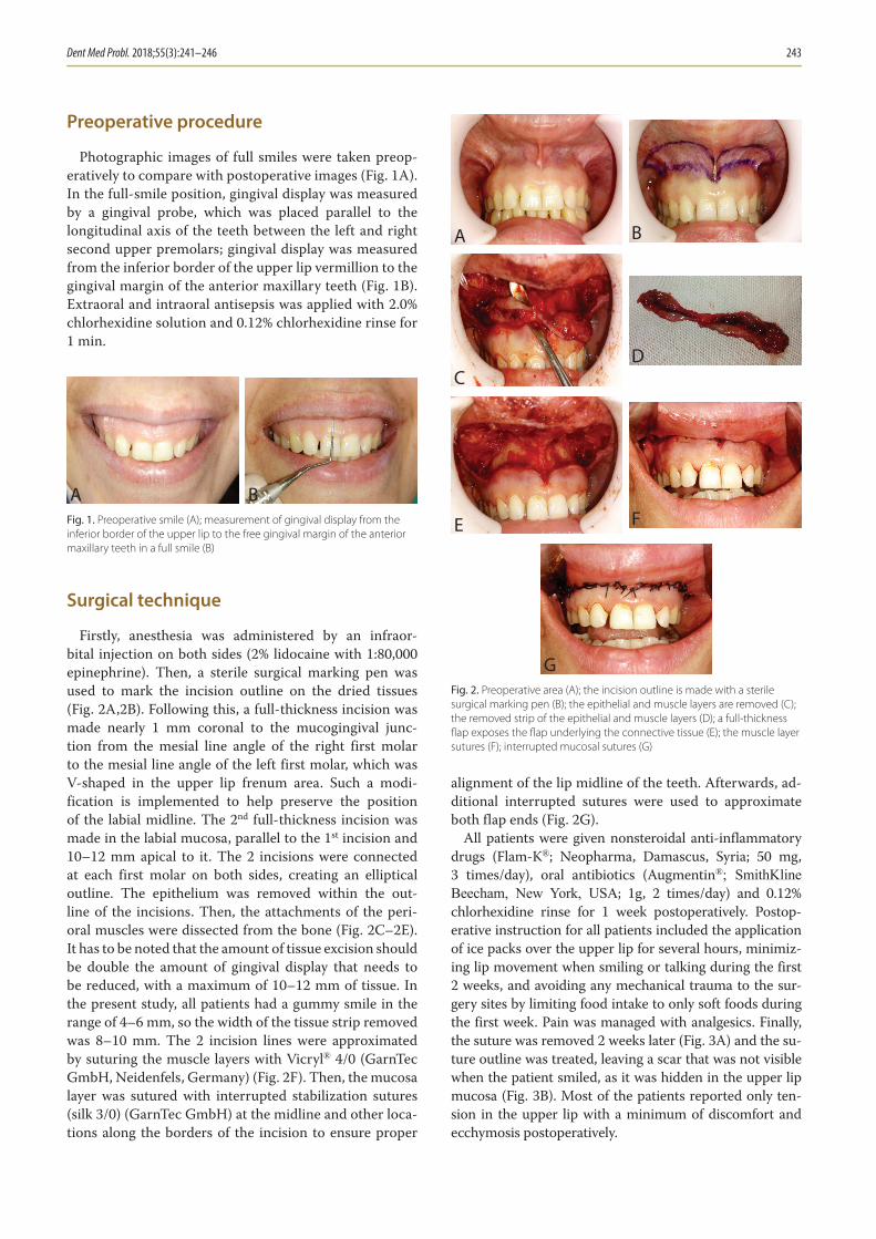
Surgical technique
Firstly, anesthesia was administered by an infraor-
bital injection on both sides (2% lidocaine with 1:80,000
epinephrine). Then, a sterile surgical marking pen was
used to mark the incision outline on the dried tissues
(Fig. 2A,2B). Following this, a full-thickness incision was
made nearly 1 mm coronal to the mucogingival junc-
tion from the mesial line angle of the right first molar
to the mesial line angle of the left first molar, which was
V-shaped in the upper lip frenum area. Such a modi-
fication is implemented to help preserve the position
of the labial midline. The 2nd full-thickness incision was
made in the labial mucosa, parallel to the 1st incision and
10–12 mm apical to it. The 2 incisions were connected
at each first molar on both sides, creating an elliptical
outline. The epithelium was removed within the out-
line of the incisions. Then, the attachments of the peri-
oral muscles were dissected from the bone (Fig. 2C–2E).
It has to be noted that the amount of tissue excision should
be double the amount of gingival display that needs to
be reduced, with a maximum of 10–12 mm of tissue. In
the present study, all patients had a gummy smile in the
range of 4–6 mm, so the width of the tissue strip removed
was 8–10 mm. The 2 incision lines were approximated
by suturing the muscle layers with Vicryl® 4/0 (GarnTec
GmbH, Neidenfels, Germany) (Fig. 2F). Then, the mucosa
layer was sutured with interrupted stabilization sutures
(silk 3/0) (GarnTec GmbH) at the midline and other loca-
tions along the borders of the incision to ensure proper
alignment of the lip midline of the teeth. Afterwards, ad-
ditional interrupted sutures were used to approximate
both flap ends (Fig. 2G).
All patients were given nonsteroidal anti-inflammatory
drugs (Flam-K®; Neopharma, Damascus, Syria; 50 mg,
3 times/day), oral antibiotics (Augmentin®; SmithKline Beecham, New York, USA; 1g, 2 times/day) and 0.12%
chlorhexidine rinse for 1 week postoperatively. Postop-
erative instruction for all patients included the application
of ice packs over the upper lip for several hours, minimiz-
ing lip movement when smiling or talking during the first
2 weeks, and avoiding any mechanical trauma to the sur-
gery sites by limiting food intake to only soft foods during
the first week. Pain was managed with analgesics. Finally,
the suture was removed 2 weeks later (Fig. 3A) and the su-
ture outline was treated, leaving a scar that was not visible
when the patient smiled, as it was hidden in the upper lip
mucosa (Fig. 3B). Most of the patients reported only ten-
sion in the upper lip with a minimum of discomfort and
ecchymosis postoperatively.
Fig. 1. Preoperative smile (A); measurement of gingival display from the
inferior border of the upper lip to the free gingival margin of the anterior
maxillary teeth in a full smile (B)
A B
Fig. 2. Preoperative area (A); the incision outline is made with a sterile
surgical marking pen (B); the epithelial and muscle layers are removed (C);
the removed strip of the epithelial and muscle layers (D); a full-thickness
fl ap exposes the fl ap underlying the connective tissue (E); the muscle layer
sutures (F); interrupted mucosal sutures (G)
G
E F
CD
A B

A. Alammar, O. Heshmeh. Lip repositioning244
Follow-up after surgery
All patients were followed up at 1, 3 and 6 months
postoperatively. The parameter that was assessed dur-
ing a full active smile (Fig. 4) was the amount of gin-
gival display from the inferior border of the upper lip
vermillion to the gingival margin of the anterior maxil-
lary teeth, from the right second premolar to the left
second one, measured with a gingival probe placed
parallel to the longitudinal axis over the mid-buccal
region of the anterior maxillary teeth. All measure-
ments were registered to the nearest millimeter. If part
of the clinical crown was covered by the lip (postoper-
atively), the gingival display level was set at zero point.
The follow-up included the recording of postoperative
complications, such as infection, dehiscence, numb-
ness, and other complications, as well as questions
to the patients to determine if the complications had
occurred or not. The postoperative pain was assessed
using the visual analog scale (VAS) during follow-up
periods. Furthermore, the whole surgical procedure
and clinical measurements, taken postopeatively at 1-,
3- and 6-month follow-ups, were set by 1 surgeon to
avoid partiality and the variance of results.
Statistical analysis
Statistical analysis was conducted using the SPSS v. 13
(IBM Corp., Armonk, USA) and the data was analyzed
using a descriptive analysis and the t-test, where p-value
<0.05 was considered significant.
ResultsThe most important complaint of most patients at
the early stage of the postoperative follow-up period
was tension feeling in the circumoral area and the up-
per lip. Specifically, 2 patients expressed blood oozing
through the suture in the initial days following the sur-
gery. Most patients expressed edema at a moderate rate,
which disappeared in 7 days postoperatively, while only
in 3 patients perioral area edema was extended to the
lower eyelids with ecchymosis, which lasted for 14 days.
Pain recurrences varied between mild pain in 5 patients
(35.7%) and moderate pain in 3 patients (21.4%), which
was controlled by means of analgesics. Flap dehiscence
appeared in 2 patients (14.2%) and numbness appeared
in 9 patients (64.2%). No infection was reported in
any of the patients. Only 2 patients complained of dry
mouth after surgery due to damage to the minor sali-
vary glands in the upper lip mucosa during surgery. The
mean rate of gingival display at baseline was 6.36 mm
and changed significantly (p < 0.05) at 1, 3 and 6 months
postoperatively. At 1, 3 and 6 months, gingival display
was 0.91 mm, 2.27 mm and 2.45 mm, respectively (Ta-
ble 1). There was a significant difference in gingival dis-
play between the results obtained after 1 and 6 months,
but there was no significant difference in gingival dis-
play between the 3- and 6-month follow-up, so the re-
sults were fairly stable. At 1, 3 and 6 months postop-
eratively, the obtained reduction was 5.45 mm, 4.09 mm
and 3.91 mm, respectively (Table 2, Fig. 5). The present
study showed that no complete relapse was recorded in
any case during the follow-up periods. However, a par-
tial relapse was recorded in 6 patients.
Fig. 3. Sutures removed after 2 weeks (A); healing site after 6 months (B)
A B
Fig. 4. Preoperative smile (A); smile at 1-month follow-up postoperatively
(B); smile at 3-month follow-up postoperatively (C); smile at 6-month
follow-up postoperatively (D)
C D
A B
Fig. 5. The mean amount of gingival display in a full smile [mm] in relation
to the time period

Dent Med Probl. 2018;55(3):241–246 245
DiscussionExcessive gingival display or a “gummy smile” is con-
sidered undesirable by many people, which is why an at-
tractive smile can improve the quality of life. The stan-
dard amount of gingival display varies between 1 mm
and 3 mm.16 When the amount of gingival display in
a full smile is ≥4 mm, the demand of most patients for an
attractive smile is increased. In the literature, many treat-
ment techniques have been reported for the manage-
ment of a gummy smile, such as botulinum toxin injec-
tions, orthodontic surgery, crown lengthening, etc.1,4,12
In about 20% of patients, a gummy smile is caused by the
hypermobility of the upper lip elevator muscles, so a my-
otomy of the lip elevator muscles can decrease a gummy
smile by reducing the function of the muscles.4 Most pa-
tients prefer minor surgical techniques for the manage-
ment of a gummy smile to major ones. The main goal
of this study was to assess the outcomes of lip reposition-
ing surgery (a full-thickness flap with a myotomy of the
lip elevator muscles) in the treatment of a gummy smile,
caused only by soft tissue disorders (short upper lip, hy-
peractive lip elevator muscles), to reduce the postopera-
tive relapse in order to maintain stable surgical results for
as long as possible and to assess the postoperative com-
plications. All patients treated in this study had gingival
display at the baseline ranging from 4 to 6 mm. The tech-
nique presented in this article involved 2 full-thickness
incisions connected together at the second upper premo-
lar area, outlining an elliptical area of soft tissue which
was removed; then, the attachments of the perioral
muscles were dissected from the bone. This included the
upper lip frenum area being V-shaped to facilitate and
maintain the position of the labial midline. The 2 inci-
sion lines were approximated by suturing the muscle lay-
ers; then, the mucosa layer was sutured with interrupted
stabilization sutures. Accordingly, our results exhibited
significant differences in the reduction of gingival display
at 1, 3 and 6 months postoperatively. The mean postop-
erative gingival display at 1, 3 and 6 months postopera-
tively was 0.91 mm, 2.27 mm and 2.45 mm, respectively.
Successful rates at 1 and 6 months were 85.4% and 61.5%,
respectively. The results were stable for up to 6 months
postoperatively. Only a partial relapse was noticed in
6 patients, without any complete relapse noted. This
can be explained by incomplete stripping of the muscles
from the bone during the surgical procedure or as a re-
sult of muscle memory reattachment to the previous
pre-bone bases. These results are consistent with other
studies concerning modified lip repositioning surgery.4,17
Our findings also correlate with a study conducted by El-
lenbogen and Swara on 21 patients with a gummy smile,
who were treated by a myotomy of the lip elevator mus-
cles through the nose.15 In their study, silicone implants
were used to prevent the relapse after surgery and they
appeared to be effective in reducing gingival display. The
most pronounced postoperative complication was the
feeling of tension in the circumoral area and the upper
lip as a result of removing during the operation a strip
of the mucosa and muscles, and then suturing. That feel-
ing disappeared within the first 2 weeks after surgery.
Pain recurrences varied between mild pain in 5 patients
(35.7%) and moderate pain in 3 patients (21.4%) dur-
ing a few days postoperatively. The pain was controlled
by analgesics. Flap dehiscence appeared in 2 patients
(14.2%) as a result of strong upper lip movement dur-
ing the first 2 weeks after surgery. The patients with flap
dehiscence were treated using disinfectant solution until
the secondary healing was achieved. Numbness in the
upper lip appeared in 9 patients (64.2%), which gradu-
ally disappeared in the first month after surgery, after
the patients were given a course of vitamin B-complex
to enhance healing. No infection was reported in any
of the patients as a result of infection control princi-
ples being followed during surgery and postoperatively.
Table 1. The mean, standard deviation, standard error, min, and max of the amount of gingival display in a full smile [mm] in the study sample
Time period Number of patients Mean Standard deviation Standard error Min Max
Preoperative 14 6.36 1.12 0.34 4 6
After 1 month 14 0.91 1.22 0.37 0 4
After 3 months 14 2.27 1.27 0.38 0 4
After 6 months 14 2.45 1.13 0.34 0 4
Table 2. Results of the interrelated-samples t-test to show signifi cant diff erences in the mean values of gingival display in a full smile [mm] at 1, 3 and 6
months postoperatively
Periods compared Difference between the 2 means t-test value Degrees of freedom p-value
Preoperative – after 1 month −5.45 −11.066 10 0.000
Preoperative – after 3 months −4.09 −8.964 10 0.000
Preoperative – after 6 months −3.91 −10.618 10 0.000
After 1 month – after 3 months 1.36 4.404 10 0.001
After 1 month – after 6 months 1.55 4.949 10 0.001

A. Alammar, O. Heshmeh. Lip repositioning246
In the literature, there have been no studies on lip reposi-
tioning surgery by means of a myotomy of the lip elevator
muscles for the management of a gummy smile and on its
postoperative complications.
ConclusionsThe surgical lip repositioning technique proposed in the
current study is a less invasive procedure, carried out under
local anesthesia, which has shown its effectiveness in reduc-
ing the amount of gingival display in a full smile through
postoperative follow-up periods; therefore, it can be used to
correct a gummy smile in patients with gingival display be-
tween 4 and 6 mm. However, additional studies with longer
follow-up periods and larger sample sizes may still be need-
ed to evaluate the effectiveness of this technique.
References 1. Iqbal C, Nandakumar K, Padmakumar TP. Laser assisted treatment
of excessive gingival display along with modified lip re-position-ing. IOSR J Dent Med Sci. 2015;14(7):28–33.
2. Peck S, Peck L, Kataja M. The gingival smile line. Angle Orthod. 1992;62(2):91–100.
3. Levine RA, McGuire M. The diagnosis and treatment of the gummy smile. Compend Contin Educ Dent. 1997;18(8):757–762,764,quiz 766.
4. Abdullah W, Khalil H, Alhindi M, Marzook H. Modifying gummy smile: A minimally invasive approach. J Contemp Dent Pract. 2014;15(6):821–826.
5. Martins AT, Sakakura CE, Correcirc BE, et al. A modified technique that decreases the height of the upper lip in the treatment of gummy smile patients: A case series study. J Dent Oral Hyg. 2012;10:21–28.
6. Pandurić DG, Blašković M, Brozović J, Sušić M. Surgical treatment of excessive gingival display using lip repositioning technique and laser gingivectomy as an alternative to orthognathic surgery. J Oral Maxillofac Surg. 2014;72(2):404.e1–11.
7. Miron H, Calderon S, Allon D. Upper lip changes and gingival expo-sure on smiling: Vertical dimension analysis. Am J Orthod Dentofa-cial Orthop. 2012;141(1):87–93.
8. Hwang W-S, Hur M-S, Hu K-S, et al. Surface anatomy of the lip ele-vator muscles for the treatment of gummy smile using botulinum toxin. Angle Orthod. 2009;79(1):70–77.
9. Livada R, Shiloah J. Gummy smile: Could it be genetic? Hereditary gingival fibromatosis. J Tenn Dent Assoc. 2012;92(1):23–26.
10. Garber DA, Salama MA. The aesthetic smile: Diagnosis and treat-ment. Periodontol 2000. 1996;11:18–28.
11. Monaco A, Streni O, Marci MC, Marzo G, Gatto R, Giannoni M. Gummy smile: Clinical parameters useful for diagnosis and thera-peutical approach. J Clin Pediatr Dent. 2005;29(1):19–25.
12. Simon Z, Rosenblatt A, Dorfman W. Eliminating a gummy smile with surgical lip repositioning. J Cosmet Dent. 2007;23(1):100–108.
13. Wei J, Herrler T, Xu H, Li Q, Dai C. Treatment of gummy smile: Nasal septum dysplasia as etiologic factor and therapeutic target. J Plast Reconstr Aesthet Surg. 2015;68(10):1338–1343.
14. Ishida LH, Ishida LC, Ishida J, Grynglas J, Alonso N, Ferreira MC. Myotomy of the levator labii superioris muscle and lip reposition-ing: A combined approach for the correction of gummy smile. Plast Reconstr Surg. 2010;126(3):1014–1019.
15. Ellenbogen R, Swara N. The improvement of the gummy smile using the implant spacer technique. Ann Plast Surg. 1984;12(1):16–24.
16. Silva CO, Ribeiro‐Júnior NV, Campos TV, Rodrigues JG, Tatakis DN. Excessive gingival display: Treatment by a modified lip reposition-ing technique. J Clin Periodontol. 2013;40(3):260–265.
17. Benlier E, Top H, Aygit AC. A new approach to smiling deformity: Cutting of the superior part of the orbicularis oris. Aesthetic Plast Surg. 2005;29(5):373–377.