Embed Size (px)
Citation preview

RESEARCH ARTICLE Open Access
Linalool isomerase, a membrane-anchoredenzyme in the anaerobic monoterpenedegradation in Thauera linaloolentis 47LolRobert Marmulla1, Barbara Šafarić1, Stephanie Markert2, Thomas Schweder2 and Jens Harder1*
Abstract
Background: Thauera linaloolentis 47Lol uses the tertiary monoterpene alcohol (R,S)-linalool as sole carbon and energysource under denitrifying conditions. The conversion of linalool to geraniol had been observed in carbon-excess cultures,suggesting the presence of a 3,1-hydroxyl-Δ1-Δ2-mutase (linalool isomerase) as responsible enzyme. To date, only asingle enzyme catalyzing such a reaction is described: the linalool dehydratase/isomerase (Ldi) from Castellanielladefragrans 65Phen acting only on (S)-linalool.
Results: The linalool isomerase activity was located in the inner membrane. It was enriched by subcellular fractionationand sucrose gradient centrifugation. MALDI-ToF MS analysis of the enriched protein identified the corresponding genenamed lis that codes for the protein in the strain with the highest similarity to the Ldi. Linalool isomerase is predictedto have four transmembrane helices at the N-terminal domain and a cytosolic domain. Enzyme activity required areductant for activation. A specific activity of 3.42 ± 0.28 nkat mg * protein−1 and a kM value of 455 ± 124 μM weredetermined for the thermodynamically favored isomerization of geraniol to both linalool isomers at optimal conditionsof pH 8 and 35 °C.
Conclusion: The linalool isomerase from T. linaloolentis 47Lol represents a second member of the enzyme class 5.4.4.4,next to the linalool dehydratase/isomerase from C. defragrans 65Phen. Besides considerable amino acid sequencesimilarity both enzymes share common characteristics with respect to substrate affinity, pH and temperatureoptima, but differ in the dehydratase activity and the turnover of linalool isomers.
Keywords: Linalool, Geraniol, Thauera, Isomerase, Allyl alcohol, Monoterpene
BackgroundMonoterpenes (C10H16), naturally occurring hydrocar-bons, are the main constituents of essential oils andbelong to the diverse group of terpenoids, of which morethan 50000 structures are known to date [1, 2]. They areproduced as secondary plant metabolites and serve diversefunctions like signaling, attraction/repellence of pollinatorsand insects, thermotolerance and are involved in allelop-athy [3, 4]. Atmospheric monoterpene emission fromplants was estimated to be 127 Tg C yr−1 [5], with half-livesof minutes to hours in the atmosphere due to their suscep-tibility to chemical and photooxidative reactions. Monoter-penes enter soils by precipitation from the atmosphere,
excretion from roots and by leaf fall [6–9]. The hydropho-bic character of monoterpenes causes cell toxicity, mainlyby accumulation into and destabilization of the cell mem-branes [10]. Below toxic concentrations, microorganismscan use monoterpenes as carbon and energy source forgrowth. Several bacteria have been described to transformmonoterpenes in the presence of oxygen as a cosubstrateapplying mono- and dioxygenases [11], but also other bio-transformations are described [12]. Linalool, a tertiarymonoterpene alcohol (Fig. 1), is the main component ofessential oils in lavender and coriander. Its chemical struc-ture prevents a direct oxidation of the hydroxyl group.Hence, another functionalization or isomerization is theinitial biological degradation reaction. Linalool is oxidizedto 8-hydroxylinalool under aerobic conditions [13, 14]. Inthe absence of oxygen, the primary alcohol geraniol isformed in linalool-grown cultures of Thauera linaloolentis
* Correspondence: [email protected] of Microbiology, Max Planck Institute for Marine Microbiology,Celsiusstr. 1, D-28359 Bremen, GermanyFull list of author information is available at the end of the article
© 2016 Marmulla et al. Open Access This article is distributed under the terms of the Creative Commons Attribution 4.0International License (http://creativecommons.org/licenses/by/4.0/), which permits unrestricted use, distribution, andreproduction in any medium, provided you give appropriate credit to the original author(s) and the source, provide a link tothe Creative Commons license, and indicate if changes were made. The Creative Commons Public Domain Dedication waiver(http://creativecommons.org/publicdomain/zero/1.0/) applies to the data made available in this article, unless otherwise stated.
Marmulla et al. BMC Biochemistry (2016) 17:6 DOI 10.1186/s12858-016-0062-0

47Lol (Fig. 1) [15]. This betaproteobacterium was isolatedon linalool as sole carbon end energy source under denitri-fying conditions [16]. A 3,1-hydroxyl-Δ1-Δ2-mutase wasproposed as novel enzymatic function initializing themineralization of linalool [15, 16].A similar reaction was described for the bifunctional
enzyme linalool dehydratase/isomerase from Castella-niella defragrans 65Phen. The enzyme catalyzes the re-versible hydration of β-myrcene to (S)-linalool and itsisomerization to geraniol. It is an oxygen-sensitive, peri-plasmatic protein of 43 kDa including a signal peptidefor export [17, 18]. The tertiary alcohol 2-methyl-3-buten-2-ol (232-MB) may also be transformed byenzyme-catalyzed isomerization reactions. 232-MB is ametabolite in bacterial degradation of fuel oxygenates.Its mineralization may proceed via an initial isomeriza-tion to 3-methyl-2-buten-1-ol (prenol) in Aquincolatertiaricarbonis L108 and Methylibium petroleiphilumPM1 [19] as well as in Pseudomonas putida MB-1 [20].The later was isolated on 232-MB as sole carbon source[20]. However, the corresponding enzymes were so farnot characterized. For intramolecular hydroxyl-grouptransfer (EC 5.4.4.x), only seven different enzyme activ-ities are described: (hydroxyamino) benzene mutase (EC5.4.4.1), isochorismate synthase (EC 5.4.4.2), 3-(hydro-xyamino) phenol mutase (EC 5.4.4.3), geraniol isomerase(EC 5.4.4.4), 9,12-octadecadienoate 8-hydroperoxide8R-isomerase (EC 5.4.4.5), 9,12-octadecadienoate 8-hydroperoxide 8S-isomerase (EC 5.4.4.6) and hydroxy-peroxy icosatetraenoate isomerase (EC 5.4.4.7).We report the enrichment of the linalool isomerase
activity in protein fractions of Thauera linaloolentis47Lol and the kinetic properties of the enzyme. A corre-sponding gene was identified in the draft genome. Wesuggest to place the linalool isomerase of Thauera lina-loolentis 47Lol as a new member in the enzyme familyof intramolecular hydroxyl group transferases (EC5.4.4.x) with the EC number 5.4.4.4 next to the geraniolisomerase function of the linalool dehydratase/isomerasefrom Castellaniella defragrans 65Phen.
Results and discussionIdentification of a candidate protein for linaloolisomeraseThe linalool dehydratase/isomerase (Ldi, NCBI:CBW30776)was used in similarity searches to identify a putative linaloolisomerase protein in a draft genome of T. linaloolentis47Lol. One protein showed a considerable similarity with
an overall amino acid identity of 20 % (positives 33 %,E-value 3E-10, NCBI:ENO87364, Additional file 1: Fig-ure S1). The corresponding gene is isolated from theadjacent genes (>150 bp) and encodes a protein of 644amino acids with a calculated molecular weight of71.8 kDa, an isoelectric point of 6.06 and a hydrophobi-city of −0.115 (GRAVY) [21]. No signal peptide waspredicted by the SignalP software. For the N-terminus,four transmembrane domains within the first 139amino acids were predicted together with a localizationin the inner membrane with the C-terminal protein foldin the cytoplasm. Conserved domains as described inPfam were not present. The specific hydrophobicityvalues (GRAVY) were 0.94 and −0.406 for the N- andC-terminal parts of the protein (amino acids 1–139 and140–644, respectively). The similarity to the Ldi was re-stricted to the C-terminal domain. Such a location atthe cytoplasmic site of the inner membrane seems tobe ideal for a catabolic enzyme acting on a hydrophobicsubstrate. This may maximize the contact with the sub-strate and reduces the intracellular concentration, butalso produces geraniol for the next catabolic enzymesthat likely depend on cytoplasmatic NAD+ as electronacceptor. In contrast, the periplasmatic location of Ldiis optimal for a defense enzyme. Myrcene is less toxicthan the alcohols and can diffuse into the environmentof the cell, thus keeping the damage at the inner mem-brane to a minimum.
Enrichment of the linalool isomerase activity (Lis)Lis activity was determined as geraniol isomerase activityand was detected in crude cell-free protein extracts alsocontaining membrane fragments after application ofhigh pressure cell disruption. Cell disintegration by os-mosis or ultra sonification retained the activity in themembrane fraction. Due to the large cytoplasmatic do-main of the candidate protein, we attempted the isola-tion as soluble enzyme from dialyzed crude extracts.The pH was increased to 9.5 to eventually increase theanionic character of the enzyme. The enzyme activitydid not bind to a DEAE column. After ammoniumsulfate addition (10 % v/v of a saturated solution), theenzyme activity was retained on a Phenyl-Sepharose col-umn and eluted with pure water. A strong binding tohydrophobic columns has also been observed for Ldi[17] and other monoterpene-transforming enzymes [22].A size determination of the eluted fraction on a size-exclusion column yielded a molecular weight of over
Fig. 1 Linalool isomerase catalyzes the reversible reaction from linalool to geraniol
Marmulla et al. BMC Biochemistry (2016) 17:6 Page 2 of 11

600 kDa, as native protein in phosphate buffer as well asin the presence of urea as denaturant. Visualization ofthe proteins on SDS-PAGE revealed the presence ofmany proteins, including abundant proteins between 60and 70 kDa and around 33 kDa (data not shown).The large size together with the predicted membrane
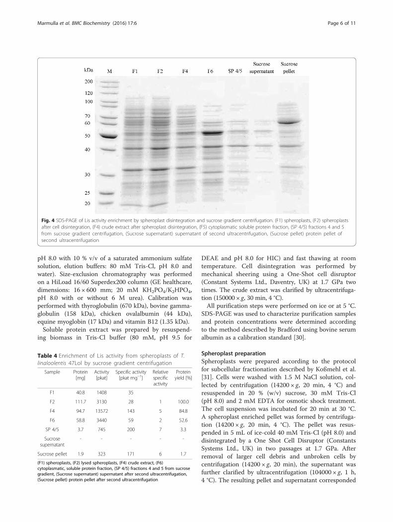
association of the Lis candidate gene suggested an alter-native purification approach, a subcellular fractionationincluding a sucrose-gradient centrifugation for the iden-tification of membrane-associated proteins. First, theouter membrane and periplasmatic proteins were re-moved by spheroplast formation. After disintegration byhigh pressure release, the inner membrane (IM) fractionwas separated from the cytosolic soluble protein (SP)fraction by ultracentrifugation (158 Svedberg). Total Lisactivity was five times higher in the membrane fractionthan in the soluble fraction (15.8 and 3.3 nkat, respect-ively) (Table 1 and Additional file 2: Figure S2). Bothfractions were separated on sucrose gradients. The Lisactivity of the membrane fraction (Fig. 2 and Table 2)was concentrated in two fractions with a sucrose densitybetween 1.17 and 1.22 g mL−1 (IM4 and IM5). For thesoluble protein fraction (Fig. 3 and Table 3), Lis activitywas also concentrated in the fractions with the afore-mentioned sucrose content (SP4 and SP5). Protein gelsrevealed an enrichment of proteins with a molecular sizebetween 60 and 70 kDa and around 32 kDa for theactive fractions from IM and SP. Lis activity was depos-ited from the active fractions (SP4 and SP5) by decreaseof the sucrose concentration and ultracentrifugation.SDS-PAGE characterized the dissolved pellet as contain-ing an approx. 60 kDa protein as most abundant protein(Fig. 4 and Table 4). This purification stage was used forenzyme kinetics.Particles with a Svedberg constant of above 158 S were
separated from the soluble fraction. High pressure celldisintegration produces a number of small membranefragments and vesicles. Small vesicles (microsomes)
settle with a sedimentation coefficient between 100 and10000 S [23]. We conclude from our observations thatthe Lis activity is located on a protein associated withthe inner membrane. Only a minor fraction of the totalenzyme activity, present as small membrane-protein ag-gregates, was enriched in the fraction that is traditionallyconsidered to contain only cytosolic proteins.The protein gels showed consistently protein(s) between
60 and 70 kDa in fractions with Lis activity. MALDI-ToFmass spectroscopy identified the protein band at approx.60 kDa from several gels as NCBI:ENO87364, the proteinthat was predicted to be a candidate for the linalool isom-erase. The discrepancy between the calculated molecularweight of 71.8 kDa and the observed weight of between60 and 70 kDa is likely a result of the hydrophobic natureof the protein. Hydrophobic proteins, including mem-brane proteins tend to bind more SDS and show a fastermigration in denaturing gel electrophoreses [24].Further purification of the Lis activity was not success-
ful, as a detergent-mediated release of the protein fromthe membrane was impaired by irreversible inhibitory ef-fects on the enzyme activity. Detergents aid solubility ofmembrane proteins but also have an adverse effect onstability and functionality of proteins [25, 26]. Terpenoidsynthases were shown to have a rather hydrophobic coreshielding the catalytic center from surrounding bulksolvent [27]. Also the Lis may have a hydrophobicprotein domain sensitive to detergents that change theconformation into a non-active state.
Heterologous gene expressionExpression of the native lis gene in E.coli BL21(DE3)yielded an induced protein band around 60 kDa (Fig. 5).No inclusion bodies were observed. The expressed proteinwas located in the membrane fraction, but Lis activity wasnot observed in crude cell extracts or in cell-free proteinfractions. Expression of a N-terminally truncated versionof the linalool isomerase, representing the cytosolic partof the enzyme only, yielded a soluble protein that also didnot show activity. The lis gene did not have rare codons.The folding in E.colimay have been incorrect.
Characterization of linalool isomerase activityThe Lis activity was enriched without a chemical reduc-tant in the buffer and enzymatically inactive. Activity wasrestored by addition of a reducing agent and under anoxicconditions in the enzyme assay. The activation could beinitiated by cationic (ferrous iron), neutral (dithiothreitol)or anionic (dithionite) compounds. However, maximumactivity was measured with 4 mM dithionite (268 pkat *mg protein−1). A large excess of reductant caused a de-crease in activity, e.g. 10 mM dithionite or 8 mM DTTwere less suitable for activation (Table 5). Lis activity was
Table 1 Enrichment of Lis activity from spheroplasts of T.linaloolentis 47Lol
Sample Protein[mg]
Activity[pkat]
Specific activity[pkat mg−1]
Relative specificactivity
Proteinyield [%]*
F1 71.9 7295 101
F2 79.0 7800 99 1.0 100.0
F3 67.6 3060 45 0.5
F4 51.8 24984 482 4.9 65.6
F5 29.1 513 18 0.2 36.8
F6 38.0 3283 86 0.9 73.3
F7 21.4 15848 741 7.5 41.3
(F1) spheroplasts, (F2) lysed spheroplasts, (F3) outer membrane and periplasmaticproteins, (F4) crude extract from spheroplasts, (F5) unbroken spheroplast and celldebris, (F6) cytoplasmatic, soluble proteins, (F7) inner membrane fraction. *F4 andF5 are derived from F2. F6 and F7 are derived from F4
Marmulla et al. BMC Biochemistry (2016) 17:6 Page 3 of 11

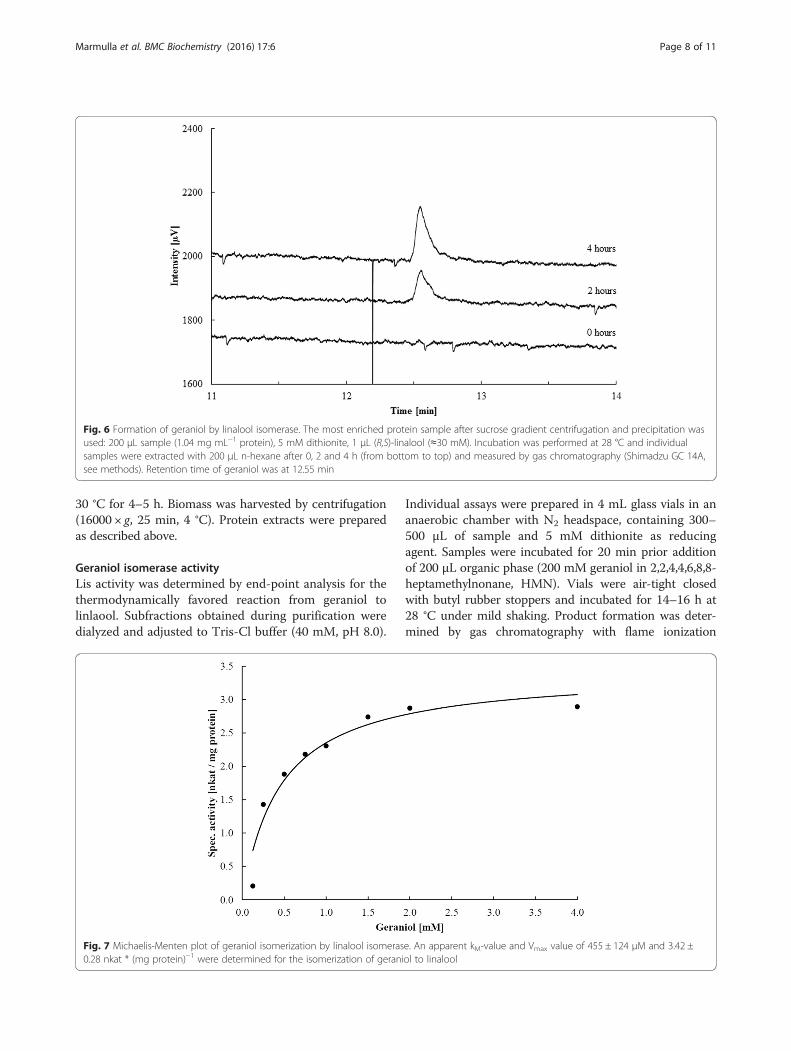
determined in the thermodynamically favorable directionfrom geraniol to linalool [15] and was observed betweenpH 7 and 9.5, with an optimum around pH 8. Thetemperature optimum was 35 °C and the activation energywas 80.4 ± 6.9 kJ mol−1. For comparison, the pH andtemperature optimum of the Ldi enzyme were at pH 9and 35 °C, respectively, and the activation energy was68.6 kJ mol−1 [17]. Geraniol formation from linalool wasdetected within 4 h of incubation (Fig. 6). Kinetic parame-ters were determined with the most enriched sample induplicates for the geraniol isomerization to linalool. Theenzyme followed Michaelis-Menten-kinetics with an
apparent kM-value of 455 ± 124 μM and a Vmax of 3.42 ±0.28 nkat * mg protein−1 (Fig. 7). A similar substrate affin-ity was determined for the linalool dehydratase/isomerase(kM 500 μM). Maximal velocity was higher for the Ldi(Vmax 410 nkat * mg protein−1) than for the Lis, however,the purification level for Lis was lower, and we do notknow whether part of the enzyme is in an inactivestate. Lis did not show a stereospecificity towards lin-alool isomers: both (R) and (S)-linalool were formed(Fig. 8). In contrast, the Ldi accepts only (S)-linaloolas a substrate [17].Earlier studies already showed the regiospecific forma-
tion of geraniol from linalool without nerol formation[15]. To confirm the regioselectivity, Lis activity wastested with nerol or citronellol and in combination withgeraniol. No activity was measured with nerol or citro-nellol alone. Linalool isomerase activity dropped toapprox. 50 % in the presence of nerol. Activity in thepresence of citronellol and geraniol was barely detect-able. Thus, the enzyme is regioselective and seems tobind nerol with a similar affinity as geraniol, whereas cit-ronellol which lacks the C2-C3 double bond is strongerbound than geraniol.UV-VIS spectroscopy in a range of 200–800 nm did
not provide evidence for the presence of cofactors.Cofactor-independent enzymes are known for allylicrearrangements that are catalyzed by acid-base mecha-nisms [28]. The isomerization of geraniol to linaloolrequires a protonation of the hydroxyl group to leave as
Fig. 2 SDS-PAGE of sucrose gradient fractions after separation of the inner membrane (IM) fraction from spheroplast disintegration. (F7) innermembrane fraction from spheroplast disintegration, (IM 1) - (IM 7) fractions 1–7 (top-to-bottom) of sucrose gradient
Table 2 Enrichment of Lis activity by sucrose gradientcentrifugation from inner membrane pellets (F7) obtained fromspheroplasts of T. linaloolentis 47Lol. 1 mL fractions from top tobottom are shown
Sample Protein[mg]
Activity[pkat]
Specific activity[pkat mg−1]
Relative specificactivity
Proteinyield [%]
Appliedsample (F7)
6.74 5925 879 1 100.0
IM 1 0.20 139 697 0.8 3.0
IM 2 1.06 238 225 0.3 15.7
IM 3 1.44 870 604 0.7 21.4
IM 4 1.92 3085 1607 1.8 28.5
IM 5 3.28 3513 1071 1.2 48.7
IM 6 1.44 1050 729 0.8 21.4
IM 7 0.02 0 0 0.0 0.3
Marmulla et al. BMC Biochemistry (2016) 17:6 Page 4 of 11

water, and a shift in electron density leading to a tertiarycarbocation intermediate which may be attacked by wateror a hydroxyl ion, resulting in the formation of linalool.
ConclusionWe identified a linalool isomerase in T. linaloolentis47Lol by partial protein purification. The gene encodes atwo-domain protein with a N-terminal anchor in theinner membrane that is characterized by four transmem-brane helices and a C-terminal cytosolic domain whichshowed considerable similarity to the linalool dehydra-tase/isomerase from C. defragrans 65Phen. The enzymeis active in the reduced state and sensitive towards
detergents. It expands the enzyme class of intramolecu-lar hydroxyl group transferases as a new member, linal-ool isomerase (5.4.4.4).
MethodsBacterial strains, cultivation conditions and biomassharvestT. linaloolentis 47Lol was cultivated under anaerobic,denitrifying conditions in artificial fresh water (AFW)medium. Medium was prepared as described by Foss etal. [29] with modifications: carbonate buffer wasreplaced by 10 mM Na2HPO4/NaH2PO4 and vitaminswere omitted. The headspace contained only nitrogen gas.1–2 mM (R,S)-linalool (>97 % purity; Sigma-Aldrich,Germany) were directly applied without carrier phase assole carbon and energy source. Cultures were incubated at28 °C under mild shaking (60 rpm). Alternatively, theywere stirred. Bacterial biomass for protein purificationwas obtained from 2 L cultures grown on 2 mM linalooland 20 mM nitrate by centrifugation (16000 × g, 25 min,4 °C). If not used directly, biomass was frozen in liquid ni-trogen and stored at −80 °C.
Purification attempts by chromatographyFirst attempts on the purification of the linalool isomer-ase were performed by classical column chromatographyusing anion exchange and hydrophobic interaction col-umns (DEAE; binding buffer: 80 mM Tris-Cl, pH 9.5,elution buffer: 80 mM Tris-Cl, pH 9.5 with 1 M NaCl;Phenyl Sepharose; binding buffer: 80 mM Tris-Cl,
Fig. 3 SDS-PAGE of sucrose gradient fractions after separation of the cytoplasmatic soluble protein fraction from spheroplast disintegration.(F6) Soluble protein fraction from spheroplast disintegration, (SP 1) - (SP 7) fractions 1–7 (top-to-bottom) of sucrose gradient
Table 3 Enrichment of Lis activity by sucrose gradientcentrifugation from cytoplasmatic soluble protein fraction (F6)obtained from spheroplasts of T. linaloolentis 47Lol. 1 mLfractions from top to bottom are shown
Sample Protein[mg]
Activity[pkat]
Specific activity[pkat mg−1]
Relative specificactivity
Proteinyield [%]
Appliedsample (F6)
9.2 1863 202 1.0 100.0
SP 1 0.6 14 24 0.1 6.1
SP 2 1.5 16 11 0.1 16.7
SP 3 3.1 165 54 0.3 33.2
SP 4 2.7 273 100 0.5 29.5
SP 5 1.2 141 114 0.6 13.3
SP 6 0.3 13 47 0.2 3.0
SP 7 0.3 0 0 0.0 2.8
Marmulla et al. BMC Biochemistry (2016) 17:6 Page 5 of 11
pH 8.0 with 10 % v/v of a saturated ammonium sulfatesolution, elution buffers: 80 mM Tris-Cl, pH 8.0 andwater). Size-exclusion chromatography was performedon a HiLoad 16/60 Superdex200 column (GE healthcare,dimensions: 16 × 600 mm; 20 mM KH2PO4/K2HPO4,pH 8.0 with or without 6 M urea). Calibration wasperformed with thyroglobulin (670 kDa), bovine gamma-globulin (158 kDa), chicken ovalalbumin (44 kDa),equine myoglobin (17 kDa) and vitamin B12 (1.35 kDa).Soluble protein extract was prepared by resuspend-
ing biomass in Tris-Cl buffer (80 mM, pH 9.5 for
DEAE and pH 8.0 for HIC) and fast thawing at roomtemperature. Cell disintegration was performed bymechanical sheering using a One-Shot cell disruptor(Constant Systems Ltd., Daventry, UK) at 1.7 GPa twotimes. The crude extract was clarified by ultracentrifuga-tion (150000 × g, 30 min, 4 °C).All purification steps were performed on ice or at 5 °C.
SDS-PAGE was used to characterize purification samplesand protein concentrations were determined accordingto the method described by Bradford using bovine serumalbumin as a calibration standard [30].
Spheroplast preparationSpheroplasts were prepared according to the protocolfor subcellular fractionation described by Koßmehl et al.[31]. Cells were washed with 1.5 M NaCl solution, col-lected by centrifugation (14200 × g, 20 min, 4 °C) andresuspended in 20 % (w/v) sucrose, 30 mM Tris-Cl(pH 8.0) and 2 mM EDTA for osmotic shock treatment.The cell suspension was incubated for 20 min at 30 °C.A spheroplast enriched pellet was formed by centrifuga-tion (14200 × g, 20 min, 4 °C). The pellet was resus-pended in 5 mL of ice-cold 40 mM Tris-Cl (pH 8.0) anddisintegrated by a One Shot Cell Disruptor (ConstantsSystems Ltd., UK) in two passages at 1.7 GPa. Afterremoval of larger cell debris and unbroken cells bycentrifugation (14200 × g, 20 min), the supernatant wasfurther clarified by ultracentrifugation (104000 × g, 1 h,4 °C). The resulting pellet and supernatant corresponded
Fig. 4 SDS-PAGE of Lis activity enrichment by spheroplast disintegration and sucrose gradient centrifugation. (F1) spheroplasts, (F2) spheroplastsafter cell disintegration, (F4) crude extract after spheroplast disintegration, (F5) cytoplasmatic soluble protein fraction, (SP 4/5) fractions 4 and 5from sucrose gradient centrifugation, (Sucrose supernatant) supernatant of second ultracentrifugation, (Sucrose pellet) protein pellet ofsecond ultracentrifugation
Table 4 Enrichment of Lis activity from spheroplasts of T.linaloolentis 47Lol by sucrose gradient centrifugation
Sample Protein[mg]
Activity[pkat]
Specific activity[pkat mg−1]
Relativespecificactivity
Proteinyield [%]
F1 40.8 1408 35
F2 111.7 3130 28 1 100.0
F4 94.7 13572 143 5 84.8
F6 58.8 3440 59 2 52.6
SP 4/5 3.7 745 200 7 3.3
Sucrosesupernatant
- - - - -
Sucrose pellet 1.9 323 171 6 1.7
(F1) spheroplasts, (F2) lysed spheroplasts, (F4) crude extract, (F6)cytoplasmatic, soluble protein fraction, (SP 4/5) fractions 4 and 5 from sucrosegradient, (Sucrose supernatant) supernatant after second ultracentrifugation,(Sucrose pellet) protein pellet after second ultracentrifugation
Marmulla et al. BMC Biochemistry (2016) 17:6 Page 6 of 11

to an inner membrane fraction (IM, F7, Additional file 1:Figure S1) and a soluble protein fraction (SP, F6, Additionalfile 1: Figure S1), respectively.
Sucrose density gradient centrifugationA linear sucrose gradient was created by overlaying a20 % (w/v) over a 70 % (w/v) sucrose solution, 3 mLeach. The tubes were incubated horizontally for 90 minto allow mixing. A 1 mL-protein sample was loaded
carefully on top of the gradient and centrifugation wasperformed in a L-70 ultracentrifuge (70.1Ti rotor,Beckmann Coulter) at 260000 × g, for 4 h at 4 °C, withslow acceleration and deceleration. The linearity of thesucrose gradient was confirmed by gravimetrical meas-urement. 1 mL fractions covering the gradient were ana-lyzed for enzyme activity and for protein content onSDS-PAGE. The two most active fractions were pooled,diluted with 40 mM Tris-Cl (pH 8.0) to a final volumeof 8 mL and a second ultracentrifugation step (260000 ×g, 4 °C, 1.5 h ≡ 42 Svedberg) was performed to removesucrose. This resulted in the formation of a proteinpellet enriched in Lis activity, which was resuspended in1 mL buffer and used for the characterization of theenzyme kinetic.
Proteomics by MALDI-ToF MSProtein samples, obtained from individual purifications,were analyzed by SDS-PAGE coupled with matrix-assistedlaser desorption/ionization time of flight (MALDI-ToF)mass spectrometry (MS). Protein bands in gels were ex-cised manually, and the Ettan Spot Handling Workstation(GE Healthcare) was used for trypsin digestion and em-bedding of the resulting peptide solutions in an α-cyano-4-hydroxycinnamic acid matrix for spotting onto MALDItargets. MALDI-ToF MS analysis was performed on anAB SCIEX TOF/TOF™ 5800 Analyzer (AB Sciex/MDSAnalytical Technologies [32]. Spectra in a mass rangefrom 900 to 3700 Da (focus 1700 Da) were recorded andanalyzed by GPS Explorer™ Software Version 3.6 (build332, Applied Biosystems) and the Mascot search engineversion 2.4.0 (Matrix Science Ltd, London, UK) using theRAST draft genome as reference.
Heterologous gene expressionThe predicted Lis gene was isolated from genomic DNAof T. linaloolentis 47Lol by means of PCR, using the Phu-sion Polymerase according to the manufacturer manual(Life Technologies, Thermo Fisher Scientific, Waltham,USA) with the primer pair pLI_NdeI_FW (TCGTACATATGATGAGCAATATGGAATCG) and pLI_BglII_RV(CGATAGATCTTCAGTGGCCCGGCTTG, annealingtemperature 59 °C). Additionally, a N-terminal truncatedversion of the gene was constructed with the primerpair pLI_NdeI_FW-truncated-N (TCGTACATATGATGCGCGGCGCCAAGC) and pLI_BglII_RV (annealingtemperature 68 °C), which had an artificial start codon(ATG) and covered the amino acids 141–644 of the Lis.The genes were cloned into the pET42a overexpressionplasmid and transformed into E. coli BL21(DE3). E. coliBL21(DE3) pET42-Lis or pET42-Lis-ΔN were grown inliquid LB medium at 37 °C and protein expression wasinduced by addition of 1 mM IPTG at an optical density(600 nm) of around 1. Cultures were further incubated at
Fig. 5 SDS-PAGE of heterologous expression of the Lis gene in E.coli BL21(DE3) pET42-Lis. (1) Cells from induced culture, (2) crudeprotein extract after cell disintegration and (3) soluble protein frac-tion after ultracentrifugation
Table 5 Influence of different reducing agents on Lis activity
Reducingagent
Reductionpotential [mV]
Concentration[mM]
Specific activity[pkat * mg protein−1]
Dithionite - 660 2 89
4 268
10 210
Dithiothreitol - 330 2 199
8 145
16 83
Cysteine - 220 5 23
10 7
Ferrous iron - 236 (Fe(OH)3/Fe2+) 5 112
Marmulla et al. BMC Biochemistry (2016) 17:6 Page 7 of 11
30 °C for 4–5 h. Biomass was harvested by centrifugation(16000 × g, 25 min, 4 °C). Protein extracts were preparedas described above.
Geraniol isomerase activityLis activity was determined by end-point analysis for thethermodynamically favored reaction from geraniol tolinlaool. Subfractions obtained during purification weredialyzed and adjusted to Tris-Cl buffer (40 mM, pH 8.0).
Individual assays were prepared in 4 mL glass vials in ananaerobic chamber with N2 headspace, containing 300–500 μL of sample and 5 mM dithionite as reducingagent. Samples were incubated for 20 min prior additionof 200 μL organic phase (200 mM geraniol in 2,2,4,4,6,8,8-heptamethylnonane, HMN). Vials were air-tight closedwith butyl rubber stoppers and incubated for 14–16 h at28 °C under mild shaking. Product formation was deter-mined by gas chromatography with flame ionization
Fig. 6 Formation of geraniol by linalool isomerase. The most enriched protein sample after sucrose gradient centrifugation and precipitation wasused: 200 μL sample (1.04 mg mL−1 protein), 5 mM dithionite, 1 μL (R,S)-linalool (≈30 mM). Incubation was performed at 28 °C and individualsamples were extracted with 200 μL n-hexane after 0, 2 and 4 h (from bottom to top) and measured by gas chromatography (Shimadzu GC 14A,see methods). Retention time of geraniol was at 12.55 min
Fig. 7 Michaelis-Menten plot of geraniol isomerization by linalool isomerase. An apparent kM-value and Vmax value of 455 ± 124 μM and 3.42 ±0.28 nkat * (mg protein)−1 were determined for the isomerization of geraniol to linalool
Marmulla et al. BMC Biochemistry (2016) 17:6 Page 8 of 11

detection (PerkinElmer Auto System XL, Überlingen,Germany). 1 μL of the HMN phase was injected onto anOptima-5 column (30 m × 0.32 mm, 0.25 μm film thick-ness; Macherey-Nagel, Germany) with hydrogen as carriergas and the following temperature program: injectionport 250 °C, detection port 350 °C, initial columntemperature 40 °C for 2 min, increasing to 100 °C at arate of 4 °C min−1, keeping 100 °C for 0.1 min, followedby an increase to 320 °C at 45 °C min−1 and hold for3 min. The split ratio was set to 1:9.The effect of detergents on Lis activity was tested for
Triton X100 and Tween20 (0.5 %, 1 % w/v), CHAPS(0.1 % w/v) and n-octyl-α-D-glucoside (0.1 %, 0.5 %,1 % w/v). Detergents were added to the soluble sphero-plast fraction (protein concentration 0.1 mg mL−1) andaforementioned enzyme assays were performed.Reducing agents were tested with a dialyzed (Visking
dialysis tubing 12–14 kDa cut-off, Serva) soluble proteinextract (4 mg mL−1 protein). The following reducingagents were added prior to the start of the assay: dithionite(2, 4 and 10 mM), dithiothreitol (2, 8 and 16 mM), cyst-eine (5 and 10 mM), or ferrous iron (5 mM).Temperature dependency on Lis activity was deter-
mined between 12 and 50 °C. Samples (300 μL,6.8 mg mL−1 protein) were pre-incubated for 20 minat the individual temperatures prior to substrateaddition. The assay was terminated after 8 h and ana-lyzed by gas chromatography. Activation energy wascalculated from the Arrhenius plot (y = −9664.3 x +36.2; R2 = 0.914).
The pH-optimum was determined by incubatingcrude cell lysate (20 mg mL−1) in a pH-range from 7 to9.5 in Tris-Cl (40 mM) applying the aforementionedenzyme assay.Kinetic parameters (kM and Vmax) were determined for
the most enriched enzyme fraction in biological duplicates(68 and 80 μg mL−1 protein; 10 to 20 μg total protein infinal assay). Samples were incubated with geraniol concen-tration from 0.125 to 4 mM, directly applied withoutcarrier phase. Both substrate and enzyme were preparedseparately with 7 mM dithionite and pre-incubated. Reac-tions were started by injecting an equal volume (200 μL)of enzyme to the substrate solution. Samples were incu-bated at 28 °C for 90 min and terminated by addition of100 mM NaOH (final concentration). 1 μL of sample wasdirectly subjected to GC analysis. Kinetic parameters werecalculated from primary data plotted in a Michaelis-Menthen-graph.Substrate specificity of the Lis was tested with gera-
niol, nerol and citronellol. A 400 μL-sample (active frac-tion after sucrose gradient, SP 4/5) was incubated with200 μL of 200 mM geraniol, nerol and citronellol inHMN as well as with 200 μL of geraniol-nerol andgeraniol-citronellol mixtures in HMN (100 mM each).Assays were prepared as aforementioned.Stereoselectivity was tested with soluble protein extract
(1.4 mg protein) and inner membrane-enriched fraction(1.6 mg protein) from spheroplast disintegration. Sampleswere treated with 5 mM dithionite and incubated with10 mM geraniol under anaerobic conditions at 28 °C for
Fig. 8 Gas chromatogram demonstration of the absence of stereoselectivity of the linalool isomerase. Soluble protein extract (1.4 mg protein;solid line) and inner membrane-enriched fraction (1.6 mg protein; dashed line) from spheroplast disintegration were incubated with geraniol,subsequently extracted with n-hexane and analyzed by gas chromatography. Retention times: (R)- and (S)-linalool 7.85 and 8.05 min,geraniol 12.45 min
Marmulla et al. BMC Biochemistry (2016) 17:6 Page 9 of 11

14 h and subsequently extracted with 200 μL n-hexane.Monoterpene analysis was performed by gas chromatog-raphy with flame ionization detector (Shimadzu GC-14A,Shimadzu Corporation) on a Hydrodex-ß-6TBDM col-umn (25 m × 0.25 mm, Macherey-Nagel, Germany) withthe following temperature program: injection port 200 °C,detection port 250 °C, initial column temperature 60 °Cfor 1 min, increasing to 130 °C at a rate of 5 °C min−1,keeping 130 °C for 0.5 min, followed by an increase to230 °C at 20 °C min−1 and hold for 4 min.
Linalool isomerase activityThe forward reaction of the linalool isomerase - linaloolto geraniol - was tested in a separate assay. 200 μL ofenriched fraction after sucrose gradient centrifugationwere treated with 5 mM dithionite and incubatedwith 1 μL (R,S)-linalool under anaerobic conditions at28 °C for 0, 2 and 4 h. Samples were extracted with 200 μLn-hexane and analyzed by GC (Shimadzu GC-14A).
UV-VIS spectrum for cofactorsThe most enriched, active protein sample (0.95 mg mL−1
protein) was analyzed by UV-VIS spectroscopy (BeckmanDU-640 spectrophotometer) in the range of 200–800 nmto detect cofactors.
Gene identification and bioinformatic analysisA draft genome for T. linaloolentis 47Lol was obtainedby merging data from two at NCBI public available draftgenomes: ASM31020 (4.199 Mbp on 220 contigs, pub-lished 2012) and ASM62130 (4.214 Mbp on 46 contigs,published 2014). Contigs were automatically mergedusing Sequencher 4.6 with a minimum match percentageof 95 % and a minimum overlap of 50 bp. The resultingdraft genome had 4.4 Mbp on 23 contigs and wasuploaded to RAST for further analysis [33, 34]. Identifi-cation of a putative gene, coding for a linalool isomerase(Lis), was performed by homology search using the linal-ool dehydratase/isomerase sequence from Castellanielladegragrans 65Phen. The identified gene was analyzed byvarious bioinformatic tools: SignalP 4.1 for prediction ofsignal peptides [35], TMHMM, SOSUI and Philius for pre-diction of transmembrane helices [36–39] and the Pfamdatabase to search for motifs and domain patterns [40].
Availability of data and materialsThe data sets supporting the results of this article areincluded within the article and its additional files.
Additional files
Additional file 1: Figure S1. Alignment of the linalool dehydratase/isomerase (NCBI:CBW30776) from C. defragrans 65Phen and the linalool
isomerase from T. linaloolentis 47Lol (NCBI:ENO87364). Color indicatessimilarity. (PNG 106 kb)
Additional file 2: Figure S2. SDS-PAGE of different fractions during theinner membrane preparation from spheroplasts. (F1) spheroplasts, (F2)spheroplasts after cell disintegration, (F3) outer membrane andperiplasmatic protein fraction, (F4) crude extract after spheroplastdisintegration, (F5) unbroken spheroplast and cell debris, (F6) cytoplasmatic,soluble protein fraction, (F7) inner membrane fraction. (PNG 539 kb)
Competing interestsThe authors declare that they have no competing interests.
Authors’ contributionRM and JH designed the study. RM and BS performed the purification andthe experiments for enzyme characterization. SM and TS conducted mass-spectrometry based protein identifications and analyzed data. RM and JHdrafted the manuscript. All authors read and approved the final manuscript.
AcknowledgmentsWe thank Dirk Albrecht for MALDI-ToF measurements. This study was financedby the Max Planck Society.
Author details1Department of Microbiology, Max Planck Institute for Marine Microbiology,Celsiusstr. 1, D-28359 Bremen, Germany. 2Institute for Pharmacy, Departmentof Pharmaceutical Biotechnology, University of Greifswald, Felix-Hausdorff-Str.3, D-17487 Greifswald, Germany.
Received: 15 October 2015 Accepted: 2 March 2016
References1. Breitmaier E. Terpenes; flavors, fragrances, pharmaca, pheromones, vol.
1st edition. Weinheim, Wiley-VCH; 2006.2. Ajikumar PK, Tyo K, Carlsen S, Mucha O, Phon TH, Stephanopoulos G.
Terpenoids: opportunities for biosynthesis of natural product drugs usingengineered microorganisms. Mol Pharm. 2008;5:167–90.
3. Dudareva N, Klempien A, Muhlemann JK, Kaplan I. Biosynthesis, function andmetabolic engineering of plant volatile organic compounds. New Phytol. 2013;198:16–32.
4. Sharkey TD, Wiberley AE, Donohue AR. Isoprene emission from plants:why and how. Ann Bot. 2008;101:5–18.
5. Günther A, Hewitt CN, Erickson D, Fall R, Geron C, Graedel T, Harley P,Klinger L, Lerdau M, McKay WA. A global model of natural volatile organiccompound emission. J Geophys Res-Atmos. 1995;100:8873–92.
6. Atkinson R, Arey J. Atmospheric degradation of volatile organic compounds.Chem Rev. 2003;103:4605–38.
7. Kainulainen P, Holopainen JK. Concentrations of secondary compounds inScots pine needles at different stages of decomposition. Soil Biol Biochem.2002;34:37–42.
8. Wilt FM, Miller GC, Everett RL, Hackett M. Monoterpene concentrations infresh, senescent, and decaying foliage of single-leaf pinyon (Pinus monophyllaTorr. and Frem.: Pinaceae) from the western great basin. J Chem Ecol.1993;19:185–94.
9. Ziemann PJ, Atkinson R. Kinetics, products, and mechanisms of secondaryorganic aerosol formation. Chem Soc Rev. 2012;41:6582–605.
10. Bakkali F, Averbeck S, Averbeck D, Waomar M. Biological effects of essentialoils - a review. Food Chem Toxicol. 2008;46:446–75.
11. Schewe H, Mirata MA, Holtmann D, Schrader J. Biooxidation of monoterpeneswith bacterial monooxygenases. Process Biochem. 2011;46:1885–99.
12. Marmulla R, Harder J. Microbial monoterpene transformations – a review.Front Microbiol. 2014;5.
13. Bell SG, Dale A, Rees NH, Wong LL. A cytochrome P450 class I electrontransfer system from Novosphingobium aromaticivorans. Appl MicrobiolBiotechnol. 2010;86:163–75.
14. Ullah AJH, Murray RI, Bhattacharyya PK, Wagner GC, Gunsalus IC. Proteincomponents of a cytochrome P-450 linalool 8-methyl hydroxylase. J BiolChem. 1990;265:1345–51.
Marmulla et al. BMC Biochemistry (2016) 17:6 Page 10 of 11

15. Foss S, Harder J. Microbial transformation of a tertiary allylalcohol: Regioselectiveisomerisation of linalool to geraniol without nerol formation. FEMS MicrobiolLett. 1997;149:71–5.
16. Foss S, Harder J. Thauera linaloolentis sp. nov. and Thauera terpenica sp.nov., isolated on oxygen-containing monoterpenes (linalool, menthol,and eucalyptol) and nitrate. Syst Appl Microbiol. 1998;21:365–73.
17. Brodkorb D, Gottschall M, Marmulla R, Lüddeke F, Harder J. Linalooldehydratase-isomerase, a bifunctional enzyme in the anaerobic degradationof monoterpenes. J Biol Chem. 2010;285:30436–42.
18. Lüddeke F, Harder J. Enantiospecific (S)-(+)-linalool formation from beta-myrcene by linalool dehydratase-isomerase. Z Naturforsch C. 2011;66:409–12.
19. Schuster J, Schäfer F, Hübler N, Brandt A, Rosell M, Härtig C, Harms H, MüllerRH, Rohwerder T. Bacterial degradation of tert-amyl alcohol proceeds viahemiterpene 2-methyl-3-buten-2-ol by employing the tertiary alcoholdesaturase function of the Rieske nonheme mononuclear iron oxygenaseMdpJ. J Bacteriol. 2012;194:972–81.
20. Malone VF, Chastain AJ, Ohlsson JT, Poneleit LS, Nemecek-Marshall M, Fall R.Characterization of a Pseudomonas putida allylic alcohol dehydrogenaseinduced by growth on 2-methyl-3-buten-2-ol. Appl Environ Microbiol. 1999;65:2622–30.
21. Kyte J, Doolittle RF. A simple method for displaying the hydropathiccharacter of a protein. J Mol Biol. 1982;157:105–32.
22. Lüddeke F, Wülfing A, Timke M, Germer F, Weber J, Dikfidan A, Rahnfeld T,Linder D, Meyerdierks A, Harder J. Geraniol and geranial dehydrogenasesinduced in anaerobic monoterpene degradation by Castellaniella defragrans.Appl Environ Microbiol. 2012;78:2128–36.
23. Kleinsmith LJ, Kish VM. Principles of cell and molecular biology, 2 edn. NewYork: HarperCollins; 1995.
24. Rath A, Glibowicka M, Nadeau VG, Chen G, Deber CM. Detergent bindingexplains anomalous SDS-PAGE migration of membrane proteins. Proc NatlAcad Sci. 2009;106:1760–5.
25. Gohon Y, Popot JL. Membrane protein-surfactant complexes. Curr OpinColloid Interface Sci. 2003;8:15–22.
26. Seddon AM, Curnow P, Booth PJ. Membrane proteins, lipids and detergents:not just a soap opera. Biochim Biophys Acta. 2004;1666:105–17.
27. Christianson DW. Structural biology and chemistry of the terpenoid cyclases.Chem Rev. 2006;106:3412–42.
28. Schwab JM, Henderson BS. Enzyme-catalyzed allylic rearrangements.Chem Rev. 1990;90:1203–45.
29. Foss S, Heyen U, Harder J. Alcaligenes defragrans sp. nov., description of fourstrains isolated on alkenoic monoterpenes ((+)-menthene, alpha-pinene, 2-carene, and alpha-phellandrene) and nitrate. Syst Appl Microbiol. 1998;21:237–44.
30. Bradford MM. Rapid and sensitive method for quantitation of microgramquantities of protein utilizing principle of protein-dye binding. Anal Biochem.1976;72:248–54.
31. Koßmehl S, Wöhlbrand L, Drüppel K, Feenders C, Blasius B, Rabus R. Subcellularprotein localization (cell envelope) in Phaeobacter inhibens DSM 17395.Proteomics. 2013;13:2743–60.
32. Wolf C, Hochgräfe F, Kusch H, Albrecht D, Hecker M, Engelmann S.Proteomic analysis of antioxidant strategies of Staphylococcus aureus:diverse responses to different oxidants. Proteomics. 2008;8:3139–53.
33. Aziz RK, Bartels D, Best AA, DeJongh M, Disz T, Edwards RA, Formsma K,Gerdes S, Glass EM, Kubal M. The RAST server: rapid annotations usingsubsystems technology. BMC Genomics. 2008;9:75.
34. Overbeek R, Olson R, Pusch GD, Olsen GJ, Davis JJ, Disz T, Edwards RA,Gerdes S, Parrello B, Shukla M. The SEED and the Rapid Annotation ofmicrobial genomes using Subsystems Technology (RAST). Nucleic Acids Res.2014;42:D206–14.
35. Petersen TN, Brunak S, von Heijne G, Nielsen H. SignalP 4.0: discriminatingsignal peptides from transmembrane regions. Nat Methods. 2011;8:785–6.
36. Sonnhammer EL, von Heijne G, Krogh A. A hidden Markov model forpredicting transmembrane helices in protein sequences. Proc Int ConfIntell Syst Mol Biol. 1998;6:175–82.
37. Hirokawa T, Boon-Chieng S, Mitaku S. SOSUI: classification andsecondary structure prediction system for membrane proteins.Bioinformatics. 1998;14:378–9.
38. Imai K, Asakawa N, Tsuji T, Akazawa F, Ino A, Sonoyama M, Mitaku S. SOSUI-GramN: high performance prediction for sub-cellular localization ofproteins in Gram-negative bacteria. Bioinformation. 2008;2:417–21.
39. Reynolds SM, Käll L, Riffle ME, Bilmes JA, Noble WS. Transmembrane topologyand signal peptide prediction using dynamic bayesian networks. PLoS ComputBiol. 2008;4:e1000213.
40. Finn RD, Bateman A, Clements J, Coggill P, Eberhardt RY, Eddy SR, Heger A,Hetherington K, Holm L, Mistry J. Pfam: the protein families database.Nucleic Acids Res. 2014;42:D222–30.
• We accept pre-submission inquiries
• Our selector tool helps you to find the most relevant journal
• We provide round the clock customer support
• Convenient online submission
• Thorough peer review
• Inclusion in PubMed and all major indexing services
• Maximum visibility for your research
Submit your manuscript atwww.biomedcentral.com/submit
Submit your next manuscript to BioMed Central and we will help you at every step:
Marmulla et al. BMC Biochemistry (2016) 17:6 Page 11 of 11