Embed Size (px)
Citation preview

LIMPING IN CHILDREN
Cláudio Santili1, Wilson Lino Júnior2, Ellen de Oliveira Goiano3, Romero Antunes Barreto Lins4, Gilberto Waisberg5, Susana dos Reis Braga6, Miguel Akkari7
1 – Associate Professor, Department of Orthopedics and Traumatology, Faculdade de Ciências Médicas, Santa Casa de São Paulo; Assistant Physician, Pediatric Orthopedics and Traumatology Group, Irmandade da Santa Casa de Misericórdia de São Paulo, São Paulo, Brazil.
2 – Assistant Physician, Pediatric Orthopedics and Traumatology Group, Irmandade da Santa Casa de Misericórdia de São Paulo, São Paulo, Brazil.3 – Interning Physician, Pediatric Orthopedics and Traumatology Group, Irmandade da Santa Casa de Misericórdia de São Paulo, São Paulo, Brazil.4 – Resident (R3) Physician, Department of Orthopedics and Traumatology, Faculdade de Ciências Médicas, Santa Casa de São Paulo, São Paulo, Brazil.5 – Assistant Physician, Pediatric Orthopedics and Traumatology Group, Irmandade da Santa Casa de Misericórdia de São Paulo, São Paulo, Brazil.6 – Master in Orthopedics, Faculdade de Ciências Médicas, Santa Casa de São Paulo; Physician, Pediatric Orthopedics and Traumatology Group, Irmandade da Santa Casa
de Misericórdia de São Paulo, São Paulo, Brazil.7 – Head, Pediatric Orthopedics and Traumatology Group, Irmandade da Santa Casa de Misericórdia de São Paulo, São Paulo, Brazil.
Study conducted at the Pediatric Orthopedics Group, Department of Orthopedics and Traumatology, Irmandade da Santa Casa de Misericórdia de São Paulo, “Fernandinho Simonsen” Pavilion (Director: Prof. Osmar Avanzi).Correspondence: Dr. Cláudio Santili, Departamento de Ortopedia e Traumatologia da Santa Casa de São Paulo – Grupo de Ortopedia e Traumatologia Pediátrica, Rua Cesário Mota Junior, 112 – 01277-900 – São Paulo, SP. E-mail: [email protected]
We declare no conflict of interest in this article
UPDATING ARTICLE
Rev Bras Ortop. 2009;44(4):290-8
ABSTRACT
Limping in children is a common complaint at pediatric, pediatric orthopaedic offices and in emergency rooms. There are several causes for this condition, and identifying them is a challenge. The older the patient, the better the anamnesis and more detailed the physical examination will be, enabling an easier medical assessment for searching the source of the disorder. In order to make the approach easier, three age groups can and should be considered. Among infants (1 to 3 years old), diagnosis will most likely be: transitory synovitis, septic arthritis, neurological disorders (mild brain palsy (BP) and muscular dystrophy), congenital hip disloca-tion (CHD), varus thigh, juvenile rheumatoid arthritis (JRA)
and neoplasias (osteoid osteoma, leukemia); in the scholar age group, between 4 and 10 years old, in addition to the diagnoses above, Legg-Calvé-Perthes disease, discoid meniscus, inferior limbs discrepancy and unspecific muscular pain; in adolescents (11 to 15 years old): slipped capital femoral epiphysis, congeni-tal hip dislocation, chondrolysis, overuse syndromes, dissecans osteochondritis, and tarsal coalition. The purpose of this study is to provide an update on how to approach pediatric patients presenting with limping, and to discuss its potential causes.
Keywords – Gait; Child; Intermittent claudication; Legg-Perthes disease; Hip; Osteochondritis dissecans; Arthritis, juve-nile rheumatoid; Arthritis, Infectious; Cerebral palsy; Synovitis
INTRODUCTION
The diagnosis of conditions that cause claudi-cation in children is a challenge(1,2), even for more experienced professionals that are accustomed to dealing with patients who are not forthcoming with information. Usually, disorders that cause abnormal gait can be divided according to the pattern of clau-dication and the age at which they appear(3). To facil-itate the pediatrician’s approach, who is usually the first doctor to be sought, three age groups should be
considered: toddlers, which includes children who are learning to walk (one to three years); school-aged children, with a more mature gait pattern (four to 10 years); and adolescents (11 to 15 years(4)).
Thus, a systematic approach should be created for each age group in order to obtain a more accu-rate evaluation geared to the conditions of the age, in particular to keep in mind what tests should be ordered in each case, optimizing the likelihood of obtaining an early diagnosis(5).

291
We must note that children start walking at around 12 months with support and that at 18 months, in general, they begin to walk independently, with movements that remain uncoordinated, maintaining an immature pattern until around five years of age, and reaching the adult pattern at age seven.
Claudication is often associated with pain, and the antalgic gait pattern observed is characterized by a rapid pace and minimal weight loading onto the painful extremity, shortening the stance phase. This is the most common type of claudication(6,7) and, in general, is easily detected by dynamic physical examination, complemented by inspection of signs of localization such as inflammation and post-traumatic symptoms, which are often present, and careful pal-pation of the affected areas in which the child senses pain. The antalgic type, secondary to trauma, will not be discussed in this article.
THE TODDLER PERIOD – FROM ONE TO THREE YEARS OF AGE
These patients are those that result in greatest diagnostic difficulty with regard to gait problems(2,8,9). Because of being of such a young age, this is a group that is not very helpful in relation to the data gathered during the interview, which is most often obtained only through the complaints of the parents, as well as the physical examination.
Another important fact that we should not forget is that children this age have an immature gait pat-tern(10,11), characterized by broad-based, increased flexion of the hips and knees, and arms beside the body with the elbow extended, all this to improve the swing phase, which is naturally unbalanced. Since they cannot increase the size of their steps because of the lack of neuromuscular maturity, toddlers increase their pace with the aim of gaining speed. All of this variation in the walking pattern should be evaluated and considered when determining whether claudica-tion is really present.
If, in fact, gait alteration is detected, the most likely diagnoses related to this group of children include transient synovitis, septic arthritis, neurological dis-orders (mild cerebral palsy (CP) and muscular dys-trophy), developmental dysplasia of the hip (DDH), coxa vara, juvenile rheumatoid arthritis (JRA), and neoplasms (osteoid osteoma, leukemia)(7,11,12).
Inflammatory/infectious disorders Transient synovitis vs. septic arthritis – These
two conditions lead to acute claudication pain, leaving doubt in their differential diagnosis(13,14); the two should be well distinguished from one another by their evolution, which is generally favorable in the case of synovitis and disastrous in the case of pyoarthritis, if appropriate action is not taken. The hips are the most commonly affected site. Transient synovitis presents with claudication that ranges from mild to moderate, but children do not normally stop performing their usual activities, however, the pain can drag on for days (about seven to ten days) until the resolution, which is gradual and occurs spontaneously in most cases. Children with septic arthritis tend to be more irritable, less collaborative, may refuse to walk in some cases, besides generally having poor overall health. Mobilization of the joint during the physical examination is usually more limited in septic arthritis, but if there is still any doubt regarding diagnosis, laboratory tests should be ordered.
The white blood cell count, erythrocyte sedimen-tation rate (ESR), and C-reactive protein (CRP) are generally within the normal range in synovitis, whereas they are high in septic arthritis. High fever (> 38.5°C) is also considered an important positive predictor of septic arthritis(14).
If they are slightly elevated or normal but bor-derline and the clinical picture is more intense, joint puncture is needed for elucidation(6,15). In septic ar-thritis, the white blood cell count in the analysis of as-pirated material is between 80,000 and 200,000 with more than 75% polymorphonuclear cells, while in transient synovitis it is around 5,000 to 15,000, with less than 25% polymorphs. Gram staining should also be sought in the analysis of the material to help in the selection of the antibacterial to be used in the case that arthritis is confirmed, even though it is known that S. aureus is the most common pathogen, until it is isolated in culture, other agents such as Group B Streptococcus, Pseudomonas aeruginosa and Hae-mophilus influenza should be considered.
Juveline rheumatoid arthritis (JRA) – The child who has mildly painful and insidious claudication at the beginning of the walking, at around two years of age, may be manifesting the early symptoms of the pauciarticular form of JRA. This form, which is also the most common, is most prevalent in girls, at
Rev Bras Ortop. 2009;44(4):290-8
LIMPING IN CHILDREN

292
a ratio of 4:1. The most frequently affected joints are the knees and ankles, and it is accompanied by swelling, local warmth, and a decreased range of joint movements.
Laboratory tests such as WBC count, ESR, rheu-matoid factor, and ANA (antinuclear antibody) may be normal during the initial clinical evaluation in up to 50% of cases, which should not rule out its diagnosis(16).
The clinical picture is usually intermittent, im-proving with rest, analgesics, and restriction of ac-tivity. However, if the swelling persists, a pediatric rheumatologist should be consulted.
Neurological disordersCerebral palsy (CP) – Most children begin to take
their first steps around 12 months of life and evolve to walking without assistance within a normal range of around 18 months. If there is a delay in walking beyond that age or if it is abnormal from the start, it is more likely that a neurological disorder is present.
The most common neurological disorder that leads to claudication when walking, which may go unnoticed before the child’s first steps, is mild cerebral palsy(6).
The muscle imbalance is smaller in these patients, which may raise doubts during diagnosis, unlike what occurs in the spastic gait characteristic of CP. However, a good clinical examination will show a limited range of motion of the knee and ankle, pres-ence of clonus and hyperreflexia, which differentiate the problem. From there, parents should be advised about the condition and the child referred to a multi-specialty treatment center, with a pediatric orthopedist managing the therapeutic guidelines.
Muscular dystrophy – In this unusual condition, a delay in the beginning of walking is accompanied by a history of repeated stumbling, frequent falls, and difficulty climbing stairs due to weakness of muscles proximal to the root of the limb (especially the gluteus maximus, gluteus medius, and quadriceps); the calf, therefore, exhibits false hypertrophy and a positive Gow-ers sign, in which the child placed in prone position and asked to get up is “climbing over his/herself” (Figure 1).
As a diagnostic test, the serum creatine phosphoki-nase (CPK) level may be requested in the early stages of dystrophy, and may be 200 to 300 times the normal level(17).
Figure 1 – Gowers sign (From Gowers WR. A manual of disease of the nervous system. London: Churchill, 1886; 1:391-4).
In general, the patient is taken to the doctor at around three to six years of age, boys are almost ex-clusively affected, as it is X-linked recessive.
There may be a positive family history, the disease is progressive and evolves slowly. Patients usually die during the second or third decade of life from respira-tory failure or cardiac arrest.
Developmental/congenital disorders Developmental dysplasia of the hip (DDH) –
When it passes unnoticed at birth and during the first months of life, persisting beyond the age of walking without a diagnosis, this condition delays the onset of walking and causes a painless kind of claudication. It may be unilateral or bilateral. When it is unilateral, the affected limb is shortened and the child walks on tiptoe, hip abduction is limited, with tension in the ad-ductors, and there may be slight flexion contracture of the hip. When it is bilateral, the child increases lumbar
Rev Bras Ortop. 2009;44(4):290-8

293
lordosis to walk, the gait resembles the duck walk, the trunk is balanced by limiting abduction, and there is bilateral insufficiency of the gluteus medius (positive Trendelenburg sign). An AP radiograph of the pelvis easily confirms the diagnosis at this age, showing the dislocation or subluxation of the hip, making other imaging tests unnecessary (Figures 2 and 3).
Coxa vara – The clinical picture here, regardless of whether the condition is congenital or develop-mental, is similar to that of DDH, although it is much less common at a ratio of 1:20. On physical exami-nation, it is distinguished from congenital hip dislo-cation by the prominence of the greater trochanter region secondary to functional muscle weakness, and internal rotation, which is also limited in the later stag-es. However, a conclusive diagnosis is made by AP radiography of the pelvis (Figure 4), where disloca-tion is not observed, though the neck is in a position almost perpendicular to the femoral diaphysis and the growth plate is vertical.
Neoplastic disordersOsteoid osteoma – It is uncommon in children
under the age of five, however, when present, diagnosis is a challenge, especially at the beginning of walking age. It causes claudication pain, predominantly at night, and may pass undetected on radiographs. Clinical examination is unremarkable, pain on palpation is unusual, however, as a general rule, there is significant improvement with the use of salicylates (aspirin), which may increase suspicion. In such cases, scintigraphy can be a valuable tool and has shown itself to be highly sensitive(18) in aiding the diagnosis and localization of the lesion.
Leukemia – It may also be responsible for clau-dication pain in children(19), even in their first steps. Moreover, its peak incidence occurs between two and five years of age, with musculoskeletal complaints in roughly 20% of cases(20).
The clinical picture, in affecting the joint and the presence of bone pain, fever, and lethargy, is similar to arthritis and osteomyelitis, with the differential di-agnosis associated with the presence of other systemic changes, such as hepatosplenomegaly, hemorrhagic suffusion, and bleeding(6).
X-rays may be normal, as in osteoid osteoma, scin-tigraphy may not register changes and laboratory tests at the initial stage can also create doubts, and there may be nonspecific elevation of ESR and peripheral leukocyte count(6,19,20). If other diagnoses are ruled out and suspicion persists, the child should be referred to a hematologist for bone marrow evaluation.
Figure 2 – AP radiograph of hip dislocation on the left
Figure 3 – X-ray in the frog position
Figure 4 – Developmental coxa vara
LIMPING IN CHILDREN
Rev Bras Ortop. 2009;44(4):290-8

294
THE SCHOOL AGE PERIOD – FOUR TO 10 YEARS OF AGE
At this age, children tend to be more cooperative during the exam and already have more mature gait patterns, facilitating the identification of disorders. In addition, complaints are considered important at this stage, when the child is more interested in playing and has no desire to move away from their usual activities.
Because of vigorous activity during the day and muscle fatigue that follows it, complaints of pain at night and during rest are common, which are generi-cally referred to as “growing pains”; however, other causes should be ruled out before reaching this con-clusion. Its diagnosis is predominantly clinical(21) and three criteria can be used to help clarify whether we are dealing with a case of “growing pains”: leg pain is bilateral, occurs only at night, and there are no com-plaints or limping during the day, often improving with massages and without medication(7).
The same diseases described above for children at the beginning of walking age should be remembered, especially transient synovitis, which is more frequent between the ages of three and eight years and is the most common cause of claudication pain throughout childhood. Besides these conditions, three others may be diagnosed during this period: Legg-Calvé-Perthes disease(22,23), discoid meniscus, and discrepancies in leg length.
Legg-Calvé-Perthes disease – Perthes disease is characterized as avascular necrosis of the femoral head, and is a condition of unknown cause and self-limiting behavior that affects children between four and eight years of age (80% of cases). It is more common in boys at a ratio of 4:1. In general, the complaint is claudication, pain is infrequent but manifests itself in the groin, thigh, or radiates to the medial knee when present.
Shortening may or may not be present on physi-cal examination, but the main sign is a limitation of the internal rotation of the hip.
AP and Lauenstein (frog position) radiographs of the pelvis must be requested and the radiographic appearance in the early stages, especially in the frog position, appears as a translucent subchondral line (Figure 5), and in the later stages, the collapse and fragmentation of the proximal femoral epiphysis of the femur can be seen, with denser areas interspersed with radiolucent areas (Figure 6).
The treatment for this condition is greatly variable and the child should be referred to the pediatric orthopedist in order to begin treatment, and should be generally directed to cease weight bearing on the affected limb with the help of crutches or a wheelchair.
Discoid meniscus – It is a congenital deformity of the meniscus, in which it is enlarged and thickened, widely or completely covering the lateral or medial tibial plateau, with the former being much more com-mon(24).
Discoid meniscus is usually a rare cause of clau-dication pain that worsens with activity. In addition, the patient usually has a total extension deficit and feels clicking in the knee(25).
The age range for which clinical complaints begin ranges from three to 12 years, but is most common between eight and 12 years of age(26,27). On physical examination there is pain on palpation of the lateral
Figure 5 – X-ray in the frog position revealing subchondral lysis (Caffey’s sign) in the right hip
Figure 6 – AP radiograph of the pelvis, when we have seen com-promise of the left hip with a decreased height of the nucleus and increased bone density with areas of rarefaction
Rev Bras Ortop. 2009;44(4):290-8
295
joint line on examination of the meniscus. X-rays may provide an indirect sign of the problem via a widening of the lateral joint space, accompanied by the flattening of the femoral condyle, but may raise doubts for even the most experienced doctors, while magnetic resonance imaging (MRI) will confirm the diagnosis in cases of strong suspicion(27).
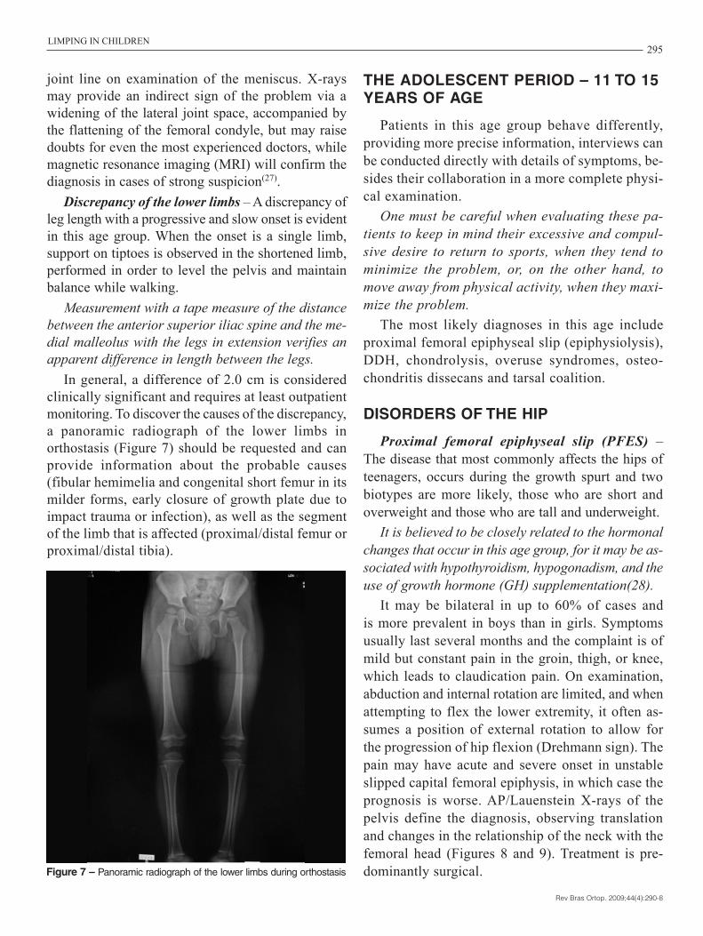
Discrepancy of the lower limbs – A discrepancy of leg length with a progressive and slow onset is evident in this age group. When the onset is a single limb, support on tiptoes is observed in the shortened limb, performed in order to level the pelvis and maintain balance while walking.
Measurement with a tape measure of the distance between the anterior superior iliac spine and the me-dial malleolus with the legs in extension verifies an apparent difference in length between the legs.
In general, a difference of 2.0 cm is considered clinically significant and requires at least outpatient monitoring. To discover the causes of the discrepancy, a panoramic radiograph of the lower limbs in orthostasis (Figure 7) should be requested and can provide information about the probable causes (fibular hemimelia and congenital short femur in its milder forms, early closure of growth plate due to impact trauma or infection), as well as the segment of the limb that is affected (proximal/distal femur or proximal/distal tibia).
THE ADOLESCENT PERIOD – 11 TO 15 YEARS OF AGE
Patients in this age group behave differently, providing more precise information, interviews can be conducted directly with details of symptoms, be-sides their collaboration in a more complete physi-cal examination.
One must be careful when evaluating these pa-tients to keep in mind their excessive and compul-sive desire to return to sports, when they tend to minimize the problem, or, on the other hand, to move away from physical activity, when they maxi-mize the problem.
The most likely diagnoses in this age include proximal femoral epiphyseal slip (epiphysiolysis), DDH, chondrolysis, overuse syndromes, osteo-chondritis dissecans and tarsal coalition.
DISORDERS OF THE HIP
Proximal femoral epiphyseal slip (PFES) – The disease that most commonly affects the hips of teenagers, occurs during the growth spurt and two biotypes are more likely, those who are short and overweight and those who are tall and underweight.
It is believed to be closely related to the hormonal changes that occur in this age group, for it may be as-sociated with hypothyroidism, hypogonadism, and the use of growth hormone (GH) supplementation(28).
It may be bilateral in up to 60% of cases and is more prevalent in boys than in girls. Symptoms usually last several months and the complaint is of mild but constant pain in the groin, thigh, or knee, which leads to claudication pain. On examination, abduction and internal rotation are limited, and when attempting to flex the lower extremity, it often as-sumes a position of external rotation to allow for the progression of hip flexion (Drehmann sign). The pain may have acute and severe onset in unstable slipped capital femoral epiphysis, in which case the prognosis is worse. AP/Lauenstein X-rays of the pelvis define the diagnosis, observing translation and changes in the relationship of the neck with the femoral head (Figures 8 and 9). Treatment is pre-dominantly surgical.Figure 7 – Panoramic radiograph of the lower limbs during orthostasis
LIMPING IN CHILDREN
Rev Bras Ortop. 2009;44(4):290-8

296
Developmental dysplasia of the hip (DDH)
Developmental dysplasia of the hip may become clinically symptomatic during adolescence, especially in cases of subluxation. The child may have appeared free of any disorder until this stage and present pain-ful discomfort only after prolonged activity, which increases in adolescence and thus begins to result in claudication pain. Physical examination may reveal no or only slight abnormalities, and diagnosis is again effected via AP radiographs of the pelvis in the frog position (Figure 10).
Chondrolysis – It is the necrosis of the hip joint cartilage. Although not a common disease, its occur-rence is most often associated with PFES and trauma,
Figure 8 – AP X-ray where asymmetry of the epiphyseal height is observed
Figure 9 – X-ray in the frog position showing a slip of the left side
Figure 10 – Subluxation of the left hip of a teenage DDH patient
but there are cases in which the cause is unknown which are not so rare. It is known that girls are five times more affected than boys, and that age of onset is between 12 and 14 years. The clinical picture is similar to other diseases that affect the hip with pain in the groin, thigh, and/or medial knee, and claudica-tion pain, which can range from mild to severe, but in this case the range of motion is limited in all direc-tions. AP and frog position radiographs of the pelvis are needed to confirm the diagnosis with imaging. The radiographic findings are reduction of the hip joint space (more than 2 mm difference between the two sides), greater disuse osteopenia, and subchondral translucency.
Treatment is aimed at the improvement of irrita-tive synovitis, since the process of joint destruction is irreversible.
DISORDERS OF THE KNEE
Overuse syndrome – Although theoretically any body part that is used in excess during exercise may fall under this concept, the knee is the most commonly affected site. In this age group, the incidence of this condition is increased in proportion to the increase of sports activity. Although pain is the most prevalent symptom, claudication is also a common sign. Stress fractures of the proximal tibia and fibula, patellar tendinitis, and apophysitis of the anterior tibial tuberosity (Osgood-Schlatter
Rev Bras Ortop. 2009;44(4):290-8

297
disease) are examples. A history of sports activity which worsens the pain, improvement with rest, and tenderness during examination raise suspicion. Radiographs can be difficult to interpret. Scintigraphy can be useful for suspected stress fracture. In the acute phase, rest, ice, and anti-inflammatory drugs are a good initial treatment.
Osteochondritis dissecans – It is a condition that affects the articular surface with the separation of a localized cartilage fragment with adjacent sub-chondral bone. It is the most common osteoarticular disease in adolescents and pain is a typical symptom that may or may not be accompanied by claudica-tion. It is also frequently associated with competitive sports. The hip and ankle may also be affected, but the knee is the main site of involvement, specifically the lateral portion of the medial femoral condyle. Physical examination is nonspecific, and the diagno-sis is most often made through radiographs (Figures 11 and 12), which should be requested in AP/profile and tunnel.
In general, conservative treatment solves the problem, but often involves radical changes in the patient’s life, with restricted physical activity for relatively long periods (six to eight weeks).
OTHER DISORDERS
Tarsal coalition – It is characterized by abnor-mal fusion between two or more tarsal bones. There are early manifestations in the first years of life, but most commonly it becomes clinically evident between 11 and 15 years of age when the initially cartilaginous coalition begins to calcify, producing pain in the sinus tarsi, in the dorsal foot, or in the longitudinal arch, and decreasing the mobility of the foot, with claudication. It may be bilateral in up to 60% of cases, manifesting itself in one foot, espe-cially during physical activity. The fibular muscles are often found contracting and spasming, leading to flat, rigid, and everted foot. When the patient is ob-served from behind and is asked to stand on the tips of the toes, the foot fails to adduct, which indicates subtalar joint stiffness. Talocalcaneal and calcaneo-navicular coalitions are the most common, and may be bony, fibrous, or cartilagenous. The diagnosis, depending on the location and type, can be made by radiographs (Figure 13), magnetic resonance imag-ing, or computed tomography (Figure 14). The treat-ment is resection of the coalition.Figure 11 – Detached subchondral fragment
Figure 12 – Tunnel X-ray showing the lesion in the lateral portion of the medial femoral condyle
LIMPING IN CHILDREN
Rev Bras Ortop. 2009;44(4):290-8

298
Figure 13 – Talocalcaneal coalition seen in an oblique foot radiograph
Figure 14 – CT scan showing coalition in the left foot and a fibrous bar in the right foot
1. Gibbons P. The limping child. Trauma. 2005;7(4):184-94. 2. Abbassian A. The limping child: a clinical approach to diagnosis. Br J Hosp Med
(Lond). 2007;68(5):246-50. 3. Leung AKC, Lemay JF. The limping child. J Pediatr Health Care. 2004;18(5):219-23. 4. Philips WA. The child with a limp. Orthop Clin North Am. 1987;18(4):489-501. 5. Sawyer JR, Kapoor M. The limping child: a systematic approach to diagnosis.
Am Fam Physician. 2009;79(3):215-24. 6. Richards BS. Claudicação na criança. In: AAOS - Atualização em conhecimentos
ortopédicos - Pediatria. São Paulo: Atheneu; 2002. p.3-10. 7. Leet AI, Skaggs DL. Evaluation of the acutely limping child. Am Fam Physician.
2000;61(4):1011-18. 8. Aronson J, Garvin K, Seibert J, Glasier C, Tursky EA. Efficiency of the bone scan
for occult limping toddlers. J Pediatr Orthop. 1992;12(1):38-44. 9. Blatt SD, Rosenthal BM, Barnhart DC. Diagnostic utility of lower extremity radio-
graphs of young children with gait disturbance. Pediatrics. 1991; 87(2):138-40.10. Beck RJ, Andriacchi TP, Kuo KN, Fermier RW, Galante JO. Changes in the
gait patterns of growing children. J Bone Joint Surg Am. 1981;63(9):1452-6.11. Boeck H, Vorlat P. Limping in childhood. Acta Orthop Belg. 2003;69(4):301-10.12. Choban S, Killian JT. Evaluation of acute gait abnormalities in preschool children.
J Pediatr Orthop. 1990;10(1):74-8.13. Luhmann SJ, Jones A, Schootman M, Gordon JE, Schoenecker PL, Luh-
mann JD. Differentiation between septic arthritis and transient synovitis of the hip in children with clinical prediction algorithms. J Bone Joint Surg Am. 2004;86(5):956-62.
14. Caird MS, Flynn JM, Leung YL, Millman JE, D’Italia JG, Dormans JP. Factors distinguishing septic arthritis from transient synovitis of the hip in children: a prospective study. J Bone Joint Surg Am. 2006;88(6):1251-7.
15. Flynn JM, Widmann RF. The limping child: evaluation and diagnosis. J Am Acad Orthop Surg. 2001;9(2):89-98.
16. MacEwen GD, Dehne R. The limping child. Pediatr Rev. 1991; 12(9):268-74.
17. Sussman M. Duchenne muscular dystrophy. J Am Acad Orthop Surg. 2002;10:138-51.
18. Kaweblum M, Lehman WB, Bash J, Strongwater A, Grant AD. Osteoid osteoma under the age of five years: The difficulty of diagnosis. Clin Orthop Relat Res. 1993;(296):218-24.
19. Tuten HR, Gabos PG, Kumar SJ, Harter GD. The limping child: a manifestation of acute leukemia. J Pediatr Orthop. 1998;18(5):625-9.
20. Rogalsky RJ, Black GB, Reed MH. Orthopaedic manifestations of leukemia in children. J Bone Joint Surg Am. 1986; 68(4):494-501.
21. Asadi-Pooya AA, Bordbar MR. Are laboratory tests necessary in making the diagnosis of limb pains typical for growing pains in children? Pediatr Int. 2007; 49(6):833-5.
22. Flynn JM, Widmann RF. The limping child: evaluation and diagnosis. J Am Acad Orthop Surg. 2001;9(2):89-98.
23. Fischer SU, Beattie TF. The limping child: epidemiology, assessment and out-come. J Bone Joint Surg Br. 1999;81(6):1029-34.
24. Kelly BT, Green DW. Discoid lateral meniscus in children. Curr Opin Pediatr. 2002;14(1):54-61.
25. Kocher MS, Klingele K, Rassman SO. Meniscal disorders: normal, discoid and cysts. Orthop Clin North Am. 2003;34(3):329-40.
26. Aichroth PM, Patel DV, Marx CL. Congenital discoid lateral meniscus in chil-dren: a follow-up study and evolution of management. J Bone Joint Surg. 1991; 73(6):932-6.
27. Connolly B, Babyn PS, Wright JG, Thorner PS. Discoid meniscus in chil-dren: magnetic resonance imaging characteristics. Can Assoc Radiol J. 1996;47(5):347-54.
28. Waisberg G, Braga SR. Epifisiolise. In: Cohen M. Tratado de ortopedia. São Paulo: Roca; 2007. p. 326-32.
REFERENCES
FINAL CONSIDERATIONS
Claudication in children is a common complaint and is difficult to diagnose. It is necessary to bear in mind the major diseases that cause this disorder according to age group and frequency of appearance, so that details may be sought in the history reported by patients and their caregivers, and to systematically carry out the
physical examination according to the conditions in question. Requests for additional tests according to the most likely causes are often indispensable for a correct diagnosis. It is important that this be done early because the treatment and prognosis of diseases take very dif-ferent and even disastrous directions in cases where there is a delay in defining treatment.
Rev Bras Ortop. 2009;44(4):290-8