Embed Size (px)
Citation preview

Lhx2 regulates a cortex-specific mechanism forbarrel formationAshwin S. Shettya, Geeta Godbolea, Upasana Maheshwaria, Hari Padmanabhana,1, Rahul Chaudharyb,Bhavana Muralidharana, Pei-Shan Houc, Edwin S. Monukid, Hung-Chih Kuoc,e, V. Remab, and Shubha Tolea,2
aDepartment of Biological Sciences, Tata Institute of Fundamental Research, Mumbai 400005, India; bNational Brain Research Centre, Manesar 122051, India;cInstitute of Cellular and Organismic Biology and eStem Cell Program, the Genomic Research Center, Academia Sinica, Taipei 115, Taiwan; and dDepartment ofPathology and Laboratory Medicine, School of Medicine, University of California Irvine, Irvine, CA 92697
Edited by Susan K. McConnell, Stanford University, Stanford, CA, and approved October 18, 2013 (received for review June 17, 2013)
LIM homeodomain transcription factors are critical regulators ofearly development in multiple systems but have yet to be exam-ined for a role in circuit formation. The LIM homeobox gene Lhx2 isexpressed in cortical progenitors during development and also inthe superficial layers of the neocortex in maturity. However, anal-ysis of Lhx2 function at later stages of cortical development hasbeen hampered by severe phenotypes associated with early lossof function. We identified a particular Cre-recombinase line thatacts in the cortical primordium after its specification is complete,permitting an analysis of Lhx2 function in neocortical lamination,regionalization, and circuit formation by selective elimination ofLhx2 in the dorsal telencephalon. We report a profound disruptionof cortical neuroanatomical and molecular features upon loss ofLhx2 in the cortex from embryonic day 11.5. A unique feature ofcortical circuitry, the somatosensory barrels, is undetectable, andmolecular patterning of cortical regions appears disrupted. Sur-prisingly, thalamocortical afferents innervate the mutant cortexwith apparently normal regional specificity. Electrophysiologicalrecordings reveal a loss of responses evoked by stimulation ofindividual whiskers, but responses to simultaneous stimulationof multiple whiskers were present, suggesting that thalamic affer-ents are unable to organize the neurocircuitry for barrel formationbecause of a cortex-specific requirement of Lhx2. We report thatLhx2 is required for the expression of transcription factor pairedbox gene 6, axon guidance molecule Ephrin A5, and the receptorNMDA receptor 1. These genes may mediate Lhx2 function in theformation of specialized neurocircuitry necessary for neocorticalfunction.
The formation of a functional brain structure is a stepwiseprocess starting with the specification of a particular region
of neuroepithelium, followed by the production of the correcttypes and numbers of neurons, and finally the assembling of thecircuitry so that innervation to and from other structures isconnected properly. The mammalian neocortex is unique be-cause of its complex six-layer architecture, and its development isparticularly complex because not only do neurons in differentlayers have unique identities and innervation patterns, but alsothe cortex as a whole is patterned into discrete regions sub-serving distinct functions. Several transcription factors known tohave roles in neocortical patterning display graded expression inthe dorsal telencephalon. Paired box 6 (Pax6), empty spiracleshomeobox 2 (Emx2), Nuclear receptor subfamily 2, group f,member 1 (NR2f1; also known as Coup transcription factor 1,COUP-TFI), and Specificity protein 8 (Sp8) are expressed ingraded pattern in the cortical ventricular zone. Sp8 and Pax6 areexpressed in a rostral (high) to caudal (low) gradient and im-part rostral identity (1–3), whereas Emx2 and NR2f1 areexpressed in the opposite pattern, caudal (high) to rostral (low),and impart caudal areal identity to the cortical primordium (4–6).LIM homeobox 2 (Lhx2) is expressed in a gradient similar to thatof Emx2 and NR2f1, but its role in cortical patterning remains tobe investigated because early loss of Lhx2 function results in se-vere defects that prevent the formation of the neocortex (7–9).
Lhx2 plays a fundamental role as a cortical selector gene,permitting the specification of the cortical primordium as a wholeby suppressing alternative fates corresponding to the hem, anti-hem, and the paleocortex. In the Lhx2-null mutant, two non-cortical structures, the hem and the antihem, expand at theexpense of the cortical primordium (7, 9, 10). Conditional de-letion of Lhx2 at embryonic day (E) 10.5 using an Emx1Credriver (11) produces ectopic paleocortex instead of neocortex(8). NestinCre acts from E11.5 and spares the neocortex (8), butit also drives recombination in subcortical regions such as thethalamus (12), preventing an analysis of Lhx2 loss of functionexclusively in the cortex.We were able to circumvent these constraints using another
Emx1Cre line (13) which we found to act a day later (E11.5) thanthe one commonly (11). This later-acting Emx1Cre line permitsthe neocortex to form despite the loss of Lhx2, permitting ananalysis of Lhx2 function in neocortical lamination and re-gionalization by selective elimination of Lhx2 in the dorsal tel-encephalon. We report that loss of Lhx2 in the dorsal telencephalonresults in a profound disruption of neocortical regional charac-teristics. Molecular and neuroanatomical features that distin-guish the somatosensory cortex—the barrels—are not detectablewhen Lhx2 is deleted in the cortical primordium. Surprisingly,thalamocortical fibers extend to the cortex and demonstrateapparently normal region specificity with respect to the somato-sensory and visual projections, indicating that a broad areal map isformed in the absence of Lhx2. Consistent with this observation,stimulation of multiple whiskers together is able to drive cortical
Significance
The somatosensory barrels are a unique feature of the rodentcortex. Each barrel represents a functional unit in which clus-tered innervation from an individual whisker connects witha ring of cortical neurons. This study reports that when a singletranscription factor, LIM homeobox 2, is deleted specifically inthe cortex, neither the barrel cores nor the cortical barrel wallsare able to form, although a rudimentary functional mappingof the somatosensory innervation does occur. Understandinghow barrels form will shed light on how functional neuro-circuitry is assembled in its final stages, and this insight may bebroadly applicable in the nervous system.
Author contributions: A.S.S., H.-C.K., V.R., and S.T. designed research; A.S.S., G.G., U.M.,H.P., C.R., B.M., P.-S.H., and V.R. performed research; E.S.M. contributed new reagents/analytic tools; A.S.S., G.G., U.M., H.P., C.R., B.M., P.-S.H., H.-C.K., V.R., and S.T. analyzeddata; and A.S.S. and S.T. wrote the paper.
The authors declare no conflict of interest.
This article is a PNAS Direct Submission.
Freely available online through the PNAS open access option.1Present address: Stem Cell and Regenerative Biology, Bauer Laboratory 103, HarvardUniversity, Cambridge, MA 02138.
2To whom correspondence should be addressed. E-mail: [email protected].
This article contains supporting information online at www.pnas.org/lookup/suppl/doi:10.1073/pnas.1311158110/-/DCSupplemental.
www.pnas.org/cgi/doi/10.1073/pnas.1311158110 PNAS | Published online November 21, 2013 | E4913–E4921
NEU
ROSC
IENCE
PNASPL
US

neurons in the mutant. However, responses evoked by the stim-ulation of an individual whisker are not seen in the mutant cortex,suggesting that the refinement of thalamocortical connectivity toform barrels fails to occur. The barrels are a prominent example ofwhat may be a broader role for Lhx2 in the cortex, the organiza-tion of normal neuroanatomical and connectional features ofmature cortical circuitry.
ResultsEmx1Cre transgenic mice have been used extensively to studythe functions of important developmental control molecules inthe cortical primordium. The advantage of such lines is that therecombinase expression, like that of Emx1 itself, is limited tothe cortical primordium in the forebrain. The timing of thisexpression generally is considered to be effective at E10.5, al-though in the most extensively used Emx1Cre line, in which Crerecombinase is knocked into the 3′ UTR as an internal ribosomeentry site (IRES)-Cre construct, the initiation of recombinase ac-tivity has been reported as early as E9.5 (11). We compared theEmx1Cre line with another such line available to us in which anIRES-Cre construct is knocked into exon 1 of the Emx1 gene,creating a null allele of Emx1 (13). These lines henceforth arereferred to as Emx1CreKJ (for the 3′UTR knockin) and Emx1CreYL
(for the exon 1 knockin).Animals homozygous for the floxed Lhx2 allele (conditional
knockout, cKO) were crossed with Emx1CreKJ or Emx1CreYL mice.Embryos from each cross were harvested at different ages andexamined for the expression of Lhx2 exon 2/3, which is lost infloxed cells. Emx1CreKJ and Emx1CreYL mice also were crossedto the membrane targeted tomato and membrane targetedGFP (mTmG) reporter line in which GFP expression is seenupon successful floxing (Fig. 1).Embryos from both Emx1CreKJ and Emx1CreYL crosses re-
veal intense Lhx2 exon 2/3 expression at E9.5. Emx1CreKJ;Lhx2cKO embryos reveal a dramatic decline in Lhx2 exon 2/3 ex-pression in the dorsal telencephalon from E10.0 onwards.Complete floxing of Lhx2 in the dorsal telencephalon is seen byE10.5–E10.75, corroborated by the mTmG reporter line whichdisplays strong GFP expression in the dorsal telencephalon(Fig. 1D).
In contrast, Emx1CreYL;Lhx2 cKO embryos reveal strong ex-pression of Lhx2 exon 2/3 in the dorsal telencephalon up toE10.5. The mTmG reporter line shows only minimal GFP ex-pression at 10.75. It is at E11.5 that the dorsal telencephalondisplays floxing of Lhx2 exon 2/3. Therefore, there is at least a 1-ddifference in the timing of action of the two Emx1Cre lines,with the Emx1CreKJ line acting earlier than the Emx1CreYL line(Fig. 1). Control embryos display intense expression of Lhx2 inthe dorsal telencephalon from E9.5 to E12.5 (Fig. S1).This difference in timing has important consequences for the
Lhx2 cKO phenotype, because there are distinct critical periodsfor different functions of Lhx2 in the cortical primordium: sup-pression of hem/antihem fate (up to E10.5) (9) and suppressionof paleocortical fate (up to E11.5) (8). Therefore, the temporaldifference in the activity of the two Emx1Cre lines would beexpected to give different Lhx2 cKO phenotypes. We tested thishypothesis by examining Emx1CreKJ;Lhx2 cKO and Emx1CreYL;Lhx2 cKO embryos (Fig. 2) and littermate controls (Fig. S2) forthe expression of antihem marker developing brain homeoboxprotein 1 (Dbx1) at E12.5 and the paleocortex marker LIM do-main only 3 (Lmo3) at P0. Emx1CreKJ;Lhx2 cKO embryos displayectopic antihem dorsally in locations that normally would corre-spond to the neocortical primordium. In contrast, Emx1CreYL;Lhx2 cKO embryos do not display ectopic antihem (Fig. 2 F–H).There is no specific marker at E12.5 for the neuroepithelialdomain that will give rise to the paleocortex, but we examinedthe postmitotic paleocortex using Lmo3 as a marker (14). InEmx1CreKJ;Lhx2 cKO mice, as previously described, the paleo-cortex appears ectopically, and the neocortex is greatly shrunken(Fig. 2 D and E) (8). In contrast, in Emx1CreYL;Lhx2 cKO brains,an entire stretch of neocortex is present (blue dashed line, Fig. 2I and J), and there is no ectopic paleocortex. In summary, theEmx1CreYL line offers a unique tool to examine the effects ofcortex-specific deletion of Lhx2 after the critical periods for hem,antihem, and paleocortical fate restriction are past.We examined Emx1CreYL;Lhx2 cKO brains at P5–P10 for two
major features of neocortical development, regional patterningof the cortex into distinct areas and the production of layer-specific neuronal fates. A unique feature of cortical area pat-terning is the barrel cortex, consisting of an array of barrels that
Fig. 1. Time points of action of two different Emx1Cre lines. (A–E) Brains from embryos carrying Emx1CreKJ together with Lhx2 cKO (A–C and E) or mTmGreporter (D), harvested at different stages. Lhx2 is expressed in the dorsal telencephalon at E9.5 but decreases by E10.0 and is lost by E10.5 (arrowheads in Band C). The mTmG reporter reveals extensive GFP expression in an E10.75 brain, an indication of Cre activity (arrowheads in D). An E11.5 brain showscomplete loss of Lhx2 expression in the dorsal telencephalon (arrowheads in E). (F–J) Brains from embryos carrying Emx1CreYL together with Lhx2 cKO (F–Hand J) or mTmG reporter (I), harvested at different stages. Lhx2 is expressed in the dorsal telencephalon at E9.5, E10.0, and E10.5 (open arrowheads in G andH). The mTmG reporter reveals only a sparse sprinkling of floxed GFP-expressing cells at E10.75 (open arrowhead in I). The dorsal telencephalon displaysextensive floxing and loss of Lhx2 expression only by E11.5 (open arrowheads in J). (Scale bars: 500 μm.)
E4914 | www.pnas.org/cgi/doi/10.1073/pnas.1311158110 Shetty et al.

receive sensory input from the whiskers. These barrels consist ofneuropil formed by the terminal arbors of thalamocortical affer-ents (the “barrel core”) synapsing onto the dendrites of spinystellate neurons in layer 4 that form the cellular “wall” of eachbarrel. Barrels appear in early postnatal life, with each barrelrepresenting a unique one-to-one association between input fromeach whisker on the contralateral snout of the animal and thecorresponding barrel (15).Cytochrome oxidase staining in tangential sections of control
cortices at P7 reveals the barrels in the somatosensory corticalarea (Fig. 3A). These patches, corresponding to the barrel cores,appear to be completely missing in Lhx2 cKO brains (Fig. 3E).Each barrel core also contains high levels of serotonin seen incontrol brains (Fig. 3 B and C) but missing in the Lhx2 cKObrains (Fig. 3 F and G). In control brains, growth-associatedprotein 43 (GAP43) expression is specifically excluded from thebarrel core and is expressed in the cellular septae in betweenthem, but no such segregation is seen in Lhx2 cKO brains (Fig. 3D and H). Molecular markers of cortical patterning also revealapparently disrupted patterning, with the somatosensory cortexmarker EphrinA5 being nearly undetectable in the Lhx2 cKObrains (Fig. 3L). LIM domain only 4 (Lmo4) and Cadherin8,markers that in controls delineate the somatosensory cortex bya sharp boundary and a gap in expression (Fig. 3 J and K), displayno such gap in the Lhx2 cKO brain (Fig. 3 M and N).Because barrel formation is an interactive process between
thalamocortical afferents and cortical neurons, we examinedprojections between the ventrobasal nucleus of the thalamus andthe presumptive somatosensory area. We injected 0.5% DiI andDiD in the somatosensory and visual cortex, respectively, of livecontrol pups at postnatal day (P) 9–P12, when barrel formation iscomplete, and made similar placements in Emx1CreYL;Lhx2 cKOpups. After 3 d of active transport, the brains were sectionedcoronally or sagittally. A total of six controls and five Lhx2cKOanimals were examined. In control brains, the DiI label wasdetected broadly in both parts of the somatosensory nuclei, theventroposterior and posterior nuclei. The DiD label was seen in
the lateral geniculate nucleus (Fig. 4). Surprisingly, a very similarpattern was seen in all 5 Lhx2 cKO brains. Thus, despite thedisruption of molecular patterning of the cortex and the loss ofthe barrels, the projections between the thalamus and cortexmaintained their area-specific patterns (Fig. 4).This finding prompted us to examine whether the thalamo-
cortical arbors made functional synaptic contacts onto the cor-tical neurons. We performed extracellular, multineuron recordingsin adult animals under urethane anesthesia to determine sponta-neous and stimulus-driven activity from the region that was shownto be innervated by the neurons from the somatosensory thalamicnuclei. Three Emx1CreYL;Lhx2 cKO and three control animalswere examined. Penetrations were made in the barrel column ofcontrols and compared with recordings from Lhx2 cKO animals atcomparable depths. Spontaneous discharges of neurons in theLhx2 cKO cortex displayed high burst rates, with large amplitudespike, whereas controls displayed a more uniform rate of spon-taneous activity (Fig. S3).We mapped the receptive fields of neurons at various depths
in multiple radial penetrations in control brains, in the area ∼1.5mm posterior and ∼3 mm lateral to bregma, corresponding toarea S1. The schema in Fig. 5A shows the somatotopic map incontrol animals that has been well described in the literature(16). Stimulation of individual whiskers in control mice gener-ated responses that mapped to the whisker barrels (blue circlesin Fig. 5A). In contrast, no responses to stimulation of individualwhiskers were obtained in the Lhx2 cKO brain. Responses wereobtained only when all the large whiskers on the contralateralwhisker pad were stimulated together (blue circles in Fig. 5 C, E,and N–P). Furthermore, these responses were restricted toa small region of the Lhx2 cKO cortex. All three Emx1CreYL;Lhx2 animals from the laboratory of Yuqing Li gave similarresults. In addition, we found robust responses to stimulation ofdifferent parts of the animals’ body, such as parts of the face,tail, and hind limb (Fig. 5 C and E). These responses were inappropriate somatotopic locations with respect to the whisker-responsive region.
Fig. 2. Ectopic antihem and paleocortex appear when Lhx2 is deleted using Emx1CreKJ but not Emx1CreYL. (A–E) Emx1CreKJ;Lhx2 cKO brains reveal ectopicantihem in the dorsal telencephalon at E12.5 (arrowheads in A–C) and ectopic paleocortex instead of neocortex at P0 (D and E). (F–J) Emx1CreYL;Lhx2 cKObrains do not reveal ectopic antihem (F–H), and the neocortex is spared (dashed line in I and J). Open arrowheads in C and H identify the normal antihem. In Eand J, the white arrow indicates the basolateral amygdaloid complex, and the white open arrow indicates the nucleus of the lateral olfactory tract, layer 2/3.(Scale bars: 500 μm.) OE, olfactory epithelium; S, septum; Str, striatum; vTel, ventral telencephalon.
Shetty et al. PNAS | Published online November 21, 2013 | E4915
NEU
ROSC
IENCE
PNASPL
US

In control brains, stimulus-evoked responses were obtained atexpected depths of penetration, consistent with the location oflayer 4 (Fig. 5 H–J). A surprising feature of evoked responses inLhx2 cKO brains was that activity usually was seen in very su-perficial levels of penetration (Fig. 5 K, L, N, and O). The shortlatencies of these responses (<10 ms after stimulus onset) areindicative of functional thalamocortical inputs to the mutantcortex, seen at levels of penetration more superficial than theexpected depth for layer 4. To understand the nature of thisdefect, we first ascertained whether layer 4 neurons and othercortical laminar-specific fates are specified and normally posi-tioned in the Emx1CreYL;Lhx2 cKO brain. All layer-specificmarkers were seen in the appropriate order, with deep-layermarkers T-box brain 1 (Tbr1), forebrain embryonic zinc fingerprotein 2 (Fezf2), and Ets-related protein 81 (ER81) displayingcomparable expression in the mutant brains (Fig. 6). The ex-pression of the layer 4 marker RAR-related orphan receptor B(RORb) and the layer 2/3 marker cut-like homeobox 2 (Cux2)reveals that, although these molecular identities are indeedspecified, these layers are thinner in the absence of Lhx2 (Fig. 6),
as is consistent with a recent report (17). Therefore it is rea-sonable that layer 4 neurons reside more superficially in the Lhx2cKO brain than in the control brain, and this more superficiallocation could explain the functional responses seen in the mu-tant. These deficiencies in the thickness of the superficial layersare reminiscent of the phenotype reported for the loss of Pax6(18). We examined Pax6 cKO brains using the same Emx1CreYL
driver. The laminar expression of Tbr1, Fezf2, ER81, RORb, andCux2 in Emx1CreYL;Pax6 cKO brains is strikingly similar to theEmx1CreYL;Lhx2 cKO phenotype (Fig. 6).This finding motivated an examination of whether Lhx2 and
Pax6 may interact in an epistatic relationship. We examinedLhx2 expression in Pax6-null mutant embryos (Pax6sey/sey) atE12.5 and found it to be comparable to that in control brains(Fig. S4). In contrast, Pax6 expression is dramatically reduced inthe absence of Lhx2. Pax6 expression is depleted in much of thedorsal telencephalon in both CreER;Lhx2 cKO brains adminis-tered tamoxifen at E10.5 and Emx1CreYL;Lhx2 cKO brains. Onlythe extreme lateral antihem region is spared and continues toexpress high levels of Pax6 even though Lhx2 has been floxed in
Fig. 3. The somatosensory cortical barrels are missing upon loss of cortical Lhx2 function. Emx1CreYL;Lhx2 cKO and littermate controls were examined at P5–P7. (A–D) Control brains display characteristic cytochrome oxidase staining in tangential sections (A) and serotonin immunostaining in sagittal sections (B andC) in the barrel cores (asterisk in C). In situ hybridization for GAP43 (D) identifies cortical neurons that form the cellular barrel walls (arrows in D) and areexcluded from the cell-poor barrel core (asterisks in C and D). (E–H) In Lhx2 cKO brains, neither cytochrome oxidase histochemistry nor serotonin immu-nostaining reveals detectable barrels, and GAP43 expression shows cortical neurons uniformly distributed with no sparing of barrel cores. (I–K) In sagittalsections of control brains, the somatosensory cortex is marked by expression of EphrinA5 and also is delineated by a gap in the expression of Lmo4 andCadherin8 (the region between the arrowheads in J and K). (L–N) Lhx2 cKO sections reveal disrupted molecular regionalization with loss of EphrinA5 from thesuperficial layers and reduced expression of EphrinA5 in the deep layers and no apparent boundaries in Lmo4 and Cadherin8 expression. A is a montage ofthree tangential section images that have been assembled to display the barrel cortex. (Scale bars: 500 μm.)
E4916 | www.pnas.org/cgi/doi/10.1073/pnas.1311158110 Shetty et al.

this region (Fig. 7B). These data indicate an interaction betweenLhx2 and Pax6 in the cortical primordium, with Lhx2 actingupstream of Pax6. To test whether this interaction may be direct,we performed ChIP from E12.5 cortex tissue. We focused ona conserved Lhx2-binding site TAATTA within the Etel region,a well-characterized telencephalon-specific enhancer of Pax6transcription located between exon 0 and exon 1 of the Pax6gene (19). Lhx2 binding to this site has been demonstrated inhuman embryonic stem cells (20). We found a 3.5-fold enrich-ment of Lhx2 binding at this site in E12.5 cortex tissue (Fig. 7D,n= 4). These results, together with the strikingly similar reducedupper-layer phenotypes seen in Lhx2 cKO and Pax6 cKO ani-mals, suggest that Lhx2 may act via Pax6 to regulate the pro-duction of cells in the superficial layers of the cortex.When Pax6 is conditionally deleted in the cortex, cytochrome
oxidase patches are seen in the barrel cortex (21). We examinedEmx1CreYL;Pax6 cKO brains for evidence of cellular barrel wallsand found that GAP43 expression does indeed display a barrel-like expression pattern in an appropriate region of the Pax6 cKOcortex (Fig. 7 G and I), reduced in size as has been previouslyreported (21, 22). Because both barrel walls and barrel cores
form in the absence of Pax6, it is unlikely that Pax6 is a majortarget of Lhx2 with respect to the regulation of barrel formation.We confirmed this notion by comparing GAP43 expression at P7in Emx1CreYL-driven single- and double-cKO mutant brains. Aspredicted, the double Lhx2 cKO;Pax6 cKO phenotype (Fig. 7K)closely resembled that seen in Lhx2 cKO (Fig. 7J) but not in Pax6cKO brains (Fig. 7 G and I).We examined mechanisms that are known to regulate synaptic
maturation, NMDA receptor (NMDAR)- and serotonin-medi-ated signaling. Barrel formation is known to require functionalNMDARs (23, 24). It also is sensitive to enhanced levels of se-rotonin (25), which may act via 5HT1b receptors expressed bythalamocortical afferents (25, 26). We examined NMDAR1 andmonoamine oxidase A (MAOA) mRNA levels in the Emx1CreYL;Lhx2 cKO cortex at P3, when the barrels have not yet formed(Fig. 8). NMDAR1 mRNA levels are reduced to 48% of con-trol levels, but the expression of MAO is unaffected, sug-gesting that Lhx2 may specifically control NMDA-dependentsignaling mechanisms.
DiscussionLIM homeodomain (LIM-HD) genes regulate key steps in thedevelopment of many systems, and the LIM-HD family membersLIM homeobox transcription factor 1α (Lmx1a), Lhx2, and Lhx5are known to be critical for the development of different com-ponents of the dorsal telencephalon. Broadly, these genes haveroles in early development, such as the specification of a partic-ular cell fate, with parallels across vertebrate and invertebratespecies: apterous is a dorsal selector gene in the Drosophila wingdisk (27); in a parallel role, Lhx2 acts as a cortical selector in themammalian telencephalon (9); Lmx1a regulates the developmentof the cortical hem (28); Lhx5 is required for the development ofthe hippocampus (29); and Lmx1b has a parallel role in the de-velopment of the isthmic organizer (30). Lhx6, Lhx7, and islet 1(Isl1) are necessary for the proper specification of striatal inter-neurons (31, 32); mec3 is required for the specification of touchreceptor neurons in Caenorhabditis elegans (33). A complex codeof Isl1, Isl2, and Lhx3 controls motor neuron subtype identity inthe vertebrate spinal cord (34). A second set of roles identifiedfor LIM-HD transcription factors involve axon guidance: Lhx2itself in thalamocortical pathfinding (35, 36) and Apterous andIsl in axon guidance of Drosophila ventral nerve cord inter-neurons (37, 38).A notable feature of this family is that its members subserve
multiple roles in different systems, and in some cases the samegene plays distinct roles at different times in the development ofa particular system. For example, Isl1 is necessary first for speci-fication of motoneurons (39) and then participates in a combina-torial code to specify the identity of particular motoneuronalsubtypes within this pool (40). The diverse roles of Lhx2 arestriking in this regard: It is required not only for erythropoiesis(41) but also for multiple stages of optic development (42). In thedorsal telencephalon, there are distinct critical periods for differ-ent functions of Lhx2. Before E10.5, Lhx2 suppresses alternativefates corresponding to the hem and antihem in the cortical pri-mordium (7, 9), and up to E11.5 it prevents the neocortex frombeing transformed into paleocortex (8). Later, during the period ofneurogenesis in the hippocampal primordium, it acts in the ven-tricular-zone progenitors to suppress astrogliogenesis (43). In theneocortical primordium, it maintains ventricular-zone progenitorsin a proliferative state (17). Lhx2 also regulates thalamocorticalpathfinding (35, 36), which is an important regulator of corticalarealization (44), thereby making it difficult to examine whetherLhx2 has a role in the development of area-specific features in thecortex independent of its role in the thalamus. We have uncovereda novel function of Lhx2 using a cKO strategy combined witha spatio-temporally controlled Cre line that acts in the dorsaltelencephalon from E11.5 (Emx1CreYL). Our results highlight the
Fig. 4. Area-specific projections are formed between the thalamus and theLhx2 cKO cortex. (A–H) DiI and DiD injections were made in discrete locationsin the cortex of P9–P12 Emx1CreYL;Lhx2 cKO pups and littermate controlsunder anesthesia, and the brains were harvested after 3 d. Whole-brainimages (E and Insets in A and C) indicate the injection sites. Sagittal (A–D) andcoronal (G and H) sections were counterstained with DAPI. Confocal images ofthe thalamus reveals DiI (red label) in the ventroposterior nucleus (VP) andposterior (Po) nucleus and DiD (white label) in the lateral geniculate nucleus(LGN) of both control (A and B) and mutant (C, D, G, and H) brains. (F) Abright-field image of a section of the Lhx2 cKO brain in E and G, revealing theinjection site of DiI in cortex. A–D, G, and H are montages of multiple imagesthat have been assembled to display the entire brain section in A, C, and G(low-magnification epifluorescence images) and the entire labeled region in B,D, and H (high-magnification confocal images). In each low-magnificationimage (A, C, and G), the corresponding high-magnification confocal image (B,D, and H, respectively) is overlaid in the appropriate location of the thalamusto indicate the region in which the label was detected. (Scale bars: 500 μm.)
Shetty et al. PNAS | Published online November 21, 2013 | E4917
NEU
ROSC
IENCE
PNASPL
US

Emx1CreYL line as a valuable tool that permits fine temporaldissection of gene function in cortical development.
Graded Transcription Factors in Cortical Arealization. Transcriptionfactors expressed in gradients in the cortical primordium are wellpositioned to regulate the patterning of the cortex into distinctareas subserving different functions. Pax6, Emx2, Sp8, andNR2f1 each are required to position these areas properly; in theabsence of any, the area map is shifted (1–6). Lhx2, which also isexpressed in a gradient, thus far has not been examined for thisrole. In Emx1CreYL;Lhx2 cKO animals, the expression of mo-lecular markers of arealization is disrupted profoundly in amanner that is not easy to interpret, except that the molecularcharacteristics of the somatosensory area appear to be missing.Nonetheless, it is clear that thalamocortical/corticothalamic axonsare able to interpret broad areal identities in appropriate positionsrelative to each other in the Lhx2 cKO cortex. Furthermore,despite gross aberrations in the expression of molecular markersthat call into question the molecular identity of the presumptivesomatosensory region, stimulation of different body parts is ableto drive neurons in the Lhx2 cKO cortex. A significant feature ofthese evoked responses is that they display a topography thatroughly parallels that in controls. Responses to stimulation of thetail and hind limb are seen in sites medial to the whisker-responsiveregion in the Lhx2 cKO, similar to the representation of the trunkand hindlimb in controls. Likewise, stimulation of parts of the faceevoked responses in sites rostral to the whisker-responsive area inLhx2 cKO animals, similar to the location of “upper lip” responses
in control animals. It is the whisker responses themselves that areprofoundly disrupted in the Lhx2 cKO animals, in that stimulationof individual whiskers failed to evoke any detectable response.However, responses were obtained to stimulation of multiplewhiskers, supporting the interpretation that this site is indeed thepresumptive barrel field of the Lhx2 cKO cortex, in which thebarrels have failed to form. In summary, the evidence indicatesthat in cortical neurons Lhx2 is necessary for the circuitry thatenables the formation of whisker-specific barrels rather than forthe specification of the somatosensory area itself.
Transcription Factors in Cortical Lamination. Several transcriptionfactors play key roles in the specification of particular layer-specific neuronal identities. In Emx1CreYL;Lhx2 cKO animals, allcortical layers appear to be specified. However, superficial layerneurons are reduced in number upon cortex-specific loss of Lhx2,apparently because the early exit of ventricular-zone progenitorsfrom the cell cycle depletes the progenitor pool (17). Recently, ithas been shown that Lhx2 regulates neuronal differentiation inhuman embryonic stem cells by promoting the expression of Pax6as well as Cerberus1, an antagonist of Wnt and bone morpho-genic protein signaling (20). We find Pax6 expression in thecortical primordium to be critically dependent on Lhx2. To-gether with the similar reduction of superficial layers seen in theEmx1CreYL;Lhx2 cKO and the Emx1CreYL;Pax6 cKO pheno-types, this finding suggests that Lhx2 may act via Pax6 for thecontrol of neurogenesis in the cortical ventricular zone.
Fig. 5. Aberrant evoked responses and a rudimentary functional map in the adult Lhx2 cKO cortex. (A, C, and E) Schematic representations of a flattenedcontrol cortex (A) and Lhx2 cKO cortices from two different animals (C and E). The lesion sites with reference to which the recording sites were localized aremarked. The key for the symbols in A, C, and E is shown in G. (B, D, and F) Cytochrome oxidase staining in tangential sections of the same cortices, showing thelesion sites (white arrows). (H–J) Evoked responses to principal whisker stimulation in control brains at different depths of penetration. (Scale bars: 500 μm.)(K–P) Electrophysiological recordings in adult control and Emx1CreYL;Lhx2 cKO mice. (K–M) Evoked responses to stimulation of skin above the whisker pad.(N–P) Stimulation of multiple whiskers simultaneously in Lhx2 cKO animals at different depths of penetration. Note the peristimulus time histogram revealsresponses at short latencies (<10 ms) after stimulus onset in control and mutants, indicative of direct input from the thalamus. Aud, auditory; FP, forepaw; HL,hindlimb; LJ, lower jaw; TR, trunk; UL, upper lip; Vis, visual; WB, whisker barrels.
E4918 | www.pnas.org/cgi/doi/10.1073/pnas.1311158110 Shetty et al.

One explanation for the lack of cortical barrels in the Lhx2cKO brain might be that barrel formation requires a minimumnumber of layer 4 cortical neurons. Comparison with the Pax6cKO brain is useful in this regard. Upon cortex-specific loss ofPax6, a reduction in superficial layer neurons, similar to thedefect in the Lhx2 cKO brain (17), is seen because of the pre-mature exit of ventricular zone progenitors from the cell cycle(18). However, whisker-specific cytochrome oxidase-expressingbarrel cores (21, 22) as well as cellular barrel walls (this study) doform in the absence of Pax6. Therefore, a decrease in corticalneuronal number may not by itself explain the loss of barrelformation in the Lhx2 cKO. Furthermore, because barrels doform despite the loss of Pax6, this transcription factor may not bethe critical mediator for the function of Lhx2 in regulatingbarrel formation.
The Regulation of Somatosensory Barrel Formation. The barrel fieldis an organizational hallmark of the rodent cortex. Layer 4 spinystellate neurons form the cellular wall of each barrel and extenddendrites in a polarized manner into the barrel core, which isinnervated by whisker-specific, clustered thalamocortical affer-ents. Thalamocortical axons are thought to presegregate intobarrel-specific clusters just as they enter the cortex (45). Thissegregation can be independent of cellular barrel-wall formation.For example, barrel formation critically requires NMDA-medi-ated signaling, so that neither cellular barrel walls nor thala-mocortical afferent clusters are seen in NMDAR-null mutants
(24). However, cortex-specific NMDAR1 cKO mutants (Emx1Cre;NMDAR1 cKO) display cytochrome oxidase patches correspond-ing to the large whiskers but no cellular barrel walls (23). Thisobservation indicates that NMDAR1 function in the cortex iscritical for the formation of the barrel walls but not for the seg-regation of whisker-specific thalamocortical afferents.Our results show that Lhx2 is required for normal levels of
NMDAR expression in the cortex. However, in Emx1CreYL;Lhx2cKO animals, cortex-specific deletion of Lhx2 causes the loss ofboth cellular barrel walls and cytochrome oxidase-positive patches,suggesting that NMDA-regulated mechanisms may mediate Lhx2function only partially in barrel formation. What mechanismsmight mediate the role of Lhx2 in the clustering of thalamocorticalaxons? The expression of EphrinA5, an axon-guidance moleculespecifically expressed in the somatosensory cortex, is greatly re-duced upon loss of Lhx2. In particular, expression of EphrinA5appears to be lost almost completely in the superficial layer.EphrinA5 has been shown to regulate thalamocortical axonbranching in cortical slices (46), and this mechanism maycontribute to the loss of cytochrome oxidase-positive patchesin the Lhx2 cKO.Barrel formation is dependent on many synaptic proteins
and activity-regulated molecules such as the receptor mGlur5,adenylate cyclase1, phospholipase C β1, synaptic Ras GTPaseactivating protein 1, and Rab3-interacting molecule 1 and 2 (47,48). Cortex-specific knockouts of particular transcription factorssuch as CCCTC-binding factor (CCTF), neurogenic differentiation
Fig. 6. Loss of Lhx2 produces cortical lamination phenotypes similar those seen with loss of Pax6. P7 brains were examined with a panel of layer-specificmarkers. Control (A–E) and Emx1CreYL;Lhx2 cKO (F–J) sections reveal layer-specific markers expressed in appropriate relative positions, but the superficiallayers in the Lhx2 cKO cortex are significantly reduced. (K–Y) The same panel of markers was used to compare control sections (K–O) with Emx1CreYL;Lhx2cKO (P–T) and Emx1CreYL;Pax6 cKO (U–Y) sections. Lhx2 cKO and Pax6 cKO brains display a similar thinning of the Cux2- and RORb-expressing superficiallayers. Deep layers, marked by Tbr1, Fezf2, and ER81, appear similar to controls. (Scale bars: 500 μm.)
Shetty et al. PNAS | Published online November 21, 2013 | E4919
NEU
ROSC
IENCE
PNASPL
US

2 (NeuroD2), and DNAmethyltransferase 1 (Dnmt1) (49–51) alsodisplay impaired or deficient barrel formation. In particular,CCTF regulates several members of the protocadherin (Pcdh)cluster, many of which have been implicated in the control ofdendritic morphogenesis and synapse formation that may be crit-ical to barrel formation (49). A link between Lhx2 and Pcdh10bhas been discovered in the zebrafish diencephalon, where Lhx2and Lhx9 suppress Wnt signaling and the expression of Pcdh10b iscritical for patterning and boundary formation (52). These find-ings motivate further studies aimed at examining whether Lhx2interacts with CCTF or members of the protocadherin cluster toregulate cortical barrel formation.
In summary, we report a cortex-specific role for Lhx2 in theformation of area-specific neurocircuitry specializations, of whichthe somatosensory barrels may be the most prominent example inrodents. Multiple direct or indirect downstream targets maymediate this function of Lhx2, such as Pax6 (this study), theNotch signaling pathway (17, 43), axon guidance molecules suchas Robo1 (36) and Ephrin A5 (this study), and the receptorNMDAR1 (this study), a major regulator of synaptic plasticity.This work extends the known functions of Lhx2 in fundamentalstages of corticogenesis, positioning it as a master regulator offorebrain development.
Materials and MethodsMice. The different mice mutant strains along with their sources are detailedin SI Materials and Methods.
Histochemistry. In situ hybridization was performed as described in ref. 7.Cytochrome oxidase histochemistry was done as previously described (53).The sources, concentrations, and protocols for the antibodies used in thisstudy (rabbit anti-serotonin, biotinylated goat anti-GFP, and goat anti Lhx2)are detailed SI Materials and Methods.
Imaging. The different epifluorescence and confocal microscopes and theimage-analysis procedures used are described in SI Materials and Methods.
Lipophilic Dye Labeling. Dye labeling was performed via injections of lipo-philic carbocyanine dye in the cortex and is detailed in SI Materialsand Methods.
Electrophysiology. Multineuronal activity from adult control and Emx1CreYL;Lhx2 cKO mice was recorded using standard protocols (54, 55) as detailed inSI Materials and Methods.
ChIP. Mouse E12.5 cortical tissue was used for ChIP using Lhx2 antibody asdetailed in SI Materials and Methods.
Fig. 7. Lhx2 regulates Pax6 in the dorsal telencephalon. Sections of control (A) and Lhx2 cKO (B and C) brains at E12.5. (A) In control brains, Pax6 is expressedin a medial (low) to lateral (high) gradient. (B and C) When Lhx2 is removed by tamoxifen administration to CreER;Lhx2 cKO animals at E10.5 (B) or by crossingto Emx1CreYL (C), much of the Pax6 expression in the dorsal telencephalon is lost or greatly reduced by E12.5, except in the antihem (AH) region at the lateraledge of the pallium which is maintained (B and C). (D) Lhx2 binding to its conserved site within the Etel enhancer region of Pax6 in E12.5 cortical tissue in vivo.ChIP using Lhx2 antiserum displays 3.5-fold enrichment over control IgG. Error bars represent the mean ± SEM. *P < 0.05. (E) A schematic representation ofthe Lhx2-binding site in the Etel enhancer. (F–K) GAP43 expression reveals barrel walls with unstained barrel cores (white arrowheads) in tangential (F) andcoronal (H) sections of control P7 brains. In Emx1CreYL;Pax6 cKO mutant brains, a reduced barrel field is seen which displays GAP43 expression in barrel wallsin tangential (G) and coronal (I) sections. In contrast, there is a complete absence of barrel-like cytoarchitecture in P7 Emx1CreYL;Lhx2 cKO (J) and double-mutant Emx1CreYL;Lhx2 cKO;Pax6 cKO (K) brains. (Scale bars: 100 μm in A–C and 500 μm in F–K.)
Fig. 8. Lhx2 regulates the expression of synaptic plasticity molecules in thedorsal telencephalon. MAO and NMDAR1 levels were determined by quanti-tative real-time PCR analysis in tissue harvested at P3 (control, n = 3; Emx1CreYL;Lhx2 cKO, n = 4). Statistical analysis was performed using the Student t test.MAO levels were comparable in control and mutant cortices, whereas NMDAR1levels in the mutant were 48% of the control levels. Error bars represent SEM.*P < 0.05.
E4920 | www.pnas.org/cgi/doi/10.1073/pnas.1311158110 Shetty et al.

ACKNOWLEDGMENTS. We thank B. Anderson (Lmo4), A. Pierani (Dbx1),E. Grove (Cadherin 8), R. Hevner (Tbr1), J. Macklis (Fezf2), R. Neve (GAP 43),S. McConnell (ER81), T. Rabbitts (Lmo3), C. Ragsdale (RORb), E. Monuki(Cux2), and G. Saunders (Pax6) for gifts of plasmid DNA; Elizabeth Grove,Pushkar Joshi, Lakshmi Subramanian, and Vidita Vaidya for suggestions andinput; Raghu Ram Katreddi and Kuldeep Tripathi for help with histology;Vidita Vaidya and Ankit Sood for help with dye tracing; Rachel Cinco forassistance with harvests; Shital Suryavanshi and the animal house staff of the
Tata Institute for Fundamental Research (TIFR) for excellent support; and San-jeev Galande for guidance with the ChIP experiments performed at the Centerfor Excellence in Epigenetics facility at the Indian Institute of Science Educationand Research-Pune. This work was supported by intramural funds from TIFR (toS.T.), a Sarojini Damodaran travel award (TIFR Endowment Fund) (to A.S.S.),and Wellcome Trust-Department of Biotechnology India Alliance Early CareerFellowships (to B.M. and G.G.). S.T. is a recipient of the Shanti Swarup Bhatna-gar award (Council of Scientific and Industrial Research, Government of India).
1. Bishop KM, Goudreau G, O’Leary DD (2000) Regulation of area identity in themammalian neocortex by Emx2 and Pax6. Science 288(5464):344–349.
2. Sahara S, Kawakami Y, Izpisua Belmonte JC, O’Leary DD (2007) Sp8 exhibits reciprocalinduction with Fgf8 but has an opposing effect on anterior-posterior cortical areapatterning. Neural Dev 2:10.
3. Zembrzycki A, Griesel G, Stoykova A, Mansouri A (2007) Genetic interplay betweenthe transcription factors Sp8 and Emx2 in the patterning of the forebrain. Neural Dev2:8.
4. Armentano M, et al. (2007) COUP-TFI regulates the balance of cortical patterningbetween frontal/motor and sensory areas. Nat Neurosci 10(10):1277–1286.
5. Faedo A, et al. (2008) COUP-TFI coordinates cortical patterning, neurogenesis, andlaminar fate and modulates MAPK/ERK, AKT, and beta-catenin signaling. CerebCortex 18(9):2117–2131.
6. Hamasaki T, Leingärtner A, Ringstedt T, O’Leary DD (2004) EMX2 regulates sizes andpositioning of the primary sensory and motor areas in neocortex by direct specifica-tion of cortical progenitors. Neuron 43(3):359–372.
7. Bulchand S, Grove EA, Porter FD, Tole S (2001) LIM-homeodomain gene Lhx2 regu-lates the formation of the cortical hem. Mech Dev 100(2):165–175.
8. Chou SJ, Perez-Garcia CG, Kroll TT, O’Leary DD (2009) Lhx2 specifies regional fate inEmx1 lineage of telencephalic progenitors generating cerebral cortex. Nat Neurosci12(11):1381–1389.
9. Mangale VS, et al. (2008) Lhx2 selector activity specifies cortical identity and sup-presses hippocampal organizer fate. Science 319(5861):304–309.
10. Monuki ES, Porter FD, Walsh CA (2001) Patterning of the dorsal telencephalon andcerebral cortex by a roof plate-Lhx2 pathway. Neuron 32(4):591–604.
11. Gorski JA, et al. (2002) Cortical excitatory neurons and glia, but not GABAergicneurons, are produced in the Emx1-expressing lineage. J Neurosci 22(15):6309–6314.
12. Tronche F, et al. (1999) Disruption of the glucocorticoid receptor gene in the nervoussystem results in reduced anxiety. Nat Genet 23(1):99–103.
13. Jin XL, et al. (2000) Emx1-specific expression of foreign genes using “knock-in” ap-proach. Biochem Biophys Res Commun 270(3):978–982.
14. Bulchand S, Subramanian L, Tole S (2003) Dynamic spatiotemporal expression of LIMgenes and cofactors in the embryonic and postnatal cerebral cortex. Dev Dyn 226(3):460–469.
15. Woolsey TA, Van der Loos H (1970) The structural organization of layer IV in thesomatosensory region (SI) of mouse cerebral cortex. The description of a cortical fieldcomposed of discrete cytoarchitectonic units. Brain Res 17(2):205–242.
16. Chapin JK, Lin CS (1984) Mapping the body representation in the SI cortex of anes-thetized and awake rats. J Comp Neurol 229(2):199–213.
17. Chou SJ, O’Leary DD (2013) Role for Lhx2 in corticogenesis through regulation ofprogenitor differentiation. Mol Cell Neurosci 56:1–9.
18. Tuoc TC, et al. (2009) Selective cortical layering abnormalities and behavioral deficitsin cortex-specific Pax6 knock-out mice. J Neurosci 29(26):8335–8349.
19. Kammandel B, et al. (1999) Distinct cis-essential modules direct the time-space pat-tern of the Pax6 gene activity. Dev Biol 205(1):79–97.
20. Hou PS, et al. (2013) LHX2 regulates the neural differentiation of human embryonicstem cells via transcriptional modulation of PAX6 and CER1. Nucleic Acids Res 41(16):7753–7770.
21. Piñon MC, Tuoc TC, Ashery-Padan R, Molnár Z, Stoykova A (2008) Altered molecularregionalization and normal thalamocortical connections in cortex-specific Pax6 knock-out mice. J Neurosci 28(35):8724–8734.
22. Zembrzycki A, Chou SJ, Ashery-Padan R, Stoykova A, O’Leary DD (2013) Sensory cortexlimits cortical maps and drives top-down plasticity in thalamocortical circuits. NatNeurosci 16(8):1060–1067.
23. Iwasato T, et al. (2000) Cortex-restricted disruption of NMDAR1 impairs neuronalpatterns in the barrel cortex. Nature 406(6797):726–731.
24. Iwasato T, et al. (1997) NMDA receptor-dependent refinement of somatotopic maps.Neuron 19(6):1201–1210.
25. Cases O, et al. (1996) Lack of barrels in the somatosensory cortex of monoamineoxidase A-deficient mice: Role of a serotonin excess during the critical period. Neuron16(2):297–307.
26. Salichon N, et al. (2001) Excessive activation of serotonin (5-HT) 1B receptors disruptsthe formation of sensory maps in monoamine oxidase a and 5-ht transporter knock-out mice. J Neurosci 21(3):884–896.
27. Diaz-Benjumea FJ, Cohen SM (1993) Interaction between dorsal and ventral cells inthe imaginal disc directs wing development in Drosophila. Cell 75(4):741–752.
28. Chizhikov VV, et al. (2010) Lmx1a regulates fates and location of cells originatingfrom the cerebellar rhombic lip and telencephalic cortical hem. Proc Natl Acad Sci USA107(23):10725–10730.
29. Zhao Y, et al. (1999) Control of hippocampal morphogenesis and neuronal differ-entiation by the LIM homeobox gene Lhx5. Science 284(5417):1155–1158.
30. Adams KA, Maida JM, Golden JA, Riddle RD (2000) The transcription factorLmx1b maintains Wnt1 expression within the isthmic organizer. Development127(9):1857–1867.
31. Fragkouli A, van Wijk NV, Lopes R, Kessaris N, Pachnis V (2009) LIM homeodomaintranscription factor-dependent specification of bipotential MGE progenitors intocholinergic and GABAergic striatal interneurons. Development 136(22):3841–3851.
32. Marin O, Anderson SA, Rubenstein JL (2000) Origin and molecular specification ofstriatal interneurons. J Neurosci 20(16):6063–6076.
33. Way JC, Chalfie M (1988) mec-3, a homeobox-containing gene that specifies differ-entiation of the touch receptor neurons in C. elegans. Cell 54(1):5–16.
34. Tsuchida T, et al. (1994) Topographic organization of embryonic motor neurons de-fined by expression of LIM homeobox genes. Cell 79(6):957–970.
35. Lakhina V, Falnikar A, Bhatnagar L, Tole S (2007) Early thalamocortical tract guidanceand topographic sorting of thalamic projections requires LIM-homeodomain geneLhx2. Dev Biol 306(2):703–713.
36. Marcos-Mondéjar P, et al. (2012) The lhx2 transcription factor controls thalamocort-ical axonal guidance by specific regulation of robo1 and robo2 receptors. J Neurosci32(13):4372–4385.
37. Lundgren SE, Callahan CA, Thor S, Thomas JB (1995) Control of neuronal pathwayselection by the Drosophila LIM homeodomain gene apterous. Development 121(6):1769–1773.
38. Thor S, Thomas JB (1997) The Drosophila islet gene governs axon pathfinding andneurotransmitter identity. Neuron 18(3):397–409.
39. Pfaff SL, Mendelsohn M, Stewart CL, Edlund T, Jessell TM (1996) Requirement for LIMhomeobox gene Isl1 in motor neuron generation reveals a motor neuron-dependentstep in interneuron differentiation. Cell 84(2):309–320.
40. Thaler JP, Lee SK, Jurata LW, Gill GN, Pfaff SL (2002) LIM factor Lhx3 contributes tothe specification of motor neuron and interneuron identity through cell-type-specificprotein-protein interactions. Cell 110(2):237–249.
41. Porter FD, et al. (1997) Lhx2, a LIM homeobox gene, is required for eye, forebrain, anddefinitive erythrocyte development. Development 124(15):2935–2944.
42. Roy A, et al. (2013) LHX2 is necessary for the maintenance of optic identity and for theprogression of optic morphogenesis. J Neurosci 33(16):6877–6884.
43. Subramanian L, et al. (2011) Transcription factor Lhx2 is necessary and sufficient tosuppress astrogliogenesis and promote neurogenesis in the developing hippocampus.Proc Natl Acad Sci USA 108(27):E265–E274.
44. Vue TY, et al. (2013) Thalamic control of neocortical area formation in mice. J Neurosci33(19):8442–8453.
45. Agmon A, Yang LT, Jones EG, O’Dowd DK (1995) Topological precision in the thalamicprojection to neonatal mouse barrel cortex. J Neurosci 15(1 Pt 2):549–561.
46. Mann F, Peuckert C, Dehner F, Zhou R, Bolz J (2002) Ephrins regulate the formationof terminal axonal arbors during the development of thalamocortical projections.Development 129(16):3945–3955.
47. Narboux-Nême N, et al. (2012) Neurotransmitter release at the thalamocortical syn-apse instructs barrel formation but not axon patterning in the somatosensory cortex.J Neurosci 32(18):6183–6196.
48. Wu CS, Ballester Rosado CJ, Lu HC (2011) What can we get from ‘barrels’: The rodentbarrel cortex as a model for studying the establishment of neural circuits. Eur JNeurosci 34(10):1663–1676.
49. Hirayama T, Tarusawa E, Yoshimura Y, Galjart N, Yagi T (2012) CTCF is required forneural development and stochastic expression of clustered Pcdh genes in neurons.Cell Rep 2(2):345–357.
50. Ince-Dunn G, et al. (2006) Regulation of thalamocortical patterning and synapticmaturation by NeuroD2. Neuron 49(5):683–695.
51. Golshani P, Hutnick L, Schweizer F, Fan G (2005) Conditional Dnmt1 deletion in dorsalforebrain disrupts development of somatosensory barrel cortex and thalamocorticallong-term potentiation. Thalamus Relat Syst 3(3):227–233.
52. Peukert D, Weber S, Lumsden A, Scholpp S (2011) Lhx2 and Lhx9 determine neuronaldifferentiation and compartition in the caudal forebrain by regulating Wnt signaling.PLoS Biol 9(12):e1001218.
53. Wong-Riley M (1979) Changes in the visual system ofmonocularly sutured or enucleatedcats demonstrable with cytochrome oxidase histochemistry. Brain Res 171(1):11–28.
54. Welker E, Armstrong-James M, Van der Loos H, Kraftsik R (1993) The mode of acti-vation of a barrel column: Response properties of single units in the somatosensorycortex of the mouse upon whisker deflection. Eur J Neurosci 5(6):691–712.
55. Yang Z, Seif I, Armstrong-James M (2001) Differences in somatosensory processing inS1 barrel cortex between normal and monoamine oxidase A knockout (Tg8) adultmice. Cereb Cortex 11(1):26–36.
Shetty et al. PNAS | Published online November 21, 2013 | E4921
NEU
ROSC
IENCE
PNASPL
US

Supporting InformationShetty et al. 10.1073/pnas.1311158110SI Materials and MethodsMice. All animal protocols were approved by the InstitutionalAnimal Ethics Committee (Tata Institute of Fundamental Re-search, Mumbai, India) according to regulations formulated bythe Committee for the Purpose of Control and Supervision ofExperiments on Animals, India. The tamoxifen-inducible CreERline (strain name: B6; 129-Gt(ROSA)26Sortm1(cre/ERT)Nat/J; stocknumber: 004847), mTmG reporter line (strain name: Gt(ROSA)26Sortm4(ACTB-tdTomato,-EGFP)Luo; stock number: 007676), and Emx1-CreKJ line (strain name: B6.Cg-Emx1tm1(cre)Krj/J; stock number:005628) were obtained from the Jackson Laboratory. Mice fromthe floxed paired box gene 6 (Pax6) line (Pax6lox/lox) (1) werea gift from A. Stoykova (Department of Molecular Cell Biology,Max Planck Institute for Biophysical Chemistry, Goettingen,Germany). The floxed LIM homeobox 2 (Lhx2) line (Lhx2lox/lox)and Emx1CreYL lines used in this study have been described pre-viously by Mangale et al. (2). Conditional mutant mice were ob-tained by mating double-heterozygous male containing Emx1Creand standard Lhx2-null alleles (sKO) (3) (Emx1Cre;Lhx2 sKO) orthe Pax6-null allele (Emx1Cre;Pax6sey) to Lhx2lox/lox or Pax6lox/lox
homozygous females, respectively. Pregnant dams were obtainedfrom the Tata Institute animal breeding facility. Noon of the daythe vaginal plug was taken was considered embryonic day (E)0.5. Early-age embryos were staged by somite number andgenotyped by PCR.
Histochemistry.Rabbit anti-serotonin (1:200; Immunostar, catalogno. 20080) and biotinylated goat anti-GFP (1:500; Abcam, catalogno. AB6658) were used as the primary antibodies. Biotinylatedgoat anti-rabbit (1:500; Chemicon, catalog no. AP156B) was usedas the secondary antibody. The fluorophore was Streptavidin Alexa488 (1:500; Molecular Probes, catalog no. S11223).The brains were sectioned (30 μm) using a freezing microtome.
The sections were mounted on Superfrost Plus slides (ThermoScientific) and fixed in 4% (wt/vol) paraformaldehyde (PFA) for5 min at room temperature and were washed with 0.1 M phos-phate buffer (pH 7.4). Sections were blocked in 10% (vol/vol)lamb serum (Invitrogen) in 0.1 M phosphate buffer with 0.3%Triton X-100 (Sigma) for 1 h at room temperature, followed byprimary antibody treatment in 0.1 M phosphate buffer contain-ing 0.3% Triton X-100 and 2.5% (vol/vol) lamb serum overnightat room temperature. The sections then were washed in 0.1 Mphosphate buffer, followed by biotin-labeled secondary antibodyfor 2 h (1:500). The slides were washed again with 0.1 M phos-phate buffer and were incubated with fluorescent conjugates ofstreptavidin (1:500) for 1 h.Cytochrome oxidase histochemistry was performed as pre-
viously described (4). Briefly 3 mg of cytochrome C, 6 mg of 3,3′-diaminobenzidine-tetrahydrochloride, and 450 mg of sucrose wasdissolved in 10 mL 0.1 M phosphate buffer, pH 7.4. Flattenedcortices were cut in a tangential plane at 50-μm thickness andwere incubated in the above solution at 37 °C and monitored forcolor development. Then sections were washed in 0.1 M phos-phate buffer, were mounted on slides, and were imaged.
Lipophilic Dye Labeling. Postnatal day (P) 9–P12 mice were an-esthetized using Avertin. A total of 2.5% (wt/vol) Avertin [1 g/mLsolution of 2, 2, 2-Tribromoethanol, 97% in tert-amyl alcohol(99%); Sigma Aldrich, catalog nos. T4, 840–2 and 24,048–6,respectively] in 0.9% saline was injected i.p. (15 μL/g of bodyweight). A small incision was made on the head to gain access tothe brain. Then 0.3–0.5 μL of 0.5% DiI and DiD dissolved in
dimethylformamide was injected 0.5 mm deep in the somato-sensory and visual cortex, respectively. Mice were perfused with4% (wt/vol) PFA 3 d after surgery. Brains were dissected andequilibrated in 30% (wt/vol) sucrose; then 35-μm sectionswere cut on a freezing microtome, counterstained with DAPI,mounted in 30% (wt/vol) sucrose, and photographed usingepifluorescence (for low-magnification images) and confocalmicroscopy (for high-magnification images).
Tissue Collection and Quantitative PCR. Brains from P3 mice wereharvested in cold PBS. Four Lhx2 conditional mutants and threecontrol brains were harvested. They were hemispherized; then thethalamus, subpallium, and hippocampus were removed, and thecentral part of the dorsal telencephalon (the presumptive so-matosensory area) was collected for RNA isolation. Total RNAwas extracted by the TriZol (Invitrogen) method, and 800 ng to1 μg of RNA was reverse-transcribed to cDNA using Multiscribereverse transcriptase (Invitrogen). The transcript levels ofNMDAR1 and MAO were assessed by quantitative PCR usingKAPA SYBR fast reagent (KAPA Biosystems) in LightCycler480 system (Roche Applied Science). The primers used forthe quantitative PCR were as follows:
NR1 forward 5′-CGGCTCTTGGAAGATACAGC-3′NR1 reverse 5′-NR1R GTGGGAGTGAAGTGGTCGTT-3′MAO forward 5′-CACCGGTTTTGACTGCCAAG-3′MAO reverse 5′-CAGGCATTGACCCATCTGGT-3′18S forward 5′-CGCGGTTCTATTTTGTTGGT3′18S reverse 5′-TCGTCTTCGAAACTCCGACT-3′
The PCR products were verified by melt curve analysis andagarose gel electrophoresis. All PCR amplifications were carriedout in duplicate. Mean cycle threshold (Ct) values generated ineach experiment were used to obtain the standard curve, and thecDNA concentrations in the samples were computed and nor-malized to the 18S RNA. The relative expression ratios werecalculated by the comparative Ct method (5).
Electrophysiology. Multineuronal activity from adult control andEmx1CreYL;Lhx2 conditional knockout (cKO) mice (21–36 g)was recorded (6, 7) under urethane anesthesia (2 mg/kg bodyweight, i. p., 10% aqueous solution). After the fur was removed,the scalp was cleaned with 70% (vol/vol) alcohol, and Xylocaine(0.05 mL; 0.4% wt/vol) was injected s.c. above the midline. Themouse was placed in a headholder and mounted on a stereotaxicapparatus (Narishige). Core body temperature was maintainedat 37 °C controlled by a rectal thermistor (Harvard Apparatus),and a continuous flow of 100% oxygen was provided in front ofthe animals’ nose. The skin above midline was incised, anda large craniotomy (diameter ∼5 mm) was made on the lefthemisphere 0.1–5.1 mm caudal from bregma and 0.5–5.5 mmlateral from midline to expose the cortex. The intact dura wasirrigated with normal saline. A carbon-fiber microelectrode (0.8–1.0 MΩ; Kation Scientific) was inserted into the cortex throughthe intact dura using a micropositioner (Kopf Instruments).Because the mutant mice did not have distinct barrels in thecortex, the area 0.25–1.5 mm caudal to the bregma and 1.5–3.7mm lateral to the midline was mapped for responses to manualstimulation of contralateral whiskers, upper jaw, lower lip, neck,trunk, forelimb, and hindlimb. Recordings were done between
Shetty et al. www.pnas.org/cgi/content/short/1311158110 1 of 3

0 and 550 μm in depth from the pial surface, from neurons inlayers 1–5 (6).Initially penetrations were 200 μm apart until a response to
stimulation of any body part was detected on the audio monitor.When a detectable response was achieved, the density of mappingaround the responsive site was increased, and penetrations were100 μm apart. The areas of the skin that responded strongly tomanual stimulation were stimulated further with 50 stimuli (3-msduration, 0.5-ms rise time, 500-μm deflection in the rostral di-rection), at a 1-s interstimulus interval, using a piezoelectric waferactuated by a digital stimulator (DS8000; WPI). Simultaneousstimulation of large whiskers on whisker pad was done 50 times atone stimulation/s, using a handheld wooden probe. Spontaneousactivity was collected for 30–60 s for all penetrations. In controlmice the neurons in the barrel column responded to stimulation ofindividual whiskers. Hence, the principal whisker (the whisker thatgenerated the maximum response) and the four adjacent whiskers(surround whiskers) were stimulated with the piezoelectric wafer(50 stimuli, 3-ms duration, 0.5-ms rise time, 500-μm deflection inthe rostral direction, with a 1-s interstimulus interval), and theresponses of neurons were recorded from various depths. ThePlexon Multichannel Acquisition Processor was used for datacollection, and offline analyses were done using Neuroexplorersoftware. At the end of the recording session some of electrodepositions were marked with an electrolytic lesion and were lo-calized in tangential sections of the recorded hemisphere stainedfor cytochrome oxidase enzyme activity by histochemistry (8).These lesions were used to extrapolate the other recording sites.
Lhx2-ChIP. Mouse E12.5 cortical tissue was homogenized andcross-linked with 1% PFA for 8 min at room temperature; then
the cross-linking reaction was quenched with 125 mM glycine.The cells were sonicated (15 cycles: 60 s on/30 s off) in SDS lysisbuffer (S220; Covaris), generating soluble chromatin fragments100–400 bp in size. Chromatin was immunoprecipitated with 4 μggoat anti-Lhx2 (Santa Cruz Biotechnology; SC-19344) and goatanti-IgG (as a negative control) followed by pull-down usingProtein A and G Dynabeads (Invitrogen) mixed in equal pro-portions. The immunoprecipitated DNA was purified using phe-nol-chloroform-isoamyl alcohol (Ambion). The purified DNA wasquantified by quantitative PCR using SYBR FAST 2X qPCRMaster Mix (KAPA) and an ABI PRISM 7900 Sequence De-tection System (Applied Biosystems). The percentage of foldenrichment was determined as the fold enrichment of Lhx2binding over IgG control × 100. The primers used for thequantitative PCR for ETel were as follows:
ETel forward 5′- CCCCAGCGTGTGATTTGAG-3′ETel reverse 5′- CGCTGCTAGGAACCCGTT-3′
Imaging. Images were taken using a Zeiss Axioplan 2 + micro-scope, Zeiss AxioCam camera, and Zeiss Axiovision software forbright-field and epifluorescence images. Images of DiI/DiD la-beling were obtained using an Olympus Fluoview Confocal Im-aging system. Image stacks were generated by scanning at intervalsof 4.76 μm using filters of the appropriate wavelengths. The stackswere analyzed, merged, and projected using ImageJ software fromthe National Institutes of Health. Figure panels were preparedusing Adobe Photoshop CS6.
1. Ashery-Padan R, Marquardt T, Zhou X, Gruss P (2000) Pax6 activity in the lensprimordium is required for lens formation and for correct placement of a single retinain the eye. Genes Dev 14(21):2701–2711.
2. Mangale VS, et al. (2008) Lhx2 selector activity specifies cortical identity and suppresseshippocampal organizer fate. Science 319(5861):304–309.
3. Porter FD, et al. (1997) Lhx2, a LIM homeobox gene, is required for eye, forebrain, anddefinitive erythrocyte development. Development 124(15):2935–2944.
4. Wong-Riley M (1979) Changes in the visual system of monocularly sutured or enucleatedcats demonstrable with cytochrome oxidase histochemistry. Brain Res 171(1):11–28.
5. Livak KJ, Schmittgen TD (2001) Analysis of relative gene expression data using real-time quantitative PCR and the 2(-Delta Delta C(T)) Method. Methods 25(4):402–408.
6. Welker E, Armstrong-James M, Van der Loos H, Kraftsik R (1993) The mode ofactivation of a barrel column: Response properties of single units in the somatosensorycortex of the mouse upon whisker deflection. Eur J Neurosci 5(6):691–712.
7. Yang Z, Seif I, Armstrong-James M (2001) Differences in somatosensory processing inS1 barrel cortex between normal and monoamine oxidase A knockout (Tg8) adultmice. Cereb Cortex 11(1):26–36.
8. Wong-Riley MT, Welt C (1980) Histochemical changes in cytochrome oxidase of corticalbarrels after vibrissal removal in neonatal and adult mice. Proc Natl Acad Sci USA 77(4):2333–2337.
Fig. S1. (A–E) Representative coronal brain sections of early embryonic ages (E9.5–12.5) show intense Lhx2 expression in the entire cortical neuroepithelium.(Scale bars: 500 μm.)
Shetty et al. www.pnas.org/cgi/content/short/1311158110 2 of 3
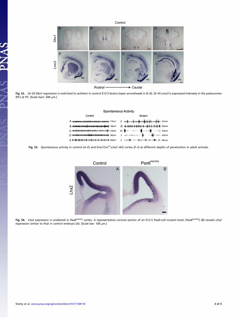
Fig. S2. (A–D) Dbx1 expression is restricted to antihem in control E12.5 brains (open arrowheads in B–D). (E–H) Lmo3 is expressed intensely in the paleocortex(PC) at P5. (Scale bars: 500 μm.)
Fig. S3. Spontaneous activity in control (A–E) and Emx1CreYL;Lhx2 cKO cortex (F–J) at different depths of penetration in adult animals.
Fig. S4. Lhx2 expression is unaltered in Pax6sey/sey cortex. A representative coronal section of an E12.5 Pax6-null mutant brain (Pax6sey/sey) (B) reveals Lhx2expression similar to that in control embryos (A). (Scale bar: 100 μm.)
Shetty et al. www.pnas.org/cgi/content/short/1311158110 3 of 3