Embed Size (px)
Citation preview
Open Access Free Rad. Antiox.
Free Radicals and Antioxidants 34 Vol 1, Issue 1, Jan-Mar, 2011
ww
w.a
ntio
x.or
g
Levels of two oxidative stress indicators of human sickle erythrocytes incubated in aqueous extracts of Anacardium occidentale, Psidium guajava and Terminalia catappa.Paul Chidoka Chikezie.
Department of Biochemistry, Imo State University, Owerri, Imo State, Nigeria.
INTRODUCTION
Oxidative stress is caused by accumulation of reactive oxygen species (ROS)[1, 2] produced as normal by-products of cellular metabolism [2, 3], exposure to ionic/electromagnetic radiations and some environmental pollutants.[4,5] These reactive species are capable of damaging diverse biomolecules and cell structures [6] in which lipids are probably the most susceptible,[7, 8] when cellular levels are not controlled by appropriate antioxidant scavenging systems.[9] Though erythrocytes of all human genotypes are particularly sensitive to oxidative stress, when compared with normal erythrocytes, sickle erythrocytes spontaneously generate approximately twice as much anion superoxide (.O
2−), hydrogen peroxide
(H2O
2) and hydroxyl radicals (.OH)[10, 11] and increasing
evidence suggest that lipid peroxidation may be an important factor in sickle cell anaemia.[12]
Specifically, sickle erythrocytes and their membranes are susceptible to endogenous free radical-mediated oxidative damage that correlates with the proportion of irreversibly sickled erythrocytes.[13] Furthermore, accumulation of hydrogen peroxide (H
2O
2) decreases the
half life of erythrocytes by increasing oxidation of polyunsaturated fatty acids of cell membrane[14] and can oxidize haemoglobin to methaemoglobin.[9, 15] Methaemoglobin does not bind reversibly with oxygen. One of the toxic end products of lipid peroxidation is malondialdehyde (MDA).[9] Sickle erythrocytes contain increased amount of MDA and evidence of amino group cross-linking by MDA, producing MDA-modified protein adducts has been demonstrated in lipid extract of sickle erythrocyte membrane preparations.[16]
Erythrocytes like other cells are supplied with diverse protective antioxidant mechanisms in order to counteract the toxic effects of ROS.[17-21] Antioxidants function as
ABSTRACTAn in vitro study was carried out to investigate levels of oxidative stress indicators of sickle erythrocytes incubated in aqueous extracts of Anacardium occidentale, Psidium guajava and Terminalia catappa for 12 h. At regular time intervals of 3 h, portions of the incubation mixtures were withdrawn and spectrophotometric method was used to assay for levels of erythrocyte malondialdehyde (MDA) and methaemoglobin (Met.Hb%). The control analysis showed that within the experimental time, erythrocyte MDA increased from 2.45±0.35 to 3.13±0.59 mmol/ml (p > 0.05; p value = 0.801176). Erythrocyte MDA concentrations in the presence of the three extracts were higher than the control samples at t = 3 h (p > 0.05; p value = 0.963253). Compared with the control samples at the given time (t) intervals, extract of T. catappa exhibited the highest capacity to cause reduction of erythrocyte MDA ([T. catappa] = 800 mg%; [MDA] = 2.89±0.33 mmol/ml; t = 12 h). Erythrocyte Met.Hb% increased from 2.42±0.55 to 2.51±0.49% (p > 0.05; p value = 0.995171) in the control samples within 12 h. Incubation of sickle erythrocytes with extract of [P. guajava] = 800 mg% for 9 h caused reduction of Met.Hb% from 2.49±0.49% to 2.29±0.45% (p > 0.05; p value = 0.983519). Extracts of A. occidentale, P. guajava and T. catappa exhibited variable capacities to hinder lipid peroxidation, but did not cause corresponding reduction in erythrocyte Met.Hb%, as shown by negative correlation between the two oxidative stress indicators in the presence of T. catappa and higher concentrations of A. occidentale and P. guajava.
Keywords: Malondialdehyde, Methaemoglobin, Erythrocyte, Anacardium occidentale, Psidium guajava, Terminalia catappa.
Phone: +2348038935327.
E-mail: p [email protected]
Levels of two oxidative stress indicators of human sickle erythrocytes incubated in aqueous extracts of Anacardium occidentale
Free Radicals and Antioxidants 35 Vol 1, Issue 1, Jan-Mar, 2011
ww
w.a
ntio
x.or
g
modulators of the cell redox homeostasis detoxifying both radical and non-radical species as well as heavy metals. Erythrocytes-reduced glutathione is one of the major non-enzymatic endogenous antioxidants protecting tissue against ROS.[9, 22] Other antioxidants are alpha-tocopherol[23, 24], uric acid [25-27], ascorbic acid[28], carotenoids[29, 30] and varieties of plant secondary metabolites such as flavonoid and related polyphenolic compounds.[31, 32] Notable erythrocyte enzymatic ROS scavenging systems include glutathione reductase,[17, 19] glutathione peroxidase,[33] glucose–6-phosphate dehydrogenase,[18, 34] superoxide dismutase,[5, 35] catalases,[36-38] peroxiredoxins, [39, 40] and NADH-methaemoglobin reductase.[21, 41-43]
Varieties of xenobiotics of plant origin such as fava beans extracts (Vicia faba) have been reported as agents that can modify the redox status of human erythrocytes especially in individuals with impaired glucose-6-phosphate dehydrogenase activity.[18,34,44]. Similarly, scavenging activities for free radicals by natural products of plant origin have been widely reported.[1, 28, 45-49]. This study has ascertained the level of oxidative stress by estimating the concentrations of two oxidative stress indicators, namely, MDA and methaemoglobin of sickle erythrocytes incubated in aqueous extracts of three medicinal plants: Anacardium occidentale, Psidium guajava and Terminalia catappa.
MATERIALS AND METHODS
Collection of Plant Specimens Fresh leaf samples of A. occidentale, P. guajava and T. catappa were harvested between July and August, 2010, from trees within the environment of Imo State University, Owerri, Nigeria. The plant specimens were identified and authenticated by Dr. F. N. Mbagwu at the Herbarium, Department of Plant Science and Biothechnology. A voucher specimen was deposited at the Herbarium for reference purposes.
Preparation of Aqueous Extracts of Plant Specimens The samples were washed under continous current of distilled water for 15 min and air dried at room temperature (24oC) for 60 min. The separate leaves were dried for 5 h in an oven at 60oC to become crispy, and ground with ceramic mortar and pestle. For each specimen, two grams (2 g) of the pulverized sample was suspended in 100 ml of distilled water and allowed to stand for 6 h at 37oC. The aqueous extracts (2 g%; w/v) of A. occidentale, P. guajava and T. catappa leaves were obtained by simple filteration method with Whatman No.
2 filter paper. The filtrates were centrifuged at 1200 x g for 5 min to remove tissue debris. The superntants were carefully harvested with Pasteur pipette into sterile test tubes and kept at 4oC in a refrigerator for at least 24 h before subsequent tests. Serial dilutions of the aqueous extracts in the order of 200, 400, 600 and 800 mg% (w/v) were used for analyses.
Collection of Blood Samples/Preparation of Erythrocyte Haemolysate Five milliliters (5.0 ml) of human venous blood samples of HbSS genotype were collected by venipuncture and stored in EDTA anticoagulant tubes. The blood samples were obtained between July and August, 2010, from nine (9) male volunteers (59-79 kg) between the age bracket of 21-34 yrs attending clinics at the Federal Medical Center (FMC), Imo State University Teaching Hospital (IMSUTH), Orlu, St. John Clinic / Medical Diagnostic Laboratories, Avigram Medical Diagnostic Laboratories, and Qualitech Medical Diagnostic Laboratories. These centers are located in Owerri, Imo State, Nigeria. The Institutional Review Board of the Department of Biochemistry, Imo State University, Owerri, Nigeria, granted approval for this study and all blood donors signed informed consent form. This study was in accordance with the ethical principles of the Declaration of Helsinki.
The erythrocytes were washed by centrifugation methods as described by Tsakiris et al.[50] Within 2 h of collection of blood samples, 2.0 ml aliquots of the samples were introduced into centrifuge test tubes containing 4.0 ml of buffer solution pH = 7.4: 250 mM tris (hydroxyl methyl) amino ethane–HCl (Tris-HCl) 140 mM NaCl 1.0 mM MgCl
2 10 mM glucose. The
erythrocytes were separated from plasma by centrifugation at 1200 x g for 10 min and washed three times by the same centrifugation method with the buffer solution. To remove platelets and leucocytes, the sediment was re-suspended in 3 ml of phosphate-buffered saline (PBS) solution, pH 7.4, and passed through a column (3.5 cm in a 30 ml syringe) of cellulose-microcrystalline cellulose (ratio w/w 1:1) as described by Kalra et al.[51] The eluted fraction was passed twice through a new column of cellulose-microcrystalline cellulose (ratio 1:1 w/w) to obtain erythrocyte suspension sufficiently devoid of leucocytes and platelets. The isolated erythrocytes were lysed by freezing/ thawing as described by Galbraith and Watts, [52] and Kamber et al.[53] The erythrocyte haemolysates were finally re-suspended in 1.0 ml of the buffer and stored at -70oC until analyses.[37]
Free Radicals and Antioxidants 36 Vol 1, Issue 1, Jan-Mar, 2011
Levels of two oxidative stress indicators of human sickle erythrocytes incubated in aqueous extracts of Anacardium occidentalew
ww
.ant
iox.
org
Experimental Design A 0.2 ml aliquot of the aqueous extracts of A. occidentale, P. guajava and T. catappa of increasing concentrations in the order: 200, 400, 600 and 800 mg% w/v were added to corresponding test tubes containing 0.8 ml of erythrocyte haemolysate (ratio 1:4 v/v). The incubation mixture was allowed to stand at a regulated temperature of 37oC in a water bath. At regular time intervals of 3 h for 12 h, aliquots of 0.2 ml of the incubation mixture were withdrawn and used for the determinations of erythrocyte MDA and methaemoglobin concentrations.
Determinations of Erythrocyte Malondialdehyde and Methaemoglobin Concentrations Determination of erythrocyte concentration of MDA was by method described by Tjahjani et al. [54] with minor modifications. A mixture of 20% trichloracetic acid (TCA) and 0.67% thiobarbituric acid (TBA) in a ratio of 2:1 was added into a test tube. A volume of 0.2 ml of erythrocyte haemolysate was introduced in the mixture and boiled for 10 min in a water bath. After cooling to 24oC, the mixture was centrifuged at 3,000 x g for 10 min. The absorbance of supernatant was read with a spectrophotometer (SPECTRONIC 20, Labtech – Digital Blood Analyzer®) at maximum wavelength (λ
max) = 532 nm. The values of
absorbance of the samples were converted to MDA concentrations using the MDA standard curve.[55]
Determination of methaemoglobin content of erythrocyte haemolysate was by modification of the method of Evelyn and Malloy,[56] as described by Akomopong et al.[57] A total of 0.4 ml of 0.5 M phosphate buffer (pH = 6.5) was added to 0.6 ml of the cell lysate, and the mixture was centrifuged at 16,000 x g for 5 min to sediment debris. A 0.7 ml aliquot of the supernatant fraction was used to measure the absorbance at λ
max = 630
nm (the absorbance maximum for methaemoglobin), and
the measurement was recorded as first reading (S1). A volume of 0.05 ml of 10 g% KCN was added, and after 5 min at 24oC, a second reading (S2) was recorded. KCN converts methaemoglobin to cyanomethaemoglobin, which does not absorb at 630 nm; hence, the difference between absorbance readings S1 and S2 represents the absorbance due to methaemoglobin. To measure total hemoglobin levels, all of the hemoglobin was converted to methaemoglobin, the absorbance of the sample at λ
max = 630 nm was recorded, and then KCN was added
to form cyanomethaemoglobin. Specifically, 0.07 ml of the supernatant fraction was diluted 10-fold into 0.6 ml of 0.1 M phosphate buffer (pH = 6.5). Next, 0.03 ml of freshly prepared 20 g% K
3Fe(CN)
6 was added and
incubated for 5 min at 24oC and an initial reading (T1) was recorded. A total of 0.05 ml of 10% KCN was subsequently added, and a second reading (T2) was recorded. The percent of methaemoglobin (Met.Hb%) in the sample was calculated as [100(S1-S2)] / [10(T1-T2)].
Statistical analysis The results were expressed in terms of arithmetic mean (X) ± standard deviation (SD). The correlation coefficients between the results were determined with Microsoft Office Excel, 2007 version and data were analyzed by Student’s t-test as described by Pearson and Hartley.[58] Values of p < 0.05 were considered statistically significant.
RESULTS
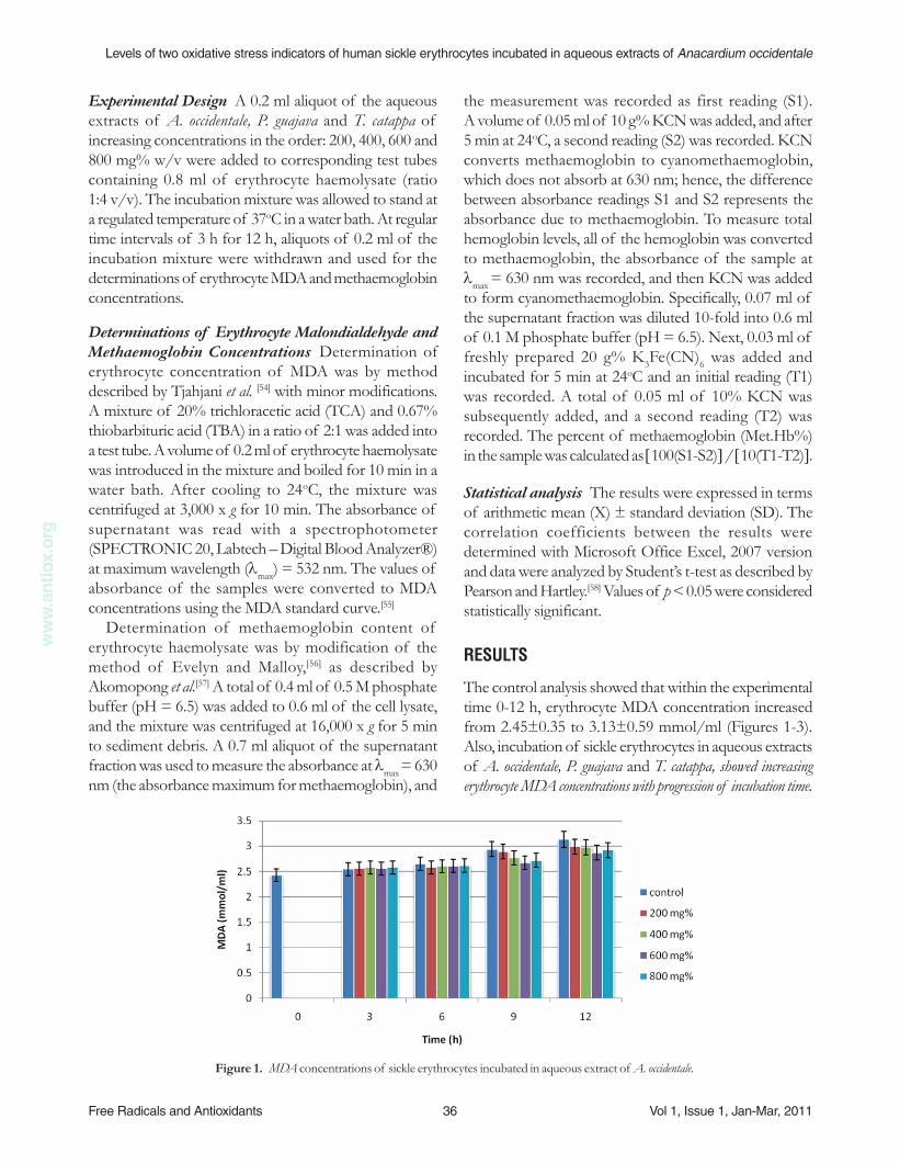
The control analysis showed that within the experimental time 0-12 h, erythrocyte MDA concentration increased from 2.45±0.35 to 3.13±0.59 mmol/ml (Figures 1-3). Also, incubation of sickle erythrocytes in aqueous extracts of A. occidentale, P. guajava and T. catappa, showed increasing erythrocyte MDA concentrations with progression of incubation time.
Figure 1. MDA concentrations of sickle erythrocytes incubated in aqueous extract of A. occidentale.
Levels of two oxidative stress indicators of human sickle erythrocytes incubated in aqueous extracts of Anacardium occidentale
Free Radicals and Antioxidants 37 Vol 1, Issue 1, Jan-Mar, 2011
ww
w.a
ntio
x.or
g
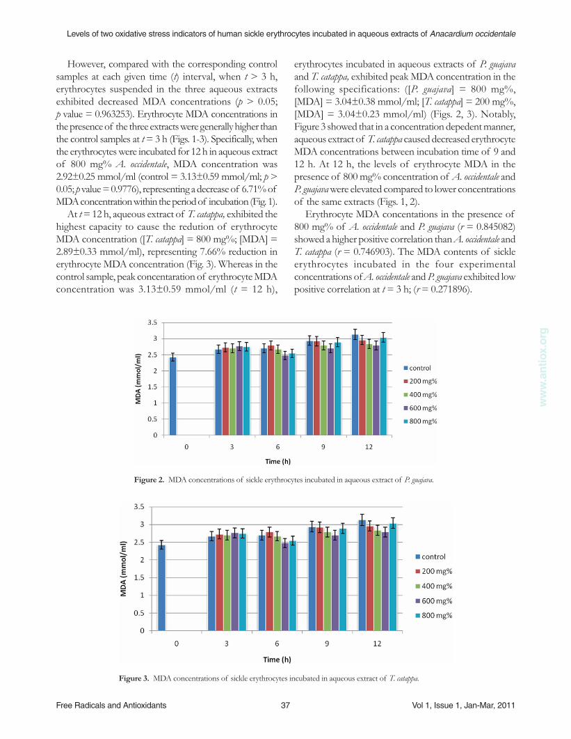
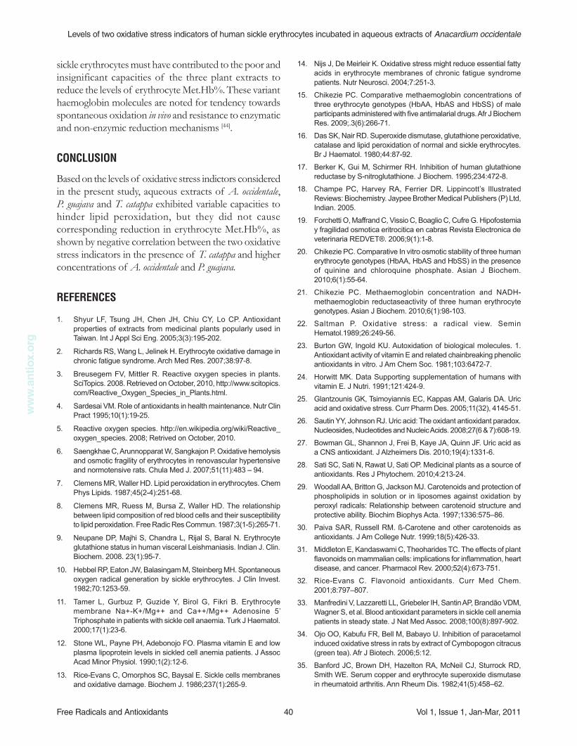
erythrocytes incubated in aqueous extracts of P. guajava and T. catappa, exhibited peak MDA concentration in the following specifications: ([P. guajava] = 800 mg%, [MDA] = 3.04±0.38 mmol/ml; [T. catappa] = 200 mg%, [MDA] = 3.04±0.23 mmol/ml) (Figs. 2, 3). Notably, Figure 3 showed that in a concentration depedent manner, aqueous extract of T. catappa caused decreased erythrocyte MDA concentrations between incubation time of 9 and 12 h. At 12 h, the levels of erythrocyte MDA in the presence of 800 mg% concentration of A. occidentale and P. guajava were elevated compared to lower concentrations of the same extracts (Figs. 1, 2).
Erythrocyte MDA concentations in the presence of 800 mg% of A. occidentale and P. guajava (r = 0.845082) showed a higher positive correlation than A. occidentale and T. catappa (r = 0.746903). The MDA contents of sickle erythrocytes incubated in the four experimental concentrations of A. occidentale and P. guajava exhibited low positive correlation at t = 3 h; (r = 0.271896).
However, compared with the corresponding control samples at each given time (t) interval, when t > 3 h, erythrocytes suspended in the three aqueous extracts exhibited decreased MDA concentrations (p > 0.05; p value = 0.963253). Erythrocyte MDA concentrations in the presence of the three extracts were generally higher than the control samples at t = 3 h (Figs. 1-3). Specifically, when the erythrocytes were incubated for 12 h in aqueous extract of 800 mg% A. occidentale, MDA concentration was 2.92±0.25 mmol/ml (control = 3.13±0.59 mmol/ml; p > 0.05; p value = 0.9776), representing a decrease of 6.71% of MDA concentration within the period of incubation (Fig. 1).
At t = 12 h, aqueous extract of T. catappa, exhibited the highest capacity to cause the redution of erythrocyte MDA concentration ([T. catappa] = 800 mg%; [MDA] = 2.89±0.33 mmol/ml), representing 7.66% reduction in erythrocyte MDA concentration (Fig. 3). Whereas in the control sample, peak concentaration of erythrocyte MDA concentration was 3.13±0.59 mmol/ml (t = 12 h),
Figure 2. MDA concentrations of sickle erythrocytes incubated in aqueous extract of P. guajava.
Figure 3. MDA concentrations of sickle erythrocytes incubated in aqueous extract of T. catappa.

Free Radicals and Antioxidants 38 Vol 1, Issue 1, Jan-Mar, 2011
Levels of two oxidative stress indicators of human sickle erythrocytes incubated in aqueous extracts of Anacardium occidentalew
ww
.ant
iox.
org
Also, in the presence of aqueous extract of [P. guajava] = 200 mg%, erythrocyte Met.Hb% value of 2.46±0.43%, at t = 9 h, was not significanly different (p > 0.05; p value = 0.999778) from the control sample (Fig. 5). In addition, 200 mg% of T. catappa caused increased erythrocyte Met.Hb% within the experimental period of 6 h (Fig. 6). Generally, aqueous extract of T. catappa exhibited low capacity to cause reduction in erythrocyte Met.Hb % (Fig. 6). Statistical analysis showed that erythrocyte Met.Hb% in the presence of 600 mg% of P. guajava and T. catappa displayed high postive correlation (r = 0.965535). Also, 800 mg% of A. occidentale and P. guajava exhibited postive correlation (r = 0.878868) with respect to erythrocyte Met.Hb%. From the data presented in Table 1, erythrocyte Met.Hb% and MDA concentration showed significant positive correlation in the presence of aqueous extracts of 200 mg% A. occidentale and 400 mg% P. guajava. Notably, erythrocytes incubated in aqueous extract of T. catappa and concentrations of A. occidentale, P. guajava > 400 mg% showed negative correlation.
Erythrocyte Met.Hb% increased from 2.42±0.55 to 2.51±0.49% in the control sample within the duration of 12 h (Figs 4-6). Although, at t = 3 h, aqueous extract of A. occidentale (excerpt 800 mg%) caused increasing of Met.Hb%, the values were not significantly different (p > 0.05) from the control samples. Further increases in incubation time (t > 3) engendered declining levels of erythrocyte Met.Hb%, except at t = 12 h (Fig. 4). Although aqueous extract of [A. occidentale] = 200 mg% caused decreased erythrocyte Met.Hb% from 2.49±0.49% to 2.34±0.65% within 6 h of incubation, the value increased to 2.55±0.43% at t = 12 hr (Fig. 4).
Incubation of sickle erythrocytes in aqueous extract of [P. guajava] = 800 mg% for 9 h caused the reduction of Met.Hb% from 2.49±0.49% to 2.29±0.45%. Incubation with [P. guajava] = 600 mg%, erythrocyte Met.Hb% gave value of 2.55±0.43% (t = 3 h), which was not significantly (p > 0.05; p value = 0.996121) higher than the control value (Fig. 5).
Figure 4. Met.Hb% of sickle erythrocytes incubated in aqueous extract of A. occidentale.
Figure 5. Met.Hb% of sickle erythrocytes incubated in aqueous extract of P. guajava.

Levels of two oxidative stress indicators of human sickle erythrocytes incubated in aqueous extracts of Anacardium occidentale
Free Radicals and Antioxidants 39 Vol 1, Issue 1, Jan-Mar, 2011
ww
w.a
ntio
x.or
g
irradation-induced oxidative stress indicators were significantly reduced in rats after orally administered with Aloe vera. Prevoius findings have attributed the capacities of Zingiber officinale, Aloe vera and Rheum rhabarbarum extracts to reduce lipid peroxidation to the presence of phenolic compounds in these plants [46, 62]. Also, membrane protective activities of diverse plant extracts have been attributed to their antioxidant content and capacity to impede membrane lipid peroxidation [47, 63, 64]. However, when compared to lower experimental concentrations of A. occidentale and P. guajava, 800 mg% concentration of the two plant extracts did not cause an anticipated reduction of lipid peroxidation (r = 0.845082) (Figs. 1, 2). Therefore, it is envisaged that higher concentrations of the two plant extracts may promote peroxidation. It is worthwhile to mention that Paiva and Russell [30] prevoiusly reported that antioxidant activity of carotenoids (including ß-carotene) may exhibit adverse effects when present in high dose.
The present results have shown that Met.Hb% of sickle erythrocytes was significantly (p < 0.05; p value = 0.489323) higher than normal physiologic concentration (MetHb% = 1.50) reported for HbAA erythrocyte genotype [15, 65]. Noteworthy, the level of erythrocyte methaemoglobin reported here is comparable to those presented by other authors [15, 21, 66]. The primary reason for the relatively high concentration of oxidized haemoglobin is also connected with excessive production and accumulation of ROS compared with other human erythrocyte genotypes [66]. The moderate reduction in Met.Hb% in sickle erythrocytes incubated in aqueous extracts of A. occidentale, P. guajava and T. catappa is attributed to their antioxidant activity as earlier discussed. However the association of certain methaemoglobinopathies such as HbM
Boston, HbM
Iwate, HbM
Hydepark and HbM
Hammersmith with
DISCUSSION
The increasing erythrocyte MDA concentrations of the control samples with time were obvious reflection of production and accumulation of ROS, engendered by normal metabolic processes in these cells. Tarmer et al. [59] had previously reported that sickle erythrocytes generate ROS spontaneously and contain high level of MDA, a by-product of lipid peroxidation. It is worthwhile to note that several abnormalities associated with sickle erythrocyte are inextricably connected with high level of oxidative stressors in this erythrocyte genotype [33, 60, 61]. From the results showed in Figures 1-3, the increasing erythrocyte MDA concentrations of the control and test samples are indication of the time-dependent progression of lipid peroxidation in both samples. Furthermore, relatively lower MDA concentrations of the test samples, compared to the control ones, at the given time intervals, revealed that lipid peroxidation was retarded by the three plant extracts in relation to their corresponding concentrations. The present findings are in concord with the report of Lam et al. [46]. They noted that gamma
Figure 6. Met.Hb% of sickle erythrocytes incubated in aqueous extract of T. catappa.
Table 1. Correlation between changes in MDA concentration and Met.Hb% of sickle erythrocyte
incubated in aqueous extracts of A. occidentale, P. guajava and T. catappa for 12 h.
Correlation coefficient (r)
[Extract] mg% A. occidentale P. guajava T. catappa
200 0.578986 0.284634 -0.40214
400 -0.42167 0.624449 -0.95828
600 -0.48163 -0.02065 -0.33534
800 -0.5715 -0.03693 -0.27915
Free Radicals and Antioxidants 40 Vol 1, Issue 1, Jan-Mar, 2011
Levels of two oxidative stress indicators of human sickle erythrocytes incubated in aqueous extracts of Anacardium occidentalew
ww
.ant
iox.
org
14. Nijs J, De Meirleir K. Oxidative stress might reduce essential fatty acids in erythrocyte membranes of chronic fatigue syndrome patients. Nutr Neurosci. 2004;7:251-3.
15. Chikezie PC. Comparative methaemoglobin concentrations of three erythrocyte genotypes (HbAA, HbAS and HbSS) of male participants administered with five antimalarial drugs. Afr J Biochem Res. 2009;.3(6):266-71.
16. Das SK, Nair RD. Superoxide dismutase, glutathione peroxidative, catalase and lipid peroxidation of normal and sickle erythrocytes. Br J Haematol. 1980;44:87-92.
17. Berker K, Gui M, Schirmer RH. Inhibition of human glutathione reductase by S-nitroglutathione. J Biochem. 1995;234:472-8.
18. Champe PC, Harvey RA, Ferrier DR. Lippincott’s Illustrated Reviews: Biochemistry. Jaypee Brother Medical Publishers (P) Ltd, Indian. 2005.
19. Forchetti O, Maffrand C, Vissio C, Boaglio C, Cufre G. Hipofostemia y fragilidad osmotica eritrocitica en cabras Revista Electronica de veterinaria REDVET®. 2006;9(1):1-8.
20. Chikezie PC. Comparative In vitro osmotic stability of three human erythrocyte genotypes (HbAA, HbAS and HbSS) in the presence of quinine and chloroquine phosphate. Asian J Biochem. 2010;6(1):55-64.
21. Chikezie PC. Methaemoglobin concentration and NADH-methaemoglobin reductaseactivity of three human erythrocyte genotypes. Asian J Biochem. 2010;6(1):98-103.
22. Saltman P. Oxidative stress: a radical view. Semin Hematol.1989;26:249-56.
23. Burton GW, Ingold KU. Autoxidation of biological molecules. 1. Antioxidant activity of vitamin E and related chainbreaking phenolic antioxidants in vitro. J Am Chem Soc. 1981;103:6472-7.
24. Horwitt MK. Data Supporting supplementation of humans with vitamin E. J Nutri. 1991;121:424-9.
25. Glantzounis GK, Tsimoyiannis EC, Kappas AM, Galaris DA. Uric acid and oxidative stress. Curr Pharm Des. 2005;11(32), 4145-51.
26. Sautin YY, Johnson RJ. Uric acid: The oxidant antioxidant paradox. Nucleosides, Nucleotides and Nucleic Acids. 2008;27(6 & 7):608-19.
27. Bowman GL, Shannon J, Frei B, Kaye JA, Quinn JF. Uric acid as a CNS antioxidant. J Alzheimers Dis. 2010;19(4):1331-6.
28. Sati SC, Sati N, Rawat U, Sati OP. Medicinal plants as a source of antioxidants. Res J Phytochem. 2010;4:213-24.
29. Woodall AA, Britton G, Jackson MJ. Carotenoids and protection of phospholipids in solution or in liposomes against oxidation by peroxyl radicals: Relationship between carotenoid structure and protective ability. Biochim Biophys Acta. 1997;1336:575–86.
30. Paiva SAR, Russell RM. ß-Carotene and other carotenoids as antioxidants. J Am College Nutr. 1999;18(5):426-33.
31. Middleton E, Kandaswami C, Theoharides TC. The effects of plant flavonoids on mammalian cells: implications for inflammation, heart disease, and cancer. Pharmacol Rev. 2000;52(4):673-751.
32. Rice-Evans C. Flavonoid antioxidants. Curr Med Chem. 2001;8:797–807.
33. Manfredini V, Lazzaretti LL, Griebeler IH, Santin AP, Brandão VDM, Wagner S, et al. Blood antioxidant parameters in sickle cell anemia patients in steady state. J Nat Med Assoc. 2008;100(8):897-902.
34. Ojo OO, Kabufu FR, Bell M, Babayo U. Inhibition of paracetamol induced oxidative stress in rats by extract of Cymbopogon citracus (green tea). Afr J Biotech. 2006;5:12.
35. Banford JC, Brown DH, Hazelton RA, McNeil CJ, Sturrock RD, Smith WE. Serum copper and erythrocyte superoxide dismutase in rheumatoid arthritis. Ann Rheum Dis. 1982;41(5):458–62.
sickle erythrocytes must have contributed to the poor and insignificant capacities of the three plant extracts to reduce the levels of erythrocyte Met.Hb%. These variant haemoglobin molecules are noted for tendency towards spontaneous oxidation in vivo and resistance to enzymatic and non-enzymic reduction mechanisms [44].
CONCLUSION
Based on the levels of oxidative stress indictors considered in the present study, aqueous extracts of A. occidentale, P. guajava and T. catappa exhibited variable capacities to hinder lipid peroxidation, but they did not cause corresponding reduction in erythrocyte Met.Hb%, as shown by negative correlation between the two oxidative stress indicators in the presence of T. catappa and higher concentrations of A. occidentale and P. guajava.
REfERENCES
1. Shyur LF, Tsung JH, Chen JH, Chiu CY, Lo CP. Antioxidant properties of extracts from medicinal plants popularly used in Taiwan. Int J Appl Sci Eng. 2005;3(3):195-202.
2. Richards RS, Wang L, Jelinek H. Erythrocyte oxidative damage in chronic fatigue syndrome. Arch Med Res. 2007;38:97-8.
3. Breusegem FV, Mittler R. Reactive oxygen species in plants. SciTopics. 2008. Retrieved on October, 2010, http://www.scitopics.com/Reactive_Oxygen_Species_in_Plants.html.
4. Sardesai VM. Role of antioxidants in health maintenance. Nutr Clin Pract 1995;10(1):19-25.
5. Reactive oxygen species. http://en.wikipedia.org/wiki/Reactive_oxygen_species. 2008; Retrived on October, 2010.
6. Saengkhae C, Arunnopparat W, Sangkajon P. Oxidative hemolysis and osmotic fragility of erythrocytes in renovascular hypertensive and normotensive rats. Chula Med J. 2007;51(11):483 – 94.
7. Clemens MR, Waller HD. Lipid peroxidation in erythrocytes. Chem Phys Lipids. 1987;45(2-4):251-68.
8. Clemens MR, Ruess M, Bursa Z, Waller HD. The relationship between lipid composition of red blood cells and their susceptibility to lipid peroxidation. Free Radic Res Commun. 1987;3(1-5):265-71.
9. Neupane DP, Majhi S, Chandra L, Rijal S, Baral N. Erythrocyte glutathione status in human visceral Leishmaniasis. Indian J. Clin. Biochem. 2008. 23(1):95-7.
10. Hebbel RP, Eaton JW, Balasingam M, Steinberg MH. Spontaneous oxygen radical generation by sickle erythrocytes. J Clin Invest. 1982;70:1253-59.
11. Tamer L, Gurbuz P, Guzide Y, Birol G, Fikri B. Erythrocyte membrane Na+-K+/Mg++ and Ca++/Mg++ Adenosine 5’ Triphosphate in patients with sickle cell anaemia. Turk J Haematol. 2000;17(1):23-6.
12. Stone WL, Payne PH, Adebonojo FO. Plasma vitamin E and low plasma lipoprotein levels in sickled cell anemia patients. J Assoc Acad Minor Physiol. 1990;1(2):12-6.
13. Rice-Evans C, Omorphos SC, Baysal E. Sickle cells membranes and oxidative damage. Biochem J. 1986;237(1):265-9.

Levels of two oxidative stress indicators of human sickle erythrocytes incubated in aqueous extracts of Anacardium occidentale
Free Radicals and Antioxidants 41 Vol 1, Issue 1, Jan-Mar, 2011
ww
w.a
ntio
x.or
g
52. Galbraith DA, Watts DC Changes in some cytoplasmic enzymes from red cells fractionated into age groups by centrifugation in FicollTM/TriosilTM gradients. Comparison of normal human and patients with Duchenne muscular dystrophy. Biochem J. 1980;191:63-70.
53. Kamber K, Poyiagi A, Delikonstantinos G. Modifications in the activities of membrane-bound enzymes during in vivo ageing of human and rabbit erythrocytes. Compreh Biochem Physiol. 1984;B.77B, 95-9.
54. Tjahjani S, Puji BSA, Syafruddin D, Agoes R, Hanggono T, Immaculata M. Oxidative stress in Plasmodium falciparum culture incubated with artemisinin. Proc ASEAN Congr Trop Med Parasitol. 2008;3:47-50.
55. Schmuck G, Roehrdanz E, Hayes RK, Kahl R. Neurotoxic mode of action of artemisinin. Antimicrob Agents Chemother. 2002;46: 821-7.
56. Evelyn K, Malloy H. Micro determination of oxyhaemoglobin, methaemoglobin and sulfhaemoglobin in a single sample of blood. J Biol Chem. 1938;126:55-66.
57. Akomopong T, Ghori N, Haldar H. In vitro activity of riboflavin against the human malaria parasite Plasmodium falciparum. Antimicrob Agents Chemothera. 2000;44 (1):88-96.
58. Pearson ES, Hartley HQ. Biometric Tables for Statistians. 3rd ed. Cambridge University Press, London. 1966.
59. Tamer L, Gurbuz P, Guzide Y, Birol G, Fikri B. Erythrocyte membrane Na+-K+/Mg++ and Ca++/Mg++ Adenosine 5’ Triphosphate in patients with sickle cell anaemia. Turk J Haematol. 2000;17(1):23-6.
60. Repka T, Hebbel RP. Hydroxyl radical formation by sickle erythrocyte membranes: role of pathologic iron deposits and cytoplasmic reducing agents. Blood. 1991;78:2753-8.
61. Dhalla NS, Elmoselbi AB, Hata T. Status of myocardial antioxidants in ischemia-reperfusion injury. Cardiovasc Res. 2000;47:446-56.
62. Aaviksaar A, Haga M, Kuzina K, Pussa T, Raal A, Tsoupras G. Hydroxystilbenes in the roots of Rheum rhaponticum. Proc Estonian Acad Sci Chem. 2003;52:99 –107.
63. Hu C, Kitts DD. Studies on the antioxidant activity of Echinacea root extract. J Agric Food Chem. 2000;48:1466-72.
64. Okpuzor J, Adebesin O, Ogbunugafor H, Amadi I. The potential of medicinal plants in sickle cell disease control: A review. Inter. J Biomed Health Sci. 2008;4(2):47-55.
65. Tietz N. Fundamentals of Clinical Chemistry W. B Saunders Co., Philadephia. 1976.
66. Van Kiujk FJ, Savannah A, Hanelman GI, Dratz EA. A new role of phospholipase A2 : Peroxiase damage. TIBS. 1987;12:31-4.
36. Goth L. A simple method for determination of catalase activity and revision of reference range. Clin Chem Acta. 1991;190:143-52.
37. Pennings HJ, Borm PJA, Evelo CTA, Wouters EFM. Changes in levels of catalase and glutathione in erythrocytes of patients with stable asthma, treated with beclomethasone dipropionate. Eur Respir J. 1999;13:1260-6.
38. Chandrasena LG, Chackrewarthy P, Perera MJ, de Silva D. Erythrocyte antioxidant enzymes in patients with cataract. Ann Clin Lab Sci. 2006;36(2):201-4.
39. Neumann CA, Krause DS, Carman CV, Das S, Dubey DP, Abraham JL, et al. Essential role for the peroxiredoxin Prdx1 in erythrocyte antioxidant defence and tumour suppression. Nature. 2003;424(6948):561-5.
40. Low FM, Hampton MB, Peskin AV, Winterbourn CC. Peroxiredoxin 2 functions as a noncatalytic scavenger of low-level hydrogen peroxide in the erythrocyte. Blood. 2007;109:2611-17.
41. Kuma F. Properties of Kinetic study of methaemoglobin reduction. J Biochem. 1981;2:50-5.
42. Huskey RJ. NADH-diaphorase enzyme action. http://www.people.virginia.edu./mrjhgu/methbenz.htm/>. 1998. Retrieved on October, 2007.
43. Mallory M. Methaemoglobinemia. Utox. Update. 2003. 5(2):1-2 http://www.uuhsc.utah.edu/poison. Retrieved on October, 2007.
44. Mayes PA. Metabolism of carbohydrates In: Harper’s Review of Biochemistry 19th ed. (Martin DW, Mayer PA, Rodwell VN. Eds). Lange Medical Publications, California. 1983.
45. Aqil F, Ahmed I, Mehmood Z. Antioxidant and free radical scavenging properties of twelve traditionally used Indian medicinal plants. Turk J Biol. 2006;30:177-83.
46. Lam RYY, Woo AYH, Leung P-S, Cheng CHK. Antioxidant actions of phenolic compounds found in dietary plants on low-density lipoprotein and erythrocytes in vitro. J. Am College Nutr. 2007;26(3):233-42.
47. Buřičová L, Réblová Z. Czech medicinal plants as possible sources of antioxidants. Czech J Food Sci. 2008;26 (2):132–8.
48. Muanda F, Koné D, Dicko A, Soulimani R, Younos C. Phytochemical composition and antioxidant capacity of three Malian medicinal plant parts. eCAM. 2009. doi:10.1093/ecam/nep109.
49. Veeru P, Kishor MP, Meenakshi M. Screening of medicinal plant extracts for antioxidant activity. J Med Plant Res. 2009;3(8):608-12.
50. Tsakiris S, Giannoulia-Karantana A, Simintzi I, Schulpis KH. The effect of aspartame metabolites on human erythrocyte membrane acetylcholinesterase activity. Pharmacol Res. 2005;53:1-5.
51. Kalra VK, Sikka SC, Sethi GS. Transport of amino acids in gamma-glutamyl transpeptidase-implanted human erythrocytes. J Biol Chem. 1981;256:55-67.