Embed Size (px)
Citation preview

203
Mycobacterium
MICROBIOLOGY
MODULEMicrobiology
Notes
20
MYCOBACTERIUM
20.1 INTRODUCTION
Mycobacterium are slender rods that sometimes show branching filamentousforms resembling fungal mycelium. In liquid cultures they form a mould-likepellicle. Hence the name ‘mycobacteria’, meaning fungus like bacteria. They donot stain readily, but once stained, resist decolourisation with dilute mineralacids. Hence they are called ‘Acid fast bacilli’. They are aerobic, nonmotile,noncapsulated and nonsporing.
OBJECTIVES
After reading this lesson, you will be able to:
describe the morphology of Mycobacterium tuberculosis & M. leprae
describe the characteristics of Mycobacterium tuberculosis & M. leprae
explain about pathogenesis of Mycobacterium tuberculosis & M. leprae
explain the laboratory diagnosis Mycobacterium tuberculosis & M. leprae
The first member of this genus to be identified was Lepra bacillus discovered byHansen. Koch (1882) isolated the mammalian tubercle bacillus and proved itscausative role in tuberculosis. In humans tuberculosis is caused by mycobacteriumtuberculosis and also by bovine type called Mycobacterium bovis.
The second human pathogenic mycobacterium is the lepra bacillus causingLeprosy. The third group of mycobacterium is a mixed group from varied sourceslike birds, cold-blooded and warm blooded animals, from skin ulcers, soil, waterand other environmental sources. They are called as atypical mycobacteria. Theyare opportunistic pathogens and can cause many types of diseases.

MICROBIOLOGY
MODULE Mycobacterium
Microbiology
204
Notes
20.2 MYCOBACTERIUM TUBERCULOSIS
Morphology
M tuberculosis is a straight or slightly curved rod, about 3 X 0.3 µm in size,occurring singly, in pairs or as small clumps. M bovis is usually straighter,shorter and stouter.
Tubercle bacilli have been described as Gram positive, even though afterstaining with basic dyes they resist decolourisation by alcohol even without theeffect of iodine. When stained with carbol fuchsin by Ziehl-Neelsen method orby fluorescent dyes they resist decolorisation by acids such as 20% Sulphuricacid as well as by alcohols. The unsaponifiable wax (mycolic acid) forms asemipermeable membrane around the cell that makes it acid fast.
Fig. 20.1: AFB smear
Fig. 20.2: Mycobacterium tuberculosi Acid-Fast stain
Culture characteristics
The bacilli grow and invitro time for generation is 14-15 hours. The optimumtemperature is 37°C and growth does not occur below 25°C or above 40°C.Optimum pH is 6.4-7.0. M tuberculosis is an obligate aerobe while M bovis ismicroaerophilic on primary isolation. M tuberculosis grows luxuriantly inculture as compared to M bovis which grows sparsely.

205
Mycobacterium
MICROBIOLOGY
MODULEMicrobiology
Notes
On solid media, dry, rough, raised, irregular colonies with a wrinkled surface aresenn and they are creamy white, becoming yellowish coloured on furtherincubation. M bovis forms flat, smooth, moist, white colonies that break upeasily.
INTEXT QUESTIONS 20.1
1. Mycobacteria means ................
2. Mycobacteria are ................ bacilli
3. Presence of ................ around cells makes it acid fast
4. Mycobacteria tuberculosis is an ................ aerobe
Resistance
Mycobacteria are not heat resistant, being killed at 60°C in 15-20 minutes.Cultures may be killed by exposure to direct sunlight for two hours. But bacilli inspectrum may remain alive for 20-30 hours. Bacilli are relatively resistant tochemical disinfectants, surviving exposure to 5% phenol, 15% sulphuric acid,3% Nitric acid, 5%oxalic acid and 4% sodium hydroxide. They are sensitive toformaldehyde and glutaraldehyde.
Biochemical reactions
Niacin test: Human tubercle bacilli form niacin when grown on an egg medium.When 10% cyanogens bromide and 4% aniline in 96% ethanol are added to asuspension of the culture, a canary yellow colour indicates a positive reaction.The test is positive with human type and negative with bovine type.
Aryl sulphatase test: This test is positive only with atypical mycobacteria. Thebacilli are grown in a medium containing 0.001 M tripotassium phenolphthaleindisulphate. 2N NaOH is added drop by drop to the culture and pink colourindicates a positive reaction.
Neutral red test: Virulent strains of tubercle bacilli are able to bind neutral red inalkaline buffer solution.
Catalase-Peroxidase tests: this is used to differentiate tubercle bacilli fromatypical mycobacteria. Most atypical mycobacteria strains are catalase positivewhile tubercle bacilli are weakly positive. Tubercle bacilli are peroxidasepositive but not atypical mycobacteria.

MICROBIOLOGY
MODULE Mycobacterium
Microbiology
206
Notes
A mixture of equal volumes of 30 volumes of H2O2 and 0.2% catechol indistilled water is added to 5 ml of test culture and are allowed to stand for fewminutes. Effervescence indicates catalase production and browning indicatesperoxidase activity.
Amidase tests: The ability to split amides has been used to differentiatemycobacteria. A 0.00165 M solution of the amide is incubated with the bacillarysuspension at 37°C and 0.1 ml MnSO4.4 H2O, 1.0 ml of phenol solution and 0.5ml hypochlorite solution are added. The tubes are placed in boiling water for 20minutes. A blue colour indicates a positive test.
Nitrate reduction test: this is positive with M tuberculosis and negative with Mbovis.
Antigenic properties: Antigens have been identified in mycobacteria. Groupspecificity is due to polysaccharides and type specificity to protein antigens.Delayed hypersensitivity develops following an infection of tubercle bacilli tothe bacillary protein. M tuberculosis stains are antigenically homogeneous but isnot useful in diagnosis or in immunity.
Bacteriophage: Mycobacteriophages have been isolated from soil, water andother environmental sources as well as from lysogenic strains.
There are four phage types A,B,C and a intermediate type between A & B as I,which is common in india.
Molecular typing: DNA fingerprinting provides a method for differentiatingbetween strains of tubercle bacilli. Restriction endonuclease treatment yieldsnucleic acid fragments of varying lengths and the pattern are strain specific. Thisrestriction fragment length polymorphism (RFLP) is used in strain typing.
Pathogenesis: Open case of pulmonary tuberculosis is the source of infection,which is most common in India. One open case may infect 25 contacts. The modeof infection is by direct inhalation of aerosolized bacilli in droplet nuclei ofexpectorated sputum. Coughing, sneezing and speaking releases numerousdroplets as many as 3000 infectious nuclei per cough. Dried bacilli in dust aremuch less infectious.
The majority of inhaled bacilli are arrested by natural defenses of the upperrespiratory tract and which reaches the lungs are ingested by alveolar macrophages.Number and virulence of the infecting bacilli, host factors including geneticsusceptibility, age, immunocompetence, stress, nutrition and coexisting illnessinfluence the outcome of the infection.
Humans have effective defence against the infection as only a tenth of theinfected develop active tuberculosis. Cell mediated immunity appears to be

207
Mycobacterium
MICROBIOLOGY
MODULEMicrobiology
Notes
effective, whereas humoral immunity is irrelevant. The key cell is the activatedCD4+ helper T cell which develops as Th-1 or Th-2 cells, releasing cytokinessuch as interferon γ (gamma) interleukins 1 and 2, toxic necrosis factor α (alpha)and others exerting different biological effects. Th-1 dependent cytokinesactivate macrophages resulting in protective immunity and containment of theinfection. Th-2 cytokines induce delayed type hypersensitivity (DTH), tissuedestruction and progressive disease.
The essential pathology in tuberculosis is the production in infected tissues of acharacteristic lesion the tubercle, this is an avascular granuloma composed of acentral zone containing giant cells with or without caseation and a peripheralzone of lymphocytes and fibroblasts.
Tuberculosis may be classified as primary and post primary.
Primary tuberculosis is the initial infection by tubercle bacilli. In endemiccountries like India this usually occurs in young children, the bacilli engulfed byalveolar macrophages multiply and give rise to a subpleural focus of tuberculouspneumonia, commonly in upper lobe, the Ghon factor. The hilar lymph nodes areinvolved. The Ghon focus together with enlarged hilar lymph node constitutesprimary complex. This occurs about 3-8 weeks from the time of infection and isassociated with the development of tuberculin hypersensitivity. In most of thecases the lesion heals spontaneously in 2-6 months leaving a calcified nodule anda few bacilli may survive and remain latent. In children with impaired immunityor other risk factors they may cause miliary, meningeal or other forms ofdisseminated tuberculosis.
The post primary type of tuberculosis is due to reactivation of latent infection orexogenous reinfection. It affects mostly in the upper lobes of the lungs, thelesions undergoing necrosis and tissue destruction, leading to cavitation. Thenecrotic materials are released through airway, to expectoration of latent sputum,which is the main source of infection.
INTEXT QUESTIONS 20.2
1. Niacin test is negative in ................
2. Aryl sulphatase test is positive in ................
3. In molecular typing ................ is used in stain typing
4. Mycobacteria gets transmitted by ................ infection
5. Primary complex constitutes of ................ & ................
MICROBIOLOGY
MODULE Mycobacterium
Microbiology
208
Notes
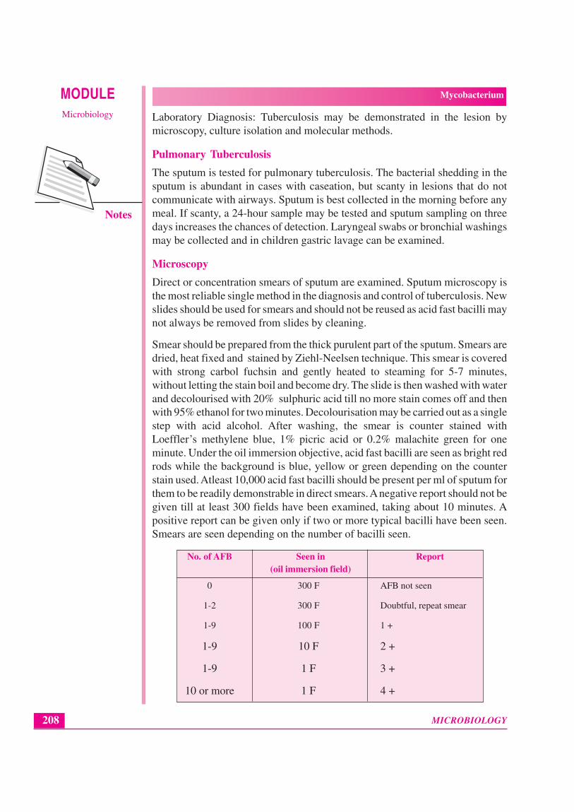
Laboratory Diagnosis: Tuberculosis may be demonstrated in the lesion bymicroscopy, culture isolation and molecular methods.
Pulmonary Tuberculosis
The sputum is tested for pulmonary tuberculosis. The bacterial shedding in thesputum is abundant in cases with caseation, but scanty in lesions that do notcommunicate with airways. Sputum is best collected in the morning before anymeal. If scanty, a 24-hour sample may be tested and sputum sampling on threedays increases the chances of detection. Laryngeal swabs or bronchial washingsmay be collected and in children gastric lavage can be examined.
Microscopy
Direct or concentration smears of sputum are examined. Sputum microscopy isthe most reliable single method in the diagnosis and control of tuberculosis. Newslides should be used for smears and should not be reused as acid fast bacilli maynot always be removed from slides by cleaning.
Smear should be prepared from the thick purulent part of the sputum. Smears aredried, heat fixed and stained by Ziehl-Neelsen technique. This smear is coveredwith strong carbol fuchsin and gently heated to steaming for 5-7 minutes,without letting the stain boil and become dry. The slide is then washed with waterand decolourised with 20% sulphuric acid till no more stain comes off and thenwith 95% ethanol for two minutes. Decolourisation may be carried out as a singlestep with acid alcohol. After washing, the smear is counter stained withLoeffler’s methylene blue, 1% picric acid or 0.2% malachite green for oneminute. Under the oil immersion objective, acid fast bacilli are seen as bright redrods while the background is blue, yellow or green depending on the counterstain used. Atleast 10,000 acid fast bacilli should be present per ml of sputum forthem to be readily demonstrable in direct smears. A negative report should not begiven till at least 300 fields have been examined, taking about 10 minutes. Apositive report can be given only if two or more typical bacilli have been seen.Smears are seen depending on the number of bacilli seen.
No. of AFB Seen in Report(oil immersion field)
0 300 F AFB not seen
1-2 300 F Doubtful, repeat smear
1-9 100 F 1 +
1-9 10 F 2 +
1-9 1 F 3 +
10 or more 1 F 4 +

209
Mycobacterium
MICROBIOLOGY
MODULEMicrobiology
Notes
When several smears are to be examined daily, fluorescent microscopy is used.Smears are stained with auramine phenol or auramine rhodamine fluorescentdyes and when examined under ultraviolet illumination, the bacilli will appear asbright rods against a dark background.
Concentration methods
(i) Petroff’s method
This method is widely used. Sputum is incubated with an equal volume of 4%sodium hydroxide solution at 37°C with frequent shaking till it becomes clear. Itis then centrifuged at 3000 rpm for 20 minutes and the sediment neutralized withN/10 HCl and used for smear, culture and animal inoculation.
A simpler method like, treating the sputum with an approximately equal volumeof a sterile solution containing 20 g cetrimonium bromide and 40 g of NaOH perlitre of distilled water. The contents are mixed with cotton swab and left to standfor five minutes. About 0.2 ml of the inoculum is smeared firmly with the swabover the entire surface of acid buffered medium.
Culture
Culture is a very sensitive diagnostic technique for tubercle bacilli, detecting asfew as 10 to 100 bacilli per ml. The concentrated material is inoculated intoatleast two bottles of IUAT-LJ medium. If the specimen is positive bymicroscopy a direct drug sensitivity test may be done. Cultures are examined forgrowth after incubation at 37°C for four days, for rapid growing mycobacteria,fungi and contaminant bacteria and atleast twice weekly thereafter. A negativereport is given if no growth occurs after 8-12 weeks. Any growth seen is smearedand tested by Ziehl Neelsen staining. For routine purposes, a slow growing, nonpigmented, niacin positive acid fast bacillus is taken as M.tuberculosis.Confirmation is by biochemical studies
Sensitivity tests
As drug resistance is an important problem in tuberculosis it is desirable to havesensitivity of isolates tested as an aid to treatment and they are of three types. Thefirst is absolute concentration method in which a number of media containingserial concentration of the drugs are inoculated and the minimum inhibitoryconcentrations calculated
The second is resistance ratio method in which two sets of media containinggraded concentrations of the drugs are inoculated. One set with the test strain andother with a standard strain of known sensitivity
The third is proportion method which indicates average sensitivity of the strain.

MICROBIOLOGY
MODULE Mycobacterium
Microbiology
210
Notes
Allergic test – Mantoux test
0.1ml of Purified Protein Derivative (PPD) containing 5 TU is injectedintradermally on the forearm with a tuberculin syringe causing a wheal. Theinjection should not be given subcutaneously but in between the layers of theskin, intradermally. The site is examined 48-72 hrs later and induration measuredat its widest point transversely. Induration of diameter 10mm or more isconsidered positive, 5mm or less is considered negative and 6-9mm equivocal.
A positive tuberculin test indicated hypersensitivity to tuberculoprotein denotinginfection with tubercle bacilli or BCG immunization, recent or past with orwithout clinical disease
INTEXT QUESTIONS 20.3
1. ................ is the most reliable single method in the diagnosis of tuberculosis
2. ................ technique is used in demonstration of tubercle bacilli
3. ................ is the common concentration method in diagnosis of tuberclebacilli
4. ................, ................ & ................ are the common sensitivity tests in thediagnosis of tubercle bacilli
5. ................ is the allergic test used in the diagnosis of tuberculosis
6. ................ is injected in allergic test
20.3 MYCOBACTERIUM LEPRAE
Leprosy is a disease recognized since vedic times in India. The person sufferingwith leprosy is considered ‘unclean’ and a social outcast. The lepra bacillus wasfirst observed by Hansen in 1868 and hence it is also called as Hansen’s disease.
Morphology
M leprae is a straight or slightly curved rod, 1-8 X 0.2-0.5 µm in size, showingconsiderable morphological variation. It is Gram positive and stains morereadily than tubercle bacillus. It is acid fast, but less so than tubercle bacillus.Hence 5% sulphuric acid instead of 20% is used for a decolourisation afterstaining with carbol fuchsin. In stained smears, live bacilli appear solid anduniformly stained, while the dead bacilli are fragmented and granular.

211
Mycobacterium
MICROBIOLOGY
MODULEMicrobiology
Notes
Fig. 20.3
The bacilli are seen, singly and in groups, intracellularly or lying free outside thecells. Mostly they appear as agglomerates, the bacteria being bound together bya lipid-like substance known as ‘globi’.
Resistance:
Lepra bacilli have been found to remain viable in a warm humid environment for9-16 days and in moist soil for 46 days. They survive exposure to direct sunlightfor 2 hours and ultraviolet light for 30 minutes.
INTEXT QUESTIONS 20.4
1. Leprosy is caused by ................
2. Leprosy is also called as ................ disease
3. Bacteria being bound with a lipid like substance known as ................
4. Leprosy is a chronic ................ disease of humans
Leprosy
Leprosy is a chronic granulomatous disease of humans primarily involving theskin, peripheral nerves and nasal mucosa but capable of affecting any tissue ororgan.
The disease may be classified into four types namely Lepromatous, tuberculoid,dimorphous and indeterminate.
Lepromatous type is seen where the host resistance is low. The bacilli are seen inlarge numbers or as globi inside lepra cells or extracellularly. This is known as‘multibacillary disease’. Superficial nodular lesions (lepromata) develop whichconsist of granulation tissue containing a dense collection of vacuolated cells indifferent stages of development from mononuclear cells to lepra cells. The

MICROBIOLOGY
MODULE Mycobacterium
Microbiology
212
Notes
nodules ulcerate, become secondarily infected and cause distortion and mutilation.Bacilli invade the mucosa of the nose, mouth and upper respiratory tract and areshed in large numbers in nasal and oral secretions. Cell mediated immunity isdeficient and the lepromin test is negative. Lepromatous type is more infectivethan the other types.
Tuberculoid leprosy is seen in patients with high degree of resistance. The skinlesions are few and sharply demarcated, consisting of macular anestheticpatches. Neural involvement occurs early leading to deformities of hand andfeet. Bacilli are scanty in the lesions and infectivity is minimal and this is knownas ‘paucibacillary disease’. Cell mediated immunity is adequate and thelepromin test is positive.
Borderline or dimorphous type refers to lesions possessing characteristics ofboth tuberculoid and lepromatous types. It may shift to the lepromatous ortuberculoid part of the spectrum depending on chemotherapy or alterations inhost resistance.
The indeterminate type is the early unstable tissue reaction which is notcharacteristic of either the lepromatous or tuberculoid type.
INTEXT QUESTIONS 20.5
1. Bacilli seen in large number is known as ................ disease
2. ................ is more infective than other types
3. Neural involvement develops early in ................ leprosy
4. Tuberculoid leprosy is also known as ................
Lepromin test
Lepromin test first described by Mitsuda, is a skin test for delayed hypersensitivity.The response to the intradermal injection of lepromin is typically biphasic,consisting of two separate events. The first is the early reaction consists oferythema and induration developing in 24-48 hours and usually remaining for 3-5 days. The second and more meaningful is the late reaction starting in 1-2weeks, reaching a peak in four weeks and gradually subsiding in the next fewweeks. The late reaction is a indication to measure cell mediated immunityinduced by injected lepromin.
The lepromin test is not used to diagnose leprosy, nor does it indicate priorcontact with lepra bacillus. The test is used for following purposes:
To classify the lesions of leprosy patients. The lepromin test is positive intuberculoid, negative in lepromatous and variable in dimorphous andindeterminate types of disease.
213
Mycobacterium
MICROBIOLOGY
MODULEMicrobiology
Notes
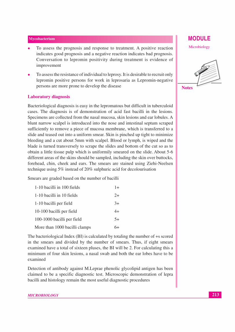
To assess the prognosis and response to treatment. A positive reactionindicates good prognosis and a negative reaction indicates bad prognosis.Conversation to lepromin positivity during treatment is evidence ofimprovement
To assess the resistance of individual to leprosy. It is desirable to recruit onlylepromin positive persons for work in leprosaria as Lepromin-negativepersons are more prone to develop the disease
Laboratory diagnosis
Bacteriological diagnosis is easy in the lepromatous but difficult in tuberculoidcases. The diagnosis is of demonstration of acid fast bacilli in the lesions.Specimens are collected from the nasal mucosa, skin lesions and ear lobules. Ablunt narrow scalpel is introduced into the nose and intestinal septum scrapedsufficiently to remove a piece of mucosa membrane, which is transferred to aslide and teased out into a uniform smear. Skin is pinched up tight to minimizebleeding and a cut about 5mm with scalpel. Blood or lymph, is wiped and theblade is turned transversely to scrape the slides and bottom of the cut so as toobtain a little tissue pulp which is uniformily smeared on the slide. About 5-6different areas of the skins should be sampled, including the skin over buttocks,forehead, chin, cheek and ears. The smears are stained using Ziehi-Neelsentechnique using 5% instead of 20% sulphuric acid for decolourisation
Smears are graded based on the number of bacilli
1-10 bacilli in 100 fields 1+
1-10 bacilli in 10 fields 2+
1-10 bacilli per field 3+
10-100 bacilli per field 4+
100-1000 bacilli per field 5+
More than 1000 bacilli clamps 6+
The bacteriological Index (BI) is calculated by totaling the number of +s scoredin the smears and divided by the number of smears. Thus, if eight smearsexamined have a total of sixteen pluses, the BI will be 2. For calculating this aminimum of four skin lesions, a nasal swab and both the ear lobes have to beexamined
Detection of antibody against M.Leprae phenolic glycolipid antigen has beenclaimed to be a specific diagnostic test. Microscopic demonstration of leprabacilli and histology remain the most useful diagnostic procedures

MICROBIOLOGY
MODULE Mycobacterium
Microbiology
214
Notes
INTEXT QUESTIONS 20.6
1. ............... is the skin test used for demonstration of delayed hypersensitivity
2. Bacteriological diagnosis is easy in ............... cases
3. ............... is used in demonstration of M.leprae bacilli
4. Specific diagnostic test in diagnosis of M.leprae is detection of ...............
WHAT YOU HAVE LEARNT
Mycobacteria tuberculosis is an obligatory aerobic, nonmotile, nonsporing,rod shaped bacterium which strains poorly by the Gram strain because itscell wall contains abundant amount of lipids. It retains Carbol Fuchsin dyeduring attempted decolourisation with acid and alcohol in Ziehl-Neelsenstaining technique. M.tuberculosis is acid and alcohol fast by ZN stainingmethod. It grows very slowly, taking several weeks to form a visible colonyon enriched culture media.
M. leprae is a obligate intercellular organism that gains access to skin andpheripheral nerve tissue. Least severe form is tuberculoid tuberculoid (TT)and the most severe form is lepromatous lepromatous
TERMINAL QUESTIONS
1. Describe the Laboratory Diagnosis of mycobacteria Tuberculosis andLeprae.
2. Explain Lepromin and Mantoux test.
ANSWERS TO INTEXT QUESTIONS
20.1
1. Fungus like bacteria
2. Acid fast bacilli
3. Mycolic acid
4. Obligate
215
Mycobacterium
MICROBIOLOGY
MODULEMicrobiology
Notes
20.2
1. M. bovine
2. Atypical mycobacteria
3. Restriction Fragment Length Polymorphism (RFLP)
4. Droplet
5. Ghon focus & hilar lymph nodes
20.3
1. Sputum microscopy
2. Ziehl-Neelson
3. Petroff’s method
4. Absolute Concentration method, Resistance ratio method & Propotionmethod
5. Mantoux test
6. Purified Protein Derivative (PPD)
20.4
1. Mycobacterium leprae
2. Hansen’s
3. Globi
4. Granulomatous
20.5
1. Multibacillary
2. Lepromatous type
3. Tuberculoid
4. Paucibacillary disease
20.6
1. Lepromin test
2. Lepromatous
3. Ziehl-Neelson technique
4. Antibodies