Embed Size (px)
Citation preview

© 2013 Pearson Education, Inc.
PowerPoint® Lecture Slides
prepared by
Meg Flemming
Austin Community College
C H A P T E R 13
The
Cardiovascular
System: Blood
Vessels and
Circulation

© 2013 Pearson Education, Inc.
Chapter 13 Learning Outcomes
• 13-1
• Distinguish among the types of blood vessels based on their
structure and function.
• 13-2
• Explain the mechanisms that regulate blood flow through blood
vessels, and discuss the mechanisms that regulate movement of
fluids between capillaries and interstitial spaces.
• 13-3
• Describe the control mechanisms that interact to regulate blood
flow and pressure in tissues, and explain how the activities of the
cardiac, vasomotor, and respiratory centers are coordinated to
control blood flow through tissues.

© 2013 Pearson Education, Inc.
Chapter 13 Learning Outcomes
• 13-4
• Explain the cardiovascular system's homeostatic response to
exercising and hemorrhaging.
• 13-5
• Describe the three general functional patterns in the pulmonary and
systemic circuits.
• 13-6
• Identify the major arteries and veins of the pulmonary circuit.
• 13-7
• Identify the major arteries and veins of the systemic circuit.

© 2013 Pearson Education, Inc.
Chapter 13 Learning Outcomes
• 13-8
• Identify the differences between fetal and adult circulation patterns,
and describe the changes in the patterns of blood flow that occur at
birth.
• 13-9
• Discuss the effects of aging on the cardiovascular system.
• 13-10
• Give examples of interactions between the cardiovascular system
and the other organ systems.
© 2013 Pearson Education, Inc.
Vascular Pathway of Blood Flow (13-1)
• Arteries leave the heart and branch into:
• Arterioles feed parts of organs and branch into:
• Capillaries, where chemical and gaseous
exchange occurs, and which drain into:
• Venules, the smallest vessels of the venous
system, which drain into:
• Veins, which return blood to the atria of the heart

© 2013 Pearson Education, Inc.
Three Layers of Vessel Walls (13-1)
1. Tunica intima (or tunica interna)
• Has endothelial lining and elastic connective tissue
2. Tunica media
• Has smooth muscle with collagen and elastic fibers
• Controls diameter of vessel
3. Tunica externa (or tunica adventitia)
• Sheath of connective tissue may anchor to other
tissues

© 2013 Pearson Education, Inc.
Figure 13-1 A Comparison of a Typical Artery and a Typical Vein.
Tunica externa
Tunica media
Tunica intima
Smooth Muscle
Endothelium
Elastic fiber
Lumen of vein
Lumen of artery
Artery and vein LM x 60
Tunica externa
Tunica media
Tunica intima
Smooth muscle
Endothelium
ARTERY VEIN

© 2013 Pearson Education, Inc.
Elastic Arteries (13-1)
• First type of arteries leaving the heart
• Examples are pulmonary trunk, aorta, and major
branches
• Have more elastic fibers than smooth muscle
• Absorb pressure changes readily
• Stretched during systole, relaxed during diastole
• Prevent very high pressure during systole
• Prevent very low pressure during diastole

© 2013 Pearson Education, Inc.
Muscular Arteries and Arterioles (13-1)
• Muscular arteries
• Examples are external carotid arteries
• Tunica media contains high proportion of smooth
muscle, little elastic fiber
• Arterioles
• Tunica media has only 1–2 layers of smooth muscle
• Ability to change diameter controls BP and flow

© 2013 Pearson Education, Inc.
Capillaries (13-1)
• Tunica interna only
• Endothelial cells with basement membrane
• Ideal for diffusion between plasma and IF
• Thin walls provide short diffusion distance
• Small diameter slows flow to increase diffusion rate
• Enormous number of capillaries provide huge surface
area for increased diffusion

© 2013 Pearson Education, Inc.
Tunica externa
Endothelium
Tunica intima
Tunica externa Tunica media
Endothelium
Tunica intima
Tunica externa
Endothelium
Endothelial cells
Basement membrane
Internal elastic layer
Endothelium
Tunica intima
Tunica media
Tunica externa
Tunica externa
Tunica media
Tunica media
Endothelium
Tunica intima
Smooth muscle cells (Tunica media)
Basement membrane
Endothelium
Large Vein
Medium-Sized Vein
Venule
Capillary
Elastic Artery
Muscular Artery
Arteriole
Figure 13-2 The Structure of the Various Types of Blood Vessels.

© 2013 Pearson Education, Inc.
Capillary Beds (13-1)
• An interconnected network of capillaries
• Entrance to bed is regulated by precapillary
sphincter, a band of smooth muscle
• Relaxation of sphincter allows for increased flow
• Constriction of sphincter decreases flow
• This occurs cyclically, referred to as vasomotion
• Control is local through autoregulation

© 2013 Pearson Education, Inc.
Figure 13-4 The Organization of a Capillary Bed.
Collateral
arteries
Arteriole
Smooth muscle cells
Section of a precapillary sphincter
Arteriovenous
anastomosis
Precapillary sphincters
Capillaries
Venule
Vein
Small venule
Capillary bed
Capillary
beds
Arteriole
Small artery
LM x 125
This micrograph shows a number of capillary beds.
KEY
Consistent
blood flow
Variable
blood flow
Features of a typical capillary bed. Solid arrows indicate consistent blood flow; dashed arrows indicate variable or pulsating blood flow.

© 2013 Pearson Education, Inc.
Alternate Routes for Blood Flow (13-1)
• Formed by anastomosis, a joining of blood
vessels
• Arteriovenous anastomosis bypasses capillary
bed, connecting arteriole to venule
• Arterial anastomosis occurs where arteries fuse
before branching into arterioles
• Ensures delivery of blood to key areas, brain, and heart
© 2013 Pearson Education, Inc.
Veins (13-1)
• Collect blood from tissues and organs and return it
to the heart
• Venules are the smallest and some lack tunica media
• Medium-sized veins
• Tunica media has several smooth muscle layers
• In limbs, contain valves
• Prevent backflow of blood toward the distal ends
• Increase venous return
© 2013 Pearson Education, Inc.
Veins (13-1)
• Large veins
• Thin tunica media and thick collagenous tunica externa
• Thinner walls than arteries because of low pressure

© 2013 Pearson Education, Inc.
Figure 13-5 The Function of Valves in the Venous System.
Valve opens above
contracting muscle
Valve closes below
contracting muscle
Valve
closed
Valve
closed
© 2013 Pearson Education, Inc.
Checkpoint (13-1)
1. List the five general classes of blood vessels.
2. A cross section of tissue shows several small,
thin-walled vessels with very little smooth muscle
tissue in the tunica media. Which type of vessels
are these?
3. What effect would relaxation of precapillary
sphincters have on blood flow through a tissue?
4. Why are valves found in veins, but not in
arteries?

© 2013 Pearson Education, Inc.
Maintaining Adequate Blood Flow (13-2)
• Flow maintains adequate perfusion of tissues
• Normally, blood flow equals cardiac output (CO)
• Increased CO leads to increased flow through
capillaries
• Decreased CO leads to reduced flow
• Capillary flow influenced by pressure and
resistance
• Increased pressure increases flow
• Increased resistance decreases flow

© 2013 Pearson Education, Inc.
Pressure (13-2)
• Liquids exert hydrostatic pressure in all directions
• A pressure gradient exists between high and low
pressures at different points
• Circulatory pressure, high in aorta vs. low in venae
cavae
• Arterial pressure is blood pressure
• Capillary pressure
• Venous pressure
• Flow is proportional to pressure gradients
© 2013 Pearson Education, Inc.
Resistance (13-2)
• Any force that opposes movement
• Circulatory pressure must be high enough to
overcome total peripheral resistance
• Highest pressure gradient exists in arterioles due to
high peripheral resistance
• Vascular resistance
• Viscosity
• Turbulence

© 2013 Pearson Education, Inc.
Vascular Resistance (13-2)
• Largest component of peripheral resistance
• Caused mostly by friction between blood and vessel
walls
• Amount of friction due to length and diameter of vessel
• Length doesn't normally change
• The longer the vessel, the higher the resistance
• Arteriolar diameter is primary source of vascular
resistance
• The smaller the diameter, the greater the resistance

© 2013 Pearson Education, Inc.
Viscosity (13-2)
• Due to interactions between molecules and
suspended materials in a liquid
• Low-viscosity fluids flow at low pressures
• High-viscosity fluids flow only under high pressures
• Blood viscosity is normally stable
• Changes in plasma proteins or hematocrit can alter
viscosity and, therefore, flow

© 2013 Pearson Education, Inc.
Turbulence (13-2)
• Eddies and swirls in fluid flow
• In smooth-walled vessels turbulence is low
• Slow flow near the walls, faster flow in center
• Injured or diseased vessels or heart valves show
increase in turbulence and decrease in flow
• Turbulent blood flow across valves produces the
sound of heart murmurs

© 2013 Pearson Education, Inc.
Interplay of Pressure and Resistance (13-2)
• Blood pressure is maintained by hormonal and
neural mechanisms
• Adjusting diameter of arterioles to specific organs:
• Regulates peripheral resistance
• Regulates flow
• Allows for matching flow and perfusion to tissue needs

© 2013 Pearson Education, Inc.
Blood Pressure (13-2)
• Arterial pressures fluctuate
• Systolic pressure (SP) is peak and occurs during
ventricular contraction
• Diastolic pressure (DP) is the minimum and occurs at
the end of ventricular relaxation
• Recorded as systolic over diastolic (e.g., 120/80
mm Hg)
• Pulse is alternating changes in pressures

© 2013 Pearson Education, Inc.
Pulse Pressure (13-2)
• The difference between systolic and diastolic
pressures
• Pulse pressure = SP – DP
• Diminishes over distance, eliminated at the capillary
level
• Arterial recoil or elastic rebound occurs during diastole
• Adds additional push or squeeze on blood
• Results in fluctuation of pressures

© 2013 Pearson Education, Inc.
Systolic
Pulse pressure
Blood
pressure
(mm Hg)
Diastolic
Ao
rta
Ela
sti
c
art
eri
es
Mu
sc
ula
r
art
eri
es
Art
eri
ole
s
Ca
pil
lari
es
Ve
nu
les
Me
diu
m-
siz
ed
ve
ins
La
rge
ve
ins
Ve
na
e c
ava
e
120
100
80
60
40
20
0
Figure 13-6 Pressures within the Systemic Circuit.

© 2013 Pearson Education, Inc.
Capillary Pressures (13-2)
• Drops from 35 to 18 mmHg along capillary length
• Capillaries are permeable to ions, nutrients,
wastes, gases, and water
• Capillary pressures cause filtration out of
bloodstream and into tissues
• Some materials are reabsorbed into blood
• Some materials are picked up by lymphatic vessels

© 2013 Pearson Education, Inc.
Four Functions of Capillary Exchange (13-2)
1. Maintains constant communication between
plasma and IF
2. Speeds distribution of nutrients, hormones, and
gases
3. Assists movement of insoluble molecules
4. Flushes bacterial toxins and other chemicals to
lymphatic tissues for immune response

© 2013 Pearson Education, Inc.
Mechanisms of Capillary Exchange (13-2)
• Diffusion of solutes down concentration gradients
• Filtration down fluid pressure gradients
• Osmosis down osmotic gradient
• Water is filtered out of capillary by fluid or
hydrostatic pressures
• Water is reabsorbed into capillary due to osmotic
pressure
© 2013 Pearson Education, Inc.
Capillary Exchange and Pressure Balances
(13-2)
• Capillary hydrostatic pressure (CHP) is high at
arteriolar end, low at venous end
• Tends to push water out of plasma into tissues at
arteriolar end, favoring filtration
• Blood osmotic pressure (BOP) is higher than in
interstitial fluid
• As CHP drops over length of capillary, BOP remains the
same, favoring reabsorption

© 2013 Pearson Education, Inc.
3.6 L/day flows
into lymphatic
vessels
Return to
circulation
24 L/day
No net fluid
movement 20.4 L/day
35
Hg
25 mm Hg
CHP > BCOP
Fluid forced
out of capillary
CHP = BCOP
No net
movement
of fluid
BCOP > CHP
Fluid moves
into capillary
mm Hg
25 mm
Hg
25 mm
Hg
25 mm
Hg
18 mm
Reabsorption
Venule
Filtration
Arteriole
KEY
CHP (Capillary
hydrostatic pressure)
BOP (Blood
osmotic pressure)
Figure 13-7 Forces Acting Across Capillary Walls.
© 2013 Pearson Education, Inc.
Venous Pressure (13-2)
• Gradient is low compared to arterial side
• Large veins provide low resistance ensuring
increase in flow despite low pressure
• When standing, blood flow must overcome gravity
• Muscular compression pushes on outside of veins
• Venous valves prevent backflow
• Respiratory pump due to thoracic pressures

© 2013 Pearson Education, Inc.
Checkpoint (13-2)
5. Identify the factors that contribute to total
peripheral resistance.
6. In a healthy individual, where is blood pressure
greater: at the aorta or at the inferior vena cava?
Explain.
7. While standing in the hot sun, Sally begins to feel
light-headed and then faints. Explain what
happened.

© 2013 Pearson Education, Inc.
Homeostatic Regulation of Perfusion (13-3)
• Affected by:
• Cardiac output, peripheral resistance, and blood
pressure
• Regulated to ensure blood flow changes occur at:
• Appropriate time, in right location, and without negative
effect on pressure and flow to vital organs
• Accomplished through:
• Autoregulation, neural and hormonal input

© 2013 Pearson Education, Inc.
Autoregulation of Perfusion (13-3)
• Immediate and localized changes in:
• Vasoconstrictors, factors that stimulate
constriction
• Vasodilators, factors that promote dilation
• Tissue temperature, low O2 or pH, high CO2 cause:
• Capillary sphincter dilation causing:
• Peripheral resistance decrease causing:
• Increase in flow through capillary beds

© 2013 Pearson Education, Inc.
Neural Control of Blood Pressure and Perfusion
(13-3)
• Triggered by changes in arterial pressure or blood
gas levels
• Cardiovascular (CV) centers in medulla oblongata
• Adjust cardiac output
• Vasomotor center in medulla oblongata
• Controls diameter of arterioles and peripheral resistance
• Controls venoconstriction

© 2013 Pearson Education, Inc.
Figure 13-9 Short-Term and Long-Term Cardiovascular Responses.
Autoregulation
HOMEOSTASIS DISTURBED
• Physical stress (trauma,
high temperature)
• Chemical changes
(decreased O2 or pH,
increased CO2 or
prostaglandins)
• Increased tissue activity. Inadequate
local blood
pressure and
blood flow
Local decrease
in resistance
and increase in
blood flow HOMEOSTASIS
RESTORED
HOMEOSTASIS
If autoregulation is ineffective
Normal
blood pressure
and volume
HOMEOSTASIS
RESTORED
Neural and Hormonal Mechanisms
Endocrine
response (see
Figure 13-12a)
Long-term increase
in blood volume
and blood pressure
Stimulation of
receptors sensitive
to changes in
systemic blood
pressure or
chemistry
Activation of
cardiovascular
centers in the
medulla
oblongata
Short-term elevation
of blood pressure
by sympathetic
stimulation of the
heart and peripheral
vasoconstriction
Neural
mechanisms
Endocrine mechanisms
Start

© 2013 Pearson Education, Inc.
Baroreceptor Reflexes (13-3)
• Receptors monitor degree of stretch
• Aortic sinuses
• Located in pockets in walls of ascending aorta
• Aortic reflex adjusts flow through systemic circuit
• Carotid sinuses
• Very sensitive to ensure adequate flow to, and perfusion
of, brain

© 2013 Pearson Education, Inc.
Figure 13-10 The Baroreceptor Reflexes of the Carotid and Aortic Sinuses.
Responses to Increased
Baroreceptor Stimulation
Baroreceptors
stimulated
Cardioinhibitory
centers stimulated
Cardioacceleratory
centers inhibited
Vasomotor center
inhibited
Decreased
cardiac
output
Vasodilation
occurs
HOMEOSTASIS DISTURBED
Rising blood pressure
HOMEOSTASIS RESTORED
Blood pressure declines
Start
Start
HOMEOSTASIS
Normal range
of blood pressure
HOMEOSTASIS DISTURBED
HOMEOSTASIS RESTORED
Falling blood pressure
Blood pressure rises
Baroreceptors
inhibited
Vasoconstricti-
on occurs
Responses to Decreased
Baroreceptor Stimulation
Vasomotor center
stimulated
Cardioacceleratory
centers stimulated
Cardioinhibitory
centers inhibited
Increased
cardiac
output

© 2013 Pearson Education, Inc.
Chemoreceptor Reflexes (13-3)
• Receptors
• Sensitive to changes in carbon dioxide, oxygen, and pH
in blood and CSF
• Located in carotid and aortic bodies, medulla
oblongata
• Decrease in pH or plasma O2, increase in plasma CO2
stimulate increase in heart rate and arteriolar
constriction
• Result is increase in BP

© 2013 Pearson Education, Inc.
Hormonal Control of Cardiovascular
Performance (13-3)
• Short-term
• E and NE trigger rapid increase of cardiac output and
vasoconstriction
• Long-term
• Antidiuretic hormone (ADH), angiotensin II, EPO
• Raise BP when too low
• Atrial natriuretic peptide (ANP)
• Lowers BP when too high

© 2013 Pearson Education, Inc.
Antidiuretic Hormone and Cardiovascular
Regulation (13-3)
• Released from posterior pituitary in response to:
• Decrease in blood volume
• Increase in blood osmolarity
• Presence of angiotensin II
• Results in:
• Vasoconstriction
• Conserving water by kidneys, increasing blood volume
© 2013 Pearson Education, Inc.
Angiotensin II and Cardiovascular Regulation
(13-3)
• When BP decreases, kidney secretes renin
• Cascade of reactions forms angiotensin II
• Angiotensin II
• Stimulates CO, arteriolar constriction
• Immediately increases BP
• Stimulates release of ADH and aldosterone
• Stimulates thirst center

© 2013 Pearson Education, Inc.
Erythropoietin and Cardiovascular Regulation
(13-3)
• Released by kidney when:
• BP drops
• Plasma oxygen drops
• Stimulates:
• RBC production
• Increases blood volume

© 2013 Pearson Education, Inc.
Atrial Natriuretic Peptide and Cardiovascular
Regulation (13-3)
• Released by atrial walls when BP increases
• From stretch of atrial wall due to more venous return
• Effects
• Increases sodium (and therefore water) loss by kidneys
• Reduces thirst
• Blocks release of ADH, aldosterone, E, NE
• Stimulates arteriolar dilation

© 2013 Pearson Education, Inc.
Decreasing blood
pressure and
volume
Start
HOMEOSTASIS
DISTURBED
Blood pressure and volume fall
Short-term
Long-term
Sympathetic activation
and release of adrenal
hormones E and NE
Endocrine Response
of Kidneys
Renin release leads
to angiotensin II
activation
Erythropoietin (EPO)
is released
Increased cardiac output and peripheral vasoconstriction
HOMEOSTASIS
Normal blood pressure and
volume
Angiotensin II Effects
Antidiuretic hormone
released
Aldosterone secreted
Thirst stimulated
Increased red blood
cell formation
HOMEOSTASIS
RESTORED
Blood pressure and volume rise
Increased blood pressure
Increased blood volume
Factors that compen-
sate for decreased
blood pressure and
volume
Combined Short-Term
and Long-Term Effects
Figure 13-12a The Hormonal Regulation of Blood Pressure and Blood Volume.

© 2013 Pearson Education, Inc.
Increasing blood
pressure and
volume
Atrial natriuretic
peptide (ANP)
released by
the heart
HOMEOSTASIS
DISTURBED
Rising blood
pressure and
volume
Factors that compensate for increased
blood pressure and volume
HOMEOSTASIS
Normal
blood pressure and volume
Responses to ANP
Increased Na+ loss in urine
Increased water loss in urine
Reduced thirst
Inhibition of ADH, aldosterone,
epinephrine, and
norepinephrine release
Peripheral vasodilation
Combined Effects
Reduced blood
volume
HOMEOSTASIS
RESTORED
Declining blood
pressure and
volume
Figure 13-12b The Hormonal Regulation of Blood Pressure and Blood Volume.

© 2013 Pearson Education, Inc.
Checkpoint (13-3)
8. Describe the actions of vasodilators and
vasoconstrictors.
9. How would slightly compressing the common
carotid artery affect your heart rate?
10.What effect would vasoconstriction of the renal
artery have on systemic blood pressure and
blood volume?

© 2013 Pearson Education, Inc.
Four Cardiovascular Responses to the Stress of
Exercise (13-4)
1. Extensive vasodilation
• Increased O consumption
• Causes lower peripheral resistance
• Resulting in increased flow
2. Increased venous return
• Due to skeletal muscle and respiratory "pumps"

© 2013 Pearson Education, Inc.
Four Cardiovascular Responses to the Stress of
Exercise (13-4)
3. Increased cardiac output
• Frank-Starling principle due to increased venous return
• Arterial pressures are maintained
• Increased CO balances out decrease in peripheral resistance
4. Shunting of blood flow away from nonessential
organs
• Ensures adequate perfusion of heart and skeletal
muscles

© 2013 Pearson Education, Inc.
Short-Term Cardiovascular Response to
Hemorrhage (13-4)
• Loss of blood causes decrease in BP
• Carotid and aortic reflexes increase cardiac output and
peripheral resistance
• Venoconstriction accesses venous reserve
• Sympathetic activation triggers arteriolar constriction
• All mechanisms function to elevate BP

© 2013 Pearson Education, Inc.
Long-Term Cardiovascular Response to
Hemorrhage (13-4)
• May take several days to restore blood volume to
normal
• Fluids are accessed from interstitial space
• ADH and aldosterone promote fluid retention
• Thirst increases
• EPO triggers RBC production
• All mechanisms lead to increase in volume and BP

© 2013 Pearson Education, Inc.
Checkpoint (13-4)
11. Why does blood pressure increase during
exercise?
12. Name the immediate and long-term problems
related to the cardiovascular response to
hemorrhaging.
13. Explain the role of aldosterone and ADH in
long-term restoration of blood volume.

© 2013 Pearson Education, Inc.
Three Functional Patterns of the Cardiovascular
System (13-5)
1. Distribution of arteries and veins nearly identical
except near heart
2. Single vessel may undergo name changes as it
crosses anatomical boundaries
3. Anastomoses of arteries and veins reduce threat
of temporary blockage of vessel to organ

© 2013 Pearson Education, Inc.
Brain
Upper limbs
Pulmonary circuit
(arteries)
Pulmonary circuit (veins)
Lungs
RA LA
Systemic circuit
(arteries)
Left ventricle Right
ventricle Systemic
circuit (veins)
Kidneys
Spleen
Liver Digestive
organs
Gonads
Lower limbs
Figure 13-13 An Overview of the Pattern of Circulation.

© 2013 Pearson Education, Inc.
Checkpoint (13-5)
14. Identify the two circuits of the cardiovascular
system.
15. Identify the three general functional patterns of
the body's blood vessels.

© 2013 Pearson Education, Inc.
The Pulmonary Circuit (13-6)
• Blood exits right ventricle through pulmonary
trunk
• Branches into left and right pulmonary arteries
• Enter lungs, arterial branching nearly parallels
branching of respiratory airways
• Smallest arteriole feeds capillary surrounding alveolus
• Oxygenated blood returns to left atrium through
left and right, superior and inferior pulmonary
veins

© 2013 Pearson Education, Inc.
Ascending aorta
Superior vena cava
Right lung
Right pulmonary
arteries
Right pulmonary
veins
Aortic arch
Pulmonary trunk
Left lung
Left pulmonary
arteries
Left pulmonary
veins
Alveolus
Alveolar
capillary
O2
CO2 Inferior vena cava
Descending aorta
Figure 13-14 The Pulmonary Circuit.
© 2013 Pearson Education, Inc.
Checkpoint (13-6)
16. Name the blood vessels that enter and exit the
lungs, and indicate the relative oxygen content of
the blood in each.
17. Trace the path of a drop of blood through the
lungs, beginning at the right ventricle and ending
at the left atrium.

© 2013 Pearson Education, Inc.
The Systemic Circuit (13-7)
• Supplies oxygenated blood to all non-pulmonary
tissues
• Oxygenated blood leaves left ventricle through
aorta
• Returns deoxygenated blood to right atrium
through superior and inferior venae cavae, and
coronary sinus
• Contains about 84 percent of total blood volume

© 2013 Pearson Education, Inc.
Figure 13-15 An Overview of the Major Systemic Arteries.
Vertebral
Right subclavian
Brachiocephalic trunk
Aortic arch
Ascending aorta
Celiac trunk
Brachial
Radial
Ulnar
External iliac
Palmar arches
Popliteal
Posterior tibial
Anterior tibial Fibular
Plantar arch
Right common carotid
Left common carotid
Left subclavian Axillary
Descending aorta Diaphragm Renal Superior mesenteric
Gonadal Inferior mesenteric Common iliac Internal iliac
Deep femoral
Femoral
Dorsalis pedis

© 2013 Pearson Education, Inc.
The Aorta (13-7)
• Ascending aorta is first systemic vessel
• Begins at aortic semilunar valve
• Left and right coronary arteries branch off near
base of aorta
• Aortic arch curves across top of heart
• Descending aorta drops down through
mediastinum

© 2013 Pearson Education, Inc.
Three Elastic Arteries of the Aortic Arch (13-7)
1. Brachiocephalic trunk
• Branches to form right common carotid artery and
right subclavian artery
2. Left common carotid
3. Left subclavian
• This is an example of non-mirror-image
arrangement
• From here on, arteries are the same on both sides of
the body
• Designation of right and left not necessary

© 2013 Pearson Education, Inc.
Subclavian Arteries (13-7)
• Supply arms, chest wall, shoulders, back, and CNS
• Internal thoracic artery
• Vertebral artery
• Thyrocervical trunk
• Supply pericardium, chest, neck, shoulder, CNS
• Becomes axillary artery
• Brachial artery
• Radial and ulnar arteries
• Form anastomoses, the superficial and deep palmar arches.
• Digital artery
• Supply upper limbs

© 2013 Pearson Education, Inc.
Figure 13-16 Arteries of the Chest and Upper Limb.
Thyrocervical trunk
Right subclavian
Axillary
Deep brachial
Intercostal arteries
Brachial
Radial
Ulnar
Palmar arch
Digital arteries
Right common carotid
Vertebral Left common carotid
Brachiocephalic trunk Left subclavian Aortic arch Ascending aorta Descending aorta
Heart
Internal thoracic
Descending aorta

© 2013 Pearson Education, Inc.
The Carotid Arteries (13-7)
• Common carotids ascend up into the neck and
divide
• External carotid artery
• Supplies pharynx, esophagus, larynx, and face
• Internal carotid artery
• Enters skull, supplies brain and eyes

© 2013 Pearson Education, Inc.
Blood Supply to the Brain (13-7)
• Two pathways
• Vertebral arteries enter skull and fuse to form one
basilar artery
• Posterior cerebral artery
• Posterior communicating artery
• Cerebral arterial circle
• Ring-shaped anastomosis encircling the infundibulum of the
pituitary

© 2013 Pearson Education, Inc.
Anterior cerebral
Middle cerebral
Cerebral arterial circle
Posterior cerebral
Basilar
Internal carotid
Carotid sinus
Vertebral
Thyrocervical trunk
Subclavian Internal thoracic
Second rib
Common carotid
Brachiocephalic trunk
Branches of the
External Carotid
Superficial temporal
Maxillary
Occipital
Facial External carotid
The general circulation pattern of arteries supplying the neck and superficial structures of the head
Figure 13-18a Arteries of the Neck, Head, and Brain.

© 2013 Pearson Education, Inc.
Cerebral Arterial Circle
Anterior communicating
Anterior cerebral
Posterior communicating
Basilar
Vertebral
Anterior cerebral
Internal carotid (cut)
Middle cerebral
Posterior cerebral
The arterial supply to the brain
Posterior cerebral
Figure 13-18b Arteries of the Neck, Head, and Brain.

© 2013 Pearson Education, Inc.
Major Arteries of the Trunk (13-7)
• Descending aorta
• Thoracic aorta within thoracic cavity
• Abdominal aorta after passing through diaphragm
• Phrenic artery
• First branch off abdominal aorta
• Supplies diaphragm

© 2013 Pearson Education, Inc.
Unpaired Arteries of Digestive Organs (13-7)
• Supply blood to all digestive organs
• Celiac trunk
• Left gastric artery
• Splenic artery
• Common hepatic artery
• Superior mesenteric artery
• Inferior mesenteric artery

© 2013 Pearson Education, Inc.
Paired Major Arteries of the Trunk (13-7)
• Gonadal arteries
• Testicular in male, ovarian in female
• Adrenal arteries
• Supply adrenal glands
• Renal arteries
• Supply kidneys
• Lumbar arteries
• Supply spinal cord and abdominal wall

© 2013 Pearson Education, Inc.
Iliac Arteries (13-7)
• Abdominal aorta branches to the:
• Common iliac artery
• Branches to:
• Internal iliac artery
• Supplies pelvis
• External iliac artery
• Supplies lower limbs

© 2013 Pearson Education, Inc.
Figure 13-19b Major Arteries of the Trunk.
THORACIC
AORTA
Bronchial arteries
Pericardial arteries
Esophageal arteries
Mediastinal arteries
Un
pair
ed
(m
ult
iple
)
Conducting passages of respiratory tract
Pericardium
Esophagus
Mediastinal structures
Intercostal arteries (paired, segmental)
Vertebrae, spinal cord, back muscles, body wall, and skin
Superior phrenic arteries
Diaphragm
Left gastric
Stomach, adjacent portion of esophagus
Splenic Spleen, stomach, pancreas
Common hepatic
Liver, stomach, gallbladder, duodenum, pancreas
Superior mesenteric
Pancreas, small intestine, appendix, and first two-thirds of large intestine
Inferior mesenteric
Last third of large intestine (left third of transverse colon, descending colon, sigmoid colon, and rectum)
Inferior phrenic arteries
Diaphragm, inferior portion of esophagus
Adrenal arteries
Adrenal glands
Celiac trunk
ABDOMINAL
AORTA
Renal arteries
Kidneys
Gonadal arteries
Gonads (testes or ovaries)
Lumbar arteries (paired, segmental)
Vertebrae, spinal cord, and abdominal wall
Right common iliac
Pelvis and right lower limb
Left common iliac
Pelvis and left lower limb
Right external iliac
Right internal iliac
Pelvic muscles, skin, viscera of pelvis (urinary and reproductive organs), perineum, gluteal region, and medial thigh
Left internal iliac
Left external iliac
Superior gluteal
Hip muscles, hip joint
Obturator
Ilium, hip and thigh muscles, hip joint and femoral head
Internal pudendal
Lateral rotators of hip; rectum, anus, perineal muscles, external genitalia
Lateral sacral
Skin and muscles of sacrum
Un
pair
ed
(sin
gle
)
Pair
ed
A flowchart showing major arteries of the trunk

© 2013 Pearson Education, Inc.
THORACIC
AORTA
Bronchial arteries
Pericardial arteries
Esophageal arteries
Mediastinal arteries
Un
pa
ire
d (
mu
ltip
le)
Conducting passages of respiratory tract
Pericardium
Esophagus
Mediastinal structures
Intercostal arteries (paired, segmental)
Vertebrae, spinal cord, back muscles, body wall, and skin
Superior phrenic arteries
Diaphragm
A flowchart showing major arteries of the trunk
Pa
ire
d
Figure 13-19b Major Arteries of the Trunk. (1 of 3)

© 2013 Pearson Education, Inc.
A flowchart showing major arteries of the
trunk
Left gastric
Stomach, adjacent portion of esophagus
Splenic Spleen, stomach, pancreas
Common hepatic
Liver, stomach, gallbladder, duodenum, pancreas
Superior mesenteric
Pancreas, small intestine, appendix, and first two-thirds of large intestine
Inferior mesenteric
Last third of large intestine (left third of transverse colon, descending colon, sigmoid colon, and rectum)
Inferior phrenic arteries
Diaphragm, inferior portion of esophagus
Adrenal arteries
Adrenal glands
Celiac trunk
ABDOMINAL
AORTA
Renal arteries
Kidneys
Gonadal arteries
Gonads (testes or ovaries)
Un
pair
ed
(sin
gle
)
Pair
ed
Lumbar arteries (paired, segmental)
Vertebrae, spinal cord, and abdominal wall
Figure 13-19b Major Arteries of the Trunk. (2 of 3)

© 2013 Pearson Education, Inc.
Right common iliac
Pelvis and right lower limb
Left common iliac
Pelvis and left lower limb
Right external iliac
Right internal iliac
Pelvic muscles, skin, viscera of pelvis (urinary and reproductive organs), perineum, gluteal region, and medial thigh
Left internal iliac
Left external iliac
Superior gluteal
Hip muscles, hip joint
Obturator
Ilium, hip
and thigh
muscles, hip
joint and
femoral head
Internal pudendal
Lateral rotators of hip; rectum, anus, perineal muscles, external genitalia
Lateral sacral
Skin and muscles of sacrum
A flowchart showing major arteries of the trunk
Figure 13-19b Major Arteries of the Trunk. (3 of 3)

© 2013 Pearson Education, Inc.
Lower Limb Arteries (13-7)
• External iliac artery forms:
• Deep femoral artery
• Femoral artery
• Popliteal artery
• Anterior tibial, posterior tibial, and fibular arteries
• Two anastomoses connect anterior tibial
• Dorsalis pedis arteries and two branches of posterior tibial
• Dorsal arch on top of foot
• Plantar arch on bottom of foot

© 2013 Pearson Education, Inc.
Figure 13-20 An Overview of the Major Systemic Veins.
Vertebral
External jugular
Subclavian Axillary
Cephalic
Basilic Brachial
Hepatic veins
Median cubital
Radial
Median antebrachial Ulnar
Palmar venous arches
Digital veins
Great saphenous
Popliteal
Small saphenous
Fibular
Plantar venous arch
Dorsal venous arch
Internal jugular
Brachiocephalic
Superior vena cava
Intercostal veins
Inferior vena cava Renal Gonadal Lumbar veins
Left and right common iliac
External iliac
Internal iliac
Femoral
Deep femoral
Posterior tibial Anterior tibial
KEY
Superficial veins
Deep veins

© 2013 Pearson Education, Inc.
Systemic Veins (13-7)
• Venous network returns blood to heart
• Arteries and veins run parallel, often similar names
• Major veins in neck and limbs different than
arteries
• Arteries are located deep
• Veins usually a set of two
• One deep and the other superficial
• Aids in body temperature control

© 2013 Pearson Education, Inc.
The Superior Vena Cava (13-7)
• SVC
• Receives blood from:
• Head and neck
• Upper limbs, shoulders, and chest

© 2013 Pearson Education, Inc.
Venous Return from Head and Neck (13-7)
• Small veins in brain drain into dural sinuses
• Largest is superior sagittal sinus
• Internal jugular veins
• External jugular veins
• Collect blood from superficial head and neck
• Vertebral veins
• Collect blood from cervical spinal cord and posterior
skull

© 2013 Pearson Education, Inc.
Figure 13-21 Major Veins of the Head and Neck.
Temporal
Maxillary
Facial
Internal jugular
Right brachiocephalic
Left brachiocephalic
Superior vena cava Internal thoracic
Right subclavian
Vertebral External jugular
Dural sinuses
Great cerebral
Superior sagittal sinus

© 2013 Pearson Education, Inc.
Venous Return from the Upper Limbs and
Chest (13-7)
• Digital vein drains into venous network in palms
• Cephalic vein
• Basilic vein
• Median cubital
• Connects cephalic and basilic veins
• Site of venous blood sample tap

© 2013 Pearson Education, Inc.
Venous Return from the Upper Limbs and
Chest (13-7)
• Deeper forearm veins are radial veins and ulnar
veins
• Brachial vein joins basilic vein to form:
• Axillary vein
• Subclavian vein
• Meet and merge with internal and external jugular veins
• Creates large brachiocephalic vein SVC
• Azygos vein drains chest wall SVC

© 2013 Pearson Education, Inc.
Inferior Vena Cava (13-7)
• IVC
• Collects blood from organs below diaphragm

© 2013 Pearson Education, Inc.
Venous Return from the Lower Limbs (13-7)
• Plantar veins on the sole of the foot
• Plantar venous arch drains into:
• Anterior tibial vein
• Posterior tibial vein
• Fibular vein
• Dorsal venous arch drains into:
• Great saphenous vein and small saphenous vein

© 2013 Pearson Education, Inc.
Venous Return from the Lower Limbs (13-7)
• Behind knee small saphenous, tibial, and fibular veins
connect
• Popliteal vein
• Femoral vein
• Great saphenous and deep femoral vein join femoral vein
• External iliac vein
• Joins internal iliac vein to become common iliac vein
• IVC

© 2013 Pearson Education, Inc.
Veins of the Abdominopelvic Organs (13-7)
• As IVC ascends toward heart it collects blood
from:
• Lumbar vein
• Gonadal vein
• Renal and adrenal veins
• Phrenic vein
• Hepatic vein

© 2013 Pearson Education, Inc.
Figure 13-22 The Venous Drainage of the Abdomen and Chest.
SUPERIOR VENA CAVA
Mediastinal veins
Esophageal veins
Azygos
Internal thoracic
Hepatic veins
Renal veins
Gonadal veins
Lumbar veins
Common iliac
External iliac
Internal iliac
Superficial veins
Deep veins
KEY
Digital veins
Palmar venous arches
Ulnar
Basilic Median antebrachial
Radial
Cephalic
Median cubital
Adrenal veins
Phrenic veins
Basilic
INFERIOR VENA CAVA
Intercostals Brachial
Hemiazygos
Cephalic Axillary
Brachiocephalic Highest intercostal Subclavian
External jugular
Internal jugular Vertebral

© 2013 Pearson Education, Inc.
Figure 13-23a A Flowchart of the Tributaries of the Superior and Inferior Venae Cavae.
Right
vertebral
Right
external
jugular
Right
internal
jugular
Collects blood
from cranium, spinal
cord, vertebrae
Left
vertebral
Left
internal
jugular
Collects blood
from cranium, face,
and neck
Left
external
jugular
Collects blood from
neck, face, salivary
glands, scalp
Right
subclavian
Right
axillary
Veins of the
right upper
limb
Right
intercostal
veins
Collect blood
from vertebrae
and body wall
Right
brachiocephalic
Left and
right internal
thoracic
veins
Collect blood
from structures
of anterior
thoracic wall
Left
brachiocephalic
Left
subclavian
Mediastinal
veins
Collect blood
from the
mediastinum
KEY
Superficial veins
Deep veins
Azygos
SUPERIOR
VENA CAVA
RIGHT
ATRIUM
Collect blood
from the
esophagus
Esophageal
veins
Hemiazygos
Left
intercostal
veins
Collect blood
from vertebrae
and body wall
Through
highest
intercostal vein
Left
axillary
Left
brachial
Collects blood from
forearm, wrist, and
hand
Collects blood from lateral surface of upper limb
Collects blood from medial surface of upper limb
Left cephalic Left basilic
Interconnected by median cubital vein and median
antebrachial network
Left
radial
Radial
side of
forearm
Left
ulnar
Ulnar
side of
forearm
Venous network
of wrist and hand
Tributaries of the superior vena cava

© 2013 Pearson Education, Inc.
RIGHT
ATRIUM
INFERIOR
VENA CAVA
Hepatic veins
Gonadal veins
Lumbar veins
Collect blood from the liver
Collect blood from the gonads (testes or ovaries)
Collect blood from the spinal cord and body wall
Phrenic
veins
Adrenal
veins
Renal
veins
Collect blood from the diaphragm
Collect blood from the adrenal glands
Collect blood from the kidneys
Right
common
iliac
Left
common
iliac
Right external
iliac
Blood from
veins in right
lower limb
Collect blood from the pelvic muscles, skin, urinary and reproductive organs of pelvic cavity
Right internal iliac
Left internal iliac
Left external
iliac
Blood from
veins in left
lower limb
Superior
gluteal
veins
Internal
pudendal
veins
Obturator
veins
Lateral
sacral
veins
Tributaries of the inferior vena cava
Figure 13-23b A Flowchart of the Tributaries of the Superior and Inferior Venae Cavae.
© 2013 Pearson Education, Inc.
Hepatic Portal System (13-7)
• Portal system is two capillary beds in series
connected by portal vessel
• Blood going through capillaries of digestive organs
absorbs nutrients, some wastes, some toxins
• Blood is processed by liver before entering
general circulation
© 2013 Pearson Education, Inc.
Hepatic Portal System (13-7)
• Capillaries from:
• Lower large intestine inferior mesenteric vein
• Spleen, stomach, pancreas splenic vein
• Stomach, small and large intestines superior
mesenteric vein
• All three hepatic portal vein
• Blood from gastric vein and cystic vein added
• Blood enters liver capillaries hepatic vein
IVC

© 2013 Pearson Education, Inc.
Figure 13-24 The Hepatic Portal System.
Inferior vena cava
Hepatic veins
Hepatic portal Cystic
Superior mesenteric
Colic veins
Ascending colon
Intestinal veins
Superior rectal veins
Small intestine
Sigmoid veins
Descending colon
Inferior mesenteric
Left colic
Splenic
Gastroepiploic veins
Spleen
Gastric veins
Aorta
Esophagus
Liver Stomach
Pancreas

© 2013 Pearson Education, Inc.
Checkpoint (13-7)
18. A blockage of which branch of the aortic arch would
interfere with blood flow to the left arm?
19. Why would compression of the common carotid
arteries cause a person to lose consciousness?
20. Grace is in an automobile accident, and her celiac
trunk is ruptured. Which organs will be affected
most directly by this injury?
21. Describe the general distribution of major arteries
and veins in the neck and limbs. What functional
advantage does this distribution provide?

© 2013 Pearson Education, Inc.
Fetal Circulation (13-8)
• Biggest difference is sources of respiratory and
nutritional support
• All nutrients and blood gases supplied from
mother through diffusion across placenta
• Placenta is unique part of uterine wall
• Maternal and fetal circulatory systems in close contact

© 2013 Pearson Education, Inc.
Placental Blood Supply (13-8)
• Low O2 fetal blood flows through umbilical
arteries
• At placenta:
• CO2 and wastes cross to mother
• O2 diffuses into fetal blood
• Returns to fetal circulation through umbilical vein
• Some blood goes to liver
• Rest goes to IVC through ductus venosus
© 2013 Pearson Education, Inc.
Fetal Circulation in the Heart and Great Vessels
(13-8)
• Foramen ovale
• An interatrial opening
• Flap that acts as one-way valve from right to left atrium
• Allows blood to bypass pulmonary circuit
• Ductus arteriosus
• Short vessel that takes most of blood from right ventricle
directly to aortic arch of systemic circuit
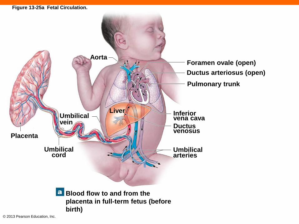
© 2013 Pearson Education, Inc.
Foramen ovale (open)
Ductus arteriosus (open)
Pulmonary trunk
Aorta
Placenta
Umbilical vein
Umbilical cord
Liver Inferior vena cava
Ductus venosus
Umbilical arteries
Blood flow to and from the
placenta in full-term fetus (before
birth)
Figure 13-25a Fetal Circulation.

© 2013 Pearson Education, Inc.
Blood flow through the heart of a
newborn baby after delivery
Inferior vena cava
Ductus arteriosus (closed)
Pulmonary trunk
Left atrium
Foramen ovale (closed)
Right atrium
Left ventricle
Right ventricle
Figure 13-25b Fetal Circulation.

© 2013 Pearson Education, Inc.
Circulatory Changes at Birth (13-8)
• Infant takes first breath
• Pulmonary vessels expand
• Ductus arteriosus contracts
• Blood flows into pulmonary trunk
• Remnants convert to ligamentum arteriosum
• Flap across foramen ovale closes
• Residual indentation is the fossa ovalis

© 2013 Pearson Education, Inc.
Checkpoint (13-8)
22. Name the umbilical vessels that constitute the
placental blood supply.
23. A blood sample taken from the umbilical cord
contains high levels of oxygen and nutrients, and
low levels of carbon dioxide and waste products. Is
this sample from an umbilical artery or from the
umbilical vein? Explain.
24. Name the structures that are vital to fetal circulation
but cease to function at birth. What becomes of
each of these structures?

© 2013 Pearson Education, Inc.
Effects of Aging on Blood (13-9)
• Lower hematocrit
• Formation of a thrombus, or stationary blood clot
• Can detach becoming an embolism
• Pooling of blood in veins of leg
• Due to ineffective venous valves

© 2013 Pearson Education, Inc.
Effects of Aging on the Heart (13-9)
• Reduction in maximum cardiac output
• Changes in nodal and conducting cells
• Reduction of elasticity of cardiac skeleton
• Progressive atherosclerosis
• Serious if found in coronary circulation
• Replacement of damaged cardiac muscle with
scar tissue

© 2013 Pearson Education, Inc.
Effects of Aging on the Vessels (13-9)
• Arteriosclerosis or thickening and toughening of
wall
• Inelastic walls of arteries less tolerant of pressure
increase
• Can lead to local dilation, an aneurysm
• Calcium salts deposited on walls
• Can lead to stroke or myocardial infarction
• Thrombi can form at atherosclerotic plaques

© 2013 Pearson Education, Inc.
Checkpoint (13-9)
25. Identify components of the cardiovascular
system that are affected by age.
26. Define thrombus.
27. Define aneurysm.

© 2013 Pearson Education, Inc.
Cardiovascular System Linked to All Other
Systems (13-10)
• Cardiovascular system supplies all others with:
• Oxygen
• Hormones
• Nutrients
• White blood cells
• Removes:
• Carbon dioxide and metabolic wastes

© 2013 Pearson Education, Inc.
Figure 13-26
SYSTEM INTEGRATOR
Stimulation of mast cells produces
localized changes in blood flow and
capillary permeability
Provides calcium needed for normal cardiac muscle
contraction; protects blood cells developing in red
bone marrow
Skeletal muscle contractions assist in moving blood
through veins; protects superficial blood vessels,
especially in neck and limbs
Controls patterns of circulation in peripheral
tissues; modifies heart rate and regulates blood
pressure; releases ADH
Erythropoietin (EPO) regulates production of RBCs; several hormones elevate blood pressure; epinephrine stimulates cardiac muscle, elevating heart rate and contractile force
The section on vessel distribution demonstrated the extent of the anatomical connections between the cardiovascular system and other organ systems. This figure summa- rizes some of the physiological relationships involved. The most extensive communication occurs between the cardiovascular and lymphatic systems. Not only are the two systems physically interconnected, but cells of the lymphatic system also move from one part of the body to another within the vessels of the cardiovascular system. We examine the lymphatic system in detail, including its role in the immune response, in the next chapter.
The CARDIOVASCULAR
System
In
te
gu
-
me
nta
ry
Sk
ele
ta
l
Mu
sc
ula
r
Ne
rvo
us
En
do
cr-
in
e
Cardiovascular System Body System Cardiovascular System Body System
Delivers immune system cells to injury sites; clotting response seals breaks in skin surface; carries away toxins from sites of infection; provides heat
Transports calcium and phosphate for bone deposition; delivers EPO to red bone marrow, parathyroid hormone, and calcitonin to osteoblasts and osteoclasts
Delivers oxygen and nutrients, removes carbon
dioxide, lactic acid, and heat during skeletal
muscle activity
Endothelial cells maintain blood–brain barrier;
helps generate CSF
Distributes hormones throughout the body; heart
secretes ANP
In
te
gu
-
Me
nta
ry
(P
ag
e 1
38)
Sk
ele
ta
l
(Pag
e 1
88)
Mu
sc
ula
r
(Pag
e 2
41)
Ne
rvo
us
(P
ag
e 3
02)
En
do
cr-
In
e
(Pag
e 3
76)
Re
sp
ira
-
to
ry
(Pag
e 5
32)
Lym
ph
-
atic
(Pa
ge 5
00)
Dig
es-
tive
(Pag
e 5
72)
Urin
ary
(Pag
e 6
37)
Re
pro
du
-
ctive
(Pag
e 6
71)

© 2013 Pearson Education, Inc.
Checkpoint (13-10)
28. Describe what the cardiovascular system
provides for all other body systems.
29. What is the relationship between the skeletal
system and the cardiovascular system?


