Embed Size (px)
Citation preview
laserinternational magazine of laser dentistry4
2013
i s sn 2193-4665 Vol. 5 • Issue 4/2013
| researchEr,Cr:YSGG laser and radial firing tips in highly compromised endodontic scenarios
| case reportThe efficacy of combined low intensity laser therapy and medication on xerostomia
| industry reportLasers in aesthetic dentistry

©BIOLASE, Inc. All rights reserved. For use by licensed professionals only. BIOLASE, WaterLase, iPlus, and Deep Pocket Therapy with New Attachment are trademarks of BIOLASE, registered in the U.S. and other countries.
FOLLOW US!
| www.biolase.com | +1.949.361.1200
The WaterLase iPlus system is simply amazing. It is our
most advanced, most powerful and simplest WaterLase
yet. But don’t take our word for it. See for yourself at AMAZEDBYWATERLASE.COM!
BE AMAZED BYWATERLASE
®
CONTACT US TO LEARN MORE ABOUT WATERLASE IPLUS!OR VISIT AMAZEDBYWATERLASE.COM TO VIEW INSTANT REACTIONS FROM DOCTORS AND PATIENTS!
GO TO BIOLASE.COM TO FINDYOUR LOCAL DISTRIBUTOR.
BREAKS THE SPEED BARRIER- Up to 100/pulses per second- Patented tech delivers 600 mJ/pulse- As fast as a high-speed drill
ILASE 940nm DIODE LASER DOCKING STATION- Adds dual wavelength convenience - First totally wireless diode laser- Battery operated with fi nger operation
INTUITIVE USER INTERFACE- 52 illustrated procedure pre-sets- Touch-screen controls- Greatly simplifi es the learning curve
BIOLOGICALLY FRIENDLY DENTISTRY- No micro-fractures or thermal damage- More precise, minimally invasive- Relies on water and light to cut
Biolase_A4_laser213.pdf 1Biolase_A4_laser213.pdf 1 05.04.13 17:5605.04.13 17:56

editorial I
I 03laser4_2013
Dear colleagues,
In order to fulfill its claim to be a scientific-oriented, science-based dental society, the Ger-man Society for Laser Dentistry has chosen the motto “Evidence-based Dentistry” for this year’sannual congress. It is undisputable that laser application has not met the qualitative demandsof evidence-based dentistry for many years. Instead, laser dentistry has developed from thera-peutically useful and successful applications. In spite of this, already since it was founded, DGLhas always aimed at supporting scientific-based laser applications in dentistry. This objective,the associated efforts and gradual successes have finally led to DGL being fully integrated withinthe German Society of Dental, Oral and Craniomandibular Sciences (DGZMK) which is the sci-entific umbrella organization of all dental societies.
DGZMK can thus be trend-setting for all societies of laser dentistry which want to gain sup-port and recognition by the established dental societies of their countries.
Last year, DGL supported in an exemplary way the scientific work of colleagues who have beenbusy revising investigations in evidence-based dentistry which were conducted in 2006. There-fore, this year’s congress features scientifically established national and international speakers,who will provide both DGL members and visitors of the congress with the state-of-the-art of ev-idence-based laser applications in dentistry.
With this in mind, I wish the German Society for Laser Dentistry all the best for their upcom-ing annual congress.
Kind regards,
Prof. Dr Norbert Gutknecht
DGL and its role in evidence-baseddentistry
Prof. Dr Norbert Gutknecht
Editor-in-Chief

I content
04 I laser4_2013
page 36 page 40 page 46
page 16 page 22 page 24
I editorial
03 DGL and its role in evidence-based dentistry| Prof. Dr Norbert Gutknecht
I research
06 Laser therapy in dentistry| Dr Kirpa Johar
10 Er,Cr:YSGG laser and radial firing tips in highly compromised endodontic scenarios| Miguel Rodrigues Martins et al.
16 Er:YAG laser in the bonding and debonding steps of orthodontic treatment| Prof. Carlo Fornaini
I case report
22 The efficacy of combined low intensity laser therapy andmedication on xerostomia| Dr Sawanya Taboran et al.
I industry report
24 A combined device for optimal soft tissue applications inlaser dentistry| Hans J. Koort et al.
30 Lasers in aesthetic dentistry | Dr Ilay Maden et al.
I economy
34 Gain power at your laser clinics!| Dr Anna Maria Yiannikos
I health
36 The right to be pain free| Dr Michael Sultan
I interview
40 Biolase could become the next Intuitive Surgical
42 Dentures produced using 3-D printing versus casting andmilling
I meetings
46 Jean-Paul Rocca new President of IPTA| Prof. Carlo Fornaini
47 International events 2013 & 2014
I news
38 Manufacturer News
48 News
I about the publisher
50 | imprint
Cover image courtesy of Fotona,www.fotona.com

LI-PB77692EN A4 Advertisment 2, LiteTouch award print.pdf 1LI-PB77692EN A4 Advertisment 2, LiteTouch award print.pdf 1 28.10.13 10:4628.10.13 10:46

_Introduction
The Therapeutic laser is a new tool which can be aboon to the dental practice. Laser Therapy is alsoknown as Photo-Biomodulation. It is based on theconcept that certain low level doses of specific coher-ent wavelengths can turn on or turn off certain cellu-lar components or functions. Administering lasertherapy to patients helps in healing, reducing pain,swelling and controlling oral infections.
The wavelengths used for Laser Therapy have poorabsorption in water and thus penetrate soft and hardtissues from 3 mm up to 15 mm. Therapeutic lasersgenerally operate in the visible and the infrared spec-trum, 600–900 nm wavelength. The energy used is in-dicated in Joule (J), which is the number of milliwatts
x the number of seconds of irradiation. High powersurgical lasers can be defocused and arranged to giveenergy densities of the same values as the former.Thus, a therapeutic laser could be defined as a laserusing energy densities below the threshold where ir-reversible changes in cells occur.
_Mechanism of action
The principle of using laser therapy is to supply di-rect biostimulative light energy to the body’s cells.Cellular photoreceptors can absorb low-level laserlight and pass it on to mitochondria, which promptlyproduce the cell’s fuel, ATP. The most beneficial effectof laser therapy is wound healing. Studies have shownthe evidence of accumulated collagen fibrils and elec-tron dense vesicles intracytoplasmatically within the
I research
Fig. 1_Bio stimulation headpiece
used for Intra-Oral therapy.
Fig. 2_Preoperative (a), laser-
assisted biostimulation (b), after
three days (c), after one week (d).
Laser therapy in dentistryAuthor_Dr Kirpa Johar, India
06 I laser4_2013
Fig. 1 Fig. 2a
Fig. 2b Fig. 2c
[PICTURE: ©PIXEL EMBARGO]

PROFESSIONAL EDUCATION PROGRAMMES
Master of Science (M.Sc.) inLasers in Dentistry
Batch: EN2014 - Fulltime (new) or Module based
Become part of the International Dental Elite
• Create new economic potential for your practice
• Two year career-accompanying postgraduate pro-
gramme at the University of Excellence RWTH Aachen
• Combination of lectures, skill training sessions, live ops,
tutorials, and practical workshops
• Internationally accepted and accredited by the German
Governent, the European Union, the Washington Accord
and the Bologna Process
• Science-based and practice-orientated on highest na-
tional and international level
• Increased patient satisfaction: minimal contact reduced
vibration and pain
RWTH International Academy
Kackertstraße 10 I 52072 Aachen I Germany
phone +49 241 80 23543 I fax +49 241 80 92525
www.academy rwth-aachen.de
AALZ GmbH
Pauwelsstraße 17 I 52074 Aachen I Germany
phone +49 241 47 57 13 10 I fax +49 241 47 57 13 29
www.aalz.de
Next Start: 22 September 2014 Aachen, Germany
4 semesters
AALZ_DGL_A4_laser313.pdf 1AALZ_DGL_A4_laser313.pdf 1 23.09.13 15:1523.09.13 15:15

I research
08 I laser4_2013
laser stimulated fibroblasts as compared with un-treated areas. Increased microcirculation can be ob-served with the increased redness around the woundarea.
The analgesic effects can be understood with someevidence suggesting that laser therapy may have sig-nificant neuropharmalogic effects on the synthesis,release and metabolism of a range of neurochemicals,including serotonin and acetylcholine at the centrallevel and histamine and prostaglandin at the periph-eral level.
_Common indications for laser therapyin dentistry
Alveolitis
Laser phototherapy (LPT) directly after extractionhelps to prevent alveolitis. If it is already established,4–5 J before and after the alveolus is debrided andplugged with medication is recommended. Irradiatethe alveolus and its surrounding area directly. If thealveolitis is very painful, then 15–20J can be used.
Anaesthetics
Some patients are difficult to anaesthetize. By ad-ministering 2–3 J over the apex, circulation is in-creased and the anaesthetic is more quickly absorbed.This also means that the duration is reduced. The du-ration of numbness in the lip after anaesthesia cantherefore be reduced by LPT. This can be advantageousin paediatrics.
Bleeding
A laser is useful in the treatment of postoperativebleeding. Although the mechanism is unclear, litera-ture shows that LPT brings about initial vasoconstric-tion that is followed by vasodilatation.
Pulpal analgesia
In selected patients, using the 660-nm laser probecan achieve adequate pulpal analgesia. Successfulanalgesia may allow a dentist to use a high speed drillto prepare a class II restoration without the need forany local anaesthesia.
Treatment consists of placing the laser probe on theocclusal surface of a primary molar for one to two min-utes. In permanent teeth, placing the probe for oneminute next to gingival tissue over the roots of thetreated tooth also contributes to successful analgesia.
Trauma
Trauma to a primary anterior tooth may compro-mise the tooth’s vitality and result in requiring either a pulpotomy or extraction in a four- to six-week period.
A tooth or teeth which have been significantly dis-placed may respond positively if treatment is begunwithin a few hours of trauma. Treatment consists ofplacing the laser over the injured tooth for a period ofone minute on the facial root area and one minute onthe lingual or palatal root area. An additional treat-ment in 24 to 36 hours may improve the chance ofsuccessfully healing the tooth.
Healing of soft tissue trauma
Patients who fall and receive facial lacerations andswelling benefit from placing the laser/light-emittingdiode (LED) unit over the area for approximately threeminutes and placing the 660-nm or 808-nm probeover the most injured area for one to two minutes,helping to heal the lesions more quickly and with lesspost trauma discomfort. Additional treatment 24 to36 hours later may be needed to reduce the discom-fort and improve healing.
Fig. 3_Laser-assited
haemostasis (a), after first session of
laser therapy (b), after one week (c),
immediately after extraction (d).
Fig. 2d Fig. 3a
Fig. 3b Fig. 3c

research I
I 09laser4_2013
Fig. 4_Extraoral biostimulation (TMJ)
– with high power laser therapy (a),
extraoral biostimulation (TMJ) – with
a biostimulation hand piece (b).
Post extraction and bone healing therapy
It is useful to irradiate the area before and after anextraction. Irradiating before the extraction with 1 Jat the injection site and 2 J right below the apices in-duces a transient but useful effect. After extraction,an additional 2 J/cm2 on the alveolar and gingival tis-sues is needed to control the swelling and inflamma-tion. Less postoperative pain and better healing can beexpected.
Apthous ulcer
Laser therapy for the treatment of apthae can berecommended for its pain relieving effect and theshortened healing time. The treatment dosage is thesame for herpes and apthae; 2 J/cm2 applied near con-tact. This is repeated the following day.
Mucositis
Patients undergoing radiation therapy or chemo -therapy develop mucositis. Mucositis is painful andmay force the oncologist to reduce the dosage or num-ber of sessions. Red laser light has been shown to re-duce the severity of mucositis and can be used pro-phylactically before radiation.
Trigeminal neuralgia
Laser phototherapy has been documented to havea pain relieving effect on trigeminal neuralgia. Studieshave shown that patients treated with low level ther-apy are more likely to be relieved of pain by the end ofthe first year.
Tempromandibular joint dysfunction
Problems in the tempromandibular joint region arequite suitable for laser therapy. For arthritic cases, thetreatment is concentrated to the joint area, in myo-genic cases the muscular insertions and trigger pointsare treated. Laser therapy should always be used incombination with conventional treatment but will im-prove the outcome of the treatment. Tempro-mandibular joint-pain biostimulation of the TMJ andmasseter muscles is effective for pain relief, especiallyin situations of acute locking.
Periodontitis
The use of laser therapy helps to control the symp-toms and conditions of periodontitis. The anti-inflam-
matory effect slows or stops the deterioration of peri-odontal tissues and reduces the swelling to facilitate thehygiene in conjunction with scaling, root planning,curettage or surgical treatment. As a result, there is anaccelerated healing and less post operative discomfort.
_Laser therapy in medically compromised patients
Pacemaker
As pacemakers are electronic and cased in metalthey are not influenced by light. Hence, low level lasertherapy on patients with pacemakers should not beconsidered a total contraindication.
Cancer
Laser phototherapy can provide palliative treat-ment to cancer patients. LPT is a viable option for paincontrol and general stimulation in these patients.
Epilepsy
Pulsed visible light, particularly at pulse frequen-cies in the 5–10 Hz range can cause epileptic attacks,one should obviously be careful with instruments thatuse flashing visible light. However, it is rare for thera-peutic lasers to have pulsing visible light.
_Conclusion
LPT has been found to accelerate wound healingand reduce pain, by stimulating oxidative phosphory-lation in mitochondria and modulating inflammatoryresponses. The enhanced cell metabolic functions seenafter laser therapy are the result of activation of pho-toreceptors within the electron transport chain of mi-tochondria. Because of the many advantages lasertherapy provides, it is gaining momentum as an irre-placeable treatment modality in modern dental prac-tice._
Fig. 4bFig. 4a
Dr Kirpa Johar
Director Laser Dentistry Research And Review
Bangalore, India
www.ldrr.org
_contact laser

I research
Figs. 1 & 2_Initial clinical aspect and
X-ray of the tooth 1.6 with an active
fistula.
Figs. 3 & 4_Initial panoramic view.
_Introduction
As bacterial contamination is considered the primaletiologic factor for the development of pulpal and peri-apical lesions, to obtain the root canal system free of irritants has been showing to be the primordial en-dodontic therapy goal.1-3
The idea that an absence of cultivable microbes at thetime of obturation will favor healing is consistent withthe notion that microorganisms are the primary causeof persistent apical periodontitis.4Accordingly, other in-vestigators have suggested that the presence of mi-crobes at the time of root filling will adversely affect theoutcomes.5-7 The bactericidal effects of conventional ir-rigation strategies during and after root canal prepara-tion with solutions such sodium hypochlorite (NaOCl)have been studied by numerous investigators, the idealconcentration and temperature of NaOCl in root canaltherapy remains as controversy and topic of debatewithin endodontists.8-11
In fact, it is known that the bactericidal effective-ness of NaOCl is limited to a depth of 100 µm. How-ever, heavy E. faecalis infection was found 800 µmdeep into the canal lumen and other bacterial propa-gation into the dentinal tubules may reach up to1.100 µm in depth.12,13
During canal enlargement proceedings, a smearlayer is mechanically produced, covering the instru-mented walls of the main root canal. Together with thepossibility that the smear layer itself may be infected, itcan also protect the bacteria harbored in the dentinaltubules by reducing root dentin permeability from 25%to 49 %.14 Hence, it is generally accepted that the com-plete removal of the smear layer would be consistentwith the elimination of irritants from the root canal sys-tem.15
Whether adequate microbial control can be ob-tained in one appointment is an ongoing source of con-troversy. Although there are multiple scientific argu-ments to prefer multiple appointments in root canaltherapy of infected teeth with apical periodontitis, clin-ical research to date has been equivocal.16,17 Althoughcalcium hydroxide paste is one of the most commonlyused intracanal medications for multiple appointmentroot canal therapy, it’s effectiveness against several mi-croorganisms commonly associated with persistentapical periodontitis remains questionable.18,19
Though, newer treatment strategies designed toeliminate microorganisms from the root canal systemshould be considered in order to penetrate the dentinaltubules and destroy the microorganisms beyond thehost defense mechanisms. Alternative possibilities such
Er,Cr:YSGG laser and radialfiring tips in highly compro-mised endodontic scenariosAuthors_Miguel Rodrigues Martins, Manuel Fontes Carvalho, Irene Pina-Vaz, José Capelas,
Miguel André Martins, Portugal & Norbert Gutknecht, Germany
10 I laser4_2013
Fig. 2 Fig. 3Fig. 1

research I
as ozone treatment, ultrasonic and laser-assisted treat-ments are being suggested as suitable methods toachieve endodontic disinfection, possibly overcomingthe limitations of commonly used chemical solutions aswell as any hazardous effects.20-23
The goals for the adjunctive application of erbiumlasers in root canal therapy are: the ability of infraredlight to interact with water and efficiently remove thesmear layer and debris from the root canal walls, to-gether with the ability of light to propagate into thedentinal tubules further than any chemical solution,providing deep disinfection.24
The goal of Er,Cr:YSGG laser-assisted endodontictreatment (LAET) is to provide successful long-termoutcomes, namely in cases of persistent infections orper-operative obstacles (e.g. isthmus, recurrent canals,internal resorptions, root canal perforations, or wideapical constrictions) which are often associated to ei-ther lower or compromised clinical expectations.
_Chronic apical periodontitis and apical cysts
Following the formation of a periapical inflamma-tory lesion secondary to pulpal necrosis, chronic apicalperiodontitis (granuloma) is considered the next step inthe progression of these inflammatory events showingthe replacement of adjacent tissue by inflammatorycells, typically containing fibrous tissue and cholesterolcrystals.25
Over time, due to inflammatory stimulation and pro-liferation of the epithelial rests of Malassez, an inflam-matory cyst can develop around the root apex and
through the bone. If the lumen of the cyst is continuouswith the infection source at the pulpal entry, it may notbe a self-sustained (“pocket” cyst); this will heal follow-ing infection source elimination. On the other hand, ifthe cyst is completely encased by epithelium and re-moved from the source of infection, it may be a self-sus-tained (“true” cyst) and become refractory to treatmentexcept by surgical excision.26
Cysts mostly appear as round or pear-shaped,unilocular, radiolucent lesions in the periapical region.They are usually classified when they become biggerthan 1 cm in diameter, being bordered by a thin rim ofcortical bone. Cysts may displace adjacent teeth orcause mild root resorption.27
The differentiation between radicular cysts andgranulomas is difficult or impossible by traditional radi-ographic techniques, even if several radiographic fea-tures have been proposed to make this distinction. Thesemay include the lesion size and the presence of a ra-diopaque rim lining the cystic lesion. While the proba-bility of a lesion being a cyst may increase with its size, areliable diagnosis still remains based on histology.28, 29
Although being very prevalent, the location of peri-apical lesions in the oral cavity was found quite similarin different populations. The majority is found in the an-terior maxilla (46,5–47,3 %) followed by the posteriormaxilla (20,7–28,7 %), posterior mandible (15,3–18,3%), and anterior mandible (8,7–14,3%).30,31
The stages in development and healing of chronicapical periodontitis, granulomas and cysts are, depend-ing on several circumstances, reflected by changes inthe radiographic appearance of periapical areas. Gener-
Figs. 5–7_Initial CT scan.
Figs. 8 & 9_Initial CT scan with
three-dimensional reconstruction
showing bone fenestration and sinus
tract.
I 11laser4_2013
Fig. 4 Fig. 5 Fig. 6
Fig. 7 Fig. 8 Fig. 9

I research
Fig. 10_Intermediate panoramic
view, after gutta-percha removal.
Fig. 11_Final X-ray.
Fig. 12_Ten months follow-up.
Figs. 13 & 14_Two years follow-up.
Figs. 15 & 16_Two years follow-up
CT scan.
ally, the prognosis for complete healing of endodonti-cally treated teeth with diagnosis of apical periodonti-tis is approximately 10 %–15 % lower than teeth with-out apical periodontitis.32, 33Thus, if with ideal conditionsfor root canal therapy the success rate can reach over 90 %, for teeth with periapical radiolucencies, the suc-cess rate can decrease to 80 %.34 So, it may be consid-ered that the real challenge for endodontists is toachieve the disinfection of the complete root canal sys-tem in teeth associated with chronic apical periodonti-tis.
_The role of Er,Cr:YSGG laser in endodontics
The rapid development of laser technology as well asa better understanding of laser interaction with biolog-ical tissues has broadened the spectrum of possible laserapplications in endodontics. The development of newdelivery systems, including thin and flexible fibers aswell as newly designed endodontic tips, has enabled theuse of this technology in almost all range of endodon-tic procedures.
According to the wavelength and tip configuration,they are applied to disinfect strongly curved root canalsand those susceptible to small enlargement. Due to ei-ther absorption or transmission properties in dentin,laser energy was found to be still effective in deep den-tine layers adjacent to the canal lumen as well as in periapical regions.35,36
Generally, lasers may be especially recommended inthe following situations: teeth with purulent pulpitis or
pulp necrosis, periapical lesions, abscesses, lateralcanals, reabsorption of the apex caused by inflamma-tion or trauma and for teeth retreatments with lowprognosis of success.
The Er,Cr:YSGG laser shows high absorption coeffi-cients in hydroxyapatite and water so that germ reduc-tion would theoretically take place predominantly in themain canal(s). However, apart from being useful to re-move organic tissue and smear layer through cavita-tional effects, researchers reported that dentinaltubules may act as light optical conductors and there-fore erbium lasers could still be considered effective forroot canal disinfection up to a depth of 500 µm.35
Although in vitro investigations may support the useof Er,Cr:YSGG laser in endodontics, few clinical trialshave been reported regarding the potential benefits andlong-term outcomes after such treatments.24
_Radial firing tips
Up to now, endodontic fibers have bare tips so theenergy is transmitted forward with a relatively small di-vergence. This limitation required the clinician to movethe fiber in a withdrawing and rotating action in orderto attempt a uniform coverage of the root canal walls.Thus, with bare fibers, it was found almost impossible toobtain uniform coverage of the canal surface along withreproducible results.37
The direct emission of the laser from the tip of the op-tical fiber near the root end may also result in the trans-mission of the irradiation beyond the apical foramenleading to undesirable effects on either teeth in closeproximity to the mental foramen or the mandibularnerve.38 Most lasers have then commonly reported dis-advantages: (1) most of the laser energy is directed onlyin the axial direction, and little energy can be obtainedperpendicular to the fiber; and (2) many wavelengthscannot eliminate the smear layer and the bacteria in theroot canal wall, making the use of lasers less applicable.
To overcome concerns related to the energy emis-sion in axial direction and not towards the canal wall, theunique emission profile obtained for the Er,Cr:YSGG
12 I laser4_2013
Fig. 10 Fig. 11 Fig. 12 Fig. 13 Fig. 14
Fig. 16Fig. 15

research I
laser radial firing tips (RFT) played a significant role in in-creasing the efficiency of laser delivery for endodonticapplication. Not only does the beam expansion by thetip geometry reduce emissions in the forward direction,but it favours homogeneous energy distribution alongthe root canal wall.39, 30
The debriding action of a laser in endodontics hasshown to be better when delivered through conicalfibers than with bare fibers as the divergent laser energywill interact with the canal walls, causing direct and in-direct ablation through photomechanical effects. Infact, erbium lasers have been demonstrating to induceshock waves in aqueous solutions inside root canalsand radial firing tips positively influence their config-uration. Hence, through the activation of aqueous so-lutions (e.g. water, EDTA) the Er,Cr:YSGG laser inducesprimary and secondary cavitation effects, useful fordebris and smear layer removal.41-44
However, the best results in terms of thoroughroot canal disinfection with the Er,Cr:YSGG laser areachieved while operating in dry conditions, relying onthe fact that—without water inside the main rootcanal—the ability of such wavelength to penetrateinto the dentinal tubules is increased.35, 40 Today, lim-ited literature addresses the clinical outcome of en-
dodontic therapy using RFTs without the aid of anychemical substances.24 This clinical case study aims torepresent an interesting proof of concept for the ben-efits of using radial firing tips in highly compromisedteeth with apical pathology.
_Clinical case
A female patient (S.F.), 33 years old, presented ahistory of recurrent sinusitis and multiple antibioticadministrations. A previous endodontic treatmentwas performed within the past two years. At this time,she was referred by her dentist to an oral surgeon forcyst ablation and tooth extraction under generalanaesthesia. An active fistula on the apical-buccalarea of the tooth 1.6 was detected; vertical percussionwas also found positive (Fig. 1). A non-surgical laser-assisted endodontic retreatment was proposed priorto surgery for cyst ablation. A written informed con-sent was previously signed. The endodontic retreat-ment was performed in two appointments accordingto the protocol described in Martins et al.24 During thefirst appointment, initial carious excavation was per-formed and the resin filling was removed. Rubber damisolation was obtained and the access cavity prepared.The working length (WL) was electronically estab-lished as 1 mm short of the biological apex of the root.
AD
Desobturation and root canal preparation were per-formed with both ProTaper® retreatment and treat-ment files respectively (DENTSPLY Maillefer, Switzer-land). Both Mesio-Buccal and Disto-Buccal canals wereprepared up to a #F3 file, while the palatal canal was pre-pared up to an #F4 file. Irrigation was performed with2.0ml of saline solution between files. After root canalenlargement, the main canals were filled with distilledwater and laser irradiation was performed with the2,780 nm Er,Cr:YSGG laser (Waterlase MD; Biolase Tech-nology, Inc, San Clement, CA) and a 270µm in diameterradial firing tip (RFT2 Endolase, Biolase Technology, Inc;calibration factor of 0.55) with panel settings of 0.75 W,20 Hz (37,5 mJ), 140 µs pulse, 0% water and air. The tipwas placed at the working length and irradiation wasperformed approximately at the speed of 2 mms-1 untilthe most coronal part of each canal was reached.
The irradiation procedure was repeated four times(two with the canal filled with distilled water and two indry conditions), resting approximately 15 seconds be-tween each irradiation. This protocol was described byMartins et al.2 Finishing the first appointment, a sterilecotton pellet was placed in the pulp chamber, and theaccess cavity was sealed with a reinforced zinc-oxideeugenol intermediate restorative material (IRM—inter-mediate restorative material, DENTSPLY). On the secondappointment, which took place 15 days after the firstvisit, the patient was inquired for symptoms historysuch as pain, sensitivity to percussion, or swelling. Asnone of these clinical conditions were registered, apicalpatency was confirmed, the main canals were filled withdistilled water and laser irradiation was now performedwith a 320 µm radial firing tip (RFT3 Endolase, BiolaseTechnology, Inc; calibration factor of 0.85) with panelsettings of 1.25 W, 20 Hz (62.5 mJ), 140 µs pulse, 0 % wa-ter and air. The irradiation protocol was identical to thefirst appointment.
After irradiation, canals were irrigated with 5.0ml ofsaline solution during approximately 1 minute of finalrinsing and dried with sterile paper points, checking forthe absence of any suppuration or exudate. Root canalswere filled with a single gutta-percha tapered conetechnique, a resin-based sealer (TopSeal, DENTSPLY)and vertical compaction.
_Discussion and conclusion
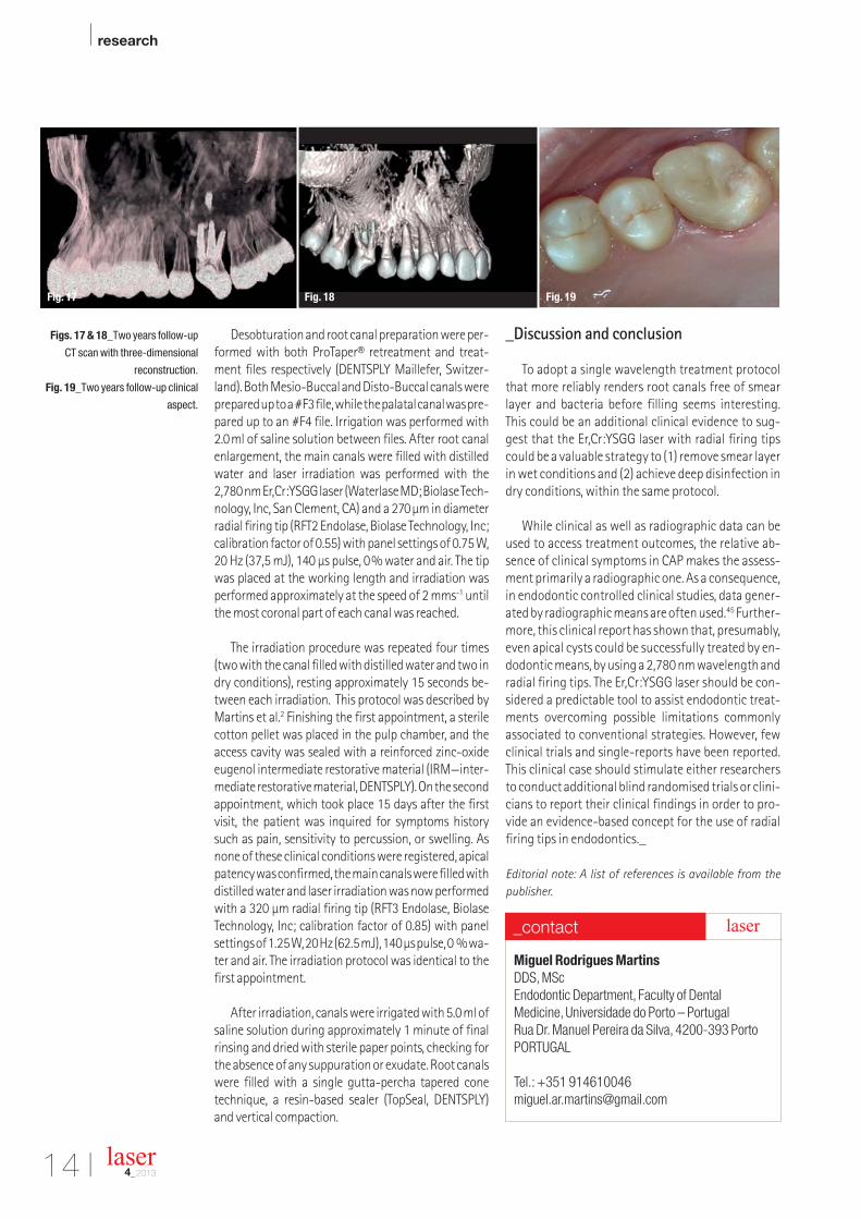
To adopt a single wavelength treatment protocolthat more reliably renders root canals free of smearlayer and bacteria before filling seems interesting.This could be an additional clinical evidence to sug-gest that the Er,Cr:YSGG laser with radial firing tipscould be a valuable strategy to (1) remove smear layerin wet conditions and (2) achieve deep disinfection indry conditions, within the same protocol.
While clinical as well as radiographic data can beused to access treatment outcomes, the relative ab-sence of clinical symptoms in CAP makes the assess-ment primarily a radiographic one. As a consequence,in endodontic controlled clinical studies, data gener-ated by radiographic means are often used.45 Further-more, this clinical report has shown that, presumably,even apical cysts could be successfully treated by en-dodontic means, by using a 2,780 nm wavelength andradial firing tips. The Er,Cr:YSGG laser should be con-sidered a predictable tool to assist endodontic treat-ments overcoming possible limitations commonlyassociated to conventional strategies. However, fewclinical trials and single-reports have been reported.This clinical case should stimulate either researchersto conduct additional blind randomised trials or clini-cians to report their clinical findings in order to pro-vide an evidence-based concept for the use of radialfiring tips in endodontics._
Editorial note: A list of references is available from the
publisher.
I research
Figs. 17 & 18_Two years follow-up
CT scan with three-dimensional
reconstruction.
Fig. 19_Two years follow-up clinical
aspect.
14 I laser4_2013
Fig. 18 Fig. 19Fig. 17
Miguel Rodrigues Martins
DDS, MScEndodontic Department, Faculty of Dental Medicine, Universidade do Porto – PortugalRua Dr. Manuel Pereira da Silva, 4200-393 PortoPORTUGAL
Tel.: +351 [email protected]
_contact laser

22 International Speakers
Lectures on implantology, oral surgery, aesthetics, endodontics, laser dentistry
Workshop / Live demonstations
Hands-on courses
Visit free exhibition area on 2.500 sqm
Enjoy open buffet lunch from the chef masters
Online congress booking is available at
dentalistanbul.com
ISTANBUL WELCOMES DENTAL PROFESSIONALS
To book your booth please contact [email protected]
Dental_Istanbul_A4_2013.pdf 1Dental_Istanbul_A4_2013.pdf 1 23.08.13 11:1423.08.13 11:14

I research
Fig. 1_Non-treated enamel.
Fig. 2_Acid-treated enamel.
Fig. 3_Laser-treated enamel.
Fig. 4_Before the irradiation: the
center is marked on each tooth.
Fig. 5_The screen positioned on the
tooth.
Fig. 6_Irradiation of the tooth.
_Introduction
The Er:YAG laser was first proposed in 1990 byHibst and Keller to ablate hard dental tissues. Todayit is employed in conservative dentistry as an alter-native to rotating instruments.1, 2 A study based onpatient questionnaires demonstrated that, in termof satisfaction, Er:YAG dental treatment representsan effective technique that may improve patientcooperation and diminish fears associated with thedental office, particularly in pediatric patients.3 Thisis also a reason to suggest its application in the fieldof orthodontics, where cooperation and good rela-tionships between the patient and operator arestrictly necessary for full success of a treatment. Inthis paper we describe the utilisation of the Er:YAGlaser in the bonding and debonding steps of ortho-dontic treatments.
_Enamel preparation
Proper conditioning of the enamel surface isnecessary for the bonding of orthodontic attach-ments to teeth. In orthodontics, as in other fieldsof dentistry, the most common method of enamelpreparation is acid phosphoric etching. The acidetching process prepares the surface by selectiveremoval of inter-prismatic mineral structure,while organic materials are less affected.
The resultant rough and micro-fissured surfaceis very useful for the retention of adhesive resins,but these structures are also more vulnerable tocaries formation. Acid etching removes and de-mineralises the most superficial and protectivelayer of enamel and makes the teeth more suscep-tible to long-term acid attack, especially when
Er:YAG laser in thebonding and debonding stepsof orthodontic treatmentAuthor_Prof. Carlo Fornaini, Italy
16 I laser4_2013
Fig. 5 Fig. 6
Fig. 2 Fig. 3
Fig. 4
Fig. 1

AEEDC_A4_2014.pdf 1AEEDC_A4_2014.pdf 1 14.02.13 12:0314.02.13 12:03

I research
18 I laser4_2013
resin monomers cannot sufficiently fill the de-mineralised area due to saliva contamination or airbubbles.4 Since the prevalence of white spot le-sions is very high among orthodontic patients, theprevention of enamel demineralisation is of greatimportance in orthodontics.5
There has been extensive research to find an al-ternative conditioning method to overcome themain disadvantage of phosphoric acid etching, i.e.the potential for producing decalcification. Someresearchers have worked on conditioning enamelwith poly-acrylic acid or pre-treatment of theenamel surface with a sandblast of aluminium ox-
ide to reduce the rate of enamel loss during etch-ing,6, 7 however, these methods failed to achieveadequate bond strength to resist intraoral forces.8
_Laser in orthodontics
Er:YAG laser preparation has become one effec-tive alternative to acid etching of enamel. Laseretching is painless and does not involve either vi-bration or heat; also, the easy handling of the ap-paratus makes this treatment highly attractive forroutine clinical use.9
The employment of a laser with orthophos-phoric acid etching to enhance the strength adhe-sion of composite resins has been proposed by sev-eral authors in conservative dentistry, as well as forbracket bonding in orthodontics.10 An in vitro studyat our university11 on 36 human extracted molars,divided into three groups on the basis of enamelconditioning (acid only, laser only and laser plusacid) and analysed by traction tests by measuringthe force necessary to detach the brackets, gavethe results reported below (Tab.1 and 2).
Recently, another interesting in vitro study,12
based on strength analysis by traction test and mor-phological analysis by SEM and Atomic Force Micro-scope, showed the same effects with Er:YAG irradia-tion alone as with acid etching. This was obtained byusing the so-called “QSP” mode (Fotona, Ljubljana,Slovenia) in which each pulse is split into severalshorter pulses that follow each other at an optimallyfast rate. In this way, a specific surface roughness isachieved, representing a real alternative to acid etch-ing. Microscopic observation of the samples ob-
Fig. 7_Bonding procedure
completed.
Fig. 8_The thermo-formed tray
inserted into the model.
Fig. 9_Thermo-formed tray with
holes for laser irradiation.
Fig. 10_Laser irradiation.
Fig. 11_Bonding procedure
completed.
Fig, 12_X-Runner, Fotona.
Table 1_Values (in MPa) of the force
necessary to detach the brackets.
Fig. 7 Fig. 8 Fig. 9
Fig. 10 Fig. 11 Fig. 12

laser 4/13
OFFICE STAMP
NAME/E-MAIL
LASER START UP
201322ndANNUALCONGRESS OF THE DGL e.V.
NOVEMBER 15–16, 2013// BERLIN,GERMANY//MARITIM HOTEL
PLEASE FAX THIS FORM+49 341 48474-390
Further information about:
❏ LASER START UP 2013 ❏ 22nd ANNUAL CONGRESS OF THE DGL e.V
November 15–16, 2013, Berlin, Germany
DOWNLOADPROGRAMME AND REGISTRATION FORM
SPONSOR

I research
Table 2_Comparison of the three
samples groups.
Fig. 13_Irradiation of the central
incisor.
Fig. 14_Irradiation of the premolars.
Fig. 15_Bonding procedure
completed.
tained by the first study (University of Parma) (Figs. 1–3) demonstrated that the enamel laser irra-diation creates micro-fissures that are ideal for resinpenetration.13
The surface produced by laser irradiation is alsoacid resistant. Laser irradiation of the enamel modi-fies the calcium-phosphate ratio and leads to theformation of more stable and less acid-soluble com-pounds, thus reducing the susceptibility to caries at-tack.14, 15
Because water spraying and air drying are notneeded with laser etching, time can be saved.16, 17
From a clinical standpoint, saving chair time also im-proves adhesion because it reduces the risk of sali-vary contamination.
Moreover, other authors have underscored theresult by using lasers to prepare the enamel surfaceto make it more resistant to decay18 due to the mod-ification of the hydroxyapatite crystals. Additionally,it is very important in the prevention of decalcifica-tion zones around the brackets, particularly in pa-tients with scanty oral hygiene.19
In recent years, several techniques have been pro-posed with the same goal: to prepare a very small
surface of enamel, exactly of the same dimension ofthe bracket, in line with the concept of modern “min-imally invasive dentistry”.
The first method20 consisted of the use of a ceramic screen with a central window; the disad-vantage consisted of the necessity to move thescreen from one tooth to another for irradiation, af-ter first marking the centre of each with a pencil(Figs. 4–7).
Parameters: Laser source: Er:YAG, 2940 nm (Fidelis Plus III, Fotona)
Pulse duration: MSPEnergy: 80 mJ defocusedFrequency: 18 HzHandpiece: R02, 4/6 water/air spray
An evolution of this technique was performedwith the introduction of thermo-formed individualtrays,21 which, after placement into the mouth, al-lowed for the irradiation of all the teeth of the arch(Figs. 8–11).
Parameters: Laser source: Er:YAG, 2940 nm (Fidelis Plus III, Fotona)
Pulse duration: MSPEnergy: 80 mJ defocusedFrequency: 18 HzHandpiece: R02-C, 4/6 water/air spray
With the introduction of digitally-controlledtechnology in laser dentistry, which led to the reali-sation and commercialisation of the “X-Runner”laser handpiece (Fotona, Ljubljana, Slovenia), themethod became significantly easier and faster, with-out the need to employ screens and/or trays.22 In fact,using the laser system’s touch screen, it is very sim-ple to program the size and dimensions required, andthen automatically irradiate an area equivalent tothe bracket surface (Figs. 12–14).
_Debonding
Enamel damage, whether in the form of enamelfractures or cracks, detracts from the aesthetics ofthe tooth and may require costly restorative treat-ment. It may even compromise the tooth’s integrityby increasing the risk of eventual tooth fracture.
20 I laser4_2013
Fig. 14 Fig. 15Fig. 13

research I
When the required force for bracket removalexceeds the cohesive strength of the enamel, frac-ture of the enamel surface is inevitable. With theintroduction of ceramic brackets in the mid-1980s,the problem became more important: in fact, thelow fracture toughness of ceramics may causepartial or complete bracket fracture during re-moval. This precludes reuse of the same bracket ata corrected position and may result in eye damage,ingestion or aspiration of bracket fragments. Inaddition, removal of a bracket fragment on thetooth may require the use of diamond burs, aprocess that is time consuming and can damagethe pulp and enamel surface.23, 24
Since the early 1990s, lasers have been used ex-perimentally for the debonding of ceramic brack-ets. The use of lasers eliminates problems such asenamel tear outs, bracket failures, and pain that areencountered during conventional ceramic bracketremoval techniques.25
Additionally, lasers have the advantage of de-creasing the debonding force and operation time.In most previous studies, carbon dioxide lasers,whose wavelength is more easily absorbed by theceramic brackets, had been preferred for debond-ing.25 In others,26 Nd:YAG was proposed, althoughwith this wavelength, approximately 69–75 % ofthe incident light reached the enamel surface,which has the potential to cause pain or damage tothe tooth structure.
Oztoprak et al. preferred the Er:YAG laser since ithas a lower thermal effect than the Nd:YAG or CO2
lasers. They stated that the Er:YAG laser is effectivefor reducing the shear bond strengths of orthodon-tic polycrystalline ceramic brackets from high valuesto levels that are safe for removal from the teeth.27
All these methods described are based on ther-mal softening of the resin by the beam, but are ac-tive only in the case of ceramic brackets. The tech-nique we propose may be used both on ceramicand metallic brackets, and consists of the utilisa-tion of a H14-C handpiece with chiselled fiber tip
(LightWalker AT, Fotona, Ljubljana, Slovenia). It isassumed that the vibrations produced by thephoto-mechanical effects of this wavelength playthe main role in the process of bracket detachment.
The fiber tip is placed tangentially to the crownsurface and inserted between bracket and enamelas close to the metal bracket as possible at a 45 de-gree angle. This way, the laser energy is directed tothe adhesive. Ten laser pulses are delivered at eachside of the bracket. After that, the metal bracket isremoved, with a very low strength, with the help ofa spatula normally used to mix the cement. In thisway, there are no complications during thedebonding procedure and no damage to theenamel surface. As the energy is set relatively lowin MSP mode, there is also no danger for intra-pul-pal temperature rise. Patients report absolutely nostress during the procedure (Figs. 16–18).
Parameters: Laser source: Er:YAG, 2,940 nm(LightWalker AT, Fotona)
Pulse duration: MSPEnergy: 80 mJFrequency: 10 HzHandpiece: H14-C with chiseled fiber tip,
4/6 water/air spray
Editorial note: A list of references is available from the
publisher.
I 21laser4_2013
Fig. 16 Fig. 17 Fig. 18
Prof. Dr Carlo Fornaini
MD, DDS, MSc
Dental School, Faculty of Medicine,
University of Parma,
Via Gramsci 14
43126 Parma, Italy
Tel.: +39 0521 292759
Fax: +39 0523 986722
www.fornainident.it
_contact laser

I case report
Fig. 1_Dry buccal mucosa observed
in the first visit.
Fig. 2_Dried floor of the mouth
observed in the first visit.
Fig. 3_After the first episode of LILT,
still some erythematous areas in
vermillion border of the lower lip are
observed in the first visit.
Fig.4_LILT irradiating to the
vermillion border of lower lip.
_Abstract
Xerostomia causes patient suffering from thelimitation of daily life activities. Still, there has beenno definite treatment for this, particularly its vari-ant induced by aging. This case report presents themethod of treating xerostomia using low intensitylaser therapy and medication.
_Introduction
Xerostomia is an oral symptom of dry mouth,which is a major complaint of many elderly indi-viduals. It results in impaired food and beverage in-take, altered taste, difficulties in eating, chewingand swallowing, which can affect the quality of aperson’s life. Although suffering patients seekmedical help, it usually provides no adequate re-
lief.1 Not many patients were able to tolerate thestrange taste of artificial saliva or the side effect ofsweating from taking pilocarpine; a medication foractivating salivation. Symptomatic treatment byusing saliva substitute for moistening and lubrica-tion of the oral mucosa was the only managementfor this condition. But is there a different, the non-invasive method that could be used for treating xe-rostomia?
In 2010, Kato et al. conducted a pilot study prov-ing a significant and lasting reduction in burningmouth symptoms from the group treated by low in-tensity laser therapy (LILT).2 In the same year, VidovicJuras et al. reported an application of LILT to xeros-tomia patients' major salivary glands to stimulatesalivation.3 This report presents a combined med-ication and LILT for treating xerostomia with satis-factory results.
_Case report
A 60-year-old woman had suffered from oraldryness interfering with her life style daily for threeyears. There had been no therapy including vitaminsupplement and more water intake to relief thesesymptoms.
Past medical history
The patient suffered from anaemia due to nutri-tional deficiency. She was treated by taking ferroussulphate and folic acid. Her menopause had startedabout ten years ago. She was allergic to penicillin,pollen and adverse weather. The patient took an an-tihistamine agent, Zyrtec, one tablet per day.
Oral examination
Extraoral examination showed a dry and crackedlower lip. Intraoral examination found dryness of
The efficacy of combined lowintensity laser therapy andmedication on xerostomiaAuthors_Dr Sawanya Taboran & Assoc. Prof. Dr Sajee Sattayut, Thailand
22 I laser4_2013
Fig. 1 Fig. 2
Fig. 3 Fig. 4

case report I
Fig. 5_After ten LILT sessions;
remarkably less erythematous areas
are observed in the vermillion border
of lower lip.
Fig. 6_Moist buccal mucosa
observed during the tenth visit.
Fig. 7_Moist floor of the mouth
observed in the tenth visit.
Fig. 8_LILT irradiation of the skin
above the parotid gland area.
I 23laser4_2013
the oral mucosa (Fig. 1), including the floor the ofmouth (Fig. 2), atrophic tongue and erythematousat the vermillion border of the lower lip with bitingfrom the lower anterior teeth (Fig. 3).
Treatments and their results
The first visit:
Treatment: Zyrtec was limited. The patient wasadvised to sip water as necessary.
Result: Two weeks later, the patient could sensemoisture in the oral cavity and dysphagia less pro-nounced. Intraoral examination found a significantamount of saliva in comparison to the last visit.
Impression: Drug-induced xerostomia.
The second to the tenth visits (one treatment ses-sion per week): Erythematous sections at the ver-million border of the lower lip were treated with lowintensity laser (Fig. 4), 820 nm, 100 mW, 2 J, 20 sec-onds, continuous wave for 18 points. Vitamin C andB complexes were prescribed.
Result: The patient was more satisfied with theresult after the second treatment session with LILT.The moist lip (Fig. 5), oral mucosa (Fig. 6) and floor ofmouth (Fig. 7) were observed. Less erythematous ar-eas were noticed at the vermillion border of thelower lip after the second time of treatment withLILT. They disappeared after the tenth visit.
The sixth to eleventh visits: Low intensity lasertherapy 820 nm, 100 mW, 2 J, 20 seconds, continu-ous wave for 22 points in the left and right parotidgland area (Fig. 8).
Result: There was an increase in the whole stimu-lated salivary flow rate from 0.06 ml/min in the firstvisit to 0.08 ml/min after the second treatment and0.10 ml/min after the fourth treatment.
_Discussion
The satisfying clinical outcome of treating xerosto-mia and atrophic mucosa caused by aging and side ef-fects of drugs was found by using combined medica-tions and LILT. LILT can be used to reduce burningmouth symptoms and to stimulate major salivaryglands to produce more saliva. This can be explainedby the effect of LILT for biomodulation in terms of ini-tiating healing mechanism and immune response.4
_Conclusion
This case report has shown that using vitamin Cand B complex supplements combined with low in-tensity laser therapy can improve the clinical impair-ment of xerostomia and can also increase saliva se-cretion in elderly patients._
Fig. 5 Fig. 6
Fig. 7 Fig. 8
Assoc Prof. Dr Sajee Sattayut
Chairman of Lasers in Dentistry Research Group
Faculty of Dentistry
Khon Kaen University, Khon Kaen City, 40002
Thailand
Tel.: +66 815442460
Fax: +66 43348153
_contact laser

_Introduction
The use of alternating electric current for blood-less interventions in the oral soft tissues has beenestablished for nearly a century, first in the form ofthe electric knife (cauter), then later as radiofre-quency devices. Laser devices were introduced in the1980s as new, additional tools and they havestrongly gained in importance today. One particularquestion often emerges: What are the similaritiesbetween diode lasers and radiofrequency genera-tors and what makes them different? What is theirvalue for dental applications?
_Similarities
The laser light as well as the electric current of theradiofrequency device make use of the spatially con-centrated and very fast heating of cells in the tissue.Thus they both are able to cut and coagulate in a pre-cise way. Bleeding is stopped with high efficacy, al-lowing the surgeon a clear view of the surgical field.
_Differences
While the laser radiation is passed through anoptical fiber and emitts from the fiber tip to the tis-sue surface, the radiofrequency current is directed
through a metal electrode into the tissue. The maindifference: A priori, the laser fiber can not be in-serted deeply into the tissue to produce a cut. Thelaser radiation emitts from the front end of a fiberand thus heats only the uppermost layer of the tis-sue and ablates it. Therefore, the tissue must be re-moved in a layer-by-layer procedure to get into thedepth.
In contrast, the metal electrode of a radiofre-quency device can be inserted into the tissue in onestep in an intended depth. The radiofrequency cur-rent heats the targeted area simultaneously anduniformly to the entire physical length of the elec-trode. It allows a one-step precise, stressless and al-most athermic procedure with an excellent tactilefeel. The cutting speed especially in larger anddeeper areas is much faster than with a laser.
In intraoral use, the radiofrequency technologyis positively received because the local increase intemperature is less than 60 to 80 °C. Using a laser oran electric knife, respectively, a temperature in-crease of more than 400 °C must be considered.
_Useful applications
The strength of a laser lies in the treatment of su-perficial soft tissues, such as the removal or re-shaping of thin skin layers: i.e. exposure of over-grown implants, gingiva streamlining, in periodon-tic treatments, in endodontics as well as in specialapplications like bleaching, photodynamic therapy(PDT) and low level laser therapy (LLLT).
A significant disadvantage, however, can beseen in deeper surgical applications. The oral tissueis very thin, it is delicate and has complicated struc-tures and in addition it usually is in close proximityto jaw bones and tooth structures. Laser radiation
I industry report
Fig 1_Cutting of tissue with
radiofrequency: The tissue is
removed with only one precise,
uniform cut in the entire length of the
inserted electrode. The metal
electrode remains cold at its
frequency of 2.2 MHz.
Fig. 2_The cutting process with a
laser is a layer-by-layer removal of
superficial tissue.
A combined device for opti-mal soft tissue applications inlaser dentistryAuthors_Hans J. Koort, Dr Michael Hopp, Germany & Prof. Dr D. Gabric Panduric, Croatia
24 I laser4_2013
Fig. 2Fig. 1

industry report I
Fig. 3_Clinical appearance of the palatal fibroepithelial
polyp and inflammatory papillary hyperplasia of the
hard palate.
Fig. 4_Immediate postsurgical view.
Fig. 5_Follow up, third day after surgery.
Fig. 6_Clinical appearance of the mucocele at the lower
lip, before surgery.
Fig. 7_Surgical procedure performed using diode laser.
Fig. 8_Immediate postsurgical view.
Fig. 9_Closing of the incision lines with bipolar forceps
using radiofrequency technology.
Fig. 10_Clinical appearance of the maxillary giant cell
granuloma.
Fig. 11_Removal of giant cell granuloma with
radiofrequency.
Fig. 12_Bleeding control using bipolar forceps
immediately after excision.
Fig. 13_Follow-ups, two weeks after surgery.
Fig. 14_Follow up, five weeks after surgery.
Fig. 15_Clinical appearance of metastasis at the hard
palate.
Fig. 16_Immediate postsurgical view, bleeding control
using bipolar forceps.
Fig. 17_Wound covered with a dressing.
I 25laser4_2013
Fig. 3 Fig. 4 Fig. 5
Fig. 6 Fig. 7 Fig. 8
Fig. 9 Fig. 10 Fig. 11
Fig. 12 Fig. 13 Fig. 14
Fig. 15 Fig. 16 Fig. 17

I industry report
26 I laser4_2013
Fig. 18_Pre-surgical clinical view.
Fig. 19_Re-contouring of the soft tissue using
diode laser.
Fig. 20_Application of aPDT before abutment fixation.
Fig. 21_Immediate postsurgical view.
Fig. 22_Application of photosensitiser gel through
periodontal space and central ridge incision line.
Fig. 23_Distribution of photosensitiser gel around the
periimplantitis area.
Fig. 24_aPDT, using red 660 nm diode laser.
Fig. 25_Control radiograph, four weeks after finishing
the treatment.
Fig. 26_Presurgical clinical view.
Fig. 27_Radiofrequency electrode cuts the carcinoma.
Fig. 28_Immediate postsurgical view.
Fig. 29_Follow-up after five days.
Fig. 30_Follow-up, five weeks after surgery.
Fig. 31_Pre-surgical view of hemangioma in the
lower lip.
Fig. 32_About ten single punctures with a thin needle
electrode (see magnification).
Fig. 18 Fig. 19 Fig. 20
Fig. 21 Fig. 22 Fig. 23
Fig. 24 Fig. 25 Fig. 26
Fig. 27 Fig. 28 Fig. 29
Fig. 30 Fig. 31 Fig. 32

industry report I
is not only absorbed in the tissue and converted intoheat, it also will be partially transmitted through thetissue and may cause unpredictable and undesiredside effects in adjacent healthy areas. Furthermore,the cutting speed of a laser beam is limited by thefact that the tissue can only be removed in layers.Neither increasing the laser power nor using pulsedlaser radiation can eliminate this problem.
_A promising approach
The combination of a diode laser and a modernradiofrequency generator in one unit is a usefuland perfect tool for an extensive soft tissue man-agement. The laser can treat the relatively thin andcomplicated oral tissue very selectively and showspromising and successful results in periodontics,endodontics and implant surgery. The radiofre-quency technology on the other hand, because ofits significantly higher cutting speed and perfectcoagulation, has benefits in oral surgery. Tissue willbe heated and cut simultaneously, homogeneouslyand very fast in the entire length of the insertedmetal electrode. Damage to adjacent healthy areasare unlikely. If they do occur, they are predictableand can be planned. Photodynamic therapy (PDT),low level laser therapy (LLLT) and tooth bleachingmake new, additional treatments possible. At the
Department of Oral Surgery at the University of Zagreb, Croatia, a clinical study was performed todemonstrate the use of the combined LaserHF®unit, consisting of a 975 nm diode laser with 8 Wpower (cw, pulsed), a 2.2 MHz radiofrequency gen-erator with 50 W power (monopolar and bipolar)and a 660 nm diode laser with 100 mW power (cw),in various treatments of oral soft tissue lesions(Figs. 1 & 2). To all patients, local anaesthesia wasadministered before the procedures. Patients inthis study showed significantly less oedema andhematoma as well as significantly less pain andhigher satisfaction compared to conventionallytreated patients (p < 0.05).
_Case presentation
Out of these patients we present the followingcases:
Case 1
A female patient aged 67 presented with palatalfibroepithelial polyp and inflammatory papillaryhyperplasia of the hard palate (Fig. 3). The soft tissuesurgery was performed with a radiofrequency loopelectrode at 35 W and diode laser 975 nm with apower of 5 W in continuous mode (cw). Low levellaser therapy (LLLT) at 660 nm followed for 90 s im-
I 27laser4_2013
Please contact Claudia Jahn
AD
[PIC
TUR
E: ©
SU
KIY
AKI]

I industry report
Fig. 33_Following up after five days.
Fig. 34_Leukoplakic, exophytic
growing alteration (12 x 25 mm) at
the left side of the tongue.
Fig. 35_The tissue was very deeply
removed from the tongue muscle to
prevent any recurrences.
Fig. 36_Due to the repeated growth
and the unclear etiology, the tissue
was deeply removed from the tongue
muscle to prevent any recurrences.
Fig. 37_This method allows an
almost pressure-free work, resulting
in straight-cut edges in the muscle.
Fig. 38_Increasing epithelialisation
two weeks postoperatively
Fig. 39_An impressing macroscopic
scar-free result eight months after
surgery.
Fig. 40_Alterations like thermal
induced vacuoles in the striated
muscle could not be seen after using
radiofrequency technology.
Fig. 41_Histological comparison of
the thermal reaction zone produced
with a 980 nm laser at 3 W cw. The
broad and partially melted reaction
zone is a result of a significant
temperature effect
mediately after the surgical (Fig. 4) procedure wasperformed. Neither side effects nor complicationsoccurred after surgery (Fig. 5).
Case 2
A female patient aged 23 presented with a mu-cocele in the lower lip (Fig. 6). The surgery was per-formed with the laser 975 nm at a power of 5 W incw-mode (Figs. 7 & 8). The lesion was closed using abipolar radiofrequency forceps (Fig. 9). LLLT applica-tion 660 nm with a power setting of 90 mW followedimmediately for 90 s.
Case 3
A male patient presented with a giant cell gran-uloma of the upper jaw, central type in the frontalregion of the maxilla (Fig. 10). The surgery was per-formed with radiofrequency cutting-mode (35 W)and laser 975 nm, 5 W cw. LLLT application followedimmediately after surgical procedure (660 nm, 90 mW for a period of 90 s, Figs. 11 & 12). Follow-upswere taken two (Fig. 13) and five weeks after surgery(Fig. 14).
Case 4
A male patient, aged 82, presented with a metas-tasis of adenocarcinoma of the kidney (Fig. 15). Sur-gery was performed using radiofrequency cutting(35 W), coagulation grade 3 as well as laser 975 nm,5 W cw, in combination (Figs. 16 & 17).
Case 5
A female patient aged 53 presented with a peri-implant mucositis (Fig. 18). Re-contouring of theperiimplant mucosal tissue took place one week af-ter the second surgical phase (Fig. 19). Defixation ofthe final abutment due to periimplant mucositiswas performed with laser 975 nm at 4 W cw. Anti-bacterial PDT was performed after surgery (Figs. 20& 21). No side effects or complications regarding theimplant-bone interface after surgery were reported.Systemic antibiotic therapy was also included.
Case 6
A female patient with periimplantitis: Treatmentof initial periimplantitis using closed technique wasperformed with systemic antibiotic therapy (Figs. 22& 23) and aPDT (660 nm, 100 mW, 3 x 10 s) for tenconsecutive days (Figs. 24 & 25).
Case 7
Male patient, aged 66, presented with verrucouscarcinoma (Fig. 26). The surgery was performed withradiofrequency 35 W, coagulation grade 3—ellipticloop electrode and laser 975 nm, 5 W cw, in combi-nation (Figs. 27–30).
Case 8
An 11-year-old girl presented with a haeman-gioma in the lower lip (Fig. 31). Low power radiofre-quency application was conducted with 15 W, co-agulation grade 2. A fine needle electrode (diameter0.2 mm, length 5 mm) has been inserted into the tis-sue at about 10 points around the lesion, resultingin shrinkage of the tissue (Figs. 32 & 33). This treat-ment is not possible using the laser device.
Case 9
A 33-year-old patient presented with a leukopla-kic, exophytic growing alteration (12 x 25 mm) at theleft side of the tongue (Fig. 34). Considering the par-
28 I laser4_2013
Fig. 33 Fig. 34 Fig. 35
Fig. 40 Fig. 41
Fig. 36 Fig. 37 Fig. 38 Fig. 39

industry report I
ticular topography and anticipated depth of the le-sion, the treatment was performed with radiofre-quency at 2.2 MHz and a power of 50 W, using littlecoagulation (grade 1–2) simultaneously (Figs. 35–37). Monopolar operation was chosen with the neu-tral electrode on the shoulders of the patient. Witha laser such a deep surgical operation would not bepossible. After anaesthesia of the left N. lingual, thesurgical area was marked with a fine, very thin elec-trode and the cutting line was defined in the rangeof the healthy mucosa. Follow-ups were taken twoweeks (Fig. 38) and eight months (Fig. 39) after sur-gery.
The healing was without complications or seri-ous swelling, but with moderate post-operativepain. There were no functional limitations, form andfunction of the tongue have been preserved or fullyrestored. The histological evaluation of the excisedtissue showed no changes; only in the direct sectionarea, a low thermal reaction zone was found (Figs.40 & 41).
_Conclusion
For precise applications in dental soft tissues,especially surgical incisions, scalpel, laser and ra-diofrequency are appropriate tools. The rightchoice must be made by the dentist and is based onvarious criteria: If the lesions are relatively smalland if they are more in the depth of the tissue, andno previous histopathologic evaluation is possibleor useful, laser or radiofrequency are suitable de-vices. In contrast to the scalpel, a flushing out of ab-normal cells and distant metastasis can be sub-stantially averted. Besides, the scalpel always in-duces some mechanical stress to tissues which maylead to unprecise healing and thus cannot alwaysclaim a good cosmetic result. On the opposite, ther-mally produced incisions made by radiofrequencyare fast and stress-free, providing excellent cos-metic results. They also deliver convincing results inmore complicated procedures in the depth of thetissue. With appropriate coagulation, just minortissue alterations are found, thus promising a goodhealing. The laser is more to be seen as a tool withstrong importance in the treatment of superficiallesions due to its mode of layer-by-layer operation.Apart from treatments in periodontics, in endodon-tics, in PDT, Bleaching and LLLT, the laser is a perfectinstrument for reshaping and smoothing ofwound edges, for removal of overgrown implants,trimming of gingiva, drying and shrinking of tissueand of course even for small superficial surgery(Fig. 42).
Laser and radiofrequency technology showsome delay in the epithelial regeneration. A wound
takes some more time to re-epithelialise than fol-lowing conventional surgery with a scalpel. How-ever, they offer a minimally invasive technique withan intention to make surgical applications less ex-tensive. They may reduce the need for general anes-thetics or hospital care, and therefore can lower theoverall costs.
Considering the potential applications, the com-bination of a diode laser with a radiofrequency de-vice meets the desire for a perfect system in the den-tal soft tissue management. The LaserHF® (by Hager& Werken, Germany, Fig. 43) is the worldwide firstcombination device. It consists of a 975 nm laserwith a power of up to 8 W, combined with a 2.2 MHzradiofrequency generator—monoploar and bipolar—with a power of 50 W. An additional 660 nm laserwith a power of 100 mW completes the device astherapy supplement for photodynamic (PDT) andlow level laser therapy._
Fig. 42_The laser is not perfect in
oral surgery and the radiofrequency
is not suitable in many other dental
applications. However, in the
combined device, they both can
perform as perfect tools for the dental
soft tissue management.
Fig. 43_The LaserHF® device.
I 29laser4_2013
Hans-H. Koort
MedLas Consult
Auf der Schleide 18
53225 Bonn, Germany
www.medlas.com
_contact laser
Fig. 43
Fig. 42

I industry report
30 I laser4_2013
Figs. 1a & b_Crown lengthening
before treatment (a).
Crown lengthening 6 months
after treatment (b).
Figs. 2a & b_Gingival
depigmentation before treatment (a).
Gingival depigmentation 7days
after treatment (b).
_The exciting possibilities offered by theworld of aesthetic dentistry are being experiencedby an increasing number of dentists and patientsworldwide as new materials, techniques and toolsbecome widely available.
One such example is the laser, of which thereare many for use in dentistry. To appreciate laserdentistry to its fullest, however, it is necessary to gain accurate information from unbiasedsources, firstly about laser safety and the physicsof laser–tissue interaction, and secondly aboutthe specific uses of lasers in practice.
The most commonly used laser types in den-tistry are erbium lasers (with two variants: 2,940 nm Er:YAG and 2,780 nm Er,Cr:YSGG),neodymium lasers (1,064 nm Nd:YAG) and diodelasers (810, 940 or 980 nm). Each type of laser hasa different wavelength, and each wavelength has a different interaction with the specific bodytissue being treated.
Crown lengthening or gingival levelling (Figs.1a & b) is a routine procedure for laser-assistedaesthetic interventions. All lasers can be used for
this procedure; however, there are two main ad-vantages of using erbium lasers in crown length-ening. First of all, they can be used without anaes-thesia, as they do not cause thermal damage tothe tissue. This results in a stable gingival heightafter the procedure.
Since diode and Nd:YAG lasers work in a morethermal manner, a longer healing time should be expected for the tissue to settle. A prerequisitefor success in crown lengthening is, of course, torespect biologic width. If there is less than 3 mmbetween the desired gingival level and the bone,the bone level must be decreased. While this ispossible to do with erbium lasers (even flapless),neither diode nor Nd:YAG lasers are suitable forthis, since they are only capable of removing softtissue.
The same conditions are applicable for uncov-ering implants with erbium lasers, so it is possibleto take the impressions for prosthetic procedureson the same day or within a short period. If it is necessary to remove bone or soft tissue for either indication, erbium lasers with adjustablepulse duration (referred to as VSP technology in
Fig. 2a Fig. 2b
Fig. 1a Fig. 1b
Lasers in aesthetic dentistryAuthors_Drs Ilay Maden, Zafer Kazak & Özge Erbil Maden, Turkey

industry report I
I 31laser4_2013
Fotona lasers, for example) are the only optionwithout raising a flap.
Tooth preparation for crowns and bridges withlasers is not yet as efficient as one might like it tobe; however, new research and technological im-provements are ongoing. A diode or Nd:YAG lasercan still be helpful during prosthetic work fortroughing before taking impressions or desensi-tising prepared teeth if required. It is also possibleto reduce or eliminate dentine hypersensitivitydue to periodontal treatment or gingival reces-sion by either modulating the nerve endings orblocking the dentinal tubules using a laser.
Another aesthetic treatment is gingival depig-mentation (Figs. 2a & b), which can also be per-formed by using long pulses of erbium or diodelasers. It is possible to de-epithelialise the surface,as the pigmentation is usually in the basal layer.
Erbium lasers are safer, since they do not pen-etrate the tissue. The effect is only superficial, andthis is exactly where the pigmentation is.
Diode lasers penetrate more deeply, especiallyif one is not careful and tries to remove tissue that is lighter in colour. As with other treatments,erbium lasers allow the tissue to heal faster; however, there can be mild bleeding during the operation.
Class V caries removal for composite fillingscan easily be performed with an erbium laser,quickly, painlessly, and without any thermal side-
effects, especially if the pulse durations are shortenough—typically between 50 and 100 micro -seconds (Fig. 3). The shorter the pulse duration is, the more effective the laser energy will be at re-moving hard tissue. The margins of the cavity caneven be bevelled for better aesthetic appearanceand long-term colour stability if the laser is effi-cient enough to remove small amounts of soundenamel when needed.
Lasers also work selectively to only removecarious tissue, which has more water contentthan sound hard dental tissue. Surface modifica-tion can also be done with the erbium laser aftercavity preparation for repairing composite fill-ings, or even for restoration cementation. One big advantage is that anaesthesia is not generally required when bloodless gingivectomy is used touncover the borders of the carious lesion with anerbium laser using pulse durations of between600 and 1,000 microseconds (Figs. 4a & b).
Fig. 3_SEM image of hard
dental tissue after ablation
with an erbium laser.
Figs. 4a & b_Clinical case
before treatment (a).
Immediately after gingivectomy
and carious lesion removal (b).
Figs. 5a & b_Before (a) and after
TouchWhite procedure (b).
Fig. 3
Fig. 5a Fig. 5b
Fig. 4a Fig. 4b
I industry report
32 I laser4_2013
For tooth whitening, lasers can also be used for activation of the bleaching gel (Figs. 5a & b),which decreases the treatment time as well aspost-operative sensitivity. As the laser is absorbedby the appropriate gel, the heat is only superficialand the contact time is decreased, leading to lessor no sensitivity.
The Er:YAG laser beam is uniquely absorbed inthe water molecules that are contained in all gels.The more water content (the more the gel is “soft”and a bit runny), the better the resulting bleach-ing interaction (known as the TouchWhite™ pro -cedure, patented by Fotona). The colour is not of importance for this interaction, unlike withNd:YAG and diode lasers, which are absorbedmore efficiently in pigments and therefore re-quire specially coloured gels to be effective.
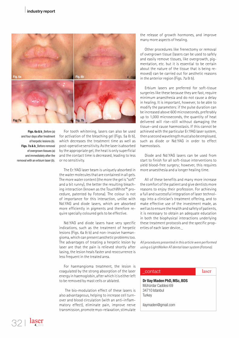
Nd:YAG and diode lasers have very specific indications, such as the treatment of herpetic lesions (Figs. 6a & b) and non-invasive haeman-gioma, which can present aesthetic problems too.The advantages of treating a herpetic lesion bylaser are that the pain is relieved shortly after lasing, the lesion heals faster and reoccurrence isless frequent in the treated area.
For haemangioma treatment, the lesion is coagulated by the strong absorption of the laserenergy in haemoglobin, after which it is either leftto be removed by mast cells or ablated.
The bio-modulation effect of these lasers isalso advantageous, helping to increase cell turn-over and blood circulation (with an anti-inflam-matory effect), eliminate pain, improve nervetransmission, promote myo-relaxation, stimulate
the release of growth hormones, and improvemany more aspects of healing.
Other procedures like frenectomy or removalof overgrown tissue (lasers can be used to safelyand easily remove tissues, like overgrowth, pig-mentation, etc. but it is essential to be certainabout the nature of the tissue that is being re-moved) can be carried out for aesthetic reasons in the anterior region (Figs. 7a & b).
Erbium lasers are preferred for soft-tissue surgeries like these because they are fast, requireminimum anaesthesia and do not cause a delay in healing. It is important, however, to be able tomodify the parameters: if the pulse duration canbe increased above 600 microseconds, preferablyup to 1,000 microseconds, the quantity of heatdelivered will rise—still without damaging the tissue—and cause haemostasis. If this cannot beachieved with the particular Er:YAG laser system,then a second wavelength must also be employed,such as diode or Nd:YAG in order to effecthaemostasis.
Diode and Nd:YAG lasers can be used fromstart to finish for all soft-tissue interventions toyield blood-free surgery; however, this requiresmore anaesthesia and a longer healing time.
All of these benefits and many more increasethe comfort of the patient and give dentists morereasons to enjoy their profession. For achieving a full and successful integration of laser technol-ogy into a clinician’s treatment offering, and tomake effective use of the investment made, aswell as to ensure the health and safety of patients,it is necessary to obtain an adequate education in both the biophysical interactions underlyingthese treatment protocols and the specific prop-erties of each laser device._
All procedures presented in this article were performed
using a LightWalker AT dental laser system (Fotona).
Figs. 6a & b_Before (a)
and four days after treatment
of herpetic lesions (b).
Figs. 7a & b_Before removal
of overgrown tissues (a)
and immediately after the
removal with an erbium laser (b).
Dr Ilay Maden PhD, MSc, BDS
Mühürdar Caddesi 69
34710 Istanbul
Turkey
_contact laser
Fig. 7a Fig. 7b
Fig. 6a Fig. 6b

October 9-14, 2014 | San Antonio, Texas, USA
Education: October 9-12 | Exhibition: October 9-11
To learn more, visit ADA.org/meeting.
ExhibitionResearch and purchase
dental products and services
at a discount
ConnectionsMingle with colleagues
from across the world
EducationParticipate in challenging
CE courses that fit into your
schedule and budget
7975Z_Cos_Dentistry_Ad.pdf 17975Z_Cos_Dentistry_Ad.pdf 1 30.10.13 11:0530.10.13 11:05

I economy
Fig. 1_Relation between
satisfaction and loyalty.
Source: Professor J. Heskett,
Harvard Business School.
_Introduction
During this issue we are going to explore the first el-ement of marketing mix which is the package in ourcase the service that we deliver in our offices. Dentaltreatment is an intangible service therefore a very im-portant role is played by the quality.
Do you know how quality is perceived in the mind ofour patients? First by the past experiences our patientshave had from us or from other colleagues. Secondfrom the word of mouth, which is our most powerfulmarketing tool, what others are saying about us and fi-nally, from the personal needs that our patients have.As an example we can start with their need of experi-encing minimal pain during a treatment. Many getafraid or annoyed by the sound of the drill; in this casethey will prefer to choose a laser dentist. The above arefactors that we have to meet during the services thatwe offer. We should understand that we do not only de-liver services but a culture; the culture and mentality ofour clinic through the experience that our patients re-
ceive. They do not only expect from us to be just gooddoctors. If we think like this we are wrong! They expectfrom us to be the best, period. Our patients will not justcompare us with other dentists but with all the serviceexperiences that they have like as in a hotel or in arestaurant.
Our competitors are everyone when it comes to ourpatients; the manager in a restaurant, the receptionistin a hotel. Our patients hold in mind a mental picture ofhow they think they should be treated and this picturebecomes their standard by which their experiences arejudged.
_Law of memorable event: determines dissatisfaction and loyalty
When nothing makes either a good or a bad impres-sion on you, your feelings are neutral. It takes some-thing memorable to turn ordinary satisfactory experi-ence into something special. Dissatisfaction comesfrom something bad that you experienced and you re-member. Furthermore, loyalty is generated by memo-rable things that happen, that we weren’t expecting.And if our patients do not remember us, why shouldthey choose to continue to come to us?
As we can see from Figure 1, we have three zoneswhen we interrelate satisfaction and loyalty:1. The zone of defection, which includes the terrorists—
all the dissatisfied patients that discourage othersfrom visiting us. This zone gets narrower as the rela-tionship between dentist and patient matures andgoodwill is added in the emotional bank account.
2. The zone of indifference, where the patients are sat-isfied but they might visit other colleagues as well fortheir treatment or they will not refer us to others.
3. Finally the zone of affection containing the apostles—the very satisfied patients, the loyal patients thattalk favourably about our services and their experi-ences and they will spread through the word ofmouth how thrilled they are with us, encouragingfriends and relatives coming to us for their dentaltreatments.
34 I laser4_2013
Gain power at your laser clinics!Author_Dr Anna Maria Yiannikos, Germany/Cyprus

economy I
A question that might arise is: how can we minimizethe zone of defection?
Exit interviews or measuring regularly our patient’ssatisfaction with surveys can have an extreme impacton our profits since through them we can learn in whichareas we lack in so to improve.
And here comes another question to consider: Howmany of us administrate patient satisfaction surveys,how often and when do we imply them? As mentionedbefore, dental treatment is an intangible service (=wecannot measure it like a product). Therefore, the qualityplays a very important role.
How do patients measure the quality of our practice?Patients cannot judge how well we can fill a root
canal or how nicely we polish a composite filling—in-stead, quality is effectively perceived from our patientsby the below factors:1. Appearance: the way our office and our staff look. 2. Security of our clinic: In which way do we increase our
patient’s confidence that they belong in a safe placeand can relax and trust us for the best? Some exam-ples are the existence of safety signs and flash lampsoutside the doors, laser safety diploma, safety glassesthat we provided to them during laser treatments, ac-creditations we have gained like ISO 9001(for quality)or OSHAS 18000(for health and safety). These are justsome of the options that we have that constantly re-mind our patients of our best interest in the quality.
3. Reliability: Our ability to keep a consistent level of alaser treatment. Is our treatment plan performed astold from the beginning? If we promise our patientsthat they will have a painless treatment, will this webe able to keep our word? Our patients must know thetruth from the beginning. No promises must be giventhat cannot be kept in order for our patient to trustand rely on us.
4. Responsiveness: Do we respond quickly to their prob-lems? Or their phone calls? Are our staff memberswilling to answer their questions with care and seri-ousness expressing simultaneously their knowledgeand skills?
5. Competence: Our patients ought to understand thatwe are not only skillful and that we do not only havethe equipment but we also posses the knowledge toprovide laser treatments.
6. Courtesy: We have to be polite not only with our pa-tients, but also show our friendliness towards our em-ployees as well: “Treat your employees as your pa-tients and your patients as employees”.
7. Credibility: We ought to be honest and have a goodreputation
8. Access: People value nice people since this make themhappy. We cannot describe to them how nice we or ourstaff is, they have to see for themselves—we have towalk the walk through internal and external acts ofniceness. We should be approachable and ease to
contact. If, for example, we have a secretary that is atall time distracted and cannot be reached from ourpatients, this will create a bad impression for our en-tire clinic.
9. Communication: We must know what, when and howto say something. We must master verbal, non verbalcommunication skills so as to show them that we un-derstand them with sympathy and compassion; un-derstand not only their needs, but also their fears.
Let’s look at the following example: If we are ex-plaining the treatment to Mr Smith like this: “Mr Smith,today we are going to make a cavity preparation usingVSP, 20 Hz with water and air using our Er:Yag laser. Areyou ok with that?” What do you believe that we are going to achieve through this? Loose him instantly as alistener!
Let’s close the referral to the first element of market-ing mix by remembering the Gucci family motto: “Qual-ity is remembered long after the price is forgotten”. In thenext issue, we are going to talk about the second ele-ment of marketing mix, which is the PRICE.
A fruit for thought: Let’s not forget who our patientsare. All of the people that we meet daily, the reception-ist at a hotel, the bank cashier, our hairdresser, all thepeople that we come into contact with can spread theword of how wonderful or miserable we are… In whichof the two groups do you want to belong to? I leave thisdecision up to you.
Last as a closure allow me to proudly announce thelaunch of DBA mastership course in Europe under theumbrella of AALZ—RWTH Aachen University Campus.A mini MBA designed only for dentists scheduled tobegin on the 1st of May 2014. It is a course for the den-tists that want to gain their power back! For thosethat have the desire to master the skills of manage-ment and administration for their own clinics, wheremany of the above and many more matters will betaught in more detail. Looking forward to your ques-tions and requests for further information at:[email protected]_
I 35laser4_2013
Dr Anna Maria Yiannikos
Adjunct Faculty Member ofAALZ at RWTH Aachen Uni-versity Campus, GermanyDDS, LSO, MSc, MBA
_contact laser

36 I laser4_2013
I health
_Pain is defined by the World Health Organiza-tion (WHO) as “an unpleasant sensory or emotionalexperience associated with actual or potential tissuedamage, or described in terms of such damage.”
While recognising its existence, what the WHOdoesn’t mention is that pain is, of course, entirely sub-jective, which is one of the reasons that it is such a chal-lenge and a major global public health issue. We prob-ably know far more about pain and its treatment thanever before, yet there is a disconnect between havingthat knowledge and using it to treat and manage pain.
I believe passionately that dental professionals ingeneral, and endodontists in particular, should com-mit to the right of every patient to be free of pain and,through our work as compassionate professionals, tounderstanding acute pain management, if we are toprovide real health and emotional benefits for our patients.
During the 2010–11 “Global Year Against AcutePain”, the International Association for the Study ofPain published a paper that points to inadequate education of health-care practitioners as one of the
The right to be pain freeAuthor_ Dr Michael Sultan, UK

main reasons for underestimating the seriousnessof, and failing to recognise treatment options for,acute pain.
It is clear therefore that, despite huge advances ina vast array of sophisticated medical and non-clinicaltreatment options, we are part of the problem so wemust become part of the solution.
By increasing our own awareness and understand-ing of the issues surrounding the assessment andtreatment of acute pain, we can, in turn, help educateour colleagues in the use of anaesthetics and anal-gesics so they are better placed to offer informationand help to their patients, many of whom are reluctantto use painkillers for fear of unpleasant side-effects oreven, addiction.
Pain is both physical and emotional, which is thereason that it is fundamentally important to recognisethat it is subjective and that different people will have
different pain thresholds and, indeed, vastly differentcapacity to deal with it. Interestingly, the Australianand New Zealand College of Anaesthetists puts at thevery top of its list in a statement on Patients’ Rights to Pain Management : “The right to be believed, recog-nising that pain is a personal experience and that thereis a great variability among people in their response to different situations causing pain.”
Acute pain is the awareness of noxious signalsfrom damaged tissue and is complicated not only bysensitisation in the periphery but also by changes inthe central nervous system. A person’s emotionalstate can often have a significant influence on painand increase the level of distress and impact on qual-ity of life. Pain is hugely debilitating and makes life extremely miserable for millions of people every day,and there are many underlying cultural, economicand social reasons that should also be taken into consideration.
I firmly believe that the dental profession mustwork with the government, policymakers and cam-paigners to ensure that every patient has access topain-free dentistry. In some cases, this will mean thatNHS patients will receive treatment from private dental specialists, an issue raised by the Steele report,which suggests that poorer patients are forced to settle for extractions and dentures rather than toothpreservation, with root-canal treatments the preserveof the rich.
While there is no legally enshrined right to be painfree, there are those who believe that the interna-tionally established and recognised rights to healthinclude that by implication and inference. We can atleast encourage greater awareness, better educationand knowledge sharing, as well as raising patient expectations to be pain free._
I 37laser4_2013
health I
Fig. 1_By understanding the
use of anaesthetics and analgesics,
dental staff are better placed to offer
information and help to their patients.
Fig. 2_Every patient has the
right to be pain free.
Dr Michael Sultan (BDS, MSc, DFO, FICD) is a specialist in endodontics and the Clinical Director of Endo Care, a group ofspecialist practices. Michael qualified at University of Bristol in1986. He worked as a general dental practitioner for five yearsbefore commencing specialist studies at Guy’s Hospital, London.He completed his MSc in Endodontics in 1993 and worked asan in-house endodontist in various practices before setting upin Harley St., London, in 2000. He was admitted to the specialist
register in endodontics in 1999 and has lectured extensively to postgraduate dentalgroups, as well as presented endodontic courses at the Eastman Dental Institute’s CPDdepartment, University of London. He has been involved in numerous dental groups andhas been chairperson of the Alpha Omega International Dental Fraternity. In 2008, he became clinical director of EndoCare.
ΩΩΩΩΩΩΩΩΩΩΩΩΩΩΩΩΩ laser
Fig. 1
Fig. 2

I manufacturer _ news
38 I laser4_2013
LiteTouch™ 2013 Users Meeting & Clinical Trainingwill take place during November 17 at Maritim hotel inBerlin, right after the 22ndAnnual Congress of DGL. Lim-ited seats are still available at www.synerondental.de.The event will include laser dentistry clinical trainingand R.O.I. management by international renownedKOL as well as a Laser dentistry brainstorming and ex-perience sharing. The 2013 LiteTouch™ Users Meet-ing is a unique opportunity to experience and feel theSyneron Dental Laser Family exclusive environment.Syneron Dental Lasers is a leading international com-pany providing dental laser technology for both hard &soft tissue dental treatments. The company achievedsignificant global market share and transformed theway practitioners perform dental treatments today,with its Laser-in-the-Handpiece™ innovation, the first
all-tissue, non-fiber, non-articulatedarm Er:YAG dental laser equipment.
The LiteTouch system allows den-tists to focus on their performance ofthe clinical treatment and on theirpatients, rather than being con-stantly concerned about causingdamage to the laser fiber. Thus, den-tists can quickly and easily integrateLiteTouch™ into their daily routines and enjoy thecomplete expression of their dental mastery. Lite-Touch™ is used in a variety of dental treatments suchas cavities & restorations, pocket debriment, calculusremoval, apicoectomy, implant site preparations, peri-implantitis treatment, gingival reconturing & children’s
pits and fissures among other treat-ments. Syneron Dental Lasers wasrecognized and distinguished withfour international awards, Red Dot De-sign Award (2013), A’ Design Award(2013), Best in Bizz Awards 2013EMEA, Deloitte’s 2011 EMEA Technol-ogy Fast 500.
Syneron Dental Lasers
Tavor Building, Industrial Zone
20692 Yokneam Illit, Israel
www.synerondental.de
Syneron Dental Lasers
LiteTouch™ Users Meeting & Clinical Training in Germany
As the world leader in dental lasers, BIOLASE has created astrong technology lineup starting with the most advancedall-tissue laser, WaterLase iPlus. Beginning October 2013,BIOLASE is celebrating the 15th Anniversary of WaterLase.This laser has helped practices achieve both clinical excel-lence and patient satisfaction. WaterLase iPlus eliminatesthe need for anesthetic, allows for multi-quadrant dentistryin a single visit, and can expand your ROI. The iPlus helps toeliminate microfractures associated with the traditional drill,as well as thermal damage and cross-contamination risks.Also from BIOALSE is the EPIC diode which has three uniquemodes—soft tissue surgery, whitening and pain therapy. Ithas the power, portability, and high performance for greaterpatient comfort. iLase is the first personal diode laser. With nofoot pedal, power cord or external controls, this laser was de-signed to be affordable.
Biolase Europe GmbH
Paintweg 10a
92685 Floss, Germany
Tel.: +49 9603 8080
Fax: +49 9603 2360
www.biolase.com
BIOLASE
15th Anniversary of Waterlase
Manufacturer News
AD

P R O F E S S I O N A L M E D I C A L C O U T U R E
E X P E R I E N C E O U R E N T I R E C O L L E C T I O N O N L I N E
WWW.CROIXTURE.COM
A4-wbleeds.pdf 1A4-wbleeds.pdf 1 11.09.13 17:1911.09.13 17:19
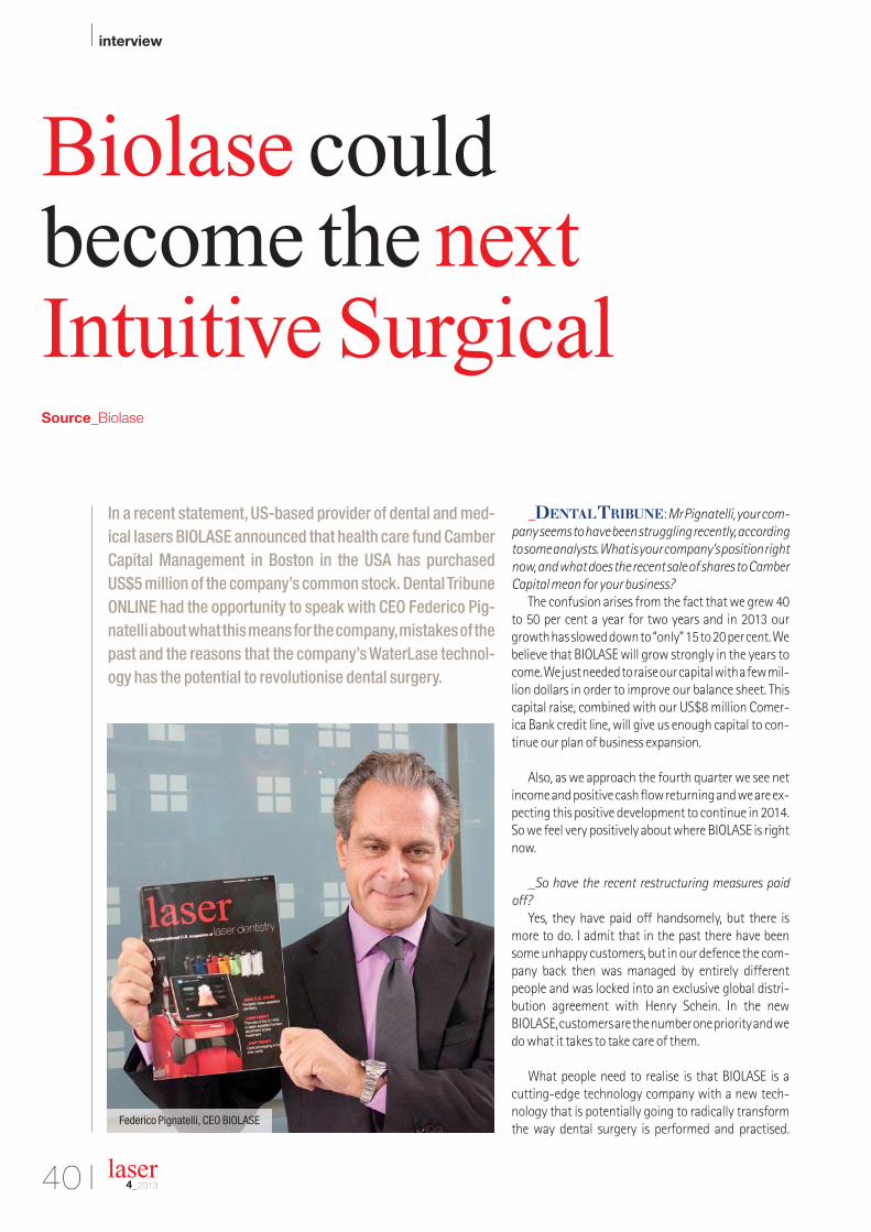
I interview
_DENTALTRIBUNE: Mr Pignatelli, your com-pany seems to have been struggling recently, accordingto some analysts. What is your company’s position rightnow, and what does the recent sale of shares to CamberCapital mean for your business?
The confusion arises from the fact that we grew 40to 50 per cent a year for two years and in 2013 ourgrowth has slowed down to “only” 15 to 20 per cent. Webelieve that BIOLASE will grow strongly in the years tocome. We just needed to raise our capital with a few mil-lion dollars in order to improve our balance sheet. Thiscapital raise, combined with our US$8 million Comer-ica Bank credit line, will give us enough capital to con-tinue our plan of business expansion.
Also, as we approach the fourth quarter we see netincome and positive cash flow returning and we are ex-pecting this positive development to continue in 2014.So we feel very positively about where BIOLASE is rightnow.
_So have the recent restructuring measures paidoff?
Yes, they have paid off handsomely, but there ismore to do. I admit that in the past there have beensome unhappy customers, but in our defence the com-pany back then was managed by entirely different people and was locked into an exclusive global distri-bution agreement with Henry Schein. In the new BIOLASE, customers are the number one priority and wedo what it takes to take care of them.
What people need to realise is that BIOLASE is a cutting-edge technology company with a new tech-nology that is potentially going to radically transformthe way dental surgery is performed and practised.
Biolase could become the next Intuitive SurgicalSource_Biolase
40 I laser4_2013
In a recent statement, US-based provider of dental and med-ical lasers BIOLASE announced that health care fund CamberCapital Management in Boston in the USA has purchasedUS$5 million of the company’s common stock. Dental TribuneONLINE had the opportunity to speak with CEO Federico Pig-natelli about what this means for the company, mistakes of thepast and the reasons that the company’s WaterLase technol-ogy has the potential to revolutionise dental surgery.
Federico Pignatelli, CEO BIOLASE
interview I
As a new step in informing the marketplace about WaterLase, we have recently embarked on a social media and press campaign to reach out to millions ofpatients to educate them about the many advantagesof being treated with BIOLASE’s technology.
We are particularly glad to have Dr Fred Moll, the co-founder of Intuitive Surgical, who values our technol-ogy such that he joined our board of directors recently.He is a legend in the medical field because with his com-pany he transformed the way surgery is approachedthrough the use of robotics. Thanks to a visionary likehim, today tens of thousands of patients with cancercan be treated in a much more precise way than ever be-fore.
We believe BIOLASE has a technology that is so ad-vanced and revolutionary that the company could be-come the next Intuitive Surgical. That is because withWaterLase technology we can transform surgical den-tistry for hundreds of thousands of dental practicesaround the world, while providing better and safer carefor patients.
_Why do you think lasers and particularly Water-Lase will be the technology of choice in dentistry for thefuture?
If you think about it, dentistry has not really changedvery much since the dental drill was invented by theEgyptians 7,000 years ago. The principle of removingtissue by mechanical rotation has remained the samesince that time, with the only major change in the past70 years being the attachment of a high-speed engine.With WaterLase technology, we are able to make use ofthe most basic element of human tissue, water. The hu-man body in its entirety consists of 60 per cent water,so water can be found in almost all tissue. Dentine, forexample, has 20 per cent water in it. By energising wa-ter molecules with a laser, tissue can be cut withoutpain, heat, abrasion, vibration, or the risk of microfrac-tures. At the same time, it is also much more precise.Clinically, this is much better dentistry.
Furthermore, there is no need for any anaestheticfor the patient; 99 per cent of patients can be treatedwithout using Novocaine. How wonderful is that? Ontop of that, laser energy kills bacteria, viruses and fun-gus, and that provides another advantage for dentists,since it is almost impossible and certainly very costly tohave surgical instrumentation like dental burs and en-dodontic files fully sterilised, and too costly to use newinstrumentation for every patient to be treated.
_With all these advantages, why does it seem thatthe technology has not been adopted widely by dentistsyet?
That is not exactly true. Since the introduction ofWaterLase technology 15 years ago, we have sold over
10,000 units worldwide, 6,000 of which were in the USAalone. The main challenge however is education. Den-tists need to be better educated about the return on in-vestment and the system’s extensive clinical advan-tages in comparison with conventional dentistry.
In fact, only two and a half years ago, WaterLasetechnology for the very first time broke the speed bar-rier, which means that it now cuts as fast as a conven-tional dental drill, sometimes even faster. Furthermore,it allows impressive treatment and cutting of soft tis-sue, which is something a dental drill cannot do. Theseadditional options mean that dentists no longer needto refer patients to a specialist for these tasks, therebyboosting revenue in the practice.
_Where do you see the technology in the next five toten years?
In contrast with other market-leading systems ortechnologies, such as Sirona’s CEREC, WaterLase is pro-tected by over 100 patents, which will allows us to pro-tect our competitive advantage. The adoption cycle ofnew technologies is growing increasingly shorter andmore advanced technologies like WaterLase will rapidlyfind their way into dental practices. Dentists that do notupgrade their practices will likely begin to lose patients,become uncompetitive and lag behind. You cannotfight technology; you cannot fight innovation. If youdo, you are doomed to be left out of the market.
We regularly ask patients whether they would like tobe treated by a conventional dentist or high-tech den-tist, and almost 100 per cent of patients would like to betreated by a high-tech dentist. Therefore, we believethat WaterLase will be part of most dental practices inthe near future._
Thank you very much for the interview.
Editorial note: Dental Tribune is the international coopera-tion partner of laser international magazine of laserdentistry .
I 41laser4_2013
Biolase Europe GmbH
Paintweg 1092685 FlossGermany
Tel.: +49 9603 8080Fax: +49 9603 2360
_contact laser

I interview
_Mr. Spitzer, you refer to Unicim as a digital pro-duction center. What do you mean by that?
Unicim combines traditional production meth-ods with digital CAD/CAM manufacturing, such asmetal laser melting or powder-based plastic lasersintering. With rapid manufacturing methods,you can select the most functional and affordabledental prosthetic solution based on your cus-tomer's needs, be it crowns and bridges, frame-works, primary and secondary structures or im-plant supra-constructions.
_Can you give us an idea of the process for cre-ating dental restorations out of metal by using theadditive manufacturing technology?
Once the 3-D CAD data is complete, the supportstructures are set up with the help of a data pro-cessing software. Various software solutions areavailable for this purpose. One of the most com-mon is CAMbridge, which requires license fees.
Alternatively, there is AutoFab Mlab, which is li-cense-free and allows you to assign specific meas-urements. With Concept Laser systems, the cus-tomer is able to choose freely and is not bound byany software. The processed data is then trans-mitted to the machine via the network or USB portand the construction job is started. With thisprocess, you can finish a project fully automati-cally overnight. Once complete, the componentsare removed from the building board and refin-ished. After manually removing the support struc-tures, the surface is then microblasted with alu-minum oxide and, in the case of bridges, the crownedges are thinned down.
_Will milling and casting soon be a thing of thepast in dental prosthetics?
Milling and casting will remain part of the stan-dard repertoire of dental laboratories for trainingand application. Additive manufacturing options
Dentures produced using 3-D printing versus casting and milling
42 I laser4_2013
Non-precious metal alloys (NEM) are enjoying increased de-mand in dental technology. Additive manufacturing with lasermelting ensures the uniformity and accuracy of ceramic-ve-neered, non-precious metal restorations created from dustand light. Are the traditional manufacturing processes of den-tal technicians, such as casting and milling, making a come-back? We spoke with Master Dental Technician Dieter Spitzerof Unicim, a manufacturer of dental restorations based inBerschis in the Swiss canton of St. Gallen.
Fig. 1_Tailor-made dental technologies: Master Dental Technician Dieter Spitzer offers
traditional manufacturing along with CAD/CAM methods, such as laser melting of metals for
dental restorations.
All images courtesy of Concept Laser GmbH, Lichtenfels, Germany.
interview I
will offer many advantages in the future and re-duce production risk enormously. Unfortunatelythey are still far too rarely seen in practice by den-tists and dental technicians. Some of this has to dowith the old school mentality of doing everythingmanually. The dental laboratory of the future willbe more of a hybrid: milling and casting where de-sirable but with additive manufacturing as a topalternative. "Add on versus take away," I like to callit. In summary, the casting process, from creatingthe cast object to the finished product, is usuallyvery time consuming and can lead to distortion,especially with large-span restorations. With ad-ditive technology, we achieve contour accuracymore easily than with milling. Our workplaces indental technology are also cleaner thanks toCAD/CAM: less dust, less bonding agent, glue andoutgassing. Ultimately, the deciding factor isquality. Compared to casting and milling, additiveprinting processes are creating entirely new waysof thinking in terms of production, workflow andthe products themselves.
_How are these changes expressed?We need to look at different levels here. First is
the transition from manual craftsmanship tohigh-precision, high-accuracy industrial CAD/CAM production. Milled non-precious metalrestorations have significant disadvantages dueto material consumption: high production costsand system-related lower quality in terms of fitand shape retention. During casting, we also en-counter disadvantages in terms of low materialdensity, mold costs, production time and rework.Nearly all of these disadvantages disappear withlaser melting. By using proven materials like re-manium star CL and rematitan CL from Dentau-rum with our Mlab cusing R, we have been verysatisfied with the quality of our system-manufac-tured products. In the case of large-volumerestorations, any excess tension that arises can bealleviated through subsequent heat treatment,thus avoiding any potential distortion. Of course,the same applies to cobalt-chromium alloys or ti-tanium.
_You mentioned changes in the products. Whatchanges were you referring to?
I'm quite optimistic. I'll describe a couple ofthem. First, the geometric flexibility of prostheticsis enabling a new way of looking at shapes or func-tions. In the future, imagine restorations withchannels into which medications can be fed. Thedentist or orthodontist can provide treatment,and the patient won't have to deal with tempo-raries. The second major change is the selectivedensity of a component made possible by theprocess. Thus, for example, bridges with more than
ten sections can not only be manufactured in aone-step process tension-free, they can also beincreasingly applied in heavily utilized areas, suchas cantilevers, edges or brace elastics. In modelcasting, that's not always an easy problem tosolve. Geometric freedom is a genuine plus for us,as it opens up new possibilities for restoration de-sign. For example, brace elements can be mademuch finer while retaining sufficient mechanicalproperties.
These new options also increase the longevityof dental product. In casting or milling, we have todeal with cost, material waste and lower materialdensity; in casting especially, we have oversizeddimensions and much lower material densities.With cast restorations, breakage is always an is-sue. But it doesn't have to be that way. Anotherbenefit is the ability to create combinationsthrough module or multicomponent constructionmethods. Base elements implanted into the jaw-bone are used as primary structures. An additivelymanufactured foundation element is then putinto place as a secondary structure, onto which asecure, durable veneer such as HeraCeram is ap-plied. Another aspect relates to new materials,such as non-precious metal titanium...
_Titanium would be hard and biocompatible...Titanium is the ideal material for allergy suffer-
ers, for example. In combination with laser melt-ing and veneering, we can maximize its biologicalbenefits. From a visual standpoint, titaniumrestorations offer a risk-free silver-gray luster.Manufacturers of non-precious metal alloys havespread pseudoscientific criticism regarding itsaesthetics.
Low-dose fluoride in toothpaste or mouth-wash, for example, has no impact on appearance.We can't deny the reality that titanium has notonly caught up to non-precious metal alloys in im-portance, it has surpassed them. This is preciselywhy, in 2012, Unicim invested in a Mlab cusing Rsystem for titanium applications from ConceptLaser, which allows us to process reactive titaniummaterial in a closed system. The unit can be usedwith dental materials certified under the GermanMedical Devices Act, such as rematitan CL fromDentaurum. Because of the high amount of mate-rial waste, milling-based processing of titanium istoo expensive and casting is highly impractical.
_What are some of the problems that arise inthe casting of titanium?
The reaction of titanium with oxygen causes aso-called alpha-case layer to form on the outside.This leads to embrittlement of the surface and
I 43laser4_2013

I interview
Fig. 2_Crowns and bridges
manufactured using laser
melting technology.
Fig. 3_Cast parts manufactured
with LaserCUSING.
must be removed. If not removed, it can leads toproblem with the adhesion of veneering. WithLaserCUSING, no alpha-case layer forms. Thismakes laser melting with titanium powder excel-lent for processing. The very fine-grained mi-crostructure of the laser-fused parts of this tita-nium alloy allows greater firmness than with con-ventional castings. The dentist receives a high-performance, long-life alternative that's easy towork on and more affordable than a preciousmetal solution. Finally, dentists and patients canbenefit from a quality product that is both durableand natural in appearance.
_How does titanium compare in terms of price?The price of the Dentaurum titanium powder
we use is currently around 595 euros per kilogram;a 4-unit bridge weighing 4 grams thus costs EUR2.40 in materials alone.
_Why has laser melting been so slow to catchon in the dental industry?
The reasons for this are many. The process is rel-atively new, so the learning curve is enormous. Thefact that the quality of laser-fused products is bet-ter than conventionally manufactured dentalrestorations remains largely unknown. Its reputa-tion continues to be tarnished by ignorance ormisconceptions. Keep in mind, too, that dentaltechnician training takes four years in Switzer-land, and theoretical instruction is slow to incor-porate new technologies. In addition, Swiss den-tal labs are very small.
The Association of Swiss Dental Technicians es-timates some 1,200 centers, many of which oper-ate with just 1 or 2 people. Therefore, investmentsin laser melting are carefully considered. Unicim,as a digital production center, acts as a serviceprovider, supplying other laboratories. Right now
I see it as more of an outsourcing topic while wewait for it to take hold in the market.
_What is the position of dentists regarding thisissue?
Interest is undoubtedly growing, not least be-cause it's impossible to ignore the technical, time-saving and affordability benefits. But we also needto look at the process chain. To prepare the data formanufacturing, the STL format is required. STLdata from different scanners can be processed us-ing the CAMbridge or AutoFab Mlab data process-ing software available from Concept Laser. Nowa-days, conventional dental impressions form thebasis for CAD data. The accuracy of the data de-pends on the preciseness of the work performedthe dentist. Higher accuracy is essential. A high-quality intraoral scanner costs dentists todayabout 20,000 Swiss francs (CHF). If we had com-plete data migration from the dentist to the den-tal laboratory, we would be one step further. In thelong term, however, that is unavoidable. Qualityassurance and documentation needs will makeopen, manufacturer-independent data transferan increasingly critical requirement. Especially interms of affordability, the topic of laser melting isbecoming more important._
44 I laser4_2013
Concept Laser GmbH
An der Zeil 8, 96215 LichtenfelsGermany
Tel.: +49 9571 949238Fax: +49 9571 949249
www.concept-laser.de
_contact laser
Fig. 2 Fig. 3

The editors of laser would like to thank all authors for dedicating their time and
efforts to this year’s issues.
laser������������������� �� ������������12013
� � �� ������� ��������������������������������������������� ��������� ���� ��� �
| �������������������������������������������
| �������������������������������������������� �����
| �����������������!����������������"������������������������!������������������
Issue 1/2013
| Dr Jonas de Almeida Rodrigues, Brazil| Cristiane Meira Assunção, Brazil| Dr Georg Bach, Germany| Prof. Dr Reiner Biffar, Germany| Prof. Dr Carlo Fornaini, Italy| Dr Michael Hopp, Germany | Dr Darius Moghtader, Germany| Joanna Tatith Pereira, Brazil| Dr Avi Reyhanian, Israel| Renata Schlesner Oliveira, Brazil
laser������������������� �� ������������22013
� � �� ������� ��������������������������������������������� ��������� �������� �
| ��������������� ����������������� �����
| ������������ ���� �������� ������������ ��� ��
| ���������������������� �� �����������#���������� ��� �
Issue 2/2013
| Dr Ralf Borchers, MSc, Germany| Prof. Dr Carlo Fornaini, Italy| George Freedman, Canada| Dr Shachi Goenka, India| Dr Fay Goldstep, Canada| Prof. Dr Norbert Gutknecht, Germany| Dr Vivek Hegde, India| Dr Paresh Jain, India| Hans J. Koort, Germany| Samah S. Mehani, Egypt| Dr Ana Minovska, Bulgaria| Laura Navasaityte, Bulgaria| Birute Rakauskaite, Bulgaria| Ali M. Saafan, Egypt| Dr Sucheta Sathe, India| Prof. Dr Georgi Tomov, Bulgaria| Dr Naresh Thukral, India| Nermin M.Yussif, MSc, Egypt
laser������������������� �� ������������32013
���� ������� ��������������������������������������������� ��������� �������� �
| ������������������������������������������������������������������
| ��������������������������������������������������������
| �������������� ������������������������������������������������������������������������
Issue 3/2013
| Dr Glenn A. van As, Canada| Dr Georg Bach, Germany| José Luis Capote Femenias, Cuba| Res. Asst. Dt. Necla Aslı Kocak, Turkey| Prof. (Shandong University, China) | Dr Frank Liebaug, Germany| Dr Darius Moghtader, Germany| Giovanni Olivi, Italy| Matteo Olivi, Italy| Emin Selim Pamuk, Turkey| Dr Wariya Panprasit, Thailand| Prof. Dr Georgios Romanos, USA| Pedro J. Muñoz Sánchez, Cuba| Sajee Sattayut, Thailand| Prof. Dr Georgi Tomov, Bulgaria| Jan Tunér, Sweden| Dr Blagovesta Yaneva, Bulgaria| Prof. Dr Aslan Yasar Gokbuget, Turkey| Dr Anna Maria Yiannikos, Germany/Cyprus| Dr Ning Wu, Germany
laser������������������� �� ������������42013
� � �� ���������� ��������������������������������������������� ��������� ��������� �
| �� ������ �������������������������$������������%�������������������$�����������
| � ������������������%���������������&���$����$%������$����%�����������$�������'���$����
| ������������� �����������$��$������$��$%
Issue 4/2013
| Prof. Carlo Fornaini, Italy| Prof. Dr Norbert Gutknecht, Germany| Prof. Dr Michael Hopp, Germany| Dr Kirpa Johar, India | Zafer Kazak, Turkey| Hans J. Koort, Germany| Dr Ilay Maden, Turkey| Özge Erbil Maden, Turkey| Miguel Martins, Portugal| Prof. Dr D. Gabrić Pandurić, Croatia| Assoc. Prof. Dr Sajee Sattayut, Thailand| Dr Michael Sultan, UK| Dr Sawanya Taboran, Thailand| Dr Anna Maria Yiannikos, Germany/Cyprus
Please contact Claudia Jahn

_On the 19 and 20 of September in Vilnius, thecapital of Lithuania, the “World Laserology Congress”was celebrated with the participation of the most im-portant scientific societies involved with the utilisa-tion of the laser technology in medicine: ISLSM (In-ternational Society for Laser in Surgery and Medi-cine), IPTA (International Photo-Therapy Associa-tion), WFSLMS (World Federation Societies for Laserin Medicine and Surgery), WFLD (World Federationfor Laser in Dentistry) and EMDOLA ACADEMY.
The President of the Congress, Prof. AurelijaVaitkuviene, also President of IPTA, decided to pro-pose a program based on the interaction of physicsand medicine in all the specialities and, for these rea-son, she invited as speakers people who are amongthe best experts of the field in the world.
So, Prof. Toshio Oshiro from Japan, General Sec-retary of WFSLMS and Editor of the journal “LaserTherapy”, Prof. Jouzas Vaitkus, President of theLithuanian Physical Society, Prof. Leonardo Longofrom Florence, President of WFSLMS and IALMS, Prof.Juanita Anders from Bethesda University, PresidentElect of the American Society for Laser in Medicineand Surgery, Prof. Chang Jen Cheng, President ofLaser and Photonics Medicine of the Republic ofChina, Prof. Krishna Rau from India, Past President of
WFLSMS, Prof. Carlo Fornaini from Italy, PresidentElect of WFLD and Prof. Jean-Paul Rocca from France,Past President of WFLD and Honorary President ofEMDOLA Academy, showed to the participants thestate-of-the-art of the application of the laser tech-nology in each medical and surgical field by severalimportant lectures.
Among others, lectures were held on the therapyof LLLT on the paresis of facial nerve (Anders), thelaser effect on the cerebral palsy (Asagai), the lasertreatment of scars (Oshiro), the laser action on the re-generation of the mandibular nerve lesions (Longo),The IntraOral Laser Welding (Fornaini) and the Edu-cational Program in Laserology (Rocca).
During the congress, the assembly of IPTA electedthe new president in the person of Prof. Jean PaulRocca, the director of the TELEO Laboratory of theUniversity of Nice, an expert on the interaction be-tween Er:YAG and hard dental tissues.
Prof. Rocca, in acceptating of the new position,announced that the next IPTA Congress will be heldin Nice in June 2015, joint to the Emdola AcademyCongress. All our good wishes for a good and suc-cessful work in the association go to Prof. Rocca andthe new Board of IPTA._
I meetings
Jean-Paul Rocca new President ofIPTAAuthor_Prof. Carlo Fornaini, Italy
46 I laser4_2013
Prof. Rocca, new President of IPTA (right), with Prof. Vaitkuviene,
Past President (center) and Prof. Oshiro, Treasurer (left).[PICTURE: ©LA.ANELE]

International events
meetings I
I 47laser4_2013
2013
Greater New York Dental Meeting
New York, USA
29 December 2013 to 4 January 2014
www.gnydm.com
2013 Dental Istanbul
Istanbul, Turkey
7–8 December 2013
www.dentalistanbul.com
2014
AEEDC Dubai 2014
18th UAE International Dental Conference
& Arab Dental Exhibition
Dubai, UAE
4–6 February 2014
www.aeedc.com
ALD 2014 Academy of Laser Dentistry
Scottsdale, AZ, USA
27 February to 1 March 2014
www.laserdentistry.org
Pacific Dental Conference 2014
Vancouver, Canada
6–8 March 2014
www.pdconf.com
Academy of Osseointegration
Annual Meeting 2014
Seattle, WA, USA
6–8 March 2014
www.osseo.org
WFLD — 5th Congress
Paris, France
3–4 July 2013
www.wfld.info

NEWS
A new study conducted by researchers at the De-partment of Dental Public Health at King’s CollegeLondon has found that some dental patients mayneed to consult a psychologist before undergoingtreatment. In a study with 60 participants, the re-searchers found that higher satisfaction with ap-pearance before dental aesthetic treatment affectedpatients’ satisfaction after treatment significantly.
For the study, all participants were asked to assesssatisfaction with their appearance before and afterdental work according to a predefined scale. Addi-tionally, they completed a personality test prior to theprocedure. Among other findings, the researchersfound that participants who were most satisfied withtheir appearance before receiving dental aesthetictreatment were also the happiest patients aftertreatment. On the other hand, neuroticism seemed topersist after treatment in those patients who wererather dissatisfied before.
“We found that it is in patients’ and dentists’ interestto ensure that patients receiving aesthetic dentalwork start from as high a point of satisfaction withtheir current appearance as possible. This will en-hance the chances that they will be satisfied with theresult of the treatment,” the researchers concluded.The findings were presented on Wednesday at theBritish Psychological Society’s Division of HealthPsychology Annual Conference, which was heldfrom 11 to 13 September in Brighton.
Predictors of satisfaction with
Aesthetic dentalwork
Researchers from the UKhave evaluated the oralhealth status of athleteswho took part in the2012 Olympic Gamesheld in London. Throughsystematic oral check-ups, the researchersfound that 55 per cent of the302 athletes surveyed suf-fered from dental caries. In ad-dition, more than 75 per cent had gingivi-tis, with 15 per cent showing signs of periodontitis. They found signs of dentalerosion in about 45 per cent of athletes.Although 42 per cent of the participantssaid that their oral health status botheredthem, nearly half of them (47 per cent)had not undergone a dental examinationor professional cleaning in the previousyear, the researchers reported. Almost 9 percent had never been to the dentist. While 28per cent of athletes stated that they believedthat their oral health affected their quality oflife, about 18 per cent said that it was having
a negative impact on their training and perform-ance. According to the researchers, the findings corro -
borate those of previous studies, which sug-gested that poor oral health in sportsmen
might be a result of frequent carbohydrateintake, a reduced immune function owingto intensive training, and a lack of aware-
ness about the link between oral health,overall health and physical perform-
ance.
The study was conducted by re-searchers at University College Lon-
don’s Eastman Dental Institute at thedental clinic in the London 2012 ath-
letes’ village. The study population com-prised athletes from Africa, the Americas
and Europe competing in 25 different sports,including track and field, boxing and hockey.The study, titled “Oral health and impact onperformance of athletes participating in theLondon 2012 Olympic Games”, was pub-lished online on 24 September in the BritishJournal of Sport Medicine ahead of print.
Olympic athletes with
Poor oral health
The UK Department of Health announced its plansto introduce language tests for European medicalprofessionals wishing to work in the UK. The newregulation will give the General Medical Council(GMC) the power to impose checks where con-cerns arise about a doctor’s English language com-petence.
According to the department, the new proposalsare designed to complement and strengthen theexisting language controls imposed on medicalstaff through the Responsible Officers Regulations,Performers Lists Regulations and other checks un-dertaken at a local level, which were introduced inApril this year.
Health Minister Dr Dan Poulter explained that theGMC will be entitled to carry out assessments be-fore allowing European doctors from outside the UKto treat patients in a hospital or general practice. Bycarrying out tests at a national level, the ministerhopes to prevent doctors who do not have the nec-essary knowledge of English from treating pa-tients. To be accepted on a centrally held list, gen-eral practitioners will have to demonstrate theirability to communicate in English. The new rulesare expected to come into effect in 2014.
Foreign dentists
Face language tests in UK
48 I laser4_2013
[PICTURE: ©WOAISS]
[PICTURE: ©ARTFAMILY]
[PICTURE: ©BUTTET]

I 49laser4_2013
Dentophobia is equally common in both men andwomen. However, a new study has found that the levelof disgust they experience with regard to dental treat-ment may differ significantly. In the study, 36 individu-als with dentophobia (18 men and 18 women) and 36non-dentophobic controls were asked to rate theirarousal, disgust and fear while looking at images ofdental treatment scenes and images depicting neutralscenes. Simultaneously, their heart rate and the activ-ity of the musculus levator labii, a muscle used in pro-
totypical facial disgust expression, producing nosewrinkles and upper lip retraction, were recorded elec-tronically. Overall, male and female participants did notdiffer in their self-ratings. However, there were signifi-cant differences in facial expressions. According to theresearchers, only dentophobic women showed en-hanced disgust-related facial activity, indicating thattargeting of disorder-specific disgust might be of greatimportance in the therapy of dentophobic women.
Although dentophobic men perceived dental treat-ment scenes as equally disgusting as did women, theydisplayed significantly lower disgust-related facial ac-tivity. However, the researchers suggested that maleparticipants might have been more successful in in-hibiting behavioural reactions. The study, titled “Canyou read my pokerface? A study on sex differences indentophobia”, was published online on 12 Septemberin the European Journal of Oral Sciences and will ap-pear in the October issue. It was conducted at the Uni-versity of Graz.
Dentophobia
More common in women than men
People with poor oral hygiene or gum disease maybe at a greater risk of developing Alzheimer’s dis-ease, a study led by the University of Central Lan-cashire’s (UCLan) School of Medicine and Dentistryhas found. For their study, the researchers exam-ined samples from the donated brains of ten peo-
ple without dementia and ten people with demen-tia. The research found the presence of productsfrom Porphyromonas gingivalis in the brains of de-mentia patients.
The research benefited from donated brain sam-ples provided by Brains for Dementia Research, abrain donation scheme supported by Alzheimer’sResearch UK and Alzheimer’s Society. Finding P.gingivalis in the brains of dementia sufferers is sig-nificant, as its presence in the brains of Alzheimer’sdisease patients has not been documented previ-ously and the finding adds to a growing body of ev-idence that suggests an association between poororal health and dementia.
These published research findings from humanbrain specimens are further supported by recentunpublished research on periodontal disease fromthe same group using animal models, which wascarried out in collaboration with the University ofFlorida. This animal work has confirmed that P. gin-givalis in the mouth finds its way to the brain onceperiodontal disease has become established.
Poor dental health
May lead to Alzheimer’s
Psychologists from Germany have suggested that eat-ing popcorn disrupts the way people process and re-member brand names. In a recently published study,participants were invited to a movie theatre and wereshown a block of foreign commercials prior to the film.Half of the participants were given popcorn to eat, while
the other participants chewed gum or ate a single sugarcube. When the participants were presented with im-ages of products one week after the cinema session,those who had eaten the sugar cubes exhibited higherpreference and physiological responses for the brandsadvertised. In contrast, the participants who had con-sumed popcorn or gum during the commercialsshowed no evidence of advertising effects, the re-searchers said. According to researchers from the Uni-versity of Cologne, the advertised products were lessfamiliar to the participants owing to the fact that theirmouth was obstructed when they were watching thecommercials. Prior studies have shown that subvocalpronunciation, a covert mechanism of the mouth, isvery important to the perception of information aboutnew brands. The researchers said that each time a per-son encounters a new name the lips and the tongue au-tomatically simulate the pronunciation of the name.Because they were chewing, the participants could notinternally train the articulation of the brands’ names.
The researchers concluded that advertising might re-main unsuccessful in situations involving oral interfer-ence, such as snacking or talking. The study, titled“Popcorn in the cinema: Oral interference sabotagesadvertising effects”, was published online on 29 Sep-tember in the Journal of Consumer Psychology aheadof print.
Chewing popcorn may disturb
Moviegoers’ percep-tion of commercials
[PICTURE: ©MYPOKCIK]
[PICTURE: ©ZAMETALOV]
[PICTURE: ©KTSDESIGN]

I about the publisher _ imprint
50 I laser4_2013
Copyright Regulations
_laser international magazine of laser dentistry is published by OEMUS MEDIA AG and will appear in 2013 with one issue every quarter. The magazine and all articles and illustrations therein are protected by copyright. Any utilization without the prior consent of editor and publisher is inad-mi ssible and liable to prosecution. This applies in particular to duplicate copies, translations, microfilms, and storage and processing in electronic systems.
Reproductions, including extracts, may only be made with the permission of the publisher. Given no statement to the contrary, any submissions to theeditorial department are understood to be in agreement with a full or partial publishing of said submission. The editorial department reserves the right tocheck all submitted articles for formal errors and factual authority, and to make amendments if necessary. No responsibility shall be taken for unsolicitedbooks and manuscripts. Articles bearing symbols other than that of the editorial department, or which are distinguished by the name of the author, representthe opinion of the afore-mentioned, and do not have to comply with the views of Oemus Media AG. Responsibility for such articles shall be borne by the author.Responsibility for advertisements and other specially labeled items shall not be borne by the editorial department. Likewise, no responsibility shall be assumedfor information published about associations, companies and commercial markets. All cases of consequential liability arising from inaccurate or faulty representation are excluded. General terms and conditions apply, legal venue is Leipzig, Germany.
Publisher
Torsten R. Oemus [email protected]
CEO
Ingolf Dö[email protected]
Members of the Board
Jürgen [email protected]
Lutz V. [email protected]
Editor in Chief
Norbert [email protected]
Coeditors in Chief
Samir NammourJean Paul Rocca
Managing Editors
Georg BachLeon Vanweersch
Division Editors
Matthias FrentzenEuropean Division
George RomanosNorth America Division
Carlos de Paula EduardoSouth America Division
Toni ZeinounMiddle East & Africa Division
Loh Hong SaiAsia & Pacific Division
Senior Editors
Aldo Brugneira JuniorYoshimitsu AbikoLynn PowellJohn FeatherstoneAdam StabholzJan TunerAnton Sculean
Editorial Board
Marcia Martins Marques, Leonardo Silberman,Emina Ibrahimi, Igor Cernavin, Daniel Heysselaer,Roeland de Moor, Julia Kamenova, T. Dostalova,Christliebe Pasini, Peter Steen Hansen, Aisha Sul-tan, Ahmed A Hassan, Marita Luomanen, PatrickMaher, Marie France Bertrand, Frederic Gaultier,Antonis Kallis, Dimitris Strakas, Kenneth Luk, Mukul Jain, Reza Fekrazad, Sharonit Sahar-Helft,Lajos Gaspar, Paolo Vescovi, Marina Vitale, CarloFornaini, Kenji Yoshida, Hideaki Suda, Ki-Suk Kim,Liang Ling Seow, Shaymant Singh Makhan, Enri-que Trevino, Ahmed Kabir, Blanca de Grande, JoséCorreia de Campos, Carmen Todea, Saleh GhabbanStephen Hsu, Antoni Espana Tost, Josep Arnabat,Ahmed Abdullah, Boris Gaspirc, Peter Fahlstedt,Claes Larsson, Michel Vock, Hsin-Cheng Liu, SajeeSattayut, Ferda Tasar, Sevil Gurgan, Cem Sener,Christopher Mercer, Valentin Preve, Ali Obeidi,Anna-Maria Yannikou, Suchetan Pradhan, RyanSeto, Joyce Fong, Ingmar Ingenegeren, Peter Klee-mann, Iris Brader, Masoud Mojahedi, Gerd Volland, Gabriele Schindler, Ralf Borchers, StefanGrümer, Joachim Schiffer, Detlef Klotz, HerbertDeppe, Friedrich Lampert, Jörg Meister, ReneFranzen, Andreas Braun, Sabine Sennhenn-Kirch-ner, Siegfried Jänicke, Olaf Oberhofer, ThorstenKleinert
Editorial Office
Georg [email protected]
Claudia [email protected]
Executive Producer
Gernot [email protected]
Designer
Sarah [email protected]
Customer Service
Marius [email protected]
Published by
OEMUS MEDIA AGHolbeinstraße 2904229 Leipzig, GermanyTel.: +49 341 48474-0Fax: +49 341 [email protected]
Printed by
Löhnert Druck Handelsstraße 1204420 Markranstädt, Germany
laser
international magazine of laser dentistryis published in cooperation with the World Federation for Laser Dentistry (WFLD).
WFLD Headquarters
University of Aachen Medical FacultyClinic of Conservative DentistryPauwelsstr. 30, 52074 Aachen, GermanyTel.: +49 241 808964Fax: +49 241 [email protected]
laserinternational magazine of laser dentistry

laserinternational magazine of laser dentistry
I would like to subscribe to laser international magazine of
laser dentistry (4 issues per year) for € 44 including shippingand VAT for German customers, € 46 including shipping and VATfor customers outside Germany, unless a written cancellation issent within 14 days of the receipt of the trial subscription. Thesubscription will be renewed automatically every year until a writ-ten cancellation is sent to OEMUS MEDIA AG, Holbeinstr. 29,04229 Leipzig, Germany, six weeks prior to the renewal date.
Last Name, First Name
Company
Street
ZIP/City/County
E-mail Signature
Signature
OEMUS MEDIA AGHolbeinstraße 29, 04229 Leipzig, Germany, Tel.: +49 341 48474-0, Fax: +49 341 48474-290, E-Mail: [email protected]
Reply via Fax +49 341 48474-290 to OEMUS MEDIA AG or per E-mail to [email protected]
Notice of revocation: I am able to revoke the subscription within 14 days after my order by sending a writtencancellation to OEMUS MEDIA AG, Holbeinstr. 29, 04229 Leipzig, Germany.
lase
r 4/1
3
" Subscribe now!
You can also subscribe via www.oemus.com/abo

instantly adjustable spot size and shape
precise coverage of large areas
lightweight, ergonomic design
for Fotona LightWalker AT laser systems
The first digitally controlled dental laser handpiece
X-Runner
The universe at your fingertips.92734/1
92734_X_Runner_A4_LAHA_magazine_v1.pdf 192734_X_Runner_A4_LAHA_magazine_v1.pdf 1 26.04.13 16:3226.04.13 16:32