Embed Size (px)
Citation preview
Large chondral fragment of the lateral femoral condyle treatedwith arthroscopic internal fixation in an elite young athlete
A case report
Mitchell W. Beckert* and Robert G. Klitzman
Department of Orthopaedics, Indiana University, Indianapolis, IN, USA
Received 28 July 2019, Accepted 20 December 2019, Published online 8 January 2020
Abstract – Focal chondral lesions in the adolescent population create a particular challenge for orthopedic surgeons,and currently there exists no consensus on proper treatment. Numerous techniques for addressing focal chondraldefects are employed in both pediatrics and adults, including fragment excision, debridement and fixation, bonemarrow stimulation and microfracture techniques, cell-based options, as well as chondral and osteochondral grafts.Although historical evidence is mixed, recent reports of primary fixation of displaced cartilage fragments have shownfavorable results. We present a case of reduction and fixation of a large displaced cartilage lesion in an elite youngtennis player. Our results, in addition to other reports mentioned in this manuscript, highlight the importance ofconsidering primary fixation of large chondral lesions when amenable to repair.
Introduction
Focal chondral lesions in the adolescent population create aparticular challenge for orthopedic surgeons due to their limitedhealing potential [1]. Cartilage restoration techniques have seensignificant advances in the last decade; however, their role inpediatric patients is not well-defined due to the lack of high-quality, long-term data [2, 3]. While displaced osteochondrallesions amenable to fixation have shown good outcomes[4–6], the success of fixation when cartilage fragments aredevoid of subchondral bone is rarely described and previouslybeen questioned [7]. The evidence for reduction and internalfixation of large cartilage fragments is limited to case reports[8–15], and the purpose of this study is to contribute yet anothercase to the literature of successful primary fixation of a largechondral fragment in the knee, here in a highly active youngpatient.
Both the patient and parents provided written informedconsent to the submission of this case report.
Case report
An 11-year-old elite level male tennis player presented forevaluation of acute right knee pain after jumping and land-ing on his feet during a tennis tournament. The patient was
accompanied to clinic by his father, who reported immediatepain, swelling, and locking of his knee after landing. He wasunable to continue participation or bear weight on the affectedleg. The patient denied any history consistent with patellar sub-luxation or dislocation. At the time the patient was the numbertwo ranked tennis player in the United States for his age, andhad traveled across the country to compete against players fromall over the world. He was otherwise healthy and had no signif-icant past medical, surgical, or relevant family history. He was avery active competitive athlete in the seventh grade.
The patient was first evaluated at an outside facility, whereMRI demonstrated a large chondral defect sitting in the femoralnotch. He subsequently was referred to a pediatric orthopedistwho placed him in a knee immobilizer, recommended not tobear weight on the affected side, and referred the patient tosee us in the orthopedic sports medicine clinic. The patientwas 5 feet 1 inch tall, and weighed 44.6 kg at the time of exam-ination. On physical exam, his right knee demonstrated a mildeffusion. Range of motion testing was deferred to avoid furthercartilage damage; however, the patient was comfortably restingat approximately 90� of flexion and reported being able to bearweight on the right leg. He was neurovascularly intact.Examination of the contralateral knee was within normal limits,with range of motion negative 3–135�.
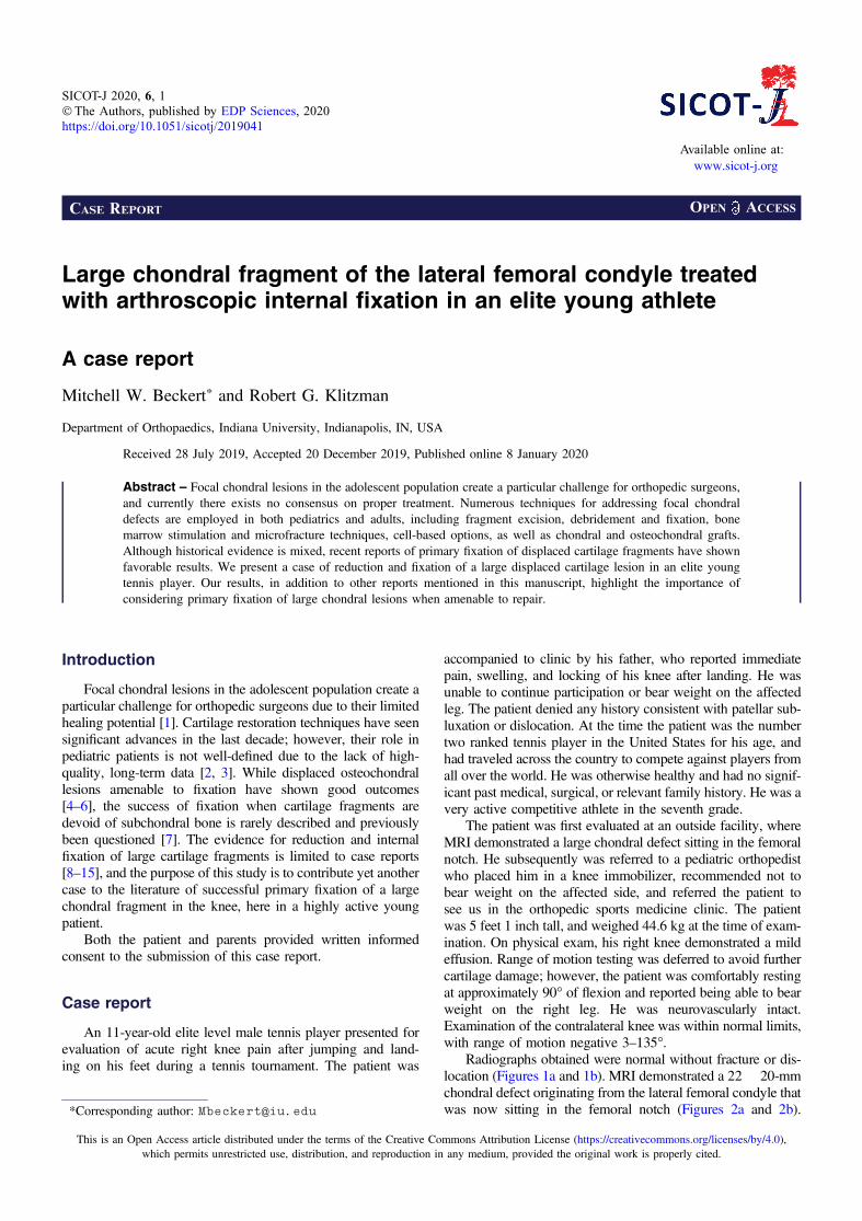
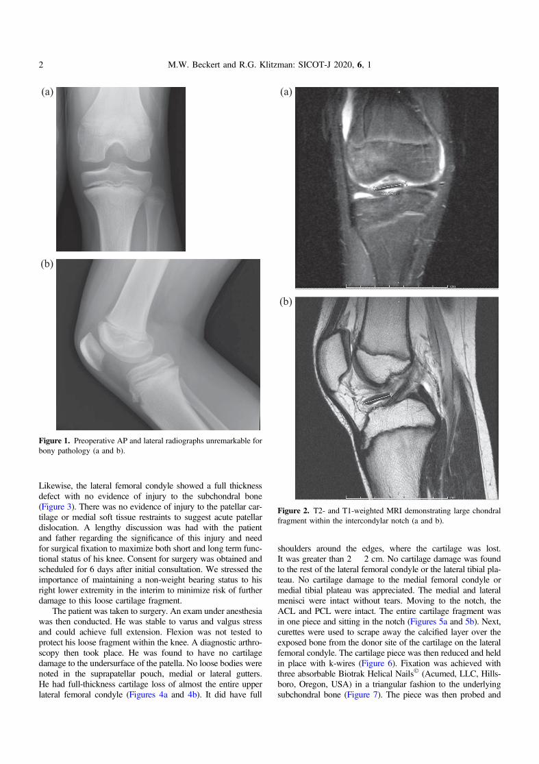
Radiographs obtained were normal without fracture or dis-location (Figures 1a and 1b). MRI demonstrated a 22� 20-mmchondral defect originating from the lateral femoral condyle thatwas now sitting in the femoral notch (Figures 2a and 2b).*Corresponding author: [email protected]
SICOT-J 2020, 6, 1�The Authors, published by EDP Sciences, 2020https://doi.org/10.1051/sicotj/2019041
Available online at:www.sicot-j.org
This is an Open Access article distributed under the terms of the Creative Commons Attribution License (https://creativecommons.org/licenses/by/4.0),which permits unrestricted use, distribution, and reproduction in any medium, provided the original work is properly cited.
OPEN ACCESSCASE REPORT
Likewise, the lateral femoral condyle showed a full thicknessdefect with no evidence of injury to the subchondral bone(Figure 3). There was no evidence of injury to the patellar car-tilage or medial soft tissue restraints to suggest acute patellardislocation. A lengthy discussion was had with the patientand father regarding the significance of this injury and needfor surgical fixation to maximize both short and long term func-tional status of his knee. Consent for surgery was obtained andscheduled for 6 days after initial consultation. We stressed theimportance of maintaining a non-weight bearing status to hisright lower extremity in the interim to minimize risk of furtherdamage to this loose cartilage fragment.
The patient was taken to surgery. An exam under anesthesiawas then conducted. He was stable to varus and valgus stressand could achieve full extension. Flexion was not tested toprotect his loose fragment within the knee. A diagnostic arthro-scopy then took place. He was found to have no cartilagedamage to the undersurface of the patella. No loose bodies werenoted in the suprapatellar pouch, medial or lateral gutters.He had full-thickness cartilage loss of almost the entire upperlateral femoral condyle (Figures 4a and 4b). It did have full
shoulders around the edges, where the cartilage was lost.It was greater than 2 � 2 cm. No cartilage damage was foundto the rest of the lateral femoral condyle or the lateral tibial pla-teau. No cartilage damage to the medial femoral condyle ormedial tibial plateau was appreciated. The medial and lateralmenisci were intact without tears. Moving to the notch, theACL and PCL were intact. The entire cartilage fragment wasin one piece and sitting in the notch (Figures 5a and 5b). Next,curettes were used to scrape away the calcified layer over theexposed bone from the donor site of the cartilage on the lateralfemoral condyle. The cartilage piece was then reduced and heldin place with k-wires (Figure 6). Fixation was achieved withthree absorbable Biotrak Helical Nails� (Acumed, LLC, Hills-boro, Oregon, USA) in a triangular fashion to the underlyingsubchondral bone (Figure 7). The piece was then probed and
(b)
(a)
Figure 1. Preoperative AP and lateral radiographs unremarkable forbony pathology (a and b).
(a)
(b)
Figure 2. T2- and T1-weighted MRI demonstrating large chondralfragment within the intercondylar notch (a and b).
2 M.W. Beckert and R.G. Klitzman: SICOT-J 2020, 6, 1

found to be stable. Temporary fixation pins were removed,and the knee was ranged through a full arc of motion. Thepatella was observed to track well over the fixed piece. Hewas placed in a knee brace locked in extension immediatelypostoperatively.
Figure 3. T1-weighted MRI showing full-thickness cartilage defectat the lateral femoral condyle.
(a)
(b)
Figure 4. Intraoperative arthroscopic images showing large full-thickness cartilage defect at the lateral femoral condyle withoutdisruption of the underlying subchondral bone (a and b).
(a)
(b)
Figure 5. Intraoperative arthroscopic images showing the large loosechondral fragment. This was found in one large piece within theintercondylar notch, without underlying bony attachment (a and b).
Figure 6. Provisional fixation of the chondral fragment usingk-wires.
Figure 7. Definitive fixation of the chondral fragment using threebioabsorbable pins in a triangular fashion.
M.W. Beckert and R.G. Klitzman: SICOT-J 2020, 6, 1 3
The patient was discharged from the hospital the same daywith crutches and strict non-weight bearing status, using thebrace during ambulation. The patient returned to clinic 2 weekspostoperatively where sutures were removed, and a continuouspassive motion device was given to help improve his kneerange of motion. He was given no restrictions to knee rangeof motion. At 6 weeks postoperatively the patient was allowedto begin bearing weight on his right leg with the brace locked inextension using crutches. Formal physical therapy was initiatedat this time. At 8 weeks postoperatively the patient was allowedto walk with his brace unlocked with the assistance of crutches.At 10 weeks postoperatively he began walking in his bracewithout crutches. At 3 months postoperatively the patient
reported beginning to hit tennis balls with his feet flat on theground without pivoting on his right leg. At 4 months postop-eratively the patient had weaned out of his brace and continuedto hit tennis balls from a stationary position. He was allowed toslowly increase his activities at this time starting with a light jogand subsequently cleared to return to full activities at 6 monthspostoperatively.
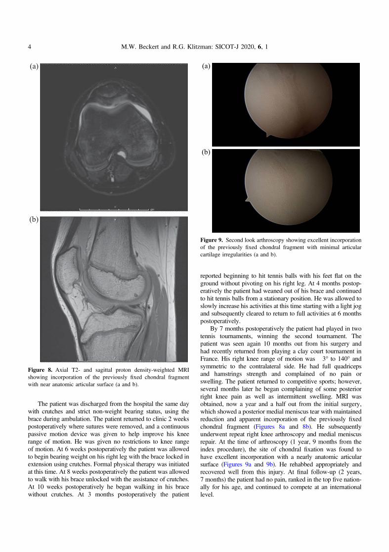
By 7 months postoperatively the patient had played in twotennis tournaments, winning the second tournament. Thepatient was seen again 10 months out from his surgery andhad recently returned from playing a clay court tournament inFrance. His right knee range of motion was �3� to 140� andsymmetric to the contralateral side. He had full quadricepsand hamstrings strength and complained of no pain orswelling. The patient returned to competitive sports; however,several months later he began complaining of some posteriorright knee pain as well as intermittent swelling. MRI wasobtained, now a year and a half out from the initial surgery,which showed a posterior medial meniscus tear with maintainedreduction and apparent incorporation of the previously fixedchondral fragment (Figures 8a and 8b). He subsequentlyunderwent repeat right knee arthroscopy and medial meniscusrepair. At the time of arthroscopy (1 year, 9 months from theindex procedure), the site of chondral fixation was found tohave excellent incorporation with a nearly anatomic articularsurface (Figures 9a and 9b). He rehabbed appropriately andrecovered well from this injury. At final follow-up (2 years,7 months) the patient had no pain, ranked in the top five nation-ally for his age, and continued to compete at an internationallevel.
(a)
(b)
Figure 9. Second look arthroscopy showing excellent incorporationof the previously fixed chondral fragment with minimal articularcartilage irregularities (a and b).
(a)
(b)
Figure 8. Axial T2- and sagittal proton density-weighted MRIshowing incorporation of the previously fixed chondral fragmentwith near anatomic articular surface (a and b).
4 M.W. Beckert and R.G. Klitzman: SICOT-J 2020, 6, 1

Discussion
Osteochondral injuries in the pediatric population are well-documented and most commonly caused by trauma, oftenfollowing acute patellar dislocation, or a result of osteochondri-tis dissecans (OCD) [16–22]. Although osteochondral lesionsusually have some degree of subchondral bone attachment, acartilage fragment can occur in isolation. Previous biomechan-ical studies have shown decreased resistance to shear stress atthe osteochondral junction in adolescents compared to adults,making adolescents more susceptible to this type of injury[23, 24]. When a displaced fragment is purely cartilaginous,it creates a difficult dilemma for the treating physician withno clear consensus option. Various treatment options forchondral defects include fragment excision, debridement andfixation, bone marrow stimulation and microfracture tech-niques, cell-based options, as well as chondral and osteochon-dral grafts [2]. Goals of treatment are focused on restoringarticular congruity of the joint surface and preventing futureosteoarthritis [2, 21, 22].
Fixation has historically been indicated for the classic osteo-chondral defect with a true osseous component, while excisionwith or without restorative procedures reserved for the carti-lage-only fragment [25, 26]. Several authors have consideredthese cartilage fragments “unsalvageable” and routinely exciseduring surgery and are discarded [8, 25, 27–33]. Maletius andLundberg reported poor healing potential in two cases of chon-dral fragment fixation, subsequently questioning the efficacy ofthis technique [7]. Recent reports, however, have shownsuccessful fixation of purely cartilaginous lesions [8–15]. Allcases were performed with open arthrotomies, except one caseof a 22-year-old patient who underwent fixation of a 1.5 cm2
cartilage fragment with arthroscopy [15]. Previously describedmethods of fixation of these cartilage fragments include bonepegs, chondral darts, bioabsorbable nails, and headless screws,of which can be augmented by fibrin glue or suture [8–15].
Here we contribute another case of a large displaced osteo-chondral lesion completely devoid of bone that was success-fully treated by native fragment reduction and fixation.Additionally, to our knowledge, this is the only case of success-ful fixation of a chondral-only fragment of this size by arthro-scopic means.
Conclusion
Large displaced cartilage fragments in the pediatric kneewithout attachment of subchondral bone create a difficult prob-lem for the orthopedic surgeon and patient. This case and othercase reports previously mentioned highlight the importance ofconsidering primary fixation as a treatment option for adoles-cents with this specific injury pattern. For young patients, thesenior author (RGK) recommends arthroscopic reduction andfixation of displaced chondral and osteochondral lesions thatare amenable to fixation to restore the native articular congruity.
Conflicts of interest
The authors have no conflicts of interest to report.
References
1. Sophia Fox AJ, Bedi A, Rodeo SA (2009) The Basic Science ofArticular Cartilage: Structure, Composition, and Function.Sports Health 1, 461–468.
2. Moran CJ, Pascual-Garrido C, Chubinskaya S, et al. (2014)Restoration of articular cartilage. J Bone Joint Surg Am 96,336–344.
3. Magnussen RA, Dunn WR, Carey JL, Spindler KP (2008)Treatment of focal articular cartilage defects in the knee:A systematic review. Clin Orthop Relat Res 466, 952–962.
4. Dines JS, Fealy S, Hollis PG, Warren RF (2008) Outcomes ofosteochondral lesions of the knee repaired with a bioabsorbabledevice. Arthroscopy, 24, 62–68.
5. Chotel F, Knorr G, Simian E, Dubrana F, Versier G, FrenchArthroscopy Society (2011) Knee osteochondral fractures inskeletally immature patients: French multicenter study. OrthopTraumatol Surg Res 97, S154–S159.
6. Walsh SJ, Boyle MJ, Morganti V (2008) Large osteochondralfractures of the lateral femoral condyle in the adolescent:Outcome of bioabsorbable pin fixation. J Bone Joint Surg Am90, 1473–1478.
7. Maletius W, Lundberg M (1994) Refixation of large chondralfragments on the weight-bearing area of the knee joint: A reportof two cases. Arthroscopy, 10, 630–633.
8. Siparsky PN, Bailey JR, Dale KM, et al. (2017) Open reductioninternal fixation of isolated chondral fragments without osseousattachment in the knee. Ortho J Sports Med 5, 1–8.
9. Song KW, Min BW, Bae KC, et al. (2015) Chondral fracture ofthe lateral femoral condyle in children with different treatmentmethods. J Pediatr Orthop B 25, 43–47.
10. Chan MC, King JJ, Farmer KW (2014) Fixation of chondralfracture of the weight-bearing area of the lateral femoral condylein an adolescent. Knee Surg Sports Traumatol Arthrosc 22,1284–1287.
11. Morris JK, Weber AE, Morris MS (2016) Adolescent femoralchondral fragment fixation with poly-L-lactic acid chondraldarts. Orthopedics 39, e362–e366.
12. Nakamura N, Horibe S, Iwahashi T, et al. (2004) Healing of achondral fragment of the knee in an adolescent after internalfixation. A case report. J Bone Joint Surg Am, 86, 2741–2746.
13. Nakayama H, Yoshiya S (2014) Bone peg fixation of a largechondral fragment in the weight-bearing portion of the lateralfemoral condyle in an adolescent after internal fixation. A casereport. J Med Case Rep 8, 316.
14. Uchida R, Toritsuka Y, Yoneda K, et al. (2012) Chondralfragment of the lateral femoral trochlea of the knee inadolescents. Knee 19, 719–723.
15. Anderson CN, Magnussen RA, Block JJ, et al. (2013) Operativefixation of chondral loose bodies in osteochondritis dissecans inthe knee: A report of 5 cases. Orthop J Sports Med 1, eCollection.
16. Seeley MA, Knesek M, Vanderhave KL (2013) Osteochondralinjury after acute patellar dislocation in children and adoles-cents. J Pediatr Orthop 33, 511–518.
17. Kramer DE, Pace JL (2012) Acute traumatic and sports-relatedosteochondral injury of the pediatric knee. Orthop Clin NorthAm 43, 227–236.
18. Duthon VB (2015) Acute traumatic patellar dislocation. OrthopTraumatol Surg Res 101, S59–S67.
19. Ries Z, Bollier M (2015) Patellofemoral instability in activeadolescents. J Knee Surg 28, 265–277.
M.W. Beckert and R.G. Klitzman: SICOT-J 2020, 6, 1 5

20. Nomura E, Inoue M, Kurimura M (2003) Chondral andosteochondral injuries associated with acute patellar dislocation.Arthroscopy 19, 717–721.
21. Giacomo Z, Giovanni D, Matteo M (2014) Osteochondritisdissecans of the knee. Joints 2, 29–36.
22. Accadbled F, Vial J, Sales de Gauzy J (2018) Osteochondritis dis-secans of the knee. Orthop Traumatol Surg Res 104, S97–S105.
23. Broom ND, Oloyede A, Flachsmann R, Hows M (1996)Dynamic fracture characteristics of the osteochondral junctionundergoing shear deformation. Med Eng Phys 18, 396–404.
24. Flachsmann R, Broom ND, Hardy AE, Moltschaniwskyj G (2000)Why is the adolescent joint particularly susceptible to osteochon-dral shear fracture? Clin Orthop Relat Res 381, 212–221.
25. Hopkinson WJ, Mitchell WA, Curl WW (1985) Chondralfractures of the knee: Cause for confusion. Am J Sports Med 13,309–312.
26. Salzmann GM, Niemeyer P, Hochrein A, et al. (2018) Articularcartilage repair of the knee in children and adolescents. Orthop JSports Med 6, 1–12.
27. Dory MA (1983) Chondral fracture of the anterior intercondylargroove of the femur. Clin Rheumatol 2, 175–177.
28. Milgram JW, Rogers LF, Miller JW (1978) Osteochondralfractures: Mechanisms of injury and fate of fragments. AJR: AmJ Roentgenol 130, 651–658.
29. Ahstrom JP Jr (1965) Osteochondral fracture in the kneejoint associated with hypermobility and dislocation of thepatella. Report of eighteen cases. J Bone Joint Surg Am 47,1491–1502.
30. Makin M (1951) Osteochondral fracture of the lateral femoralcondyle. J Bone Joint Surg Am 33, 262–264.
31. Matthewson MH, Dandy DJ (1978) Osteochondral fractures ofthe lateral femoral condyle: A result of indirect violence to theknee. J Bone Joint Surg Br 60, 199–202.
32. Rorabeck CH, Bobechko WP (1976) Acute dislocation of thepatella with osteochondral fracture: A review of eighteen cases.J Bone Joint Surg Br 58, 237–240.
33. Sledge SL (2001) Microfracture techniques in the treatment ofosteochondral injuries. Clin Sports Med 20, 365–377.
Cite this article as: Beckert MW & Klitzman RG (2020) Large chondral fragment of the lateral femoral condyle treated with arthroscopicinternal fixation in an elite young athlete. SICOT-J 6, 1
6 M.W. Beckert and R.G. Klitzman: SICOT-J 2020, 6, 1