Embed Size (px)
Citation preview

Lab on a Chip
COMMUNICATION
Cite this: Lab Chip, 2016, 16, 1549
Received 25th February 2016,Accepted 24th March 2016
DOI: 10.1039/c6lc00261g
www.rsc.org/loc
One-step generation of cell-laden microgels usingdouble emulsion drops with a sacrificial ultra-thinoil shell†
Chang-Hyung Choi,‡a Huanan Wang,‡*ab Hyomin Lee,a June Hwan Kim,a
Liyuan Zhang,a Angelo Mao,ac David J. Mooneyac and David A. Weitz*a
Cell-laden microgels with highly uniform sizes have significant
potential in tissue engineering and cell therapy due to their
capability to provide a physiologically relevant three-dimensional
(3D) microenvironment for living cells. In this work, we present a
simple and efficient microfluidic approach to produce monodis-
perse cell-laden microgels through the use of double emulsion
drops with an ultra-thin oil shell as the sacrificial template. Specifi-
cally, the thin oil shell in double emulsion spontaneously dewets
upon polymerization of the innermost precursor drop and subse-
quent transfer into an aqueous solution, resulting in direct disper-
sion of microgels in the aqueous phase. Compared to conven-
tional single emulsion-based techniques for cell encapsulation,
this one-step approach prevents prolonged exposure of cells to
the oil phase, leading to high-throughput cell encapsulation in
microgels without compromising the cell viability. Moreover, this
approach allows us to culture cells within a 3D microgel which
mimics the extracellular matrix, thus enabling long-term cell func-
tionality. This microfluidic technique represents a significant step
forward in high-throughput cell microencapsulation technology
and offers a potentially viable option to produce cell-laden micro-
gels for widespread applications in tissue engineering and cell
therapies.
Introduction
Hydrogels consisting of a biocompatible and biodegradablepolymeric matrix provide a physiologically relevant three-dimensional (3D) microenvironment for living cells.1–5 How-ever, cells embedded within macroscopic hydrogels suffer
from restricted intracellular communications and nutrient ex-change due to the limited diffusion rate and distance of extra-cellular molecules through the crosslinked network.6 In con-trast, microgels on the microscopic scale are more suitablecarriers for cell encapsulation and 3D culture, as they easemass transport and facilitate higher control over the environ-mental cues of extracellular matrices.7–9 Thus, high-throughput encapsulation of cells in microgels can potentiallyenable major advances in tissue engineering and cell therapystrategies.1,7 Such microgels loaded with living cells can serveas building blocks that allow assembly into complex tissuemimics,10 or can act as carriers for controlled delivery in celltherapy.11 However, it is critical to control both the size andsize distribution of the delivery vehicles as they can affect bio-availability; moreover, they also determine the behavior of theencapsulated cells.5,12 This demands techniques to producemicrogels that are both highly controlled in size and struc-ture, and in which cells can be easily encapsulated whileretaining very high viability.
Several microfabrication techniques to produce cell-ladenmicrogels have been proposed. These approaches make useof photolithography,13 micromolding9,14 and centrifuge-based drop makers,15 providing many advantages such asscalability and precise control over particle size and shape.However, these conventional techniques are limited by the in-herent nature of batch processing, resulting in low through-put. Recent advances in microfluidic techniques enable pre-cise control of immiscible multiphase flows, providingcontinuous and rapid production of monodisperse microgelswith controllable sizes. For example, water-in-oil (w/o) emul-sion drops that are formed using a T-junction16 or throughflow-focusing17 can be used as templates for producingmonodisperse microgels with a variety of polymerizationschemes.18–20 However, this approach is not efficient due tothe use of a continuous oil phase, which can significantly de-crease the viability of immobilized cells due to prolonged ex-posure to the oil and surfactants that can be potentially cyto-toxic.8,21 In addition, an extra washing step is needed to
Lab Chip, 2016, 16, 1549–1555 | 1549This journal is © The Royal Society of Chemistry 2016
a John A. Paulson School of Engineering and Applied Sciences, Harvard University,
Cambridge, MA 02138, USA. E-mail: [email protected] School of Life Science and Biotechnology, Dalian University of Technology,
Dalian 116023, PR ChinacWyss Institute for Biologically Inspired Engineering, Harvard University,
Cambridge, MA 02138, USA
† Electronic supplementary information (ESI) available. See DOI: 10.1039/c6lc00261g‡ These authors contributed equally to this work.
Publ
ishe
d on
24
Mar
ch 2
016.
Dow
nloa
ded
by H
arva
rd U
nive
rsity
on
01/0
5/20
16 2
0:25
:02.
View Article OnlineView Journal | View Issue

1550 | Lab Chip, 2016, 16, 1549–1555 This journal is © The Royal Society of Chemistry 2016
break the emulsion and transfer the resulting microgel parti-cles into an aqueous medium for use, which makes this ap-proach time- and labor-consuming. Alternatively, water-in-water emulsion drops, an aqueous two-phase system, can beused as a template to produce microgels in an aqueous me-dium, avoiding the extra washing steps.22,23 However, thissystem is limited to specific combinations of two immiscibleaqueous solutes, such as dextran and polyethylene glycol,which precludes the widespread use of this technique for cellencapsulation. Thus, it still remains a challenge to producemonodisperse microgels that can effectively encapsulate cellswhile retaining their viability, and new techniques to accom-plish this are required.
In this work, we report a simple and efficient microfluidicapproach to produce monodisperse microgels by utilizing anultra-thin oil shell of double emulsion drops as a sacrificialtemplate. Using a glass capillary device, we form a coaxialflow of an aqueous prepolymer solution surrounded by an oilphase that is subsequently emulsified into a continuousaqueous phase, resulting in monodisperse double emulsiondrops, as shown schematically in Fig. 1a. Upon UV exposure,the innermost drops composed of the prepolymer solutionare selectively solidified, and the drops are then immediatelytransferred to an aqueous solution (Fig. 1b). The surroundingthin oil shell in double emulsion spontaneously dewetsupon polymerization of the innermost drop and subsequenttransfer into an aqueous solution, thereby separating theoil phase from the microgels and directly dispersingthem into the aqueous solution. We further demonstratethat this one-step approach can be extended to achievescalable production of cell-laden microgels while retaininghigh cell viability by preventing prolonged exposure ofcells to the oil phase. By utilizing a biocompatible and biode-gradable polymer as the extracellular matrix, we providethe encapsulated cells with a physiologically relevant 3D
microenvironment and enable long-term cell functionalitywithin the gel matrix.
Results and discussionFormation of double emulsion drops with an ultra-thin sacri-ficial oil shell
To make double emulsion drops with an ultra-thin sacrificialoil layer, we use a glass capillary microfluidic device com-posed of two tapered circular capillaries inserted into asquare capillary.24 We use n-octadecyltrichlorosilane to makethe circular injection capillary wall hydrophobic. In addition,a small tapered capillary is inserted into the injection capil-lary to facilitate simultaneous injection of two immisciblefluids. Another circular capillary is inserted into the squarecapillary at the other side to confine the flow near the injec-tion tip, thereby increasing the flow velocity; this is treatedwith PEG-silane to make the capillary wall hydrophilic. Theassembly of the capillary microfluidic device is illustratedschematically in Fig. 1a.
An aqueous prepolymer solution is injected through thesmall tapered capillary to form the innermost drop, while anoil phase is injected through the injection capillary. The co-injection of these two immiscible fluids leads to a coaxialflow that consists of an ultra-thin oil layer surrounding theinnermost fluid of the prepolymer phase due to the strong af-finity of the oil phase to the hydrophobic wall of the injectioncapillary. An additional aqueous phase is injected throughthe interstices of the square and injection capillaries fromthe same side with the injection capillary. The coaxial flowfrom the injection capillary is emulsified in a dripping re-gime by the continuous aqueous phase at the exit of the in-jection capillary, resulting in the formation of monodispersedouble emulsion drops with an ultra-thin oil layer, as shownin Fig. 1b-1. Upon exposure to UV illumination, the inner-most drops, containing the prepolymer solution, are selec-tively solidified, forming microgels in the core as shown inFig. 1b-2. Collecting these microgels in an aqueous solutionwithout any surfactants causes the ultra-thin oil layer todewet from the surface of the microgels, resulting in the for-mation of cell-laden microgels directly in water, without theadditional washing step, as shown in Fig. 1b-3.
Production of monodisperse microgels through dewetting ofthe oil shell
To produce the microgels, we prepare monodisperse doubleemulsion drops with an ultra-thin oil layer using a capillarymicrofluidic device operating in the dripping regime,25 asshown in the optical image in Fig. 2a. To accomplish this, weuse an aqueous solution of 10% polyethyleneglycol diacrylate(PEG-DA) for the innermost prepolymer phase, mineral oilwith 0.5% Span80 as the middle oil phase and an aqueoussolution of 5% PVA as the continuous phase. The stream ofdouble emulsion drops is exposed to UV illumination; thisleads to selective polymerization of the innermost prepolymerdrops. To visualize how the microgels are formed from the
Fig. 1 One-step production of microgels through the use of doubleemulsion drops with an ultra-thin oil layer. (a) Schematic illustrationshowing a glass capillary microfluidic device for the preparation ofdouble emulsion drops. The innermost drops are solidified to formmicrogels upon UV exposure. (b) Schematic illustration showing thedetailed procedure to form solid microgels from double emulsiondrops through a dewetting process of the oil layer.
Lab on a ChipCommunication
Publ
ishe
d on
24
Mar
ch 2
016.
Dow
nloa
ded
by H
arva
rd U
nive
rsity
on
01/0
5/20
16 2
0:25
:02.
View Article Online

Lab Chip, 2016, 16, 1549–1555 | 1551This journal is © The Royal Society of Chemistry 2016
double emulsions, we label the ultra-thin oil layer with anoil-soluble fluorescent dye (Nile red). Close examination ofthe microgels upon collection in an aqueous solution withoutany surfactants reveals that the ultra-thin oil layer completelyengulfs the microgels. However, after 1 min, the thin oil layerstarts to gradually dewet from the surface of the microgels,leading to the segregation of oil drops on the surface of themicrogels and ultimately to the separation of the oil dropsfrom the microgels, as shown in Fig. 2b. We attribute thedewetting process to the significant change in the interfacialtension among the fluids comprising the emulsions whenthey are transferred into an aqueous solution in the absenceof surfactants. Specifically, the lack of surfactants causes theoil layer in double emulsions to destabilize, making it sepa-rate from the innermost phase and thus forming oil-freemicrogels in a consistent manner. The separated oil dropsimmediately migrate to the upper region of the vial due to buoy-ancy (ρmineral oil = 0.840 g mL−1 and ρ10% PEG-DA aq. = 1.01 g mL−1),as shown in Fig. 2c; this facilitates the collection of microgels,as shown in Fig. 2d. The resulting microgels are monodis-perse with a coefficient of variation of 3%, as shown by thesize distribution curve in Fig. 2e.
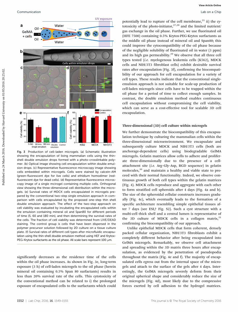
Encapsulation of mammalian cells with high viability
Compared to conventional single-emulsion techniques formicrogel preparation, this thin-shell double emulsion ap-
proach avoids the time- and labor-consuming washing stepand achieves direct transfer of the resulting microgels fromthe oil phase into the aqueous phase; this leads to the one-step production of microgels in a continuous manner. Impor-tantly, this approach significantly reduces the residence timeof the microgels within the oil phase from hours to minutes.This improves the viability of encapsulated cells by avoidingprolonged exposure of the cells to the potentially harmful oiland surfactants.21 To demonstrate this, we compare the via-bility of encapsulated cells using the conventional singleemulsion technique with the current one-step thin-shell dou-ble emulsion one. We encapsulate living Madin–Darby caninekidney epithelial (MDCK) cells using a biocompatible poly-mer (polyethyleneglycol diacrylate, PEG-DA, or gelatin meth-acrylate, GelMA) as the microgel material. To prepare thesemicrogels, we first dissolve the hydrogel precursor and thephoto-initiator (Irgacure 2959) into cell culture medium (i.e.DMEM). Then, we disperse the cells into this solution with aconcentration of 1 × 10⁶ cells per ml. For cell encapsulation,we use mineral oil containing 0.5% Span 80 surfactants orfluorinated oil (HFE 7500) containing 0.5% Krytox-PEG-Krytox surfactants as the middle oil phase. An aqueous solu-tion of 5% PVA is used as the continuous phase to form dou-ble emulsion drops. A schematic illustration of the cell en-capsulation procedure based on double emulsions with asacrificial oil shell is shown in Fig. 3a. We successfully dem-onstrate the feasibility of this technique for cell encapsula-tion as shown in the optical image in Fig. 3b. Upon UV illu-mination (λ ∼ 365 nm for 2 s), cells are immobilized as thegel matrix is solidified. Collection of the cell-loaded micro-gels in an aqueous solution of cell culture media (DMEM)leads to spontaneous dewetting of the oil phase on the sur-face of microgels, allowing autonomous transfer of microgelsinto the aqueous phase. The resulting microgels contain mul-tiple cells and have an average diameter of 244 μm, as shownin the fluorescence microscopy images in Fig. 3c and d. Thenumber of encapsulated cells per microgel and the size ofmicrogels can be modulated by varying the concentration ofthe cell suspension, the nozzle size of the capillary, and thefluid flow rates.8,26 We observe reduced monodispersity ofthe cell-laden microgels (see ESI,† Fig. S1) compared to thecell-free ones; this can be attributed to the inhomogeneity ofthe cell suspension, leading to variability in the drop sizeduring emulsification and subsequent break-up.
The rapid dewetting allows spontaneous transfer of theresulting cell-laden microgels into cell-culture media, there-fore avoiding extensive exposure of living cells to the oilphase which can depress the viability and metabolic activityof the cells, and providing unperturbed nutrient exchangethrough the hydrogel layer. Indeed, we observe that cells en-capsulated in microgels using the one-step double emulsiontechnique exhibit survival rates comparable to those of cellscultured on a tissue culture plate (polystyrene) as evidencedby the Live/Dead staining assay performed after 3 h of micro-fluidic cell encapsulation (Fig. 3c and d). In contrast, cell via-bility within microgels from the single emulsion approach
Fig. 2 Monodisperse microgels produced by a glass capillarymicrofluidic device. (a) Optical image showing the continuousformation of double emulsion drops in a dripping mode. (b) A series ofconfocal images showing the dynamic behavior of the oil layer as thepolymerized drops are transferred in an aqueous solution (DI water).The estimated thickness of the oil shell is approximately 1 μm usingimage analysis. (c–d) Optical images showing the resulting microgelson the bottom surface and the oil layer collected on the top surfaceafter the dewetting process. (e) Size distribution of the resultingmicrogels. All scale bars represent 100 μm.
Lab on a Chip Communication
Publ
ishe
d on
24
Mar
ch 2
016.
Dow
nloa
ded
by H
arva
rd U
nive
rsity
on
01/0
5/20
16 2
0:25
:02.
View Article Online
1552 | Lab Chip, 2016, 16, 1549–1555 This journal is © The Royal Society of Chemistry 2016
significantly decreases as the residence time of the cellswithin the oil phase increases. As shown in Fig. 3e, long-termexposure (3 h) of cell-laden microgels to the oil phase (hereinmineral oil containing 0.5% Span 80 surfactants) results inless than 20% survival rate of the cells. This cytotoxicity ofthe conventional method can be related to i) the prolongedexposure of encapsulated cells to the surfactants which could
potentially lead to rupture of the cell membrane,21 ii) the cy-totoxicity of the photo-initiator,27,28 and the limited nutrient/gas exchange in the oil phase. Further, we use fluorinated oil(HFE 7500) containing 0.5% Krytox-PEG-Krytox surfactants asthe middle oil phase instead of mineral oil and Span80; thiscould improve the cytocompatibility of the oil phase becauseof the negligible solubility of fluorinated oil in water (3 ppm)and its high gas permeability.29 We observe that all three celltypes tested (i.e. myelogenous leukemia cells (K562), MDCKcells and NIH/3T3 fibroblast cells) exhibit desirable survivalrates after encapsulation (Fig. 3f), confirming the biocompati-bility of our approach for cell encapsulation for a variety ofcell types. These results indicate that the conventional single-emulsion approach is not suitable for scale-up production ofcell-laden microgels since cells have to be trapped within theoil phase for a period of time to collect enough samples. Incontrast, the double emulsion method enables continuouscell encapsulation without compromising the cell viability,which can serve as a cost-effective tool for scalable 3D cellencapsulation.
Three-dimensional (3D) cell culture within microgels
We further demonstrate the biocompatibility of this encapsu-lation technique by culturing the mammalian cells within thethree-dimensional microenvironment. We encapsulate andsubsequently culture MDCK and NIH/3T3 cells (both areanchorage-dependent cells) using biodegradable GelMAmicrogels. Gelatin matrices allow cells to adhere and prolifer-ate three-dimensionally due to the presence of a cell-attachment site (i.e. Arg–Gly–Asp, RGD sequence) in gelatinmolecules,30 and maintain a healthy and viable state to pro-ceed with their normal functionality. Indeed, we observe con-tinuous growth of both cell types within the gelatin matrices(Fig. 4). MDCK cells reproduce and aggregate with each otherto form stratified cell spheroids after 4 days (Fig. 4a and b).The size of the spheroidal cellular constructs increases gradu-ally (Fig. 4c), which eventually leads to the formation of aspecific architecture resembling simple epithelial tissues af-ter 7 days (see ESI,† Fig. S2). Such a cyst structure with amulti-cell thick shell and a central lumen is representative ofthe 3D culture of MDCK cells in a collagen matrix,31
confirming the biocompatibility of our approach.Unlike epithelial MDCK cells that form coherent, densely
packed cellular organization, NIH/3T3 fibroblasts exhibit acompletely different behavior after being encapsulated intoGelMA microgels. Remarkably, we observe cell attachmentand spreading within the 3D matrix three hours after encap-sulation, as evidenced by the penetration of pseudopodiathroughout the matrix (Fig. 4e and f). The majority of encap-sulated cells egress out from the internal space of the micro-gels and attach to the surface of the gels after 4 days. Inter-estingly, the GelMA microgels severely deform from theiroriginal spherical shape and considerably reduce the size ofthe microgels (Fig. 4d), most likely due to the compressiveforces exerted by cell adhesion to the hydrogel matrices.
Fig. 3 Production of cell-laden microgels. (a) Schematic illustrationshowing the encapsulation of living mammalian cells using the thin-shell double emulsion drops formed with a photo-crosslinkable poly-mer. (b) Optical image showing cell encapsulation within double emul-sion drops. (c) Representative fluorescence microscopy image showingcells embedded within microgels. Cells were stained by calcein-AM(green-fluorescent dye for live cells) and ethidium homodimer (red-fluorescent dye for dead cells). (d) Representative fluorescence micros-copy image of a single mcirogel containing multiple cells. Orthogonalview showing the three-dimensional cell distribution within the micro-gels. (e) Survival rates of MDCK cells encapsulated in microgels pre-pared by the conventional two-step single emulsion approach in com-parison with cells encapsulated by the proposed one-step thin shelldouble emulsion approach. The effect of the two-step approach oncell viability was evaluated by incubating the encapsulated cells withinthe emulsion containing mineral oil and Span80 for different periodsof time (5, 60 and 180 min), and then determining the survival rates ofthe cells. The fraction of cell viability was determined from LIVE/DEADstaining. The control group is cells that have been dispersed in thepolymer precursor solution followed by 2D culture on a tissue cultureplate. (f) Survival rates of different cell types after microfluidic encapsu-lation using the thin-shell double emulsion method using HEF and Krytox-PEG-Krytox surfactants as the oil phase. All scale bars represent 100 μm.
Lab on a ChipCommunication
Publ
ishe
d on
24
Mar
ch 2
016.
Dow
nloa
ded
by H
arva
rd U
nive
rsity
on
01/0
5/20
16 2
0:25
:02.
View Article Online

Lab Chip, 2016, 16, 1549–1555 | 1553This journal is © The Royal Society of Chemistry 2016
Moreover, the growth of fibroblasts eventually bridges andaggregates the neighboring microgels. These results confirmthat our approach could provide an efficient platform to cre-ate these cell-laden microgels that can serve not only as amicroscopic platform for the study of long-term cell function-ality in a 3D microenvironment, but also as building blocksfor the bottom-up assembly of complex cellularized con-structs with a tissue-specific architecture.
ExperimentalPreparation of double emulsions with a sacrificial ultra-thinoil shell
To produce double emulsion drops, we inject an 8–10% aque-ous solution of polyethyleneglycol diacrylate (PEG-DA, Mn
700, Sigma-Aldrich) and gelatin-methacrylate (GelMA)through a small tapered capillary with a typical flow rate of1000 μL hr−1. A mineral oil (Sigma-Aldrich) containing 0.5%Span80 (Sigma-Aldrich) as a non-ionic surfactant or HFE-7500 (3 M) containing 0.5% Krytox-PEG-Krytox (Ran Biotech-nologies, Inc.) is simultaneously supplied through the injec-tion capillary with a typical flow rate of 1000 μL hr−1. Anaqueous solution of 5% polyIJvinyl alcohol) (PVA) (Mw 13000–23000, Sigma-Aldrich) is injected through the interstices ofthe square and collection capillaries with a typical flow rateof 15 000 μL hr−1. These rates ensure that drop formation isrestricted to the dripping regime.
Preparation of the microfluidic device and drop generation
We use a glass capillary microfluidic device to producedouble emulsion drops with an ultra-thin oil layer. We pre-pare an injection capillary by tapering a 560 μm inner diame-ter cylindrical glass capillary (1B100-6, World PrecisionInstruments, Inc.) to 50 μm inner diameter; to make theinner wall hydrophobic or fluorophilic, we treat it withn-octadecyltrimethoxyl silane (Aldrich) or heptadecafluoro-1,1,2,2 tetrahydrodecyl trichlorosilane (Gelest, Inc.), respec-tively, for 10 minutes and subsequently wash it with ethanol.We insert the injection capillary into a square capillary (AITGlass) whose inner width (1.05 mm) is slightly larger thanthat of the outer diameter of the injection capillary (1 mm).Next, we prepare a small tapered glass capillary (10 μm innerdiameter) by heating and pulling a cylindrical capillary byhand using a gas torch; this capillary is inserted into the in-jection capillary for simultaneous injection of two immisciblefluids. Finally, a cylindrical collection capillary is insertedinto the square capillary from the other end; we also treatthis collection capillary with 2-[methoxyIJpolyethyleneoxy)-propyl] trimethoxy silane (Gelest, Inc.) to make the capillarywall hydrophilic. During drop generation, the volumetric flowrate is controlled by syringe pumps (Harvard Apparatus) andthe production of emulsion drops is observed using aninverted microscope equipped with a high-speed camera(Phantom V9.0). The experimental setup is shown in Fig. S3.†
Cell encapsulation and 3D culture
Myelogenous leukemia cells (K562), Madin–Darby canine kid-ney epithelial (MDCK) cells and NIH/3T3 fibroblast cells areobtained from ATCC®, and are cultured for 6 days in prolifer-ation medium (Dulbecco's modified Eagle medium (DMEM,Sigma-Aldrich), supplemented with 10% v/v fetal bovine se-rum (FBS, Gibco), at 37 °C, 95% relative humidity and 5%CO2. The medium is refreshed every three days of culture. Atconfluence, cells are washed twice with PBS, detached usingtrypsin/EDTA (0.25% w/v trypsin/0.02% EDTA) for 5 min andresuspended in cell culture media. GelMA and the photo-initiator (Irgacure 2959) are dissolved in DMEM followed bysterilization and filtration. All devices are sterilized prior touse by exposure to ultraviolet illumination (λ ∼ 254 nm) for60 min. Cell-laden microgels are prepared by encapsulating
Fig. 4 3D cell culture in GelMA microgels. (a) Representativefluorescence microscopy images of the cross-section of the cell-ladenmicrogels and (b) corresponding 3D reconstruction showing thegrowth of MDCK cells embedded within the microgels at different timepoints during 3D culture. Cells were stained with calcein (green-fluo-rescent dye for live cells) and ethidium homodimer (red-fluorescentdye for dead cells). (c) The size of the cell clusters (characterized bythe diameter of the cell spheroids) formed within the GelMA microgelsat different time points during 3D cell culture. (d) The diameter of thecell-laden microgels (characterized by the diameter of the microgels)at different time points during 3D cell culture. (e) Representative fluo-rescence microscopy images of the cross-section of the cell-ladenmicrogels and (f) corresponding 3D reconstruction showing NIH/3T3fibroblast cells embedded within the microgels at different time pointsduring 3D culture. All scale bars represent 100 μm.
Lab on a Chip Communication
Publ
ishe
d on
24
Mar
ch 2
016.
Dow
nloa
ded
by H
arva
rd U
nive
rsity
on
01/0
5/20
16 2
0:25
:02.
View Article Online
1554 | Lab Chip, 2016, 16, 1549–1555 This journal is © The Royal Society of Chemistry 2016
the cells suspended in DMEM containing 10% w/v of PEG-DAor GelMA and 1% w/v photo-initiator in double emulsiondrops. Then these double emulsion drops are in situ photo-polymerized in UV for 2 s to form cell-laden microgels. Thecell-laden microgels are collected using a cell strainerfollowed by re-dispersion in cell culture media. The mediumis refreshed every three days of culture. Cell survival is deter-mined using a LIVE/DEAD Viability/Cytotoxicity Kit (Molecu-lar Probes). To this end, the gels are washed in sterile PBSfor 30 min at 37 °C prior to incubation for 30 min at roomtemperature with 2 mM calcein-AM (red-fluorescent dye fordead cells) and 4 mM ethidium homodimer (green-fluores-cent dye for live cells) in PBS solution. After incubation, thegels are rinsed in PBS and examined using a Leica SP5 confo-cal laser scanning microscope with a 40× water-immersionobjective lens.
Conclusions
In this paper, we present a one-step microfluidic approach to pro-duce monodisperse microgels by utilizing double emulsiondrops with an ultra-thin oil shell as a sacrificial template.This approach allows the production of microgels in a simpleand efficient manner, avoiding the extra washing procedurerequired for conventional single emulsion-based approaches.In addition, we demonstrate the advantages of this methodby encapsulating and culturing living mammalian cells inthree-dimensional microgel matrices; we achieve high cell vi-ability by avoiding potential cytotoxicity from prolonged expo-sure to oil and surfactants. We anticipate that the cell-ladenmicrogels with tunable sizes and compositions fabricatedusing this microfluidic platform are not limited to UV-induced polymerization but can also be formed using otherpolymerization schemes such as chemical diffusion andtemperature-induced solidification. Moreover, this approachallows us to manipulate cells at the microscopic scale basedon microfluidics and to observe long-term cell functionalitywithin the microgel matrices. This microfluidic techniquerepresents a significant step forward in high-throughput cellmicroencapsulation technology and offers a potentially viableoption to produce cell-laden microgels for widespread appli-cations in tissue engineering and cell therapy.
Acknowledgements
This work was supported by the National Institute of Health(R01 EB014703 and P01GM096971), the Basic Science Re-search Program through the National Research Foundation ofKorea (NRF) funded by the Ministry of Education, Scienceand Technology (2013R1A6A3A03065122), a Rubicon postdoc-toral fellowship from the Netherlands Organization for Scien-tific Research (no. 825.12.018), and the National Natural Sci-ence Foundation of China (no. 51503208). C.-H. Choi and H.Wang contributed equally to this work. J. H. Kim is currentlyaffiliated with Buckingham Browne and Nichols.
References
1 D. E. Discher, D. J. Mooney and P. W. Zandstra, Science,2009, 324, 1673–1677.
2 J. L. Drury and D. J. Mooney, Biomaterials, 2003, 24, 4337–4351.3 A. S. Hoffman, Adv. Drug Delivery Rev., 2002, 54, 3–12.4 L. Klouda and A. G. Mikos, Eur. J. Pharm. Biopharm.,
2008, 68, 34–45.5 D. Seliktar, Science, 2012, 336, 1124–1128.6 G. D. Nicodemus and S. J. Bryant, Tissue Eng., Part B,
2008, 14, 149–165.7 A. Khademhosseini and R. Langer, Biomaterials, 2007, 28,
5087–5092.8 Y. Tsuda, Y. Morimoto and S. Takeuchi, Langmuir, 2010, 26,
2645–2649.9 J. Yeh, Y. Ling, J. M. Karp, J. Gantz, A. Chandawarkar, G.
Eng, J. Blumling Iii, R. Langer and A. Khademhosseini,Biomaterials, 2006, 27, 5391–5398.
10 F. Yanagawa, H. Kaji, Y.-H. Jang, H. Bae, D. Yanan, J.Fukuda, H. Qi and A. Khademhosseini, J. Biomed. Mater.Res., Part A, 2011, 97A, 93–102.
11 J. K. Oh, R. Drumright, D. J. Siegwart and K. Matyjaszewski,Prog. Polym. Sci., 2008, 33, 448–477.
12 S. Allazetta and M. P. Lutolf, Curr. Opin. Biotechnol.,2015, 35, 86–93.
13 Y. Du, E. Lo, S. Ali and A. Khademhosseini, Proc. Natl. Acad.Sci. U. S. A., 2008, 105, 9522–9527.
14 H. Tekin, T. Tsinman, J. G. Sanchez, B. J. Jones, G. Camci-Unal, J. W. Nichol, R. Langer and A. Khademhosseini, J. Am.Chem. Soc., 2011, 133, 12944–12947.
15 K. Maeda, H. Onoe, M. Takinoue and S. Takeuchi, Adv.Mater., 2012, 24, 1340–1346.
16 W. H. Tan and S. Takeuchi, Adv. Mater., 2007, 19, 2696–2701.17 H. Zhang, E. Tumarkin, R. Peerani, Z. Nie, R. M. A. Sullan,
G. C. Walker and E. Kumacheva, J. Am. Chem. Soc.,2006, 128, 12205–12210.
18 T. Rossow, J. A. Heyman, A. J. Ehrlicher, A. Langhoff, D. A.Weitz, R. Haag and S. Seiffert, J. Am. Chem. Soc., 2012, 134,4983–4989.
19 C.-H. Choi, J.-H. Jung, Y. Rhee, D.-P. Kim, S.-E. Shim andC.-S. Lee, Biomed. Microdevices, 2007, 9, 855–862.
20 C.-H. Choi, J.-H. Jung, T.-S. Hwang and C.-S. Lee, Macromol.Res., 2009, 17, 163–167.
21 G. Dehghan Noudeh, P. Khazaeli, S. Mirzaei, F. Sharififarand S. Nasrollahosaiani, J. Biol. Sci., 2009, 9, 423–430.
22 Y. Song, Y. K. Chan, Q. Ma, Z. Liu and H. C. Shum, ACSAppl. Mater. Interfaces, 2015, 7, 13925–13933.
23 I. Ziemecka, V. van Steijn, G. J. M. Koper, M. Rosso, A. M.Brizard, J. H. van Esch and M. T. Kreutzer, Lab Chip,2011, 11, 620–624.
24 S.-H. Kim, J. W. Kim, J.-C. Cho and D. A. Weitz, Lab Chip,2011, 11, 3162–3166.
25 A. S. Utada, A. Fernandez-Nieves, H. A. Stone and D. A.Weitz, Phys. Rev. Lett., 2007, 99, 094502.
26 S. Utech, R. Prodanovic, A. S. Mao, R. Ostafe, D. J. Mooneyand D. A. Weitz, Adv. Healthcare Mater., 2015, 4, 1628–1633.
Lab on a ChipCommunication
Publ
ishe
d on
24
Mar
ch 2
016.
Dow
nloa
ded
by H
arva
rd U
nive
rsity
on
01/0
5/20
16 2
0:25
:02.
View Article Online

Lab Chip, 2016, 16, 1549–1555 | 1555This journal is © The Royal Society of Chemistry 2016
27 P. Panda, S. Ali, E. Lo, B. G. Chung, T. A. Hatton, A.Khademhosseini and P. S. Doyle, Lab Chip, 2008, 8,1056–1061.
28 C. G. Williams, A. N. Malik, T. K. Kim, P. N. Manson andJ. H. Elisseeff, Biomaterials, 2005, 26, 1211–1218.
29 K. C. Lowe, J. Fluorine Chem., 2002, 118, 19–26.30 H.-W. Kang, Y. Tabata and Y. Ikada, Biomaterials, 1999, 20,
1339–1344.31 L. E. O'Brien, M. M. P. Zegers and K. E. Mostov, Nat. Rev.
Mol. Cell Biol., 2002, 3, 531–537.
Lab on a Chip Communication
Publ
ishe
d on
24
Mar
ch 2
016.
Dow
nloa
ded
by H
arva
rd U
nive
rsity
on
01/0
5/20
16 2
0:25
:02.
View Article Online