Embed Size (px)
Citation preview
LAB #2:
GROSS & INTERNAL CNS II
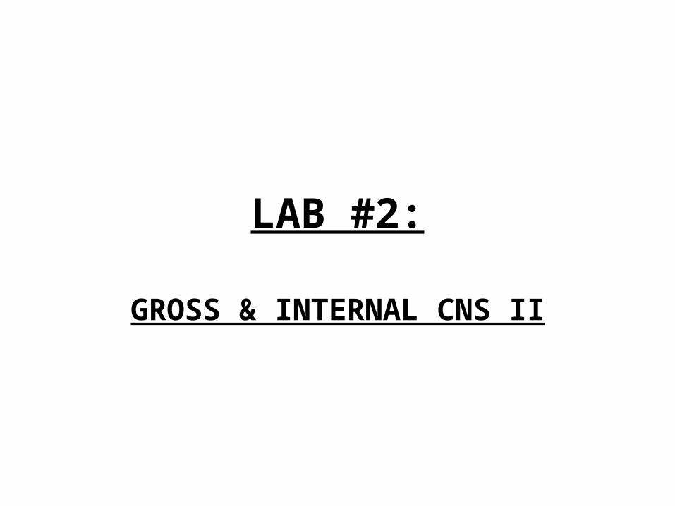
5-11 Midrostral Medulla
IV Ventricle
Tegmentum
Periventricular Zone
Pyramidal
tract
Inferior Olive
Hypoglossal Nucleus Vestibular Nuclei
Reticular Formation
Dorsal Motor Nucleus of Vagus
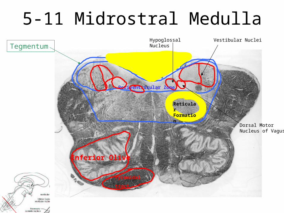
Midrostral Medulla
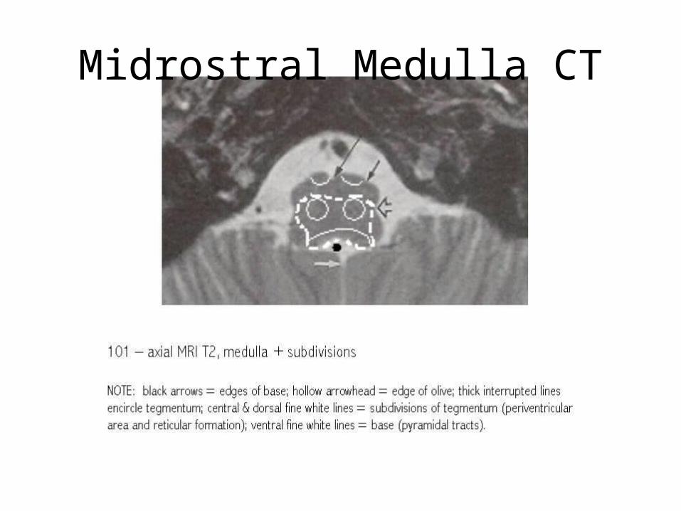
Midrostral Medulla CT
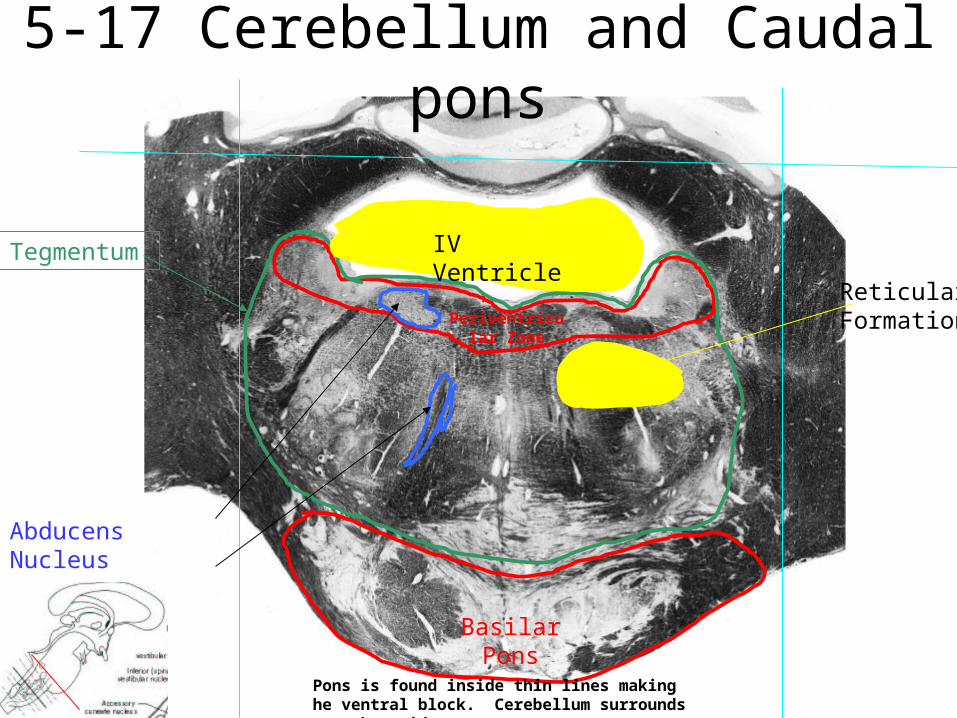
5-17 Cerebellum and Caudal pons
Basilar Pons
Periventricular Zone
Reticular Formation
Abducens Nucleus
Root of Abducens n.
Tegmentum IV Ventricle
Pons is found inside thin lines making he ventral block. Cerebellum surrounds on other sides
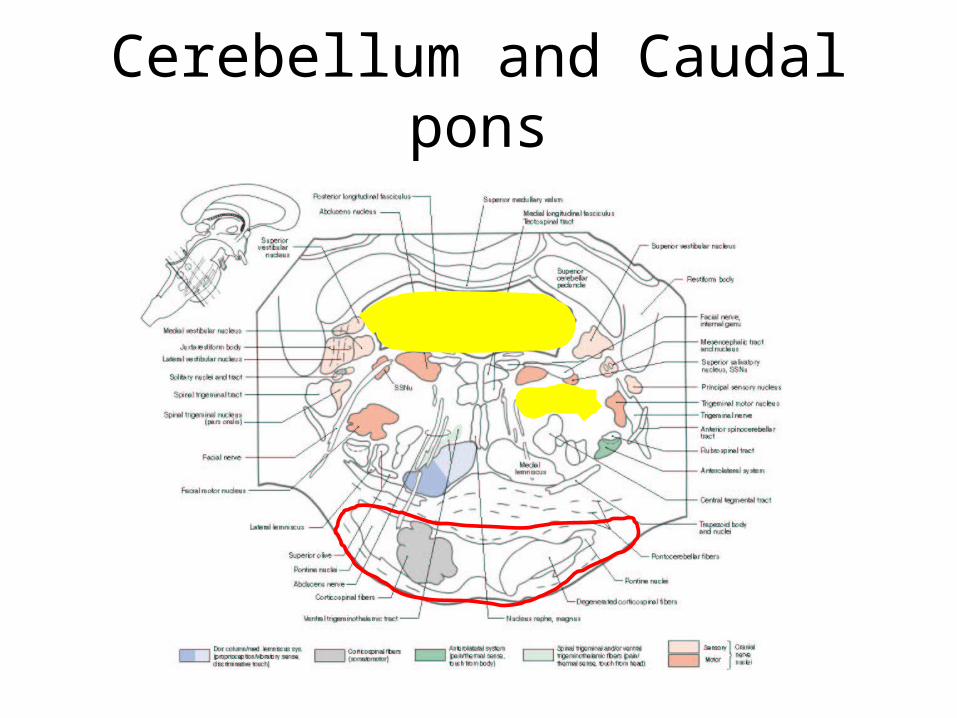
Cerebellum and Caudal pons
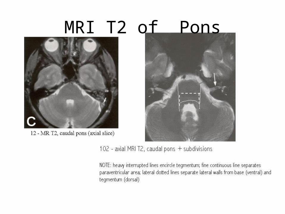
MRI T2 of Pons
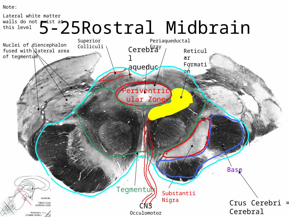
5-25Rostral Midbrain
Periventricular Zone
Substantii NigraTegmentum
Base
Reticular Formation
Note:
Lateral white matter walls do not exist at this level
Nuclei of diencephalon fused with lateral area of tegmentum
Cerebral aqueduct
Crus Cerebri = Cerebral Peduncle
Superior Colliculi
CN3 Occulomotor
Periaqueductal Gray
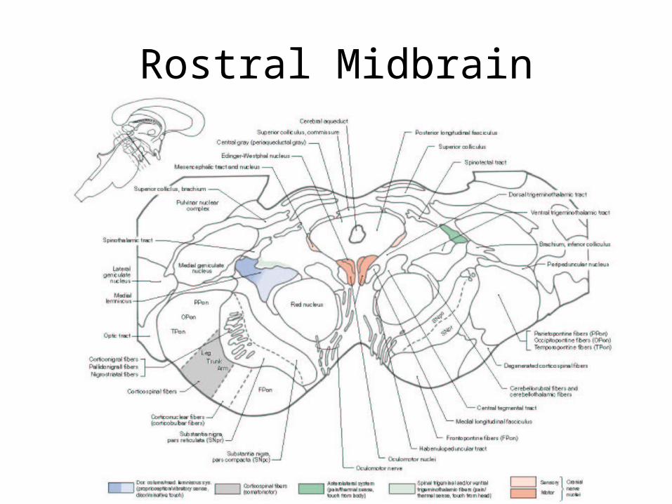
Rostral Midbrain
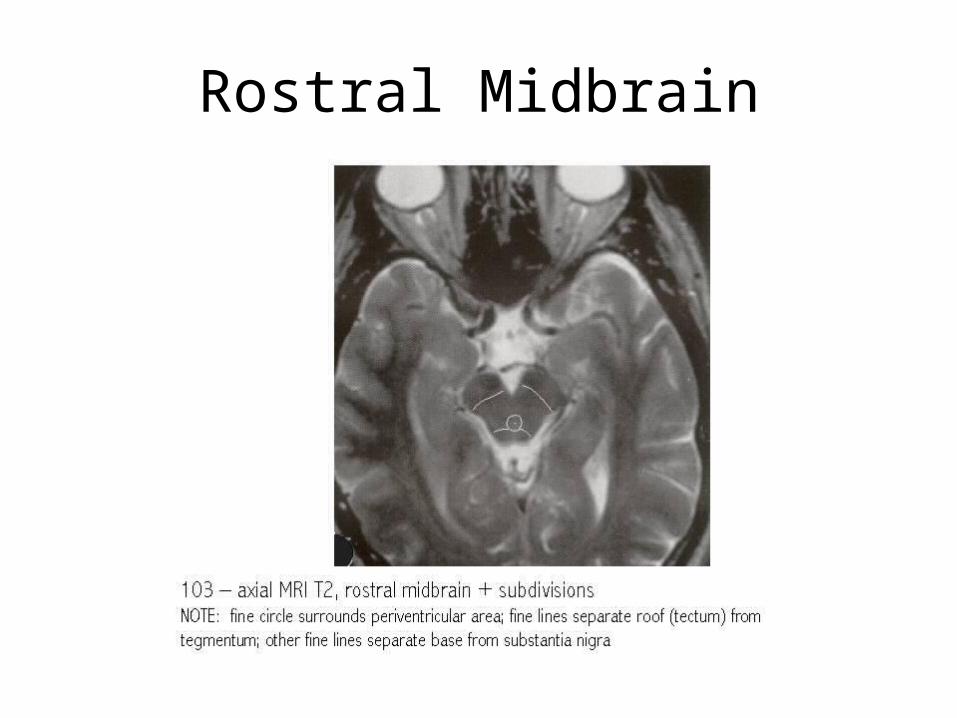
Rostral Midbrain
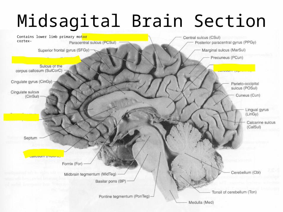
Midsagital Brain SectionContains lower limb primary motor cortex-
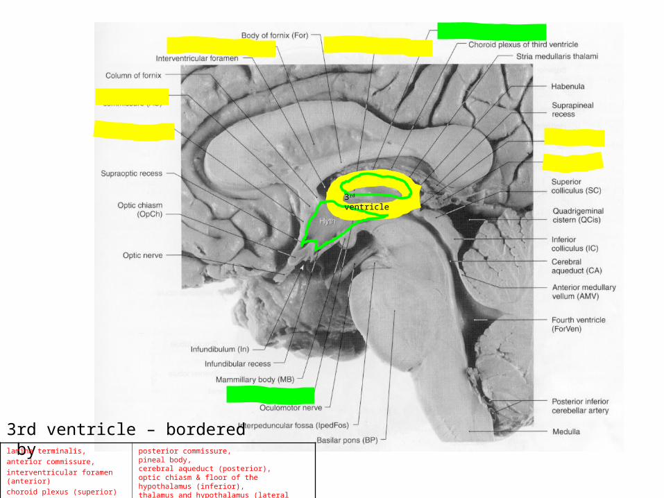
3rd ventricle
3rd ventricle – bordered bylamina terminalis,
anterior commissure,
interventricular foramen (anterior)
choroid plexus (superior)
posterior commissure, pineal body,cerebral aqueduct (posterior), optic chiasm & floor of the hypothalamus (inferior), thalamus and hypothalamus (lateral walls)
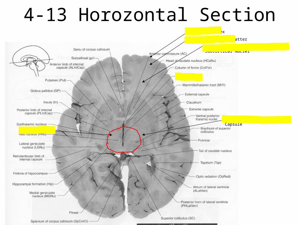
4-13 Horozontal Section
Diencephalon
Posterior Limb of Internal Capsule
Subcortical White Matter
Deep White Matter – Containing Subcortical Nuclei
Cerebral Cortex
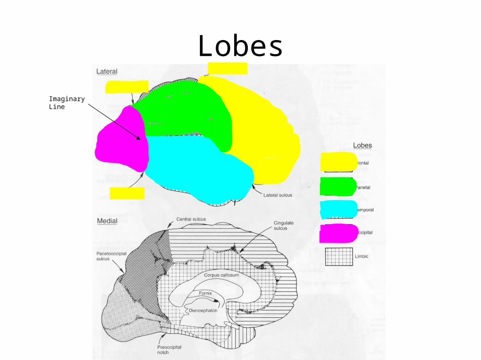
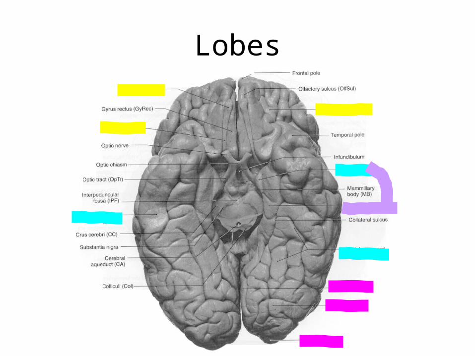
Lobes
Imaginary Line
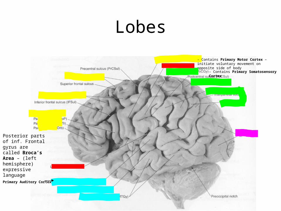
Posterior parts of inf. Frontal gyrus are called Broca’s Area – (left hemisphere) expressive language
- Contains Primary Motor Cortex – initiate voluntary movement on opposite side of body
Lobes
- Contains Primary Somatosensory Cortex
Primary Auditory Cortex
Lobes
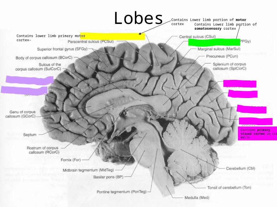
Contains lower limb primary motor cortex-
Lobes Contains Lower limb portion of somatosensory cortex
Contains primary visual cortex in its walls
Contains Lower limb portion of motor cortex
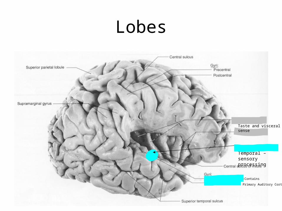
Lobes
Planum Temporal – sensory processing
-Contains
Primary Auditory Cortex
Taste and visceral sense
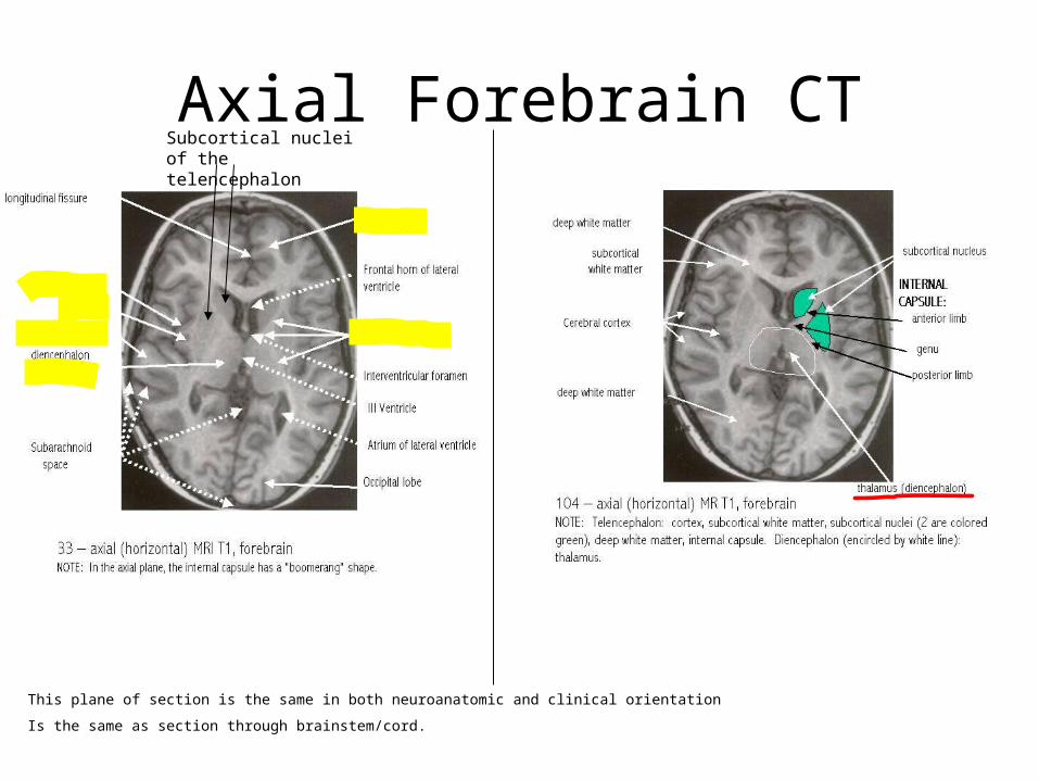
This plane of section is the same in both neuroanatomic and clinical orientation
Is the same as section through brainstem/cord.
Axial Forebrain CTSubcortical nuclei of the telencephalon
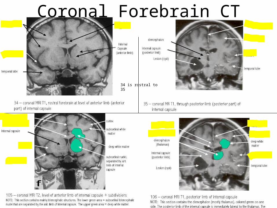
34 is rostral to 35
Coronal Forebrain CT
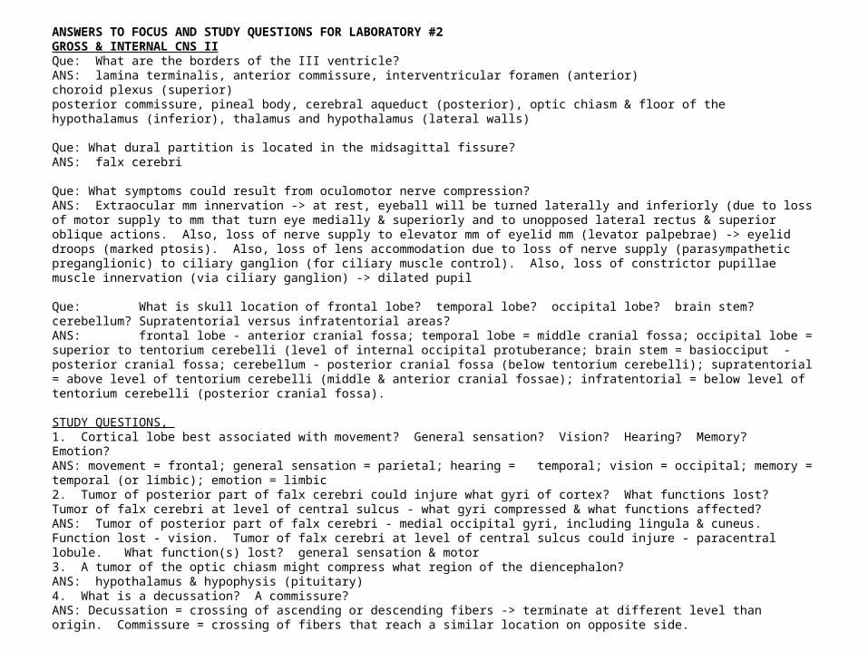
ANSWERS TO FOCUS AND STUDY QUESTIONS FOR LABORATORY #2GROSS & INTERNAL CNS IIQue: What are the borders of the III ventricle?ANS: lamina terminalis, anterior commissure, interventricular foramen (anterior)choroid plexus (superior) posterior commissure, pineal body, cerebral aqueduct (posterior), optic chiasm & floor of the hypothalamus (inferior), thalamus and hypothalamus (lateral walls)
Que: What dural partition is located in the midsagittal fissure?ANS: falx cerebri
Que: What symptoms could result from oculomotor nerve compression?ANS: Extraocular mm innervation -> at rest, eyeball will be turned laterally and inferiorly (due to loss of motor supply to mm that turn eye medially & superiorly and to unopposed lateral rectus & superior oblique actions. Also, loss of nerve supply to elevator mm of eyelid mm (levator palpebrae) -> eyelid droops (marked ptosis). Also, loss of lens accommodation due to loss of nerve supply (parasympathetic preganglionic) to ciliary ganglion (for ciliary muscle control). Also, loss of constrictor pupillae muscle innervation (via ciliary ganglion) -> dilated pupil
Que: What is skull location of frontal lobe? temporal lobe? occipital lobe? brain stem? cerebellum? Supratentorial versus infratentorial areas? ANS: frontal lobe - anterior cranial fossa; temporal lobe = middle cranial fossa; occipital lobe = superior to tentorium cerebelli (level of internal occipital protuberance; brain stem = basiocciput - posterior cranial fossa; cerebellum - posterior cranial fossa (below tentorium cerebelli); supratentorial = above level of tentorium cerebelli (middle & anterior cranial fossae); infratentorial = below level of tentorium cerebelli (posterior cranial fossa).
STUDY QUESTIONS, 1. Cortical lobe best associated with movement? General sensation? Vision? Hearing? Memory? Emotion?ANS: movement = frontal; general sensation = parietal; hearing = temporal; vision = occipital; memory = temporal (or limbic); emotion = limbic2. Tumor of posterior part of falx cerebri could injure what gyri of cortex? What functions lost? Tumor of falx cerebri at level of central sulcus - what gyri compressed & what functions affected?ANS: Tumor of posterior part of falx cerebri - medial occipital gyri, including lingula & cuneus. Function lost - vision. Tumor of falx cerebri at level of central sulcus could injure - paracentral lobule. What function(s) lost? general sensation & motor3. A tumor of the optic chiasm might compress what region of the diencephalon?ANS: hypothalamus & hypophysis (pituitary)4. What is a decussation? A commissure? ANS: Decussation = crossing of ascending or descending fibers -> terminate at different level than origin. Commissure = crossing of fibers that reach a similar location on opposite side.