Embed Size (px)
Citation preview

19 Periventricular White Matter Abnormalities on
Magnetic Resonance Imaging

CLINICAL IMAGAGINGAN ATLAS OF DIFFERENTIAL DAIGNOSIS
EISENBERG
DR. Muhammad Bin Zulfiqar PGR-FCPS III SIMS/SHL

• Fig SK 19-1 Prominent Virchow-Robin spaces.

• Fig SK 19-2 Deep white matter ischemia. Multiple areas of increased signal intensity around the ventricles and in the deep white matter.
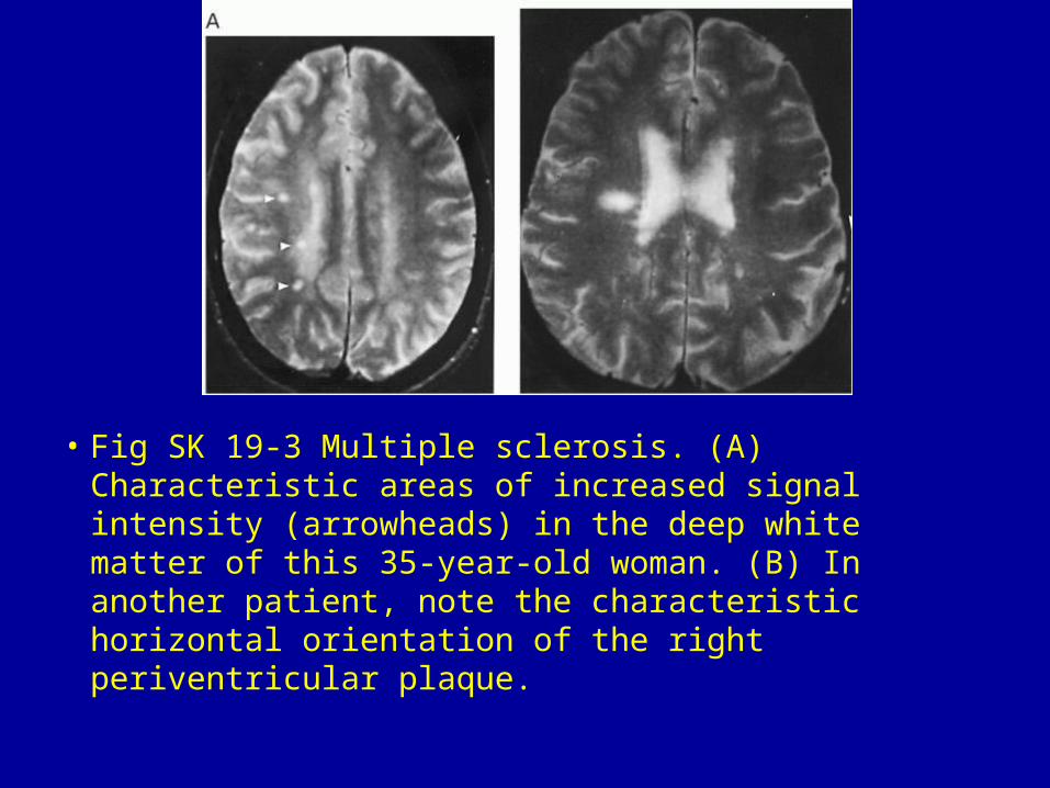
• Fig SK 19-3 Multiple sclerosis. (A) Characteristic areas of increased signal intensity (arrowheads) in the deep white matter of this 35-year-old woman. (B) In another patient, note the characteristic horizontal orientation of the right periventricular plaque.

• Fig SK 19-4 Radiation injury. Symmetric foci of high signal intensity in the periventricular white matter on this T2-weighted image.
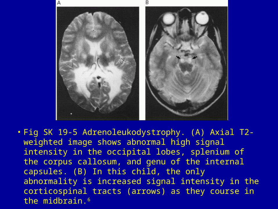
• Fig SK 19-5 Adrenoleukodystrophy. (A) Axial T2-weighted image shows abnormal high signal intensity in the occipital lobes, splenium of the corpus callosum, and genu of the internal capsules. (B) In this child, the only abnormality is increased signal intensity in the corticospinal tracts (arrows) as they course in the midbrain.6
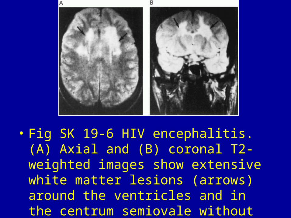
• Fig SK 19-6 HIV encephalitis. (A) Axial and (B) coronal T2-weighted images show extensive white matter lesions (arrows) around the ventricles and in the centrum semiovale without evidence of mass effect.28

• Fig SK 19-7 Migraine. Focal area of hyperintensity in the left frontal white matter in a woman with classic migraine headaches.29


![Untitled-1 [] › admin › uploads › CARF Bulletin Issue 50.pdfduring antenatal period also can help picking up cervical cancer or cervical abnormalities early on. Magnetic resonance](https://img.dokumen.tips/doc/110x75/5f03030b7e708231d4071a00/untitled-1-a-admin-a-uploads-a-carf-bulletin-issue-50pdf-during-antenatal.jpg)