Embed Size (px)
Citation preview

FEMALE REPRODUCTIVE SYSTEMKristina C. Erasmo, M.D.

FEMALE REPRODUCTIVE SYSTEM External genitalia
ClitorisLabia majoraLabia minoraVestibular glands
Internal genitaliaOvaries OviductsUterusVagina


OVARY Paired, slightly
flattened, ovoid organs
Produce ova and hormones
Simple squamous or cuboidal epithelium (“germinal epithelium”)
Tunica albuginea – layer of dense CT beneath the epithelium

OVARY Cortex
Thick peripheral zoneStroma – CT substance of cortex,
Fibroblasts, collagenous fibers surrounded by amorphous intercellular substance
Ovarian follicles
MedullaVascular inner zone, pale-stainingLoose CT, abundant blood vessels


OVARIAN CYCLE Refers to structural and physiological
changes that the ovarian follicles and the stroma that surrounds them undergo during the menstrual cycle
Hormonal control:FSHLH


OVIDUCT (FALLOPIAN TUBES) Pair of muscular
tubes Passageway for
ovum on its way to the uterus and the sperms on their way to fertilizing the ovum
4 parts: Infundibulum Ampulla Isthmus Pars interstitialis

OVIDUCT (FALLOPIAN TUBES) Infundibulum
Funnel-shaped area related to the ovary and opens into peritoneal cavity
Fimbriae Ampulla
Expanded intermediate portion (2/3 of length)
Isthmus Narrow, slender part w/c
connects the FT to uterus
Pars interstitialis Part of the tube within
the uterine wall

OVIDUCT: LAYERS Mucosa
Forms numerous folds
Simple columnar epithelium Ciliated cells Non-ciliated
cellsLamina propria
Muscular layer (OLIC)
Serosa
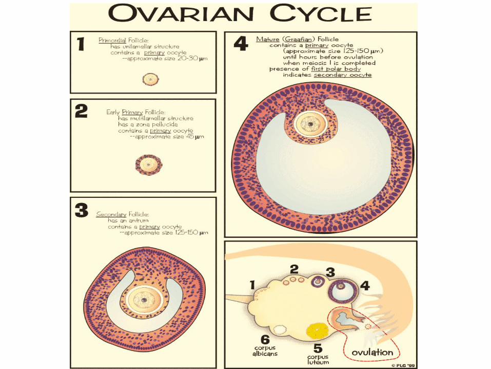
OVIDUCT Thickness and specific
characteristics of histologic layers vary with the segments
Infundibulum and ampulla:Tunica mucosa: thick,
highly developed. Isthmus:
Tunica mucosa: thinnerTunica muscularis:
thicker

UTERUS Pear-shaped,
hollow pelvic organ Receives fertilized
ovum and nourishes the embryo throughout its dev’t
2 parts:Corpus uteri (body)
FundusCervix
Portio vaginalis
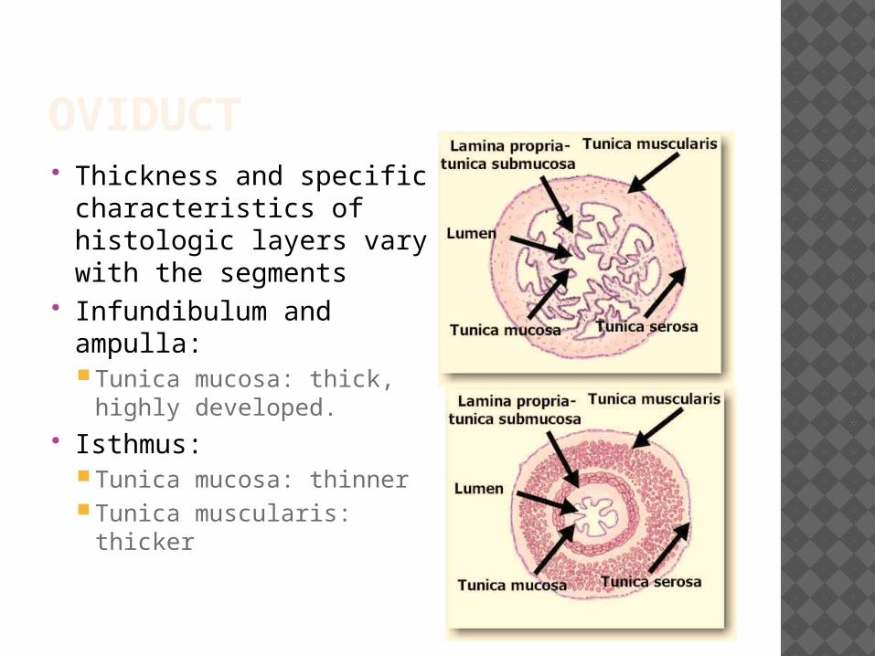
UTERUS: LAYERS Endometrium
Innermost (mucosa)
MyometriumMiddle, thickestSmooth muscles
Serosa/AdventitiaSerosa: over
fundus, posterior aspect of uterus

ENDOMETRIUM Simple columnar
epithelium Lamina propria:
CT, endometrial glands
Functional layer – superficial 2/3
Basal layer – deeper 1/3

MYOMETRIUM Thickest layer of
the corpus uteri Smooth muscle
cells arranged in bundles separated by CT

UTERINE CYCLE a.k.a. endometrial cycle, menstrual
cycle Refers to continuous sequence of
morphological and physiological changes that the endometrium undergoes in response to ovarian hormones
Proliferative (follicular) phase Secretory (luteal) phase Menstrual phase


PROLIFERATIVE PHASE Governed by estrogen (ovarian follicles) Coincides with ovarian follicle growth
(under the influence of FSH in first ½ of menstrual cycle)
Epithelial and stromal cells undergo mitosis
Glands in lamina propria Blood vessels Ground substance Thickness


SECRETORY PHASE Governed by progesterone (from corpus
luteum, under LH influence) Occurs during the 15th day of menstrual
cycle Glands become tortuous and secretory Blood vessels Thickest endometrium (due to glandular
secretion and edema of stroma)
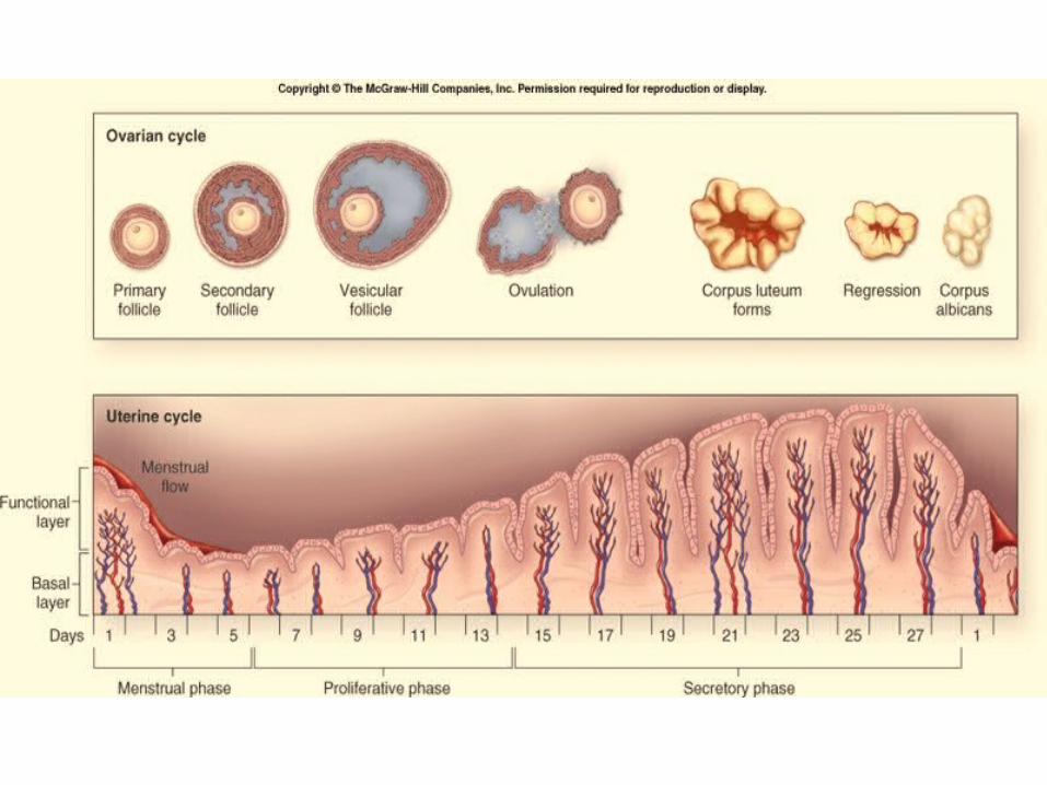

MENSTRUAL PHASE 2 weeks after ovulation (no fertilization) Decrease in ovarian hormones No glandular secretion Blood vessels constrict → close → open Functional layer exfoliates Basal layer intact


CERVIX Cylindrical inferior
portion of uterus Parts:
Cervical canal Internal osExternal osPortio vaginalis
(ectocervix)
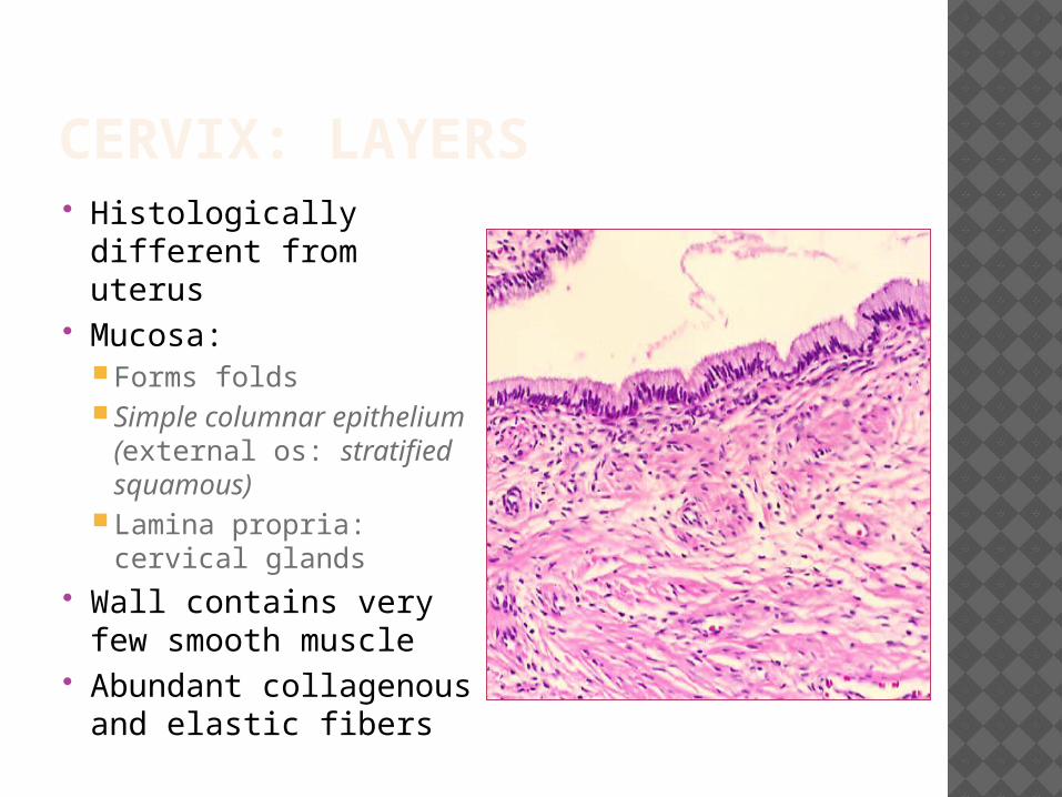
CERVIX: LAYERS Histologically different
from uterus Mucosa:
Forms folds Simple columnar
epithelium (external os: stratified squamous)
Lamina propria: cervical glands
Wall contains very few smooth muscle
Abundant collagenous and elastic fibers


VAGINA Fibromuscular
tube Extends from
vestibule of external genitalia to the cervix
Normally, collapsed (anterior and posterior walls are in contact with each other)

VAGINA: LAYERS Mucosa
Thrown into folds (rugae)
Stratified squamous non-keratinized epithelium
Lamina propria: dense CT, no glands
Muscularis (OLIC) Adventitia
Thin CT Nerve bundles, venous
plexus

EXTERNAL GENITALIA Clitoris Labia minora Labia majora Vestibular glands
Major vestibular glands (Bartholin’s glands)Minor vestibular glands
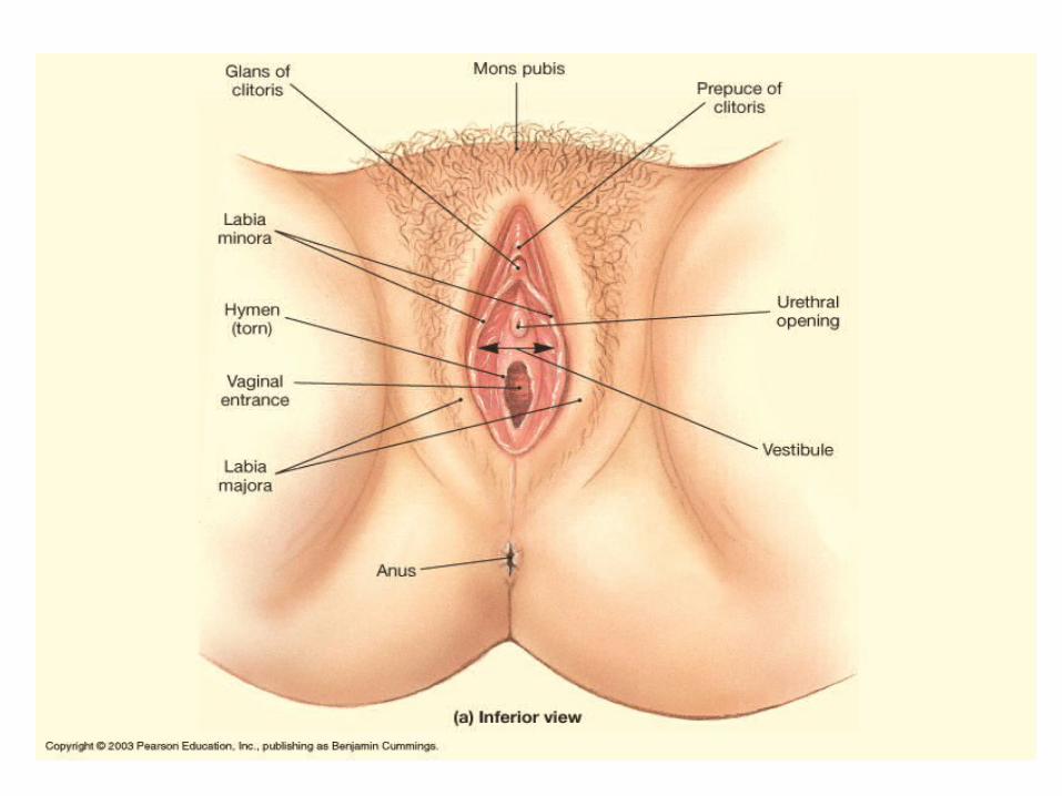

EXTERNAL GENITALIADescription Epithelium
Clitoris Homologue of male penis,2 erectile cavernous bodies (corpora cavernosa)
Stratified squamous with specialized nerve endings
Labia minora
Form the lateral walls of the vestibule
Stratified squamous epithelium with highly vascular CT underneathNumerous sebaceous glands, no hair follicles
Labia majora
Cover the labia minoraHomologue of the male scrotum
Stratified squamous epithelium

EXTERNAL GENITALIADescription
Major vestibular glands (Bartholin’s)
Pair of larger glands in the lateral walls of vestibule, open on the inner surface of labia minoraMucus-secreting
Minor vestibular glands
Located under the urethral opening and near the clitorisMucus-secreting
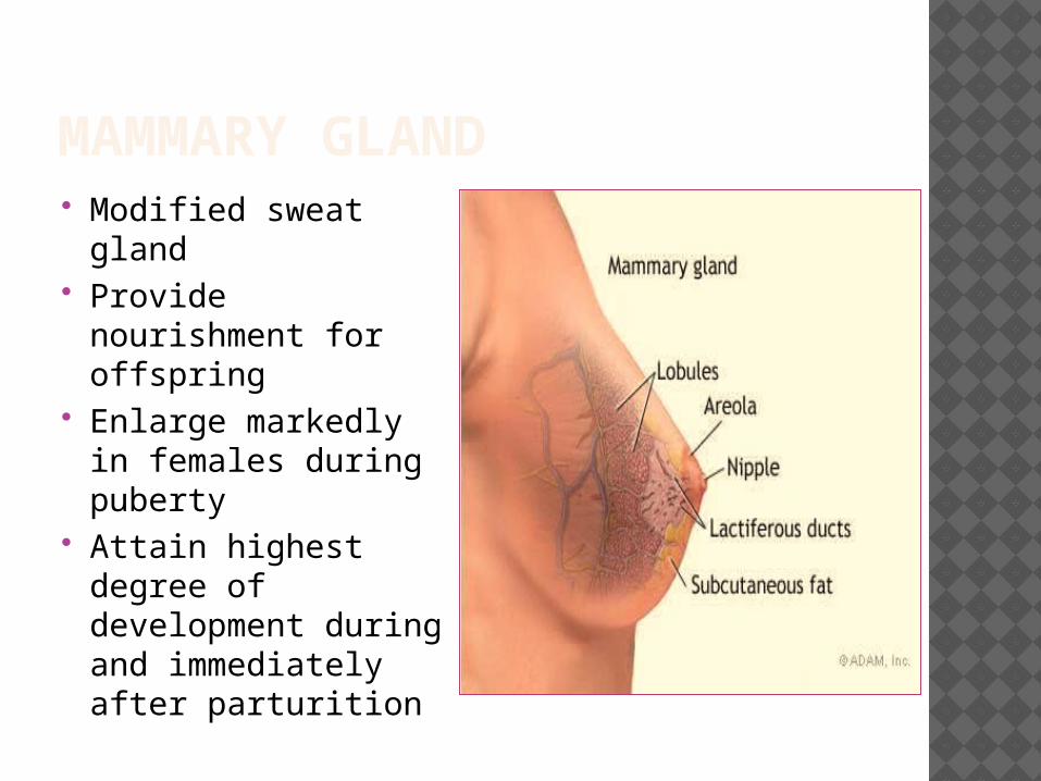
MAMMARY GLAND Modified sweat
gland Provide nourishment
for offspring Enlarge markedly in
females during puberty
Attain highest degree of development during and immediately after parturition

MAMMARY GLAND: LOBES 15-20
lobes/mammary gland divided by interlobar CT
Intralobular ducts → interlobular ducts → lactiferous duct and sinusStratified cuboidal
epithelium

MAMMARY GLAND: AREOLA AND NIPPLE Lactiferous ducts
open into nipple Skin of nipple:
highly pigmented, dermis contains many smooth muscle
Areola – surrounds nipple, contains sweat and sebaceous glands, hair, glands of Montgomery


![Het gynaecologisch onderzoek 3BACH.ppt [Compatibiliteitsmodus] · 14/02/2011 2 Onderzoekshouding Stuit tot op de rand Genitalia externa 1. Clitoris 2. Labia majora 3. Labia minora](https://img.dokumen.tips/doc/110x75/5cd3d37488c993de288b48e0/het-gynaecologisch-onderzoek-3bachppt-compatibiliteitsmodus-14022011-2.jpg)