Embed Size (px)
Citation preview
8/17/2019 Knee Knee Collateral Ligaments
http://slidepdf.com/reader/full/knee-knee-collateral-ligaments 1/8
Compliments of:
A Patient’s Guide to
Collateral Ligament Injuries
Houston Methodist6565 Fannin StreetHouston, TX 77030Phone: 713-790-3333
Houston Methodist
8/17/2019 Knee Knee Collateral Ligaments
http://slidepdf.com/reader/full/knee-knee-collateral-ligaments 2/8
2
A Patient's Guide to Collateral Ligament Injuries
Compliments of:
DISCLAIMER: The information in this booklet is compiled from a variety of sources. It may not be complete or timely. It does not cover all diseases, physical conditions, ailments or treatments. The information should NOT be used in place of a visit with your health care provider, nor should you disregard the advice of your health care provider because of any information you read in this booklet.
All materials within these pages are the sole property of Medical Multimedia Group, LLC and are used herein by permission. eOrthopod is aregistered trademark of Medical Multimedia Group, LLC.
Houston Methodist
Houston Methodist6565 Fannin StreetHouston, TX 77030Phone: 713-790-3333http://www.methodistorthopedics.com
Houston Methodist
8/17/2019 Knee Knee Collateral Ligaments
http://slidepdf.com/reader/full/knee-knee-collateral-ligaments 3/8
3
A Patient's Guide to Collateral Ligament Injuries
Compliments of:
Introduction
The collateral ligaments are commonly
injured parts of the knee. An injury to these
ligaments usually involves a significantforce, such as a fall while skiing or a direct
impact to the side of the leg.
This guide will help you understand
• where the collateral ligaments are
located
• how a collateral ligament injury causes
problems
• how doctors treat the condition
Anatomy
Where are the collateral ligaments, and what
do they do?
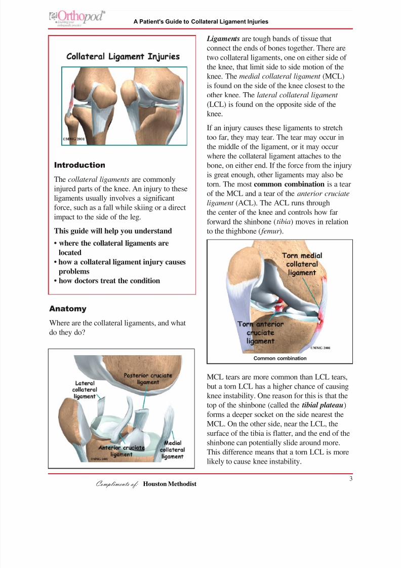
Ligaments are tough bands of tissue that
connect the ends of bones together. There are
two collateral ligaments, one on either side of
the knee, that limit side to side motion of the
knee. The medial collateral ligament (MCL)
is found on the side of the knee closest to the
other knee. The lateral collateral ligament
(LCL) is found on the opposite side of the
knee.
If an injury causes these ligaments to stretch
too far, they may tear. The tear may occur in
the middle of the ligament, or it may occur
where the collateral ligament attaches to the
bone, on either end. If the force from the injury
is great enough, other ligaments may also be
torn. The most common combination is a tear
of the MCL and a tear of the anterior cruciate
ligament (ACL). The ACL runs through
the center of the knee and controls how far
forward the shinbone (tibia) moves in relation
to the thighbone ( femur ).
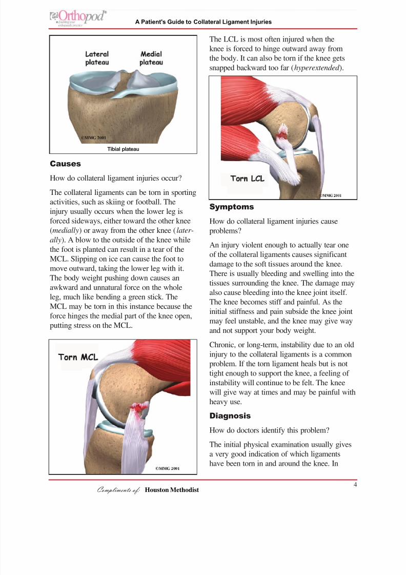
MCL tears are more common than LCL tears,
but a torn LCL has a higher chance of causing
knee instability. One reason for this is that thetop of the shinbone (called the tibial plateau)
forms a deeper socket on the side nearest the
MCL. On the other side, near the LCL, the
surface of the tibia is flatter, and the end of the
shinbone can potentially slide around more.
This difference means that a torn LCL is more
likely to cause knee instability.
Common combination
Houston Methodist
8/17/2019 Knee Knee Collateral Ligaments
http://slidepdf.com/reader/full/knee-knee-collateral-ligaments 4/8

8/17/2019 Knee Knee Collateral Ligaments
http://slidepdf.com/reader/full/knee-knee-collateral-ligaments 5/8
5
A Patient's Guide to Collateral Ligament Injuries
Compliments of:
some cases, there is too much pain and muscle
spasm to completely tell what is damaged
in your knee. Your physician may suggest a
period of rest with a knee splint and then reex-
amine the knee in five to seven days. This will
allow some of the initial pain and spasm to
decrease, and the exam may be more reliable.
X-rays may be required to rule out the possi-
bility that any bones have been damaged.
Stress X-rays may be useful to confirm that
one of the collateral ligaments has been torn.
Stress X-rays are plain X-rays taken with
someone attempting to open the side of the
joint that is suspected of being unstable. The
X-rays will show a widening of the joint space
on that side if instability is present.
Magnetic resonance imaging (MRI) maybe ordered if there is evidence that multiple
injuries have occurred, including injury to the
ACL or meniscus (a special type of ligament
in the knee joint). The MRI machine uses
magnetic waves rather than X-rays to create
pictures that look like slices of the knee.
This test does not require any needles or
special dye and is painless. If there is uncer-
tainty in the diagnosis following the history
and physical examination, or if other injuriesin addition to the collateral ligament tear are
suspected, an MRI scan will probably be
suggested.
Treatment
How do doctors treat collateral ligament
injuries?
Nonsurgical Treatment
An isolated injury to the LCL or MCL rarely
requires surgical repair or reconstruction.Partial tears to the LCL, such as Grade 1 or
Grade II injury, are usually treated by reduced
activity and allow the ligament healed with
or without a brace for several weeks. Most
doctors opt not to immobilize the knee in a
cast when the MCL is torn. Some doctors
prefer to issue their patients a knee brace after
the injury if there is significant pain and insta-
bility.
Initial treatments for a collateral ligament
injury focus on decreasing pain and swelling in
the knee. Rest and anti-inflammatory medica-
tions, such as aspirin, can help decrease thesesymptoms. You may need to use crutches until
you can walk without a limp.
Most patients receive physical therapy
treatments for collateral ligament injuries.
Therapists may treat swelling and pain with
the use of ice, electrical stimulation, and rest
periods with your leg supported in elevation.
Exercises are used to help you regain normal
knee movement. Range-of-motion exer-
cises should be started right away with
the goal of helping you swiftly regain full
knee movement. This includes the use of a
stationary bike, gentle stretching, and careful
pressure applied to the joint by the therapist.
Exercises are also used to improve the strength
of the quadriceps muscle on the front of the
thigh. As your symptoms ease and strength
improves, you will be guided through
advancing stages of exercise.
When you get full knee movement, your
strength is improving, and your knee isn't
giving way, you'll be able to gradually get
back to your work and sport activities. Some
doctors prescribe the use of a functional brace
for athletes who intend to return quickly to
their sport. These braces support the knee and
protect the collateral ligaments.
Patients who continue having periods of
swelling or instability in the knee may needsurgery to correct their problem.
Surgery
If other structures in the knee are injured,
surgery may be required. Some surgeons
feel that a combination of an ACL tear and
an MCL tear should be treated surgically.
Houston Methodist

8/17/2019 Knee Knee Collateral Ligaments
http://slidepdf.com/reader/full/knee-knee-collateral-ligaments 6/8
8/17/2019 Knee Knee Collateral Ligaments
http://slidepdf.com/reader/full/knee-knee-collateral-ligaments 7/8
7
A Patient's Guide to Collateral Ligament Injuries
Compliments of:
surgery. Severe tears or ruptures of the LCL
are the trickiest, because they tend to leave
the knee joint the most unstable, and patients
with this condition typically don't do well with
nonsurgical care.
After Surgery
Rehabilitation proceeds cautiously after
surgery of the collateral ligaments, and treat-
ments will vary depending on the type of
surgical procedure that was used. Some
surgeons have their patients use a continuous
passive motion (CPM) machine after surgery
to help the knee begin to move and to alleviate
joint stiffness.
Most patients are prescribed a hinged knee
brace to wear when they are up and about.
Surgeons occasionally cast the leg after recon-
struction surgery of the LCL.
Patients are strongly advised to follow the
recommendations about how much weight to
place on the leg while standing or walking.
After a ligament repair, patients will be
instructed to put little or no weight on their
foot when standing or walking for up to six
weeks. Weight bearing may be restricted for
up to 12 weeks after a ligament reconstruction.
Patients usually take part in formal physical
therapy after collateral ligament surgery.
The first few physical therapy treatments are
designed to help control the pain and swelling
from the surgery. The goal is to help you
regain full knee motion as soon as possible.
Physical therapists will also work with patientsto make sure they are using crutches safely
and only bearing the recommended amount of
weight while standing or walking.
As the rehabilitation program evolves, more
challenging exercises are chosen to safely
advance the knee's strength and function.
Ideally, patients will be able to resume their
previous lifestyle activities. Some patients may
be encouraged to modify their activity choices,
especially if an allograft procedure was used.
The physical therapist's goal is to help you
keep your pain under control, ensure safe
weight bearing, and improve your strength and
range of motion. When you are well under
way, regular visits to the therapist's office
will end. Your therapist will continue to be a
resource, but you will be in charge of doing
your exercises as part of an ongoing home
program.
Houston Methodist

8/17/2019 Knee Knee Collateral Ligaments
http://slidepdf.com/reader/full/knee-knee-collateral-ligaments 8/8
8
A Patient's Guide to Collateral Ligament Injuries
Compliments of:
Notes
Houston Methodist