Embed Size (px)
Citation preview

Version 1 Last Updated 20 January 2014
Instructions for Use
For quantitative detection of SMN ERα (Survival Motor NeuronEstrogen receptor alpha) in cell and tissue extracts of Human, hamster and and mouse origin.
This product is for research use only and is not intended for diagnostic use.
ab128499ab136947 – SMN ELISA KitEstrogen Receptor Alpha ELISA Kit

Discover more at www.abcam.com 1
Table of ContentsINTRODUCTION1. BACKGROUND 22. ASSAY SUMMARY 34
GENERAL INFORMATION3. PRECAUTIONS 454. STORAGE AND STABILITY 565. MATERIALS SUPPLIED 566. MATERIALS REQUIRED, NOT SUPPLIED 677. LIMITATIONS 688. TECHNICAL HINTS 78
ASSAY PREPARATION9. REAGENT PREPARATION 910. STANDARD PREPARATIONS 1011. SAMPLE COLLECTION AND STORAGE 111212. SAMPLE PREPARATION 121313. PLATE PREPARATION 1517
ASSAY PROCEDURE14. ASSAY PROCEDURE 1618
DATA ANALYSIS15. CALCULATIONS 182016. TYPICAL DATA 182117. TYPICAL SAMPLE VALUES 192218. ASSAY SPECIFICITY 1925
RESOURCES19. TROUBLESHOOTING 202620. NOTES 2127

Discover more at www.abcam.com 2
INTRODUCTION
1. BACKGROUNDAbcam’s SMN Estrogen receptor alpha ELISA (Enzyme-Linked Immunosorbent Assay) kit is an in vitro enzyme-linked immunosorbent assay for the quantitative measurement of SMN ERα in Human, hamster and mouse cell and tissue extracts.
An Rabbit anti-Human SMN ERα antibody has beenis pre-coated onto 96-well plates, . Sstandards or test samples are added to the wells, and incubated at room temperature. The wells are washed and a polyclonal detector antibody specific to SMNERa is added, followed by incubation at room temperature. After further washing, a along with an horseradish peroxidase (HRP) conjugated anti-species secondary antibody-SMN antigen and a polyclonal rabbit antibody specific to SMNis added to each well and incubated at room temperature. After incubation the excess reagents are washed away. TMB substrate is added to each well and after a short incubation the enzyme reaction is stopped and the yellow color generated is read at 450 nm. The intensity of the yellow coloration is inversely directly proportional to the amount of SMNERα captured in the plate.
Estrogen receptor alpha (ERα) belongs to the nuclear receptor (NR)
super-family of structurally related ligand-inducible transcription
factors. NRs act in combination with other transcription factors to
regulate the expression of gene networks involved in cell growth and
development, apoptosis, homeostasis, inflammation, lipid
metabolism, the reproductive cycle and other fundamental biological
processes. ERα is also a well-established marker of breast cancer
hormone sensitivity, and the quantification of estrogen receptors in
breast tumors has been routinely performed in clinical laboratories to
aid in the selection between hormonal and chemotherapy and also to
predict prognosis. Because of ERα’s critical role in cell biology, it is

Discover more at www.abcam.com 3
INTRODUCTION
important to measure the total amounts of ERα contained in different
cell types and tissues. Traditional methods for monitoring ERα
protein levels, such as Western blotting, EMSA,
immunohistochemistry (IHC) and reporter gene assays, are time
consuming and not suitable to high-throughput applications.
Survival Motor Neuron (SMN) is a ~38 kDa protein produced chiefly by the SMN1 gene, located on the telomeric portion of chromosome 5q1‐4. A nearly identical centromeric copy of the gene (SMN2) also produces a small amount of full‐length SMN protein, but due to a translationally silent CT transition that results in alternative splicing of the pre‐mRNA, most of the resulting SMN is truncated, causing reduced protein stability and lower overall SMN levels. Deletion or mutation of the SMN1 gene results in a reduced level of full‐length SMN protein and manifests as a range of neuromuscular phenotypes in Humans as the disease spinal muscular atrophy (SMA). SMA is characterized by muscle weakness and atrophy, functional disability and is the most common lethal genetic disease of infants and toddlers. Approximately one in 35 adults is a carrier of the SMN1 mutation. The incidence of SMA is 1 in 6,000 to 1 in 10,000 live births.
SMN protein is present in the cell cytoplasm, and also in the nucleus where it is concentrated in “gem” structures associated with Cajal bodies. SMN protein is a constituent of Gemin‐containing complexes, and is thought to participate in many aspects of RNA metabolism. SMN complexes have been shown to mediate the assembly of uridine‐rich small nuclear ribonucleoproteins (snRNPs), which in turn act as critical components of spliceosomes.

Discover more at www.abcam.com 4
INTRODUCTION
2. ASSAY SUMMARY
Prepare all reagents, samples and standards as instructed.

Discover more at www.abcam.com 5
INTRODUCTION
Add standard or sample to each well used. Incubate at room temperature.
Wash and add prepared detection antibody to each well. Incubate at room temperature.
Wash and add prepared antibody-HRP conjugate. Incubate at room temperature.
Add TMB Substrate to each well. Incubate at room temperature. Add Stop Solution to each well. Read immediately.

Discover more at www.abcam.com 6
GENERAL INFORMATION
3. PRECAUTIONSPlease read these instructions carefully prior to beginning the assay. Stop Solution 2 is a 1 normal (1N) hydrochloric acid solution. This
solution is caustic; care should be taken in use.
The activity of the Horseradish peroxidase conjugate is affected by nucleophiles such as azide, cyanide and hydroxylamine.
We test tThis kit us tested for’s performance with a variety of samples, however it is possible that high levels of interfering substances may cause variation in assay results
The SMN standard should be handled with care due to the unknown effects of the antigen.

Discover more at www.abcam.com 7
GENERAL INFORMATION
4. STORAGE AND STABILITYAll components should be kept at 4ºC except the standard which must be stored at -20ºC until the kit’s expiration date..noted below. Reagents are stable for 6 months at stated storage conditions.
5. MATERIALS SUPPLIED
Item Amount
StorageCondition
(Before Prior to
Preparation)ERα Detection AntibodyMicroplate coated with anti-SMN monoclonal antibody.
96 wells13 µL 4ºC
HRP-Conjugated AntibodyRabbit polyclonal anti-Human SMN antibody 6 µL10 mL 4ºC
MCF-7 Nuclear ExtractAssay Buffer 13 13 µL100 mL -80ºC4ºC
Diluent BufferAnti-rabbit IgG-HRP conjugate 1022 mL -20ºC4ºC10X Wash Buffer10X Wash Buffer Concentrate 22100 mL 4ºC
Developing SolutionHuman SMN standard Standard (lyophilized)
11 mL2 Vials -20ºC
Stop SolutionTMB Substrate 110 mL 4ºC
96-well assay plateStop solution 2 96 wells10 mL 4ºC
Plate sealExtraction Reagent 4 100 mL 4ºC

Discover more at www.abcam.com 8
GENERAL INFORMATION
6. MATERIALS REQUIRED, NOT SUPPLIEDThese materials are not included in the kit, but will be required to successfully utilize this assay:
Deionized or distilled water.
Precision pipets for volumes between 10 μL and 1,000 μL.
Disposable polypropylene test tubes for dilution of samples and standards.
Repeater pipettes for dispensing 100 μL.
Disposable beakers for diluting buffer concentrates.
Graduated cylinders.
A microplate shaker.
Adsorbent paper for blotting.
Microplate reader capable of reading at 450 nm.
Hemocytometer for cell counts.
Cover slip for hemocytometer.
Trypan Blue 0.4%
Mechanical homogenizer or manual dounce homogenizer
Phosphate buffered saline.
Protease inhibitor cocktail (PIC)
Phenylmethlysulphonyl fluoride (PMSF),

Discover more at www.abcam.com 9
GENERAL INFORMATION
7. LIMITATIONS Assay kit intended for research use only. Not for use in diagnostic
procedures Do not mix or substitute reagents or materials from other kit lots or
vendors. Kits are QC tested as a set of components and performance cannot be guaranteed if utilized separately or substituted
8. TECHNICAL HINTS Standards must be made upprepared in polypropylene tubes
Pre-rinse the pipette tips with the reagent, use fresh pipette tips for each sample, standard and reagent
Pipette standards and samples to the bottom of the wells
Add the reagents to the side of the well to avoid contamination
This kit uses break-apart microtiter strips, which allow the user to measure as many samples as desired. Unused wells must be kept desiccated at 4°C in the sealed bag provided. The wells should be used in the frame provided
Prior to addition of substrate, ensure that there is no residual wash buffer in the wells. Any remaining wash buffer may cause variation in assay results
If inhibitors other than those recommended are used, the
end user is responsible for assay validation. In some

Discover more at www.abcam.com 10
GENERAL INFORMATION
cases, some protease inhibitor cocktails may cause
performance differences.
Developing Solution must be warmed to room
temperature prior to use. Avoid direct exposure to
intense light during storage. The Developing Solution
may develop a yellow hue over time which does not
affect product performance. Blue color present in the
solution indicates it has been contaminated and must be
discarded. Prior to use, transfer the amount of
Developing Solution required for the assay into a
secondary container, avoid direct exposure to intense
light and leave at room temperature for at least 1 hour.
After use, discard any remaining solution that was
transferred into the secondary container.
Stop Solution - Prior to use, transfer the amount of Stop
Solution required for the assay into a secondary
container. After use, discard remaining Stop Solution.
WARNING: The Stop Solution is corrosive. Wear
personal protective equipment when handling, i.e. safety
glasses, gloves and labcoat.
This kit is sold based on number of tests. A ‘test’ simply refers to a single assay well. The number of wells that contain sample, control or standard will vary by product. Review the protocol completely to confirm this kit meets your requirements. Please contact our Technical Support staff with any questions

Discover more at www.abcam.com 11
ASSAY PREPARATION
9. REAGENT PREPARATIONEquilibrate all reagents and samples to room temperature (18 - 25°C) prior to use.
9.1 1X Wash Buffer10 Prepare the amount of 1X Wash Buffer required for the assay by
diluting 1:10 in ultrapure water. For every 10 ml of 1X Wash
Buffer required, dilute 1 ml 10X Wash Buffer into 9 ml distilled
water. Mix gently to avoid foaming. The 1X Wash Buffer may be
stored at 4°C for one week.
10.1 1X ERα Detector Antibody10.2 Prepare the 1X ERα Detector Antibody by diluting stock ERα
detecting antibody 1:400 in Diluent Buffer. Prepare 50 µL per well.
10.3 Example; for 24 wells prepare 1,200 µL of 1X ERα Detector Antibody by adding 3 µL of ERα detecting antibody to 1,197 µL of Diluent Buffer. Mix thoroughly and gently.
10.4 1X HRP Conjugated Secondary AntibodyPrepare the 1X HRP-Conjugated Secondary Antibody by diluting stock HRP-Conjugated Secondary Antibody 1:1000 with Diluent Buffer. Prepare 50 µL per well. Example; for 24 wells prepare1,200 µL of 1X ERα Detector Antibody by adding 1.2 µL of ERα detecting antibody to 1,198.8 µL of Diluent Buffer. Mix thoroughly and gently.Prepare the 1X wash Wash buffer Buffer by diluting 50 mL of the supplied Wash Buffer Concentrate with 950 mL of distilled water. This can be stored at room temperature until the kit’s expiration date, or for 3 months, whichever comes first.
10.5 Addition of PIC to Extraction Reagent 4 + Protease Inhibitor CocktailAdd protease inhibitors inhibitor cocktail to extraction Extraction reagent Reagent 4 prior to use. Add 0.5 μL of PIC per mL of Extraction Reagent 4 extraction reagent and add

Discover more at www.abcam.com 12
ASSAY PREPARATION
PMSF to a final concentration of 1 mM. Do not store Extraction Reagent 4 extraction reagent with protease inhibitors.
10.CONTROL STANDARD PREPARATIONS
10.1 Nuclear ExtractThe MCF-7 nuclear extract is provided as a positive
control to ensure that the kit reagents are functional. Sufficient extract is provided for 20 reactions. This extract is optimized to provide a strong signal when used at 5 µg/well. We recommend aliquoting the extract in 5 µl fractions and storing at -80ºC. Avoid multiple freeze/thaw cycles of the extract10.2 Standard Preparation (not included)Standards for quantitation are not included with the kit. However, for those who wish to quantify the amount of ERα in their samples we recommend to begin with a 100 ng/µL stock of recombinant protein

Discover more at www.abcam.com 13
ASSAY PREPARATION
and preparing a serial dilution in Diluent Buffer. Prepare serially diluted standards immediately prior to use. Always prepare a fresh set of standards for every use. Diluted Pin21 SMNERα standards should be used within 1 hour of preparation.
10.1 Label seven tubes with numbers 1 - 7. 10.2 Allow the SMNERα standard to equilibrate to room
temperature. Add 5 µL of the stock 100 ng/µL stock ERα standard to 245 µL of Diluent Buffer in tube number 1. Mix gently and thoroughly.
10.3 Reconstitute one vial of SMN standard with by adding 1 mL of Assay Buffer 13. Mix thoroughly and gently. Hold at room temperature for 5 minutes. This is the 3,200 pg/mL Standard #1 Solution (see table below). to create a 3,200 pg/mL Standard 1.
10.3.1 Label eight tubes with numbers 1 2 – 78 and BO. 10.4 Pipette: Add
10.4.1 1,000 μL appropriate diluent into tube 1. 10.4.2 125250 μL Diluent BufferAssay Buffer 13 to all
each other tubes (2–7 and BO).tube.10.5 Prepare a 3,200 pg/mL Standard 1 by adding 1 mL of Assay
Buffer to lyophilized standard. Vortex thoroughly, Wait 5 minutes and vortex again prior to use.
10.6 Prepare a 1,600 pg/mL Standard #2 by transferring 250125 μL from Standard 1 to tube 2. Mix thoroughly and gently.
10.7 Prepare Standard #3 by transferring 250 125 μL from Standard 2 to tube 3. Mix thoroughly and gently.
10.8 Using the table below as a guide, repeat for tubes 4 through 7.
10.9 BO Standard #8 contains no protein and is the Blank Activity control.

Discover more at www.abcam.com 14
ASSAY PREPARATION
Standard#
Sample toDilute
Volume to
Dilute(µL)
Volume of
Diluent (µL)
StartingConc.
(npg/mL)
Final Conc.
(npg/mL)
1 100 ng/mL Stock 5 245 100 2.0
2 Standard 1 250 250 2.03,200 1.01,6003 Standard 2 250 250 1.01,600 0.58004 Standard 3 250 250 0.5800 0.254005 Standard 4 250 250 0.25400 0.1252006 Standard 5 250 250 0.125200 0.0631007 Standard 6 250 250 0.063100 0.03150
BO8 None - 250 - 0-

Discover more at www.abcam.com 15
ASSAY PREPARATION
11.SAMPLE COLLECTION AND STORAGE This assay is compatible with Human, hamster and mouse
SMNERα samples. Prior to assay, frozen samples should be brought slowly to 4°oC (on ice) and centrifuged, if necessary, to isolate residual cell debris. Samples diluted sufficiently into the assay buffer can be read directly from a standard curve.
A minimum 1:4 dilution is recommended for cell lysates and 1:8 dilution is recommended for tissue extracts. This is the minimum recommended dilution to remove matrix interference in the assay.

Discover more at www.abcam.com 16
ASSAY PREPARATION
12.SAMPLE PREPARATION10 The amount of nuclear extract sample to use for the assay is 2.5 µg to 50 µg diluted in Diluent Buffer per well. This procedure can be used for a confluent cell layer of 75 cm2 (100 mm dish). The yield is approximately 0.5 mg of nuclear proteins for 107 cells.
Sample Preparation
Wash cells with 10 ml of ice-cold PBS/PIB.
Add 10 mL of ice-cold PBS/PIB and scrape the cells off the dish
with a cell lifter.
Transfer the cells into a pre-chilled 15 mL tube and spin at 300 x g
for 5 minutes at 4°C.
Resuspend the pellet in 1 mL of ice-cold HB buffer by gentle
pipetting and transfer the cells into a pre-chilled 1.5 mL tube.
Allow the cells to swell on ice for 15 minutes.
Add 50 µL 10% Nonidet P-40 (0.5 % final) and mix by gentle
pipetting.
Centrifuge the homogenate for 30 seconds at 4°C in a
microcentrifuge.
Discard the supernatant (which contains the cytoplasm and RNA)
carefully without disturbing the pellet. Resuspend the nuclear
pellet in 50 µL Complete Lysis Buffer and rock the tube gently
on ice for 30 minutes on a shaking platform.
Centrifuge for 10 minutes at 14,000 x g at 4°C and save the
supernatant (nuclear extract). Aliquot and store at -80°C. Avoid
freeze/thaw cycles.
Discover more at www.abcam.com 17
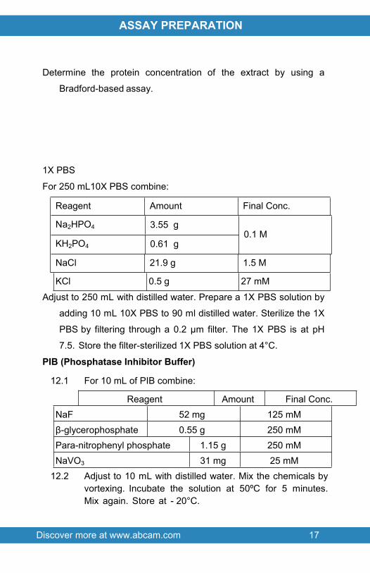
ASSAY PREPARATION
Determine the protein concentration of the extract by using a
Bradford-based assay.
1X PBS
For 250 mL10X PBS combine:
Reagent Amount Final Conc.
Na2HPO4 3.55 g
KH2PO4 0.61 g0.1 M
NaCl 21.9 g 1.5 M
KCl 0.5 g 27 mM
Adjust to 250 mL with distilled water. Prepare a 1X PBS solution by
adding 10 mL 10X PBS to 90 ml distilled water. Sterilize the 1X
PBS by filtering through a 0.2 µm filter. The 1X PBS is at pH
7.5. Store the filter-sterilized 1X PBS solution at 4°C.
PIB (Phosphatase Inhibitor Buffer)
12.1 For 10 mL of PIB combine:
Reagent Amount Final Conc.NaF 52 mg 125 mMβ-glycerophosphate 0.55 g 250 mMPara-nitrophenyl phosphate 1.15 g 250 mMNaVO3 31 mg 25 mM
12.2 Adjust to 10 mL with distilled water. Mix the chemicals by vortexing. Incubate the solution at 50ºC for 5 minutes. Mix again. Store at - 20°C.

Discover more at www.abcam.com 18
ASSAY PREPARATION
1X PBS/PIBPrior to use, add 500 µL of PIB in 10 ml of 1X PBS. Mix gently and
thoroughly. Do not store.
HB (Hypotonic Buffer)
Reagent Amount Final Conc.Hepes, pH 7.5 0.24 g 20 mMNaF 12 mg 5 mMNa2MoO4 5 µL 10 µMEDTA 10 µL
13 Adjust pH to 7.5 with 1 N NaOH. Adjust volume to 50 mL with
distilled water. Sterilize by filtering through a 0.2 µm filter. Store
the filter- sterilized solution at 4°C
Lysis Buffer
Reagent Amount Final Conc.Hepes, pH 7.5 0.24 g 20 mMNaCl 1.17 g 400 mMEDTA 1.15 mg 0.1 mMNaF 21 mg 10 mMNa2MoO4 0.12 mg 10 µMNaVO3 6.1 mg 1 mMGlycerol 10 mL 20%PNPP 0.23 g 10 mMBeta-Glycerophosphate 0.11 g 10 mM

Discover more at www.abcam.com 19
ASSAY PREPARATION
Adjust pH to 7.5 with 1 N NaOH. Adjust volume to 50 mL with
distilled water. Store at 4°C. Just before use, make up
Complete Lysis Buffer by adding 1 µL of 1 M DTT and 10 µl
of Protease Inhibitor Cocktail per mL of Lysis Buffer.
12.1 Peripheral Blood Mononuclear Cell (PMBC) Collection12.1.2 Collect blood samples using standard venipuncture
into tubes. Invert tubes 8 to 10 times to mix anticoagulant additive with blood. Blood samples should be centrifuged within two hours of blood collection. Centrifuge tube/blood samples at room temperature (18–25ºC) for 20 minutes at 1,500 to 1,800 RCF.
12.1.3 After centrifugation, mononuclear cells and platelets will be in a whitish layer just under the plasma layer. Immediately process the PBMCs, by aspirating approximately half of the plasma without disturbing the cell layer. Collect the cell layer and transfer to a 15 mL conical centrifuge tube with cap.
12.1.4 Add PBS to the PBMCs to bring the volume to 15 mL. Cap tube and invert to mix cells.
12.1.5 Centrifuge tube for 15 minutes at 300 RCF, 4ºC. Aspirate supernatant without disturbing the cell pellet.
12.1.6 Resuspend cell pellet in residual PBS by gently vortexing or tapping tube with index finger.
12.1.7 Add PBS to resuspended pellet to bring volume to 10 mL. Cap tube and invert to mix cells.
12.1.8 Centrifuge tube for 15 minutes at 300 RCF, 4ºC. Aspirate supernatant without disturbing the cell pellet.
12.1.9 Repeat washing steps 8 and 9 for a total of 3 washes.

Discover more at www.abcam.com 20
ASSAY PREPARATION
12.1.10 Assay immediately or freeze down in freezing media and store in liquid nitrogen.
12.2 PBMC Thawing12.2.2 Remove vials containing frozen cells from liquid
nitrogen and place in 37ºC water bath.12.2.3 Remove vials form water bath when no ice crystals
remain12.2.4 Transfer cell solution to 15 mL conical centrifuge tube
with cap.12.2.5 Add PBS to the PBMCs to bring the volume to 15 mL.
Cap tube and invert to mix cells.12.2.6 Centrifuge tube for 15 minutes at 300 RCF, 4ºC.
Aspirate supernatant without disturbing the cell pellet.12.2.7 Resuspend cell pellet in 2 mL PBS for performing cell
counts.
12.3 Cell Counts with Hemocytometer12.3.2 Transfer 50 μL of cell suspension to a solution
containing 75 μL PBS and 125 μL12.3.3 Trypan blue. Vortex the trypan-blue cell solution.12.3.4 With the cover slip in place, transfer a small amount of
trypan blue-cell suspension to a chamber on the hemocytometer. Ensure that the entire area under the cover slip contains the staining solution before removing any excess staining solution from the edge of the cover slip.
12.3.5 Both chambers of the hemocytometer must contain staining solution before performing cell counts.

Discover more at www.abcam.com 21
ASSAY PREPARATION
12.3.6 Place hemocytometer on the microscope and count the number of trypan-blue excluding (viable) cells in the 4 outer squares. If there are less than 10 cells or more than 100 cells per square, repeat the procedure adjusting to an appropriate dilution factor.
12.3.7 Calculate the cell concentration as follows:Cell concentration per milliliter = Total cell count in 4 squares x 2,500 x 5 (dilution factor) Total cell count = Cell concentration per milliliter x 2.0 mL (cell suspension)
12.3.8 Centrifuge cell suspension for 10 minutes at 300 RCF, 4ºC. Aspirate supernatant without disturbing cell pellet.
12.3.9 Proceed to cell lysis immediately.
12.4 Cell Lysis12.4.2 Resuspend cell pellet in Extraction Reagent 4,
containing protease inhibitors.12.4.3 Add 1 mL of extraction reagent per 108 cells. See
Reagent Preparation Section for addition of protease inhibitors to Extraction Reagent 4.
12.4.4 Incubate cell suspension on ice for 30 minutes for complete lysis.
12.4.5 Transfer cell lysis to 1.5 mL centrifuge tube. Centrifuge cell lysates for 10 minutes at 14,000 RCF, 4ºC.
12.4.6 Clarified lysates may be assayed immediately, or aliquoted and stored at -70ºC.
12.4.7 No degradation of SMN in cell lysate was observed after 2 freeze-thaw cycles.
12.5 Cell Lysate Sample Handling 12.5.2 If cell lysates were frozen prior to assay, the frozen
lysate samples should be brought slowly to 4ºC (on ice) and, if residual precipitate is present, centrifuge to

Discover more at www.abcam.com 22
ASSAY PREPARATION
isolate residual cell debris. Samples diluted sufficiently into the assay buffer can be read directly from a standard curve. A minimum 1:4 dilution is recommended for cell lysates to remove matrix interference in the assay.
12.6 Mouse Tissue Homogenization12.6.2 Prepare Extraction Reagent 4 with protease inhibitors.
Recommended protease inhibitors are 0.5 μL of PIC8340 per mL of reagent and PMSF to a final concentration of 1 mM.
12.6.3 Transfer tissue sample to appropriate sized tube for homogenization with 1mL of prepared Extraction Reagent 4.
12.6.4 For mechanical homogenizer, disrupt the tissue with three pulses of 3-4 seconds each. For manual dounce homogenizer, complete a minimum of 5 passes of the pestle past the buffer/tissue volume, or until tissue appears completely homogenized. Keep samples on ice while completing all preparations.
12.6.5 Pellet out tissue/cellular debris via centrifugation at 14,000g for 10 minutes at 4°C and transfer supernatant to a clean tube.
12.6.6 Measure the protein content of the supernatant using the biorad Biorad DC protein assay.
12.6.7 Prepare tissue homogenates for use in the SMN assay by diluting the extracted samples in assay buffer. Samples must be diluted at least 1:8. Dilute brain, muscle, and spinal cord tissue samples to final assay protein concentrations of 25 μg/mL, 50 μg/mL and 100 μg/mL, respectively.

Discover more at www.abcam.com 23
ASSAY PREPARATION
13.PLATE PREPARATION The 96 well plate strips included with this kit are supplied ready to
use. It is not necessary to rinse the plate prior to adding reagents Unused well strips should be returned to the plate packet and
stored at 4°C For each assay performed, a minimum of 2 wells must be used as
blanks, omitting primary antibody from well additions For statistical reasons, we recommend each sample should be
assayed with a minimum of two replicates (duplicates) Well effects have not been observed with this assay. Contents of
each well can be recorded on the template sheet included in the Resources section

Discover more at www.abcam.com 24
ASSAY PROCEDURE
14.ASSAY PROCEDURE Equilibrate all materials and prepared reagents to room
temperature prior to use It is recommended to assay all standards, controls and
samples in duplicate Determine the appropriate number of microwell strips required for
testing samples, controls and blanks in duplicate. If less than 8 wells in a strip need to be used, cover the unused wells with a portion of the plate sealer while you perform the assay. The content of these wells are stable at room temperature if kept dry and, therefore, can be used later for a separate assay. Store the unused strips in the aluminum pouch at 4°C. Use the strip holder for the assay.13 Prepare all reagents, working standards, and samples as
directed in the previous sections.14.2 Add 100 μL of each Standard s 1 through 7 and BO into the
appropriate wells.14.3 Add 50100 μL of the prepared sSamples diluted in Diluent
Buffer or protein standards (if used) into the appropriate wells.
14.4 Add 2 μL of the provided MCF-7 nuclear extract to 48 μL of Diluent Buffer to the appropriate number of wells for the controls.
14.5 Add 50 μL of Diluent Buffer to the appropriate number of wells for the blank control.
14.614.7 Seal the plate and incubate for 1 hour minutes on a rocking
plate shaker at 1500 rpm and at room temperature.14.8 Empty the contents of the wells and wash by adding 2300
µL of 1X Wash Buffer to every well. Repeat the wash 23 more times for a total of 34 Washes. After the final wash, empty or aspirate the wells, and firmly tap the plate on a lint free paper towel to remove any remaining wash buffer.

Discover more at www.abcam.com 25
ASSAY PROCEDURE
14.9 Add 5100 μL of the 1X ERα Detector Antibody anti-SMN monoclonal detection antibody to every well.
14.10 Seal the plate and incubate for 1 hour on a plate shaker at 500 rpm and at room temperature with gentle agitation.
14.11 Wash as described in step 14.665. 14.1214.13 Add 1050 µL of the 1X HRP Conjugated Secondary
Antibody anti-rabbit IgG – HRP conjugate to all wells.14.14 Seal the plate and incubate for 30 minutes1 hour on a plate
shaker (~500) at room temperature with gentle agitation.14.15 During this incubation, place the Developing Solution at
room temperature. 14.16 .14.17 Wash as described in step 14.665 but with a total of 4
washes.. 14.18 Transfer the amount of Developing Solution required for
the assay into a secondary container. 14.19 Add 100 µl Developing Solution to all wells being used.14.20 Incubate 2-10 minutes at room temperature protected from
direct light. Monitor the blue color development in the sample and positive control wells until it turns medium to dark blue. Blank wells should remain faint to light blue. Avoid overdevelopment.Add 100 μL TMB substrate solution to each well.
14.2114.22 Seal the plate and incubate for 30 minutes on a plate
shaker at 500 rpm and at room temperature.14.23 Add 100 μL Stop Solution to each well. In presence of the
acid, the blue color turns yellow.14.24 Read absorbance on a spectrophotometer within 5
minutes at 450 nm with a reference wavelength of 655 nm. Blank the plate reader according to the manufacturer’s instructions using the blank wells.
Discover more at www.abcam.com 26
ASSAY PROCEDURE
14.25 Read the O.D. absorbance at 450 nm, preferably with correction between 570 and 590 nm.

Discover more at www.abcam.com 27
ASSAY PROCEDURE
15.CALCULATIONS
If you have generated a standard curve using a recombinant ER
protein, average the duplicate readings for each standard, control,
and sample and subtract the optical density (OD) obtained from the
zero standard. Plot the OD for the standards against the quantity
(ng/well) of the standards and draw the best fit curve.
A four parameter algorithm (4PL) provides the best fit, though other equations can be examined to see which provides the most accurate (e.g. linear, semi-log, log/log, 4 parameter logistic). Interpolate protein concentrations for unknown samples from the standard curve plotted. Samples producing signals greater than that of the highest standard should be further diluted and reanalyzed, then multiplying the concentration found by the appropriate dilution factor.
Calculate the average net Optical Density (OD) bound for each standard and sample by subtracting the average Blank OD from the average OD bound:
Average Net OD = Average Bound OD - Average BO Blank OD
Plot the average Net OD for each standard versus SMN concentration in each standard. Sample concentrations may be calculated off of Net OD values using the desired curve fitting

Discover more at www.abcam.com 28
ASSAY PROCEDURE
16.TYPICAL DATAData provided for demonstration purposes only.
Measuring cellular ERα levels. Different amounts of nuclear
extracts from MCF-7 and MDA-MB-23 cells were analyzed for
cellular levels of ERα using the Estrogen Receptor Alpha ELISA Kit.
This data is provided for demonstration purposes only.

Discover more at www.abcam.com 29
DATA ANALYSIS
Example of aA ne standard curve using recombinant protein (not included with kit). wA new standard curve must be generated for each assay performed.
ERα
Net
SMN Conc.(pg/mL) Net OD
0 0.0880.088

Discover more at www.abcam.com 30
DATA ANALYSIS
50 0.1242.679
100 0.171.482
200 0.2580.789
400 0.4340.434
800 0.7890.258
1,600 1.4820.17
3,200 2.6790.124
17.TYPICAL SAMPLE VALUESSENSITIVITY –The sensitivity of the assay, defined as the concentration of SMN measured at 2 standard deviations from the mean of 20 replicates of zero standard along the standard curve, was determined to be 50 pg/mL.detection limit of the assay is > 0.6 µg nuclear extract/well.
LINEARITY OF DILUTIONRANGE OF DETECTION –Estrogen Receptor Alpha ELISA Kit provides quantitative results from 0.6 to 10 µg of nuclear extract/well.The minimum required dilution for several common samples was determined by serially diluting samples into the assay buffer and identifying the dilution at which linearity is observed.
DilutionHuman cell
lysate (%)
Mouse Brain
Tissue(%)
Mouse Muscle Tissue
(%)
Mouse Spinal Cord Tissue
(%)
Neat - - - -1:2 - >LOD 74 65

Discover more at www.abcam.com 31
DATA ANALYSIS
1:4 98 >LOD 84 821:8 102 >LOD 90 88
1:16 105 >LOD 99 891:32 100 94 100 971:64 - 100 <LOD 106
RECOVERY –After diluting each sample matrix to its minimum required dilution, recombinant Human SMN was spiked at high, medium, and low concentrations. The recovery of the standard in spiked samples was compared to the recovery of identical spikes in the assay buffer. The mean and range of percent recovery at the three concentrations are indicated below for each matrix.
Mean Spike and Recovery Results
Sample Matrix(# of samples)
Minimum Required Dilution
Spike Concentration(pg/mL)
Average % Recovery
(range)
1667 100 (88 - 116)
667 100 (88 - 116)Human PBMC
lysate(n=5)
1:4
267 99 (79 - 134)
1250 84 (83‐84)
250 86 (85‐87)Mouse Brain Extract (n=2) ≥1.8a
50 104 (96‐112)
1250 79 (76‐81)
250 88 (85‐90)Mouse muscle
extract(n=2)
≥1.8b
50 125 (103‐146)
Mouse spinal cord
≥1.8c 1250 68 (66‐69)

Discover more at www.abcam.com 32
DATA ANALYSIS
250 68 (67‐68)extract(n=2) 50 44 (39‐48)
a = Dilute mouse brain tissue extract such that the final protein concentration in the assay sample is 25μg/mL with a minimum dilution of 1:8.b = Dilute mouse muscle tissue extract such that the final protein concentration in the assay sample is 50μg/mL with a minimum dilution of 1:8.c = Dilute mouse spinal cord tissue extract such that the final protein concentration in the assay sample is 100μg/mL with a minimum dilution of 1:8.
PARALLELISM –A parallelism experiment was carried out to determine if the recombinant Human SMN standard accurately determines SMN concentrations in biological matrices. To assess parallelism, values for Human PBMC lysate and mouse tissue extract was obtained from a standard curve using four parameter logistic curve fitting. The observed concentration was plotted against the dilution factor. Parallelism of the curves demonstrates that the antibody binding characteristics are similar enough to allow the accurate determination of analyte levels in diluted samples.

Discover more at www.abcam.com 33
DATA ANALYSIS
PRECISION –Intra‐assay precision was determined by assaying 20 replicates of three buffer controls containing SMN in a single assay.
SMN(pg/mL) % CV
928 0.8322 1.0122 3.2

Discover more at www.abcam.com 34
DATA ANALYSIS
Inter‐assay precision was determined by measuring buffer controls (n=12) of varying SMN concentrations in multiple assays over several days.
SMN(pg/mL) % CV
983 7.1378 8.9134 11.4
18.ASSAY SPECIFICITYEstrogen Receptor Alpha ELISA Kit detects ERα from human, mouse and hamster origin. This assay is not recommended for use with samples from rat origin. Cross-reactivity with other species has not been determined.This kit detects SMN protein of both Human and mouse origin. Other species have not been tested.

Discover more at www.abcam.com 35
RESOURCES
19.TROUBLESHOOTING
Problem Cause Solution
Inaccurate pipetting Check pipettes
Poor standard curve Improper standards
dilution
Prior to opening, briefly spin the stock standard tube and dissolve the powder thoroughly by gentle
mixing
Incubation times too brief
Ensure sufficient incubation times; change to overnight
standard/sample incubationLow Signal
Inadequate reagent volumes or improper
dilution
Check pipettes and ensure correct preparation
Samples give higher value than the highest standard
Starting sample concentration is too
high.
Dilute the specimens and repeat the assay
Plate is insufficiently washed
Review manual for proper wash technique. If using a plate washer,
check all ports for obstructionsLarge CV
Contaminated wash buffer Prepare fresh wash buffer
Low sensitivity
Improper storage of the kit
Store the all components as directed.

Discover more at www.abcam.com 36
RESOURCES
20.NOTES

Discover more at www.abcam.com 37
RESOURCES

Discover more at www.abcam.com 38
RESOURCES

Discover more at www.abcam.com 39
RESOURCES

RESOURCES 40
UK, EU and ROWEmail: [email protected] | Tel: +44-(0)1223-696000
AustriaEmail: [email protected] | Tel: 019-288-259
FranceEmail: [email protected] | Tel: 01-46-94-62-96 GermanyEmail: [email protected] | Tel: 030-896-779-154 SpainEmail: [email protected] | Tel: 911-146-554 SwitzerlandEmail: [email protected] Tel (Deutsch): 0435-016-424 | Tel (Français): 0615-000-530
US and Latin AmericaEmail: [email protected] | Tel: 888-77-ABCAM (22226)
CanadaEmail: [email protected] | Tel: 877-749-8807
China and Asia Pacific Email: [email protected] | Tel: 108008523689 (中國聯通) JapanEmail: [email protected] | Tel: +81-(0)3-6231-0940 www.abcam.com | www.abcam.cn | www.abcam.co.jp
Copyright © 2013 Abcam, All Rights Reserved. The Abcam logo is a registered trademark.
All information / detail is correct at time of going to print.