Embed Size (px)
Citation preview

12
American Journal of Botany 102 ( 1 ): 12 – 20 , 2015 ; http://www.amjbot.org/ © 2015 Botanical Society of America
American Journal of Botany 102 ( 1 ): 12 – 20 , 2015 .
The mechanical properties of plant cell walls play crucial roles in the regulation of the growth of cells while ensuring their structural integrity. The tension response is of great im-portance because it is thought that turgor pressure regulates the tensile state of the cell wall to control its expansive growth ( Cosgrove, 2005 ; Albersheim et al., 2010 ; Geitmann, 2010 ). Water is one of the major components of the primary cell wall and is thought to play a signifi cant role in mechanical properties of the cell wall ( Tang et al., 1999 ; Blewett et al., 2000 ; Ulvskov et al., 2005 ; Saito et al., 2006 ; Evered et al., 2007 ; Moore et al., 2008 ; Hansen et al., 2011 ). To understand how water’s pres-ence affects the molecular architecture of the cell wall, includ-ing the interactions among its major structural constituents, and consequently, how it alters the higher-scale (such as micro- and macroscales) mechanical responses, quantitative measure-ment of the cell wall without confounding effects from cell geometries and interactions with middle lamellae are needed. Vanstreels et al. (2005) and Saito et al. (2006) showed the
signifi cance of water’s role in the mechanical responses of plant cell wall tissues or larger samples. Additionally, changes in moisture alter the structure and physical properties of cellulose ( Salmén, 2004 ), hemicellulose, and pectin ( Ha et al., 1997 ; Moore et al., 2008 ). In addition, water is known to induce struc-tural changes in the primary cell wall ( Ha et al., 1997 ; Thimm et al., 2000 ). Especially, increased moisture induces the soften-ing of bacterial cellulose, hemicellulose, and pectin composites ( Dammström et al., 2005 ; Bader et al., 2011 ). In summary, wa-ter is well known to affect the mechanical responses of primary cell walls. Despite the claimed importance of water’s status in plant growth, quantitative studies on the effects of water on the primary cell wall, where the actual growth takes place, are scarce. To that end, quantitative experiments, which can quan-tify changes in mechanical properties of primary cell wall at different hydration states, can provide essential information on water’s role and contribution on the mechanics of the expansive cell wall growth.
The studies to date have been limited by the plant samples, which include other elements than those in the cell wall ( Ha et al., 1997 ; Blewett et al., 2000 ; Saito et al., 2006 ; Evered et al., 2007 ). The challenge is that the plant tissue includes many components and several extracellular features that interact with water, and, as such, the effect of water on the cell wall is non-trivial to isolate. Therefore, it is essential to conduct stretching experiments to enable reliable and repeatable measurements of the cell wall mechanical response. However, sample prepa-ration, manipulation, and experimentation on test samples at the micron scale is challenging ( Castillo-Leó n et al., 2012 ),
1 Manuscript received 14 June 2014; revision accepted 10 December 2014. The authors thank the Center for LignoCellulose Structure and
Formation, an Energy Frontier Research Center funded by the U.S. Department of Energy, Offi ce of Science, Offi ce of Basic Energy Sciences under Award Number DE-SC0001090 for fi nancial support.
4 Author for correspondence (e-mail: [email protected]); present address: School of Engineering, University of British Columbia, Kelowna, BC V1V1V7, Canada; phone: 1-250-807-8040, fax: 1-250-807-9850
doi:10.3732/ajb.1400273
MULTISCALE STRESS–STRAIN CHARACTERIZATION OF ONION OUTER EPIDERMAL TISSUE IN WET AND DRY STATES 1
KEEKYOUNG KIM 2–4 , HOJAE YI 2 , M. SHAFAYET ZAMIL 2 , M. AMANUL HAQUE 3 , AND VIRENDRA M. PURI 2
2 Department of Agricultural and Biological Engineering, Pennsylvania State University, University Park, Pennsylvania 16802 USA; and 3 Department of Mechanical and Nuclear Engineering, Pennsylvania State University, University Park, Pennsylvania
16802 USA
• Premise of the study: Quantitative measurements of water’s effects on the tension response of plant tissue will assist in under-standing the regulatory mechanism underlying expansive growth. Such measurements should be multiscale in nature to account for plants’ hierarchical structure.
• Methods: Outer onion epidermal tissues were cut and bonded to uniaxial displacement-controlled mechanical loading devices to apply and measure the force on the sample. Fluorescent polystyrene beads (500 nm in diameter) were dispersed on the sample surface under various levels of tensile load conditions to obtain displacement maps with a confocal fl uorescent micro-scope. The resulting strain was measured using a digital image correlation technique by tracking individual bead displace-ments. The applied forces were obtained by measuring the displacement of the calibrated force-sensing device. Tissue- and cell-scale mechanical properties were quantifi ed by calculating the applied stress and the corresponding global and local strains.
• Key results: The Young’s modulus values of individual cell walls of dehydrated and rehydrated samples were 3.0 ± 1.0 GPa and 0.4 ± 0.2 GPa, respectively, and are different from the Young’s modulus values of the global tissue-scale dehydrated and rehydrated samples, which were 1.9 ± 0.3 GPa and 0.08 ± 0.02 GPa, respectively. Poisson’s ratio increased more than 3-fold due to hydration.
• Conclusion: The results on global, cell-to-cell, and point-to-point mechanical property variations suggest the importance of the mechanical contribution of extracellular features including the middle lamella, cell shape, and dimension. This study shows that a multiscale investigation is essential for fundamental insights into the hierarchical deformation of biological systems.
Key words: cell wall mechanics; digital image correlation; dry and wet states; multiscale stress–strain; onion epidermal tissue; tensile testing.

KIM ET AL.—MULTISCALE STRESS–STRAIN CHARACTERIZATION OF TISSUE 13January 2015]
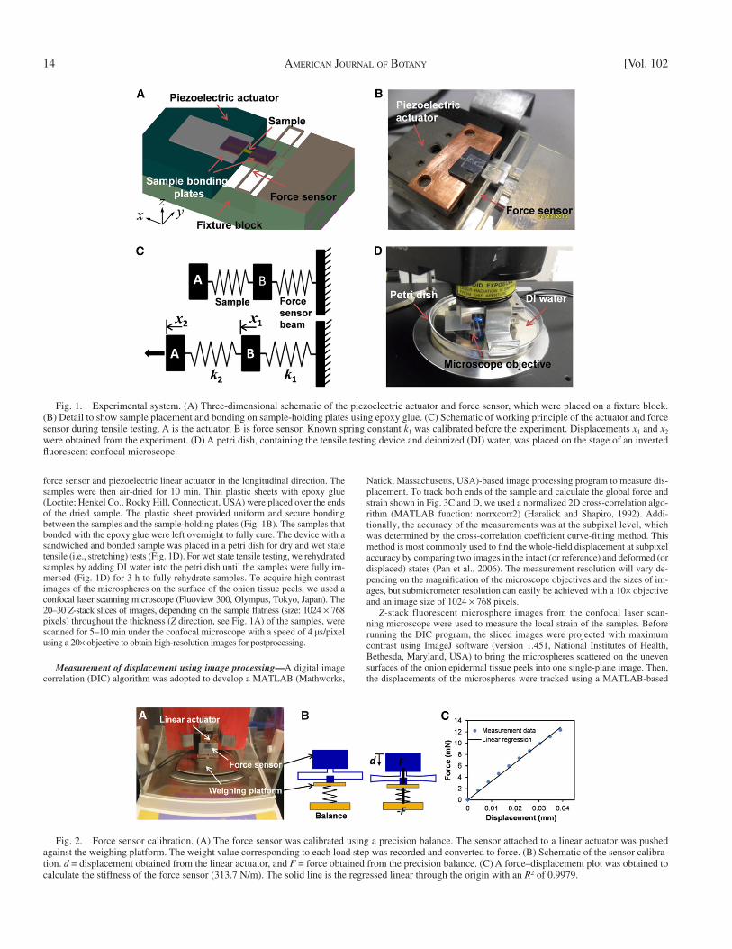
is known. The piezoelectric actuator and force sensor were mounted on a fi xture block to enable the expansion-type displacement of the piezoelectric actuator to be transferred to a sample-holding plate of the force sensor as shown in Fig. 1A and B . Figure 1C shows a schematic of the working principle of the experimental system. The force balance equation at the sample-holding plate of the force sensor is 1 1 2 2 1F k x k x x , where F is the loading force, k 1 is the spring constant of the force sensor, which was known, x 1 is the displacement of the sample-holding plate, k 2 is the spring constant of the sample, and x 2 is the displacement of the linear actuator. Displacements x 1 and x 2 were obtained from the experiment and used to calculate the applied forces and the mechanical properties of the samples.
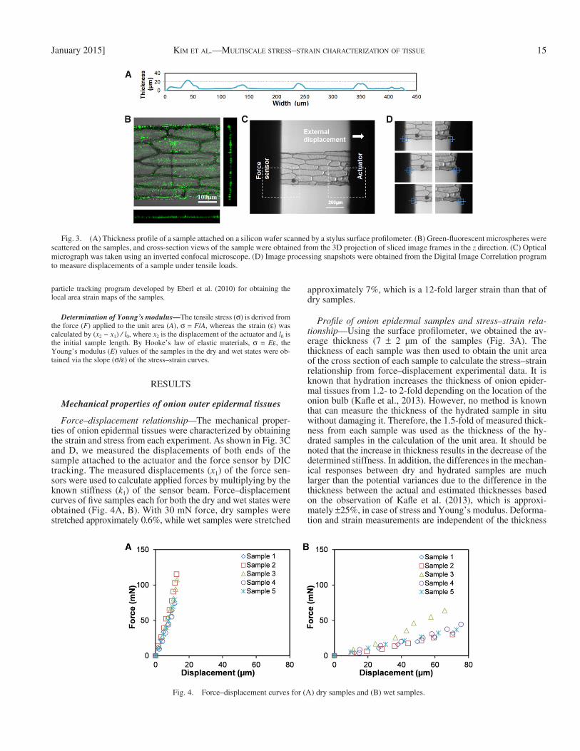
Force sensor calibration — The acrylic force sensors were calibrated using a precision balance (CP26, Sartorius AG, Goettingen, Germany). Figure 2A and B show the experimental setup and a schematic of the calibration of the serpentine beam springs of the force sensors. The force sensor was bonded to a piezoelectric linear stage. For calibrating the force sensor, the piezoelectric stage was moved in 4-µm increments against the weighing platform of the bal-ance. The weight values corresponding to displacements of the linear stage were converted to force data. As shown in Fig. 2C , the stiffness of the force sensor (313.7 N/m) was derived by the slope of the obtained force-displacement curve.
Sample preparation — The outer epidermal tissues were extracted from yel-low onions ( Allium cepa ) that were purchased from a local grocery store. The onion was selected as a test material due to accessibility and the ease of sample preparation. The ease of sample preparation is essential because the prepared sample stays close to the native state compared with other model plants, which require a more sophisticated sample preparation. The outer epidermal cell wall profi le was peeled away from the middle scale of the onion to expose the peri-clinal single-wall layer of the epidermal tissue. Following procedures of Kafl e et al. ( 2014) , we used an optical microscope to make sure that the epidermal cell wall profi le thus prepared only comprised split open cells, i.e., no intact cells were present. Subsequently, the cell wall profi le patch was rinsed gently with tap water at room temperature to wash away the protoplasmic liquid. Immediately thereafter, the cell wall profi le patch was used as a consistent source of plant cell wall fragments for all of the tests. It should be noted that cell walls exposed by the peeling of epidermal cells are expected to experience some degree of perturbation in the water content equilibrium due to the loss of contact with the cytoplasm, i.e., exposed cell walls will no longer able to exchange molecules and ions with the cytoplasm, which can affect the water equilibrium between cell wall and cytoplasm. In addition, it is also possible for part of the residual cytoplasmic protein to be adsorbed to the cell wall despite the rinsing step. The extent of this potential disturbance to the mechanical properties of the cell wall profi le patch cannot be measured due to the lack of an effective method to prepare samples without such disruptions. Nonetheless, this aspect should be considered in studying the result with respect to the mechanics of the native cell wall.
A thin slice of onion from the middle scale was cut with two sandwiched blades with a width of 300 µm. To measure the thickness of the samples, we placed them on a silicon wafer, and the samples were uniformly attached and dried by the cohesion and adhesion forces of water molecules while the mois-ture was evaporated at room temperature for 30 min. As shown in Fig. 3A , the thickness of the samples was measured by a stylus surface profi lometer (AlphaStep 500, KLA-Tencor, Milpitas, California, USA) in a transverse direction relative to each sample.
The measured samples were then labeled by polystyrene microspheres. Fluorescein isothiocyanate (FITC)-labeled carboxylate polystyrene micro-spheres (Fluoresbite, Polyscience, Warrington, Pennsylvania, USA) 500 nm in diameter were dispersed on the surface of the epidermal tissue. We prepared the polystyrene microspheres beforehand in deionized (DI) water by diluting them 10 times to adjust the concentration. The microsphere solution was sonicated in an ultrasonic bath for 10 min to disperse the microspheres in the solution. Then 20 µL of the solution was pipetted onto the epidermal tissue sample, and this step was followed by a 10-min wait for the microspheres to adhere to the onion epidermal tissue ( Fig. 3B ). A carboxylate group on the surface of the micro-spheres plays a role to covalently bind to proteins on the surface of the onion epidermal tissue and provides for a secure attachment during the wet state ex-periment. The samples were washed three times with DI water to remove exces-sive microspheres.
Stretching samples under tensile loading — After placing the samples on the tensile test device, the fl uorescent microsphere-labeled samples spanned the
especially due to the thickness of the cell wall. Recently, a bio-mechanical experimental protocol was developed ( Zamil et al., 2013 ), which enables the determination of the cell wall’s me-chanical properties without considering extracellular features such as different cell sizes, multiple cell mechanical interac-tions, middle lamella. One limitation of this unique approach was that the sample preparation and the experiment are per-formed inside a vacuum chamber, which dries the cell wall sample rapidly. Therefore, an improved test procedure is needed to investigate the effect of water on the cell wall. The measured response of wet samples will yield a more accurate representation of the onset of cell wall expansion in both the major (longitudinal) and minor (transverse) growth directions.
Comparisons of the mechanical responses of near-native (or hydrated) samples and dried samples will elucidate the effect of water on the mechanical behavior of cell walls. When a test sample is prepared from a plant, dehydration is almost inevita-ble. In this study, the effect of water content of onion outer epidermal tissue comprising a two-dimensional array of broken cell walls was examined quantitatively under tensile loading using a microelectromechanical system (MEMS) device with a novel experimental protocol. The test setup comprises a piezo-electric-based actuation motor and a folded spring MEMS force sensor device fabricated by a 3D prototyping technique. The new experimental protocol adapts to a larger or macroscale sample (wall profi le of a cluster of approximately 25 cell walls) excised from onion epidermal tissue, which can be prepared without the aid of electron microscopy and be examined with an optical microscope and at any environmental condition. For achieving suffi ciently high image resolution and to facilitate both the microscale (subcellular) and macroscale (tissue) inves-tigation, the samples were tagged with fl uorescent micro-spheres, and tests were carried out using a confocal laser scanning microscope. The images were analyzed using a digital image correlation (DIC)-based technique to investigate the ten-sile stretching responses both at local and global scales. This approach resolves the problem of vacuum exposure associated with investigating plant cell walls at the subcellular scale pro-posed by Zamil et al. (2013) . To quantify the effect of water on the mechanical properties of samples consists of primary plant cell walls, we measured tension deformations using air-dried and rehydrated samples of onion epidermal cell wall patch.
MATERIALS AND METHODS
The essential experimental framework to investigate the fundamental issues introduced in the previous section should have the capability of characterizing both cellular and subcellular level mechanical properties and be adaptable to various moisture contents. The most rigorous requirement is, therefore, the ap-plication and measurement of micro- to nanoscale forces and displacements on micrometer-size biological samples. Additionally, sample extraction, prepara-tion, and experimental conditions should maintain the natural state of the bio-logical test material as closely as possible. In this study, a novel experimental setup followed by sophisticated and robust postprocessing of the acquired data in microscale forces and displacements was implemented to enable us to quan-tify and compare the mechanical properties of onion epidermal cell walls in the wet state, which closely mimics the natural state, and the dry state.
Experimental setup — The experimental system consists of a force sensor and a piezoelectric linear actuator stage (AG-LS25, Newport Co., Irvine, Cali-fornia, USA) with a displacement resolution of 430 nm per step. The force sensor made of an acrylic-based polymer composite was fabricated by 3D pro-totyping. The sensor comprises a mechanical structure with serpentine beams and a sample gripping pad whose spring constant (force per unit displacement)
14 AMERICAN JOURNAL OF BOTANY [Vol. 102
Natick, Massachusetts, USA)-based image processing program to measure dis-placement. To track both ends of the sample and calculate the global force and strain shown in Fig. 3C and D , we used a normalized 2D cross-correlation algo-rithm (MATLAB function: norrxcorr2) ( Haralick and Shapiro, 1992 ). Addi-tionally, the accuracy of the measurements was at the subpixel level, which was determined by the cross-correlation coeffi cient curve-fi tting method. This method is most commonly used to fi nd the whole-fi eld displacement at subpixel accuracy by comparing two images in the intact (or reference) and deformed (or displaced) states ( Pan et al., 2006 ). The measurement resolution will vary de-pending on the magnifi cation of the microscope objectives and the sizes of im-ages, but submicrometer resolution can easily be achieved with a 10 × objective and an image size of 1024 × 768 pixels.
Z -stack fluorescent microsphere images from the confocal laser scan-ning microscope were used to measure the local strain of the samples. Before running the DIC program, the sliced images were projected with maximum contrast using ImageJ software (version 1.451, National Institutes of Health, Bethesda, Maryland, USA) to bring the microspheres scattered on the uneven surfaces of the onion epidermal tissue peels into one single-plane image. Then, the displacements of the microspheres were tracked using a MATLAB-based
force sensor and piezoelectric linear actuator in the longitudinal direction. The samples were then air-dried for 10 min. Thin plastic sheets with epoxy glue (Loctite; Henkel Co., Rocky Hill, Connecticut, USA) were placed over the ends of the dried sample. The plastic sheet provided uniform and secure bonding between the samples and the sample-holding plates ( Fig. 1B ). The samples that bonded with the epoxy glue were left overnight to fully cure. The device with a sandwiched and bonded sample was placed in a petri dish for dry and wet state tensile (i.e., stretching) tests ( Fig. 1D ). For wet state tensile testing, we rehydrated samples by adding DI water into the petri dish until the samples were fully im-mersed ( Fig. 1D ) for 3 h to fully rehydrate samples. To acquire high contrast images of the microspheres on the surface of the onion tissue peels, we used a confocal laser scanning microscope (Fluoview 300, Olympus, Tokyo, Japan). The 20–30 Z -stack slices of images, depending on the sample fl atness (size: 1024 × 768 pixels) throughout the thickness ( Z direction, see Fig. 1A ) of the samples, were scanned for 5–10 min under the confocal microscope with a speed of 4 µs/pixel using a 20 × objective to obtain high-resolution images for postprocessing.
Measurement of displacement using image processing — A digital image correlation (DIC) algorithm was adopted to develop a MATLAB (Mathworks,
Fig. 1. Experimental system. (A) Three-dimensional schematic of the piezoelectric actuator and force sensor, which were placed on a fi xture block. (B) Detail to show sample placement and bonding on sample-holding plates using epoxy glue. (C) Schematic of working principle of the actuator and force sensor during tensile testing. A is the actuator, B is force sensor. Known spring constant k 1 was calibrated before the experiment. Displacements x 1 and x 2 were obtained from the experiment. (D) A petri dish, containing the tensile testing device and deionized (DI) water, was placed on the stage of an inverted fl uorescent confocal microscope.
Fig. 2. Force sensor calibration. (A) The force sensor was calibrated using a precision balance. The sensor attached to a linear actuator was pushed against the weighing platform. The weight value corresponding to each load step was recorded and converted to force. (B) Schematic of the sensor calibra-tion. d = displacement obtained from the linear actuator, and F = force obtained from the precision balance. (C) A force–displacement plot was obtained to calculate the stiffness of the force sensor (313.7 N/m). The solid line is the regressed linear through the origin with an R 2 of 0.9979.
KIM ET AL.—MULTISCALE STRESS–STRAIN CHARACTERIZATION OF TISSUE 15January 2015]
approximately 7%, which is a 12-fold larger strain than that of dry samples.
Profi le of onion epidermal samples and stress–strain rela-tionship— Using the surface profi lometer, we obtained the av-erage thickness (7 ± 2 µm of the samples ( Fig. 3A ). The thickness of each sample was then used to obtain the unit area of the cross section of each sample to calculate the stress–strain relationship from force–displacement experimental data. It is known that hydration increases the thickness of onion epider-mal tissues from 1.2- to 2-fold depending on the location of the onion bulb ( Kafl e et al., 2013 ). However, no method is known that can measure the thickness of the hydrated sample in situ without damaging it. Therefore, the 1.5-fold of measured thick-ness from each sample was used as the thickness of the hy-drated samples in the calculation of the unit area. It should be noted that the increase in thickness results in the decrease of the determined stiffness. In addition, the differences in the mechan-ical responses between dry and hydrated samples are much larger than the potential variances due to the difference in the thickness between the actual and estimated thicknesses based on the observation of Kafl e et al. (2013) , which is approxi-mately ± 25%, in case of stress and Young’s modulus. Deforma-tion and strain measurements are independent of the thickness
particle tracking program developed by Eberl et al. (2010) for obtaining the local area strain maps of the samples.
Determination of Young’s modulus — The tensile stress ( σ ) is derived from the force ( F ) applied to the unit area ( A ), σ = F / A , whereas the strain ( ε ) was calculated by ( x 2 − x 1 ) / l 0 , where x 2 is the displacement of the actuator and l 0 is the initial sample length. By Hooke’s law of elastic materials, σ = E ε , the Young’s modulus ( E ) values of the samples in the dry and wet states were ob-tained via the slope ( σ / ε ) of the stress–strain curves.
RESULTS
Mechanical properties of onion outer epidermal tissues
Force–displacement relationship— The mechanical proper-ties of onion epidermal tissues were characterized by obtaining the strain and stress from each experiment. As shown in Fig. 3C and D , we measured the displacements of both ends of the sample attached to the actuator and the force sensor by DIC tracking. The measured displacements ( x 1 ) of the force sen-sors were used to calculate applied forces by multiplying by the known stiffness ( k 1 ) of the sensor beam. Force–displacement curves of fi ve samples each for both the dry and wet states were obtained ( Fig. 4A, B ). With 30 mN force, dry samples were stretched approximately 0.6%, while wet samples were stretched
Fig. 3. (A) Thickness profi le of a sample attached on a silicon wafer scanned by a stylus surface profi lometer. (B) Green-fl uorescent microspheres were scattered on the samples, and cross-section views of the sample were obtained from the 3D projection of sliced image frames in the z direction. (C) Optical micrograph was taken using an inverted confocal microscope. (D) Image processing snapshots were obtained from the Digital Image Correlation program to measure displacements of a sample under tensile loads.
Fig. 4. Force–displacement curves for (A) dry samples and (B) wet samples.

16 AMERICAN JOURNAL OF BOTANY [Vol. 102
values) increased during application of the forces. Poisson’s ratio at 30 mN of force was calculated as 0.11 ± 0.06 for the dry samples and 0.35 ± 0.22 for the wet samples ( Fig. 6C ).
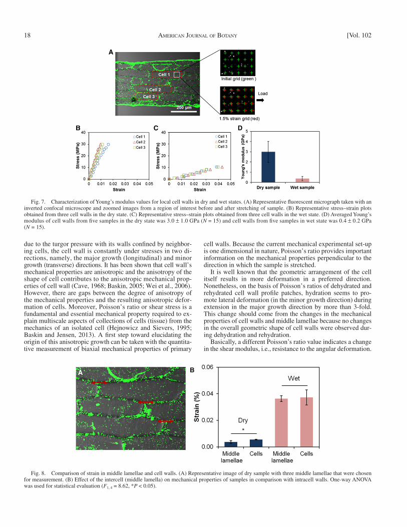
Intercell and intracell mechanical properties — By generat-ing and tracking grid points correlated to fl uorescent microspheres ( Fig. 7A ) attached to the samples, we obtained strains within single cell walls from fi ve wet and fi ve dry samples. With 30 mN force, the average strain of cell walls in the dry state was 0.4 ± 0.1% and in the wet state 2.4 ± 1.3%. The strain of cell walls in the wet state was 6-fold higher than for cell walls in the dry state. Under the assumption of the same stress level being applied to the cell walls, the averaged Young’s modulus of three cell walls from each of the fi ve samples in the dry state was 3.0 ± 1.0 GPa ( N = 15), while for the wet state it was 0.4 ± 0.2 GPa ( N = 15).
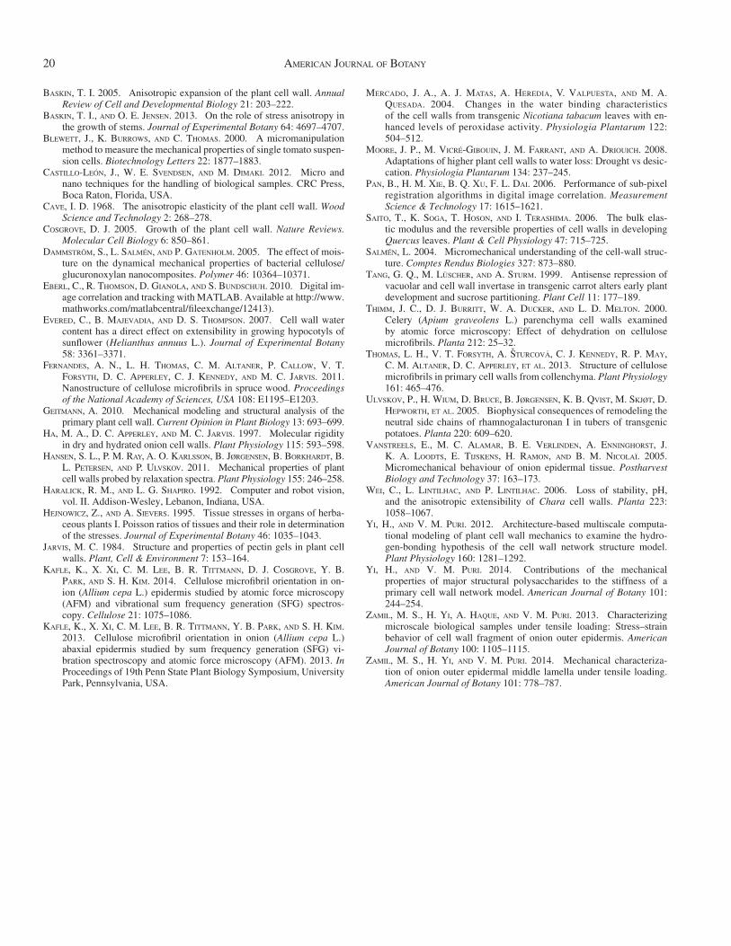
To investigate the intercell’s (middle lamella) effect on the mechanical properties of the samples, we selected an epidermal tissue in the dry state and in the wet state, respectively, and compared the strains on cell walls and the middle lamellae at a force of 30 mN ( Fig. 8A and B ). To measure the strains on the intracell wall region (middle lamellae), fl uorescent micro-spheres, which were deposited on an intracell wall region aligned in the same direction of tensile experiment, were se-lected and tracked in the tensile direction, i.e., along the major growth direction. The strain value of cell walls in the dry state was 0.55 ± 0.01%, while for the intracell wall region, it was 0.40 ± 0.10%. A one-way ANOVA was used to statistically evaluate the data. The cell strain was 1.4-fold higher than the intracell wall region strain, which was a signifi cant difference
changes. The tensile stress–strain curves are shown in Fig. 5A and B .
Young’s modulus and Poisson’s ratio— Young’s modulus of the samples in the dry state was 1.9 ± 0.3 GPa, while Young’s modulus of the samples in the wet state was 0.08 ± 0.02 GPa. Therefore, the samples in the dry state were 20-fold stiffer than the samples in the wet state. The higher value of dry vs. wet samples is consistent with the values reported in the literature. In a recent study by Zamil et al. (2013) , the modulus of vacuum-exposed onion outer epidermal cell wall fragments was 3.7 GPa in the major growth direction, whereas the modulus of wet onion epidermal tissue was reported by Vanstreels et al. (2005) to be 0.056 GPa. Our results show similar trends and magnitudes.
Poisson’s ratio of samples, which is the negative ratio of transverse to axial strain, was characterized. The power of the current approach is that one can calculate not only Young’s modulus, but also Poisson’s ratio from the same experiment. The same test results provide a complete set of elastic parame-ters, which has several advantages. The most important ones are (1) our approach avoids the use of two different experimen-tal set-ups, which can lead to inconsistencies resulting from boundary conditions and (2) the effi cient use of time and re-sources. Nonetheless, it should be noted that Poisson’s ratio due to the change in the thickness of the cell wall profi le patch was not determined because the thickness is much smaller than other dimensions resulting in negligible strain when compared with other dimensions.
Figure 6A and B show axial and transverse strains of the sam-ples in the dry and wet states. The transverse strains (negative
Fig. 5. Characterization of Young’s modulus values for epidermal tissues in the dry and wet state. (A) Stress–strain plots from dry samples and (B) stress–strain plots from wet samples. (C) Young’s modulus of tissues in the dry state was 1.9 ± 0.3 GPa, 0.08 ± 0.02 GPa in the wet state as. Dry state tissues are 20-fold stiffer than wet state tissues.

KIM ET AL.—MULTISCALE STRESS–STRAIN CHARACTERIZATION OF TISSUE 17January 2015]
air-drying process removes most of the free water from the onion cell wall, the spacing between cell wall constituents, in-cluding structural polysaccharides, will be reduced. This phe-nomenon was noted by Evered et al. (2007) ; when moisture was reduced in excised etiolated sunfl ower hypocotyl samples, the extension rate and, ultimately, elongation of the hypocot-yls decreased. The authors attributed this change to changes in the spacing between the cell wall components due to different water contents. A similar argument was presented by Ha et al. (1997) . From nuclear magnetic resonance (NMR) measure-ments of air-dried and rehydrated onion tissue samples, Ha et al. (1997) showed that hydration increased the molecular mo-bility of pectic polysaccharides. They argued that the glassy pectic matrix must carry part of the stresses within and between the layers and ruled out the participation of xyloglucan owing to its smaller fraction compared with the pectic polysaccharides. In summary, they suggested that hydration of onion cell wall mainly affects the pectic matrix and implied that dehydration of the cell wall will collapse the interlayer spacing so that the mi-crofi brils from adjacent layers come close to or even touch one another as the wall shrinks in thickness. Recently, the presence of such direct contact between cellulose microfi brils in cell wall has been observed especially when the cell wall is dehydrated ( Fernandes et al., 2011 ; Thomas et al., 2013 ). Overall, the rea-son for the decrease in the stiffness of the cell wall upon rehy-dration is that dehydration or rehydration of air-dried sample alters the architectural structure of the cell wall at the molecular level, especially the spacing between cell wall components.
Effect of the hydration in mechanical anisotropic responses — When the primary wall of a cell is subject to the outward load
( F 1, 4 = 8.62, P < 0.05). The strain value of the cell walls in the wet state was 3.6 ± 0.2%, and for the intracell wall region, it was 3.7 ± 0.5. This result does not refl ect a signifi cant difference ( F 1, 4 = 0.07, P > 0.05). In summary, intracell wall region de-formed less than cell wall, when the cell wall was in a dry state, while there was no difference when the cell was in the wet state.
DISCUSSION
Role of hydration in mechanical stiffness of the cell wall profi le patch — More than 70% of the fresh mass of a primary cell wall is accounted for by water ( Albersheim et al., 2010 ). In addition to the aqueous phase’s biochemical role in the forma-tion of cell wall structure, water is thought to make a signifi cant mechanical contribution to growth control ( Mercado et al., 2004 ; Ulvskov et al., 2005 ; Evered et al., 2007 ). Because the mechanical properties of a cell wall are primary determinants of its extensibility, it is essential to measure the quantitative stress–strain responses of the cell wall. When a plant cell wall sample is excised, prepared for, and subjected to mechanical testing in ambient conditions, it is inevitable that it loses moisture. When moisture is lost from the cell wall, the molecular structure of the cell wall changes, as does the way the load is borne. Nonethe-less, the effect of the amount of water present in the cell wall on the stress–strain measurement has yet to be established.
A major observation of this study was that the wet sample was 20-fold weaker (i.e., lower strength) than the air-dried sam-ple. At this point, it is worth discussing plausible origins of changes in mechanical properties of samples of primary cell walls when the hydration state changes. Assuming that the natural
Fig. 6. Measurement of transverse strains and comparison between axial strain and transverse strain for (A) a dry sample and (B) a wet sample. (C) The Poisson’s ratio of dry samples was 0.11 ± 0.06 and wet samples 0.35 ± 0.22.
18 AMERICAN JOURNAL OF BOTANY [Vol. 102
cell walls. Because the current mechanical experimental set-up is one dimensional in nature, Poisson’s ratio provides important information on the mechanical properties perpendicular to the direction in which the sample is stretched.
It is well known that the geometric arrangement of the cell itself results in more deformation in a preferred direction. Nonetheless, on the basis of Poisson’s ratios of dehydrated and rehydrated cell wall profi le patches, hydration seems to pro-mote lateral deformation (in the minor growth direction) during extension in the major growth direction by more than 3-fold. This change should come from the changes in the mechanical properties of cell walls and middle lamellae because no changes in the overall geometric shape of cell walls were observed dur-ing dehydration and rehydration.
Basically, a different Poisson’s ratio value indicates a change in the shear modulus, i.e., resistance to the angular deformation.
due to the turgor pressure with its walls confi ned by neighbor-ing cells, the cell wall is constantly under stresses in two di-rections, namely, the major growth (longitudinal) and minor growth (transverse) directions. It has been shown that cell wall’s mechanical properties are anisotropic and the anisotropy of the shape of cell contributes to the anisotropic mechanical prop-erties of cell wall ( Cave, 1968 ; Baskin, 2005 ; Wei et al., 2006 ). However, there are gaps between the degree of anisotropy of the mechanical properties and the resulting anisotropic defor-mation of cells. Moreover, Poisson’s ratio or shear stress is a fundamental and essential mechanical property required to ex-plain multiscale aspects of collections of cells (tissue) from the mechanics of an isolated cell ( Hejnowicz and Sievers, 1995 ; Baskin and Jensen, 2013 ). A fi rst step toward elucidating the origin of this anisotropic growth can be taken with the quantita-tive measurement of biaxial mechanical properties of primary
Fig. 7. Characterization of Young’s modulus values for local cell walls in dry and wet states. (A) Representative fl uorescent micrograph taken with an inverted confocal microscope and zoomed images from a region of interest before and after stretching of sample. (B) Representative stress–strain plots obtained from three cell walls in the dry state. (C) Representative stress–strain plots obtained from three cell walls in the wet state. (D) Averaged Young’s modulus of cell walls from fi ve samples in the dry state was 3.0 ± 1.0 GPa ( N = 15) and cell walls from fi ve samples in wet state was 0.4 ± 0.2 GPa ( N = 15).
Fig. 8. Comparison of strain in middle lamellae and cell walls. (A) Representative image of dry sample with three middle lamellae that were chosen for measurement. (B) Effect of the intercell (middle lamella) on mechanical properties of samples in comparison with intracell walls. One-way ANOVA was used for statistical evaluation ( F 1, 4 = 8.62, * P < 0.05).

KIM ET AL.—MULTISCALE STRESS–STRAIN CHARACTERIZATION OF TISSUE 19January 2015]
disappeared when samples were rehydrated. This observation suggests that hydration enables the epidermal tissue to deform in a homogeneous way and, as a result, minimizes the possibility of separations between cell walls during expansive growth. Considering that the middle lamella’s mechanical strength can be considered to be higher than the strength of the cell wall ( Zamil et al., 2014 ), this observation suggests that the middle lamella’s stiffness should be controlled to accommodate a cell wall’s expansion to allow for the overall growth of a plant without mechanical failure. It is also possible that the hydration of onion epidermal tissue samples may signifi cantly affect cell to cell adhesion, which is also regulated by the middle lamella. Overall, this result implies that the regulation of the stiffness between adjacent cell walls may play a more important role in plant growth regulation than in the regulation of the cell wall’s stiffness.
Conclusion — Water is an essential component of a growing cell wall. Despite its recognized importance from the stand-point of the mechanics of a growing cell wall, quantitative mea-surements of the effect of water on a cell wall’s mechanical responses without the confounding effect from the cells’ geom-etries and their interactions by the middle lamella have gener-ally been lacking. This study used a microelectromechanical system device to conduct tensile tests on onion epidermal tissue samples. The results show that the mechanical responses of in-dividual cell walls at a subcellular scale are discernable from the mechanical responses of the overall epidermal tissue at the tissue scale.
Dehydration increases the stiffness of the onion epidermal tissue and the individual cell wall as seen in the observation of rehydration’s adverse contribution to stiffness. This observa-tion suggests that hydration of the primary cell wall induces alterations in the cell wall’s molecular structure that are respon-sible for load transfer and bearing and in the properties of inter-cellular regions, i.e., middle lamellae. Considering the order of magnitude changes in stiffness upon rehydration, it is thought that water may alter the molecular interactions between struc-tural polysaccharides in addition to functioning as a spacer be-tween polysaccharides. With 30 mN force, the change in the strain of the cell wall was approximately 6-fold, whereas the change in the strain of the epidermal tissue was 12-fold upon rehydration. In addition, the presence of water also altered the lateral deformation more than 3-fold compared with the longitu-dinal deformation. These observations suggest that the regula-tion of the mechanical strength of cell-to-cell interactions may play a more important role in the mechanics of plant growth than the regulation of the mechanical stiffness of the cell wall.
While this study does not provide defi nitive proof of the role of water in the mechanical behavior of the cell wall, it does provide insight into the quantitative effect of water based on the cell wall’s molecular structure. In conjunction with studies on the effects of water on the molecular structure of the cell wall, this study provides insight into how such structural changes of the cell wall due to water can be interpreted from a mechanical perspective.
LITERATURE CITED
ALBERSHEIM , P. , A. DARVILL , K. ROBERTS , R. SEDEROFF , AND A. STAEHELIN . 2010 . Plant cell walls . Garland Science, New York, New York, USA.
BADER , T. K. , K. HOFSTETTER , C. HELLMICH , AND J. EBERHARDSTEINER . 2011 . The poroelastic role of water in cell walls of the hierarchical composite “softwood” . Acta Mechanica 217 : 75 – 100 .
Although the deformation of living tissue is much more com-plex due to its three axes of dimension and is confounded by neighboring cells and turgor, we can infer the result of the change in mechanical properties of the cell wall. In the intact state, the lateral deformation is restricted by adjacent cell walls, and therefore, a higher Poisson’s ratio indicates a lower shear modulus. In other words, a lower shear modulus indicates more fl exibility of cell wall, allowing concurrent lateral deforma-tion. Because dehydration adversely affects lateral deformation, regions of the cell wall with less moisture will grow less in the transverse direction; instead, the cell will require further loosen-ing by enzymatic or chemical actions to achieve the same growth as in the areas with higher moisture. For a growing cell, the con-sequences of the loss of moisture could adversely affect its growth, which is consistent with the observation of Evered et al. (2007) that less moisture resulted in a decreased extension rate.
Changes in the mechanical stiffness due to rehydration — An important aspect of the decrease in stiffness of the rehydrated cell wall profi le patch is the amount of the decrease, which is in an order of magnitude. From computational modeling by Yi and Puri (2014) , this amount of change can occur due to the stiffness of the interactions between structural polysaccharides. In other words, the result suggests that rehydration changes the way the cell wall components interact to carry a load. The results of Yi and Puri (2014) suggested that the change in stiffness of the polysaccharides, which bear loads, results in a linear change in the overall stiffness of the cell wall. Conversely, the stiffness equivalent of the interactions between polysaccharides results in a change in the overall stiffness of the cell wall network at an order of magnitude scale. This result suggests that the differ-ences in stiffness between air-dried and rehydrated onion epi-dermal tissues are more likely due to a change in the way the polysaccharides interact with each other to transfer and bear loads than to changes in mechanical properties of major loading components in the primary cell wall. This argument comple-ments the results of Ha et al. (1997) on the change in mobility of polysaccharides in onion epidermal tissues in which decreas-ing mechanical stiffness of the cell wall was attributed to pectin or other polysaccharides with increased mobility due to the pres-ence of water. Although the model of Yi and Puri (2012) lacks pectic polysaccharides, it can be inferred that such large changes in the overall stiffness come from changes in the stiffness of the interactions of load-bearing components, rather than the stiff-ness of these load-bearing members themselves.
There is a notable difference in the changes in stiffness upon rehydration between the epidermal tissue sample and the local cell walls. With 30 mN force, the epidermal tissue’s strain de-creased by 12 folds due to the rehydration, while local cell walls exhibited only 6-fold decrease in the strain when rehy-drated. This observation indicates that dehydration or rehydra-tion affects the stiffness of the tissue more than the individual cell wall. Considering that the onion epidermal tissue consists of cell walls in addition to middle lamellae, the middle lamella might experience more dramatic changes in the mechanical re-sponses than the cell wall. The middle lamella is composed mostly of pectin polysaccharides, which has higher affi nity to the water than do other major polysaccharides in the primary cell wall ( Jarvis, 1984 ). Therefore, it is reasonable that the mid-dle lamella is more strongly affected by hydration.
Moreover, there was a signifi cant difference ( P < 0.05) in strains between the cell wall and intracell wall region (middle lamellae), when samples were in the dried state. This difference
20 AMERICAN JOURNAL OF BOTANY
BASKIN , T. I. 2005 . Anisotropic expansion of the plant cell wall. Annual Review of Cell and Developmental Biology 21 : 203 – 222 .
BASKIN , T. I. , AND O. E. JENSEN . 2013 . On the role of stress anisotropy in the growth of stems. Journal of Experimental Botany 64 : 4697 – 4707 .
BLEWETT , J. , K. BURROWS , AND C. THOMAS . 2000 . A micromanipulation method to measure the mechanical properties of single tomato suspen-sion cells. Biotechnology Letters 22 : 1877 – 1883 .
CASTILLO-LEÓN , J. , W. E. SVENDSEN , AND M. DIMAKI . 2012 . Micro and nano techniques for the handling of biological samples . CRC Press, Boca Raton, Florida, USA.
CAVE , I. D. 1968 . The anisotropic elasticity of the plant cell wall. Wood Science and Technology 2 : 268 – 278 .
COSGROVE , D. J. 2005 . Growth of the plant cell wall. Nature Reviews. Molecular Cell Biology 6 : 850 – 861 .
DAMMSTRÖM , S. , L. SALMÉN , AND P. GATENHOLM . 2005 . The effect of mois-ture on the dynamical mechanical properties of bacterial cellulose/glucuronoxylan nanocomposites. Polymer 46 : 10364 – 10371 .
EBERL , C. , R. THOMSON , D. GIANOLA , AND S. BUNDSCHUH . 2010 . Digital im-age correlation and tracking with MATLAB. Available at http://www.mathworks.com/matlabcentral/fi leexchange/12413 ).
EVERED , C. , B. MAJEVADIA , AND D. S. THOMPSON . 2007 . Cell wall water content has a direct effect on extensibility in growing hypocotyls of sunfl ower ( Helianthus annuus L.). Journal of Experimental Botany 58 : 3361 – 3371 .
FERNANDES , A. N. , L. H. THOMAS , C. M. ALTANER , P. CALLOW , V. T. FORSYTH , D. C. APPERLEY , C. J. KENNEDY , AND M. C. JARVIS . 2011 . Nanostructure of cellulose microfi brils in spruce wood. Proceedings of the National Academy of Sciences, USA 108 : E1195 – E1203 .
GEITMANN , A. 2010 . Mechanical modeling and structural analysis of the primary plant cell wall. Current Opinion in Plant Biology 13 : 693 – 699 .
HA , M. A. , D. C. APPERLEY , AND M. C. JARVIS . 1997 . Molecular rigidity in dry and hydrated onion cell walls. Plant Physiology 115 : 593 – 598 .
HANSEN , S. L. , P. M. RAY , A. O. KARLSSON , B. JØRGENSEN , B. BORKHARDT , B. L. PETERSEN , AND P. ULVSKOV . 2011 . Mechanical properties of plant cell walls probed by relaxation spectra. Plant Physiology 155 : 246 – 258 .
HARALICK , R. M. , AND L. G. SHAPIRO . 1992 . Computer and robot vision, vol. II . Addison-Wesley, Lebanon, Indiana, USA.
HEJNOWICZ , Z. , AND A. SIEVERS . 1995 . Tissue stresses in organs of herba-ceous plants I. Poisson ratios of tissues and their role in determination of the stresses. Journal of Experimental Botany 46 : 1035 – 1043 .
JARVIS , M. C. 1984 . Structure and properties of pectin gels in plant cell walls. Plant, Cell & Environment 7 : 153 – 164 .
KAFLE , K. , X. XI , C. M. LEE , B. R. TITTMANN , D. J. COSGROVE , Y. B. PARK , AND S. H. KIM . 2014 . Cellulose microfi bril orientation in on-ion ( Allium cepa L.) epidermis studied by atomic force microscopy (AFM) and vibrational sum frequency generation (SFG) spectros-copy. Cellulose 21 : 1075 – 1086 .
KAFLE , K. , X. XI , C. M. LEE , B. R. TITTMANN , Y. B. PARK , AND S. H. KIM . 2013 . Cellulose microfi bril orientation in onion ( Allium cepa L.) abaxial epidermis studied by sum frequency generation (SFG) vi-bration spectroscopy and atomic force microscopy (AFM). 2013. In Proceedings of 19th Penn State Plant Biology Symposium, University Park, Pennsylvania, USA.
MERCADO , J. A. , A. J. MATAS , A. HEREDIA , V. VALPUESTA , AND M. A. QUESADA . 2004 . Changes in the water binding characteristics of the cell walls from transgenic Nicotiana tabacum leaves with en-hanced levels of peroxidase activity. Physiologia Plantarum 122 : 504 – 512 .
MOORE , J. P. , M. VICRÉ-GIBOUIN , J. M. FARRANT , AND A. DRIOUICH . 2008 . Adaptations of higher plant cell walls to water loss: Drought vs desic-cation. Physiologia Plantarum 134 : 237 – 245 .
PAN , B. , H. M. XIE , B. Q. XU , F. L. DAI . 2006 . Performance of sub-pixel registration algorithms in digital image correlation. Measurement Science & Technology 17 : 1615 – 1621 .
SAITO , T. , K. SOGA , T. HOSON , AND I. TERASHIMA . 2006 . The bulk elas-tic modulus and the reversible properties of cell walls in developing Quercus leaves. Plant & Cell Physiology 47 : 715 – 725 .
SALMÉN , L. 2004 . Micromechanical understanding of the cell-wall struc-ture. Comptes Rendus Biologies 327 : 873 – 880 .
TANG , G. Q. , M. LÜSCHER , AND A. STURM . 1999 . Antisense repression of vacuolar and cell wall invertase in transgenic carrot alters early plant development and sucrose partitioning. Plant Cell 11 : 177 – 189 .
THIMM , J. C. , D. J. BURRITT , W. A. DUCKER , AND L. D. MELTON . 2000 . Celery ( Apium graveolens L.) parenchyma cell walls examined by atomic force microscopy: Effect of dehydration on cellulose microfi brils. Planta 212 : 25 – 32 .
THOMAS , L. H. , V. T. FORSYTH , A. Š�TURCOV Á� , C. J. KENNEDY , R. P. MAY , C. M. ALTANER , D. C. APPERLEY , ET AL . 2013 . Structure of cellulose microfi brils in primary cell walls from collenchyma. Plant Physiology 161 : 465 – 476 .
ULVSKOV , P. , H. WIUM , D. BRUCE , B. JØRGENSEN , K. B. QVIST , M. SKJØT , D. HEPWORTH , ET AL . 2005 . Biophysical consequences of remodeling the neutral side chains of rhamnogalacturonan I in tubers of transgenic potatoes. Planta 220 : 609 – 620 .
VANSTREELS , E. , M. C. ALAMAR , B. E. VERLINDEN , A. ENNINGHORST , J. K. A. LOODTS , E. TIJSKENS , H. RAMON , AND B. M. NICOLAÏ . 2005 . Micromechanical behaviour of onion epidermal tissue. Postharvest Biology and Technology 37 : 163 – 173 .
WEI , C. , L. LINTILHAC , AND P. LINTILHAC . 2006 . Loss of stability, pH, and the anisotropic extensibility of Chara cell walls. Planta 223 : 1058 – 1067 .
YI , H. , AND V. M. PURI . 2012 . Architecture-based multiscale computa-tional modeling of plant cell wall mechanics to examine the hydro-gen-bonding hypothesis of the cell wall network structure model. Plant Physiology 160 : 1281 – 1292 .
YI , H. , AND V. M. PURI . 2014 . Contributions of the mechanical properties of major structural polysaccharides to the stiffness of a primary cell wall network model. American Journal of Botany 101 : 244 – 254 .
ZAMIL , M. S. , H. YI , A. HAQUE , AND V. M. PURI . 2013 . Characterizing microscale biological samples under tensile loading: Stress–strain behavior of cell wall fragment of onion outer epidermis. American Journal of Botany 100 : 1105 – 1115 .
ZAMIL , M. S. , H. YI , AND V. M. PURI . 2014 . Mechanical characteriza-tion of onion outer epidermal middle lamella under tensile loading. American Journal of Botany 101 : 778 – 787 .