Embed Size (px)
Citation preview

Serving Advanced Technology
Scanning Electron Microscope
JSM-6510 series
JSM-6510 series Catalog

Invitation to the nano-world
The lowest magnification lets you observe more about 25mm wide area to locate an area of interest. You can change magnification continuously without change of focus so that finding areas of interest at high magnifications is easy and quick. At high magnifications you can observe nano structures easily.
The scanning electron microscope (SEM) has a wide magnification range to observe from an overall view similar to an image by naked eye to nano-structures at very high magnification.The SEM can be equipped with analytical capability to find elements in a sub-micron area. The SEM is now an indispensable tool in research and industry.
Specimen: Surface mount IC
1 JSM-6510 series
JSM-6510 series Lineup
JSM-6510: Multi purpose high performance SEMJSM-6510 is a high performance multi-purpose scanning electron microscope with the unique high precision optics. It is equipped with a large specimen chamber for up to 150 mm diameter specimen.The operation software is designed for multi-user environment. It is easy to learn and ensures comfortable operation.
JSM-6510LV: Observation of non-conductive specimensThe low vacuum SEM JSM-6510LV is suitable for observation of nonconductive specimens, specimens containing small amount of water or oil, and specimens which outgases in vacuum. The unique detector detects a shadowed image, a composition image, and a topographic image, which can be selected simply on the operation GUI. One can switch to the high vacuum SEM mode in a short time.
JSM-6510A: Non-destructive elemental analysisThe JSM-6510A with JEOL EDS embedded lets you observe and analyze a specimen smoothly. The detector does not require liquid nitrogen for cooling and ready for analysis in a few minutes after turning on the system.
JSM-6510LA: Observation and analysis of non-conductive specimensThe JSM-6510LA is equipped with the low vacuum mode and an embedded JEOL EDS. You can observe and analyze non-conductive specimens.
Scanning Electron Microscope brings the nano-world close to you
500 m

Scanning Electron Microscope
JSM-6510 seriesInvitation to the nano-world
Specimen: Evaporated gold particles 30kV Specimen: Evaporated gold particles 1kVSpecimen: Unhulled rice
JSM-6510 series 2
Scanning Electron Microscope brings the nano-world close to you
JSM-6510LA
1 m1 mm 200 nm

You start observation when the specimen chamber has been evacuated. You do not need much time to learn the simple to understand operation menu.Simply click the HT icon to turn ON the electron source. The adjustments of the electron source are fully automated and you can start observation immediately.
Custom iconYou can customize the icons to display the frequently used functions. The customized operation environment is set up as you log-on.
Operation keyboard*All the operation functions can be performed by a mouse. You can add the operation knobs as an option. You can operate the frequently adjusted functions by knobs.
Full live image displayYou can display the live image in full size. This may be good when more than one person are observing.
Example of customized icons. The icons for EDS operations can be added.
Easy to Understand Operation
Operation navi The easy t o unde r s t and operation procedures are displayed on the operation navi area by selecting the function by the tab at the bottom.
3 JSM-6510 series
u
p
* Optional

Navigation is Easy
Specimen setting naviThe procedure to set a specimen is displayed in the operation navigation area below the live image area. You can simply follow this procedure.
Stage navigation system (SNS)*The optional stage navigation system takes color image of the specimen and uses it for navigation of the specimen stage.SEM images can be pasted for navigation as well.
Chamber scope*You can mount the chamber scope to observe the inside of the specimen chamber. The image by the chamber camera is displayed on the operation monitor.
JSM-6510 series 4
* Optional

Standard recipe
High Quality Images
Standard recipeThe operation conditions recommended by JEOL application laboratory help you optimize the SEM instantly.
Custom recipeWhen you have found a good operation condition for your specimen, you can save it in the custom recipe and use it repeatedly.You can also save the operation conditions shared by multiple users.
Seamless auto-biasThe gun bias voltage adjusts the brightness of the electron source. The seamless auto-bias sets the optimum bias automatically over the entire range of the accelerating voltage.
Stigma memoryJEOL's unique stigma memory automatically corrects astigmatism after a change of accelerating voltage or working distance. It makes selection of optimum accelerating voltage for your application simple and quick.
Select an accelerating voltageThe appearance of an SEM image varies depending on the accelerating voltage. It is recommended to select the suitable voltage (electron energy) for your specimen by the density and size of structures.
High accelerating voltage lowers the contrast (25kV)
The optimum accelerating voltage for high quality image (5kV)
Specimen : Ceramic
The low voltage is suitable for observation of fine structures (1.5kV)
5 JSM-6510 series
5 m

Optimizing the probe currentThe contrast of the image is higher as the probe current is increased. But the damage by the electron irradiation or heat increases. The large probe current lowers the image resolution.It is important to optimize the probe current depending on the magnification.
Zoom condenser lensThe condenser lens adjusts the probe current. The zoom condenser lens keeps the focus during change of probe current. It makes quick to optimize the probe current.
Small probe current Medium probe current Large probe current
Electron gun
Zoom condenser lensFirst condenser lens
Second condenser lens
Super conicalobjective lens
1×10 -12A 1×10 -10A 3×10 -9A
1×10 -12A 1×10 -10A 3×10 -9A
Zoom condenser lens on JSM-6510
Non-zoom condenser lens
←→
Therefore, The focus does not change.
JSM-6510 series 6
When the probe current is changed, the focused p r o b e b y t h e f i r s t condenser moves up or down but the position of the focused probe by the second condenser lens moves very little.
5 m
5 m

Image archivingThe digital images are saved in bmp, tiff, or jpeg format. You can save acquired images automatically by specifying the directory and file name of the images. You can display a saved image on the live image area or on a separate window.
Image acquired in a shorter timeYou can acquire an image of 1280 3 960 pixels in 40 or 20second. This may be recommended for a routine work when efficiency is important.
保存画像を見ながら次の視野の観察ができます
Image acquired in a shorter exposure time (40second)
Acquisition of Images
Image acquisitionSimply a click on the "Photo" icon acquires a digital image. The acquired images are displayed on the operation navigation area below the live image area. When the SEM is equipped with a motorized specimen stage, you can go back to the position where the image was taken.
7 JSM-6510 series
u
Operation navi area
100 m

Information generated from a specimenA higher density material generates more backscattered electrons than a lighter density material. A backscattered electron image shows the distribution of different materials in a specimen. The shadowed image is a composition image with shadow caused by surface roughness.
High sensitivity backscattered electron detector*The unique JEOL made high sensitivity backscattered electron detector forms composition image, topographic image, and shadowed image. The detector is mounted on the bottom of the objective lens. You do not need to insert or retract the detector.
Objective lens
Backscattered electrona
specimen
backscattered electron detector
Composition image Secondary electron image
Shadowed image Topography image
REF (filtered) image Metal (polish + etched)
Enhancement of surface topographySmall steps on the surface of specimen are enhanced by the REF image detected by the secondary electron detector. This image can be displayed together with a composition image of backscattered electrons.
* Standard on JSM-6510LV/JSM-6510LA
Composition Contrast
JSM-6510 series 8
20 m 20 m
20 m 20 m
10 m

Dual live imageYou can display 2 live images such as a secondary electron image and a backscattered electron image. You can survey a specimen while observing surface morphology and composition distribution. Each live image is same size as the normal live image.
Signal mixingTwo kinds of images are added and displayed on the main image area. The two original images are displayed on the reference image areas. The mixing ratio of each image can be adjusted. The example shows the mixing of SE and BSE Composition images.
Specimen : Copper dendrite and tungsten powder Left : SE image Right : BSE composition image
Mixing images
SEI BEIW
Side by Side Display of Morphology and Composition
SMile movieThe SMile Movie records and plays live images. The format is AVI.
Split live imageOne live image area is divided into two halves side-by-side or top and bottom. Each half is displayed with a user selectable image.
Flexible windowA user selectable portion on the main image is displayed with an image other than the main image. The selected area can be moved on the main image area. A click on Photo icon records the flexible window image and 2 original images.
9 JSM-6510 series
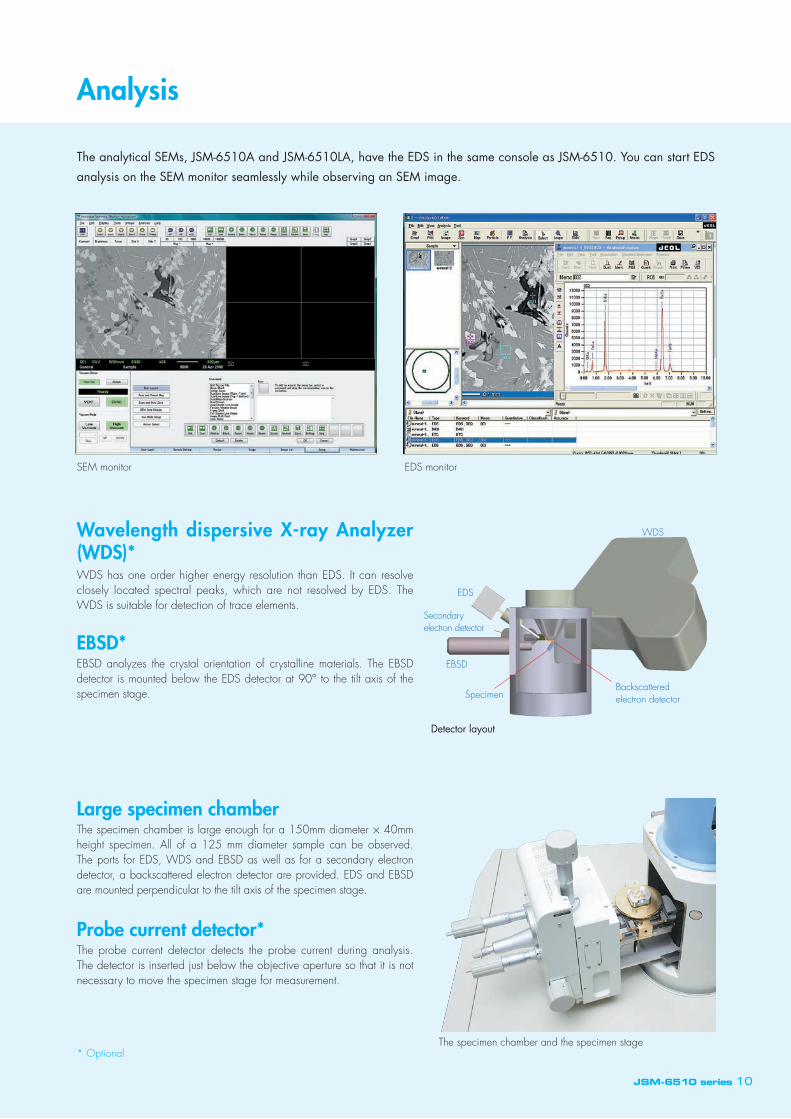
The analytical SEMs, JSM-6510A and JSM-6510LA, have the EDS in the same console as JSM-6510. You can start EDS analysis on the SEM monitor seamlessly while observing an SEM image.
Wavelength dispersive X-ray Analyzer (WDS)*WDS has one order higher energy resolution than EDS. It can resolve closely located spectral peaks, which are not resolved by EDS. The WDS is suitable for detection of trace elements.
Large specimen chamberThe specimen chamber is large enough for a 150mm diameter × 40mm height specimen. All of a 125 mm diameter sample can be observed. The ports for EDS, WDS and EBSD as well as for a secondary electron detector, a backscattered electron detector are provided. EDS and EBSD are mounted perpendicular to the tilt axis of the specimen stage.
Probe current detector*The probe current detector detects the probe current during analysis. The detector is inserted just below the objective aperture so that it is not necessary to move the specimen stage for measurement.
EBSD*EBSD analyzes the crystal orientation of crystalline materials. The EBSD detector is mounted below the EDS detector at 90° to the tilt axis of the specimen stage.
SEM monitor EDS monitor
The specimen chamber and the specimen stage* Optional
Analysis
JSM-6510 series 10
Specimen
Secondary electron detector
Backscattered electron detector
EDS
WDS
EBSD
Detector layout

Measurement
Measurement A variety of measurement functions are provided. You can manually measure features on a SEM image on the monitor and save.
Length of line, size of free shape, diameter
Area of polygon
Angle between 2 lines
Count of features
Measurement functions
Measurement on SMile View™* The standard report editing software, SMile View™ has a measurement function. Measurement results are pasted on a SEM image on the SMile View™ layout sheet. The measurement accuracy can be calibrated by using the SEM image of standard or reference specimen taken at the same magnification for measurement. The measurement results can be displayed as a list or pasted on Excel sheet.
* Standard on JSM-6510A/JSM-6510LA
11 JSM-6510 series
™

* Optional
SEM has larger depth of focus as well as higher resolution than a light microscope so that it is suitable for observation of complicated morphology. The three dimensional image software* developed by JEOL constructs a bird's eye view image from a pair of stereo images. You can measure height of surface morphology form the bird's eye view image.
Three dimensional imageA pair of stereo images is acquired by changing tilt angle of the specimen stage by 4 to 8 degrees. You can view three-dimensional image from these images.
Acquisition of stereo pair imagesThe function to help acquisition of stereo pair images is provided. The second image is acquired by comparing to the first image. It is simple and easy to acquire a stereo pair.
Stereo image acquisition menu of JSM-6510
Bird's eye view image
Height profile
JSM-6510 series 12
Observation of 3D image on the three dimensional image softwareYou can add red and blue color to stereo pair images. You can observe depth when you look the colored images through a pair of glass with blue and red filter.
A pair of stereo images in two colors
Three dimensional image softwareThe three dimensional image software constructs a bird's eye view image from a pair of stereo images. You can measure height of surface morphology from the bird's eye view image.
Stereo pair

Observation without Conductive Coating
JSM-6510LV and JSM-6510LA are equipped with the low vacuum mode and enable you to observe non-conductive specimens. You can analyze a specimen by EDS using high accelerating voltages without worrying about charging.
Backscattered electron detectorThe conventional secondary electron detector, Everhart Thornley detector, does not function in the low vacuum environment. A backscattered electron detector is widely used instead. You can observe the morphology and composition of a specimen.
Secondary electron detector for low vacuum mode*The secondary electron detector dedicated to the low vacuum mode.
Low vacuum systemThe pressure in the specimen chamber can be varied from 10 Pa to 270 Pa without changing the size of the orifice. JSM-6510LA/JSM-6510LV has 2 vacuum systems, one high-vacuum system and one low-vacuum system dedicated to the low vacuum specimen chamber.The objective lens apertures are placed in high vacuum and kept clean for a long period of time.
Principle of low vacuum SEMA small amount of air is introduced into the specimen chamber. These air molecules, oxygen and nitrogen, are ionized by the incident electrons. These ions neutralize electrons on the surface of the specimen and eliminate charging so that a non conductive specimen can be observed.
Morphology and composition are observed Morphology is observed clearly
The vacuum system dedicated to low vacuum specimen chamber is equipped.
* Optional
Charging Generation of ions
13 JSM-6510 series
VV1
VV5
V4
V6 V2V1
V8
VV7VV3 VV2
RP1RP2
DP
VV6
Electron gun
Objective lensaperture
Backscatteredelectrondetector Orifice
(Needle valve)
LVcontrollerPirani
gauge
Motordrive
Foreline trap
Scattered electron Ion Electron on a specimen
No charging Neutralization of charge
10 m10 m
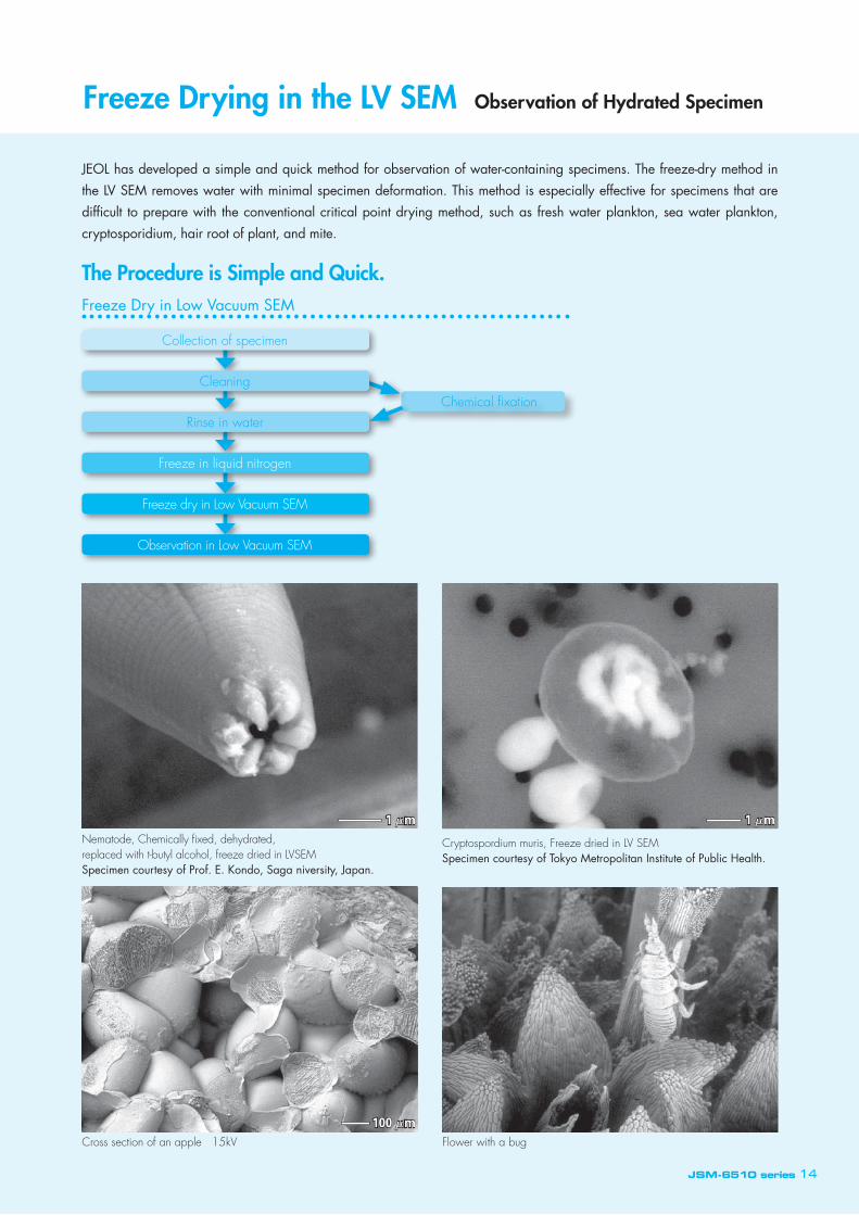
Observation without Conductive Coating Freeze Drying in the LV SEM Observation of Hydrated Specimen
JEOL has developed a simple and quick method for observation of water-containing specimens. The freeze-dry method in the LV SEM removes water with minimal specimen deformation. This method is especially effective for specimens that are difficult to prepare with the conventional critical point drying method, such as fresh water plankton, sea water plankton, cryptosporidium, hair root of plant, and mite.
Freeze Dry in Low Vacuum SEM
The Procedure is Simple and Quick.
Flower with a bugCross section of an apple 15kV
Nematode, Chemically fixed, dehydrated, replaced with t-butyl alcohol, freeze dried in LVSEMSpecimen courtesy of Prof. E. Kondo, Saga niversity, Japan.
Cryptospordium muris, Freeze dried in LV SEMSpecimen courtesy of Tokyo Metropolitan Institute of Public Health.
JSM-6510 series 14
Chemical fixation
Collection of specimen
Rinse in water
Freeze in liquid nitrogen
Freeze dry in Low Vacuum SEM
Observation in Low Vacuum SEM
Cleaning
1 m 1 m
100 m

Specimen Navigationa
Eucentric large-specimen stageThe eucentric specimen stage has minimum shift of observation area and focus when ti l t ing. The stage is sui table for observation of a rough surface from a variety of directions.The stage is eucentric over the entire range of working distance from 8mm for high magnification work, 10mm for analysis, and longer working distance for large depth of focus.Focus change during X, Y, or rotation of a specimen with tilt is small so that surveying a large specimen is done efficiently.It is possible to completely cover a 125 mm diameter sample.
Tilt of large specimenThe high conical shape of the objective lens provides great flexibility in tilting a large specimen. Combination with the eucentric specimen stage makes tilting of a specimen quite easy.
Motorized specimen stage*A variety of motor controlled specimen stages are available as option. You can select one from 2 axes (X, Y), 3 axes (X, Y, R or X, Y, Z), and 5 axes (X, Y, R, Z, T) controls. The functions explained on this page are available with the optional motor controlled specimen stages.
Continuous moveA click and hold on the shift icon or X, Y, R, T or Z button on the motorized specimen stage menu moves the specimen continuously. Tilting the joy stick on the optional operation knob set does the same.
Click center zoomA click on a feature on the live image moves the feature to the center of the live image. You can set to magnify an image after shift of a feature.
Eucentric rotationThe eucentric rotation rotates a specimen around the current observation area.
Frame step moveEach click on the frame-step-move icon shifts a specimen at a user preset interval to survey a large area efficiently
Saving position coordinateUnlimited specimen positions can be saved to move to these areas later.
Motorized specimen stage menu
* Optional
Super conical objective lens
EDS
Super conical objective lens
EDS Take offangle 35°
Specimen tiltSpecimen
WD=10mm
Montage 10 × 9 images (512 × 384 pixels each) Specimen : ButterflySpecimen courtesy of Prof. Matsuda, Kumamoto National College of Technology
15 JSM-6510 series

High Brightness LaB6 Gun*
Turbo molecular pump (TMP) *
JSM-6510 series SEM uses the high performance and reliable diffusion pump (DP). With the DP it is necessary to heat the heater for approximately 25 minutes before the DP is fully operational. The DP also requires cooling water.An air-cooled TMP is available as an option for a user who wants to use the SEM immediately after turning it on or to eliminate the use of cooling water.The vacuum system is completely identical except TMP being used in place of DP. The TMP is not exposed to the air during specimen exchange. The inside of the SEM is kept in vacuum while the SEM is turned off.The specimen chamber of the low vacuum mode is pumped by the dedicated rotary pump while the high vacuum region is pumped by the TMP.
LaB6 GunThe LaB6 gun is brighter than the tungsten hairpin gun. The electron source of the LaB6 gun is smaller so that a higher quality image with better sharpness can be obtained. The improvement is more significant at the lower accelerating voltages. The LaB6 gun has an advantage in the observation of fine surface structures.The expected life is around 500 hours, which is approximately 5 times longer than that of the tungsten hairpin gun. The LaB6 gun is suitable for a study such as the automated particle or gun shot residue analysis, which takes a long time.The LaB6 requires higher vacuum than the tungsten hairpin gun for its stable operation. An ion pump is equipped on the gun chamber to create a higher vacuum for the LaB6 gun. The conventional tungsten hairpin gun can also be used in the gun chamber equipped with the ion pump.
Yogurt bacteria 3kV Original magnification ×25,000
Ceramic 10kV Original magnification ×30,000
* Optional
JSM-6510 series 16
VV1
VV5
V4
V6 V2V1
V8
VV7VV3 VV2
RP1RP2
TMP
VV6
Electron gun
Objective lensaperture
Backscatteredelectrondetector Orifice
(Needle valve)
LVcontrollerPirani
gauge
Motordrive
Foreline trap
Low vacuum systemHigh vacuum systemEvacuation system of Low Vacuum SEM equipped with TMP
1 m 0.5 m

Effective Reports Created Quickly & Easily
It is important to center the filament tip to the small aperture of the Wehnelt cap to ensure the best performance. JEOL provides factory pre-centered filaments, which are centered by JEOL. A user does not have to center a filament. The lens is designed to easily attain high vacuum to minimize contamination to the objective apertures.
Maintenance Videos
Maintenance procedures in movieThe procedures to replace filament, objective apertures, and orifice are explained in easy to follow movies displayed on the operation navi area.
SMile View™* SMile View™ creates an effective report with SEM images and EDS analysis data. You can design layout freely. Simply drag and paste thumb nail images to a layout sheet and a report sheet is done. The edited SMile View™ layout sheet can be sent to Microsoft Word and edited as the Word document. Images in bmp, tiff, jpeg, and meta file are compatible with the SMile View™.
Magnification on SMile View™ layout sheetThe magnification indicated on JSM-6510 series SEM is correct when the width of an image is 128mm. When you paste images on a SMile View™ layout sheet, this is the default size. You can freely change the size of images on the layout sheet. The correct magnifications of images on a layout sheet are automatically calculated and printed on the layout
Factory pre-centered filament
Wehnelt
17 JSM-6510 series
Replacement of filament
* Standard on JSM-6510A/JSM-6510LA

Scanning Electron Microscope
JSM-6510 series
Installation Layout (JSM-6510LV)
Principal Options Installation Requirements
Principal Specifications
JSM-6510LV
ResolutionHV mode 3.0 nm(30 kV)、8 nm(3 kV)、15 nm(1 kV)
LV mode ※1 4.0 nm(30 kV)
Magnification × 5 to × 300,000 (on 128 mm × 96 mm image siza)
Preset magnifications 5 step, user selectable
Standard recipe Built in
Custom recipe Operation conditions (Optics, Image mode, LV pressure*1) Specimen stage
Image modeSecondary electron image, REF image, Composition*1, Topography*1,
Shadowed*1
Accelerating voltage 0.5 kV to 30 kV
Filament Factory pre-centered filament
Electron gun Fully automated, manual override
Condenser lens Zoom condenser lens
Objective lens Super conical objective lens
Objective lens apertures 3 stages, XY fine adjustable
Stigmator memory Built in
Electrical image shift ± 50 μm (WD = 10 mm)
Auto functions Focus, brightness, contrast, stigmator
Specimen stageEucentric large-specimen stageX: 80 mm, Y: 40 mm, Z: 5 mm to 48 mm, Tilt: −10° to 90°, Rotation: 360°
Reference image
(Navigator*3) 4 images
Specimen exchange Draw out the stage
Maximum specimen 150 mm diameter
PC IBM PC/AT compatible
OS Windows® 7
Monitor 19 inch LCD, 1 or 2*2
Frame store 640 × 480, 1,280 × 960, 2,560 × 1,920, 5,120 × 3,340
Dual live image Built in
Full size image display Built in
Pseudo color Built in
Multi image display 2 images, 4 images
Digital zoom Built in
Dual magnification Built in
Network Ethernet
Measurement Built in
Image format BMP, TIFF, JPEG
Auto image archiving Built in
Pumping system Fully automated, DP: 1, RP: 1 or 2*1
Switching vacuum mode*1 Through the menu, less than 1 minute
LV Pressure*1 10 to 270 Pa
JED-2300 EDS*2 Built in
l Backscattered electron detector*1
l Low vacuum secondary electron detector
l Energy dispersive X-ray analyzer (EDS)
l Wave length dispersive X-ray analyzer (WDS)
l EBSD
l Airlock chamber
l Chamber scope
l Operation keyboard
l LaB6 electron gun
l Report creation software (SMile View™)*2
l Operation console (750 mm wide, 900 mm wide)
l Motor controlled stage (2 axes, 3 axes, 5 axes)
Power Single-phase, 100V AC, 50/60Hz, 3.0kVA Voltage regulation within ±10 % ( svoltage drop at 3.0 kVA within 3%)Grounding terminal One, 100 ohms or lessCooling water Faucet One, 14 mm OD or ISO 7/1 Rc 1/4 internal thread Drain One, 25 mm or more ID, or ISO 7/1 Rc 1/4 internal thread Flow rate 2L / min Pressure 0.05 to 0.2 MPa Temperature 20 ± 5 °CEnvironment Temperature 20 ± 5 °C Humidity 60 % or lessFloor space 2,000(W) × 2,500(D) × 1,800(H) mm or moreWeight Approx. 435 kg ( JSM-6510 ), Approx. 435 kg ( JSM-6510LA )Door width 850 mm or more
*1: Standard on JSM-6510LA and JSM-6510LV
*2: Standard on JSM-6510LA and JSM-6510A
*3: Available when the motorized specimen stage is provided.
Specifications subject to change without notice.
Windows operating system is trademark or registered trademark of Microsoft Corporation (US)
in United States and other countries.
JSM-6510 series 18

No. 1305K250C Printed in Japan, Kp


![Gem fall 2017[6510]](https://img.dokumen.tips/doc/110x75/5a66f76f7f8b9a68588b48bd/gem-fall-20176510.jpg)