Embed Size (px)
Citation preview

Differential Detergent Extraction of Mycobacterium marinum CellEnvelope Proteins Identifies an Extensively Modified Threonine-RichOuter Membrane Protein with Channel Activity
Aniek D. van der Woude,a,b Kozhinjampara R. Mahendran,c Roy Ummels,a Sander R. Piersma,d Thang V. Pham,d Connie R. Jiménez,d
Karin de Punder,e Nicole N. van der Wel,e Mathias Winterhalter,c Joen Luirink,b Wilbert Bitter,a,b Edith N. G. Houbena,b
Department of Medical Microbiology and Infection Control, VU University Medical Center, Amsterdam, the Netherlandsa; Department of Molecular Microbiology, Instituteof Molecular Cell Biology, VU University, Amsterdam, the Netherlandsb; School of Engineering and Science, Jacobs University Bremen, Bremen, Germanyc; Department ofMedical Oncology, OncoProteomics Laboratory, VU University Medical Center, Amsterdam, the Netherlandsd; Division of Cell Biology-B6, Netherlands Cancer Institute-Antoni van Leeuwenhoek Hospital (NKI-AVL), Amsterdam, the Netherlandse
A striking characteristic of mycobacteria is the presence of an unusual outer membrane which forms a thick permeability barrierand provides resistance to many antibiotics. Although specialized proteins must reside in this layer, only few mycolate outermembrane (MOM) proteins have been identified to date. Their discovery is complicated by difficulties in obtaining good separa-tion of mycobacterial inner and outer membranes. During our efforts to identify novel mycobacterial outer membrane proteins(MOMPs), we discovered that we can enrich for MOMPs using differential solubilization of mycobacterial cell envelopes. Subse-quently, these different fractions were analyzed by nano liquid chromatography-tandem mass spectrometry (nanoLC-MS/MS).This proteomic analysis confirmed that our marker proteins for inner membrane and MOM were found in their expected frac-tions and revealed a few interesting candidate MOMPs. A number of these putative MOMPs were further analyzed for their ex-pression and localization in the cell envelope. One identified MOMP, MMAR_0617 of Mycobacterium marinum, was purifiedand demonstrated to form a large oligomeric complex. Importantly, this protein showed a clear single-channel conductance of0.8 � 0.1 ns upon reconstitution into artificial planar lipid bilayers. The most surprising feature of MMAR_0617 is a long C-ter-minal threonine-rich domain with extensive modifications. In summary, we have identified a novel mycobacterial outer mem-brane porin with unusual properties.
The architecture of the cell envelope of Mycobacterium tubercu-losis has been an enigma for many years. This extraordinary
hydrophobic cell envelope contains, in addition to a relativelynormal cytoplasmic or inner membrane (IM), a polymer of pepti-doglycan and arabinogalactan, which is covalently linked to beta-hydroxy branched-chain fatty acids of considerable size, the my-colic acids. These mycolic acids are the key components of theatypical outer membrane, which has been called the mycolateouter membrane (MOM) (1). Although the existence of a secondmembrane was already suggested by Minnikin (2), it was onlyrecently unequivocally shown as a lipid bilayer structure by cryo-electron tomography (3–5). As the observed thickness of theMOM is 7 to 8 nm, mycolic acids are proposed to be present in afolded conformation (4, 6), which is in agreement with recentLangmuir assays (7). The outer leaflet of this membrane is prob-ably formed by an array of unusual mycolate- or mycobacterium-specific (glyco)lipids, such as trehalose dimycolate, phtiocerol di-mycoserosate, and sulfolipids.
The MOM forms an exceptionally dense barrier, with evenlower permeability than most Gram-negative bacteria (8). In anal-ogy with the Gram-negative outer membrane, specific outermembrane proteins should be present to accommodate processessuch as nutrient uptake, protein secretion, and membrane biogen-esis. Although more than 60 integral outer membrane proteinshave been described for the Gram-negative Escherichia coli (9), noteven a few mycobacterial outer membrane proteins (MOMPs) areknown. Channel activity, indicative of specific porin proteins, hasbeen identified in cell extracts of Mycobacterium bovis BCG. Theseexperiments showed large cation-selective channels with a single-
channel conductance of 4.0 ns and small anion-selective channelswith single-channel conductance of approximately 0.8 ns (10).However, the nature of these proteins was never established. Thebest-studied MOMP is the major Mycobacterium smegmatis porinMspA. This protein forms an extremely stable octameric goblet-like �-barrel with a central 10-nm channel (11). Although thesefeatures significantly differ from the general monomeric or trim-eric porins found in Gram-negative bacteria (12), all these pro-teins have in common is that they cross the membrane using a�-barrel structure. Unfortunately, no homologues of MspA arefound in M. tuberculosis. For this species, the only proteins de-scribed to reside in the MOM are MctB (mycobacterial coppertransport protein B; Rv1698) (13) and OmpATb (Rv0899). MctBis a putative porin (14) implicated to be involved in copper efflux(13), whereas OmpATb shows homology with the periplasmicdomain of E. coli OmpA and has been the subject of a continuousdebate about its function, localization, and structure. Originally,OmpATb has been described as a surface-accessible porin with
Received 19 December 2012 Accepted 22 February 2013
Published ahead of print 1 March 2013
Address correspondence to Edith N. G. Houben, [email protected].
Supplemental material for this article may be found at http://dx.doi.org/10.1128/JB.02236-12.
Copyright © 2013, American Society for Microbiology. All Rights Reserved.
doi:10.1128/JB.02236-12
2050 jb.asm.org Journal of Bacteriology p. 2050–2059 May 2013 Volume 195 Number 9
Dow
nloa
ded
from
http
s://j
ourn
als.
asm
.org
/jour
nal/j
b on
25
Nov
embe
r 20
21 b
y 42
.98.
49.1
87.
channel activity (15), but recently these results have been ques-tioned (16).
Other mycolic acid-producing bacteria, such as Corynebacte-rium glutamicum and Nocardia sp., provide further knowledge ofthe proteins residing in the MOM. Channel-forming porins re-sembling MspA have been identified in both these species (17). Inaddition, porins have been identified that are proposed to have an�-helical structure (18) or modified by O-mycoloylation (19).These results indicate that the classical model of outer membraneproteins as (predominantly) being �-barrels might not apply to allmycolate outer membrane proteins. Recently, various attemptswere made to define proteins in the cell envelope or cell wall frac-tions of different strains of M. tuberculosis, M. bovis BCG, andMycobacterium avium (20–22), but none were dedicated to thespecific identification of MOMPs. In this study, we developed amethod to specifically enrich for MOM proteins by differentialdetergent extraction of mycobacterial cell envelopes.
MATERIALS AND METHODSBacterial strains and culture conditions. Mycobacterium marinum strainE11 (23) and M. bovis BCG were routinely grown at 30°C in Middlebrook7H9 (Difco) liquid medium and on Middlebrook 7H10 plates (Difco)supplemented with 10% Middlebrook ADC and 0.05% Tween 80 (BDBiosciences) or only oleic acid-albumin-dextrose-catalase (OADC), re-spectively. Escherichia coli strain DH5� was used for amplification andmanipulation of plasmid DNA. Antibiotic concentration used was 50�g/ml hygromycin.
Subcellular fractionation procedure. A culture of 200 to 500 ml of M.marinum or M. bovis BCG was grown to mid-log phase. Prior to lysis,bacteria were killed by the addition of 100 �g/ml ciprofloxacin (Sigma) tothe culture medium and incubation for an hour at 30°C. Out of severalkilling procedures tested, this was the one with the smallest effect on cellenvelope isolation. Cells were harvested at 4°C by centrifugation at 4,000 � g,washed three times with phosphate-buffered saline (PBS), and resus-pended in PBS supplemented with proteinase inhibitor (Complete,Roche), 1 mM ethylenediamine tetra acetic acid (EDTA), and 0.1 mg/mlDNase. Cells were broken either by a three-time passage through a Frenchpress (12,000 lb/inch) or two passages through a One-Shot cell disrupter(Constant Systems). After the first passage, 1 mM dithiothreitol (DTT)was added to the lysate. Lysates were subjected to low-speed centrifuga-tion for 5 min at 3,000 � g to remove unbroken cells, which was repeatedtwice. Subsequently, cytosolic and cell envelope fractions were separatedby centrifugation at 27,000 � g. The obtained pellet was resuspended inPBS and centrifuged again at 27,000 � g to obtain the final cell envelopefraction. In addition, the soluble fraction of the first centrifugation step,representing the cytosol fraction, was subsequently centrifuged at 100,000 �g to further purify this fraction.
Detergent extraction of cell envelope proteins. Cell envelope frac-tions were resuspended in PBS or PBS containing either 1% (wt/vol) n-oc-tyl-�-D-glucopyranoside (OBG; Calbiochem), 0.5% (vol/vol) GenapolX-80 (Fluka), 1% (wt/vol) sodium-lauryl sarcosinate (Sarkosyl, Sigma),or 1% (vol/vol) Triton X-100 (Sigma), all above critical micelle concen-tration (CMC) values, and incubated for 30 min at room temperature.Subsequently, samples were centrifuged twice at 27,000 � g to separate thesoluble from the insoluble fraction. The final soluble fraction was sub-jected to TCA precipitation, whereas the pellet fraction was washed byPBS. Both fractions were dissolved in SDS solubilization buffer (50 mMTris-HCl [pH 6.8], 5 mM EDTA, 10% [vol/vol] glycerol, 2% [wt/vol]SDS, 100 mM DTT, and bromophenol blue) and boiled at 95°C prior toanalysis by SDS-PAGE and immunoblotting.
SDS-PAGE, blue native PAGE, and immunostaining. Samples wereloaded on SDS-PAGE gels (Bio-Rad/Hoefer) or NativePAGE gels (Invit-rogen) and subsequently visualized by staining with Coomassie GT-250(Bio-Rad) or PageSilver silver staining kit (Fermentas), by immunoblot-
ting, or by incubation with horseradish peroxidase (HRP)-conjugatedConA (Sigma). Antisera used for immunostaining are rabbit antisera re-active to FtsH (24) or EccC5 (25) and mouse monoclonal antibodiesagainst the influenza hemagglutinin (HA) epitope (HA.11; Covance), theHis6 epitope (Roche), or GroEL2 (CS44; Colorado State University). Rab-bit antiserum recognizing MctB was raised against a peptide correspond-ing to amino acids 123 to 135 of MctB (Rv1698), prepared by Innovagen(Sweden). The presence of bound HRP-conjugated secondary antibodieswas detected via chemiluminescence (Pierce) using a charge-coupled-de-vice (CCD) camera (Bio-Rad). Provided QuantityOne software (Bio-Rad) was used to (semi)quantify signals obtained via immunostaining.
Mass spectrometric analysis. Protein lanes from Coomassie brilliantblue (CBB)-stained SDS-PAGE gels were excised and prepared for nanoliquid chromatography-tandem mass spectrometry (nanoLC-MS/MS)analysis as previously described (5, 26), using Scaffold 3 for protein iden-tification.
Bioinformatic analysis. Proteins were quantified by spectral countingand exported to Microsoft Excel (see Table S2 in the supplemental mate-rial). All identified proteins were annotated and functionally classifiedusing MarinoList (27). Prediction of possible signal sequences and trans-membrane domains (TMDs) was performed using the SignalP 3.0 andTMHMM version 2.0, respectively. Subsequently, identified proteinswere ordered according to the number of spectral counts detected in thedetergent-solubilized fraction compared to the total number of counts.Putative MOMPs (i.e., �90% extracted in three biological replicates)were subjected to BLASTp analysis. Additionally, spectral counts werenormalized by dividing the spectral counts per protein by the sum of allcounts per sample, multiplied by the average sum across all samples. Sub-sequently, the fold change (Fc) and P values between counts for detergentpellet and detergent supernatant were calculated using the beta binominaltest for spectral count data (28).
Molecular cloning. Selected M. marinum genes mmar_3191 (ftsQ),mmar_4637 (ompA), mmar_2503 (mctB), dppA, mmar_0617, mmar_4057,mmar_4366, and mmar_5387 were amplified by PCR using Pfu polymerase(Fermentas) and specific anchored primers containing restriction sites and aC-terminal HA tag (YPYDVPDYA) or His6 tag (see Table S1 in the supple-mental material). After restriction with NheI/XbaI/SpeI and BamHI/BglII(for specific details, see Table S1), the resulting PCR fragments were ligatedinto NheI/BamHI-digested pSMT3-LipYtb (29), directly behind the hsp60promoter. Resulting vectors were checked by restriction digestion and nucle-otide sequence analysis of the relevant part.
Analysis of putative MOMPs. Putative MOMPs were tested for heatmodifiability by resuspending mycobacterial cell envelopes in solubiliza-tion buffer containing either 2% SDS and 100 mM DTT or 0.2% SDS andno DTT, incubating these samples at 95°C or 37°C, respectively, and thenrunning them on a polyacrylamide gel lacking SDS. To examine proteasesensitivity of MOMPs, isolated cell envelopes were either directly incu-bated with different concentrations of proteinase K (Sigma) or trypsin(Sigma) or solubilized by 1% OBG prior to protease treatment. Proteaseactivity was inhibited after incubation for 30 min at room temperatureusing phenylmethylsulfonyl fluoride or trypsin inhibitor (Sigma).
Immunogold electron microscopy (EM). Fixation of bacterial cells,cryosectioning, and immunogold labeling of fixed cells were essentiallydone as described in reference 5. The HA.11 mouse monoclonal antibody(Covance), bridging rabbit anti-mouse antibody (Dako) and protein Aattached to 10 nm gold (University Utrecht), was used to visualize theHA-tagged MOMPs. Localization of gold particles was quantified double-blind. For each strain, a representative 50 to 200 cells were counted forgold particles, which were assigned to three different compartments: in-tracellular (cytosol), associating with the observed membrane layers(membrane), or localized external to the membrane layers, including theelectron transparent zone (cell wall).
Purification of MMAR_0617-His. Cell envelope preparations of M.marinum E11 with or without pSMT3::MMAR_0617-His or pSMT3::MMAR_0617�C-His were incubated for 30 min at room temperature in
Identification of Novel Porin in Mycobacterium marinum
May 2013 Volume 195 Number 9 jb.asm.org 2051
Dow
nloa
ded
from
http
s://j
ourn
als.
asm
.org
/jour
nal/j
b on
25
Nov
embe
r 20
21 b
y 42
.98.
49.1
87.
phosphate buffer (20 mM Na phosphate [pH 7.8], 500 mM NaCl) con-taining 1% (vol/vol) Triton X-100 (Sigma) and 10 mM imidazole andcentrifuged at 27,000 � g to obtain the solubilized proteins. Subsequently,these protein samples were loaded on HisTrap HP columns (GE Health-care) equilibrated with wash buffer (phosphate buffer containing 10 mMimidazole and 1% [vol/vol] Triton X-100) coupled to an ÄKTA FPLCsystem (Amersham Biosciences) at 0.5 ml/min. Proteins were eluted byapplying a linear gradient of 40 to 160 mM imidazole (buffer containingeither 1% or 0.1% [vol/vol] Triton X-100). Fractions containing theNi2�-purified protein were optionally precipitated using ammoniumsulfate and further purified by size exclusion chromatography using aSuperdexTM 200 HR 10/30 column (Amersham Pharmacia no. 17-1088-01).In a separate approach, HA-labeled MMAR_0617 was isolated from solubi-lized cell envelope proteins using the HA tag immunoprecipitation (IP)/coimmunoprecipitation (Co-IP) kit (Pierce). Supplied buffers were supple-mented with 1% (vol/vol) Triton X-100 to avoid protein aggregation.
Single-channel reconstitution and ion conductance measurements.Virtually solvent-free lipid bilayers were formed as previously described(30, 31). 1,2-Diphytanoyl-sn-glycero-3-phosphatidylcholine (DPhPC),the most widely used lipid for the characterization of reconstituted chan-nels, was used for membrane formation in a Teflon cell with an approxi-mately 100-�m-diameter aperture in the 25-�m-thick Teflon partition. Abuffer containing 1 M KCl and 10 mM HEPES, pH 7.4, served as theelectrolyte. Standard silver-silver chloride electrodes from World Preci-sion Instruments (WPI) were placed in each chamber to detect the ioncurrent. Small amounts of purified MMAR_0617 stock solution wereadded to the cis side of the chamber (ground electrode). The addition ofproteins into the aqueous phase resulted in a rapid dilution of the deter-gent below the CMC, causing channels to spontaneously insert into thelipid bilayers. An ion current flowing through the channel was induced byapplication of a transmembrane voltage. Conductance measurementswere recorded by using an Axopatch 200B amplifier (Axon Instruments,Foster City, CA). The signal was filtered using a four-pole low-pass Besselfilter at a frequency of 10 kHz and sampled at 50 kHz, acquired using aDigidata 1440A digitizer, and analyzed using the pClamp 10.0 software(Axon Instruments, Foster City, CA). All measurements were repeated 10times with different purification batches to establish confidence about thereproducibility.
RESULTSDevelopment of a differential detergent extraction method toenrich for MOMPs. Several techniques have been developed forthe subcellular fractionation of mycobacteria, but none of themhave been successfully used to isolate and identify mycobacterialouter membrane proteins (32). In this work, we used a variety ofdifferent methods to identify inner and outer membrane proteinsof M. marinum. As markers for successful fractionation, anti-bodies were used directed against the MOM protein MctB andagainst the inner membrane (IM) proteins EccC5 and FtsH(24); the latter antiserum additionally recognizes the cytosolicATPase MMAR_0752 (33). The most commonly used method toisolate different cell envelope fractions is by differential centrifu-gation (34–36). Using our markers, we could show, in contrast toa previous study (36), that both IM and MOM proteins were pel-leted by a centrifugation step at 27,000 � g (Fig. 1B). We also triedto separate IM and MOM proteins using sucrose gradient centrif-ugation (37), but this approach was not successful (results notshown). Next, we performed “shaving” of M. marinum cells usingtrypsin-coated beads that specifically cleave exposed protein do-mains (38). This procedure identified only abundant cytosolic andsecreted proteins, such as GroEL and EsxA, and not any known orputative membrane proteins (results not shown). Specific labelingof surface proteins by whole-cell biotinylation using hydrophilic
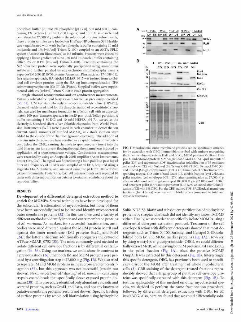
Sulfo-NHS-SS-biotin and subsequent purification of biotinylatedproteins by streptavidin beads did not identify any known MOMPeither. Finally, we succeeded to specifically isolate MOMPs using adifferential detergent extraction protocol. Treatment of the cellenvelope fraction with different detergents showed that most de-tergents, such as Triton X-100, Sarkosyl, and Genapol X-80, solu-bilized both IM and MOM marker proteins (Fig. 1A). However,by using n-octyl-�-D-glucopyranoside (OBG), we could differen-tially extract MctB, while leaving both IM proteins FtsH and EccC5
in the pellet fraction (Fig. 1A). Also, the putative MOMPOmpATb was extracted by this detergent (Fig. 1B). Interestingly,this specific detergent, OBG, has previously been used to specifi-cally disrupt the MOM after treatment of whole mycobacterialcells (3). CBB staining of the detergent-treated fractions repro-ducibly showed that a large group of putative cell envelope pro-teins was specifically extracted with this detergent (Fig. 1B). Totest the applicability of this method on other mycobacterial spe-cies, we decided to perform the same fractionation procedure,followed by differential detergent extraction with OBG, for M.bovis BCG. Also, here, we found that we could differentially solu-
FIG 1 Mycobacterial outer membrane proteins can be specifically enrichedfor by extraction with OBG. Immunoblots probed with antisera recognizingthe inner membrane proteins FtsH and EccC5, MOM proteins MctB and Om-pATb, and cytosolic proteins MMAR_0752 and GroEL2. (A) Equal amounts ofpellet (DP) and supernatant (DS) fractions after solubilization of M. marinumcell envelope (CE) with Sarkosyl (S), Triton X-100 (T100), Genapol X-80 (G),and n-octyl-�-D-glucopyranoside (OBG). (B) Immunoblot of fractions corre-sponding to equal OD units of total lysate (T), soluble fraction (cyt1 27k), andpellet fraction (cell envelopes [CE], 27k) after centrifugation at 27,000 � g,after an additional centrifugation step at 100,000 � g (cyt2 100k and P 100k),and detergent pellet (DP) and supernatant (DS) were obtained after solubili-zation of CE with 1% OBG. For the CBB-stained SDS-PAGE gel, all membranefractions (last 4 lanes) were loaded in 3-fold excess compared to total andcytosolic fractions.
van der Woude et al.
2052 jb.asm.org Journal of Bacteriology
Dow
nloa
ded
from
http
s://j
ourn
als.
asm
.org
/jour
nal/j
b on
25
Nov
embe
r 20
21 b
y 42
.98.
49.1
87.
bilize MctB and OmpATb, while leaving the major fraction ofFtsH in the detergent pellet (see Fig. S1A in the supplementalmaterial).
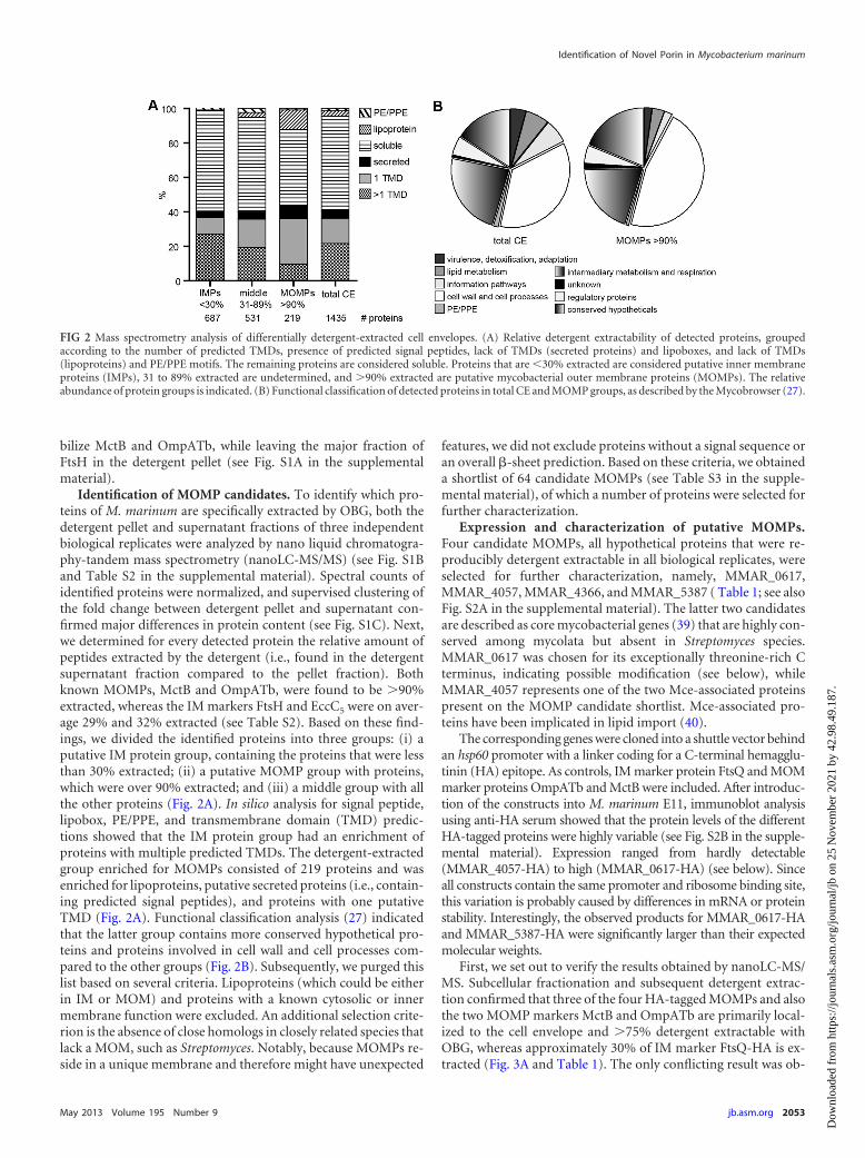
Identification of MOMP candidates. To identify which pro-teins of M. marinum are specifically extracted by OBG, both thedetergent pellet and supernatant fractions of three independentbiological replicates were analyzed by nano liquid chromatogra-phy-tandem mass spectrometry (nanoLC-MS/MS) (see Fig. S1Band Table S2 in the supplemental material). Spectral counts ofidentified proteins were normalized, and supervised clustering ofthe fold change between detergent pellet and supernatant con-firmed major differences in protein content (see Fig. S1C). Next,we determined for every detected protein the relative amount ofpeptides extracted by the detergent (i.e., found in the detergentsupernatant fraction compared to the pellet fraction). Bothknown MOMPs, MctB and OmpATb, were found to be �90%extracted, whereas the IM markers FtsH and EccC5 were on aver-age 29% and 32% extracted (see Table S2). Based on these find-ings, we divided the identified proteins into three groups: (i) aputative IM protein group, containing the proteins that were lessthan 30% extracted; (ii) a putative MOMP group with proteins,which were over 90% extracted; and (iii) a middle group with allthe other proteins (Fig. 2A). In silico analysis for signal peptide,lipobox, PE/PPE, and transmembrane domain (TMD) predic-tions showed that the IM protein group had an enrichment ofproteins with multiple predicted TMDs. The detergent-extractedgroup enriched for MOMPs consisted of 219 proteins and wasenriched for lipoproteins, putative secreted proteins (i.e., contain-ing predicted signal peptides), and proteins with one putativeTMD (Fig. 2A). Functional classification analysis (27) indicatedthat the latter group contains more conserved hypothetical pro-teins and proteins involved in cell wall and cell processes com-pared to the other groups (Fig. 2B). Subsequently, we purged thislist based on several criteria. Lipoproteins (which could be eitherin IM or MOM) and proteins with a known cytosolic or innermembrane function were excluded. An additional selection crite-rion is the absence of close homologs in closely related species thatlack a MOM, such as Streptomyces. Notably, because MOMPs re-side in a unique membrane and therefore might have unexpected
features, we did not exclude proteins without a signal sequence oran overall �-sheet prediction. Based on these criteria, we obtaineda shortlist of 64 candidate MOMPs (see Table S3 in the supple-mental material), of which a number of proteins were selected forfurther characterization.
Expression and characterization of putative MOMPs.Four candidate MOMPs, all hypothetical proteins that were re-producibly detergent extractable in all biological replicates, wereselected for further characterization, namely, MMAR_0617,MMAR_4057, MMAR_4366, and MMAR_5387 ( Table 1; see alsoFig. S2A in the supplemental material). The latter two candidatesare described as core mycobacterial genes (39) that are highly con-served among mycolata but absent in Streptomyces species.MMAR_0617 was chosen for its exceptionally threonine-rich Cterminus, indicating possible modification (see below), whileMMAR_4057 represents one of the two Mce-associated proteinspresent on the MOMP candidate shortlist. Mce-associated pro-teins have been implicated in lipid import (40).
The corresponding genes were cloned into a shuttle vector behindan hsp60 promoter with a linker coding for a C-terminal hemagglu-tinin (HA) epitope. As controls, IM marker protein FtsQ and MOMmarker proteins OmpATb and MctB were included. After introduc-tion of the constructs into M. marinum E11, immunoblot analysisusing anti-HA serum showed that the protein levels of the differentHA-tagged proteins were highly variable (see Fig. S2B in the supple-mental material). Expression ranged from hardly detectable(MMAR_4057-HA) to high (MMAR_0617-HA) (see below). Sinceall constructs contain the same promoter and ribosome binding site,this variation is probably caused by differences in mRNA or proteinstability. Interestingly, the observed products for MMAR_0617-HAand MMAR_5387-HA were significantly larger than their expectedmolecular weights.
First, we set out to verify the results obtained by nanoLC-MS/MS. Subcellular fractionation and subsequent detergent extrac-tion confirmed that three of the four HA-tagged MOMPs and alsothe two MOMP markers MctB and OmpATb are primarily local-ized to the cell envelope and �75% detergent extractable withOBG, whereas approximately 30% of IM marker FtsQ-HA is ex-tracted (Fig. 3A and Table 1). The only conflicting result was ob-
FIG 2 Mass spectrometry analysis of differentially detergent-extracted cell envelopes. (A) Relative detergent extractability of detected proteins, groupedaccording to the number of predicted TMDs, presence of predicted signal peptides, lack of TMDs (secreted proteins) and lipoboxes, and lack of TMDs(lipoproteins) and PE/PPE motifs. The remaining proteins are considered soluble. Proteins that are �30% extracted are considered putative inner membraneproteins (IMPs), 31 to 89% extracted are undetermined, and �90% extracted are putative mycobacterial outer membrane proteins (MOMPs). The relativeabundance of protein groups is indicated. (B) Functional classification of detected proteins in total CE and MOMP groups, as described by the Mycobrowser (27).
Identification of Novel Porin in Mycobacterium marinum
May 2013 Volume 195 Number 9 jb.asm.org 2053
Dow
nloa
ded
from
http
s://j
ourn
als.
asm
.org
/jour
nal/j
b on
25
Nov
embe
r 20
21 b
y 42
.98.
49.1
87.
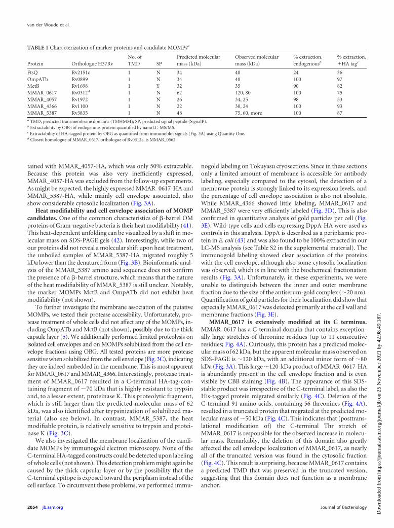
tained with MMAR_4057-HA, which was only 50% extractable.Because this protein was also very inefficiently expressed,MMAR_4057-HA was excluded from the follow-up experiments.As might be expected, the highly expressed MMAR_0617-HA andMMAR_5387-HA, while mainly cell envelope associated, alsoshow considerable cytosolic localization (Fig. 3A).
Heat modifiability and cell envelope association of MOMPcandidates. One of the common characteristics of �-barrel OMproteins of Gram-negative bacteria is their heat modifiability (41).This heat-dependent unfolding can be visualized by a shift in mo-lecular mass on SDS-PAGE gels (42). Interestingly, while two ofour proteins did not reveal a molecular shift upon heat treatment,the unboiled samples of MMAR_5387-HA migrated roughly 5kDa lower than the denatured form (Fig. 3B). Bioinformatic anal-ysis of the MMAR_5387 amino acid sequence does not confirmthe presence of a �-barrel structure, which means that the natureof the heat modifiability of MMAR_5387 is still unclear. Notably,the marker MOMPs MctB and OmpATb did not exhibit heatmodifiability (not shown).
To further investigate the membrane association of the putativeMOMPs, we tested their protease accessibility. Unfortunately, pro-tease treatment of whole cells did not affect any of the MOMPs, in-cluding OmpATb and MctB (not shown), possibly due to the thickcapsule layer (5). We additionally performed limited proteolysis onisolated cell envelopes and on MOMPs solubilized from the cell en-velope fractions using OBG. All tested proteins are more proteasesensitive when solubilized from the cell envelope (Fig. 3C), indicatingthey are indeed embedded in the membrane. This is most apparentfor MMAR_0617 and MMAR_4366. Interestingly, protease treat-ment of MMAR_0617 resulted in a C-terminal HA-tag-con-taining fragment of 70 kDa that is highly resistant to trypsinand, to a lesser extent, proteinase K. This proteolytic fragment,which is still larger than the predicted molecular mass of 62kDa, was also identified after trypsinization of solubilized ma-terial (also see below). In contrast, MMAR_5387, the heatmodifiable protein, is relatively sensitive to trypsin and protei-nase K (Fig. 3C).
We also investigated the membrane localization of the candi-date MOMPs by immunogold electron microscopy. None of theC-terminal HA-tagged constructs could be detected upon labelingof whole cells (not shown). This detection problem might again becaused by the thick capsular layer or by the possibility that theC-terminal epitope is exposed toward the periplasm instead of thecell surface. To circumvent these problems, we performed immu-
nogold labeling on Tokuyasu cryosections. Since in these sectionsonly a limited amount of membrane is accessible for antibodylabeling, especially compared to the cytosol, the detection of amembrane protein is strongly linked to its expression levels, andthe percentage of cell envelope association is also not absolute.While MMAR_4366 showed little labeling, MMAR_0617 andMMAR_5387 were very efficiently labeled (Fig. 3D). This is alsoconfirmed in quantitative analysis of gold particles per cell (Fig.3E). Wild-type cells and cells expressing DppA-HA were used ascontrols in this analysis. DppA is described as a periplasmic pro-tein in E. coli (43) and was also found to be 100% extracted in ourLC-MS analysis (see Table S2 in the supplemental material). Theimmunogold labeling showed clear association of the proteinswith the cell envelope, although also some cytosolic localizationwas observed, which is in line with the biochemical fractionationresults (Fig. 3A). Unfortunately, in these experiments, we wereunable to distinguish between the inner and outer membranefraction due to the size of the antiserum-gold complex (20 nm).Quantification of gold particles for their localization did show thatespecially MMAR_0617 was detected primarily at the cell wall andmembrane fractions (Fig. 3E).
MMAR_0617 is extensively modified at its C terminus.MMAR_0617 has a C-terminal domain that contains exception-ally large stretches of threonine residues (up to 11 consecutiveresidues; Fig. 4A). Curiously, this protein has a predicted molec-ular mass of 62 kDa, but the apparent molecular mass observed onSDS-PAGE is 120 kDa, with an additional minor form of 80kDa (Fig. 3A). This large 120-kDa product of MMAR_0617-HAis abundantly present in the cell envelope fraction and is evenvisible by CBB staining (Fig. 4B). The appearance of this SDS-stable product was irrespective of the C-terminal label, as also theHis-tagged protein migrated similarly (Fig. 4C). Deletion of theC-terminal 91 amino acids, containing 56 threonines (Fig. 4A),resulted in a truncated protein that migrated at the predicted mo-lecular mass of 50 kDa (Fig. 4C). This indicates that (posttrans-lational modification of) the C-terminal Thr stretch ofMMAR_0617 is responsible for the observed increase in molecu-lar mass. Remarkably, the deletion of this domain also greatlyaffected the cell envelope localization of MMAR_0617, as nearlyall of the truncated version was found in the cytosolic fraction(Fig. 4C). This result is surprising, because MMAR_0617 containsa predicted TMD that was preserved in the truncated version,suggesting that this domain does not function as a membraneanchor.
TABLE 1 Characterization of marker proteins and candidate MOMPsa
Protein Orthologue H37RvNo. ofTMD SP
Predicted molecularmass (kDa)
Observed molecularmass (kDa)
% extraction,endogenousb
% extraction,�HA tagc
FtsQ Rv2151c 1 N 34 40 24 36OmpATb Rv0899 1 N 34 40 100 97MctB Rv1698 1 Y 32 35 90 82MMAR_0617 Rv0312d 1 N 62 120, 80 100 75MMAR_4057 Rv1972 1 N 26 34, 25 98 53MMAR_4366 Rv1100 1 N 22 30, 24 100 93MMAR_5387 Rv3835 1 N 48 75, 60, more 100 87a TMD, predicted transmembrane domains (TMHMM); SP, predicted signal peptide (SignalP).b Extractability by OBG of endogenous protein quantified by nanoLC-MS/MS.c Extractability of HA-tagged protein by OBG as quantified from immunoblot signals (Fig. 3A) using Quantity One.d Closest homologue of MMAR_0617, orthologue of Rv0312c, is MMAR_0562.
van der Woude et al.
2054 jb.asm.org Journal of Bacteriology
Dow
nloa
ded
from
http
s://j
ourn
als.
asm
.org
/jour
nal/j
b on
25
Nov
embe
r 20
21 b
y 42
.98.
49.1
87.

The excessive number of Thr residues in the C-terminal do-main MMAR_0617 could be posttranslationally modified by O-glycosylation, as has been described for, e.g., the M. tuberculosisantigens Apa (44) and Mpb83 (45). Glycosylation was analyzed byincubating blots with horseradish peroxidase (HRP)-conjugatedconcanavalin A (ConA). The clear binding of this lectin to the120-kDa band indicates the presence of �-linked glucose or, morelikely, mannose residues on MMAR_0617 (Fig. 4B). A faint 120-kDa reacting band was observed in the negative control, whichmight represent endogenous MMAR_0617. Cell envelope-solubi-lized MMAR_0617-HA was also tested for its sensitivity to mildalkaline hydrolysis, which releases ester-linked modifications and
O-linked carbohydrates. This treatment resulted in a distinct shiftin molecular mass of 20 kDa of the 120-kDa product ofMMAR_0617 (Fig. 4D), further indicating that MMAR_0617 isindeed extensively modified. The latter treatment also affectedConA reactivity of the protein (not shown), whereas the controlprotein MctB remained unaffected (Fig. 4D). Finally, MS/MSanalysis of purified MMAR_0617 resulted in 38% peptide cover-age (Fig. 4A), revealing the peptides without posttranslationalmodifications. The Thr-rich tail was not identified in this analysis,but it should be noted that a large part of the C-terminal domainlacks trypsin digestion sites and is therefore probably too large tobe detected by mass spectrometry, even without modifications. In
FIG 3 Candidate MOMPs localize to the detergent extractable cell envelope fraction. (A to C) Immunoblots were probed with antisera recognizing FtsH,MMAR_0752, or the HA epitope. (A) Subcellular fractions of M. marinum expressing HA-tagged versions of the control proteins and the candidate MOMPs.Total lysate (T), soluble lysate (cyt), detergent pellet (DP), and supernatant (DS) obtained after treatment of cell envelopes with PBS or 1% OBG were loaded ina 1:1:5:5:5:5 ratio. (B to C) Heat modifiability and protease accessibility of putative MOMPs. (B) Cell envelope fractions were denatured in 2% SDS at 95°C (�)or incubated in 0.2% SDS at 37°C () and loaded on an SDS-free PAGE gel. (C) MOMPs were treated with different concentrations of proteinase K (protK) ortrypsin, without or after detergent extraction with OBG. (D) Representative Tokuyasu cryosections of M. marinum expressing MMAR_0617-HA orMMAR_5387-HA probed with HA antiserum followed by goat anti-mouse conjugate labeled with 10-nm gold particles. Scale bar is 100 nm. Particles are markedwith color coding similar to that described in panel E. (E) Quantification of gold particles assigned to different subcellular compartments (see Material & Methodsfor definitions).
Identification of Novel Porin in Mycobacterium marinum
May 2013 Volume 195 Number 9 jb.asm.org 2055
Dow
nloa
ded
from
http
s://j
ourn
als.
asm
.org
/jour
nal/j
b on
25
Nov
embe
r 20
21 b
y 42
.98.
49.1
87.

conclusion, the putative MOMP MMAR_0617 is extensivelymodified, likely at the Thr-rich C terminus, by O-linked glycosylgroups.
MMAR_0617 forms an oligomer with channel-forming ac-tivity in lipid bilayer experiments. To examine whetherMMAR_0617 could function as a porin in the MOM, the His-tagged version of this protein was purified from M. marinum cellenvelopes using Ni-nitrilotriacetic acid (NTA) affinity chroma-tography, and channel activity was analyzed by reconstitution ofpurified protein into planar lipid membranes. As negative con-trols, the same purifications were performed for wild-type (WT)strains or strains expressing MMAR_0617-His with the C-termi-nal deletion (see Fig. S3A in the supplemental material). Analysisof purified MMAR_0617-His by blue native PAGE indicated thatthis protein forms two major oligomeric complexes with apparentmolecular masses of 200 and 400 kDa (see Fig. S3B). Loweringthe Triton X-100 concentration to 0.1% during the purification(see Materials and Methods) slightly decreased the apparent mo-lecular mass of both complexes (see Fig. S3B). Further purificationof these eluates by gel filtration chromatography also confirmedthat MMAR_0617 forms an oligomeric complex of 400 kDa (seeFig. S3C).
Next, we used the planar lipid bilayer technique to characterizethe single-channel properties of Ni-NTA-purified MMAR_0617-His, both before (not shown) and after gel filtration (Fig. 5A).Channel properties, such as conductance, selectivity, and gating,were investigated. When MMAR_0617 was reconstituted into sta-ble 1,2-diphytanoyl-sn-glycero-3-phosphatidylcholine (DPhPc)lipid bilayers, it formed a stable channel that allowed a specific ioncurrent under applied transmembrane voltage. Notably, no activ-
ity was recorded in control experiments using the Ni-NTA puri-fications from the empty strain or expressing MMAR_0617�C-His (results not shown). Single MMAR_0617 channels in thebilayer membrane showed a characteristic conductance of 0.8 �0.1 ns in 1 M KCl (Fig. 5B), which was consistently observed usingindependent biological replicates. Moreover, the channel showeda slight asymmetry in the channel conductance with respect to thepolarity of the applied voltage, i.e., at negative voltage, the channelconductance is slightly higher. However, the exact physical orien-tation of the channel could not be detected in this type of mea-surements. Further analysis of the data showed that MMAR_0617forms a highly voltage-sensitive channel. In addition, singleMMAR_0617 channels showed asymmetry in the channel closurewith respect to the polarity of the applied voltage. For example, atpositive voltage above 100 mV, the channel fluctuated betweenseveral different subconductance states, whereas at negative volt-age, the channel remained in one open steady state (Fig. 5B). Atpresent, this mechanism of voltage sensitivity is not understood.The corresponding current amplitude histograms showed that theMMAR_0617 channel fluctuates between two states at positivevoltage, whereas it remains mostly in one open state at negativevoltage (Fig. 5C). The ion selectivity was determined with reversalpotential measurements after applying a 10-fold salt gradient (0.1M KCl cis side/1 M KCl trans side). We used the Goldman-Hodg-kin-Katz (GHK) equation to determine the ratio of cation andanion permeability. The calculated PK/Cl ratio around 2 indi-cates that the channel is slightly cation selective.
To further exclude that other proteins that copurify withMMAR_0617-His are responsible for the observed channel activ-ity, we also purified C-terminally HA-labeled MMAR_0617 using
FIG 4 Extensive modification of MMAR_0617. (A) Amino acid sequence of MMAR_0617. Highlighted in gray are peptide sequences that were detected byMS/MS analysis and therefore do not contain modifications. Underlined are trypsin digestion sites, and the Thr-rich C-terminal domain that is deleted in thetruncated MMAR_0617�C is indicated in bold. (B) CBB staining and immunoblots using the HA antibody and horseradish peroxidase (HRPO)-conjugatedConA of cell envelope fractions (CE) without () and with (�) MMAR_0617-HA (indicated with *). (C) Immunoblot of total lysate (T), cytosol (cyt), and CEof M. marinum expressing MMAR_0617-His or MMAR_0617�C-His loaded in a 1:1:1 ratio. (D) Immunoblot of subcellular fractions of M. marinum expressingMMAR_0617-HA probed with anti-FtsH, anti-MctB, and anti-HA antibodies; T, cyt, CE, detergent pellet (DP), and supernatant (DS) obtained after solubili-zation of CE with 1% OBG and DS treated with 0.15 M NaOH (DS NaOH) loaded in a 1:1:3:5:5:5 ratio.
van der Woude et al.
2056 jb.asm.org Journal of Bacteriology
Dow
nloa
ded
from
http
s://j
ourn
als.
asm
.org
/jour
nal/j
b on
25
Nov
embe
r 20
21 b
y 42
.98.
49.1
87.

a different technique, i.e., using beads coated with HA antibodies(see Fig. S4A in the supplemental material). Also, this purifiedMMAR_0617-HA preparation clearly showed channel activity inlipid bilayers (see Fig. S4B), although the channels fluctuated be-tween different conductance states with very high noise. This rel-ative instability might be caused by the HA tag or the purificationprocedure that involved elution at low pH. In conclusion, in threedifferent MMAR_0617 purifications, we have observed thatMMAR_0617 exhibits channel activity in artificial lipid bilayers,which indicates it forms a pore in the MOM.
DISCUSSION
Recent studies have revealed that mycobacteria contain a secondlipid bilayer, the mycolate outer membrane (MOM) (3–5). Thepresence of this impermeable lipid bilayer indicates that channelproteins are necessary for transport of proteins and nutrientsacross the MOM. In this study, we have tried various methods todiscover novel MOMPs, using marker proteins to ascertain suc-cessful separation. We failed to separate inner and outer mem-branes by differential centrifugation (36) or sucrose-gradient cen-trifugation of isolated cell envelope fractions. The latter method
has been successfully used for C. glutamicum, for which two frac-tions of different density were observed in a gradient of 35 to 56%sucrose (46). However, multiple attempts for M. marinum alwaysshowed one fraction at a density of 31 to 34%, which containedboth inner and outer membrane proteins (not shown). This dis-parity might reflect a difference in composition of the corynebac-terial and mycobacterial MOM, perhaps caused by the length oftheir mycolic acids (i.e., mycobacterial mycolic acids are consid-erably longer). In the end, fractionation of IM and MOM proteinsby differential detergent extraction with OBG was successful.Analysis of the obtained fractions by nanoLC-MS/MS identified anumber of putative MOMP candidates. One of these candidates,MMAR_0617, indeed exhibited channel activity in lipid bilayerexperiments, indicating it is a novel porin protein.
Protease accessibility experiments showed that MMAR_0617is embedded in the membrane, probably via its C-terminal do-main. Thus far, it is unknown how this protein is transportedacross the IM; it does not contain an N-terminal signal sequencenor the recently identified general type VII secretion motif (47).The MS/MS analysis of MMAR_0617 purified from cell envelopesalso indicates that no N-terminal processing takes place. Recon-
FIG 5 Purified MMAR_0617-His shows channel activity in lipid bilayer experiments. (A) Silver-stained SDS-PAGE gel containing Ni-NTA-purifiedMMAR_0617-His in 1% Triton X-100 (T100) before () and after gel filtration chromatography. Obtained fractions 20, 21, 22, and 33 (negative control) wereTCA precipitated before loading. A background signal of 15 kDa was observed in all fractions, which likely represents coprecipitated T100. MMAR_0617 isindicated by an asterisk, and the fraction used for single-channel activity recordings is marked by an arrow. (B and C) Single-channel activity recordingsrepresentative for three biological replicates. Experimental conditions were 1 M KCl, 10 mM HEPES, pH 7.4, at room temperature. (B) Ionic currents througha single channel at 100 mV and �100 mV. At 100 mV, the channel exists in one open conductance state, whereas at �100 mV, the channel fluctuates betweendifferent conductance states. (C) Corresponding amplitude histograms.
Identification of Novel Porin in Mycobacterium marinum
May 2013 Volume 195 Number 9 jb.asm.org 2057
Dow
nloa
ded
from
http
s://j
ourn
als.
asm
.org
/jour
nal/j
b on
25
Nov
embe
r 20
21 b
y 42
.98.
49.1
87.
stitution of this protein into planar lipid membranes clearlyshowed it has a single-channel conductance of 0.8 � 0.1 ns. Fur-thermore, MMAR_0617 was shown to be highly voltage sensitive,exhibiting different conductance states when positive voltageswere applied. Importantly, gel filtration chromatography and bluenative PAGE analysis indicated that the channel is formed by anoligomer of MMAR_0617. Bioinformatic analysis with JPred3(48) revealed that the protein contains some �-strands but alsoseveral �-helices (not shown). It thus seems unlikely thatMMAR_0617 has a �-barrel structure comparable to MspA (11).The channel conductance is also considerably lower than that ofMspA. Accordingly, the high expression level of MMAR_0617does not significantly affect bacterial viability or antibiotic sensi-tivity (not shown). Interestingly, under a different purificationcondition (0.1% instead of 1% Triton X-100 during the Ni puri-fication and before the gel filtration step), we observed two chan-nel activities. We additionally measured channels with higherconductance of 4 to 10 ns (see Fig. S5 in the supplemental mate-rial), although less frequent than the 0.8-ns channel activity.This larger channel was voltage dependent, a behavior that is sim-ilar to MspA (49). Using the same sample, we also observed thatfew channels reconstituted into lipid bilayers fluctuated betweendifferent subconductance states with very high noise (data notshown). Importantly, in preparations using 1% Triton X-100 orincluding further purification by gel filtration in the presence of0.1% Triton X-100, such channels were never observed, indicatingthat these channel activities are due to contaminating proteins.Future research will focus on identifying the proteins that are re-sponsible for these activities.
MMAR_0617 is an unusual channel-forming protein. Its Thr-rich C terminus seems to be highly modified, resulting in an apparentmolecular mass of 120 kDa on SDS-PAGE, while the predicted mo-lecular mass is 62 kDa. Although we have not yet been able to specif-ically determine which modifications are present, the available evi-dence points toward O-linked glycosylation. Bacterial O-linkedglycosylation is usually restricted to short stretches of glycans. There-fore, it seems unlikely that the increase in molecular mass of 60 kDa ofMMAR_0617 is due solely to glycosylation. Interestingly, two outermembrane proteins of C. glutamicum, PorA and PorH, have recentlybeen described to be modified by O-mycoloylation (19). Similarly,MMAR_0617 could contain O-linked mycolic acids, which wouldalso explain that the C terminus of MMAR_0617 is responsible forthe cell envelope interaction of this protein. Future studies will bededicated to map the modifications of MMAR_0617.
Interestingly, although M. tuberculosis does not contain an or-thologue of MMAR_0617, several conserved proteins have a sim-ilar organization, i.e., these proteins (Rv0312, Rv0538, andRv2198c) have a single predicted TMD in the middle of the pro-tein followed by a Thr-rich C-terminal domain. The gene encod-ing Rv0312 has also been described to be essential for growth (50).Furthermore, proteomic analysis showed that these proteins arelocated in the cell envelope (51). Their orthologues MMAR_0884and MMAR_3358 are also highly detergent extractable (see TableS2 in the supplemental material). These data could indicate that allthese Thr-rich proteins are in fact MOMPs.
By differential detergent solubilization, we could separate in-ner membrane proteins from a specific group of proteins thatcontains MOMPs. Curiously, the OMPs of Gram-negative bacte-ria generally resist detergent solubilization, and detergent treat-ment in these bacteria is used to specifically solubilize inner mem-
brane proteins (52, 53). This discrepancy could indicate majorstructural differences between mycobacterial and Gram-negativeouter membrane proteins, which reflects the difference in compo-sition of their respective outer membranes. Alternatively, we haveidentified a specific relatively detergent-sensitive subpopulationof MOMPs, whereas others do resist detergent solubilization. Thismight be related to specific lipid patches, in which this MOMPsubpopulation resides. Interestingly, most of the candidateMOMPs that we tested showed an increased molecular mass onSDS-PAGE, indicative of posttranslational modification (Table1). We also confirmed a modification of MMAR_5387, as thisprotein showed a minor shift in molecular mass upon treatmentwith NaOH, indicating it contains O-linked modifications (notshown).
In conclusion, we have developed a method to specifically sol-ubilize specific MOMPs, which led to the discovery of a novelmycobacterial channel-forming protein with extensive modifica-tions.
ACKNOWLEDGMENTS
This work was supported by funding from ECHO grant (to A.D.V.D.W.)and VENI grant (to E.N.G.H.), both from the Netherlands Organizationfor Scientific Research.
We thank Ben Appelmelk and Yann Guerardel for helpful discussionsand Annette Dreisbach, Brigitte Wevers, and Hilde Brouwers for technicalsupport.
REFERENCES1. Bou Raad R, Meniche X, de Sousa-d’Auria C, Chami M, Salmeron C,
Tropis M, Labarre C, Daffe M, Houssin C, Bayan N. 2010. A deficiencyin arabinogalactan biosynthesis affects Corynebacterium glutamicum my-colate outer membrane stability. J. Bacteriol. 192:2691–2700.
2. Minnikin DE. 1991. Chemical principles in the organization of lipid com-ponents in the mycobacterial cell envelope. Res. Microbiol. 142:423– 427.
3. Hoffmann C, Leis A, Niederweis M, Plitzko JM, Engelhardt H. 2008.Disclosure of the mycobacterial outer membrane: cryo-electron tomogra-phy and vitreous sections reveal the lipid bilayer structure. Proc. Natl.Acad. Sci. U. S. A. 105:3963–3967.
4. Zuber B, Chami M, Houssin C, Dubochet J, Griffiths G, Daffe M. 2008.Direct visualization of the outer membrane of mycobacteria and coryne-bacteria in their native state. J. Bacteriol. 190:5672–5680.
5. Sani M, Houben EN, Geurtsen J, Pierson J, de Punder K, van Zon M,Wever B, Piersma SR, Jimenez CR, Daffe M, Appelmelk BJ, Bitter W,van der Wel N, Peters PJ. 2010. Direct visualization by cryo-EM of themycobacterial capsular layer: a labile structure containing ESX-1-secretedproteins. PLoS Pathog. 6:e1000794. doi:10.1371/journal.ppat.1000794.
6. Bhamidi S, Scherman MS, Jones V, Crick DC, Belisle JT, Brennan PJ,McNeil MR. 2011. Detailed structural and quantitative analysis revealsthe spatial organization of the cell walls of in vivo grown mycobacteriumleprae and in vitro grown M. tuberculosis. J. Biol. Chem. 286:23168 –23177.
7. Villeneuve M, Kawai M, Kanashima H, Watanabe M, Minnikin DE,Nakahara H. 2005. Temperature dependence of the Langmuir monolayerpacking of mycolic acids from Mycobacterium tuberculosis. Biochim. Bio-phys. Acta 1715:71– 80.
8. Jarlier V, Nikaido H. 1990. Permeability barrier to hydrophilic solutes inMycobacterium chelonei. J. Bacteriol. 172:1418 –1423.
9. Molloy MP, Herbert BR, Slade MB, Rabilloud T, Nouwens AS, Wil-liams KL, Gooley AA. 2000. Proteomic analysis of the Escherichia coliouter membrane. Eur. J. Biochem. 267:2871–2881.
10. Lichtinger T, Heym B, Maier E, Eichner H, Cole ST, Benz R. 1999.Evidence for a small anion-selective channel in the cell wall of Mycobacte-rium bovis BCG besides a wide cation-selective pore. FEBS Lett. 454:349 –355.
11. Faller M, Niederweis M, Schulz GE. 2004. The structure of a mycobac-terial outer-membrane channel. Science 303:1189 –1192.
12. Koebnik R, Locher KP, Van Gelder P. 2000. Structure and function ofbacterial outer membrane proteins: barrels in a nutshell. Mol. Microbiol.37:239 –253.
van der Woude et al.
2058 jb.asm.org Journal of Bacteriology
Dow
nloa
ded
from
http
s://j
ourn
als.
asm
.org
/jour
nal/j
b on
25
Nov
embe
r 20
21 b
y 42
.98.
49.1
87.

13. Wolschendorf F, Ackart D, Shrestha TB, Hascall-Dove L, Nolan S,Lamichhane G, Wang Y, Bossmann SH, Basaraba RJ, Niederweis M.Copper resistance is essential for virulence of Mycobacterium tuberculosis.Proc. Natl. Acad. Sci. U. S. A. 108:1621–1626.
14. Siroy A, Mailaender C, Harder D, Koerber S, Wolschendorf F, Danil-chanka O, Wang Y, Heinz C, Niederweis M. 2008. Rv1698 of Mycobac-terium tuberculosis represents a new class of channel-forming outer mem-brane proteins. J. Biol. Chem. 283:17827–17837.
15. Senaratne RH, Mobasheri H, Papavinasasundaram KG, Jenner P, LeaEJ, Draper P. 1998. Expression of a gene for a porin-like protein of theOmpA family from Mycobacterium tuberculosis H37Rv. J. Bacteriol. 180:3541–3547.
16. Song H, Huff J, Janik K, Walter K, Keller C, Ehlers S, Bossmann SH,Niederweis M. Expression of the ompATb operon accelerates ammoniasecretion and adaptation of Mycobacterium tuberculosis to acidic environ-ments. Mol. Microbiol. 80:900 –918.
17. Klackta C, Knorzer P, Riess F, Benz R. 2010. Hetero-oligomeric cell wallchannels (porins) of Nocardia farcinica. Biochim. Biophys. Acta 1808:1601–1610.
18. Ziegler K, Benz R, Schulz GE. 2008. A putative alpha-helical porin fromCorynebacterium glutamicum. J. Mol. Biol. 379:482– 491.
19. Huc E, Meniche X, Benz R, Bayan N, Ghazi A, Tropis M, Daffe M. 2010.O-mycoloylated proteins from Corynebacterium: an unprecedented post-translational modification in bacteria. J. Biol. Chem. 285:21908 –21912.
20. Malen H, Berven FS, Softeland T, Arntzen MO, D’Santos CS, De SouzaGA, Wiker HG. 2008. Membrane and membrane-associated proteins inTriton X-114 extracts of Mycobacterium bovis BCG identified using a com-bination of gel-based and gel-free fractionation strategies. Proteomics8:1859 –1870.
21. Malen H, De Souza GA, Pathak S, Softeland T, Wiker HG. 2011.Comparison of membrane proteins of Mycobacterium tuberculosis H37Rvand H37Ra strains. BMC Microbiol. 11:18.
22. He Z, De Buck J. 2010. Localization of proteins in the cell wall of Myco-bacterium avium subsp. paratuberculosis K10 by proteomic analysis. Pro-teome Sci. 8:21.
23. Puttinaowarat S, Thompson KD, Adams A. 2000. Mycobacteriosis:detection and identification of aquatic Mycobacterium species. Fish Vet. J.5:6 –21.
24. van Bloois E, Dekker HL, Froderberg L, Houben EN, Urbanus ML, deKoster CG, de Gier JW, Luirink J. 2008. Detection of cross-links betweenFtsH, YidC, HflK/C suggests a linked role for these proteins in qualitycontrol upon insertion of bacterial inner membrane proteins. FEBS Lett.582:1419 –1424.
25. Houben ENG, Bestebroer J, Ummels R, Wilson L, Piersma SR, JimenezCR, Ottenhoff TH, Luirink J, Bitter W. 2012. Composition of the typeVII secretion system membrane complex. Mol. Microbiol. 86:472– 484.
26. Piersma SR, Fiedler U, Span S, Lingnau A, Pham TV, Hoffmann S,Kubbutat MH, Jimenez CR. 2010. Workflow comparison for label-free,quantitative secretome proteomics for cancer biomarker discovery:method evaluation, differential analysis, and verification in serum. J. Pro-teome Res. 9:1913–1922.
27. Kapopoulou A, Lew JM, Cole ST. 2011. The MycoBrowser portal: acomprehensive and manually annotated resource for mycobacterial ge-nomes. Tuberculosis (Edinb.) 91:8 –13.
28. Pham TV, Piersma SR, Warmoes M, Jimenez CR. 2010. On the beta-binomial model for analysis of spectral count data in label-free tandemmass spectrometry-based proteomics. Bioinformatics 26:363–369.
29. Daleke MH, Cascioferro A, de Punder K, Ummels R, Abdallah AM, vander Wel N, Peters PJ, Luirink J, Manganelli R, Bitter W. 2011. Con-served Pro-Glu (PE) and Pro-Pro-Glu (PPE) protein domains target LipYlipases of pathogenic mycobacteria to the cell surface via the ESX-5 path-way. J. Biol. Chem. 286:19024 –19034.
30. Mahendran KR, Chimerel C, Mach T, Winterhalter M. 2009. Antibiotictranslocation through membrane channels: temperature-dependent ioncurrent fluctuation for catching the fast events. Eur. Biophys. J. 38:1141–1145.
31. Montal M, Mueller P. 1972. Formation of bimolecular membranes fromlipid monolayers and a study of their electrical properties. Proc. Natl.Acad. Sci. U. S. A. 69:3561–3566.
32. Niederweis M, Danilchanka O, Huff J, Hoffmann C, Engelhardt H.2010. Mycobacterial outer membranes: in search of proteins. Trends Mi-crobiol. 18:109 –116.
33. Daleke MH, van der Woude AD, Parret AH, Ummels R, de Groot AM,Watson D, Piersma SR, Jimenez CR, Luirink J, Bitter W, Houben EN.2012. Specific chaperones for the type VII protein secretion pathway. J.Biol. Chem. 287:31939 –31947.
34. Lee BY, Hefta SA, Brennan PJ. 1992. Characterization of the majormembrane protein of virulent Mycobacterium tuberculosis. Infect. Immun.60:2066 –2074.
35. Hirschfield GR, McNeil M, Brennan PJ. 1990. Peptidoglycan-associatedpolypeptides of Mycobacterium tuberculosis. J. Bacteriol. 172:1005–1013.
36. Rezwan M, Laneelle MA, Sander P, Daffe M. 2007. Breaking down thewall: fractionation of mycobacteria. J. Microbiol. Methods 68:32–39.
37. Niederweis M, Maier E, Lichtinger T, Benz R, Kramer R. 1995. Identi-fication of channel-forming activity in the cell wall of Corynebacteriumglutamicum. J. Bacteriol. 177:5716 –5718.
38. Dreisbach A, van der Kooi-Pol MM, Otto A, Gronau K, Bonarius HP,Westra H, Groen H, Becher D, Hecker M, van Dijl JM. 2011. Surfaceshaving as a versatile tool to profile global interactions between humanserum proteins and the Staphylococcus aureus cell surface. Proteomics 11:2921–2930.
39. Marmiesse M, Brodin P, Buchrieser C, Gutierrez C, Simoes N, VincentV, Glaser P, Cole ST, Brosch R. 2004. Macro-array and bioinformaticanalyses reveal mycobacterial ‘core’ genes, variation in the ESAT-6 genefamily and new phylogenetic markers for the Mycobacterium tuberculosiscomplex. Microbiology 150:483– 496.
40. Pandey AK, Sassetti CM. 2008. Mycobacterial persistence requires theutilization of host cholesterol. Proc. Natl. Acad. Sci. U. S. A. 105:4376 –4380.
41. Burgess NK, Dao TP, Stanley AM, Fleming KG. 2008. Beta-barrelproteins that reside in the Escherichia coli outer membrane in vivo dem-onstrate varied folding behavior in vitro. J. Biol. Chem. 283:26748 –26758.
42. Nikaido H. 2003. Molecular basis of bacterial outer membrane permea-bility revisited. Microbiol. Mol. Biol. Rev. 67:593– 656.
43. Olson ER, Dunyak DS, Jurss LM, Poorman RA. 1991. Identification andcharacterization of dppA, an Escherichia coli gene encoding a periplasmicdipeptide transport protein. J. Bacteriol. 173:234 –244.
44. Dobos KM, Khoo KH, Swiderek KM, Brennan PJ, Belisle JT. 1996.Definition of the full extent of glycosylation of the 45-kilodalton glycopro-tein of Mycobacterium tuberculosis. J. Bacteriol. 178:2498 –2506.
45. Michell SL, Whelan AO, Wheeler PR, Panico M, Easton RL, EtienneAT, Haslam SM, Dell A, Morris HR, Reason AJ, Herrmann JL, YoungDB, Hewinson RG. 2003. The MPB83 antigen from Mycobacterium boviscontains O-linked mannose and (1¡3)-mannobiose moieties. J. Biol.Chem. 278:16423–16432.
46. Marchand CH, Salmeron C, Bou Raad R, Meniche X, Chami M, MasiM, Blanot D, Daffe M, Tropis M, Huc E, Le Marechal P, DecottigniesP, Bayan N. 2012. Biochemical disclosure of the mycolate outer mem-brane of Corynebacterium glutamicum. J. Bacteriol. 194:587–597.
47. Daleke MH, Ummels R, Bawono P, Heringa J, Vandenbroucke-GraulsCM, Luirink J, Bitter W. 2012. General secretion signal for the mycobac-terial type VII secretion pathway. Proc. Natl. Acad. Sci. U. S. A. 109:11342–11347.
48. Cole C, Barber JD, Barton GJ. 2008. The Jpred 3 secondary structureprediction server. Nucleic Acids Res. 36:W197–W201.
49. Engelhardt H, Heinz C, Niederweis M. 2002. A tetrameric porin limitsthe cell wall permeability of Mycobacterium smegmatis. J. Biol. Chem. 277:37567–37572.
50. Sassetti CM, Rubin EJ. 2003. Genetic requirements for mycobacterialsurvival during infection. Proc. Natl. Acad. Sci. U. S. A. 100:12989 –12994.
51. Malen H, Pathak S, Softeland T, de Souza GA, Wiker HG. 2010.Definition of novel cell envelope associated proteins in Triton X-114 ex-tracts of Mycobacterium tuberculosis H37Rv. BMC Microbiol. 10:132.
52. Filip C, Fletcher G, Wulff JL, Earhart CF. 1973. Solubilization of thecytoplasmic membrane of Escherichia coli by the ionic detergent sodium-lauryl sarcosinate. J. Bacteriol. 115:717–722.
53. Schnaitman CA. 1971. Solubilization of the cytoplasmic membrane ofEscherichia coli by Triton X-100. J. Bacteriol. 108:545–552.
Identification of Novel Porin in Mycobacterium marinum
May 2013 Volume 195 Number 9 jb.asm.org 2059
Dow
nloa
ded
from
http
s://j
ourn
als.
asm
.org
/jour
nal/j
b on
25
Nov
embe
r 20
21 b
y 42
.98.
49.1
87.