Embed Size (px)
Citation preview
1
Jasmine MunkMaster’s ThesisPaper05/2009
Examining Spinal Osteoarthritis at The Engelbert Site
Introduction
I examined vertebral osteoarthritis (OA) in a Late Woodland cemetery population at the
Engelbert site. The collection is currently housed at the New York State Museum in the Native
American Graves Repatriation Act (NAGPRA) lab under curator Lisa Anderson. The collection
contains 135 burials, some interments containing more than one individual. I examined 35 well
preserved adult skeletons (22 males and 13 females), assessing their vertebrae for the presence
and severity of osteophytosis, sclerosis, eburnation, and facet apophyseal remodeling. I
compared young and old individuals within each age category, and across age categories. I also
compared younger skeletons with older spinal columns within each sex category.
Overall, my data did not show any clear, consistent pattern of OA between males and
females at Engelbert. Although I did find many significant differences in osteophytosis and facet
remodeling between males and females when examining entire vertebral segments, my sample
sizes might have been too small overall. Although many of my findings were interesting and
thought provoking, it is important to keep in mind that more research must be conducted to make
any conclusions about the relationship between osteoarthritis and physical activity.
2
Settlement and Subsistence at the Engelbert Site
The Engelbert site, located northeast of Nichols New York, situated on a large gravel hill,
was owned by the Engelbert family before the cemetery was partially disturbed in 1967 and
1968, when gravel was removed from the top of the knoll to aid in construction of the Southern
Tier Expressway. However, during gravel removal, the Perini Corporation realized that the knoll
was overflowing with valuable archaeological remains and artifacts. Thus, construction was
postponed, and archaeologists were called to the site for immediate salvage excavation. Over the
course of the project, archaeologists unearthed 135 Native American burials, approximately 600
pit features, refuse pits, as well as thousands of pot sherds and stone tools (Elliot et al. 1970).
The Engelbert site was occupied as early as 2000 B.C. (Elliott et al. 1970) (Figures1, 2
&3). Archaeologists surmised that Engelbert was first occupied by archaic semi-nomadic
peoples closely related to the Lamoka (described below), around 2,000 B.C. Excavation yielded
stone net-weights and projectile points suggesting that archaic Native Americans at the site were
engaged in both hunting and fishing. Dolores Elliott and William D. Lipe, head archaeologists
of the Engelbert project, hypothesized that groups moved between different campsites
throughout the area utilizing seasonal resources such as fish, deer, nuts, berries, and other
periodic flora and fauna. Crude stone choppers, pestles, hammerstones, milling stones, polished
stone celts and adzes were also found, suggesting that groups at the site were processing plant
materials (Elliot et al. 1970). Judging by soil striation and artifact assemblages, Engelbert was
abandoned and then reoccupied during the Late Woodland (1100-1500 AD). For a while
following the reoccupation, villages occupying the knoll maintained an Owasco culture
(described below). Later occupants to the site were probably Susquehannock, based on
diagnostic pottery and other artifacts. Late Woodland groups occupied the site year round, as is

3
evidenced by post molds similar to those left behind from traditional Iroquoian longhouses.
Pottery, as well as maize, beans, charcoal, hearths, animal bones, flint chips, and refuse pits all
suggest that people at Engelbert were farming and relatively sedentary during this time period
(Elliot et al. 1970). The Engelbert hummock was almost certainly a strategically convenient
location; as the site was located on an elevated knoll, ancient peoples may have been able to
view intruders and herds of game from a distance, and this probably drew many different groups
over time.
Burial Practices at the Engelbert Site
The 135 Owasco and Late Woodland burials excavated around the site were strewn about
the knoll without any clear pattern (Elliot et al. 1970). A majority of the interments were placed
in deep circular pits with individuals’ legs flexed tightly against their chests. A smaller
percentage of the interments were in more shallow pits, and less tightly flexed. Few grave goods
were found among the burials. However, researchers were able to roughly date the entombments
using pottery and other diagnostic artifacts. Most burials were dated from between 1110 -1350
A.D., falling within the Owasco period, and a handful of burials dated to the Proto-
Susquehannock, spanning from the early 1500’s through the 1600’s. These individuals were
dated by the diagnostic shell-tempered pottery accompanying them in their graves- a style of
ceramic characteristic of the early historic Susquehannock Indians. Some of the Proto-
Susquehannock burials also contained artifacts made from sheet copper, which was probably
obtained through trade with Europeans along the Atlantic coastline during the 1500’s (Elliott et
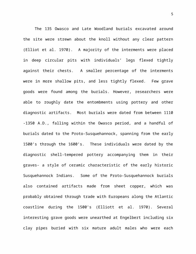
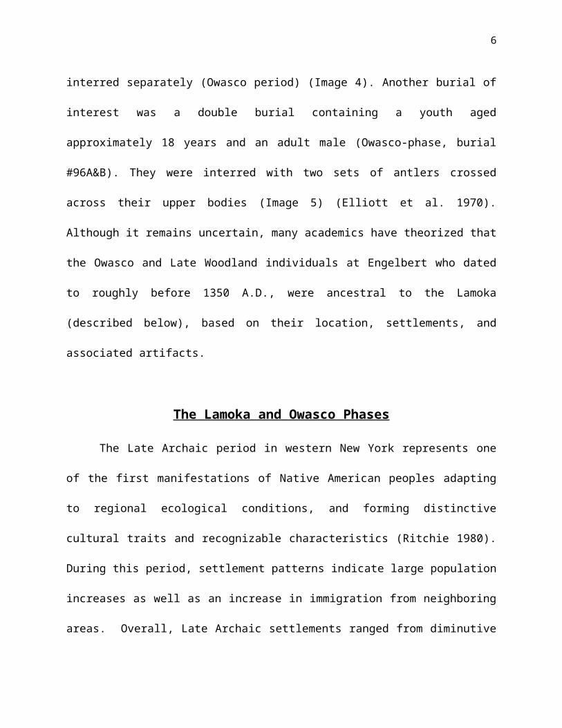
al. 1970). Several interesting grave goods were unearthed at Engelbert including six clay pipes
buried with six mature adult males who were each interred separately (Owasco period) (Image

4
4). Another burial of interest was a double burial containing a youth aged approximately 18 years
and an adult male (Owasco-phase, burial #96A&B). They were interred with two sets of antlers
crossed across their upper bodies (Image 5) (Elliott et al. 1970). Although it remains uncertain,
many academics have theorized that the Owasco and Late Woodland individuals at Engelbert
who dated to roughly before 1350 A.D., were ancestral to the Lamoka (described below), based
on their location, settlements, and associated artifacts.
The Lamoka and Owasco Phases
The Late Archaic period in western New York represents one of the first manifestations
of Native American peoples adapting to regional ecological conditions, and forming distinctive
cultural traits and recognizable characteristics (Ritchie 1980). During this period, settlement
patterns indicate large population increases as well as an increase in immigration from
neighboring areas. Overall, Late Archaic settlements ranged from diminutive sites characterized
by sparse surface scatters to larger, more substantive sites, that were likely occupied throughout
the year (Ritchie 1980). Many sites from this time period were situated adjacent to lakes, rivers,
and marshes, where groups could readily have access to aquatic resources (Ritchie 1980).
Most Late Archaic sites in western and central New York fall within the “Laurentian
Tradition”. The most well know of these is the Lamoka phase (Ritchie 1980). “The Lamoka
Phase”, best known for the Lamoka Lake Site (4999 B.C.-3000 B.C.), located near Tyrone in
Schuyler County, New York, was the first identifiable Archaic hunting and gathering culture in
the northeastern United States (Fenton 1998: 124). The Lamoka also hold an important role in
Northeastern archaeology as the first archaeologically distinguishable culture to occur after the
5
Paleo-Indian period, but before Woodland cultures that utilized and manufactured pottery (Fiedel
1992: 89).
Characteristic artifacts found at Lamoka sites include bone antler tools for use in flint
knapping, flutes, whistles, food processing tools such as mortars, mullers, hammerstones, pestles,
beveled adzes, and celts (Fenton 1998: 124). Distinctive Lamoka projectile points have also
been found around the northeast. Lamoka projectile points are generally “small, narrow, thick
points with weak to moderately pronounced side notches, or straight stemmed with slight,
usually sloping shoulders” (Ritchie 1971: 29). Lamoka points were likely used on spears,
javelins, daggers, and other throwing implements. Lamoka artifacts suggest that subsistence
consisted primarily of hunting and gathering as opposed to horticulture or agriculture (Ritchie
1971: 30). White tailed deer, wild turkey, passenger pigeons, squirrels, acorns, hickory, turtles,
frogs, mussels, as well as other aquaculture were staple food sources (Fiedel 1992: 89). The
Lamoka Lake site contained net weights, fishhooks, and other fishing implements, yet
interestingly fish bones at the site were rare. Burial remains at Lamoka settlements are also
sparse, usually flexed, and without grave goods (Ritchie 1980).
Judging by diagnostic pottery sherds, Engelbert’s cemetery likely contained Archaic
phase individuals ancestral to the Lamoka as well as peoples with an Owasco-like culture (Fiedel
1992: 89). The transitional period (1500 B.C. to 1000 B.C), between the Lamoka phase and the
Owasco phase is characterized by a gradual shift in material culture, which in turn spurred
significant lifestyle and subsistence strategy shifts throughout the northeast (Ritchie et al. 1973).
William A. Ritchie (1944) distinguished Lamoka culture from later Owasco based on
Owasco pottery (known for its “cord on cord” technique), and the emergence of agriculture
6
(Fenton 1998: 124). The emergence of pottery was also coupled with ceremonial objects such as
pendants, ornaments, pipes, gorgets, and large triangular projectile points known as Levanna
style (Image 6). Levanna points were eventually replaced by smaller Madison points for more
efficient use of the bow and arrow (Tuck 1971). The shift also coincides with the transition from
round dwellings to larger Iroquoian longhouses (Tuck 1971). Besides changing subsistence
strategies and architecture, burials also became increasingly complex with the addition of
ceremonial grave goods (Ritchie et al. 1973). Owasco cultures were known for their strategically
positioned camps, commonly located on high ground, near river deltas, and marshes in order to
ambush prey during the hunt. They were also one of the first Northeastern Indian groups to begin
farming the “three sisters”, corn, beans, and squash (Tuck 1978). Although much is known
about this time period in comparison to proceeding phases, archaeologists still have many
questions about the Owasco’s interrelationship with the Iroquois (Ritchie 1980).
The Susquehannock
After occupation by groups with relations to the Lamoka and Owasco, Englebert was
likely occupied by Susquehannock peoples as is evidenced by many of the distinctive artifacts
and shell-tempered pottery unearthed at the site (Dolores et al. 1970). The Susquehannock
people, archaeological distinguishable from the Iroquois Five Nations around A.D. 1550, were
indigenous to the Susquehanna River Valley. Archaeologists and cultural anthropologists
believe that their homeland extended from Southern New York and Pennsylvania, to the
beginning stretch of the Susquehanna River in Maryland, reaching northwards as far as the end
of the Chesapeake Bay (Kent 1984: 13). During the mid 1500’s, the Susquehannock peoples

7
were living in disjointed hamlets between Binghamton and the Wyoming Valley (Jennings 1978:
362). Settlement records show that the Susquehannock abandoned this area before 1570 A.D.
and established a new home in Lancaster, Pennsylvania. Historians question whether the
Susquehannock were driven out of the Binghamton area by hostile relations with the Iroquois
Five Nations, or whether they relocated to Pennsylvannia to take advantage of trading
opportunities with Europeans and other Native Americans (Jennings 1984: 362).
The Susquehannock’s link to the Iroquois Nations has been ambiguous at best, but
researchers have posited several scenarios based on both cultural and archaeological evidence
(Gillette et al. 1993). English Captain John Smith’s 1608 account calls the Susquehannock the
“Sasquesahanaough”, as they were referred to by one of Smith’s native informants (Gillette et al.
1993). Smith’s map of the Chesapeake Bay in 1612 showed six Indian villages along the
Susquehanna River attributed to the Susquehannock. However, many modern archaeologists
claim that Smith’s native information may have been incorrect, since there has only been one
archaeologically confirmed Susquehannock village found in the Chesapeake Bay dating to the
17th century (Gillette et al. 1993). The Susquehannocks were later identified by Champlain as
the “Carantouannais” in the mid 1950’s- a New French version of the Iroquoian word
“Skahentawaneh”, translated as ‘big grassy flat’ (Jennings 1978: 362). Although, scholars
dispute that the “Carantouan” were actually the Susquehannocks. Later French traders referred to
the Susquehannock by their Iroquoian name, “Gandastogue” or the Huron “Andaste” (Jennings
1978: 362). In addition, some researchers suggest that the Susquehannock may have been
referred to by the early French traders as the “Carantouannais”- the same people as the
“Atrakwaeronnon”, who the Iroquois are documented as overpowering in 1652 (Jennings 1978:
362). Overall, many archaeologists disagree with this theory, instead arguing that the 1652

8
skirmish was likely between the Iroquois Five Nations and allies of the Susquehannock further
south.
European diseases (smallpox and measles), internal and external conflict, as well as
migration eventually took its toll on the Susquehannock, and many speculate that some
Susquehannocks assimilated into the Iroquois Five Nations (Gillette et al. 1978). By all accounts,
the Susquehannock population was diminished significantly by 1698 when only fifty men
remained (Jennings 1978: 363). Unfortunately, the scarcity of Susquehannock sites makes it
difficult for researchers to draw concrete conclusions about Susquehannock chronology and
interrelationships with other tribes in the region (Gillette et al. 1993).
On the whole, the Susquehannock are characterized as culturally very similar to the
Iroquois, utilizing gendered divisions of labor and engaging in maize horticulture.
Susquehannock women likely tended to the fields and corn production, while men hunted,
traded, fished, and served as warriors (Jennings 1978: 364). The majority of ethnographic
accounts about gendered divisions of labor from the seventeenth century come from male
European missionaries (Amott et al. 1996: 34). Many European sources describe Native
American women as “beasts of burden” that were made to endure terribly long and strenuous
days out in the fields harvesting maize, beans, and squash. Many Europeans
saw Native American men’s tasks as more leisurely and light-weight, since fishing, fighting, and
hunting were all regarded as enjoyable upper-class pastimes (Amott et al. 1996: 34). One
interesting account was taken by a young woman who was adopted by the Seneca Iroquois after
her family had been killed by a warring group of Natives (Amott et al. 1996: 34). She wrote that
in the summer season, all of the women planted and tended to the maize harvest together (Amott
et al. 1996: 34).

9
Likewise, in southeastern North America, historians purport that European missionaries
and colonists substantially underestimated the role of women’s labor in Native society. They
claim that the “invisibility of women’s work” lead many European accounts to characterize
American Indian populations in northern Florida, Georgia, Alabama and Mississippi, as
primarily hunters and gatherers, even though Native populations relied heavily on maize
agriculture (Scarry et al. 2005: 260). As in the northeast, women’s work consisted of preparing
fields, harvesting, weeding, and creating storage. Men would sometimes assist in these duties,
but mostly focused on hunting, fishing, warrior duties, and often burning stumps and underbrush
from the fields, or trenching plots for planting (Scarry et al. 2005: 260). Although different
Native American societies during the Late Woodland and early historic period were similar in
terms of gendered divisions of labor, there were also undoubtedly regional differences (Amott et
al. 1996: 35)
Archaeologically, the Susquehannock have been characterized and described based on
only a handful of sites with distinctive pottery and other artifacts. In 1936 Donald Cadow began
a culture history chronology on Susquehannock pottery, drawing interrelationships between the
Susquehannocks and the Iroquois. In the late 1950’s, John Witthoft revised Cadow’s
chronologies incorporating additional research and collections. One of the most distinctive
Susquehannock pottery types is Schutlz Incised (Image 8), named after the Schultz site located in
Lancaster Pa. Another diagnostic artifact commonly found at Susquehannock sites in context
with shell-tempered Susquehannock pottery are native clay pipes used for smoking tobacco.
Early Susquehannock pipes are almost indistinguishable from those of the Iroquois, but after
1650 they become more differentiated. The earliest pipes have an Owasco base and are known
as trumpet pipes without incised or impressed designs (Image 9). The next evolutionary stage is

10
the addition of the ring bowl, which is believed to have developed independently from the
Owasco base alongside the trumpet style (Kent 1978: 145). The last motif in the Susquehannock
sequence is the tulip bowl, which evolved from ring bowls (Image 10) (Kent 1978: 145). Later
adult male burials at the Engelbert site were found with both ring bowl and trumpet bowl pipes
(Image 4). Susquehannock sites such as Washington Boro (Washington Boro, Pa.), Schultz, and
Strickler (Strickler, Pa.) also have steatite pipes, many of which were adorned with animal and
human motifs (Image 11). One particular pipe, unearthed at the Strickler site in the northeast
cemetery with a sub adult interment, had a human-form complete with an anus that was drilled
through the pipe, allowing a smoker to cover the lit bowl and blow smoke through the orifice
(Kent 1978: 153). Kalolen pipes used to smoke calumet have also been found at Susquehannock
sites, likely imported from Midwestern Indians. Interestingly, Susquehannock native pipe
production comes to an abrupt halt after the group’s military and political upheaval in 1675
(Kent 1978: 157).
Other archaeological artifacts characteristic of Susquehannocks sites include a variety of
lithics, and items made from bone and antler (Kent 1978: 150- 300). Kent notes that triangular
projectile points with isosceles to equilateral outlines are common at Susquehannock sites
between 1500 AD -1600 AD. Raw materials were usually cherts derived from local quarries
with exotic materials such as jasper, chalcedony, rhyolite and quartzite rarely seen (Kent 1978:
157). Refined scrapers are also rare at Susquehannock sites, and the ones found have been rather
crude.
An extensive list of bone and antler tools have also been found at Susquehannock sites
including turtle-shell cups and rattles, fishhooks, awls, knife handles, pressure-flaking tools,
chisels made of beaver teeth, harpoons, conical arrow points, smoking pipes, scraping tools,

11
spoons, and needles (Kent 1978: 176). Some of the most artistically impressive Susquehannock
artifacts, however, have been their intricately detailed bone and antler combs donning turtles,
bears, wolves, humans, and geometric patterns (Image 11). Wray (1963: 45) suggests that these
combs may represent legends or clan affiliations (Kent 1978: 177).
Overall, Susquehannock culture is little understood and researchers are still striving to
find out more about their relationship with the Iroquois and their interaction with European
traders. Ethnographic information and historical documentation is important not only for
understanding subsistence strategies and settlement patterns, but for reconstructing social values,
norms, and gendered divisions of labor (Derevenski 2000). Bioarchaeologists commonly turn to
ethnographic resources to reconstruct the activity patterns and life ways of past cultures and
populations. For example, small circular notches between the teeth may mean little until a
biological anthropologist learns that individuals from that particular culture habitually smoked
from pipes with long spherical stems. Likewise, biological anthropologists are able to use
historical data to interpret osteological markers on bone. Many researchers believe that OA is a
specifically good indicator of physical activity over an individual’s lifetime. While many
researchers strongly believe that the patterning and distribution of OA can help to reconstruct
activity patterns, many other researchers disagree with this correlation (Knüsel et al. 1997: 481).
While researchers may disagree on the exact causes of OA, there is no doubt that the condition is
both highly prevalent and debilitating.
The Paleopathological Identification and Assessment of Osteoarthritis
Periodontal disease and OA are the two most prevalent pathological conditions observed
by physical anthropologists who examine ancient skeletal remains (Larsen 1997:163).

12
Osteoarthritis, also known as degenerative joint disease (DJD), is a common condition that
affects the synovial joints (Roberts et al. 2005: 136). Clinicians and physical anthropologists
assess four major criteria for the presence and severity of OA: osteophytosis (bone spurs on the
joint margin), sclerosis (new bone growth on the joint surface), eburnation (shininess and luster
of the joint facet caused by constant rubbing), porosity of the joint surface, and plastic
remodeling of the apophyseal facets (reshaping and warping of the joint facets caused by
compression and load-bearing) (Waldron 1995: 386). OA can affect many parts of the body
including hips, knees, spine, elbows, hands, feet, temporomandibular joints, and more (Waldron
1995: 386). OA is often regarded as a unique pathology because it is one of the only diseases
that has “operational criteria” for its diagnosis and evaluation, meaning that OA can be accessed
through a demonstrated process using a set of validation tests, while other pathologies are often
more elusive, and difficult to conclusively diagnose (Waldron 1995: 385). Paleopathologists
note that the disease is often complicated, as there are several etiologies and it can be
confounded by the presence of other arthritic conditions like rheumatoid arthritis (RA)
(described below) (Roberts et al. 2005: 133). However, compared to other pathologies that can
be recognized through bony indicators and markers, OA is relatively straightforward to diagnose
and identify.
OA is caused by the deterioration of the soft porous cartilage that coats and pads the
synovial joint surface. When cartilage breaks down, the synovial joint becomes inflamed and
creates friction between bony surfaces (Bridges 1992: 67-68). Often, complete destruction of the
joint impedes the production of synovial fluid that lubricates the joint surface, further increasing
apophyseal friction (i.e. friction at the articulations located between the joint facets of vertebrae),
tenderness, and joint pain (Bridges 1992: 67-68). Traditional OA indicators on bony remains are
13
peripheral osteophytes, sclerosis, eburnation and pitting. Osteophytes (or marginal lipping), are
bone spurs or outgrowths that erupt around the margins of joint surfaces due to friction and
compression.
To increase the load distribution on apophyseal joints, apophyseal joints often form osteophytes
in an attempt to compensate for excess stress (Roberts et al. 2005: 135). Osteophytes may occur
on the vertebral body, the joint facets, or at the insertion sites of ligaments and tendons (Roberts
et al. 2005: 135). However, it remains unclear whether osteophytosis occurs concurrently with
cartilage deterioration (Dequeker et al. 1997: 358). Once OA has destroyed joint cartilage, the
underlying bone may become sclerotic (hard), and eburnated (shiny and polished from friction
and rubbing). As the cartilage deteriorates further, the surrounding bones often becomes porous
and pitted, allowing synovial joint fluid to flow into the pitted bone beneath the destabilized
cartilage, forming subchondral cysts that may further complicate the condition (Roberts et al.
2005: 136).
The Etiology of Osteoarthritis
Today, paleopathological data is widely considered to be the most reliable and concrete
indication that ancient populations suffered from specific ailments. Although historical and
ethnographic documentation may focus on the diseases of past peoples, it is common for these
sources to focus on dramatic, highly visible conditions, while more ordinary commonplace
ailments of OA are overlooked (Roberts et al. 2005: 1). In past decades, biological
anthropologists have come to realize that bioarchaeological data has the potential to uncover past
activity patterns, lifestyles, diets, and population structures. In addition, physical anthropologists
now know more about human pathologies than ever. For example, researchers can now identify

14
myriad human diseases that manifest in bone including Paget’s disease, dental defects such as
periodontal disease, porotic hyperostosis, tuberculosis, developmental disorders, osteoporosis,
OA and many others (Roberts et al. 2005: 5).
Over the decades, researchers have come to accept that OA has a complex and
multifaceted etiology that may be caused by a multitude of factors (Lieverse et al. 2006: 1).
Some researchers believe that occupational stresses and other daily activities are probable causes
of most types of OA. However, age, sex, body build, nutrition, and heredity may also be
contributing factors to varying degrees (Lieverse et al. 2006: 1). Epidemiologists have found
that certain ethnic groups, age groups, and sexes experience higher incidences of specific types
of OA, indicating they may be predisposed to cartilage deterioration (Lieverse et al. 2006: 1).
Overall, it has been found that as individuals age, their probability of developing OA increases as
biomechanical stress and diminished durability of joint tissues are compounded (Larsen 1997:
163). Recent clinical studies show that 1-5 percent of US citizens under age 45 have some
degree of either OA or rheumatoid arthritis (RA) (Roberts et al. 2005: 133).
RA is a chronic inflammatory disease affecting the lining and synovium of the joints.
The synovial lining first becomes inflamed and swollen, which may cause pain and stiffness.
Next, rapid division of cells causes the synovium to thicken (Arthritis Foundation 2009).
Eventually inflamed cell enzymes destroy bone and cartilage causing joints to become
misaligned, malformed and non-functioning (Arthritis Foundation 2009). Rather than being
related to physical activity, RA is believed to be a disorder of the immune system. It is also a
systemic disease, which means it has the capacity to affect other organs in the body as well
(Arthritis Foundation 2009). Thus, although the etiologies of RA and OA are both quite
different, the two diseases may manifest in bone quite similarly (Larsen 1997: 163). However,

15
RA usually affects many different joints bilaterally, such as the hands, wrists, feet, knees, and
neck. This is unlike OA, where individuals often have symptoms in fewer areas, and often
unilaterally, depending on activity patterns (Larsen 1997: 160-165).
Although studies show that age is a contributing factor for OA specifically, the
degeneration of joint tissues may be seen at any age if extreme stressors are present (Lowell
1994). Studies show that it is uncommon for individuals below the age of 30 to experience OA
in developed societies. However, in rural populations, where children and teens are more likely
to take on strenuous labor, even young adults may have elevated rates of OA (Lowell 1994).
Although this research shows that age is a contributing factor for OA, the degeneration of joint
tissues may be seen at any age if extreme stressors are present (Lowell 1994).
Sex is another factor that may greatly influence the development and severity of OA.
Many studies show that woman likely have a predisposition towards developing OA compared to
men (Oliveria et al. 1995: 1134). Studies show that the prevalence of OA in women remains
steady until the age of 50, and which point the incidence of OA skyrockets. Interestingly, men
have a higher prevalence of OA before the age of 50, at which point new cases drop off sharply
(Oliveria et al. 1995: 1134). Furthermore, women are more likely than men to have OA in
multiple joints (Oliveria et al. 1995: 1134). Many researchers believe that female hormones,
specifically estrogen and progesterone may play a role in a woman’s increased chance of
developing OA (Zhang et al. 1998:1867). Doctor’s often refer to “menopausal arthritis”, which
is characterized by rapidly progressing OA of the hands during menopause suggesting that
estrogen deficiency at middle age may play an important role in female OA (Zhang et al.
1998:1867). In Zhang et al.’s 1998 study “Estrogen Replacement Therapy and Worsening of
Radiographic Knee Osteoarthritis”, they found that the use of postmenopausal estrogen

16
replacement therapy may protect women against post menopausal OA progression (Zhang et al.
1998: 1972). Likewise, Okma-Keuleun et al. 2001 preformed a qualitative study in the
Netherlands, examining the onset of generalized OA in older women. They found that all of the
participants in their study group with generalized OA had previous gynecologic problems
ranging from miscarriages, difficulty becoming pregnant, giving birth to one or multiple disabled
children, having hysterectomies, or having to become sterilized due to high blood pressure
(Okma-Keuleun et al. 2001: 187).
However, other studies refute that that sex hormones have any specific effect on the
presence, or progression of OA (Cauley 1993; HL MA 2007). However, other researchers argue
that serum sex hormones have little to no effect on OA. In Cauley et al.’s (1993) study, they
found no association between endogenous estrogen or androgen levels and severity of hand
osteoarthritis, even when other factors such as obesity were taken into account (Cauley 1993:
1172). Furthermore, H.L Ma et al. (2007), found that when examining sex hormones in relation
to mouse knee histology, researchers found that ovarian hormones seemed to decrease the
severity of OA in female mice, yet testosterone exacerbated OA in males (H.L. Ma et al. 2007:
695).
Physical build and nutrition also play a pivotal role in an individual’s susceptibility to
developing OA. Physical anthropologists agree that an individual’s unique anatomical traits may
significantly affect the onset of OA and other degenerative ailments like osteoporosis. It is also
generally agreed upon that some individuals may be more prone to OA due to their body
physiques. Kalichman et al. (2005) examined the correlation between skeletal aging traits in the
hand, and physical characteristics including weight, skeleton size, and muscle development.
They found that ectomorphy (measures of longitudinal body characteristic), endomorphy
17
(measures of fatness), mesomorphy (measures of muscular development), and other physical
characteristics were significantly influential in assessing skeletal aging characteristics such as
osteoarthritis and osteoporosis (Kalichman et al. 2005: 894).
Body mass also has a significant effect on OA, especially for females. Studies also show
that obesity causes greater concentrations of leptin in cartilage, which may in turn encourage
osteophyte formation in females who have smaller joints than males (Weiss 2007: 441).
Researchers such as Sharma et al. (2000) have also found that obesity increases the severity of
certain kinds of OA. Sharma et al. (2000) found that obesity was strongly linked to OA of the
knee in patients with varus knees (knees pointing outwards), but not individuals with valgus
knees (knees pointing inwards) (Sharma et al. 2000: 568). Thus, obesity combined with certain
physical characteristics might exacerbate OA. However, many researchers argue that high body
mass is a modern phenomenon, rarely seen in prehistory (Bridges 1992). Nutritional stress, such
as vitamin C and D deficiency is also thought to increase the progression of OA. Vitamin D is an
important factor in normal bone growth and studies show that high D levels offer some
protective effect against hip and knee OA (Cimmino et al. 2005: 33). McAlindon et al. (1996)
also found that vitamin C intake reduced the progression of knee OA in patients studied by 3-
fold (McAlindon et al. 1996: 353). Four dietary deficiencies have conclusively been identified in
the progression of OA: selenium deficiency, iodine deficiency, water pollution with organic
material and fulvic acid, and grain contamination with mycotoxin-producing fungi (Cimmino et
al. 2005: 33).
Genetic factors are also thought to contribute to the severity and onset of OA in both
sexes. Holderbaum et al. (1999), claim that non-trauma initiated and idiopathic (arising
spontaneously or from an unknown cause) OA has been clearly observed to appear in familial
18
clusters, meaning that the incidence of OA is likely a complex combination of both genetics and
common environmental factors such as physical activity, diet, and nutrition (Holderbaum et al.
1999: 397). According to Holderbaum and colleagues, the first clue that OA might have a strong
genetic link was the discovery of type II collagen (COL2A1) in association with early onset OA
accompanied with mild spondylepiphyseal dysplasia (SED) in a Michigan family (Holderbaum
et al. 1999: 398). Interestingly, other families were also identified with the same unique
mutation (Holderbaum et al. 1999: 399). All of the families found to contain the mutation shared
a common Celtic ancestry, and were probably all related to a single founder (Holderbaum et al.
1999: 400). A similar mutation was also discovered in a family located in the Chiloe Islands off
the coast of South America. Overall, Holderbaum et al. believes that these findings are powerful
evidence that OA is hereditary to some degree. However, they note that OA does not seem to
have any clear or straightforward cause. Rather it is likely the result of many different factors
working together, with genetics playing a major role (Holderbaum et al. 1999: 403).
Holderbaum et al. (1999) also believe that twin studies are a valid form of identifying
hereditary traits. Recent studies have compared monozygous (identical) and dizygous (non-
identical) twins in an attempt to locate a genetic component for OA. Holderbaum et al. (1999)
writes that twin studies show an increased correlation of radiographic OA of the hands and knees
among monozygotic twins at a rate of 35% to 65%. This means that 35-65% of identical twins
show similar rates of hand OA compared to their twin suggesting a genetic factor (or the
interplay of genetics and environment). They also note that in recent studies of hand and knee
OA in parents and adult children, genetic modeling demonstrated that the traits were likely
explained by a “Mendelian recessive trait with significant polygenic or environmental factors”
(Holderbaum et al. 1999: 402). Holderbaum et al. notes that considering the many different
19
protein-protein, protein-glycoprotein, and protein-cell interactions that occur within cartilage, it
would be highly unusual if all the mutations that caused OA were genetic (Holderbaum et al.
1999: 402).
In addition, a study conducted in Iceland found that Icelandic patients who had hip OA
that eventually led to hip replacement surgery were more related to one another than matched
controls drawn at random from a population-wide scan (Ingvarsson et al. 2000: 2785).
Ingvarsson et al. noted that it is unlikely that common environmental factors affected familial
clusters since the correlations ran over many generations and branched out from nuclear families
(Ingvarsson et al. 2000: 2791). Overall, nine genetic loci have been found that influence specific
parts of different joint systems. Some loci incite osteophyte development, while others seem to
influence cartilage loss (Ingvarsson et al. 2000: 2791). In addition, a specific nucleotide mutation
in the Cartilage Intermediate Layer Protein (CILP) locus is thought by many researchers to have
a noteworthy effect on an individual’s risk of developing OA of the lumbar region of the spine
(Weiss 2007: 440). Genetic susceptibility is also thought to be a greater issue for females than
males, because estrogen receptor genes may intensify the effects of the CILP polymorphism.
While many researchers stand by twin studies, which claim a large genetic influence for certain
types of OA, other researchers argue that the aforementioned experiments grossly overestimate
heredity (Weiss 2007: 440).
Many researchers claim that twin studies may well produce inflated heritability values
since environmental factors may be more significant for both identical and fraternal twins (Weiss
2007: 440). Samsbrook et al. (1999) conducted a study in which they examined OA through MRI
examining the cervical and lumbar spine of 172 monozygotic and 154 dizygotic twins selected at
random. They found that overall heritability was 74% at the lumbar spine, and 73% at the

20
cervical spine. Even though they adjusted results to account for age, weight, height, smoking,
occupational manual work, and exercise they still note that twin studies may produce biased
results because subjects with a given disease self-select themselves for the study (Samsbrook et
al. 1999: 370). Other osteolotists claim that while cervical and lumbar disc morphology (disc
height and bulge), and lumbar osteophyte growth, seem to be heritable, twin studies simply fail
to control for all of the various environmental factors shared by participants in such studies
(Sambrook et al. 1999: 366). Many researchers also stress that while genetics may certainly
have an important link to OA, it is more likely that genetics affects the severity of OA when
present and not simply the presence or absence of OA (Weiss 2007: 440).
Osteoarthritis of the Spine
Spinal column OA is universal among past populations, but the disease’s extent and
severity differ widely across different cultures (Roberts 2005: 141). Because of the spine’s “S”
shaped curvature, some segments experience more biomechanical stress than other sections
during normal bipedalism (Brown et al. 2008). The human spine is convex posteriorly in the
thoracic region, and concave posteriorly in the lumbar and cervical sections. Because of this
pattern of curvature, the fifth cervical, eighth thoracic, and fourth lumbar vertebrae are the most
stressed during daily activities and over one’s lifetime (Roberts 2005: 139). This occurs because
these vertebrae are located furthest from the body’s center of gravity, thus the transfer of weight
resting upon them is greater (Derevenski et al. 2000:350).
Between each vertebral body is an intervertebral disc, composed of a fibrous capsule
called the annulus fibrous, which is filled with gelatinous fluid known as the nucleus pulposus
(Roberts 2005:140). In cases of spinal OA, continuous stress causes the nucleus pulposus to
infiltrate the annulus fibrous, causing the capsule to fracture and rupture. This trauma causes
21
osteophytes to erupt around the margins of the vertebral body in an attempt to form a wider
platform to handle physical stress- a process known as osteophytosis (Roberts 2005: 140). As
the gelatinous fluid continues to break through the fibrous capsule, bony growth around the
vertebral body continues, forming a lip. In particularly severe cases of spinal OA, the vertebrae
may fuse together in a condition known as ankylosis. This occurs when bony outgrowths
between vertebral bodies join (Roberts 2005: 140). In addition to osteophytosis, cartilage
between vertebral bodies may also deteriorate, causing the fibrous bone to become pitted, porous
and weakened (Roberts 2005: 140).
Many researchers argue that apophyseal facet remodeling may be an even better gauge of
mechanical stressors than other “traditional” criteria such as osteophytosis, sclerosis, eburnation
and pitting (Derevenski et al. 2000: 338). Facet remodeling occurs when the inferior and
superior facets of the apophyseal joint become buttressed up against the margin of the adjoining
facets, becoming warped and misshapen. The superior and inferior facets then form a lock and
and key, or puzzle piece effect suggesting that the facets were shaped and deformed by
biomechanical stresses and compression placed on the spine (Derevenski et al. 2000: 338).
Derevenski et al. (2000) notes that articular facet remodeling is the bone’s response to excess
stress when vertebral slipping occurs because of disc compression, collapse, or general
deterioration (Derevenski et al. 2000: 338). Derevenski et al. (2000) argues that many biological
anthropologists have had a tendency to focus exclusively on osteophyte growth, sclerosis, and
eburnation as their main criteria for diagnosing and assessing OA, because these traditional
markers are convenient and allow “ease of identification” (Derevenski et al. 2000: 338).
Nevertheless, Derevenki and her colleagues argue that facet remodeling should be added to the

22
list of commonly used OA markers, adding that it may be used as a more sensitive early indicator
of activity related OA (Derevenski et al. 2000: 338).
Osteoarthritis as a Biomechanical Indicator of Past Activities
It has been argued that sexual dimorphism in OA patterning and distribution may
demonstrate gendered divisions of labor within a given population. Past anthropologists have
pointed out that at hunter gatherer Native American sites such as Indian Knoll, Mobridge, and
Grasshopper Pueblo, males tended to have more appendicular arthritis, while females exhibited
more temporomandibular OA, likely from softening hide and other materials using their teeth
(Bridges 1992: 75). In contrast, studies of several California hunter-gatherer groups have shown
that males showed more OA of the hand, shoulder, and elbow, while women exhibited more
arthritis of the knee and vertebrae, suggesting gendered divisions of labor. Bridges (1994) also
examined spinal OA in a prehistoric group from northwestern Alabama. She found that
patterning and distribution of OA mostly followed the natural curvature of the spine, with most
osteophytosis occurring in the lumbar region. However, she also found a large amount of
cervical OA, suggesting tumpline use and burden carrying using the head and upper neck. Thus,
she concludes that while most OA is likely caused by natural bipedalism, it can be modified by
habitual behavior (Bridges 1994: 92). Moreover, many groups such as several hunter-gather
groups from Alabama, and prehistoric Illinois groups (for example, Dixon Mounds) have shown
little sexual dimorphism in OA patterning and distribution, suggesting more egalitarian
workloads, and less division of tasks (Bridges 1992: 75).
Many researchers argue that biomechanical pathologies like OA may be used in
conjunction with ethnographic information and known gendered divisions of labor to evaluate

23
the impact of daily activities and lifestyles on synovial joints (Leiverse et al. 2006: 1). For
example, some claim that by examining the distribution and patterning of OA, anthropologists
can gain a more thorough understanding of the subsistence strategies and daily activities of past
populations (Derevenski 2000: 334). Physical anthropologists like Lieverse et al. (2007) argue
that OA and activity are clearly related. Lieverse and colleagues note that human populations
that have historically high levels of activity also have correspondingly high levels of OA. In
addition, they claim that patterning and distribution of OA may reflect specific biomechanical
activities over time (Lieverse et al. 2007: 1).
However, some researchers disagree about whether traditional OA criteria such as
osteophytosis, sclerosis, and eburnation of the vertebral bodies and apophyseal joints are actually
valuable indicators of activity related stresses (Rojas-Sepúlveda et al. 2008; Derevenski et al.
2000: 334). Many studies claim that activity related stresses do not leave diagnostic signs on the
spine, but instead manifest in predictable patterning of OA in accordance with the curvature of
the vertebral column (Bridges 1992 & 1994; Knüsel et al. 1997). Consequently, many physical
anthropologists reject the activity-related findings of past OA studies, which rely exclusively on
traditional markers. Patricia Bridges (1992 & 1994), and Knüsel et al. (1997), argue that many
times these observations cannot be directly linked to specific activities. Several scholars claim
that degeneration of the apophyseal facets are a more effective gauge of lifestyle stresses than
traditional markers of OA such as osteophytosis, sclerosis and eburnation. Derevenski et al.
(2000) points out that plastic change of the apophyseal facets may be the first signs of OA,
occurring before traditional markers. During plastic remodeling of the vertebral facets joint
surfaces become modified in response to biomechanical stress. Thus, she claims that facet
24
remodeling may represent a precursor to more well-known indicators of OA (Derevenski et al.
200: 338).
Derevenski et al. compared spinal OA in two populations in England: the 16 th-19th
century site of Ensay, and a skeletal collection from the medieval site of Wharram Percy
(Derevenski et al. 2000: 333). Fortunately, Derevenski and colleagues had access to informative
ethnographic documentation, which allowed them to explore possible causes of the spinal OA
observed in each group. Overall, Derevenski et al. (2000) found that two populations she studied
had predictable patterning and distribution of osteophytosis on the vertebral bodies in accordance
with the spine’s natural curvature, but differences in facet remodeling. She found that the
sample from Ensay had more facet remodeling than the skeletons from Wharram Percy. In the
Ensay population, women and men had distinct gendered divisions of labor, and the differences
in OA distribution and patterning between the sexes was marked (Derevenski 2000: 352). In
comparison, men and women from Wharram Percy were more similar in their distribution of
facet remodeling, and showed less variation between the sexes. These results matched
ethnographic documents reporting that the population did not have gendered divisions of labor as
distinct as those existing in Ensay (Derevenski 2000: 352).
Derevenski et al. found that remodeling was a more effective gauge of activity related
stress for both populations in their study than traditional markers of OA (Derevenski 2000: 352).
Derevenski et al.’s study showed that both populations showed biomechanically “normal”
osseous changes of the spine as would be expected from normal vertebral curvature. However,
they found significant differences between the two populations and between men and women
within each group by examining apophyseal facet remodeling (Derevenski 2000: 351). For
example, Ensay females had a block of vertebrae situated around T1 that showed statistically

25
significant amounts of facet remodeling, and facet sclerosis and eburnation. This was likely due
to carrying creels, which transforms the normal curvature of the spine from an “S” shape, to a
more hooked position, putting pressure on the upper thoracic region, and taking pressure off the
lumbar. Interestingly, females from Ensay also showed less vertebral body OA in the lumbar
region than other groups in the study (Derevenski et al. 2000: 351). Thus, they argue that
osseous remodeling of the apophyseal facets might therefore be a better indicator of activity
related stress in humans than traditional markers that focus primarily on the vertebral bodies
(Derevenski et al. 2000: 337).
Brown et al. examined this assertion in their 2008 article, where they used cadaver
spines to test the hypothesis that degenerative changes in human apophyseal joints are related to
high levels of compressive load-bearing. Brown et al. exposed 36 thoracic-lumbar segments
aged 64-92 years to a high impact compressive force of 1.5 knots (kN). The distribution of the
compressive force was then measured within each segment through a pressure transducer, and
these sums were calculated independently from the initial 1.5 kN force to obtain total
compressive load-bearing by the apophyseal joints alone (Brown et al. 2008: 318). Brown and
colleagues found that in elderly individuals, apophyseal joint-bearing was positively linked to
severe DJD in both spinal cartilage and bone half of the time. From these findings, Brown et al.
concluded that activities involving significant amounts of compressive load bearing may indeed
produce pathological indicators that can assist in investigations of sex or age related lifestyle
differences (Brown et al. 2008: 324)
Despite these findings, some researchers claim that even apophyseal facet remodeling is a
poor indicator of occupational stress, often citing genetic research (especially twin studies) in
their arguments. For example, Knüsel et al. (1997) claims that degenerative changes in the

26
vertebral column are always the result of biomechanical stresses exerted on the spine by
bipedalism: “…this difference was produced as a response to erect posture during bipedal
locomotion, reflecting vertebral curvatures, rather than differing occupational stress” (Knüsel et
al. 1997: 481). Thus, they posit that the stresses from most physical activities are likely
confounded by natural wear and tear due to natural locomotion. However, they do note that in
cases of considerable biomechanical compressive force, such as in the case of the ancient
Harappans who used their heads for weight bearing, substantial compression of the head and
neck may lead to OA of the cervical spine (Knüsel et al. 1997: 494). However, Knüsel et al.
(1997) argue that the pelvic girdle and upper limbs may be better areas to judge activity related
stress through OA patterning and distribution compared to the spinal column (Knüsel et al. 1997:
494).
Overall, there remains marked disagreement among researchers over whether OA of the
spine can be directly linked to activity related stresses. Furthermore, there have been relatively
few OA studies carefully examining apophyseal facet remodeling. To contribute to ongoing
research on OA, and particularly on plastic remodeling of the vertebral facets, I examined spinal
OA in a Native American population, looking at traditional markers of OA on the vertebral
bodies and facets, as well as apophyseal remodeling.
Research Introduction and Sample Selection
Using a cemetery collection from the Engelbert site, I examined OA of the spine,
assessing osteophytosis, sclerosis, eburnation, and plastic remodeling of the facets. Given
evidence (described above) that there was gendered division of labor in this population, my goal

27
was to investigate the patterning and distribution of OA between men and women at the
Engelbert site.
My data sample consisted of adult members of the Englebert cemetery collection. One
hundred and thirty five individuals were unearthed at Englebert. Of these bodies, I included 35
mature skeletons (22 male and 13 female) dating from between ~1350 B.C. -1000 B.C., with
acceptable completeness and preservation. I did not include any burials that were theorized to be
clearly Schultz (1525-1575) or Proto-Susquehannock (after 1350) because these burials were so
much younger than the other interments, and may have represented different cultural practices. I
separated both males and females into age categories: 18-35, and 35+. For aging and sexing my
specimens, I relied on museum records from past analyses of the collection. As in Lieverse et
al.’s 2007 study, I only included vertebrae that exhibited acceptable preservation of the joint
surfaces and margins (Lieverse et al. 2007). Many researchers, such as Derevenski et al. (2000),
who examine spinal OA, only include individuals who have either complete segments of the
spine (C1-C7, T1-T6, T7-T12, or L1-L5), or complete spines. However, I chose to emulate
Rojas-Sepúlveda et al. (2008), and conduct my research vertebrae by vertebrae. All of my
research was non-destructive, non-invasive, and macroscopic. I examined each vertebra for
osteophytosis of the vertebral bodies and apophyseal joints, as well as sclerosis, eburnation, and
plastic remodeling of the superior and inferior articular facets.
Data and Methodology
Each individual apophyseal facet was scored separately for the presence, absence, and
severity of osteophytosis, sclerosis and eburnation (as one category), and plastic facet

28
remodeling. Superior and inferior aspects and left and right facets were scored separately. For
scoring severity, a scale of 0-4, was used (described below), with “unobservable” also being used
as an option when the specific area was broken or poorly preserved (Rojas-Sepúlveda et al.
2008).
Assessing Osteophytosis
Osteophytosis, as described by Säger (1969) are irregular beak-like formations of new
bone on the joint surface. For assessing osteophyte growth on the vertebral body, I used a scale
from 0-4, modeled on Rojas- Sepúlveda et al. 2008’s study, which was originally derived from
Säger (1969). For assessing osteophytosis of vertebral bodies and apophyseal facets, I followed
Derevenski et al. (2000), and used Säger’s 1969 system, using a scale of 0-4 (absence of fusion
to complete fusion) of adjacent vertebrae.
Grade 1 I is characterized by small scattered hyperostosis (Säger 1969: 58). Grade 2 II
has protrusions of bone in a horizontal direction from the corpus vertebrae. Grade 3III shows the
osteophytes with a characteristics bird’s beak shape where the bone has erupted from the edge of
the vertebral body. These osteophytes may contact bone spurs on the other side of the discus
(Säger 1969: 58). Grade 4IV is given when osteophytes from adjacent vertebrae fuse forming a
bone ridge across both adjacent vertebrae (Säger 1969: 58).
Assessing Sclerosis and Eburnation
I scored sclerosis and eburnation together, since eburnation usually follows sclerosis.
Sclerosis is the formation of thickened bone on a bony surface caused by friction and
29
compression (Säger 1969: 58). After bone has become sclerotic polishing and rubbing gives the
bone a shiny, lustrous appearance. This polishing is known as eburnation. Sclerosis always
precedes eburnation, but eburnation need not always follow sclerosis (Säger 1969: 58). I used a
scale of 0-4 as follows: 0=absent, 1=sclerosis, 2=sclerosis with some eburnation visible
3=eburnation, more extensive than sclerosis and 4=extreme eburnation (Derevenski et al. 2000:
341). In practice, a score of 4 was never used.
Assessing Plastic Bone Remodeling
Each individual apophyseal facet was scored separately for the presence, absence, and
severity of articular facet remodeling. Superior and inferior aspects, and right and left facets,
were scored separately. Scoring used a scale of 0-4, with “unobservable” also being used as an
option when the specific area was broken or badly preserved (Rojas- Sepúlveda et al. 2008). I
also used Derevenski et al.’s (2008) study as a guide for my apophyseal joint scoring criteria.
Score O was given when the inferior margin of the superior articular facet is sharp and
distinct, and has no increased surface area extending onto the lamina, or downwards (Rojas-
Sepúlveda et al. 2008). Score 1 was given when the inferior margin of the superior articular facet is
indistinct and slightly warped without a sharp margin. The facet has an increased surface area extending
downwards onto the proceeding vertebra’s lamina (Rojas- Sepúlveda et al. 2008). For score 2, there is
a newly formed small bony shelf on the lamina of the proceeding vertebrae, which acts to support the
inferior articular process of the upper vertebra (Rojas- Sepúlveda et al. 2008). Score 3, as in stage 2, is
characterized by a significantly larger bony shelf extending downwards into the laminal groove and
outwards onto the transverse process of the proceeding vertebra. The superior margin of the superior
articular process is rounded anteriorally. The inferior articular facet of the preceding vertebra has

30
indistinct margins and fits together with the articular facet of the proceeding vertebrae as a lock and key,
or a puzzle piece. Lastly the facets of proceeding and preceding vertebrae are extremely warped
and fused together (Rojas- Sepúlveda et al. 2008).
In addition, I used the score of “unobservable” for individual facets that were damaged,
and a score of 4 for cases of extreme remodeling displaying fusion with adjacent vertebrae
(Rojas- Sepúlveda et al. 2008). Joanna Derevenski was also kind enough to provide me with
enlarged photographs of facet remodeling to assist with my veterbrae scoring (Images 13-16).
These snapshots were invaluable to my research, as her 2000 study was the first of its kind to put
forth specific criteria for evaluating plastic changes in the apophyseal facets of the spine.
Hypotheses
Given evidence that the population at Engelbert did have marked gendered divisions of
labor (as described above), this paper tests the following hypotheses:
1). Null hypothesis: There are no consistent differences in facet remodeling between males and
females.
Alternate hypothesis: Males and females will show clear differences with respect to remodeling
of the apophyseal facets.
Data Analysis
I created tables scoring burials by sex and age. All of my data tables are included in the
appendix. I then arranged my data into sexed age groups, organized by each vertebra, vertebral
aspect (e.g. superior right facet), and specific pathology (e.g. osteophytosis). For example, all
data for males 18-35 with osteophytosis present on the superior vertebral body (SVB) of C1 were

31
compiled into one score, and overall scores were created by counting all the C1s that were
observable, and then summing all of the C1s whose SVB showed the presence of osteophytosis.
For example, a score might look like: 1/5, with “1” as the number of C1 vertebrae with
osteophytosis present on the SVB, and “5” as the number of observable C1 vertebrae, SVB’s
present within the 18-35 age male category. After these were completed, I compared the
frequencies of markers of OA between males and females using a Chi-square test, with a
significance level of α = 0.05, and a degree of freedom of 1. I then ran power calculations to
determine whether the samples sizes were large enough to allow me to reject the null hypothesis.
I also used adjusted Bonferroni comparionscomparisons to assess whether significance values
obtained at α = 0.05 were still significant once the alpha value was divided by the number of
tests performed. Comparisons were only considered truly significant when they also remained
significant after performing the Bonferroni adjustment.
I combined categories to compare overall findings of osteophytosis, eburnation and
sclerosis, as well as remodeling for each vertebra within age categories (Tables 1-6). I then
combined data further, and compared overall segments (e.g. C1-C7, T1-T6, T7-T12, and L1-L5)
(Tables 7-8). Lastly, I compared data that included each individual only once per each vertebral
segment. For example, each individual in a certain gendered age group that had OA present on
any vertebra within the segment C1-C7 received a score of present for the entire section (Tables
11-14).
Lastly, I compared young and old individuals within each gendered category to examine
whether older members of the collection showed substantially more OA than younger skeletons
(Tables 9-10).

32
Although I scored for severity in my initial analysis, when compiling my final data I
decided to focus on presence and absence instead of the 0-4 gradation scores. However, all of
my 0-4 scoring is available on the data charts in the appendix. It would be interesting to go back
and analyze this data examining severity of vertebral OA between men and women at Engelbert.
In addition, I did not perform either intra-observer analysis or inter-observer analysis. This is an
important step for researchers compiling scoring data and should be incorporated as much as
possible into studies of this nature. Unfortunately, my time with Engelbert was limited. I cannot
go back and perform this additional step since the collection is in the process of being
repatriated.
Results and Discussion
Although I found that males and females did show some significant differences in their
patterning and distribution of OA, no clear, consistent patterns emerged. Overall, I found
significant results when comparing males and females by entire vertebral segments (Tables 10-
11). Tables 10-11 examine each segment adding up values from previous charts, potentially
counting some individuals more than once. However, Tables 10-14 count each individual only
once, considering any presence scores for a vertebra within a segment as present for the entire
segment. For most of the results that were not statistically significant, power calculations
suggest that the corresponding sample sizes were too small to adequately test the hypothesis.
Overall, I was able to reject my null hypothesis since I was able to find some difference between
male and female facet remodeling. However, more research is needed to determine if these
differences represent a consistent pattern of activity related stress.

33
In Table 10, 18-36 year old males and females had significant differences in
osteophytosis in both the T1-T6 and the T7-T12 segments. In addition, there was significant
difference between facet remodeling in the C1-C7 and T7-T12 vertebrae. In Table 11, males and
females showed significant differences in osteophytosis in the C1-C7, T7-T12, and the L1-L5
vertebral segments as well.
I also found interesting results for the comparisons of younger and older individuals
within each gendered group (Tables 9-10). I found that 35+-year-old men had more
osteophytosis than 18-35 year old individuals from C1 all the way down to T12. Older men also
showed more sclerosis in the cervical region than young men. Comparing young and old females
I had similar results, suggesting that OA is a disease that advances with age. However, like with
my other data calculations, many of these sample sizes may not have been ideal to demonstrate
consistent patterns.
Conclusion
Overall, my data allowed me to reject my null hypothesis. However, many of my results
may have been skewed due to an insufficient sample size. Although my results did not
conclusively demonstrate that traditional markers of OA are valid markers of activity related
stresses, my data did show that men and women had slightly different patterns and distributions
of OA at Engelbert. More research is needed to evaluate whether this difference can be linked to
a consistent, clear pattern of activity related stress. Thus, although my research did not reach any
definite, solid conclusions about OA as a gauge of activity related stress, it has emphasized the
need for more studies on both traditional markers of OA and facet remodeling. Spinal OA may
well be a legitimate window window into the lifestyles of past peoples. More research in the

34
fields of genetics, sex differences, cartilage studies, immunology, and environmental factors are
all needed before scientists will be able to understand the interrelationship between OA and
biomechanical habits in life.

35
Works Cited
Amott, Teresa & Matthaei, Julie1999. Race, Gender and Work: A Multi-Cultural Economic History of Women in the United States (Revised Edition). South End Press, Boston Ma.
Arthritis Foundation2009. http://www.arthritis.org/disease-center.php?disease_id=31. Accessed April 15th, 2009.
Bridges, Cimmino, Marc A. and Parodi, Massimiliano.2005. Risk Factors for Osteoarthritis. Arthritis & Rheumatism.
Patricia S.1992. Prehistoric Arthritis in the Americas. Annual Review of Anthropology 21: 67-91.
Bridges, Patricia S.1994. Vertebral Arthritis and Physical Activity in the Prehistoric Southeastern United States American Journal of Physical Anthropology 93: 83-98.
Brown, Kate Robson; Phil Pollintine; Mike A. Adams2008. Biomechanical Implications of Degenerative Joint Disease in the Apophyseal Joints of Human Thoracic and Lumbar Vertebrae, American Journal of Physical Anthropology 000: 000-00
Cauley, Jane A.; Kent Kwoh; Grace Egeland; Michael C. Nevitt; Lawrence Cooperstein; Jeffrey Rohay; Adele Towers; and James P. Gutai
1993. Serum Sex Hormones and Sevrity of Ostearthritis of the Hand. The Journal of Rheumatology 20(7): 1170-1174
Cimmino, Marco A. and Massimiliano Parodi.2005. Risk Factors for Osteoarthritis. Arthritis & Rheumatism. 29-33.
Derevenski, Joanna R. Soafer2000. Sex Differences in Activity-Related Osseous Change in the Spine and Gendered Divisions of Labor at Ensay and Wharram Percy, UK. American Journal of Physical Anthropology 111:333-354.
36
Elliot, Dolores. N., and Lipe, William1970. The Engelbert Project, sponsored by Tioga County Historical Society, Triple Cities Chapter, NYSAA, the New York State Mueum and The National Geographic Society
Felson David T; Nevitt Michael C; Yang Mei; Clancy Margaret; Niu Jingbo; Torner James C; Lewis C Elizabeth; Aliabadi Piran; Sack Burton; McCulloch Charles; Zhang Yuqing
2008. A new approach yields high rates of radiographic progression in knee osteoarthritis. The Journal of rheumatology 2008; 35(10):2047-54.
Fenton, William N.
1998. A Political History of the Iroquois Confederacy. University of Oklahoma Press, Oklahoma
Fiedel, Suart J.1992. Prehistory of the Americas. Cambridge University Press, Cambridge Ma.
Holderbaum, Daniel; Tariq M. Haqqi; Rowland W. Moskowitz1999. Genetics and Osteoarthritis- Exposing the Iceberg. Arthritis and Rheumatism 42 (3) 397- 405.
Ingvarsson, Thorvaldur; Stefan Einar Stefansson; Ingileif B. Hallgrimsdottir; Michael L. Friggie; Halldor Jonsson, Jr.; Jeff Gulcher; Helgi Jonsson; Jon Ingvar Ragnarsson; L. Stefan Lohmnader, and Kari Stef Ansson.
2000. The Inheritance of Hip Osteoarthritis in Iceland. Arthritis & Rheumatism 43 (12) 2785: 2792.
Jennings, Francis1978. “Susquehannocks” In Bruce G. Trigger, ed. Handbook of North American Indians 15 (Northeast). Washington, D.C.: Smithsonian Institution, 1978.
Kent, Barry C.1984. Anthropological Series Number 6: “Susquehanna Indians”, Commonwealth of Pennsylvannia Historical and Museum Commission Harrisburg, 1984.
Knüsel, Christopher J.; Sonia Göggel; David Lucy2007. Comparative Degenerative Joint Disease of the Vertebral Column in the Medieval Monastic Cemetery of the Gilbertine Priory of St. Andrew, Fishergate, York, England. American Journal of Physical Anthropology 103: 481-495.

37
L.H.; T.J. Blanchet; D. Peluso; B. Hopkins; E.A. Morris; D.V.M. and S.S. Glasson. 2007. Osteoarthritis severity is sex dependent in a surgical mouse model. Osteoarthritis and Cartilage 15: 695-700.
Larsen, Clark.1997. Bioarchaeology: Determining behavior from the human skeleton. Cambridge University Press, Cambridge Ma.
Lieverse, Angela R.; Andrzej W. Weber; Vladimir Ivanovich Bazaliisky; Olga Ivanova Goriunova; Nikolai Aleksandrovich Savel’ev.
2007. Osteoarthritis in Siberia’s Cis-Baikal: Skeletal Indicators of Hunter-Gatherer Adaptation and Cultural Change. American Journal of Physical Anthropology 132: 1-16.
McAlindon TE, Jacques P, Zhang Y, et al. 1996. Do antioxidant micronutrients protect against the development and progression of knee osteoarthritis? Arthritis Rheum 39:648-656.
Okma-Keulen, P., and Hopman-Rock.2001. The Onset of Generalized Ostearthritis in Older Women: A Qualitative Approach. Arthritis & Rheumatism 45: 183-190.
Oliveria, SA; DT Felson; JI Reed; P Cirilo; Walker AM. 1995. Incidence of symptomatic hand, hip and knee osteoarthritis among patients in health maintenance organization. Arthritis & Rheumatism 38: 1134-1141.
Oliveria, Susan A.; David T. Felson; Raymond A. Klein’ John I. Reed amd Alexander M. Walker
1996. Estrogen Replacement Therapy and Development of Ostearthritis. Epidemiology (7)4: 415-419.
Ritchie, W.A. New York Projectile Points: A Typology and Nomeclature1971. Albany: University of the State of New York.
Ritchie, W.A., and R.E. Funk1973 Aboriginal Settlement Patterns in the Northeast. New York State Museum and Ritchie,

38
Rojas-Sepúlveda, Claudia; Yann Ardagna; Oliver Dutour2008. Paleoepidemiology of Vertebral Degenerative Disease in a Pre-Columbian Muisca Series from Columbia. American Journal of Physical Anthropology 135: 416-430.
Ruffer, Marc1910. Remarks on the histology and pathological anatomy of Egyptian mummies. British Medical Journal 3:7.
Säger, Philip. 1969. Spondylosis cervicalis: A pathological and osteoarchaeological study of osteochondrosis intervertebralis cervicalis, arthrosis uncovertebralis, and spondylarthrosis cervicalis. Munksgaard Copenhagen.
Samsbrook, P.N.; A.J. MacGregor; T.D. Spector1999. Genetic Influences on Cervical and Lumbar Disc Degeneration. Arthritis and Rheumatism 42(2): 366-372.
Scarry, Margaret and John, F. Scarry2005. Native American “Garden Agriculture” in Southeastern North America. World Archaeology, 37 (2) 259-274.
Tuck, J.A.1971. An Archaic Cemetery at Port an Choix Newfoundland. American Antiquity 36(3) 343-358. 1995. Changes in the distribution of osteoarthritis over historical time. International Journal of Osteoarchaeology, 5(4) 385-389.
Tuck, J.A. 1978. (1978) Excavations at Cow Head, Newfoundland: An Interim Report. Etudes/Inuit/Studies 2(1):138-141.
Virchow, Rudolph.1893. The Position of Pathology among Biological Studies. Proceedings of the Royal Society of London, (53) 114-129.
W.A William A.1980 The Archaeology of New York State. Harbor Hill Books, Harrison, New York.
Waldron, T. 1995. Changes in the distribution of osteoarthritis over time. International Journal of Osteoarchaeology. 5(4): 385-389

39
Weiss, E., and R. Jurmain2007. Osteoarthritis Revisited: A Contemporary Review of Aetiology. International Journal of Osteoarchaeology 17: 437-450.
Zhang. Yuong; Timothy E. McAlindon; Marian T. Hannan; Christine E. Chaisson; Ray Klein; Peter W. F. Wilson and David T. Felson
1998. Estrogen Replacement Therapy and Worsening of Radiographic Knee Osteoarthrits: The Framingham Study. Arthritis & Rheumatism 41 (10) 1867-1873.

40
Image 1
Volunteers excavating at the Engelbert Site (Elliot et al. 1969)
41
Image 2
A view of excavations in progress June, 1968 (Elliot et al. 1969)

42
Image 3
Visitors at the Engelbert Site (Elliot et al. 1969)

43
Image 4- Adult male buried with clay pipe (Elliot et al. 1969)

44
Image 5
Adult male buried with antlers (Elliot et al. 1969)

45
Image 6
Levanna point http://www.lithicsnet.com/levanna.htm

46
Image 7- Adult male buried with trumpet clay pipe (Elliot et al. 1969)

47
Image 8
Tulip clay bowls (Kent 1984)

48
Image 9
Steatite pipe -Google Images www.dargate.com/.../247_images/247sculp.htm

49
Image 10
Google Images- www.pennarchaeologymonth.org


![Kasper Munk [Inglês]](https://img.dokumen.tips/doc/110x75/58874cd51a28ab5a628b65bb/kasper-munk-ingles.jpg)

![Aerodynamics of Airships [MAX M. MUNK]](https://img.dokumen.tips/doc/110x75/543c4389afaf9fe1338b4711/aerodynamics-of-airships-max-m-munk.jpg)