Embed Size (px)
Citation preview

January-March 2013 V
ol. 47 No. 1
Journal of Postgraduate M
edicine, Education and Research
ISS
N 2277-8969
Editor-in-Mandeep S Dhillon
Chief
Journal of
JPMER
Also available online atwww.jaypeejournals.com
www.jpmer.com
January-March 2013 Volume 47 Number 1
Postgraduate MedicineEducation and Research
The Official Journal of Postgraduate Institute of Medical Education and Research, Chandigarh, India
Bibliographic Listings:IndexCopernicus, EBSCO

Tel: +91-172-2756725, Mobile: +91-9914209725, Fax: +91-172-2747725E-mail:
Website: [email protected]
www.2ndworldcongress-ga-68.de

January-March 2013 Volume 47 Number 1 ISSN 2277-8969
Editor-in-ChiefMandeep S Dhillon Journal of Postgraduate
Medicine, Educationand Research
www.jaypeebrothers.comwww.jaypeejournals.com
The Official Journal ofPostgraduate Institute of Medical
Education and Research, Chandigarh, India
®

iiJAYPEE
Publishing Center
PublisherJitendar P Vij
Senior ManagerChetna Vohra
Managing EditorEkta Aggarwal
Editorial AssociatePankaj K Singh
Creative DesignerRadhe Shyam Singh
INTERNATIONAL EDITORIAL ADVISORY BOARD
Ajay Sandhu, San Diego, USA (Radiotherapy)
Anupam Aggarwal, Birmingham, USA (Nephrology)
Kapil Sethi, Augusta, USA (Neurology)
Patrick S Kamath, Rochestor, USA (Hepatology)
Pravin C Singal, Bethseda, USA (Molecular Medicine)
Sanjay Suri, Canada (Oral Sciences)
Suresh T Chari, Rochester, USA (Medicine)
Vikas Khanduja, Cambridge, UK (Orthopedics)
Vikas Prasad, Germany (Nuclear Medicine)
NATIONAL EDITORIAL ADVISORY BOARD
Adarsh Kohli (Psychiatry)
Aman Sharma (Internal Medicine)
Amarjeet Singh (Community Medicine)
Amod Gupta (Ophthalmology)
Ashima Goyal (Oral Health Sciences)
Baljinder Singh (Nuclear Medicine)
Biman Sakia (Immunopathology)
Daisy Sahni (Anatomy)
Dalbir Singh (Forensic Medicine)
Dheeraj Khurana (Neurology)
Kajal Jain (Anesthesia)
Lileswar Kaman (General Surgery)
LN Yaddanapudi (Statistical Analysis)
Madhu Khullar (Experimental Medicine)
Meenu Singh (Pediatrics)
Meera Sharma (Microbiology)
Nandita Kakkar (Histopathology)
Neelam Marwaha (Transfusion Medicine)
Neelam Varma (Hematology)
Niranjan Khandelwal (Radiodiagnosis and Imaging)
Rajesh Chhabra (Neurosurgery)
Rakesh Kapoor (Radiotherapy)
Rashmi Bagga (Gynecology)
Reema Bansal (Ophthalmology)
RK Dhiman (Hepatology)
Samir Malhotra (Pharmacology)
Sanjay Bhadada (Endocrinology)
Sanjeev Handa (Dermatology)
SK Jindal (Pulmonary Medicine)
Sudesh Prabhakar (Neurology)
Surinder Rana (Gastroenterology)
Editor-in-ChiefMandeep S Dhillon
Associate EditorsV Jha
S Radhika
Assistant EditorsRamandeep S VirkSarvdeep S Dhatt
Editorial CoordinatorKusum Rana
Editorial OfficeJournal/Bulletin SectionRoom No. 617, 6th Floor, Research Block APostgraduate Institute of MedicalEducation and Research, Sector-12Chandigarh-160012, India
Production OfficeJaypee Brothers Medical Publishers (P) Ltd.4838/24, Ansari Road, DaryaganjNew Delhi-110 002, IndiaPhone: +91-11-43574357Fax: +91-11-43574314e-mail: [email protected]
AdvertisementsRakesh SheoranPhone: +91-9971020680e-mail: [email protected] [email protected]
Subscriptions/ReprintsAbhinav KumarPhone: +91-9810279794e-mail: [email protected] [email protected]
Website ManagerRohit GorawaraPhone: +91-9871855331e-mail: [email protected]@jaypeebrothers.com
January-March 2013 Vol. 47 No. 1
Journal of Postgraduate Medicine,Education and Research
JPMER

iii
Contents
Journal of Postgraduate Medicine,Education and Research
January-March 2013 Volume 47 Number 1
Editorial
• Receptor-targeted Molecular Imaging and Radionuclide Therapy: A Path to personalized Medicine ........ ivBaljinder Singh, BR Mittal, Richard P Baum, Michael K Schultz
Original Articles
• Ga-68 DOTATATE PET/CT in Neuroendocrine Tumors: Initial Experience ..................................1-6Bhagwant Rai Mittal, Kanhaiyalal Agrawal, Jaya Shukla, Anish BhattacharyaBaljinder Singh, Ashwani Sood, Anil Bhansali
• Can the Standardized Uptake Values derived from Diagnostic 68Ga-DOTATATEPET/CT ImagingPredict the Radiation Dose delivered to the Metastatic Liver NET Lesions on177Lu-DOTATATE Peptide Receptor Radionuclide Therapy? .........................................................7-13Baljinder Singh, Vikas Prasad, Christiane Schuchardt, Harshad Kulkarni, Richard P Baum
• Targeted Alpha-Particle Immunotherapy with Bismuth-213 and Actinium-225 forAcute Myeloid Leukemia ................................................................................................................... 14-17Joseph G Jurcic
Review Articles
• 68Ge/68Ga Generators and 68Ga Radiopharmaceutical Chemistry on Their Wayinto a New Century ............................................................................................................................ 18-25Frank Rösch
• An Increasing Role for 68Ga PET Imaging: A Perspective on the Availability ofParent 68Ge Material for Generator Manufacturing in an Expanding Market ............................ 26-30Michael K Schultz, Patrick Donahue, Nannette I Musgrave, Konstantin ZhernosekovClive Naidoo, Anatolii Razbash, Izabella Tworovska, David W Dick, G Leonard WatkinsMichael M Graham, Wolfgang Runde, Jeffrey A Clanton, John J Sunderland
• A Bridge not too Far: Personalized Medicine with the use ofTheragnostic Radiopharmaceuticals .............................................................................................. 31-46Suresh C Srivastava
• Molecular Imaging using PET/CT Applying 68Ga-Labeled Tracers and TargetedRadionuclide Therapy: Theranostics on the Way to Personalized Medicine ................................ 47-53Harshad R Kulkarni, Richard P Baum
• Role of Endoscopic Ultrasound in Gastroenteropancreatic Neuroendocrine Tumors andUpdate on Their Treatment ............................................................................................................... 54-60Surinder Singh Rana, Vishal Sharma, Deepak Kumar Bhasin
• 90Yttrium Microsphere Radioembolization for Liver Malignancies: A Technical Overview ........... 61-64Sandeep T Laroia
• Dosimetry in Targeted Radionuclide Therapy: The Bad Berka Dose Protocol—PracticalExperience .......................................................................................................................................... 65-73Christiane Schuchardt, Harshad Kulkarni, Carolin Zachert, Richard P Baum
Commentary
• Ga-68: A Versatile PET Imaging Radionuclide ............................................................................... 74-76Jaya Shukla, BR Mittal

ivJAYPEE
EditorialReceptor-targeted Molecular Imaging and Radionuclide Therapy:A Path to personalized MedicineIt is our pride privilege and great honor to guest edit this thematic issue of the Journal of PostgraduateMedicine Education and Research (JPMER). All of us decided to take ‘Receptor-targeted MolecularImaging and Radionuclide Therapy: A Path to personalized Medicine’ as the theme of this issue of JPMER.
Since, the first World Congress on ‘68Ga Molecular Imaging and Radionuclide Therapy’ organizedunder the stewardship of Prof Richard P Baum (June, 2011, Bad Berka, Germany), we have continued towitness the proliferation of new 68Ga-labeled compounds for molecular imaging of cancer. Much asearly discoveries of differences in cancer cell metabolism led to the development of small moleculeimaging agents (e.g. 18F-FDG and 18F-FLT), these new applications for 68Ga are founded on discoveriesthat identify fundamental differences in tumor cell extracellular antigen expression, relative nonmalignantcells. These discoveries are the foundation for the next generation of molecular targets for selectivedelivery of radiation dose to tumor cells for diagnosis, monitoring and selecting patients that can benefitmost from receptor-targeted radionuclide therapies. Thus, our efforts in this area have the potential toadvance molecular imaging science toward personalized medicine.
In the second World Congress (SWC-2013) organized (28th Feb-2nd March, 2013) by the Departmentof Nuclear Medicine and PET at the Postgraduate Institute for Medical Education and Research(Chandigarh, India), leading scientists and physicians in 68Ga-based molecular imaging and peptidereceptor-targeted radionuclide therapy gathered to share their latest discoveries and technicaldevelopments that are shaping the future for this field. New applications for 68Ga are highlighted and thelatest developments in generator technologies as well as new directions for targeted radionuclide therapyare presented. The scientific deliberations of the SWC-2013 (attended by more than 500 Experts/Delegatesfrom over 30 countries) promised a brighter future for these modalities of imaging and therapy as wediscover new molecular targets that are enabling us to selectively target cancer cells. The challenge, aswe move forward, is to combine our advances in radionuclide technologies with new discoveries ofmolecular target expression in cancer cells for more precise delivery of therapies. In this way, we moveforward toward a personalized approach to molecular diagnostics and targeted therapies.
The compilation of the presentations and discussions as original and review articles in this Congressdedicated thematic issue of JPMER provides a very comprehensive document for those practicing'Receptor Targeted Molecular Imaging and Radionuclide therapy' with the common aim of achieving thegoal of providing personalized medicine to the patients.
We sincerely thank all the authors for contributing exceptionally high quality scientific original and review articles. Theguest editors also thank the JPMER editorial team (PGIMER, Chandigarh, India) for entrusting us with the task of bringing outthis thematic issue of the journal.
Sincerely yoursBaljinder Singh
Professor, Department of Nuclear MedicinePostgraduate Institute of Medical Education and Research
Chandigarh, IndiaBR Mittal
Professor and Head, Department of Nuclear MedicinePostgraduate Institute of Medical Education and Research
Chandigarh, IndiaRichard P Baum
Professor and Chairman, Clinical DirectorDepartment of Theranostics, Center for Molecular Radiotherapy and
Molecular Imaging, Zentralklinik Bad Berka, Bad Berka GermanyMichael K Schultz
Professor, Department of RadiologyDivision of Nuclear Medicine, Carver College of Medicine
The University of Iowa, 500 Newton DriveMLB180 FRRB, Iowa City, Iowa-52242, USA
Richard P Baum
BR Mittal
Michael K Schultz
Baljinder Singh

Journal of Postgraduate Medicine, Education and Research, January-March 2013;47(1):1-6 1
Ga-68 DOTATATE PET/CT in Neuroendocrine Tumors: Initial Experience
JPMER
ORIGINAL ARTICLE
Ga-68 DOTATATE PET/CT in NeuroendocrineTumors: Initial ExperienceBhagwant Rai Mittal, Kanhaiyalal Agrawal, Jaya Shukla, Anish Bhattacharya, Baljinder SinghAshwani Sood, Anil Bhansali
ABSTRACT
Introduction: Neuroendocrine tumors (NET) are a heterogeneousgroup of neoplasms, majority of which express somatostatin(SST) receptors. Recently, with the widespread use of positronemission tomography/computed tomography (PET/CT) anddevelopment of novel PET tracers like Ga-68 DOTA peptide whichspecifically bind to somatostatin receptors (SSTR), Ga-68 DOTApeptide PET/CT is used in management of NET.
Objective: To study the various indications for which Ga-68DOTATATE PET/CT scan was performed and the utility of thescans.
Materials and methods: Retrospective evaluation of thepatients data was performed who underwent Ga-68 DOTATATEPET/CT as part of their diagnostic workup between June 2011and July 2012. A total of 145 patients aged 1 to 71 years (mean:37.4 years) were studied during this period.
Results: Ga-68 DOTATATE PET scan was positive in 23/39patients referred for characterization or diagnosis, in 6/19patients for localization, in 13/24 patients for detection ofunknown NET primary, in 16/17 patients for staging, in 6/7patients for recurrence assessment, 12/12 patients for responseevaluation, 7/18 patients in restaging and in 5/5 differentiatedthyroid cancer patients with thyroglobulin elevated but negativeiodine scan.
Conclusion: Ga-68 DOTATATE PET/CT is a useful modalityin characterization, localization, detection of unknown NETprimary, staging, restaging, recurrence and response evaluationto treatment in patients with NET.
Keywords: Neuroendocrine tumors, Somatostatin, Gallium-68,DOTATATE, Positron emission tomography/computedtomography.
How to cite this article: Mittal BR, Agrawal K, Shukla J,Bhattacharya A, Singh B, Sood A, Bhansali A. Ga-68 DOTATATEPET/CT in Neuroendocrine Tumors: Initial Experience. J PostgradMed Edu Res 2013;47(1):1-6.
Source of support: Nil
Conflict of interest: None declared
INTRODUCTION
Neuroendocrine tumors (NET) are heterogeneous group ofneoplasms that arise from endocrine cells of the glands(adrenal medulla, pituitary, parathyroid) or from endocrineislets in the thyroid, pancreas, or the respiratory andgastrointestinal tract. The majority of NETs expresssomatostatin (SST) receptors. Thus, they can be effectivelyimaged with radiolabeled SST analogs. ‘Somatostatinreceptor scintigraphy (SRS)’ with In-111 and Tc-99m-
10.5005/jp-journals-10028-1049
labeled SST analogs has been accepted as favored mode ofimaging in the assessment of NET. Recently, with thewidespread use of positron emission tomography/computedtomography (PET/CT) and development of novel PETtracers (Ga-68 DOTA-peptides), specifically binding tosomatostatin receptors (SSTR) overexpressed on the surfaceof NET cells, allowed the visualization of NET with Ga-68DOTA peptide PET/CT scans. PET/CT with Ga-68 DOTApeptides has been reported to present a higher sensitivityfor the detection of well-differentiated NET than otherimaging procedures (particularly CT and SRS).1-4
We share our experience of using Ga-68 DOTATATEimaging started in the year 2011. The data was analyzedwith the aims to analyze the various indications for which68Ga-DOTATATE PET/CT scan was performed and theutility of the scans thereof.
MATERIALS AND METHODS
We retrospectively reviewed all patients who underwentGa-68 DOTATATE PET/CT in the department of nuclearmedicine, PGIMER, Chandigarh, India, as part of theirdiagnostic workup between June, 2011 and July, 2012.A total 145 patients (male 66, female 79) were enrolled inthis study. Age of the patients ranged from 1 to 71 years(mean: 37.4 years). Detailed clinical history was availablefor all patients.
Ga-68 DOTATATE was synthesized in the inhouseradiopharmacy of the Department of Nuclear Medicine.Ga-68 was eluted from a Ge-68/Ga-68 generator (ITG,Germany), and DOTATATE was labeled with Ga-68following the recommended procedure. Studies wereperformed on a dedicated PET/CT scanner (DISCOVERYSTE-16, GE Healthcare, Milwaukee, USA). Acquisition wasstarted 45 to 60 minutes after intravenous injection ofapproximately 1.5 MBq/kg body weight of Ga-68DOTATATE. Whole-body scans were acquired inoverlapped bed positions from base of skull to mid-thighwith the arms extended above the head. After transmissionscan, 3D PET acquisition was performed at 2 minutes perbed position. Additional leg view was acquired in somepatients, if indicated. CT was performed using tube currentof 80 to 150 mA, without injection of contrast media. Dataobtained from CT acquisition was used for low noiseattenuation correction of PET emission data and for fusion

2JAYPEE
Bhagwant Rai Mittal et al
of attenuation corrected PET images with correspondingCT images. Image reconstruction was done using iterativereconstruction (ordered subset expectation maximum)algorithm. Transaxial, coronal and sagittal images wereobtained after reconstruction.
RESULTS
Ga-68 DOTATATE was performed in 145 patients forvarious indications, including characterization or diagnosisin 39 of 145 patients (26.9%), localization in 19 of 145(13.1%), detection of unknown NET primary in 24 of 145patients (16.5%), staging in 17 of 145 patients (11.7%),recurrence assessment in seven of 145 (4.8%), response totreatment in 12 of 145 (8.3%), restaging in 18 of 145(12.4%), surveillance in four of 145 (2.7%) and inthyroglobulin-elevated negative iodine scan (TENIS)patients in differentiated thyroid cancer (DTC) in five of145 (3.4%). Thirty-nine patients underwent Ga-68DOTATATE PET/CT for diagnosis or characterization ofa lesion (Table 1), 19 patients for localization of the disease(Table 2). All the patients referred with suspicion ofinsulinoma (n = 5) or having suspicion for pheochromo-cytoma (n = 5) were PET negative for SSTR expression.Out of eight patients, who were referred to look for the siteof primary with suspicious tumor induced osteomalacia, sixshowed positive results in detecting the site of tumor. Onepatient with suspicion of primary aldosteronism, PET wasnegative.
Ga-68 DOTATATE PET/CT was performed in24 patients with histologically or cytologically proven NETfor detection of unknown primary (Table 3). PET/CTlocalized primary tumor sites in 13/24 patients (54%)accurately and excluded any other site/s of involvement.
Table 1: Details of patients who underwent Ga-68 DOTATATE PET scan for lesion characterization
Clinical diagnosis Total no. Positive Negative Commentsof patients
Neuroblastoma? 10 6 4 (2 positive at site Uptake also in bone in 6other than primary) and liver in one patient
Pheochromocytoma/ 15 10 5 Multiple retroperitonealparaganglioma? nodes in one patientCarcinoid? 3 2 1 Mesenteric carcinoid = 1,
bronchial carcinoid = 1Atypical hemangioma? 1 0 1Carotid body tumor? 1 1 -Nesidioblastosis? 1 1 - Diffuse uptake in pancreasParathyroid adenoma? 1 0 1Pituitary microadenoma? 1 0 1MEN 1? 1 1 - Uptake in head of pancreas
and peripancreatic nodeNET? 5 2 3 -
Total 39 23 16
NET: Neuroendocrine tumor
Table 2: Ga-68 DOTATATE PET scan results in patients forlocalization of the disease
Clinical Total no. of Positive Negativecondition patients
Insulinoma? 5 0 5Pheochromocytoma? 5 0 5Tumor-induced 8 6 2osteomalacia (TIO)Primary 1 0 1hyperaldosteronism?
Total 19 6 13
Two representative 68Ga-PET/CT scans in patients in whommetastatic NET disease was confirmed on cytology but thesite of primary disease was not known, are presented inFigures 1A to 2E. Liver was the site of presentation(metastases in 17 patients of which PET indentified the siteof primary in 6 patients.
Ga-68 DOTATATE PET/CT was performed in17 patients, who were referred for staging of histologicallyor cytologically confirmed NET (Table 4). Ga-68DOTATATE PET/CT was performed in seven patients withNET, for recurrence assessment (Table 5). PET was positivein 5/7 of patients (71%), while two patients were negativefor recurrence. Ga-68 DOTATATE PET/CT study was donein 12 patients for response evaluation (Table 6). All thepatients showed residual disease. Ga-68 DOTATATE PET/CT was performed in 18 patients with histologically orcytologically proven NET tumor for restaging after surgery(Table 7). PET/CT was positive in seven patients for SSTRexpressing residual tumors. Ga-68 DOTATATE PET/CTwas performed in five patients with TENIS syndrome infollow-up patients with DTC, for detection ofdedifferentiated tumor (Table 8). PET/CT in all five patients

Journal of Postgraduate Medicine, Education and Research, January-March 2013;47(1):1-6 3
Ga-68 DOTATATE PET/CT in Neuroendocrine Tumors: Initial Experience
JPMER
showed SRS-positive tissue. Ga-68 DOTATATE PET/CTsurveillance scan was performed in four patients with NET(Table 9). All the four patients were negative for recurrenceof disease.
DISCUSSION
NET, which constitutes a heterogeneous group ofneoplasms, are generally considered as rare tumor.5,6
However, the surveillance, epidemiology, and end Results(SEER) database analysis shows an increase in the reportedannual age-adjusted incidence of NETs from (1.09/100,000)in 1973 to (5.25/100,000) in 2004.7 Conventional imagingmodalities (USG, CT, etc.) have limitation in detection ofNET due to the small size, their variable anatomical locationand the slow metabolic rate of well-differentiated forms.Scintigraphy with In-111 and Tc-99m-labeled SST analogs,has proven useful in diagnosing SSTR-positive tumors.8,9
Table 9: Ga-68 DOTATATE PET scan results in patientsstudied for surveillance
Primary Ga-68 DOTATATE PET/CT findings
Ileal carcinoid NegativeNET NegativeMEN 1 NegativeThymic carcinoid Negative
Table 3: Ga-68 DOTATATE PET scan results in patients with histologically proven NET for localizing unknown primary site
Secondary Positive Primary identified
Liver (n = 17) 13 6 (pancreas = 2, multiple sites = 4)Scalp (n = 1) 1 1 (meningioma)Stomach (n = 3) 3 3 (stomach)Cecum (n = 1) 1 1 (cecal carcinoid)Neck node (n = 1) 1 1 (nasopharyngeal NET)Postmediastinal mass (n = 1) 1 1 (postmediastinal mass)
Table 7: Ga-68 DOTATATE PET scan results in patientsstudied for restaging
Primary No. of patients Positive Negative
Carcinoid 6 1 5Pheochromocytoma 1 – 1Paraganglioma 3 1 2NET pancreas 3 2 1NET thymus 1 – 1NET liver 1 – 1NET arytenoid 1 1 –Gastrinoma 1 1 –Pituitary carcinoma 1 1 –
Table 4: Ga-68 DOTATATE PET scan results in patients with histologically proven NET for initial staging of the disease
Primary No. of patients Positive Negative Comments
Carcinoid 4 4 – Distant metastases = 3Ganglioneuroma 1 1 – Localized diseaseNET breast 1 – 1 Primary was excisedNET pancreas 3 3 – Distant metastases = 1Neuroblastoma 6 6 – Distant metastases = 5Pituitary macroadenoma 1 1 – Distant metastases = 1Small-cell carcinoma pleural cavity 1 1 – Locoregional metastases
Table 5: Ga-68 DOTATATE PET scan results in patients for evaluation of disease recurrence
Primary No. of patients Positive Negative
Neuroblastoma 1 1 –Carcinoid 3 2 1Medullary thyroid cancer 1 1 (neck and mediastinum) –Pheochromocytoma 1 – 1Pancreatic NET 1 1 (pancreas and greater omentum) –
Table 6: Ga-68 DOTATATE PET scan results in patients studiedfor response evaluation
Primary No. of patients Positive forresidual disease
Ileal carcinoid 5 5Neuroblastoma 1 1Medullary thyroid cancer 1 1NET pancreas 3 3NET ovary 1 1NET lung 1 1
Table 8: Ga-68 DOTATATE PET scan results in patients studiedfor TENIS syndrome
Thyroglobulin level (ng/ml) Positive SST receptor scan
115.5 Cervical nodes17 Cervical nodes17.90 Remnant and cervical nodes30 Cervical nodes82 Thyroid bed soft tissue nodule
TENIS: Thyroglobulin-elevated negative iodine scintigraphy

4JAYPEE
Bhagwant Rai Mittal et al
The detection rate was reported to be between 80 and 100%in different studies. Moreover, SSTR expression has beencorrelated well with the prognosis, as SSR expressing NETshows good response to treatment with SST analogs.10
Recent introduction of PET/CT and its wide availabilityhas lead to search for several new positron emittingradiotracers. One among this is SST analogs labeled withGa-68 (Ga-68-DOTA peptides), which has severaladvantages over conventional SRS. Firstly, gallium-68 isgenerator produced and labeling of Ga-68 with DOTApeptides is relatively easy. Secondly, resolution of PET/CTimaging is far better than gamma camera, thus better
visualization of lesion is a benefit. Also, this is less time-consuming than SRS (roughly 1.5 hours, instead of up to24 hours acquisition in SRS). Finally, PET/CT providesadvantage of semiquantification of the lesions.
Ga-68 DOTA-peptides have high affinity for SSTR.11,12
SST is a small, cyclic neuropeptide that is present in neuronsand endocrine cells; it has a high density in the brain,peripheral neurons, endocrine pancreas and gastrointestinaltract. The majority of NETs express SSTR, so they can beeffectively targeted and visualized with radiolabeled SSTanalogs in vivo.12 Gastroenteropancreatic tumors (bothfunctioning and nonfunctioning), pheochromocytoma,
Figs 1A to F: A 42 years old female patient presented with abdominal pain and swelling of face, feet and hand. Ultrasonography ofabdomen revealed multiple heterogeneously echoic lesions of varying sizes in both lobes of liver which revealed NET on FNAC. Ga-68DOTATATE PET/CT scan: (A) MIP showed SSTR-expressing lesions at multiple sites which were further localized in the, (B) left lobe ofthyroid gland (likely medullary thyroid cancer), (C) left lung (likely bronchial carcinoid), (D) mediastinal lymph nodes, (E) liver and stomach,(F) head of pancreas and abdominal lymph nodes. The PET scan findings are indicative of multiple endocrine neoplasia
Figs 2A to E: Ga-68 DOTATATE PET/CT images of a 55 years old male patient with metastatic NET in liver and unknown primaryshowing multiple sites of SSTR-expressing lesions on MIP image: (A) In the rectum, (B) pararectal lymph nodes, (C) liver (D) and cervicaland mediastinal lymph nodes (E), indicative of likely primary site in rectum with widespread metastases

Journal of Postgraduate Medicine, Education and Research, January-March 2013;47(1):1-6 5
Ga-68 DOTATATE PET/CT in Neuroendocrine Tumors: Initial Experience
JPMER
paraganglioma, neuroblastoma and ganglioneuroma,medullary thyroid carcinoma, pituitary adenoma, Merkel cellcarcinoma, small-cell lung cancer usually show high SSTRexpression.4,13-19 Low receptor expression is seen in thebreast cancer, melanoma, lymphomas, prostate cancer, non-small cell lung cancer, sarcomas, renal cell carcinoma, DTC,astrocytoma, meningioma.20,21
Structurally Ga-68 DOTA peptides are made of threeparts, the radioisotope (Ga-68), chelate (DOTA) and apeptide (TOC, NOC, TATE). This later component bindsdirectly to SSTR. Six different types (1, 2A, 2B, 3, 4 and 5)of SSTR have been identified in humans. The three availabletracers (DOTA-TOC, DOTA-NOC, DOTA-TATE) differsin their ability to bind with different SST subtypes.22 Allthree can bind to SSTR 2, whereas DOTA-NOC also showsgood affinity for SSTR 3 and 5 and DOTA-TOC also bindsto SSTR 5 (although with lower affinity than DOTA-NOC).68Ga-DOTATATE presents a predominant affinity forSSTR 2.
The main clinical indication of Ga-68 DOTA-peptidesPET/CT is the imaging of NETs. It can be used in somecases of non-NET, if treatment with radiolabeled therapeuticSST analogs is considered. Ga-68 DOTA peptides imagingcan be used in NET to localize primary tumors, staging,restaging, recurrence detection4,13-19,23-25 monitor theresponse to therapy,26 to determine SSTR status to selectthe patients for SSTR radionuclide therapy.26,27 In thepresent study, Ga-68 DOTATATE PET scan was positivein 23/39 (59%) patients referred for characterization ordiagnosis, in 6/19 (31.5%) patients for localization, in13/24 (54%) patients for detection of unknown NETprimary, in 16/17 (94%) patients for staging, in 6/7 (85%)patients for recurrence assessment, 12/12 (100%) patientsfor response evaluation and 7/18 (38.8%) patients inrestaging. All the five DTC patients, who underwent 68Ga-DOTATATE PET scan for TENIS syndrome showed traceruptake, thus guiding the further management in thesepatients.
Usually no patient preparation is needed before the testand there is no need for fasting before the test, unlike FDGPET/CT study. Some experts recommend temporarywithdrawal of SST analog therapy, if possible, to avoidSSTR blockade. The time interval between withdrawal oftherapy and Ga-68 DOTA peptides scan depends on the typeof drugs used: One day is suggested for short-livedmolecules and 3 to 4 weeks for long-acting analogs.However, this issue is still controversial. The minimumrecommended administered activity for adult patient is100 MBq. Maximal tumor activity accumulation is reached50 to 90 minutes postinjection.2
Physiological tracer uptake is seen in the liver, spleen,kidneys and pituitary. The thyroid and salivary glands are
faintly visible. The prostate gland and breast glandular tissuemay show diffuse low-grade Ga-68 DOTA-conjugatepeptides uptake. The pancreas shows variable uptake ofGa-68 DOTA peptides, due to physiological presence ofSSTR 2. A potential pitfall in image interpretation may bethe uptake of tracer in the pancreatic head due toaccumulation of islets in one pancreatic region, which maymimic focal tumor disease.4 Inflammation may be theanother potential cause of pitfalls in image interpretation,since SST are expressed on activated lymphocytes, andtherefore Ga-68 DOTA peptides may be falsely positive ininflamed areas. Moreover, an accessory spleen orphysiological activity at the adrenal level should be bornein mind while interpreting the images.
One point the referring physician should be aware of, isthe positive findings on 68Ga-DOTA peptides PET/CTreflects increased density of SSTR rather than malignantdisease. Thus, a poorly differentiated NET, i.e. poorly SSTRexpressing tumor, may not show tracer uptake. Alsoheterogeneous expression of SSTR subtypes may influencethe affinity for 68Ga-DOTA peptides.
CONCLUSION
Ga-68 DOTATATE PET/CT is useful in characterization,localization, unknown NET primary, staging, restaging,recurrence and response evaluation to treatment in NET.
REFERENCES
1. Kaltsas G, Rockall A, Papadogias D, et al. Recent advances inradiological and radionuclide imaging and therapy ofneuroendocrine tumours. Eur J Endocrinol 2004;151:15-27.
2. Hofmann M, Maecke H, Borner R, et al. Biokinetics and imagingwith the somatostatin receptor PET radioligand 68Ga-DOTATOC: Preliminary data. Eur J Nucl Med 2001;28:1751-57.
3. Kowalski J, Henze M, Schuhmacher J, Macke HR, Hofmann M,Haberkorn U. Evaluation of positron emission tomographyimaging using 68Ga-DOTA-D-Phe 1-Tyr3-octreotide incomparison to [111 In]-DTPAOC SPECT: First results in patientswith neuroendocrine tumors. Mol Imag Biol 2003;5:42-48.
4. Gabriel M, Decristoforo C, Kendler D, et al. 68Ga-DOTA-Tyr3-octreotide PET in neuroendocrine tumors: Comparison withsomatostatin receptor scintigraphy and CT. J Nucl Med2007;48:508-18.
5. Modlin IM, Kidd M, Latich I, Zikusoka MN, Shapiro MD.Current status of gastrointestinal carcinoids. Gastroenterology2005;128:1717-51.
6. Taal BG, Visser O. Epidemiology of neuroendocrine tumours.Neuroendocrinology 2004;80(Suppl 1):3-7.
7. Yao JC, Hassan M, Phan A, et al. One hundred years after‘carcinoid’: Epidemiology of and prognostic factors forneuroendocrine tumors in 35,825 cases in the United States.J Clin Oncol 2008;26:3063-72.
8. Olsen JO, Pozderac RV, Hinkle G, et al. Somatostatin receptorimaging of neuroendocrine tumors with indium-111 pentetreo-tide (Octreoscan). Semin Nucl Med 1995 Jul;25(3):251-61.

6JAYPEE
Bhagwant Rai Mittal et al
9. Bombardieri E, Maccauro M, De Deckere E, Savelli G, Chiti A.Nuclear medicine imaging of neuroendocrine tumours. AnnOncol 2001;12 (Suppl 2):S51-61.
10. Lebtahi R, Cadiot G, Sarda L, et al. Clinical impact ofsomatostatin receptor scintigraphy in the management of patientswith neuroendocrine gastroenteropancreatic tumors. J Nucl Med1997;38:853-58.
11. Reubi JC. Peptide receptors as molecular targets for cancerdiagnosis and therapy. Endocr Rev 2003;24:389-427.
12. Reubi JC, Waser B. Concomitant expression of several peptidereceptors in neuroendocrine tumors: Molecular basis for in vivomultireceptor tumour targeting. Eur J Nucl Med Mol Imag2003;30:781-93.
13. Ambrosini V, Marzola MC, Rubello D, Fanti S. (68)Ga-somatostatin analogues PET and (18)F-DOPA PET in medullarythyroid carcinoma. Eur J Nucl Med Mol Imag 2010;37:46-48.
14. Conry BG, Papathanasiou ND, Prakash V, et al. Comparison of(68)Ga-DOTATATE and (18)F-fluorodeoxyglucose PET/CT inthe detection of recurrent medullary thyroid carcinoma. Eur JNucl Med Mol Imag 2010;37:49-57.
15. Kayani I, Bomanji JB, Groves A, et al. Functional imaging ofneuroendocrine tumors with combined PET/CT using 68Ga-DOTATATE (DOTA-DPhe1,Tyr3-octreotate) and 18F-FDG.Cancer 2008;112:2447-55.
16. Ambrosini V, Tomassetti P, Castellucci P, et al. Comparisonbetween 68Ga-DOTA-NOC and 18F-DOPA PET for thedetection of gastro-entero-pancreatic and lung neuroendocrinetumours. Eur J Nucl Med Mol Imag 2008;35:1431-38.
17. Fanti S, Ambrosini V, Tomassetti P, et al. Evaluation of unusualneuroendocrine tumours by means of 68Ga-DOTA-NOC PET.Biomed Pharmacother 2008;62:667-71.
18. Kayani I, Conry BG, Groves AM, et al. A comparison of 68Ga-DOTATATE and 18F-FDG PET/CT in pulmonary neuro-endocrine tumors. J Nucl Med 2009;50:1927-32.
19. Ambrosini V, Castellucci P, Rubello D, et al. 68Ga-DOTA-NOC:A new PET tracer for evaluating patients with bronchialcarcinoid. Nucl Med Commun 2009;30:281-86.
20. Klutmann S, Bohuslavizki KH, Brenner W, et al. Somatostatinreceptor scintigraphy in postsurgical follow-up examinationsof meningioma. J Nucl Med 1998;39:1913-17.
21. Henze M, Dimitrakopoulou-Strauss A, Milker-Zabel S, et al.Characterization of 68Ga-DOTA-D-Phe1-Tyr3-octreotidekinetics in patients with meningiomas. J Nucl Med 2005;46:763-69.
22. Antunes P, Ginj M, Zhang H, et al. Are radiogallium-labelledDOTA-conjugated somatostatin analogues superior to thoselabelled with other radiometals? Eur J Nucl Med Mol Imag2007;34:982-93.
23. Prasad V, Ambrosini V, Hommann M, Hoersch D, Fanti S,Baum RP. Detection of unknown primary neuroendocrinetumours (CUP-NET) using (68)Ga-DOTA-NOC receptor PET/CT. Eur J Nucl Med Mol Imag 2010;37:67-77.
24. Putzer D, Gabriel M, Henninger B, et al. Bone metastases inpatients with neuroendocrine tumor: 68Ga-DOTA-Tyr3-octreotide PET in comparison to CT and bone scintigraphy.J Nucl Med 2009;50:1214-21.
25. Ambrosini V, Nanni C, Zompatori M, et al. (68)Ga-DOTA-NOC PET/CT in comparison with CT for the detection of bone
metastasis in patients with neuroendocrine tumours. Eur J NuclMed Mol Imag 2010;37:722-27.
26. Gabriel M, Oberauer A, Dobrozemsky G, et al. 68Ga-DOTA-Tyr3-octreotide PET for assessing response to somatostatin-receptor-mediated radionuclide therapy. J Nucl Med 2009;50:1427-34.
27. Ugur O, Kothari PJ, Finn RD, et al. Ga-66 labeled somatostatinanalogue DOTA-DPhe1-Tyr3-octreotide as a potential agent forpositron emission tomography imaging and receptor mediatedinternal radiotherapy of somatostatin receptor positive tumors.Nucl Med Biol 2002;29:147-57.
ABOUT THE AUTHORS
Bhagwant Rai Mittal (Corresponding Author)
Professor and Head, Department of Nuclear Medicine and PETPostgraduate Institute of Medical Education and ResearchChandigarh India, Phone: 91-172-2756722, Fax: 91-172-2742858e-mail: [email protected]
Kanhaiyalal Agrawal
Senior Resident, Department of Nuclear Medicine and PETPostgraduate Institute of Medical Education and Research, ChandigarhIndia
Jaya Shukla
Assistant Professor, Department of Nuclear Medicine and PETPostgraduate Institute of Medical Education and Research, ChandigarhIndia
Anish Bhattacharya
Additional Professor, Department of Nuclear Medicine and PETPostgraduate Institute of Medical Education and Research, ChandigarhIndia
Baljinder Singh
Professor, Department of Nuclear Medicine and PET, PostgraduateInstitute of Medical Education and Research, Chandigarh, India
Ashwani Sood
Assistant Professor, Department of Nuclear Medicine and PETPostgraduate Institute of Medical Education and Research, ChandigarhIndia
Anil Bhansali
Professor and Head, Department of Endocrinology, PostgraduateInstitute of Medical Education and Research, Chandigarh, India

Can the Standardized Uptake Values derived from Diagnostic 68Ga-DOTATATE PET/CT Imaging Predict the Radiation Dose
JPMER
Journal of Postgraduate Medicine, Education and Research, January-March 2013;47(1):7-13 7
ORIGINAL ARTICLE
Can the Standardized Uptake Values derived fromDiagnostic 68Ga-DOTATATE PET/CT Imaging Predict theRadiation Dose delivered to the Metastatic Liver NETLesions on 177Lu-DOTATATE Peptide ReceptorRadionuclide Therapy?Baljinder Singh, Vikas Prasad, Christiane Schuchardt, Harshad Kulkarni, Richard P Baum
ABSTRACT
Introduction: Neuroendocrine neoplasms express somatostatinreceptors, enabling the use of somatostatin analogs formolecular imaging, when labeled with the positron-emitter 68Gafor receptor positron emission tomography/computedtomography (PET/CT), and targeted radionuclide therapy, whenlabeled with beta-emitters, e.g. 90Y and 177Lu.
Aim: To investigate if 68Ga-DOTATATE PET-derivedstandardized uptake values (SUV) correlate with the dosedelivered to the liver lesions following 177Lu-DOTATATEradionuclide therapy in patients with neuroendocrine neoplasms.
Materials and methods: Twelve adult (8M: 4F; mean age:55.9 ± 14.5 years; range: 23-78 years) patients with documentedneuroendocrine tumor (NET) disease and liver metastaseswere enrolled in the study. Ten patients were subjected to68Ga DOTATATE and one patient each underwent 68Ga-DOTA-TOC and 68Ga-DOTANOC diagnostic PET/CT imaging.Subsequently, on the basis of positive PET/CT scan findingsfor the metastatic NET disease, all these patients were subjectedto peptide receptor radionuclide therapy (PRRNT) with 177Lu-DOTATATE. The reconstructed PET/CT data was used tocalculate the SUVs on the identifiable liver lesions. Thescintigraphic data acquired (anterior and posterior whole bodyimages) following therapeutic doses of 177Lu-DOTATATE weresubjected to the quantitative analysis (HERMES workstationand OLINDA/EXM software) to calculate the dose delivered tothe hepatic lesions.
Results: The initial results of this preliminary study indicate poorcorrelation between SUV and the tumor dose and the linearregression analysis provided R2 values which explained only asmall fraction of the total variance.
Conclusion: The SUVs derived from 68Ga-DOTA-peptide PET/CT images should be used with caution for the prediction oftumor dose on 177Lu-DOTA-peptide therapy as there are largeintra- and interpatient variability. Further studies with largenumbers of patients are warranted to establish such a correlationbetween SUV, tumor dose and the response assessment.
Keywords: 68Ga-DOTATATE, Positron emission tomography/computed tomography, Neuroendocrine tumors, 177Lu-DOTATATE, Peptide receptor radionuclide therapy,Standardized uptake values, Dosimetry.
How to cite this article: Singh B, Prasad V, Schuchardt C,Kulkarni H, Baum RP. Can the Standardized Uptake Valuesderived from Diagnostic 68Ga-DOTATATE PET/CT ImagingPredict the Radiation Dose delivered to the Metastatic LiverNET Lesions on 177Lu-DOTATATE Peptide Receptor
10.5005/jp-journals-10028-1050
Radionuclide Therapy? J Postgrad Med Edu Res 2013;47(1):7-13.
Source of support: Nil
Conflict of interest: None declared
INTRODUCTION
A major factor in the evaluation of newer radiopharma-ceuticals used for diagnosis and treatment is the absorbeddose from internally deposited radionuclides. Metastasizedneuroendocrine tumors (NETs) have only a few treatmentoptions. As majority of the NETs or gastroenteropancreatic(GEP) tumors possess somatostatin receptors (SSTRs) andtherefore, can be diagnosed and treated with radiolabeledoctreotide analogs.1 68Ga-DOTA-[Tyr3]octreotide (DOTA-TOC), 68Ga-DOTA-[Tyr3] octreotate (DOTATATE) or68Ga-DOTA-[1-NaI3]octreotide (DOTANOC) have beenused effectively for the accurate diagnosis of NETs due tothe high affinity of these radioligands to the SSTRexpression on these tumors.2-5
Radiopeptide therapy in patients with metastasized NETsis most commonly performed by using yttrium-90 (90Y) andlutetium-177 (177Lu).6,7 90Y, being a pure -emitter, doesnot allow the direct measurements of the dosimetric data,only the indirect estimates are possible with the use of 111In-peptide that mimic the biodistribution and dose deliveryresponse of 90Y. On the contrary, 177Lu despite having-emission and good labeling efficiency with the octreotideanalogs also have gamma emission suitable for scintigraphyand appropriate dosimetry. Therefore, 177Lu-labeledDOTATOC/TATE are the most suitable radiopeptides fortreating NETs.177Lu-DOTATATE has been reported to bevery effective in the treatment of NETs in experimentalanimals and subsequently since its first clinical use inhumans.8,9 Among all the commercially available SSTRanalogs, DOTANOC is reported to have the highest affinityto SSTR-3 and 5 followed by SSTR-2.4,5 However, a recentstudy has shown that the higher affinity of DOTANOC toSSTR-3, 4, 5 leads to a higher uptake in normal tissue andtherefore results in an increase in the whole body dose ascompared to 177Lu-DOTATATE.10

Baljinder Singh et al
8JAYPEE
It is generally considered that the patients with NETmetastatic lesions having high standardized uptake values(SUV) on 68Ga-DOTA-peptide positron emission tomography(PET) have good prognosis following peptide receptorradionuclide therapy (PRRNT). But, no information exitsin the literature on the correlation between the SUV values,and the dose delivered to the target lesions on PRRT.Therefore, in the present study, we report our firstpreliminary results on the correlation between SUV (derivedfrom 68Ga-PET data) and the tumor dose delivered to theliver target lesions after the PRRT with 177Lu-DOTATATEin patients with metastatic NET disease.
MATERIALS AND METHODS
Radiochemistry
68Ga was eluted from 68Ge/68Ga generator (Eckert andZiegler, Berlin, Germany) and radiolabeled with peptidesas ready to use (intravenous) patients’ preparations wereprepared in house by the Radiopharmacy Division of theZentralklinik, Bad Berka, Germany and the detailedmethodology is described elsewhere.2
Pure salts of DOTATOC, DOTATATE and DOTANOCwere procured from JPT (JPT Peptide Technologies GmbH,Volmerstrasse 5 (UTZ) 012489, Berlin, Germany) and astandard laboratory procedure for radiolabeling peptideswith 177Lu was followed.11 Briefly, a solution of 500.0 µgof 2, 5 dihydroxybenzoic acid and 50.0 µg of thecorresponding DOTA-peptide in 50.0 µl of 0.4 M sodiumacetate buffer (pH adjusted to 5.5) was added to 1.0 GBq of177Lu (high specific activity of 80.0 Ci/mg, RNP > 99%,supplied by ITG Isotope technologies, Garching GmbH,Germany) contained in 30 µl of 0.05 M HCl. The contentswere heated at 90°C for 30 minutes and then diluted with0.9% saline solution followed by appropriate sterilefiltration. The radiochemical purity of the labeled DOTA-peptides was always greater than 99%.
Patients
Twelve adult patients (8M:4F; mean age: 55.9 ± 14.5 years;range: 23-78 years) having documented NET with livermetastases were enrolled in the study. Intense SSTRexpression on the primary tumors and metastases renderingthe patients inoperable was the inclusion criteria forconsidering the patients for PRRNT. Ten patients weresubjected to 68Ga-DOTATATE and one patient eachunderwent 68Ga-DOTATOC and 68Ga-DOTANOCdiagnostic positron emission tomography/computedtomography (PET/CT) imaging. Subsequently, based onpositive 68Ga-PET/CT scan findings for the metastatic NET
disease, all these patients were subjected to PRRNT with177Lu-DOTATATE. An informed written consent was takenfrom all the patients who participated in the study and thestudy protocol was approved by the ethics committee ofthe institute.
Prior to PET/CT imaging and PRRNT, the patients wereinstructed to intake of long-acting release preparation ofsandostatin for 4 to 6 weeks and subcutaneous treatmentwith octreotide for at least 2 days. Patients were adequatelyhydrated and just before the PET/CT acquisition wereadministered with 1.5 L of oral contrast (Gastrografindispersion).
The scanning was performed on a dual modality PET/CT (Biograph duo, Siemens Medical Solutions, Germany)at a mean postinjection (PI) time of 72.9 ± 12.0 minutes(range: 60-95 minutes) following an intravenous injectionof a mean activity of 130.0 ± 18.5 MBq (106-182 MBq) of68Ga-labeled peptide. The patients were instructed to voidthe bladder and lie supine on the table with the armsextending over the head. First a topogram from the skull tothe upper thighs was acquired over 1,024 mm axially in7-8 bed positions. After administration of 100 ml of contrast(given as IV infusion), contrast enhanced CT was acquiredin the craniocaudal direction with a 30-second delay. CTwas performed in the spiral mode using a continuousacquisition at 130 kVp, 115 mAs, 4 mm collimation, 5 mmslice width, a table feed of 8 mm per rotation at 0.8-secondrotation time and 2.4 mm slice spacing. During the CTacquisition, a limited breath hold protocol was followedand after completion of the CT acquisition, the patients wereautomatically moved to the PET start position (rear of thegantry) and 3D PET emission scanning started in thecaudocranial direction. An emission scan time of 1 to2 minutes (normalized to the height and weight of thepatient) per bed position was used with a total emissionscan time of no more than 24 minutes and a total PET/CTacquisition of about 30 minutes.
The reconstructed PET/CT images were displayed inthree (cross-sectional, coronal and sagittal) different planesand all the metastatic target lesions on the liver andelsewhere were identified by two experienced nuclearmedicine physicians and a radiologist. All the target lesionswere subjected to a quantitative analysis to calculatethe SUVmax, SUVmean and molecular tumor volume(MTV; cm3). In addition, the diagnostic CT data were usedto calculate the thickness of liver, spleen, kidney and bodythickness in the abdominal region harboring the metastaticliver disease and volumetric measurements of the targetlesions.

Can the Standardized Uptake Values derived from Diagnostic 68Ga-DOTATATE PET/CT Imaging Predict the Radiation Dose
JPMER
Journal of Postgraduate Medicine, Education and Research, January-March 2013;47(1):7-13 9
Post PRRNT Scan
Anterior and posterior whole body images were acquired,at different time intervals following an IV infusion of 177Lu-DOTATATE (mean activity: 6,711 ± 659 MBq; range:5,500-8,500 MBq), under the dual head gamma camera(MEDISO, Medical Imaging Systems, Badapast, Hungary)peaked at 208 keV; 15% energy window, scan speed 15cm/min) by using medium energy general purpose (MEGP)collimator. The first whole body scan acquired immediatelywithout allowing the patients to void represented 100% ofthe administered radioactivity. The subsequent scansacquired at 3, 20, 44 and 68 hours following radioactivityinfusion reflected only the percent fraction of the totalinjected activity. The quantitative analysis was carried outfirst on the 20 hours whole body images by drawing regionsof interest (ROIs) manually over the source organ by usinga dedicated HERMES computer system (Gold softwareversion 3.0.92, HERMES, Medical Solutions, Stockholm,Sweden). The whole body anterior and posterior scans weredisplayed (by using the HERMES, computer algorithm,Whole Body Display whole Version 3.3) and the ROIsdrawn on the 20-hour scans were applied to the scansacquired at the other four intervals. The quantification wasalways done by the same physicist under the guidance of anuclear medicine physician who decided the quantifiablelesions as ‘target lesions’ for dosimetric evaluation. For thesecalculations, always the geometric mean data normalizedfor the background were calculated which accounted for
the physical characteristics of the organ/patient and alsofor the counts due to the adjacent background or theunderlying organs. The time-activity curves were drawnwhich were fitted depending upon the nature of the curvewhether mono and/or biexponential function. Theintegration of this curve gave the total number ofdisintegrations or the residence times (equivalent to thecumulated activity) of the region. The effective half-livesof the radiopharmaceutical (177Lu-DOTATATE) wasdetermined by using the exponential fit-function by using acomputer program (Origin Pro 7.0G). Finally, the absorbedorgan and tumor doses were estimated using the residencetimes and the computer software OLINDA/EXM which usedthe S-values for the radionuclide and different phantoms.Specifically, the mean absorbed tumor doses were estimatedby using the unit density sphere module of OLINDA/EXM.Dosimetry results were obtained for the whole body, normaltissue, spleen, kidneys and for liver metastatic lesions. Anappropriate statistical analysis of the data was conducted tofind a significant correlation, if any, between the SUVvalues, volumetric data of the tumors/target lesions and thedose delivered to these target lesions.
RESULTS
The patients’ demographic details and the variousquantitative parameters on the lesions’ characterization andthe dose delivered (sV) to the target lesions are presentedin Table 1. A total of 27 liver metastatic lesions (range1-6 lesions with at least 1 lesion/patient) were visualized
Figs 1A to E: 68Ga DOTATATE PET/CT maximum intensity projection (A and B), coronal fused (C), axial CT (D) and correspondingaxial fused (E) images demonstrating multiple areas of focal tracer uptake in the liver
Baljinder Singh et al
10JAYPEE
Tab
le 1
: P
atie
nts’
dem
ogra
phic
det
ails
, rad
ionu
clid
e do
ses,
loca
tion
of li
ver
met
asta
tic le
sion
s an
d th
eir
char
acte
rizat
ion
on th
eP
ET
/CT
imag
ing
and
the
dose
del
iver
ed to
the
targ
et le
sion
s on
177 Lu
-PR
RT
Sr.
Age
Sex
68G
a-sc
anni
ngP
ostin
ject
ion
177 Lu
-DO
TATA
TE-
Lesi
on n
o.Lo
catio
nS
tand
ard
upta
keM
ean
tum
orD
ose
no.
(yea
r)P
ET
scan
ning
scan
tim
eP
RR
T th
erap
y do
seva
lue
(SU
V)
volu
me
(cm
3 )de
liver
ed(m
in)
(MB
q)to
the
tum
oraf
ter
177 Lu
-PR
RT
Rad
ioph
arm
a-D
ose
SU
Vm
axS
UV
mea
nM
TV (c
m3 )
sVce
utic
al(M
Bq)
1.23
MD
OTA
TATE
126.
065
7000
1.S
77.
25.
85.
58
(api
coce
ntra
l)2.
S6
(cau
dal)
7.6
5.4
6.9
163.
S4b
(cau
dal)
8.5
5.3
8.0
472.
65M
DO
TATA
TE12
9.0
6074
004.
S2-
apic
al10
.86.
912
.211
13.
72F
DO
TATA
TE12
2.0
6560
005.
S7/
S8
9.6
6.1
5.2
54(a
pico
dors
al)
4.54
MD
OTA
TATE
135.
080
7000
6.S
2/S
814
.87.
515
9.4
367.
S4b
(cau
dal)
11.5
7.3
63.4
815.
59M
DO
TATA
TE18
265
7000
8.S
2 (a
pica
l)10
.16.
615
.314
36.
50F
DO
TATA
TE10
665
8500
9.S
58.
66.
65.
825
37.
45M
DO
TATA
TE11
7.0
9560
0010
.S
6 (c
auda
l)17
.38.
35.
980
11.
S6
(cau
dal)
10.3
6.3
4.3
808.
66F
DO
TATO
C13
3.0
7075
0012
.S
839
.422
.614
.492
13.
S4b
37.8
226.
610
014
.S
653
.429
.411
.265
9.62
MD
OTA
NO
C12
9.0
8070
0015
.S
3 (c
auda
l)16
.18.
922
.383
10.
49M
DO
TATA
TE11
7.0
7070
0016
.S
3 (c
auda
l)11
.76.
49.
282
17.
S4a
14.2
7.9
23.8
3818
.S
5 (c
auda
l15
.18.
529
.954
cent
ral)
19.
S5
138.
162
.226
11.
78M
DO
TATA
TE13
565
7000
20.
S2/
S3
40.6
23.8
17.2
7221
.S
3 (c
auda
l)28
.917
.210
.727
12.
48F
DO
TATA
TE12
9.0
9555
0022
.S
2/S
337
.323
56.8
3823
.S
525
.115
.220
.952
24.
S3
29.5
1611
.820
25.
S6
(cau
dal)
3721
.714
.530
26.
S3
29.6
16.9
25.3
1127
.S
4a32
.220
.269
.856
Mea
n
55.9
± 8
M:4
F13
0.0
±72
.9 ±
12.
069
08 ±
787
21.4
± 1
3.1
12.6
± 7
.425
.9 ±
32.
965
± 4
9
± S
D14
.518
.5(6
0-95
)(5
500-
8500
)(7
.2-5
3.4)
(5.3
-29.
4)(5
.2-1
59.4
)(8
-253
)(2
3-78
)(1
06-1
82)

Can the Standardized Uptake Values derived from Diagnostic 68Ga-DOTATATE PET/CT Imaging Predict the Radiation Dose
JPMER
Journal of Postgraduate Medicine, Education and Research, January-March 2013;47(1):7-13 11
on PET/CT metabolic imaging and were quantifiable bothon 68Ga-DOTA-peptide PET/CT images as well on 177Lu-DOTATATE therapeutic whole body scintigraphic scans.A representative 68Ga-DOTATATE PET/CT scan (Figs 1Ato E) showing three lesions in the right liver lobe verydistinctly delineated on the transversal PET/CT fusionimage. The corresponding liver lesions are alsodemonstrated on anterior and posterior whole body 177Lu-DOTATATE images in the same patient acquired at 24 hourspostinjection (Figs 2A to D).
The average SUVmax and SUVmean for the livermetastatic lesions (n = 27) were 21.4 ± 13.1 (range: 7.2-53.4) and 12.6 ± 7.4 (range: 5.3-29.4) respectively. Themean tumor volume (MTV-cm3 by PET/CT) was 25.9 ±32.9 cm3 (range: 5.2-159.4). The mean tumor dose deliveredto the target liver lesions was 65.0 ± 49.0 sV (range: 8-253).
The SUVmax values were observed to be highly variable(7.2-53.4). For the lesion (lesion-9, patient-6, Table 1) withSUVmax of 8.6, the dose delivered was 253 sV. On the otherhand, in the lesion (lesion-14, patient-8) with SUVmax of53.4, the dose delivered was 65.0 sV.
STATISTICAL ANALYSIS
Nonparametric Spearman’s test (SPPS-16 for Windows) wasused to study the correlations between the variousparameters. No significant correlation was observedbetween the SUVmax or SUVmean with the dose delivered to
the target lesions (r = 0.039 and 0.007). Linear regressionanalysis of dose delivered with the SUVmax or mean valuesdid not reveal any significant associations (Table 2). TheR2 values were very low suggesting that the equationsexplained only a very small fraction of the total variance.
DISCUSSION
PPRNT using the somatostatin analog [177Lu-DOTA0, Try3]octreotide is a convincing treatment modality formetastasized NETs. The radionuclide in turn is retained inthe lysosomes of the tumor cells, close to the nuclei and theirradiation to these nuclei will damage DNA leading toapoptosis and necrosis of the cell.11 The maximal tissuerange of 2 mm with 177Lu appears to be more favorable forthe treatment of small metastases, while 90Y with a maximalrange of 11.3 mm has a stronger cross fire effect and seemsto have better efficiency in bigger tumors.12,13 177Lu-labeledanalogs have been reported to show less nephrotoxicity thanthe 90Y-labeled counterparts.14 In a recent study, Wehrmannet al10 have reported that 177Lu-DOTANOC due to its higheraffinity lead to a higher uptake in normal tissue and thereforeresulted in a higher whole body dose, however the uptakein tumor lesions and the mean absorbed tumor dose washigher for 177Lu-DOTATATE.10 It was thus, decided to treatour patients subsequently with 177Lu-DOTATATE and toperform dosimetry to see correlation, if any, between theSUV and the dose delivered to the metastatic liver-targetlesions. The currently used, regimens of cumulative doseof about 800 mCi of 177Lu-DOTATATE therapy in fourcycles (after 6-10 d) of 200 mCi (7,400 MBq) has beenreported to be effective in treating the metastatic NETdisease without any renal toxicity.7 With this approach,approximately, 80% of the patients having progressivedisease at the start of therapy are reported to attain stabledisease, partial or complete remission.12,15,16
The uptake of the radionuclide and thus the dosedelivered to the target metastatic NET lesions on PRRTwith DOTATATE will largely depend upon the tissuedensity of SSTR-2 as the 177Lu-DOTATATE used in thisstudy exhibit high affinity to this subtype of SSTR.4,5 Ourpreliminary findings indicated no significant correlationbetween SUV (both max and mean) and the dose deliveredto the target lesion on PRRT using 177Lu-DOTATATE.These findings thus, indicate that the absolute SUVs derived
Table 2: Linear regression analysis of the dose delivered to thetarget lesions with the SUVmax or SUVmean
Variable (SE) Significance Constant R2
SUVmax –0.629 (0.751) 0.41 78.44 0.027SUVmean –0.996 (1.33) 0.46 77.55 0.022
Figs 2A to D: 177Lu-DOTATATE whole body dual intensity anterior(A and B) and posterior (C and D) images at 24 hours post injection

Baljinder Singh et al
12JAYPEE
on the 68Ga-DOTA-peptide PET images localizingmetastatic NET lesions cannot predict the dose deliveredto these lesions. In other words, the PRRT response isindividualized and may vary as a function of histochemicalvariations or SSTR expression on the different lesions. Eventhe two lesions in the same patient are noted to exhibitdifferent response to PRRNT, which is observed to beindependent of the SUVs derived on the 68Ga-somatostatinreceptor imaging. Wehrmann et al10 have reported thatalthough the mean absorbed tumor dose was higher forDOTATATE, but the high intra- and interpatient variabilityof the dosimetry results with 177Lu DOTATATE and 177LuDOTANOC makes it obligatory to perform the individualpatient dosimetry.
The mechanism of localization of the NETs either by68Ga-DOTATATE or 177Lu-DOTATATE remains the sameas the same peptide-ligand has been used both for diagnosisand PRRT in these patients. However, the variations in theaffinity profiles (IC50) of somatostatin receptor subtypesfor different somatostatin analogs used in differentdiagnostic imaging with PET/CT or SPECT/CT have beenreported.1,4,5,17,18 These results for affinity profiles fordifferent somatostatin analogs have been summarized byPrasad et al,3 e.g. the IC50 of 90Y-DOTA-TOC and 68Ga-DOTATOC for SSTR-2 are 11.0 and 2.5 respectively. Thelower value represents higher receptor affinity and thus theaffinity of the therapeutic 90Y-DOTATOC is about fourtimes lesser as compared to the diagnostic Ga-DOTATOC.Similarly, the affinities of 68Ga-DOTATATE and177Lu-DOTATATE may also differ which can contributetoward the observed noncorrelation between the SUVs andthe amount of the dose delivered to the target lesions. Also,the variations in the SSTR expression at the time of 68Ga-PET imaging and 177Lu therapy could be another factorwhich explains the absence of any correlation between theSUVs and the dose delivered to the metastatic liver lesionson PRRT.
In a recent experimental study, Meils et al19 reportedthat a high SSTR-2 density on the tumor cells at everyPRRNT cycle is a crucial prerequisite to enable targetingof the tumor and subsequently for the internalization of theradiolabeled somatostatin analogs. These authors reporteda very strong correlation between the increased SSTRexpression following low dose 177Lu-DOTATATE therapyand the effectiveness of the subsequent high dose PRRNTin CA-20948 tumor-bearing rats.19 As indicated in thisexperimental study, thus there is a possibility of inductionof near uniform receptor expression/density by upregulationof SSTR-2 on the NET lesions/tumors by subjecting thesepatients to first low dose 177Lu-DOTATATE radionuclide
therapy. However, more detailed experimental validationof this concept is needed to establish a correlation betweenthe SSTR expression, SUVs, dose delivered to the tumorsto predict the overall response of PRRNT in metastaticNETs. The future possibility of upregulation or inductionof SSTR expression to achieve significant density of thesereceptors on the tumor surface and subsequent treatmentwith high dose 177Lu-DOTATATE may present a positivecorrelation between SUVs and the dose delivered to thetumor to predict an overall response to PRRT.
In a recent study, Ezziddin et al20 have shown thatsomatostatin receptor PET imaging may predict tumorabsorbed doses on PRRNT. However, our initial resultsindicate poor correlation between SUV and the tumor doseand the linear regression analysis provided R2 values whichexplained only a small fraction of the total variance.Therefore, with the currently used fractionation andcumulative PRRNT treatment protocol, the SUV derivedfrom 68Ga-DOTA-peptide PET images should be used withcaution for the prediction of tumor dose on 177Lu-DOTA-peptide therapy as there are large intra- and interpatientvariability. However, further studies with large numbers ofpatients are warranted to validate the results.
ACKNOWLEDGMENTS
One of the authors (Baljinder Singh) is thankful to theInternational Union against cancer (UICC, Switzerland,Geneva) for providing him the ICRETT fellowship foranalyzing the data of the present study at the Department ofNuclear Medicine/Center for PET, Zentralklinik Bad Berka,Robert-Koch-Allee-9, 99437, Bad Berka, Germany. He isalso thankful to Prof RP Baum, Director of the Department,at this center to accept him as a UICC fellow.
REFERENCES
1. Rufini V, Calcagni ML, Baum RP. Imaging of neuroendocrinetumors. Semin Nucl Med 2006;36:228.
2. Meyer GJ, Macke H, Schuhmacher J, Knapp WH, Hofmann M.68Ga-labelled DOTA-derivatised peptide ligands. Eur J NuclMed Mol Imaging 2004;31:1097-104.
3. Prasad V, Ambrosini V, Alavi A, Fanti S, Baum RP. PET/CTin neuroendocrine tumors: Evaluation of receptor status andmetabolism. PET Clinics 2008;2:351-75.
4. Wild D, Macke HR, Waser B, Reubi JC, Ginj M, Rasch H,Müller-Brand J, Hofmann M. 68Ga-DOTA-NOC a firstcompound for PET imaging with high affinity for somatostatinreceptor subtypes 2 and 5. Eur J Nucl Med Mol Imaging2005;32:724.
5. Wild D, Schmit JS, Ginj M, et al. DOTA-NOC: A high affinityligand for somatostatin receptors subtypes 2 and 5 for labelingwith various radiometals. Eur J Nucl Med Mol Imaging2003;30:1338-47.
Can the Standardized Uptake Values derived from Diagnostic 68Ga-DOTATATE PET/CT Imaging Predict the Radiation Dose
JPMER
Journal of Postgraduate Medicine, Education and Research, January-March 2013;47(1):7-13 13
6. Siegel JA, Thomas SR, Stubbs JB. MIRD Pamphlet 16:Techniques for quantitative radiopharmaceuticals biodistributiondata acquisition and analysis for use in human radiation doseestimates. J Nucl Med 1999;40:37S.
7. Pauwels SA, Barone R, Walrand S. Practical dosimetry ofpeptide receptor radionuclide therapy with 90Y-labeled analogs.J Nucl Med 2005;46:92S.
8. Erion JL, Bugaj JE, Schmidt MA, Wilhelm RR, Srinivasan A.High radiotherapeutic efficacy of 177Lu-DOTA-Y3-octreotatein rat tumor model. J Nucl Med 1999;40(S):223.
9. Kwekkeboom DJ, Bakker WH, Kam BL, et al. Treatment ofpatients with gastroentero-pancreatic (GEP) tumors with thenovel radiolabelled somtaostatin analogue [177Lu-DOATA0,Tyr3] octreotate. Eur J Nucl Med 2003;30:417.
10. Wehrmann C, Senftleben S, Zachert C, Muller D, Baum RP.Results of individual patient dosimetry in peptide receptorradionuclide therapy with 177Lu-DOTA-TATE and 177Lu-DOTA-NOC. Cancer Biotherapy and Radiopharmaceuticals2007;22:406-16.
11. Duncan JR, Stephenson MT, Wu HP, Anderson CJ. Indium-111-diethylenetriaminepentaacetic acid-octreotide is deliveredin vivo to pancreatic, tumor cell, renal and hepatocyte lysosomes.Cancer Res 1997;57:659-71.
12. Kwekkeboom DJ, Bakker WH, Kooij PP, Konijnenberg MW,Srinivasana, Erion JL, et al. 177Lu-DOTA0,Try3: Comparisonwith [111In DTPTA0 octreotide in patients. Eur J Nucl Med 2001;28:319-25.
13. Cremonesi M, Ferrari M, Bodei L, Tosi G, Paganelli G.Dosimetry in peptide radionuclide receptor therapy: A review.J Nucl Med 2006;47:1467-75.
14. Valkema R, Pauwels SA, Kvols LK, Kwekkeboom DJ, JamarF, de Jong M, et al. Long-term follow-up of renal function afterpeptide receptor radiation therapy with (90)Y-DOTA (0), Try(3)-octreotide and 177Lu-DOTA0, Try3-octreotate. J Nucl Med2005;46(S)1:83S-91S.
15. Kwekkeboom DJ, Teunissen JJ, Bakker WH, Kooij PP, deHerder WW, Feelders RA, et al. Radiolabeled somatostatinanalog [177Lu-DOTA0, Tyr3] octreotate in patients withendocrine gastroenteropancreatic tumors. J Clin Oncol 2005;23:2754-62.
16. Teunissen JJ, Kwekkeboom DJ, Krenning EP, Quality of life inpatients with gastroenteropancreatic tumors treated with [177Lu-DOTA0, Tyr3] octreotate. J Clin Oncol 2004;22:2724-29.
17. Reubi JC, Schar JC, Waser B, et al. Affinity profiles for humansomatostatin receptor subtypes SST1-SST5 of somatostatinradiotracers selected for scintigraphic and therapeutic use. EurJ Nucl Med 2000;27:273-82.
18. Ginj M, Zhang H, Waser B, Cescato R, Wild D, Erchegyi J,et al. Radiolabeled somatostatin receptor antagonists arepreferable to agonists for in vivo peptide receptor targeting oftumors. Proc Natl Acad Sci USA 2006;103: 16436-41.
19. Meils M, Forrer F, Capello A, Bijster M, Bernard BF, ReubiJC, et al. Up-regulation of somatostatic receptor density on ratCA20948 tumors escaped from low dose [177Lu-DOTA0,Try3]octreotide therapy. Q J Nucl Med Mol Imaging2007;51:324-33.
20. Ezziddin S, Lohmar J, Yong-Hing CJ, Sabet A, AhmadzadehfarH, Kukuk G, et al. Does the pretherapeutic tumor SUV in 68GaDOTATOC PET predict the absorbed dose of 177Lu octreotate?Clin Nucl Med 2012;37(6):e141-e47.
ABOUT THE AUTHORS
Baljinder Singh
Professor, Department of Nuclear Medicine and PET, PostgraduateInstitute of Medical Education and Research, Chandigarh, India
Vikas Prasad
Associate Clinical Director, Department of Nuclear Medicine/Centrefor PET, Zentralklinik Bad Berka, Robert-Koch-Allee 9, 99437, BadBerka, Germany
Christiane Schuchardt
Scientist, Department of Nuclear Medicine/Centre for PETZentralklinik Bad Berka, Robert-Koch-Allee 9, 99437, Bad BerkaGermany
Harshad Kulkarni
Resident Physician, Department of Nuclear Medicine/Centre for PETZentralklinik Bad Berka, Robert-Koch-Allee 9, 99437, Bad BerkaGermany
Richard P Baum (Corresponding Author)
Professor, Chairman and Clinical Director, Department of NuclearMedicine/Centre for PET, THERANOSTICS Center for MolecularRadiotherapy and Molecular Imaging, ENETS Center of ExcellenceZentralklinik Bad Berka, Robert-koch-Allee 9, 99437 Bad BerkaGermany, Phone: +49 364 585 2200, Fax: +49 364 585 3515e-mail: [email protected]

Joseph G Jurcic
14JAYPEE
ORIGINAL ARTICLE
Targeted Alpha-Particle Immunotherapy with Bismuth-213and Actinium-225 for Acute Myeloid LeukemiaJoseph G Jurcic
ABSTRACT
Lintuzumab, a humanized anti-CD33 antibody, targets myeloidleukemia cells and has modest activity against acute myeloidleukemia (AML). To increase the antibody’s potency yet avoidnonspecific cytotoxicity seen with -emitting isotopes, lintuzumabwas conjugated to the -emitters bismuth-213 (213Bi) andactinium-225 (225Ac). The 46-minute half-life of 213Bi limits itswidespread use. Therefore, 225Ac was also conjugated to variousantibodies using DOTA-SCN. We conducted a phase I trial of213Bi-lintuzumab and subsequently administered cytarabine with213Bi-lintuzumab in a phase I/II study. The toxicity and biologicalactivity of 225Ac-linutuzumab in patients with relapsed/refractoryAML in a phase I dose-escalation trial was determined. An initialphase I trial demonstrated the feasibility, safety and antileukemicactivity of 213Bi-lintuzumab. 213Bi-lintuzumab producedresponses in 24% of AML patients receiving doses > 37 MBq/kgafter partial cytoreduction with cytarabine. 225Ac-labeledimmunoconjugates killed in vitro at doses at least 1,000 times lowerthan 213Bi analogs. Eighteen patients with relapsed/refractory AMLreceived 18.5 to 148 kBq/kg of 225Ac-lintuzumab in a phase I study.Dose-limiting toxicities were myelosuppression lasting >35 daysin one patient and death due to sepsis in two patients. Themaximum tolerated dose (MTD) was 111 KBg/kg. Bone marrowblast reductions were seen across all dose levels. Targeted -particle immunotherapy with 213Bi- and 225Ac-lintuzumab is safe,has significant antileukemic effects, and can produce remissionsafter partial cytoreduction.
Keywords: Alpha particle, Immunotherapy, Acute myeloidleukemia.
How to cite this article: Jurcic JG. Targeted Alpha-ParticleImmunotherapy with Bismuth-213 and Actinium-225 for AcuteMyeloid Leukemia. J Postgrad Med Edu Res 2013;47(1):14-17.
Source of support: Nil
Conflict of interest: None declared
INTRODUCTION
Although standard induction therapy with cytarabine andan anthracycline produces complete remissions (CR) in 50to 70% of patients with acute myeloid leukemia (AML),but long-term survival is seen in only 20 to 40% of patients.1
Following relapse, salvage chemotherapy producesremissions in only 20 to 25% of patients. While allogeneichematopoietic cell transplantation (HCT) can result in long-term survival in approximately 30% of patients with relapsedAML, most patients are not appropriate candidates due toage, comorbidities or lack of a suitable donor.2 Theprognosis for elderly patients is even worse with a 5-yearsurvival rate of 5% for patients more than 65 years of age.3
10.5005/jp-journals-10028-1051
Therefore, new therapies are needed to improve survivaland reduce therapy-related toxicity.
Early studies showed that anti-CD33 constructscontaining particle-emitting iodine-131 or yttrium-90could eliminate large leukemic burdens but resulted inprolonged myelosuppression requiring HCT.4,5 The uniquephysical and radiobiological properties of -particles mayprovide more efficient tumor cell killing and reduce thenonspecific cytotoxic effects seen with -emitters. The-particles have a shorter range (50-80 µm compared to800-10,000 µm of -particles) and a higher linear energytransfer (LET) (100 keV/µm compared to 0.2 keV/µm of-particles).6 As few as one or two -particles can kill atarget cell. Therefore, the potential for more efficient andspecific antitumor effects with less damage to surroundingnormal tissues makes -particle immunotherapy anattractive approach for the treatment of cytoreduced orminimal disease.
Lintuzumab (HuM195) is a humanized monoclonalantibody that targets CD33, a 67-kDa cell surfaceglycoprotein expressed on most myeloid leukemia cells. Itis also found on committed myelomonocytic and erythroidprogenitors but not on pluripotent stem cells, granulocytesor nonhematopoietic tissues.7,8 Lintuzumab inducesantibody-dependent cell-mediated cytotoxicity and can fixhuman complement in vitro.9 Previous studies demonstratedthat lintuzumab can target leukemia cells in patients withoutimmunogenicity,10 eliminate minimal residual disease inacute promyelocytic leukemia,11 and produce occasionalremissions in AML.12-14 To increase the potency of theantibody but avoid the nonspecific cytotoxicity seen withthe -emitters like iodine-131 (131I) and yttrium-90 (90Y),we conjugated lintuzumab to bismuth-213 (213Bi) andactinium-225 (225Ac), the -emitters.
Alpha-Particle Immunotherapy with213Bi-Lintuzumab Preclinical Studies
213Bi (t½ = 45.6 minutes) is a radiometal that emits-particle with 8 MeV energy. Additionally, a 440 keVphoton emission accompanies 26.5% of 213Bi decays,allowing detailed biodistribution and dosimetry studies tobe performed. Bismuth-labeled lintuzumab in vitro resultedin dose- and specific activity-dependent killing of CD33+
HL60 cells. Approximately 50% of target cells were killed

Targeted Alpha-Particle Immunotherapy with Bismuth-213 and Actinium-225 for Acute Myeloid Leukemia
JPMER
Journal of Postgraduate Medicine, Education and Research, January-March 2013;47(1):14-17 15
when only two bismuth atoms were bound to the cellsurface.16
Single-Agent Phase I Trial
In our previous work, based on these preclinical data,18 patients with relapsed and refractory AML (17 patients)or chronic myelomonocytic leukemia (one patient) weretreated with 213Bi-lintuzumab.17 The drug was given as a5-minute infusion, two to four times daily in 148 to925 MBq fractions over 2 to 4 days. Because 213Bi yieldswere limited by the activity of each 225Ac/213Bi generatorand because of constraints on the specific activity that couldbe achieved for any one injection, we escalated radioactivitydoses by increasing the number of injections. Patientsreceived a total of 3 to 7 injections. Five dose levels werestudied: 10.36, 15.54, 20.72, 25.9 and 37 MBq/kg.Biodistribution and dosimetry studies were performed byobtaining camera images after the first and last dose of213Bi-lintuzumab. Using a 20% photopeak window centeredat 440 keV, thirty, 1-minute images beginning at the startof each injection followed by ten, 3-minute images werecollected.18
No significant extramedullary toxicities were seen.Grade I and II liver function abnormalities were seen infour patients (22%). The onset was typically 5 to 14 daysfollowing treatment, and these abnormalities resolved within3 to 14 days. All 17 evaluable patients developedmyelosuppression with a median time to recovery of 22 days(range: 12-41 days). Nearly all the 213Bi-lintuzumab rapidlylocalized to and was retained in areas of leukemicinvolvement, including the bone marrow, liver and spleen.Despite avidity for free bismuth, the kidneys were notvisualized. There was no significant catabolism or clearanceof the drug, confirming the stability of the construct. Themean absorbed dose per amount of injected activity to themarrow, and therefore to CD33+ target cells, was 9.8 mSv/MBq (range: 2.6-29.4 mSv/MBq). Absorbed dose ratiosbetween these sites and the whole body were 1,000-foldgreater than those seen with -emitting constructs in thisantigen system and patient population. Blood and plasmaantibody concentrations displayed typical distributionsover the first 20 to 40 minutes, followed by slower clearance over the remaining 3 hours of sample collection.17
Fourteen (93%) of 15 evaluable patients had reductionsin circulating blasts, and 14 (78%) of 18 patients hadreductions in the percentage of bone marrow blasts (Fig. 1).No patients achieved CR, likely due to large tumor burdensin heavily pretreated patients and to the relatively lowspecific activities (329-766 MBq/mg) of 213Bi-lintuzumab.Nevertheless, this study demonstrated the safety, feasibility
and antileukemic effects of 213Bi-lintuzumab and was thefirst proof-of-concept for systemic targeted -particleimmunotherapy in humans.17
Sequential Cytarabine and 213Bi-Lintuzumab
It may be hypothesized that a 1-2 log reduction in tumorburden could increase the number of 213Bi atoms deliveredto leukemia cells and produce remissions. To determine theeffects of 213Bi-lintuzumab against cytoreduced disease, theauthors conducted a phase I/II trial in which patients firstreceived a nonremittive dose of cytarabine to decrease theleukemic burden.19 Thirty-one patients with newlydiagnosed (n = 13) or relapsed/refractory (n = 18) AMLwere treated. Patients received cytarabine at a dose of 200mg/m2 daily by intravenous continuous infusion for 5 days.Within 8 days after completion of cytarabine, 2 to 4injections of 213Bi-lintuzumab (518–1, 262 MBq each) weregiven over 1 to 2 days. Four dose levels of 213Bi-lintuzumabwere administered in the phase I portion of the trial: 18.5,27.75, 37 and 46.25 MBq/kg. An additional 16 patients weretreated at the maximum tolerated dose (MTD) in the phase IIportion of the trial.
During the phase I portion, dose-limiting myelo-suppression (defined as grade IV leukopenia lasting > 35days) was seen in two of four patients treated with 46.25MBq/kg. The MTD of 213Bi-lintuzumab followingcytarabine was found to be 37 MBq/kg. Extramedullarytoxicities were mainly limited to grade I and II events,including infusion-related reactions in nine patients (29%).Transient grade III/IV liver function abnormalities were seenin five patients (16%). No patient had evidence of sinusoidalobstructive syndrome. Treatment-related deaths occurredin two of 21 patients (10%) who received the MTD.19
Fig. 1: Percentage change in bone marrow blasts aftertreatment with 213Bi-lintuzumab in 18 patients in a phase I trial
Joseph G Jurcic
16JAYPEE
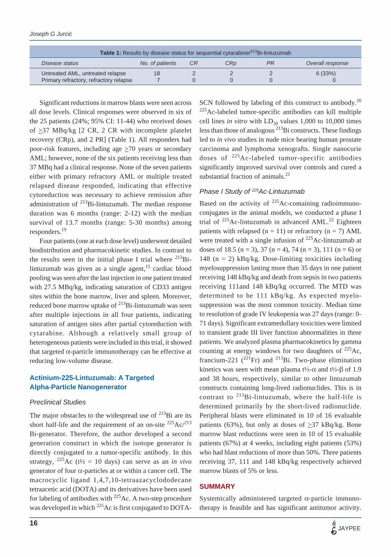
Significant reductions in marrow blasts were seen acrossall dose levels. Clinical responses were observed in six ofthe 25 patients (24%; 95% CI: 11-44) who received dosesof >37 MBq/kg [2 CR, 2 CR with incomplete plateletrecovery (CRp), and 2 PR] (Table 1). All responders hadpoor-risk features, including age >70 years or secondaryAML; however, none of the six patients receiving less than37 MBq had a clinical response. None of the seven patientseither with primary refractory AML or multiple treatedrelapsed disease responded, indicating that effectivecytoreduction was necessary to achieve remission afteradministration of 213Bi-lintuzumab. The median responseduration was 6 months (range: 2-12) with the mediansurvival of 13.7 months (range: 5-30 months) amongresponders.19
Four patients (one at each dose level) underwent detailedbiodistribution and pharmacokinetic studies. In contrast tothe results seen in the initial phase I trial where 213Bi-lintuzumab was given as a single agent,15 cardiac bloodpooling was seen after the last injection in one patient treatedwith 27.5 MBq/kg, indicating saturation of CD33 antigensites within the bone marrow, liver and spleen. Moreover,reduced bone marrow uptake of 213Bi-lintuzumab was seenafter multiple injections in all four patients, indicatingsaturation of antigen sites after partial cytoreduction withcytarabine. Although a relatively small group ofheterogeneous patients were included in this trial, it showedthat targeted -particle immunotherapy can be effective atreducing low-volume disease.
Actinium-225-Lintuzumab: A TargetedAlpha-Particle Nanogenerator
Preclinical Studies
The major obstacles to the widespread use of 213Bi are itsshort half-life and the requirement of an on-site 225Ac/213
Bi-generator. Therefore, the author developed a secondgeneration construct in which the isotope generator isdirectly conjugated to a tumor-specific antibody. In thisstrategy, 225Ac (t½ = 10 days) can serve as an in vivogenerator of four -particles at or within a cancer cell. Themacrocyclic ligand 1,4,7,10-tetraazacyclododecanetetraacetic acid (DOTA) and its derivatives have been usedfor labeling of antibodies with 225Ac. A two-step procedurewas developed in which 225Ac is first conjugated to DOTA-
SCN followed by labeling of this construct to antibody.20
225Ac-labeled tumor-specific antibodies can kill multiplecell lines in vitro with LD50 values 1,000 to 10,000 timesless than those of analogous 213Bi constructs. These findingsled to in vivo studies in nude mice bearing human prostatecarcinoma and lymphoma xenografts. Single nanocuriedoses of 225Ac-labeled tumor-specific antibodiessignificantly improved survival over controls and cured asubstantial fraction of animals.21
Phase I Study of 225Ac-Lintuzumab
Based on the activity of 225Ac-containing radioimmuno-conjugates in the animal models, we conducted a phase Itrial of 225Ac-lintuzumab in advanced AML.22 Eighteenpatients with relapsed (n = 11) or refractory (n = 7) AMLwere treated with a single infusion of 225Ac-lintuzumab atdoses of 18.5 (n = 3), 37 (n = 4), 74 (n = 3), 111 (n = 6) or148 (n = 2) kBq/kg. Dose-limiting toxicities includingmyelosuppression lasting more than 35 days in one patientreceiving 148 kBq/kg and death from sepsis in two patientsreceiving 111and 148 kBq/kg occurred. The MTD wasdetermined to be 111 kBq/kg. As expected myelo-suppression was the most common toxicity. Median timeto resolution of grade IV leukopenia was 27 days (range: 0-71 days). Significant extramedullary toxicities were limitedto transient grade III liver function abnormalities in threepatients. We analyzed plasma pharmacokinetics by gammacounting at energy windows for two daughters of 225Ac,francium-221 (221Fr) and 213Bi. Two-phase eliminationkinetics was seen with mean plasma t½- and t½- of 1.9and 38 hours, respectively, similar to other lintuzumabconstructs containing long-lived radionuclides. This is incontrast to 213Bi-lintuzumab, where the half-life isdetermined primarily by the short-lived radionuclide.Peripheral blasts were eliminated in 10 of 16 evaluablepatients (63%), but only at doses of >37 kBq/kg. Bonemarrow blast reductions were seen in 10 of 15 evaluablepatients (67%) at 4 weeks, including eight patients (53%)who had blast reductions of more than 50%. Three patientsreceiving 37, 111 and 148 kBq/kg respectively achievedmarrow blasts of 5% or less.
SUMMARY
Systemically administered targeted -particle immuno-therapy is feasible and has significant antitumor activity.
Table 1: Results by disease status for sequential cytarabine/213Bi-lintuzumab
Disease status No. of patients CR CRp PR Overall response
Untreated AML, untreated relapse 18 2 2 2 6 (33%)Primary refractory, refractory relapse 7 0 0 0 0

Targeted Alpha-Particle Immunotherapy with Bismuth-213 and Actinium-225 for Acute Myeloid Leukemia
JPMER
Journal of Postgraduate Medicine, Education and Research, January-March 2013;47(1):14-17 17
The shorter range and higher linear energy transfer of-particles compared with -particles may allow for moreefficient and selective killing of individual tumor cells.These physical properties suggest that radioimmunotherapywith -emitters may be best suited for the treatment of small-volume disease, as borne out in these clinical trials. Althoughreductions in leukemic blasts were seen when both 213Bi-and 225Ac-lintuzmab were given as single agents in phase Itrials, remissions were only seen after effective cyto-reduction. The use of 225Ac can overcome the logisticaldifficulties associated with short-lived radionuclides suchas 213Bi. Building on the encouraging results seen with 213Bi-lintuzumab for cytoreduced leukemia, we are nowconducting a multicenter phase I/II trial of 225Ac-lintuzumabin combination with low-dose cytarabine for elderly patientswith untreated AML. These studies provide the rationalefor further investigation of targeted -particleimmunotherapy for minimal residual disease or small-volume disease in a variety of malignancies.
REFERENCES
1. Cassileth PA, Harrington DP, Appelbaum FR, Lazarus HM,Rowe JM, Paietta E, et al. Chemotherapy compared withautologous or allogeneic bone marrow transplantation in themanagement of acute myeloid leukemia in first remission. NEngl J Med 1998;339:1649-56.
2. Stockerl-Goldstein KE, Blume KG. Allogeneic hematopoieticcell transplantation for adult patients with acute myeloidleukemia. In: Thomas ED, Blume KG, Forman SJ (Eds).Hematopoietic cell transplantation. Oxford: Blackwell Science,Inc 1999;823-34.
3. Howlader N, Noone AM, Krapcho M, Neyman N, Aminou R,Altekruse SF, et al. SEER Cancer Statistics Review, 1975-2009(Vintage 2009 Populations), National Cancer Institute. Bethesda,MD, http://seer.cancer.gov/csr/1975_2009_pops09/, based onNovember 2011 SEER data submission, posted to the SEERweb site, April 2012.
4. Burke JM, Caron PC, Papadopoulos EB, Divgi CR, Sgouros G,Panageas KS, et al. Cytoreduction with iodine-131-anti-CD33antibodies before bone marrow transplantation for advancedmyeloid leukemias. Bone Marrow Transplant 2003;32:549-56.
5. Jurcic JG, Divgi CR, McDevitt MR, Ma D, Sgouros G, FinnRD, et al. Potential for myeloablation with yttrium-90-HuM195(anti-CD33) in myeloid leukemia. Proc Am Soc Clin Oncol2000;19:8a (abstr 24).
6. McDevitt MR, Sgouros G, Finn RD, Humm JL, Jurcic JG,Larson SM, et al. Radioimmunotherapy with alpha-emittingnuclides. Eur J Nucl Med 1998;25:1237-47.
7. Andrews RG, Torok-Storb B, Bernstein ID. Myeloid-associateddifferentiation antigens on stem cells and their progeny identifiedby monoclonal antibodies. Blood 1983;62:124-32.
8. Griffin JD, Linch D, Sabbath K, Larcom P, Schlossman SF. Amonoclonal antibody reactive with normal and leukemic humanmyeloid progenitor cells. Leuk Res 1984;8:521-34.
9. Caron PC, Co MS, Bull MK, Avdalovic NM, Queen C,Scheinberg DA. Biological and immunological features of
humanized M195 (anti-CD33) monoclonal antibodies. CancerRes 1992;52:6761-67.
10. Caron PC, Jurcic JG, Scott AM, Finn RD, Divgi CR, GrahamMC, et al. A phase 1B trial of humanized monoclonal antibodyM195 (anti-CD33) in myeloid leukemia: Specific targetingwithout immunogenicity. Blood 1994;83:1760-68.
11. Jurcic JG, DeBlasio T, Dumont L, Yao TJ, Scheinberg DA.Molecular remission induction with retinoic acid and anti-CD33monoclonal antibody HuM195 in acute promyelocytic leukemia.Clin Cancer Res 2000;6:372-80.
12. Caron PC, Dumont L, Scheinberg DA. Supersaturatinginfusional humanized anti-CD33 monoclonal antibody HuM195in myelogenous leukemia. Clin Cancer Res 1998;4:1421-28.
13. Feldman E, Kalaycio M, Weiner G, Frankel S, Schulman P,Schwartzberg L, et al. Treatment of relapsed or refractory acutemyeloid leukemia with humanized anti-CD33 monoclonalantibody HuM195. Leukemia 2003;17:314-18.
14. Raza A, Jurcic JG, Roboz GJ, Maris M, Stephenson JJ, WoodBL, et al. Complete remissions observed in acute myeloidleukemia following prolonged exposure to lintuzumab: A phase1 trial. Leuk Lymph 2009;50:1336-44.
15. McDevitt MR, Finn RD, Ma D, Larson SM, Scheinberg DA.Preparation of -emitting 213Bi-labeled antibody constructs forclinical use. J Nucl Med 1999;40:1722-27.
16. Nikula TK, McDevitt MR, Finn RD, Wu C, Kozak RW,Garmestani K, et al. Alpha-emitting bismuth cyclohexylbenzylDTPA constructs of recombinant humanized anti-CD33antibodies: Pharmacokinetics, bioactivity, toxicity andchemistry. J Nucl Med 1999;40:166-76.
17. Jurcic JG, Larson SM, Sgouros G, McDevitt MR, Finn RD,Divgi CR, et al. Targeted alpha particle immunotherapy formyeloid leukemia. Blood 2002;100:1233-39.
18. Sgouros G, Ballangrud ÅM, Jurcic JG, McDevitt MR, HummJL, Erdi YE, et al. Pharmacokinetics and dosimetry of an-particle emitter labeled antibody: 213Bi-HuM195 (anti-CD33)in patients with leukemia. J Nucl Med 1999;40:1935-46.
19. Rosenblat TL, McDevitt MR, Mulford DA, Pandit-Taskar N,Divgi CR, Panageas KS, et al. Sequential cytarabine and-particle immunotherapy with bismuth-213-lintuzumab(HuM195) for acute myeloid leukemia. Clin Cancer Res2010;16:5303-11.
20. McDevitt MR, Finn RD, Sgouros G, Ma D, Scheinberg DA. An225Ac/213Bi generator system for therapeutic clinicalapplications: Construction and operation. Appl Rad Isotopes1999;50:895-904.
21. McDevitt MR, Ma D, Lai LT, Simon J, Borchardt P, Frank RK,et al. Tumor therapy with targeted atomic nanogenerators.Science 2001;294:1537-40.
22. Jurcic JG, Rosenblat TL, McDevitt MR, Pandit-Taskar N,Carrasquillo JA, Chanel SM, et al. Phase I trial of the targetedalpha-particle nano-generator actinium-225 (225Ac)-lintuzumab(anti-CD33; HuM195) in acute myeloid leukemia (AML). J ClinOncol 2011;29:(suppl; abstr 6516).
ABOUT THE AUTHOR
Joseph G JurcicProfessor, Department of Clinical Medicine; Director, HematologicMalignancies, Columbia University Medical Centre, 177 Fort Washington6-435 New York, NY-10032, USA, e-mail: [email protected]

18JAYPEE
Frank RöschREVIEW ARTICLE
68Ge/68Ga Generators and 68Ga RadiopharmaceuticalChemistry on Their Way into a New CenturyFrank Rösch
ABSTRACT68Ga faces a renaissance initiated by the development of new68Ge/68Ga radionuclide generators, sophisticated 68Garadiopharmaceuticals, preclinical research and state-of-the-artclincial diagnoses via positron emission tomography/computedtomography (PET/CT). A new type of 68Ge/68Ga generatorbecame commercially available in the first years of the 21stcentury, with eluates based on hydrochloric acid. Thesegenerators provided ‘cationic’ 68Ga instead of ‘inert’ 68Ga-complexes, and opened new pathways of MeIII radio-pharmaceutical chemistry. The last decade has seen a 68Garush. Increasing interest in generator-based 68Ga radio-pharmaceuticals in diagnostic applications has beenaccompanied by its potential use in the context of dieasetreatment planning, made possible by the inherent optionexpressed by theranostics. However, widespread acceptanceand clinical application requires optimization of 68Ge/68Gagenerators both from chemical and regulatory perspectives.
Keywords: 68Ga, 68Ge, Generator, Ligands.
How to cite this article: Rösch F. 68Ge/68Ga Generators and68Ga Radiopharmaceutical Chemistry on Their Way into a NewCentury. J Postgrad Med Edu Res 2013;47(1):18-25.
Source of support: Nil
Conflict of interest: None declared
INTRODUCTION
The 68Ge/68Ga radionuclide generator, with its secularequilibrium mathematics, offers a perfect combination ofthe nuclidic parameters in terms of half-lives and emissionprofiles: t½ = 270.95 days for 68Ge and t½ = 67.71 minutesfor 68Ga, with no photon emission for 68Ge and an 89.14%positron branching for 68Ga.10,46 This was known alreadyin the middle of the 20th century, yet gallium-68 today seesa renaissance, with the development of new68Ge/68Ga radionuclide generators, sophisticated 68Garadiopharmaceuticals, and state-of-the-art clincial diagnosesvia positron emission tomography/computed tomography(PET/CT).43 Current advances represent a ‘renaissance’because 68Ga is one of the very early radionuclides appliedto PET imaging. Its application preceeds the use of fluorine-18 and even the term ‘positron emission tomorgaphy’.Moreover, the availability of this positron emitter via thefirst 68Ge/68Ga generators,16,17 lead to the development ofthe first positron scintillation camera which was created inthe beginning of the 1960s.
With the availability of the first 68Ge/68Ga generators(which provided 68Ga-EDTA eluates) and dramatically
10.5005/jp-journals-10028-1052
improved tomographic detection systems, several 68Gatracers for imaging of various diseases were investigated(mainly for imaging the human brain). Hundreds of patientswere investigated in the USA using 68Ga-EDTA, and othersfrom 1963 on.
Despite several publications describing ‘improved’ 68Ge/68Ga radionuclide generators, the impact of 68Ga imagingsubsided in the late 1970s. This was primarily a consquenceof two genertaor-related factors. Firstly, the generator designwas inadequate for the versatile synthesis of 68Ga radio-pharmaceuticals. Secondly, in view of the parallel and rapiddevelopments of the new classes of 99mTc- and 18F-labeleddiagnostics, the 68Ge/68Ga generators had only minorclinical relevance. Nevertheless, numerous papers in the1970s and 1980s described the use of inorganic matrixesand organic resins, which allow for the isolation of68Ga from 68Ge within hydrochloric acid solutions of weak(0.1-1.0 N) or strong (>1 N) concentrations respectively.
Pioneering achievement of radiochemists in Obninsk,Russia, resulted in the development of a new type of 68Ge/68Ga generator which became commercially available in thefirst years of the 21st century.40 Generator eluates based onhydrochloric acid provided ‘cationic’ 68Ga instead of ‘inert’68Ga-complexes, opening new pathways of MeIII basedradiopharmaceutical chemistry. Initially, the 68Ga cation wasintroduced into existing ligands used for magnetic resonanceimaging (MRI) and SPECT imaging probes, such as DTPA-or DOTA-based derivatives. The impressive resultsachieved using 68Ga-DOTA-octreotides for PET/CTcompared to 111 In-DTPA-octroescan paved the way towardthe clinical acceptance of this particular tracer for imagingneuroendocrine tracers, and highlighted the great potentialof the 68Ge/68Ga generator for modern nuclear medicinein general.
These advances intiated a 68Ga rush in recent times (post2002). However, the widespread acceptance and clinicalapplication of 68Ga radiopharmaceuticals requiresoptimization of 68Ge/68Ga generators both from chemicaland regulatory points of view. Furthermore, dedicatedchelators are required to broaden the possibilities of 68Galabeling to allow the use of more sensitive targeting vectors.Last but not least, this should also involve applying theconcept of 68Ga-radiopharmaceutical chemistry to anincreasing number of targeting vectors, addressing theclinically most relevant diseases.

Journal of Postgraduate Medicine, Education and Research, January-March 2013;47(1):18-25 19
68Ge/68Ga Generators and 68Ga Radiopharmaceutical Chemistry on Their Way into a New Century
JPMER
With current innovation and the favorable properties ofthe 68Ga radionuclide, it is possible that in another decadefrom now 68Ge/68Ga generator-based 68Ga diagnostics mayapproach a top three ranking in imaging (together with99mTc- and 18F-based tracer diagnostics).
This paper includes material which was presented at the1st World Congress on Ga-68 and peptide receptorradionuclide therapy (PRRNT) theranostics-on the way topersonalized medicine, Bad Berka, Germany, Junge 23-26,2011, and which were published at a later stage.5
THE EARLY YEARS (1960-1970):THE SUNRISE OF 68Ga
The First 68Ge/68Ga Radionuclide Generators
The first 68Ge/68Ga radionuclide generator was describedin 196016 and entitled: ‘A positron cow’. As the title elicits,the concept was to use a radionuclide generator for theproduction of a positron emitting radionuclide. The latterwas a new entry for radiopharmaceutical chemistry andnuclear medicine molecular imaging in vivo. The generatorchemistry involved a liquid-liquid extraction, and the wholeprocessing protocol was considerably different to that ofcurrent radionuclide generator systems. Nevertheless, avariety of 68Ga compounds were synthesized using thisgenerator design.2,7,44
Further Generator Developments: Al2O3-basedEDTA-Eluted Generators
Inherent disadvantages of the first generator lead to thedevelopment of two improved generator concepts soon after.The liquid-liquid extraction chemistry introduced byGleason was substituted for a solid phase-based ionexchange system17,50 (Fig. 1). In addition, a generatorfeaturing an improved liquid-liquid extraction was describedlater.15
The original sketch, taken from the original publicationby Yano and Anger (1964) for the second solid-phase basedgenerator is reproduced in Figure 2. These solid-phasechromatographic generators offered excellent radiochemicalcharacteristics. Using an alumina column and EDTA aseluent (10 ml 0.005 M EDTA), 68Ga was easily eluted in areproducible 95% yield without the need to introduce stableGaIII as carrier. The eluate contained as little as 1.4 × 10⁻⁵%of the parent 68Ge. Prior to in vivo injection, 0.5 ml of 18%NaCl solution was added to the eluate.
68Ge/68Ga Generators and the Development ofPositron Scintillation Cameras
This system served as a convenient and economical sourceof 68Ga-EDTA. Effectively, this radionuclide generator wasa synthesis unit of a relevant radiopharmaceutical; 68Ga-
EDTA (named ‘veronate’ at the time). 68Ga-EDTA, andlimited other 68Ga-tracers, were adapted for humanapplication quite quickly by various groups in the UnitedStates for early applications.20,21,44 Systematic applicationfor brain imaging was reported, with medical impact havingsignificant dependence on the method of detection applied.Conventional imaging appeared to be relatively difficult,with relatively high dosage of 68Ga-EDTA required forvaluable medical information to be gained.
Anger thus, started to develop the basics of positronimaging tomography2,22,23 (arguing as follows (Gottschalkand Anger 1964): … ‘We seriously question whethersatisfactory results can be obtained with the conventionalpositron scanner. Recent phantom studies indicate that thepositron scintillation camera using 68Ga-EDTA will detectlesions 1/2 the volume that can be detected by theconventional positron scanner using As74. The increase insensitivity is obtained even though the phantom was set upto simulate our clinical condition where brain pictures are
Fig. 1: Early progress in 68Ge/68Ga radionuclide generators II:From liquid-liquid extraction to solid phase-based elution
Fig. 2: Solid phase-based (alumina) 68Ga elution (using an EDTAsolution) reproduced from the original publication by Yano and Anger(1964)

20JAYPEE
Frank Rösch
obtained in 4 to 10 minutes with a dose of 350 to 750microcuries of 68Ga-EDTA. Shealy et al, however, foundthat 2 to 3 millicuries of 68Ga-EDTA was sometimes aninadequate dose with their positron scanner.’ Imagesrecorded with this new type of camera (Fig. 3)2 paved theway for routine PET imaging.
68Ga-EDTA: The Prototype PET-Pharmaceutical
Despite these new imaging features and the great successof 68Ga-EDTA molecular imaging, the fact that in practicethe generator was limited to 68Ga-EDTA was a severelimitation. The extraction of 68Ga from the thermo-dynamically very stable (log K = 21.7) 68Ga-EDTA eluatespecies was not straightforward. Yano and Anger 1964reported that, ‘attempts are being made. .. to free 68Ga ...from the EDTA complex’. A procedure was developedhowever; it was not user friendly and practical for 68Ga. Ona scale which uses 10 mg Ga carrier, the time required forextraction is 30 minutes, and the transfer yield of 60%.50
The protocol was:1. The cow is milked with 10 ml of 0.005 M EDTA solution,
and the 68Ga is collected in a 40 ml centrifuge tube.2. The 10 to 20 mg of carrier GaCl3 in HCl solution is added.3. The 0.5 ml of saturated ammonium acetate solution is
added.4. Concentrated NH4OH is added dropwise (about 1 ml)
to precipitate Ga(OH)3 at pH 6.0.5. The solution is heated in a boiling water bath for 10 minutes
to coagulate the Ga(OH)3.6. The solution is centrifuged, and the supernatant solution
is discarded.7. The Ga(OH)3 is dissolved with a minimum volume of
hot 20% NaOH.8. The solution is acidified with about 1 ml of concentrated
HCl.
HIBERNATING 68Ga MEDICAL APPLICATIONS,BUT NEW CHEMISTRY AHEAD
The impact of 68Ga imaging started to subside in the late1970s, for two main reasons. Firstly, the generator designwas inadequate in terms of the requirements for versatilesynthesis of 68Ga radiopharmaceuticals. Secondly, in viewof the parallel and rapid developments of the new classesof 99mTc- and 18F-labeled diagnostics, the generatorsavailable through the existing technology had only minorclinical relevance.
Despite this apparent decrease in interest, numerousbasic radiochemical papers in the 1970s and 1980s describedthe use of inorganic matrixes as well as organic resins,selectively adsorbing 68Ge and providing 68Ga desorbtionswithin hydrochloric acid solutions of weak (0.1-1.0 N) orstrong (>1 N) concentrations respectively.
Cationic 68Ga eluates are required to facilitate theversatile radiolabeling chemistry with 68Ga. Thus, theprimary challenge is the development of separation systemswhich provide cationic 68Ga species. GaIII exists as cationicspecies (either pure water-hydrated aquocomplexes, suchas the hexa-aqua cation Ga(H2O)6
3+, or similar monochloroor monohydroxo species). This speciation is easily achievedin solutions of hydrochloric acid of pH ranging between 0and 2 (0.01-1.0 N HCl). For this purpose, MeIVO2
– typematrixes (Me = Sn, Ti, Zr, Ce, etc.) appeared to be adequate,because they effectively adsorb the parent radionuclide68GeIV.1,25,26,33,39 Alternatively, organic resins have beendeveloped which require more concentrated HCl soltuionsfor eluting the 68Ga.3,45 Figure 4 gives a schematic overview.
COMMERCIAL ‘IONIC’ GENERATORS
Generator Eluates Delivering the Gallium Cation
Thanks to the pioneering achievement of radiochemists inObninsk (Russian Federation), a new type of 68Ge/Ga
Fig. 3: A 68Ga-EDTA brain scan acquired with the Anger positron camera circa 1962 showing the tomographic capability. The braintumor is in best focus in the left image, taken at about the level of the temporal horn2

Journal of Postgraduate Medicine, Education and Research, January-March 2013;47(1):18-25 21
68Ge/68Ga Generators and 68Ga Radiopharmaceutical Chemistry on Their Way into a New Century
JPMER
generator became commercially available in the first yearsof the 21st century.40 These generators use eluting soltuionsbased on hydrochloric acid which provide ‘cationic’ 68Ga,as opposed to ‘inert’ 68Ga-complexes, opening newpathways of MeIII-based radiopharmaceutical chemistry(Fig. 5).
The 68Ga cation was immediately introduced intoexisting ligand designs of MRI and SPECT imaging probes,namely DTPA- or DOTA-based derivatives. The impressivesuccess of utilizing 68Ga-DOTA-octreotides and PET/CTinstead of, e.g. 111In-DTPA-octroescan paved the way forclinical acceptance of this particular tracer for imagingneuroendocrine tumors, but also to the realization of thegreat potential of the 68Ge/68Ga generator for modern nuclearmedicine in general. While commercial ‘ionic’ generatorshad successfully entered clinical environments, there werequestions regarding its suitability, which became morerelevant. In particular these related to its adequacyconcerning radiation safety, legal requirements and labelingof medical tracers became more and more relevant. Themost relevant concerns are outlined:• Problem 1: The long physical half-life of the parent in
principle should give a generator shelf-life of at least1 year. However, the shelf-life of the generators did notnecessarily parallel this long physical half-life due inparticular to increasing breakthrough of 68Ge, but alsodecreasing 68Ga elution yield. 68Ge breakthroughreduction and/or removal of 68Ge from the eluatestherefore remain an important radiochemical challenge.
• Problem 2: 68Ga generator eluates are not chemicallyor radiochemically pure. Nonradioactive metals, suchas 68ZnII (as generated on the generator as decay productof 68Ga), FeIII as general chemical impurity, and 68GeIV
as breakthrough represent metals, which may compete
with 68GaIII for coordinative labeling of radiopharma-ceutical precursors. Again, this illustrates the importanceof minimizing the 68Ge content in the eluate.4,51
• Problem 3: The new generation of 68Ge/68Ga radio-nuclide generators utilize hydrochloric acid solutionsfor 68Ga elution. The relatively acidic environmentcreated many protonated functional groups of ligandsand bifunctional ligands needed for the labeling of 68Ga,which may hinder efficient radiolabeling. Finally,minimizing the pH and volume of 68Ga eluted prior tolabeling should facilitate higher radiolabeling yields.
Generator Post-Processing
Three approaches have been developed to address one ormore of these problems. Two processes include chemicalseparation strategies, which may be referred to as ‘post-processing’.35,51 The third technology involves a simplefractionation of the eluate, i.e. isolating eluate fractions withhighest 68Ga concentration.11 The methods are schematicallyillustrated in Figure 6.
In most cases, commercial generators are used in directconnection with one of the three postelution processingtechnologies mentioned. The cation exchange-based post-processing4,51 guarantees almost complete removal of themetallic impurities, in particular 68Ge. Numerousmodifications have been reported, including NaCl solutionsinstead of the solution no. 2 to desorb 68Ga from the resin,36
or by incorporating a subsequent anion exchange-basedpurification step32 to remove organic solvent prior tolabeling.
CURRENT STATE/OUTLOOK
Today, 68Ge/68Ga radionuclide generators are commerciallyavailable as TiO2–, SnO2– or organic resin-based columns.68Ga eluate yields range from about 70 to 80% for freshgenerators, with a decrease overtime. 68Ge breakthroughlevels vary between 0.01 and 0.001% (or even less) for freshgenerators, with these percentages increasing over extendedperiods of generator usage. Conjugated with post-processing
Fig. 4: 68Ge/68Ga radionuclide generator concepts developed inthe 1970s and 1980s toward ‘cationic’ generators
Fig. 5: Electrophoresis of a 0.1 N HCl 68Ga generator eluate (EZAGObninsk generator) demonstrating the presence of ‘cationic’ 68Ga(parameters: 0.1 HCl, Whatman® paper strip, l = 19 cm, t =5 minutes, 191 V, 210 mA, 40 W)

22JAYPEE
Frank Rösch
technologies, 68Ga radiopharmaceuticals are beingsynthesized routinely and safely. Thus, since the early 68Ge/68Ga radionuclide generator systems developed about halfa century ago, significant advances have been made.
These generator improvements have allowed for thesignificant development of 68Ga radiopharmaceuticalchemistry within the last decade. Despite this, almost allthe technological and chemical innovation involved belongsto the 20th century. There is room for further development,where several aspects of generator design and performance,labeling chemistry and clinical application need to beaddressed. Figure 7 illustrates some of the potential futuredirections.
Generators
Concerning solid phase-based ion exchange chromato-graphic 68Ge/68Ga radionuclide generators, someimprovements may be possible within the resin materialitself. Recent publications hint at the potential ofsophisticated nanoparticles, such as ZrIV and CeIV-systems,which are classified as nanocomposites.12,13 The rational isthat these composites may provide effective adsorption of68Ge, effective release of 68Ga, be more chemically stabilityand radiation resistant. In parallel, GMP-certified andlicensed commercial generators are required to satisfy theincreasing standards of legal authorities.
Generator Online Post-Processing
Elution of generators may be further integrated into fasterand more efficient online post-processing procedures, which
are managed by automated modules. A key issue in thisregard, is to avoid the transfer of 68Ge into 68Ga-radio-pharmaceuticals. Optionally, these post-processingtechnologies should also allow for versatile labelingprotocols. For example, the transfer from aqueous tononaqueous solutions for radiolabeling (addressing potentiallipophilic 68Ga tracers)53 or onto resin for solid phasesupported labeling reactions.
Post-processing technologies, which remove 68Ge onlinefrom the eluate, are of utmost importance, as they avoid thetransfer of critical 68Ge levels into the radiopharmaceuticalpreparation. They also guarantee the safety, which isrelevant from the legal point of view, i.e. addressing safetycriteria of routine clinical use.9 Although some of the 68Garadiopharmaceuticals used clinically, in particular 68Ga-based peptides, are purified from uncomplexed 68Ga, (whichsimultaneously removes 68Ge present), the principal strategyshould be to keep generator-derived 68Ga solutions free of68Ge before labeling. Consequently, the monographs of theEuropean Pharmacopoeia (Ph Eur) in its description of thegallium chloride (68Ga) solution for radiolabeling,Monograph N°: 2464, Strasbourg, June 2012, adds, that‘…the solution is intended for use in the preparation ofgallium-68-labeled radiopharmaceuticals, including aprocedure to reduce the level of germanium-68 below0.001% of the total radioactivity.’ This means that,necessarily, the procedure for preparation of a Ga-68 radio-pharmaceutical has to include a procedure to removegermanium-68 up to a level below 0.001%.
Only this strategy will be suitable for a kit-type 68Ga-labeling approach to parallel the 99mTc analog systems. Thiswould allow for the direct synthesis and application of 68Garadiopharmaceuticals, such as 68Ga chloride,49 68Ga
Fig. 6: Schematic representation showing an overview of post-processing technologies for commercial 68Ge/68Ga radionuclidegenerators: (1) Direct generator elution through cation-exchangecartridge, (2) desorption of purified 68Ga using HCl/acetone or HCl/ethanol mixtures, (3) generator elution into HCl reservoir,(4) subsequently elution through anion-exchange cartridge,(5) desorption of purified 68Ga using water, (6) identification of theeluate fraction representing at least two-third of the 68Ga activity,and use without further purification
Fig. 7: Sketch of some future directions related to 68Ge/68Garadionuclide generators and radiopharmaceuticals

Journal of Postgraduate Medicine, Education and Research, January-March 2013;47(1):18-25 23
68Ge/68Ga Generators and 68Ga Radiopharmaceutical Chemistry on Their Way into a New Century
JPMER
citrate28,37 (Rizello et al 2009), 68Ga apotransferrin27 or 68GaSchiff base complexes.18,19 Radiotracers, such as these areotherwise not applicable due to the nonseparable contentof 68Ge.
Ligands
The future development of new 68Ga radiopharmaceuticalsmay be facilitated by the development of new ligands andbifunctional derivatives for coordinating 68Ga specifically,i.e. ideally discriminating FeIII and ZnII, or by allowingcomplex formation under a broader range of pH. Anotherrelevant aspect is the development of ligands which complex68Ga at room temperature. Such radiolabeling characteristicsapproach the advantages of 99mTc kit-type labelingprotocols. It is also desirable to speed up complex formationand minimize the amount of labeling precursor needed,thereby increasing the specific activities of the finalradiolabeled product. Ideally new ligands should label moreefficiently than that of established DOTA or NOTAderivatives, without detriment to complex stability.Important criteria (in addition to high radiochemical yields)are listed in Table 1. Several of the promising ligandcandidates for 68Ga radiolabeling are listed in Figure 8.
Current trends in bifunctional ligand design suggest achange in the paradigm that macrocyclic chelates are theligands to go for. Recent developments describe modifiedacyclic ligands. In particular, recent ligands are derived fromknown FeIII ligands, because the two metals have similarcoordination chemistry. New classes of chelators currentlyunder development, include acyclic ligands HBED,14,48
H2DEDPA7,8,24 and tris(hydroxypyridinone) ligands,6,53 butalso research on deferoxamine34 and on sulfur-basedderivatives30 continues.
New cyclic triazacyclononane-phosphinic acidchelators38,39 have been developed, which complex 68Gavery effectively. In case of the triazacyclononane-phosphinic acid chelators (TRAP), the idea is also to createan inert coordinating core leaving three linkablefunctionalities available for versatile chemistry, allowingfor multimeric substitutions.
68Ga Radiopharmaceuticals
Novel ligand design presents the opportunity for a widerange of new tracers. The clinical application, however, willfinally depend on the classes of targeting vectors attached,beyond peptidic and nonpeptidic targeting vectors available.Imaging will hopefully address tumors, infection andinflammation, but also a variety of clinical indications andalmost all organs. This would mirror the 99mTc radio-pharmaceuticals, e.g. brain, heart, etc. In the context of thesimilarity of generator based 99mTc and 68Ga pharma-ceuticals, the preparation of those 68Ga radiopharmaceuticalsshould also be KIT-based if they are to find clinicalapplication and widespread acceptance. These developmentswill contribute to a much more intense clinical use of 68Ge/68Ga generators and the corresponding 68Ga pharmaceuticalsfor molecular imaging. Again, legal considerations applyto both the generator and the pharmaceuticals.9
Theranostics
Simultaneously to the further development of 68Ga-PECT/CT diagnostics, it is one of the unique features of 68Ga, that68Ga-PET/CT imaging may be directly linked to treatmentoptions. For some classes of GaIII bifunctional ligands, thereshould be an option to synthesize therapeutics analogs withtrivalent radiometals, such as 90Y, 177Lu, 213Bi, etc. TheDOTA-conjugated octreotide derivatives represent a perfectexample of the success of this theranostic concept.42
Table 1: Challenges for new 68Ga ligand developments
Efficient labeling should occur:• At temperatures below 100°, approaching room temperature• Over a broad range of pH, i.e. covering the pH of the generator
eluate up to physiological pH• Within short periods, i.e. within 10 minutes or less• At low amounts/concentration of the ligands (10 M or less)• In the presence of impurities, such as FeIII (as a general
impurity) and ZnII (as decay product of 68Ga), etc.
Fig. 8: Recent developments in ligand structurestailored for 68Ga

24JAYPEE
Frank Rösch
REFERENCES
1. Ambe S. Germanium-68-gallium-68 generator with alpha-ferricoxide support. Appl Radiat Isot 1988;39:49.
2. Anger HO, Gottschalk A. Localization of brain tumors with thepositron scintillation camera. J Nucl Med 1963;4:326.
3. Arino H, Skraba WJ, Kramer HH. A new 68Ge/68Ga radioisotopegenerator system. Int J Appl Radiat Isot 1978;29:117.
4. Asti M, De Pietri G, Fraternali A, Grassi E, Sghedoni R,Fioroni F, et al. Validation of 68Ge/68Ga generator processingby chemical purification for routine clinical application of 68Ga-DOTATOC. Nucl Med Biol 2008;35:721.
5. Baum RP, Roesch F (Eds). Theranostics, gallium-68, and otherradionuclides. A pathway to personalized diagnosis andtreatment. Recent results in cancer research. Springer,Heidelberg: New York, Dordrecut, London 2103;194:3-16.
6. Berry DJ, Ma Y, Ballinger JR, Tavaré R, Koers A, Sunassee K,et al. Efficient bifunctional gallium-68 chelators for positronemission tomography: Tris (hydroxypyridinone) ligands. ChemCommun 2011;47:7068.
7. Boros E, Ferreira CL, Patrick BO, Adam MJ, Orvig C. New Gaderivatives of the H2dedpa scaffold with improved clearanceand persistent heart uptake. Nucl Med Biol 2011;38:1165.
8. Boros E, Ferreira CL, Yapp DTT, Gill RK, Price EW, Adam MJ,et al. RGD conjugates of the H2dedpa scaffold: Synthesis,labeling and imaging with 68Ga. Nucl Med Biol 2012;39:785.
9. Breeman WA, Verbruggen AM. The 68Ge/68Ga generator hashigh potential, but when can we use 68Ga-labelled tracers inclinical routine? Eur J Nucl Med Mol Imag 2007;34:978.
10. Burrows TW. Nuclear Data Sheets for A = 6.8. Nuclear DataSheets 2002;97:1-127.
11. Breeman WAP, de Jong M, de Blois E, Bernard BF,Konijnenberg M, Krenning EP. Radiolabelling DOTA-peptideswith 68Ga. Eur J Nucl Med Mol Imaging 2005;32:478.
12. Chakravarty R, Shukla R, Ram R, Venkatesh M, Dash A, TyagiAK. Nanoceria-PAN composite-based advanced sorbentmaterial: A major step forward in the field of clinical-grade 68Ge/68Ga generator. Appl Mat Interfaces 2010;2:2069-75.
13. Chakravarty R, Shukla R, Ram R, Tyagi AK, Dash A, VenkateshM. Development of a nano-zirconia based 68Ge/68Ga generatorfor biomedical application. Nucl Med Biol 2011;38:575.
14. Eder E, Wangler B, Knackmuss S, LeGall F, Little M,Haberkorn U, et al. Tetrafluorophenolate of HBED-CC: Aversatile conjugation agent for 68Ga-labeled small recombinantantibodies. Eur J Nucl Med Mol Imaging 2008;35:1878-86.
15. Erhardt GJ, Welch MK. A new germanium-68/gallium-68generator. J Nucl Med 1978;19:925-29.
16. Gleason GI. A positron cow. Int J Appl Radiat Isot 1960;8:90.17. Greene MW, Tucker WD. An improved gallium-68 cow. Int J
Appl Radiat Isot 1961;12:62.18. Hsiao YM, Mathias CJ, Wey SP, Fanwick PE, Green MA.
Synthesis and biodistribution of lipophilic and monocationicgallium radiopharmaceuticals derived from N,N’-bis (3-aminopropyl)-N, N’-dimethylethylenediamine: Potential agentsfor PET myocardial imaging with 68Ga. Nucl Med Biol 2009;36:39.
19. Tarkia M, Saraste A, Saanijoki T, Oikonen V, Vähäsilta T,Strandberg M, et al. Evaluation of 68Ga-labeled tracers for PETimaging of myocardial perfusion in pigs. Nucl Med Biol 2012;39:715.
20. Gottschalk A, Anger HO. The sensitivity of the positronscintillation camera for detecting simulated brain tumors withgallium-68 EDTA. Am J Roentgen 1964;92:174.
21. Gottschalk A. Anger HO. Letter to the editor. J Nucl Med1964;5:569.
22. Gottschalk A. The early years with Hal Anger. Sem Nucl Med1996;26:171.
23. Gottschalk A. Hal Anger: Nuclear medicine´s quiet genius.J Nucl Med 2004;45:13N, 26N.
24. Koop B, Reske SN, Neumaier B. Radiochim Acta 2007;95:39.25. Kopecky P, Mudrová B, Svoboda K. The study of conditions
for the preparation and utilization of 68Ge-68Ga generator. Int JAppl Radiat Isot 1973;24:73.
26. Kopecky P, Mudrová B. 68Ge-68Ga generator for the productionof 68Ga in an ionic form. Int J Appl Radiat Isot 1974;25:263.
27. Kumar V, Boddeti DK, Evans SG, Roesch F, Howman-Giles R. Potential use of 68Ga-apo-transferrin as a PET imagingagent for detecting Staphylococcus aureus infection. Nucl MedBiol 2011;38:393.
28. Kumar V, Boddeti DK, Evans SG, Angelides S. 68Ga-Citrate-PET for diagnostic imaging of infection in rats and for intra-abdominal infection in a patient. Curr Radiopharm 2012;5:71.
29. Lewis RE, Camin LL. Germanium-68/Gallium-68 generator forthe one step elution of ionic gallium-68. J Label CompdsRadiopharm 1981;18:16.
30. Luyt LG, Katzenellenbogen JA. A Trithiolate tripodalbifunctional ligand for the radiolabeling of peptides withgallium(III). Bioconjugate Chem 2002;13:1140.
31. Loc´h C, Maziere B, Comar D. A new generator for ionicgallium-68. J Nucl Med 1980;21:171.
32. Loktionova NS, Belozub AN, Filosofov DV, Zhernosekov KP,Wagner T, Türler A, et al. Improved column-basedradiochemical processing of the generator produced 68Ga. ApplRadiat Isot 2011;69:942.
33. Malyshev KV, Smirnov VV. Gallium-68 yield from hydratedzirconium oxide-based generators. Sov Radiochem 1975;17:137.
34. Mathias CJ, Lewis MR, Reichert DE, Laforest R, Sharp TL,Lewis JS, et al. Preparation of 66Ga- and 68Ga-labeled Ga(III)-deferoxamine-folate as potential folate-receptor-targeted PETradiopharmaceuticals. Nucl Med Biol 2003;30:725.
35. Meyer GJ, Mäcke HR, Schuhmacher J, Knapp WH, HofmannM. 68Ga-labelled DOTA-derivatised peptide ligands. Eur J NuclMed 2004;31:1097.
36. Mueller D, Klette I, Baum RP, Gottschaldt M, Schultz MK,Breeman WA. Simplified NaCl based 68Ga concentration andlabeling procedure for rapid synthesis of 68Ga radiopharma-ceuticals in high radiochemical purity. Bioconjug Chem 2012;23:1712.
37. Nanni C, Errani C, Boriani L, Fantini L, Ambrosini V, Boschi S,et al. 68Ga-citrate PET/CT for evaluating patients with infectionsof the bone: Preliminary results. J Nucl Med 2010;51:1932.
38. Notni J, Hermann P, Havlíèková J, Kotek J, Kubíèek V,Plutnar J, et al. A triazacyclononane-based bifunctionalphosphinate ligand for the preparation of multimeric 68Ga tracersfor positron emission tomography. Chemistry 2010;16:7174-85.
39. Notni J, Šimeèek J, Hermann P, Wester HJ. TRAP, a powerfuland versatile framework for gallium-68 radiopharmaceuticals.Chemistry 2011;17:14718-22.
40. Razbash AA, Sevastianov YuG, Krasnov NN, Leonov AI,Pavlekin VE. Germanium-68 row of products. Proceedings ofthe 5th International Conference on Isotopes, 5ICI, Brussels,Belgium April 25-29, Medimond, Bologna 2005;147-51.
41. Rizzello A, Di Pierro D, Lodi F, Trespidi S, Cicoria G,Pancaldi D, et al. Synthesis and quality control of 68Ga citratefor routine clinical PET. Nucl Med Commun 2009;30:542.

Journal of Postgraduate Medicine, Education and Research, January-March 2013;47(1):18-25 25
68Ge/68Ga Generators and 68Ga Radiopharmaceutical Chemistry on Their Way into a New Century
JPMER
42. Roesch F, Baum RP. Generator-based PET radiopharmaceuticalsfor molecular imaging of tumours: On the way to theranostics.Dalton Transactions 2011;40:6104-11.
43. Roesch F, Riss PJ. The renaissance of the 68Ge/68Ga radionuclidegenerator initiates new developments in 68Ga radio-pharmaceutical chemistry. Curr Top Med Chem 2010;10:1633-68.
44. Schaer LR, Anger HO, Gottschalk A. Gallium edetate 68Gaexperiences in brain-lesion detection with the positron camera.AMAM 1965;198:139.
45. Schumacher J, Maier-Borst W. A new 68Ge/68Ga radioisotopegenerator system for production of 68Ga in dilute HCl. Int JAppl Radiat Isot 1981;32:31.
46. Schönfeld E. 68Ge-68Ga. Comments on evaluation–CEA 1999.ISBN 27272 02111 3.
47. Shealy CN, Aronow S, Brownell GL. Gallium-68 as a scanningagent for intracranial lesions. J Nucl Med 1964;5:161.
48. Sun Y, Anderson C, Pajeau T, Reichert D, Hancock R,Motekaitis R, et al. Indium (III) and gallium (III) complexes ofbis(aminoethanethiol) ligands with different denticities:Stabilities, molecular modeling, and in vivo behavior. J MedChem 1996;39:458-70.
49. Ujula T, Salomäki S, Autio A, Luoto P, Tolvanen T,Lehikoinen P, et al. 68Ga-chloride PET reveals human pancreaticadenocarcinoma xenografts in rats-comparison with FDG. MolImaging Biol 2010;12:259.
50. Yano Y, Anger HO. A gallium-68 positron cow for medicaluse. J Nucl Med 1964;5:484.
51. Zhernosekov KP, Filosofov DV, Baum RP, Aschoff P, Bihl H,Razbash AA, et al. Processing of generator-produced 68Ga formedical application. J Nucl Med 2007;48:1741.
52. Zhou Z, Neubert H, Liu DY, Liu ZD, Ma YM, Kong XL, et al.Iron binding dendrimers: A novel approach for the treatment ofhaemochromatosis. J Med Chem 2006;49:4171-82.
53. Zoller F, Riss PJ, Montforts FP, Rösch F. Efficient post-processing of aqueous generator eluates facilitates 68Ga-labellingunder anhydrous conditions. Radiochim Acta 2010;98:157.
ABOUT THE AUTHOR
Frank Rösch
Professor, Institute of Nuclear Chemistry, Johannes Gutenberg-University,University of Mainz, Fritz-Strassmann-Weg 2, D-55128 Mainz, GermanyPhone: +49-6131-3925302, e-mail: [email protected]

26JAYPEE
Michael K Schultz et alREVIEW ARTICLE
An Increasing Role for 68Ga PET Imaging: A Perspectiveon the Availability of Parent 68Ge Material for GeneratorManufacturing in an Expanding MarketMichael K Schultz, Patrick Donahue, Nannette I Musgrave, Konstantin Zhernosekov, Clive Naidoo, Anatolii RazbashIzabella Tworovska, David W Dick, G Leonard Watkins, Michael M Graham, Wolfgang RundeJeffrey A Clanton, John J Sunderland
ABSTRACT
The use of gallium-68 for molecular imaging is gainingmomentum world-wide. While our understanding of 68Gachemistry, generators, and associated synthesis modulesappear to have advanced to a clinically-reliable stage,uncertainty in the supply of radiopharmaceutically-suitableparent is of significant concern. In this work, we examine thecurrent supply of 68Ge in an effort to better understand thepotential for expansion of manufacturing to meet an increasingdemand for 68Ga. Although specific information on sales anddemand of 68Ge is highly business sensitive and thus guarded,our examination finds no shortage in the current supply of 68Ge.On the other hand, increases in the use of 68Ge generators forclinical applications in the United States point to the need forcontinued support for production at DOE laboratories in theUnited States to ensure a reliable supply and suggests thatnew commercial facilities may be needed to meet the increasingdemand.
Keywords: Gallium-68, Germanium-68, Generators, PETimaging, Molecular imaging.
How to cite this article: Schultz MK, Donahue P, MusgraveNI, Zhernosekov K, Naidoo C, Razbash A, Tworovska I, DickDW, Watkins GL, Graham MM, Runde W, Clanton JA,Sunderland JJ. An Increasing Role for 68Ga PET Imaging: APerspective on the Availability of Parent 68Ge Material forGenerator Manufacturing in an Expanding Market. J PostgradMed Edu Res 2013;47(1):26-30.
Source of support: Nil
Conflict of interest: None declared
INTRODUCTION
The use of gallium-68 (68Ga) for molecular imaging ofdisease has seen a marked increase over the last severalyears.1-6 Applications for 68Ga positron emissiontomography (PET) are emerging across a broad spectrumof diagnostic imaging challenges including cancer,cardiovascular disease, infection and inflammation.7-18 Theincrease in enthusiasm for 68Ga use can be ascribed toseveral factors, including: Superiority in achievable imagequality compared to other gamma-emitting radionuclides(e.g. indium-111);19 nuclear decay characteristics (i.e. halflife and positron emission branching ratio) that areconsidered favorable for clinical molecular imaging; andthe potential for on-demand production via the introductionof maturing-competitive generator and fluid handling
10.5005/jp-journals-10028-1053
technologies that are now capable of providing reliable,high-purity, on-demand 68Ga precursor in sufficientquantities for routine radiopharmaceutical production in theabsence of cyclotron operations.2,3,5,6,14,20 Thesecharacteristics promise an increasing role for 68Ga PETimaging that has great potential to expand in the UnitedStates and throughout the world. The advances of generatortechnology for 68Ga production, chemistry of gallium withinthe context of radiopharmaceuticals, and emergingapplications for 68Ga radiopharmaceuticals have beenreviewed in detail by several authors recently.2,3,5,6,21 Theobservations of these authors provide evidence that thefuture for 68Ga appears bright. However, to accelerate theuse of these exciting new diagnostic agents for clinicalapplications, concerted effort will be required to promotethe ‘promising’ status of gallium-68, as alluded to byBreeman and Verbruggen,22 to widespread routine clinicaluse in the United States.
One area of development that has the potential tosignificantly impact the trajectory of 68Ga for clinicalimaging in the United States revolves around uncertaintyas to the supply of generators and parent radionuclidegermanium-68 (68Ge). A parallel can be made, in thiscontext, to recent challenges to the use of the long-timedominant radionuclide technetium-99m (99mTc) with the lossof a single supplier of parent molybdenum-99 (99Mo).23,24
While the potential advantages of 68Ga relative to 99mTccan be debated,2 the need for a reliable source of parentradionuclide is critical to success. Thus, while 68Gachemistries and associated synthesis modules appear to haveadvanced to a clinically-reliable stage, uncertainty in thesupply of radiopharmaceutically suitable parent is ofsignificant concern. In this work, we examine the currentsupply of 68Ge in an effort to better understand the potentialfor expansion of manufacturing to meet an increasingdemand for 68Ga.
MEETING AN INCREASED DEMAND FOR 68Ga
As the potential for 68Ga-labeled compounds has becomeevident, several commercial companies have advancedgenerator technologies to meet the demand for reliable

Journal of Postgraduate Medicine, Education and Research, January-March 2013;47(1):26-30 27
An Increasing Role for 68Ga PET Imaging: A Perspective on the Availability of Parent 68Ge Material
JPMER
production of 68Ga on a routine basis.6 Several promisingtechnological platforms have been applied for thedevelopment of these generators,5 based on TiO2,2 SnO2,25
nano-zirconia,26 and organic- or silica-based solid-phasematerials.27-29 The first generators documented in theliterature were developed as early as the 1960s,2,30,31 whilethe commercial alternatives available today were initiallyintroduced in the early 1980s. The technology improvementsof these commercial alternatives have played a large role inadvancing the potential of 68Ga. Significant advancesincluded removing the need for complexing agents toselectively remove 68Ga from the generator-column andreducing the acid concentration from as high as 1 Mhydrochloric acid (HCl) to as low as 0.1 M HCl eluateconcentration to remove 68Ga as a cationic species. Theseimprovements facilitated more reliable pH adjustments forthe radiolabeling reaction with most chelator-modifiedpeptides and small molecules. Importantly for smoothtransition to routine clinical use, recently introducedcommercial generators are demonstrating excellent elutionyields (65-80%) and low initial breakthrough levels of 68Geparent on generator elution.2,5,6
While these findings and observations point to amaturing technology, which is increasingly recognizedaround the world as suitable for routine clinical operations,6
one area of concern for expanding operations in the UnitedStates (US) is the availability of parent 68Ge formanufacturing of generators and the potential for a shortfall.Specific information on the capability and capacity ofcurrent production of 68Ge parent material is not entirelytransparent. However, there is documented capacity fromat least three major sources that lend confidence to the abilityof current manufacturing to maintain a stable inventory of68Ge that can meet near-term projected demand. Four majorcenters which produce parent 68Ge for generatormanufacturing currently are: iThemba laboratories (SouthAfrica), Brookhaven and Los Alamos National Laboratories(USA) and Cyclotron Co Ltd (Obninsk, Russia). Accordingto a recent IAEA report, these facilities have productioncapacities of approximately 0.5 to 2 Ci per run.32
iThemba LABS (South Africa) has been producingchemically processed 68Ge commercially for many years.The company reports production of unprocessed 68Ge bystandard irradiation of stable Ga targets (encapsuled in Nb)via a cyclotron proton irradiation (iThemba-providedcommunication). The raw 68Ge material is then purified byway of volatilization and ion-exchange chemical processingtechniques to produce radiochemically pure 68Ge that issuitable for incorporation in 68Ge/68Ga generators. Themanufacturing capacity under current capability at iThembaLabs is estimated to be approximately 4-5 Ci (148-185 GBq)
68Ge per year, with the ability using current facilities toincrease production to nearly 8 to 10 Ci (296-370 GBq) ofprocess-purified 68Ge per year.
The second major source of 68Ge parent material is theUnited States, Department of Energy (DOE), which operatesproduction facilities at Los Alamos National Laboratory(LANL; Los Alamos, NM) and Brookhaven NationalLaboratory (BNL; Brookhaven, NY), in the United States.Production facilities in the DOE have operated since 1954with the inception of the US Atomic Energy Act, whichspecified a role for the US government in isotope productionand distribution.33 This program has grown to providedomestic supply of about 300 different isotopes (stable andradioactive), which the DOE sells for medical, commercial,research and national security applications. In fiscal year2009, the DOE reports programmatic repositioning ofisotope production to the Office of Science and revision ofthe program’s mission to include maintenance of theinfrastructure required to produce and supply isotopes(including 68Ge) and related services. The revised missionof the program further included investigation anddevelopment of improved isotope production and processingtechniques that can make new isotopes available. The DOEIsotope Program relies on appropriations and revenues fromisotope sales to fund its operations. Yearly appropriationsand sales revenues are deposited in a revolving fund thathas flexibility for carryover from fiscal year to operatefacilities, pay salaries, produce isotopes and fund otheractivities. The value of this flexibility to maintain operationsin an unconstrained manner was evidenced recently with asteep decline in the use of strontium-82 (82Sr) in 2010, whichhad accounted for over one-third of the programs totalrevenues.34 The decrease in orders for 82Sr declined steeplyand unexpectedly as a result of a recall of the cardiacimaging device that represented the majority of the isotopesuse. Through use of the revolving fund, the programdemonstrated the ability to maintain continuous operationsin spite of significant loss of current revenues. Furtherflexibility for production of 68Ge by the DOE is related tothe ability to produce at two independently operated sites(i.e. Brookhaven and Los Alamos).
The published funding appropriation for the DOEIsotope Program activities totaled nearly $20M US in fiscalyear (FY) 2011, with total revenues exceeding $26M.34 Theprogram sold isotopes and provided related services to over100 customers in FY 2011 domestically and internationally.Six of these customers account for more than 80% of salesin FY 2011. More than 95% of the program revenues wereattributed to eight isotopes: Strontium-82, californium-252,helium-3, nickel-63, strontium-90, actinium-225,lithium-6 and germanium-68. According to these reports,

28JAYPEE
Michael K Schultz et al
of total isotope sales, revenues for the DOE Isotope Programassociated with 68Ge production were nearly $2M US forFY 2011. Similar to the production route at iThemba LABS,the DOE has been irradiating Ga targets at its acceleratorsites at BNL and LANL to produce and purify raw 68Gematerial. Published values for 68Ge sales in radioactivityunits from the US DOE were over 10 Ci (370 GBq) and 11Ci (407 GBq) in FY 2009 and FY 2010 respectively.Interestingly, the modest increase in total 68Ge revenues inradioactivity units contrasts the total number of shipments,which more than doubled from 26 in 2009 to 58 shipmentsof 68Ge reported by the DOE in FY 2010. These sales areattributed, in these reports, to PET calibration sources,reflecting increased demand and economic dominance ofsolid 68Ge/68Ga calibration sources (in terms of total marketneed for 68Ge), relative to generators for 68Ga PET imagingapplications. No documented shortages of 68Ge could befound through our examination.34 These observationssuggest that the DOE has adopted strategies to increaseproduction of 68Ge in response to an growing marketdemand.
The third major source of parent 68Ge material forgenerator production is the Cyclotron Co Ltd (Obninsk,Russia), which has operated production facilities for manyyears. Recent reports by the IAEA suggest that high specificactivity material is routinely made available with capacityof approximately 2 Ci per run.32,35 The company reportssimilar production methodologies, and produced over 6 Ci(222 GBq) in the calendar years 2010 and 2011. Thecompany further reported a significant increase (up to 11Ci or 407 GBq) in shipments for the calendar year 2012(Cyclotron provided communication). The company reportsthat products were destined for generator manufacturing andcalibration sources, although further breakdown of the useof the produced 68Ge was not available at the time of thiswriting. The company further reports that in response toincreased demand, the manufacturing facilities are currentlycapable of producing up to 15 Ci (555 GBq) per year,suggesting that the Obninsk operations are poised to respondto an increased market demand.
SUMMARY AND CONCLUSION
Gallium-68 generators are a promising, maturingtechnology, which is increasingly recognized around theworld as suitable for routine clinical applications. In thisbrief perspective, the current production and availability ofparent 68Ge for manufacturing of generators has beenexamined. Currently, the vast majority of 68Ge is producedin the Unites States, South Africa and Russia. In the UnitedStates, the Department of Energy has been using their
accelerators at Brookhaven (BNL) and Los Alamos NationalLaboratories (LANL) for the production and distributionof 68Ge for many years. The operating cycles at thesefacilities complement each other to enable continuousproduction and distribution of 68Ge, with a currentproduction level of approximately 11 Ci (407 GBq). In SouthAfrica, iThemba LABS has been producing chemicallyprocessed 68Ge for many years. The company reports currentmanufacturing capacity to be approximately 4 to 5 Ci (148-185 GBq) 68Ge per annum, with ability using currentfacilities to increase production to nearly 8 to 10 Ci (296-370 GBq) of process-purified 68Ge per year. Currentproduction capacity of the third major supplier of 68Ge(Cyclotron Co, Obninsk, Russia) is reported to be up to15 Ci (555 GBq) per year, for a total estimated productioncapacity for the three major manufacturers of approximately37 Ci (1369 GBq) per year. Although specific informationon sales and demand of 68Ge is highly business sensitiveand thus guarded, currently there is no shortage in the supplyof 68Ge. On the other hand, increases in the use of 68Gegenerators for clinical applications in the United States pointto the need for continued support for production at DOElaboratories in the United States to ensure a reliable supplyand suggests that new commercial facilities may be neededto meet the increasing demand.
ACKNOWLEDGMENTS
Support for this work was provided by Nuclear RegulatoryCommission (US NRC-HQ-12-G-38-0041; MKS), theDepartment of Homeland Security, Domestic NuclearDetection Office and South Carolina University Researchand Education Fund (2012-DN-130-NF0001; MKS), TheNational Institutes of Health (1R01CA167632-01; MKS),The US Department of Energy (Battelle Research Alliance;CR00131031), the University of Iowa HoldenComprehensive Cancer Center and the University of IowaDance Marathon.
REFERENCES
1. Breeman WA, de Jong M, de Blois E, Bernard BF,Konijnenberg M, Krenning EP. Radiolabelling DOTA-peptideswith 68Ga. Eur J Nucl Med Mol Imag 2005;32(4):478-85.
2. Decristoforo C. Gallium-68-A new opportunity for PETavailable from a long shelf-life generator-automation andapplications. Curr Radiopharm 2012;5(3):212-20.
3. Fani M, Andre JP, Maecke HR. 68Ga-PET: A powerful generator-based alternative to cyclotron-based PET radiopharmaceuticals.Contrast Media Mol Imag 2008;3(2):67-77.
4. Fani M, Maecke HR. Radiopharmaceutical development ofradiolabelled peptides. Eur J Nucl Med Mol Imag 2012;39 Suppl1:11-30.
5. Prata MI. Gallium-68: A new trend in PET radiopharmacy. CurrRadiopharm 2012;5(2):142-49.

Journal of Postgraduate Medicine, Education and Research, January-March 2013;47(1):26-30 29
An Increasing Role for 68Ga PET Imaging: A Perspective on the Availability of Parent 68Ge Material
JPMER
6. Roesch F. Maturation of a key resource-the germanium-68/gallium-68 generator: Development and new insights. CurrRadiopharm 2012;5(3):202-11.
7. Afshar-Oromieh A, Haberkorn U, Eder M, Eisenhut M,Zechmann C. [68Ga]Gallium-labelled PSMA ligand as superiorPET tracer for the diagnosis of prostate cancer: Comparisonwith 18F-FECH. Eur J Nucl Med Mol Imag 2012 Jun;39(6):1085-86.
8. Azad BB, Cho CF, Lewis JD, Luyt LG. Synthesis, radiometallabeling and in vitro evaluation of a targeted PPIX derivative.Appl Radiat Isot 2012;70(3):505-11.
9. Baum RP, Prasad V, Muller D, Schuchardt C, Orlova A,Wennborg A, et al. Molecular imaging of HER2-expressingmalignant tumors in breast cancer patients using synthetic 111In-or 68Ga-labeled affibody molecules. J Nucl Med 2010;51(6):892-97.
10. De Decker M, Turner JH. Automated module radiolabeling ofpeptides and antibodies with gallium-68, lutetium-177 andiodine-131. Cancer Biother Radiopharm 2012;27(1):72-76.
11. Gemmel F, Van den Wyngaert H, Love C, Welling MM,Gemmel P, Palestro CJ. Prosthetic joint infections: Radionuclidestate-of-the-art imaging. Eur J Nucl Med Mol Imag 2012 May;39(5):892-909.
12. Ginj M, Maecke HR. Radiometallo-labeled peptides in tumordiagnosis and therapy. Met Ions Biol Syst 2004;42:109-42.
13. Gourni E, Demmer O, Schottelius M, D’Alessandria C, SchulzS, Dijkgraaf I, et al. PET of CXCR4 expression by a 68Ga-labeledhighly specific targeted contrast agent. J Nucl Med 2011;52(11):1803-10.
14. Maecke HR, Hofmann M, Haberkorn U. 68Ga-labeled peptidesin tumor imaging. J Nucl Med 2005;46(Suppl 1):172S-78.
15. Persson M, Madsen J, Ostergaard S, Ploug M, Kjaer A. 68Ga-labeling and in vivo evaluation of a uPAR binding DOTA- andNODAGA-conjugated peptide for PET imaging of invasivecancers. Nucl Med Biol 2012 May;39(4):560-69.
16. Simecek J, Schulz M, Notni J, Plutnar J, Kubicek V,Havlickova J, et al. Complexation of metal ions with TRAP(1,4,7-triazacyclononane phosphinic acid) ligands and 1, 4, 7-triazacyclononane-1, 4, 7-triacetic acid: Phosphinate-containingligands as unique chelators for trivalent gallium. Inorg Chem2012;51(1):577-90.
17. Tarkia M, Saraste A, Saanijoki T, Oikonen V, Vahasilta T,Strandberg M, et al. Evaluation of 68Ga-labeled tracers for PETimaging of myocardial perfusion in pigs. Nucl Med Biol 2012Jul;39(5):715-23.
18. Roivainen A, Jalkanen S, Nanni C. Gallium-labelled peptidesfor imaging of inflammation. Eur J Nucl Med Mol Imag 2012;39(Suppl 1):68-77.
19. Buchmann I, Henze M, Engelbrecht S, Eisenhut M, Runz A,Schafer M, et al. Comparison of 68Ga-DOTATOC PET and111In-DTPAOC (Octreoscan) SPECT in patients with neuro-endocrine tumours. Eur J Nucl Med Mol Imag 2007;34(10):1617-26.
20. Meyer GJ, Macke H, Schuhmacher J, Knapp WH, Hofmann M.68Ga-labelled DOTA-derivatised peptide ligands. Eur J NuclMed Mol Imag 2004;31(8):1097-1104.
21. Zhernosekov KP, Filosofov DV, Baum RP, Aschoff P, Bihl H,Razbash AA, Jahn M, et al. Processing of generator-produced68Ga for medical application. J Nucl Med 2007;48(10):1741-48.
22. Breeman WA, Verbruggen AM. The 68Ge/68Ga generator hashigh potential, but when can we use 68Ga-labelled tracers inclinical routine? Eur J Nucl Med Mol Imag 2007;34(7):978-81.
23. Maia S, Ayachi Hatit N, Paycha F. Situation of supply and boomof PET imaging: What is the future for technetium-99m innuclear medicine? Ann Pharm Fr 2011;69(3):155-64.
24. Marcassa C, Campini R, Zoccarato O, Calza P. Technetium-99m supply shortage. Possible solutions in every day nuclearcardiology practice. Quart J Nucl Med Mol Img 2011;55(1):103-04.
25. de Blois E, Sze Chan H, Naidoo C, Prince D, Krenning EP,Breeman WA. Characteristics of SnO2-based 68Ge/68Gagenerator and aspects of radiolabelling DOTA-peptides. ApplRadiat Isot 2011;69(2):308-15.
26. Chakravarty R, Shukla R, Ram R, Tyagi AK, Dash A,Venkatesh M. Development of a nano-zirconia based 68Ge/68Gagenerator for biomedical applications. Nucl Med Biol 2011;38(4):575-83.
27. Blom E, Koziorowski J. 68Ga-autoclabeling of DOTA-TATEand DOTA-NOC. Appl Rad Isot 2012;70(6):980-83.
28. Nakayama M, Haratake M, Ono M, Koiso T, Harada K,Nakayama H, et al. A new 68Ge/68Ga generator system using anorganic polymer containing N-methylglucamine groups asadsorbent for 68Ge. Appl Rad Isot 2003;58(1):9-14.
29. Zhernosekov KP, Nikula T. Molecule for functionalizing asupport, attachment of a radionuclide to the support andradionuclide generator for preparing the radionuclide andpreparation process. US Patent Application Publication, 2010.US 2010/0202915 A1(August 12, 2010).
30. Gleason GI. A positron cow. Int J Appl Radiat Isot 1960;8:90-94.
31. Yano Y, Anger HO. A gallium-68 positron cow for medicaluse. J Nucl Med 1964;5:484-87.
32. IAEA. Production of long lived parent radionuclides forgenerators: 68Ge, 82Sr, 90Sr and 188W. 2010. STI/PUB/1436.
33. The Atomic Energy Act. Science 1945;102(2653):441.34. Office UGA. Managing critical isotopes: DOE’s isotope program
needs better planning for setting prices and managing productionrisks. GAO Report 2012;12-591.
35. Razbash AA, Krasnov NN, Sevastianov YG, Leonov AI,Pavlikhin VE. Germanium-68 row of products. 5th InternationalConference on Isotopes 2005;146-51.
ABOUT THE AUTHORS
Michael K Schultz (Corresponding Author)
Assistant Professor, Department of Radiology, Division of NuclearMedicine, Carver College of Medicine, The University of Iowa, 500Newton Drive MLB180 FRRB, Iowa City, Iowa-52242, USAPhone: +1 (319) 335-8017, e-mail: [email protected]
Patrick Donahue
President, Blackthorn Associates, LLC, 32 Falcon Ridge DrHopkinton, Massachusetts, USA
Nannette I Musgrave
Manager, Department of Strategy and Portfolio ManagementMallinckrodt, The Pharmaceutical Business of Covidien, HazelwoodMissouri, USA
30JAYPEE
Michael K Schultz et al
Konstantin Zhernosekov
Research Scientist, ITG Isotope Technologies Garching GmbHGarching, Germany
Clive Naidoo
Head, Department of Radionuclide Production, iThemba LABS, CapeTown, South Africa
Anatolii Razbash
General Director, Cyclotron Co Ltd, Obninsk, Russia
Izabella Tworovska
Director, Department of Chemical Research and Drug DevelopmentRadioMedix, Inc, Houston, Texas, USA
David W Dick
Clinical Assistant Professor, Department of Radiology/Division ofNuclear Medicine, The University of Iowa, Iowa City, Iowa, USA
G Leonard Watkins
Clinical Professor, Department of Radiology/Division of NuclearMedicine, The University of Iowa, Iowa City, Iowa, USA
Michael M Graham
Professor and Director, Department of Radiology/Division of NuclearMedicine, The University of Iowa, Iowa City, Iowa, USA
Wolfgang Runde
Associate Director, National Isotope Development Center, USDepartment of Energy, Los Alamos, New Mexico, USA
Jeffrey A Clanton
RPH, Department of Radiology and Radiological Sciences, VanderbiltUniversity, Chattanooga, Tennessee, USA
John J Sunderland
Associate Professor, Department of Radiology, Division of NuclearMedicine, The University of Iowa, Iowa City, Iowa, USA

Journal of Postgraduate Medicine, Education and Research, January-March 2013;47(1):31-46 31
A Bridge not too Far: Personalized Medicine with the use of Theragnostic Radiopharmaceuticals
JPMER
REVIEW ARTICLE
A Bridge not too Far: Personalized Medicine with the useof Theragnostic RadiopharmaceuticalsSuresh C Srivastava
ABSTRACT
This article deals primarily with the selection criteria, production,and the nuclear, physical, and chemical properties of certaindual-purpose radionuclides, including those that are currentlybeing used, or studied and evaluated, and those that warrantfuture investigations. Various scientific and practical issuesrelated to the production and availability of these radionuclidesis briefly addressed. At brookhaven national laboratory (BNL),we have developed a paradigm that involves specific individual‘dual-purpose’ radionuclides or radionuclide pairs with emissionssuitable for both imaging and therapy, and which whenmolecularly (selectively) targeted using appropriate carriers,would allow pre-therapy low-dose imaging plus higher-dosetherapy in the same patient. We have made an attempt to sortout and organize a number of such theragnostic radionuclidesand radionuclide pairs that may thus potentially bring us closerto the age-long dream of personalized medicine for performingtailored low-dose molecular imaging (SPECT/CT or PET/CT)to provide the necessary pretherapy information onbiodistribution, dosimetry, the limiting or critical organ or tissue,and the maximum tolerated dose (MTD), etc., followed byperforming higher-dose targeted molecular therapy in the samepatient with the same radiopharmaceutical. As an example, ourpreclinical and clinical studies with the theragnostic radionuclideSn-117m are covered in somewhat greater detail.
A troublesome problem that remains yet to be fully resolvedis the lack of availability, in sufficient quantities and at reasonablecost, of a majority of the best candidate theragnosticradionuclides in a no-carrier-added (NCA) form. In this regard,a summary description of recently developed new or modifiedmethods at BNL for the production of five theragnosticradionuclide/radionuclide pair items, whose nuclear, physical,and chemical characteristics seem to show promise fortherapeutic oncology and for treating other disorders thatrespond to radionuclide therapy, is provided.
Keywords: Radionuclide therapy, Personalized medicine,Theragnostics, Theragnostic radiopharmaceuticals, Tin-117m,Radionuclide production, Cancer therapy, Cardiovasculartherapy.
How to cite this article: Srivastava SC. A Bridge not too Far:Personalized Medicine with the use of TheragnosticRadiopharmaceuticals. J Postgrad Med Edu Res 2013;47(1):31-46.
Source of support: Nil
Conflict of interest: None declared
INTRODUCTION
Nuclear medicine has experienced an exponentialresurgence of interest in radiotherapeutic procedures. Usingunsealed sources for radionuclide therapy is not a newconcept; it has been around for over 5 decades starting with
10.5005/jp-journals-10028-1054
the development of among others the treatment of thyroiddisorders with radioiodine. However, recent advances inmolecular biology have led to a better understanding ofcancer and other disease states, and parallel research hasshown promise for biological vehicles, such as monoclonalantibodies, specific proteins and peptides, and a variety ofother intelligently designed molecules, to serve as specificcarriers to deliver cell killing radiation into tumors in ahighly localized fashion. These developments have led to arenewed interest in the exciting possibility of treating humanmalignancies with the systemic administration ofradionuclides. A number of other relatively new modalities,such as the treatment of metastatic bone pain, radiationsynovectomy, bone marrow ablation, and others, have givenadditional impetus to need for research on therapeuticradionuclides tailored for specific applications.
A major advantage of radionuclides is that they emitradiation of different radiobiological effectiveness and rangeof action. This offers the possibility of choosing a nuclidethe physical and nuclear characteristics of which arematched with a particular tumor type, or the disease undertreatment. In addition, certain dual-purpose (‘theragnostic’)radionuclides that seem to offer the exciting potential ofpretherapy low dose imaging followed by higher dosetreatment in the same patient, thus possibly bringing us amajor step closer to personalized medicine, are discussedin somewhat greater detail in this article. We arereintroducing and reinforcing this relatively novel paradigmthat involves specific individual theragnostic radionuclidesor radionuclide pairs with emissions suitable for bothimaging and therapy, and which when molecularly(selectively) targeted using appropriate carriers, would allowpretherapy low dose imaging plus higher dose therapy inthe same patient. We have made an attempt to sort out andorganize a number of such theragnostic radionuclides andradionuclide pairs that may thus potentially bring us a majorstep closer to the age-long dream of personalized medicinefor performing tailored low dose molecular imaging(SPECT/CT or PET/CT) to provide the necessary pretherapyinformation on biodistribution, dosimetry, the limiting orcritical organ or tissue, and the maximum tolerated dose(MTD), etc. followed by performing higher dose targetedmolecular therapy in the same patient with the sameradiopharmaceutical. Beginning in the 1980’s, our work at
32JAYPEE
Suresh C Srivastava
Brookhaven National Laboratory (BNL) with such a ‘dual-purpose’ radionuclide, tin-117m, convinced us that it isarguably one of the most promising theragnosticradionuclides and we have continued to concentrate on thiseffort. Our results with this radionuclide are thereforecovered in somewhat greater detail in this paper.
THERAGNOSTIC RADIONUCLIDES
As mentioned above, certain radionuclides or radionuclidepairs have emissions that allow pretherapy information withlow dose imaging, followed by higher dose therapy in thesame patient.1-4 Such dual-purpose theragnosticradionuclides, for example I-131, or the pair I-124/I-131have been around and used for imaging followed by therapy,without consideration to the fact that the optimumradionuclide or the optimum radionuclide pair were notscientifically or methodically chosen with this requirementand/or with the particular disease in mind. Over 2 decadesago, when Y-90 began to be promoted for radio-immunotherapy and was undergoing rapid development, itwas considered necessary to use In-111-monoclonalantibody (mAb) as a surrogate to carry out biodistributionand imaging studies in order to predict the dosimetry andtoxicity prior to doing the therapy with Y-90-mAb, becauseof the lack of imageable photons in Y-90 emissions. Aftersome very careful studies, the Julich group5 and our owngroup at Brookhaven6,7 showed that at best, it was hazardousto do so because these were two different elements whosebiochemistry had many similarities but also many strikingdissimilarities as well. Interestingly, this practice stillcontinues, because of the lack of an alternate solution.8 Insuch a situation, it would have been best to use the positronemitter Y-86, a congener of Y-90, and therefore with thesame chemical and biochemical properties, to carry outpretherapy positron emission tomography (PET) imaging,so that imaging predicts biodistribution and dosimetry in areliable, individualized fashion, and thus also predicts as towhich patients will respond to the radionuclide therapy withY-90 and which will not. However, Y-86 until very recentlyhas not been available at all or only in insufficient quantitiesfor this purpose. This situation is now changing and in manycases, if the therapeutic isotope has no photon emission,suitable congeners for imaging are becoming more and moreavailable.
It is noteworthy that a number of such radionuclides orradionuclide pairs do exist1-4 and it would make a lot ofsense to direct their use for the development of ‘theragnosticradiopharmaceuticals’. It is important to stress that ideally,for theragnostic use, the molecularly targeted radiophar-maceutical should constitute the same dual-purpose
radionuclide with both imaging and therapeutic emissions.In the second best situation, as mentioned above for theY-90 situation, a radionuclide pair (imaging photon emitter,either gamma or positron, and a congener of the therapeuticparticle emitter, with the same electronic structure) can beused as well. One caveat here, which is a fact of life, is thateven though many theragnostic PET/therapy radionuclidepairs may have the same electronic structure, theirproduction and processing methodologies may besignificantly different leading to the fact that their chemistryand in vivo behavior may be different as well due todifferences in chemical species, charge, specific activity,etc. and/or the amount of chemical and radionuclidic andchemical impurities, which cannot be totally removed.Another caveat that one has to deal with is the issue of half-life of the imaging PET congener which in most cases mightbe much shorter than the usually (desirable) longer half-life of the therapeutic congener. In most situations, thedetermination of longer term biodistribution and dosimetrywould be crucial but this information would not beachievable using the shorter lived PET congener forpretherapy imaging.
To re-emphasize, the theragnostic radionuclides orradionuclide pairs would initially allow molecular imaging(SPECT/CT or PET/CT) to provide the required and usefulpretherapy information on biodistribution, dosimetry, thelimiting or critical organ or tissue, and the maximumtolerated dose (MTD). If the imaging results then warrantit, it would be safe and appropriate to follow-up with doseranging experiments to allow higher dose targeted moleculartherapy with the greatest effectiveness. These factors areespecially important in order to be able to do tailoredimaging plus therapy (personalized medicine) in the samepatient with the same radiopharmaceutical.1-4
There are indeed a number of individual radionuclidesthat emit both imaging photons and therapeutic electronsand which would be potentially excellent choices fortheragnostic applications (Table 1). Some of the mostpromising PET/therapy radionuclide pairs are shown inTable 2. The lists included in both Tables 1 and 2 are notall-inclusive but consist of selected radionuclides andradionuclide pairs that are or could be made available insufficient quantities to be of practical value, and which showthe required combination of nuclear, physical and chemicalproperties, and thus the greatest promise. Some other criteriafor inclusion were arbitrarily set at a minimum of 20%photon/positron emission for imaging, and a sufficientlyabundant therapeutic particle emission consisting of medium(–) to high linear energy transfer (LET) electrons. It shouldbe noted that there may be other suitable candidates from

Journal of Postgraduate Medicine, Education and Research, January-March 2013;47(1):31-46 33
A Bridge not too Far: Personalized Medicine with the use of Theragnostic Radiopharmaceuticals
JPMER
Table 2: Selected theragnostic radionuclide pairs1
Radionuclide pair T½ (d) Imaging positron, keV (%) Therapeutic particle(s)imaging/therapeutic (avg energy, keV)
Scandium-44/scandium-47 3.97/3.35 y ± 511 (99.9%) –(162)Copper-64/copper-67 0.53/2.6 y ± 511 (38%) –(141)Gallium-68/gallium-67 68 mins/3.26 y ± 511 (176%) 15 Auger, 0.04-9.5 keV, 572% 10 CE
82-291 keV, 30%Yttrium-86/yttrium-90 0.61/2.7 y ± 511 (35%) –(935)Iodine-124/iodine-131 4.2/8.0 y ± 511 (38%) –(181) i/mg of 67Zn (mb)
Table 1: Selected theragnostic radionuclides1
Radionuclide t½ (days) Principal y energy for Therapeutic particle(s) [Avg energy,imaging, keV (%) keV (%) abundance]
Scandium-47 3.35 159 (68) –(162)Copper-67 2.58 186 (40 –(141)Gallium-67 3.26 93. 184, 296 15 Auger, 0.04-9.5 keV, 572%
(40, 24, 22) 10 CE, 82-291 keV, 30%Indium-111 2.80 171, 245 (91, 94) 6 Auger, 0.13-25.6 keV, 407%
12 CE, 144-245 keV, 21%Tin-117m 14.00 159 (86) 8 CE (141 keV avg, 114%)Iodine-123 13.3 h 159 (83) 12 Auger, 23-30.4 keV, 1371%
7 CE, 0.014-32 keV, 17%Iodine-131 8.0 365 (82) –(181)Samarium-153 1.94 103 (30) –(280)Astatine-211 7.2 h 79 (21) (5867, 42%)Bismuth-213 46 min 441 (926) –(425); (98%, from Tl-209
daughter, 2% from Bi-213)
the class of alpha and Auger electron emitters,1-4,9-11 butthese have not been included in this theragnostic group ofradionuclides (except Ac-225/Bi-213) since theiravailability is either limited or not enough preclinical andclinical experimental data have thus far been available.
As mentioned above, a major problem that remains yetto be completely resolved is the lack of availability of anumber of the best candidate therapeutic radionuclides, inparticular the theragnostic radionuclides or PET/SPECTradionuclide pairs, in sufficient quantities and/or in a no-carrier-added (NCA) form. Methods have already beenstudied and developed for the production of researchquantities of certain theragnostic radionuclides andradionuclide pairs listed in Tables 1 and 2 in particular Cu-64,12 Y-86,1-4,13,14 I-124,15 and Sc-44.16 Gallium-67, Y-90,In-111, I-123, Sm-153, Lu-177 and I-131 are routinelyavailable from commercial sources. Close to sufficientquantities of Ge-68 (parent of Ga-68) are made availablemainly from the Brookhaven Linac Isotope Producer (BLIP)at Brookhaven National Laboratory (BNL)17 and from theIsotope Production Facility (IPF) at Los Alamos NationalLaboratory (LANL).
A summary description relating to the development ofnew or modified methods for the production of selectedpromising theragnostic radionuclides (Sc-47, Cu-67, Y-86,Sn-117m and Ac-225/Bi-213) is included in the following
sections. Some of these have undergone through variousstages of preclinical/clinical trials.
PRODUCTION OF SELECTEDTHERAGNOSTIC RADIONUCLIDES
Beta Emitters
Scandium-47
Two low energy reactions in the reactor that produce NCASc-47 are 47Ti-47(n, p) 47Sc and 46Ca(n, )47Ca(–) (t½ =4.54 days).18 The former reaction requires En >1 MeV, whilethe latter reaction uses thermal neutrons. Both types ofneutrons are available in the fission neutron spectrum inthe High Flux Isotope Reactor at Oak Ridge NationalLaboratory. The advantages of the 46Ca(n, )47Ca(–)reaction are: (i) The year-round availability of high fluxthermal neutrons and (ii) use of a Ca-47/Sc-47 generatorsystem to supply Sc-47 activity. The disadvantage of thisroute is the requirement of an enriched target. Calcium-46is presently available with only 30% enrichment and at avery high price, which makes target costs prohibitive.
The 47Ti(n,p) route also requires an enriched targetthough 47Ti-O2 is available with very high enrichment(94.53% Ti-47, 4.74% Ti-48, 0.35% Ti-46, 0.2% Ti-49 and0.18% Ti-50) and at more reasonable cost. The hydraulictube positions at HFIR at ORNL (with 4.6 × 10¹⁴

34JAYPEE
Suresh C Srivastava
neutrons cm2s) have adequate flux in the high energy regionof the neutron spectra to produce sufficient quantities ofSc-47 activity for theragnostic applications in radioimmuno-therapy. The reported cross-section in a fission neutronspectrum is 18.9-26 mbarn, which is comparable to thecross-section value for proton-induced reactions, such asnatTi(p, 2p) and 51V(p, 3pn).19
Several 47TiO2 targets were irradiated at HFIR forperiods ranging from a few hours to several days.19 Theexperimental results are lower than the theoretical ones,perhaps due to differences in the actual neutron spectrumwhen the irradiations occurred compared to the theoreticalneutron spectrum. High production yields using the PTPpositions at the HFIR were obtained. These results indicatethat Sc-47 can be produced at a reactor using the 47Ti(n,p)reaction in quantities sufficient for theragnostic applications.For example, at HFIR a 3.35 day (one half-life of Sc-47)irradiation of a 10g target would theoretically produce~75 Ci of Sc-47 at EOB. Both the Sc-46/Sc-47 and Sc-48/Sc-47 impurity ratios are less than 0.4% at EOB (end ofbombardment), leading to a product with greater than 99.5%radiopurity.18,19
The specific activity of Sc-47 depends essentially onthe scandium content of the enriched target material.Scandium is not a ubiquitous contaminant, such as lead orcopper, and would not be expected to be introduced duringprocessing of the target. These expectations were borne outwhen several product samples were analyzed with ICP-AEand no stable Sc was detected.19 Since scandium was notdetected in batches of target material, stable scandiumcontent can be assumed to be the reported detection limit.The specific activities are high enough for antibody labelingapplications in radioimmunotherapy. Conservativelyassuming that an average of two to three scandium-ligandcomplexes can be attached to each antibody moleculewithout loss of immunoreactivity, up to 30 mCi (1.16 GBq)of ORNL produced Sc-47 can be used to label 1 mg of atypical IgG antibody (mol. wt. ~150 kDa) given the measuredspecific activities.19
Finally, it is necessary to recycle the expensive enrichedtarget material to defray the target cost over many productionruns. A simple procedure was developed that recovers~98.5% of the oxide based on precipitation of titanium atbasic pH followed by conversion to the oxide using highertemperature.19 Titanium may be recovered and easily reusedsince the only titanium activation product is the short livedTi-51 (t½ = 5.8 minutes) produced by the 50Ti(n, ) reactionusing thermal neutrons. Other radionuclidic impurities thathave been detected in recycled material are Ta-182 (t½ =114.4 days) and Zn-65 (t½ = 244 days). These isotopes arealso produced by (n, ) reactions on minor target impuritiesof stable Ta and Zn. If required, these impurities could easilybe separated from the target material using anion-exchangechromatography before precipitation of the titanium.19
Relative production yields were also determined usingthe (p, 2n) reaction on 48TiO2 targets (98.5% 48TiO2) at theBLIP in the energy region 48 < Ep < 150 MeV.18,19 Thesetargets were irradiated to see whether the use of isotopicallyenriched Ti-48 would improve the Sc-47/Sc-44m, 46,48radioimpurity ratios compared to using natural Ti targets.There is the expected improvement in the Sc-47/Sc-44 ratiosusing enriched targets since the 49Ti(p, 2p)48Sc and 50Ti(p,2 pn)48Sc reaction pathways have been eliminated. TheSc-47/Sc-44m ratio is better at lower irradiation energies(Ep < 100 MeV); however, unfortunately the Sc-47/Sc-46ratios are worse throughout the energy region measured.Thus, this production method would not be suitable forproducing Sc-47 with sufficient purity for therapeutic use.19
Although Cu-67 has long been considered as one of theideal therapeutic/theragnostic radionuclides (vide infra), itsscaled up production with a sufficiently high specific activityis still an issue that has only partially been resolved andfurther improvements are difficult if not questionable, atthis time. With this in mind, we have proposed Sc-47 as apossible replacement for Cu-6720 as shown in Table 3.
Table 3: Sc-47 vs Cu-47 as a therapeutic radiolabel20
Cu-67 Sc-47Advantages Disadvantages Advantages Disadvantages
Half-life Good – Good –(2.58 d) (3.35 d)
Beta energy, keV Good – Good –(total, weighted avg.) (141) (163)Imageable photon (keV, %) Good – Good –
(185, 49%) (159, 68%)Specific activity – Low High –
(2-18 mCi/µg) (no-carrier added)Radiochemistry – Average Good –Ease of production – Hard (accelerator) Easy (reactor)
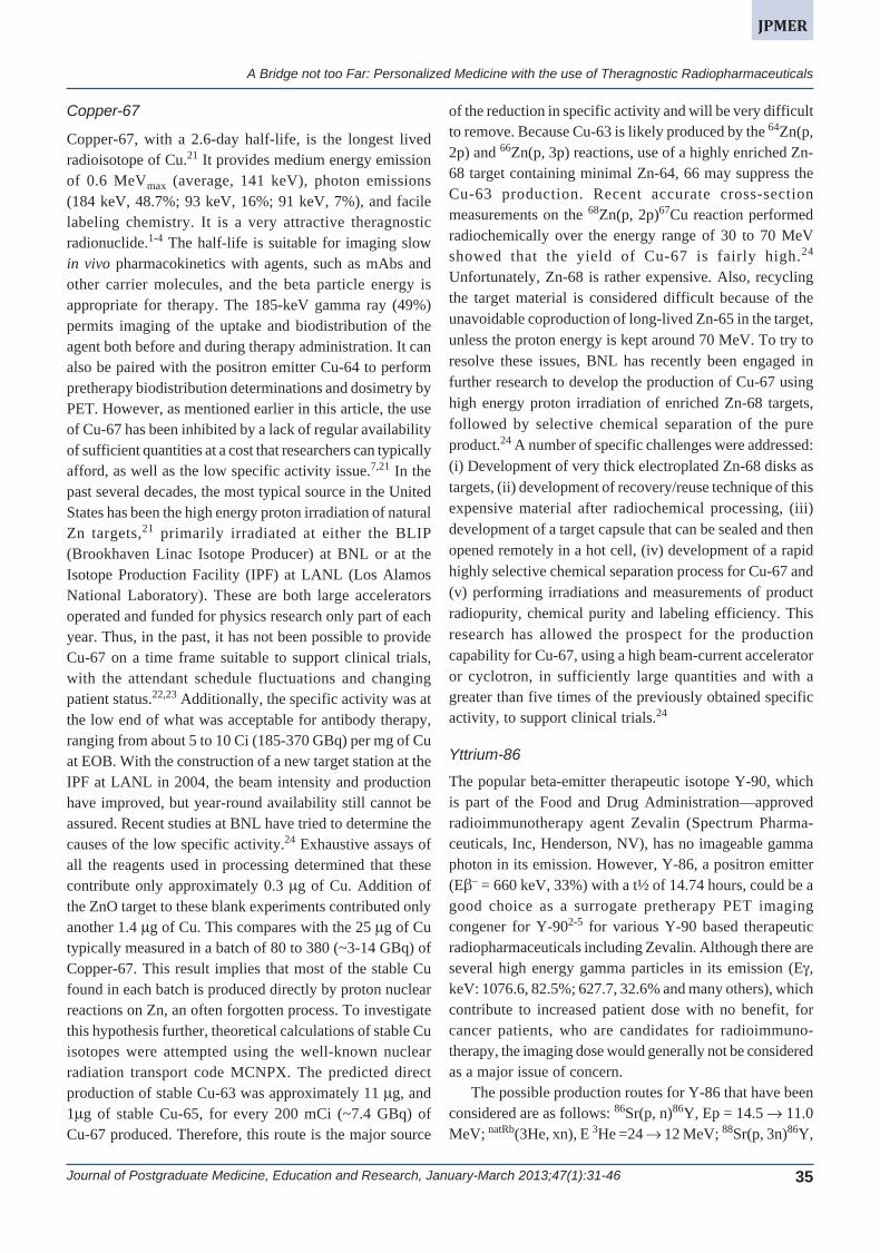
Journal of Postgraduate Medicine, Education and Research, January-March 2013;47(1):31-46 35
A Bridge not too Far: Personalized Medicine with the use of Theragnostic Radiopharmaceuticals
JPMER
Copper-67
Copper-67, with a 2.6-day half-life, is the longest livedradioisotope of Cu.21 It provides medium energy emissionof 0.6 MeVmax (average, 141 keV), photon emissions(184 keV, 48.7%; 93 keV, 16%; 91 keV, 7%), and facilelabeling chemistry. It is a very attractive theragnosticradionuclide.1-4 The half-life is suitable for imaging slowin vivo pharmacokinetics with agents, such as mAbs andother carrier molecules, and the beta particle energy isappropriate for therapy. The 185-keV gamma ray (49%)permits imaging of the uptake and biodistribution of theagent both before and during therapy administration. It canalso be paired with the positron emitter Cu-64 to performpretherapy biodistribution determinations and dosimetry byPET. However, as mentioned earlier in this article, the useof Cu-67 has been inhibited by a lack of regular availabilityof sufficient quantities at a cost that researchers can typicallyafford, as well as the low specific activity issue.7,21 In thepast several decades, the most typical source in the UnitedStates has been the high energy proton irradiation of naturalZn targets,21 primarily irradiated at either the BLIP(Brookhaven Linac Isotope Producer) at BNL or at theIsotope Production Facility (IPF) at LANL (Los AlamosNational Laboratory). These are both large acceleratorsoperated and funded for physics research only part of eachyear. Thus, in the past, it has not been possible to provideCu-67 on a time frame suitable to support clinical trials,with the attendant schedule fluctuations and changingpatient status.22,23 Additionally, the specific activity was atthe low end of what was acceptable for antibody therapy,ranging from about 5 to 10 Ci (185-370 GBq) per mg of Cuat EOB. With the construction of a new target station at theIPF at LANL in 2004, the beam intensity and productionhave improved, but year-round availability still cannot beassured. Recent studies at BNL have tried to determine thecauses of the low specific activity.24 Exhaustive assays ofall the reagents used in processing determined that thesecontribute only approximately 0.3 g of Cu. Addition ofthe ZnO target to these blank experiments contributed onlyanother 1.4 g of Cu. This compares with the 25 g of Cutypically measured in a batch of 80 to 380 (~3-14 GBq) ofCopper-67. This result implies that most of the stable Cufound in each batch is produced directly by proton nuclearreactions on Zn, an often forgotten process. To investigatethis hypothesis further, theoretical calculations of stable Cuisotopes were attempted using the well-known nuclearradiation transport code MCNPX. The predicted directproduction of stable Cu-63 was approximately 11 g, and1g of stable Cu-65, for every 200 mCi (~7.4 GBq) ofCu-67 produced. Therefore, this route is the major source
of the reduction in specific activity and will be very difficultto remove. Because Cu-63 is likely produced by the 64Zn(p,2p) and 66Zn(p, 3p) reactions, use of a highly enriched Zn-68 target containing minimal Zn-64, 66 may suppress theCu-63 production. Recent accurate cross-sectionmeasurements on the 68Zn(p, 2p)67Cu reaction performedradiochemically over the energy range of 30 to 70 MeVshowed that the yield of Cu-67 is fairly high.24
Unfortunately, Zn-68 is rather expensive. Also, recyclingthe target material is considered difficult because of theunavoidable coproduction of long-lived Zn-65 in the target,unless the proton energy is kept around 70 MeV. To try toresolve these issues, BNL has recently been engaged infurther research to develop the production of Cu-67 usinghigh energy proton irradiation of enriched Zn-68 targets,followed by selective chemical separation of the pureproduct.24 A number of specific challenges were addressed:(i) Development of very thick electroplated Zn-68 disks astargets, (ii) development of recovery/reuse technique of thisexpensive material after radiochemical processing, (iii)development of a target capsule that can be sealed and thenopened remotely in a hot cell, (iv) development of a rapidhighly selective chemical separation process for Cu-67 and(v) performing irradiations and measurements of productradiopurity, chemical purity and labeling efficiency. Thisresearch has allowed the prospect for the productioncapability for Cu-67, using a high beam-current acceleratoror cyclotron, in sufficiently large quantities and with agreater than five times of the previously obtained specificactivity, to support clinical trials.24
Yttrium-86
The popular beta-emitter therapeutic isotope Y-90, whichis part of the Food and Drug Administration—approvedradioimmunotherapy agent Zevalin (Spectrum Pharma-ceuticals, Inc, Henderson, NV), has no imageable gammaphoton in its emission. However, Y-86, a positron emitter(E– = 660 keV, 33%) with a t½ of 14.74 hours, could be agood choice as a surrogate pretherapy PET imagingcongener for Y-902-5 for various Y-90 based therapeuticradiopharmaceuticals including Zevalin. Although there areseveral high energy gamma particles in its emission (E,keV: 1076.6, 82.5%; 627.7, 32.6% and many others), whichcontribute to increased patient dose with no benefit, forcancer patients, who are candidates for radioimmuno-therapy, the imaging dose would generally not be consideredas a major issue of concern.
The possible production routes for Y-86 that have beenconsidered are as follows: 86Sr(p, n)86Y, Ep = 14.5 11.0MeV; natRb(3He, xn), E 3He =24 12 MeV; 88Sr(p, 3n)86Y,

36JAYPEE
Suresh C Srivastava
Ep = 45 37 MeV; and 90Zr(p, 2p3n)86Y, Ep = 40 30MeV.1,3-5 Out of these, it seems that the best method is the86Sr(p, n)86Y reaction, but it requires enriched 86Sr, whichis rather expensive, and economy requires its recovery andreuse.5,13,14 Recovered Sr-86 will contain radioactive Sr-85after first irradiation if the proton energy is more than14 MeV, which would mean that hot cell target assemblywill be required. At BNL, we have designed such a target,both halves of which are aluminum for better conductivity,head screws are large enough for remote manipulatorhandling and silver-coated stainless steel C-rings are usedto provide adequate seals.14 Such a target design wassuccessfully irradiated in the BLIP without any leakage andcan be opened and sealed in a hot cell.
Modification to increase active area in beam mayincrease production levels but will require more enrichedSr-86 to be used. However, if really a low energy of14 MeV is used, the amount of target material needed is nothigh. Targets of both natural SrCl2 and enriched SrCl2 wereirradiated yielding 10-13 mCi/Ah of Y-86, giving an EOBbatch yield of ~1,000 mCi in 1 hour, which is quite high.14
Chemical yields of Y-86 ranged from 82 to 93%, and afurther improvement is desirable. For labeling purposes, thischemical purity allowed for a labeling yield of 84%, andthis needs to be improved further as well. The level of amajor radionuclidic impurity, Y-87m, was a disappointing34 to 55%. The low energy route on enriched Sr-86, whichhas previously been used4,5 has allowed for better materialand is now being pursued at BNL for larger-scaleproduction. At BLIP, our first irradiation gave very lowyield because the beam spot was larger than the target area.Also, the lowest tunable beam energy at the BLIP is65.5 MeV, but the nuclear reaction cross-section peak is at~14.5 MeV. The choice of degraders seems to have resultedin too high a target entrance energy. BLIP is not an idealfacility for this low energy reaction, but we carried out theseinvestigations mainly to produce some material to developa better and quicker radiochemical processing methodologythat was short enough to allow for same-day irradiation andshipment, which has recently been accomplished.14
ALPHA, AUGER AND CONVERSIONELECTRON EMITTERS
Alpha Emitters
As mentioned earlier, targeted alpha radiation therapy canbe a very potent treatment of cancer.25 Due to comparativelyshort range of alpha particles in tissue and their high LETnature, radiation damage is mainly confined to targeted cells.The radiation burden to the surrounding healthy tissues islow when compared to beta emitters. By facilitating an
efficient delivery of alpha-emitting isotope it is possible toachieve a very effective treatment regimen for the types ofcancer where microscopic and small volume tumors arepresent. The effectiveness of alpha radioimmunotherapy hasbeen confirmed in clinical and preclinical trials.26,27
A number of alpha emitting radioisotopes have beenproposed as possible candidates. Among those are At-211,Bi-212, Bi-213, Ac-225, Pb-212, Ra-223, Tb-149 andFm-255. Some of them (Bi-212, Bi-213, Ac-225, Pb-212,Ra-223) are produced from reactor irradiations, incorporatedon generators and subsequently eluted. However, most ofthe clinical studies have been inhibited by a very limitedsupply of alpha-emitting isotopes, especially those that havetheragnostic properties, for example, astatine-211, 213Bi,radium-223 (223Ra) and 225Ac. Three of these (213Bi, 225Acand 223Ra) are presently produced from reactor neutronirradiations, incorporated on generators and subsequentlyeluted. Their parent nuclides are often fissile materials andrequire special safeguard requirements due tononproliferation concerns. On the other hand, productionof At-211, Tb-149 and Fm-255 require alpha beam(At-211), 600 MeV protons (Tb-149) or neutron irradiationof Curium (Fm-255). Only a very few facilities in the worldhave such capabilities, and most of these because of theirother primary missions are not generally available forisotope production.1
Astatine-211
Astatine-211 (t½ = 7.2 hours) decays through two paths,each emitting an alpha particle. The mean alpha energy is apotent 6.8 MeV. It is produced by the 209Bi(,2n) reactionand typically separated from the target by dry distillationinto a chloroform solution.28,29 A difficulty in this processis that the cyclotron energy must be carefully controlled tominimize the coproduction of astatine-210. This isotope(t½ = 8.1 hours) decays to Po-210 (t½ = 138 d) which is analpha particle emitting bone seeker in vivo and could causeconsiderable hematological toxicity. The half-life of At-211is relatively long compared to the other alpha emitters thus,allowing time for chemistry of labeling and use formolecular carriers with slower tumor uptake. However, itis more easily catabolized and shed after cell internalizationof the labeled antibody/antigen complex than the metallicalpha emitters.30 The main problem that inhibits clinicaltrials with this isotope is the relatively low nuclear yieldand very limited number of cyclotrons capable of producingastatine-211. This situation may be somewhat improved inthe coming years when the ARRONAX cyclotron in Nantes,France, as expected, goes into routine operations in early2013.31

Journal of Postgraduate Medicine, Education and Research, January-March 2013;47(1):31-46 37
A Bridge not too Far: Personalized Medicine with the use of Theragnostic Radiopharmaceuticals
JPMER
Actinium-225/Bismuth-213 System
Targeted alpha radiation therapy with alpha emittersAc-225 and Bi-213, as is the case with astatine-211 (videsupra), is also a very promising treatment of cancer.25
If selective efficient delivery of these alpha-emittingisotopes to tumor cells is achieved, they can become veryeffective for the treatment of the types of cancer wheremicroscopic and small volume tumors are present. Theeffectiveness of alpha radioimmunotherapy using theseisotopes has already shown promise in preclinical andclinical trials.26,27 The results of the only human clinicaltrial involving an alpha-emitting isotope have demonstratedfeasibility and antileukemic effects of Bi-213-HuM195, ahumanized anti-CD33 mAb, specifically designed to targetmyeloid leukemia cells. In this study, 78 to 93% of thesubjects showed reduction in circulating and bone marrowblasts.28 A clinically approved Ac-225/Bi-213 generatorcapable of producing 25 to 100 mCi of Bi-213, suitable forantibody labeling, was developed32 and continues to beimproved upon. The antibodies and peptides labeled withthe generated Bi-213 have successfully been used innumerous preclinical and clinical trials.26,27,32,33 Asmentioned above, most of the clinical studies have beeninhibited by a very limited supply of these and other alpha-emitting isotopes, especially those that have theragnosticproperties, for example, At-211, Bi-213, Ra-223 andAc-225.
The in vivo use of targeted NCA Ac-225, in its ownright, has received a great deal of attention as well.34,35
Because of its ability to produce 4 alpha-emitting daughters,its overall potency to destroy cells is higher. The isotopehas been referred to as an in vivo alpha particlenanogenerator because its power proliferates beyond thesize of the atom.35 In addition, the applications of Ac-225were further extended to encapsulation in liposomes36 andmetallofullerenes.37 At the Memorial Sloan–KetteringCancer Center, a clinical trial for radioimmunotherapy ofacute myeloid leukemia with both Bi-213 and Ac-225 hasrecently been carried out.38 As mentioned earlier in the text,Ac-225 has been used for radioimmunotherapy both as apotent alpha emitter attached to an antibody and as agenerator for the daughter alpha emitter Bi-213 (t½ = 45.7minutes). The decay of Bi-213 is accompanied by emissionof 440.45 keV (I = 25.9%) gamma photon, which allowsSPECT imaging to study pretherapy biodistribution,pharmacokinetics and dosimetry of the labeledradiopharmaceuticals. The current availability of Ac-225is very limited and insufficient to support ongoing andproposed clinical trials. Currently, there are two suppliersof Ac-225. One is the US Department of Energy’s ORNL
and the other is the Institute for Transuranium Elements inKarlsruhe, Germany. Both suppliers obtain thorium-229from uranium-233 that was produced as part of US moltensalt breeder reactor program. The current stock of purifiedTh-229 at ORNL is 150-mCi. In an 8-week period, about100 mCi of Ac-225 grows in from that supply, separatedand provided for clinical trials. Because Th-229 decays toAc-225 through intermediate daughter Ra-225, Ra can beisolated to provide additional quantities (up to 20 mCi) ofAc-225.39-41 Similarly, the Institute of TransuraniumElements can separate 43.2 mCi of Ra-225 and 39.4 mCi ofAc-225 from their stock.40 It is widely acknowledged thatthe anticipated growth in demand for Ac-225 will soonexceed the levels extractable from the available uranium-233.39
At BNL, our intent is to carry out future investigationsin an effort to increase the supply of Ac-225. This wouldinvolve using the high energy proton spallation reaction232Th(p, 2p6n)225Ac on natural Thorium. This method hasrecently been studied and developed at other places, inparticular, at the Institute of Nuclear Research (INR) inTroitsk, Russia, using accelerated protons at energiesbetween 90 and 145 MeV in their 160-MeV protonaccelerator.41 This investigation has pointed out that theyield of Ac-225 increases monotonically with the protonenergy. Therefore, the BLIP at BNL (200 MeV) would becapable of producing almost twice as much with an incidentenergy on the target of 192 MeV, with 110 µA beamintensity. The expected outcome of the BNL effort wouldbe the method for Ac-225 production with quality(radionuclidic and chemical purity) suitable for applicationsboth as a radiotherapeutic agent (Ac-225) and as a generatorof Bi-213 (theragnostic use). The proposed work willinvolve development of an optimized Th-232 target forirradiation, development of the chemical procedure fordissolution and processing of irradiated target to selectivelyseparate Ac-225, and carrying out preliminary labelingstudies of conventional targeting molecules with Ac-225/Bi-213 to demonstrate the efficacy of the final purifiedradioisotope(s) for the eventual protein/antibody labelingapplications.
At BNL, the method could potentially yield Curieamounts of Ac-225 in a 10-day irradiation period at theBLIP. The produced Ac-225 can be used for radio-immunotherapy as well as for generation of Bi-213 in aquality suitable for clinical trials. Shorter-lived radioisotopesAc-228, 226, 224 will be coproduced in quantitiescomparable with those of Ac-225, but the activities of theseisotopes will saturate out during the 10-day irradiation. Theradionuclidic purity of the Ac-225 can be improved by

38JAYPEE
Suresh C Srivastava
allowing a cool-off period for the target. Ten days (onet½ of Ac-225) after EOB, the coproduced Ac radionuclideswill comprise 1% of the activity of Ac-225. Among these,Ac-227, because of its 21.77-year half-life, is the onlyradioimpurity that might be of some concern as far asnoninvasive imaging is concerned but, in principle, of lessconcern for theragnostic use. However, this issue wouldrequire further investigation and additional studies.
Small quantities of Ac-225 can also be prepared by thereaction 226Ra(p, 2n)225Ac over an energy range of 13 to 20MeV.42 The cross-section for this reaction is calculated toreach a peak of 700 mb at 16 MeV but total yield is limitedby the small energy range (and target thickness) where thisreaction is favorable. Similarly the photonuclear reaction226Ra(, n)225Ra (t½ = 14.9 days) followed by beta decay toAc-225 is feasible but requires an extremely large Ra target(~40g) that represents radiological safety issues andavailability concerns.
Thus, Ac-225, when produced by the BNL method,could substantially add to the very limited present supplyof this promising alpha emitter. According to experimentaland theoretical extrapolations, and assuming certain targetdimensions, radiochemical processing losses, and so forth,the projected yields from a 10-day irradiation at the 5 highenergy accelerators in the world are estimated (from thestudies at INR) to be as follows (in Curies; experimental,and theoretical values within parentheses): BLIP at BNL,3.1 (4.9); INR in Russia, 2.6 (4.1); IPF at LANL, 0.7 (1.3);Tri-University Meson Facility (TRIUMF) in Canada,0.5 (0.7); and Arronax in France, 0.2 (0.7), giving a total of~7 (11) curies.41 This would allow detailed preclinical andclinical studies to determine the therapeutic potential ofAc-225 itself as well as of the theragnostic daughterBi-213. Many of the presently available bifunctionalchelating agents as well as those based on cyclohexylethyl-enediamine tetraacetic acid (CDTA) and 1,4,7,10-tetraazacyclo-dodecane-1,4,7,10-tetraacetic acid (DOTA)derivatives, developed earlier at BNL43,44 should allowlinking Ac-225 and Bi-213 with protein bifunctionalchelating agent conjugates that will have high in vivothermodynamic and kinetic stability. It would, of course,serve as a generator of Bi-213 similarly to Ac-225, producedby the decay from Th-229.
Auger and Conversion Electron Emitters
The production of metastable nuclei, such as conversionelectron emitter Sn-117m and Auger electron emitter Pt-195m via neutron radiative capture reactions is characterizedby small neutron cross-sections and, hence, low productionrates. Metastable nuclei typically have excitation energies
on the order of 100 keV, and large differences in angularmomentum from ground states (most metastable nuclei havehigh angular momentum). An alternative route for producingthese types of metastable nuclei is through neutron inelasticscattering where the cross-section of the AZ(n, n’)AmZreaction is, in some cases, substantially higher than the cross-section for the (A-1)Z(n, )AmZ route. As has been shown forthe case of Sn-117m45 the magnitude of gain in the cross-section may compensate for the relatively lower fast neutronflux from a well-moderated fission spectrum. Note that theexcitation energy of metastable nuclei will represent thethreshold for inelastic scattering. Large research reactors,such as HFIR at ORNL, and HFBR (now closed down) inthe United States, and at the Research Institute of AtomicReactors (RIAR) in Dimitrovgrad, Russia, with significantepithermal and fast neutron fluxes, have been well suitedfor these types of reactions.
A systematic study of the production of Sn-117m, andPt-195m in the hydraulic tube facility of the HFIR wasreported.46 In all three cases, the yields from the [n, n’]reactions were higher than those obtained from the [n, ]reactions. The relative gains in the specific activity of theunfiltered (no Cd filter) targets were 1.4, for Pt-195m, and3.3 for Sn-117m. The excitation energies for Sn-117m andPt-195m are 314.6 and 259.2 keV respectively. Since, thethresholds for these inelastic scattering reactions are wellabove the cadmium cutoff, Cd filters did not have any effecton the yield of these reactions. The corresponding cross-sections for the inelastic neutron scattering reactions forthe production of Sn-117m, and Pt-195m are 222 ± 16, and287 ± 20 mb respectively. A value of 176 ± 14 mb for thecross-section of Sn-117[n, n’]Sn-117m reaction obtainedat HFBR was reported earlier.45
Conversion Electron Emitter Tin-117m
The most common method for Sn-117m production is basedon irradiation of tin with neutrons using the 117Sn(n,n )117mSn reaction.45 However, since this reactor productionmethod is based on the inelastic neutron scattering reactionusing enriched Sn-117 as a target, it results in Sn-117mwith a low specific activity of about 20 Ci/g.47 Stanniccomplexes with Sn-117m of such specific activity,particularly Sn-117m4+-DTPA, effectively reduce pain frombone metastases without adverse reactions related to bonemarrow, as demonstrated by the results presented in Tables 4and 5.48 However, such low specific activity does not allowscaling up the therapeutic doses for treating bone metastases.Also it is not acceptable for radioimmunotherapy and formany other applications.
Although it is not possible to achieve a higher than about22mCi/mg of specific activity in the reactor-produced tin-
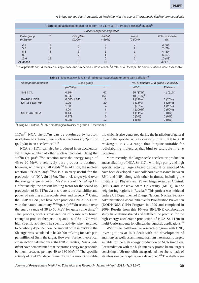
Journal of Postgraduate Medicine, Education and Research, January-March 2013;47(1):31-46 39
A Bridge not too Far: Personalized Medicine with the use of Theragnostic Radiopharmaceuticals
JPMER
117m47 NCA tin-117m can be produced by protonirradiation of antimony via nuclear reactions (p, 2p3n) or(p, 2p5n) in an accelerator.45,49
NCA Sn-117m can also be produced in an acceleratorvia a large number of other nuclear reactions. Using the115mIn (, pn)117mSn reaction over the energy range of45 to 20 MeV, a relatively pure product is obtained;however, with very small yields.50 In addition, the nuclearreaction 116Cd(, 3n)117mSn is also very useful for theproduction of NCA Sn-117m. The thick target yield overthe energy range 47 20 MeV is about 150 Ci/Ah.Unfortunately, the present limiting factor for the scaled upproduction of Sn-117m via this route is the availability andpower of existing alpha accelerators and targetry.51 Usingthe BLIP at BNL, we have been producing NCA Sn-117mwith the natural antimony(natSb)(p, xn)117mSn reaction overthe energy range of 38 to 60 MeV for quite some time.45
This process, with a cross-section of 5 mb, was foundenough to produce therapeutic quantities of Sn-117m withhigh specific activity. The specific activity when assumedto be wholly dependent on the amount of Sn impurity in theSb target was calculated to be 30,000 mCi/mg for each partper million of Sn in the target. However, further theoreticalcross-section calculations at the INR in Troitsk, Russia (videinfra) have demonstrated that the proton energy range shouldbe much broader, perhaps 40 to 130 MeV.49 The specificactivity of Sn-117m depends mainly on the amount of stable
tin, which is also generated during the irradiation of naturalSb, and the specific activity can vary from ~1000 to 3000mCi/mg at EOB, a range that is quite suitable forradiolabeling molecules that bind to saturable in vivoreceptors.
More recently, the larger-scale accelerator productionand availability of NCA Sn-117m with high purity and highspecific activity, targets based on natural or enriched Sbhave been developed in our collaborative research betweenBNL and INR, along with other institutes, including theInstitute for Physics and Power Engineering in Obninsk(IPPE) and Moscow State University (MSU), in theneighboring regions in Russia.49 This project was initiatedunder a US Department of Energy/National Nuclear SecurityAdministration/Global Initiative for Proliferation Prevention(DOE/NNSA GIPP) Program in 1999 and completed in2009. Results from this 10-year BNL/INR collaborativestudy have demonstrated and fulfilled the promise for thehigh energy accelerator production of NCA Sn-117m inmulti-Curie amounts for clinical theragnostic applications.49
Within this collaborative research program with BNL,investigations at INR dealt with the development ofantimony as wells as antimony/titanium intermetallic targetssuitable for the high energy production of NCA tin-117m.For irradiation with the high-intensity proton beam, targetsconsisting of Sb-monolith encapsulated into shells made ofstainless steel or graphite were developed.49 The shells were
Table 5: Myelotoxicity levels1 of radiopharmaceuticals for bone pain palliation48
Radiopharmaceutical Dose group No. of patients with grade 2 toxicity(mCi/Kg) n WBC Platelets
Sr-89 Cl2 0.154 67 25 (37%) 41 (61%)0.040 161 48 (31%)2
Re-186 HEDP 0.500-1.143 12 2 (17%) 3 (25%)Sm-153 EDTMP 1.00 20 3 (15%) 5 (25%)
1.50 4 3 (75%) 1 (25%)3.00 6 4 (100%) 2 (50%)
Sn-117m DTPA 0.143 9 1 (11%) 0 (0%)0.179 5 0 (0%) 0 (0%)0.286 12 1 (8%) 0 (0%)
1Using NCI criteria; 2Only hematological toxicity at grade > 2 mentioned
Table 4: Metastatic bone pain relief from Tin-117m DTPA: Phase II clinical1 studies48
Patients experiencing relief
Dose group n2 Complete Partial None Total response(MBq/kg) (100%) (>50%) (0-50%) (%)
2.6 5 0 3 2 3 (60)5.3 9 3 4 2 7 (78)6.6 5 3 1 1 4 (80)8.5 9 2 4 3 6 (67)10.6 12 4 6 2 10 (83)
All doses 40 12 18 10 30 (75)1Total patients 57; 54 received a single dose and 3 received 2 doses each; 2A total of 40 therapeutic administrations were assessable

40JAYPEE
Suresh C Srivastava
filled with metallic antimony as follows. Powdered metallicantimony was inserted into the shells under inert nitrogenatmosphere, and the filled shell was heated at 645 to 660°C.It was found that at higher temperature liquid antimonyreacts with iron and can destroy the stainless steel shellduring the target preparation or irradiation. Graphite targetshell is more reliable since there is no noticeable reactionbetween graphite and antimony, and graphite has higherheat conductivity than stainless steel [80-120 W/(m·K) vs20 W/(m·K) at 300°C], providing effective target cooling.After antimony melting and shrinkage, the shell was againfilled with powder of antimony and heated to melt again.The target with metallic shell containing a monolith of 29gmetallic antimony (a cylinder 9 mm thick, 30 mm indiameter) was hermetically sealed with a threaded plug. Thegraphite shell targets (plates 32 × 76 × 5 mm, 3 mm of Sbthick each, 19g Sb each) were hermetically sealed with acover by means of high temperature radiation resistant glueand electroplated outside with nickel in order to protectgraphite from reaction with water radiolysis products duringirradiation.49
The irradiated Sb-targets were chemically processed atthe hot cell facility at IPPE in Obninsk. The stainless steelshell was dissolved in concentrated HCl while the graphiteshell was opened mechanically. The freed Sb monolith wasdissolved in concentrated HCl with gradual addition of smallamount concentrated HNO3. The major amount of Sb (inthe form Sb5+) was removed from the solution (10-11 MHCl) by extraction with dibutyl ether. Final purification ofNCA Sn-117m from the rest of Sb, coproduced Te-118,119m, 121m and In-111, 114m was achieved bychromatography on SiO2 in 0.5 M sodium citrate (Na3Cit)solution. Tin isotopes remained adsorbed on SiO2 while Sband radioisotopes of Te and In were washed out. Then thecolumn was washed with 0.5 M Na3Cit solution and wateracidified by citric acid. Finally, tin was eluted from thecolumn with 6 M HCl. Radionuclidic purity of the finalsolution and the specific activity of Sn-117m in the finalproduct were determined using appropriate countingprocedures (spectral emission analysis), and throughinductively coupled plasma (ICP) spectrometry. Amongother isotopes of tin coproduced with Sn-117m, Sn-113 isthe most important impurity. It has a long half-life (115 d),relatively high energy -rays (392 keV) and cannot bechemically separated from Sn-117m. But it is possible toreduce Sn-113 impurity up to almost 1% by lowering theinitial proton energy. The optimum range of between55 and 20 MeV has been demonstrated to be most effective,although at the expense of somewhat reduced Sn-117mproduct.52
THERAGNOSTIC STUDIES WITH Sn-117m
Sn-117m (t½ = 14.0 days; 159 keV, 86%) showsconsiderable promise as a theragnostic radionuclide due toits nuclear, physical and chemical characteristics. Asmentioned earlier in this paper, it is certainly one of thebest radionuclides for the development of theragnosticradiopharmaceuticals, in particular, for nuclear oncology.In contrast to most other therapeutic beta emitters, Sn-117mdecays via isomeric transition, with the emission of threemajor mono energetic conversion electrons (127, 129 and152 keV; abundance, 65, 12 and 26% respectively). Theseemissions with a very high LET have short discretepenetration ranges of between 0.22 (127 keV) and 0.29 mm(152 keV) in water. Therefore, Sn-117m when selectivelytargeted should be effective for therapy of metastatic diseaseand for certain other inflammatory conditions (e.g.atherosclerotic disease), causing much reduced myelo-suppression and greatly reduced dose to normal organs.53
This is schematically represented in Figure 1 as well as inFigure 254 which shows whole-body autoradiographiesperformed in mice and rats after the administration of Sn-117m(4+)-diethylenetriamine pentaacetic acid (DTPA), andthe highly selective targeting and high uptake in bone bythis agent, but not in bone marrow or other organs,suggesting the expected effectiveness of high-LET lowenergy conversion electrons to produce intense radiationdose within a very short distance without affecting normaltissues, in particular, the radiation-sensitive bone marrow.53
Figure 3 shows whole body images in a prostate cancerpatient, who had developed extensive bone metastases. Scanon the left was obtained using the standard Tc-99m MDP(methylene diphosphonate) imaging agent and the one onthe right consists of scans obtained using tin-117m(4+)DTPA, an agent which as mentioned above has shown greatpromise for treatment of metastatic pain in prostate and
Fig. 1: A schematic comparison of energy types fortherapeutic radionuclides1

Journal of Postgraduate Medicine, Education and Research, January-March 2013;47(1):31-46 41
A Bridge not too Far: Personalized Medicine with the use of Theragnostic Radiopharmaceuticals
JPMER
therapy with 18.6 mCi (688 MBq) of Sn-117m DTPA, andthe autopsy was carried out 6 hours after death.55 Figures5A and B displays both a picture and the bone scintigraphyof three longitudinal coronal slices (5 mm thick) ofthoracolumbar vertebrae, again from the same patient as inFigures 4A and B. The uptake of Sn-117m DTPA is bothdiffuse (T8) and heterogeneous (L1). Partial involvementwithin a single vertebra indicates nonuniform distributionof radioactivity within the lesion, suggesting heterogeneousdistribution of metastatic foci.55 Thus, the unique therapeuticemissions of Sn-117m also imply that their therapeuticeffectiveness will be highly localized and should be lesion-specific and thus more effective. The use of Sn-117m DTPAis also very worth considering for an early-stage disease atwhich time, there would be the possibility of slowing oreven stopping the progression of further metastatic lesions.Moreover, having the 159-keV photon (86%), which isvery similar to that of technetium-99m, Sn-117m is perfectfor pretherapy imaging for dosimetry and for otherinformation in the same patient as well as for monitoringthe results of treatment.55 All clinical studies thus, far forbone pain palliation using Sn-117m DTPA were carried outusing reactor-produced Sn-117m, with a specific activityranging between 8 and 20 Ci/g of Sn. More recently, several50- to 150-mCi samples of NCA Sn-117m produced at INRas well as at the University of Washington alpha cyclotron,have successfully been used and evaluated at BNL, and incollaboration with the Saint Joseph’s Translational ResearchInstitute in Atlanta and at other institutions, for effectivenessfor theragnostic applications in animal models.56,57
Tin-117m, in addition to being a good therapeutic agentfor cancer, also shows promise for the noninvasivemolecular imaging and treatment of active atheromatousdisease [vulnerable plaque (VP), also known as thin-capfibroatheroma (TCFA)], in the coronary arteries and otherareas of vasculature through the use of (i) coronary stentselectroplated with Sn-117m or (ii) specific Sn-117m-labeledmolecules systemically targeted to VP components.
In the United States, there are, on an average, about875,000 to 1,000,000 new heart attacks every year, and theseare caused by two different types of atherosclerotic disease:Luminal calcified plaque and extraluminal activeatheromatous disease leading to the formation of VP(Fig. 6). Vulnerable (unstable) plaque forms below thelumen in coronary artery walls, and inflammation is the maindriver. VP is usually covered by a thin cap on the lumenside. When ruptured, the thin cap ‘releases’ highlythrombogenic material activating clotting cascadeand inducing thrombosis. Approximately more than120 substances are expressed in ruptured VP (including
Fig. 2: Autoradiography in rats (top) and in mice (middle and bottom)demonstrates selective targeting and high uptake of tin-117m DTPAin bone and not in bone marrow or other organs54
Fig. 3: Whole body images in a prostate cancer patient, who haddeveloped extensive bone metastases. Scans on the left (posterior,anterior views) were obtained using the standard Tc-99m MDP boneimaging agent. Scans on the right are (anterior, posterior) obtainedusing tin-117m DTPA which is promising for treatment of metastaticbone pain and potentially of osseous metastases48
breast cancer patients.48 These two scans are almostidentical, demonstrating the specificity of tin-117m DTPAlocalization in bone and in much higher concentration inbone metastases, in fact quantitatively even much betterthan the Tc-99m MDP. Figures 4A and B,55 which are froman actual sample of bone tissue from a prostate cancerpatient, who was treated with Sn-117m DTPA for bone painpalliation and who donated his body for research after deathcorroborates the same result as in Figures 2 and 3. Thislatter patient had died of his primary cancer 47 days after

42JAYPEE
Suresh C Srivastava
collagen) and this rupture then results in thrombosis, whichcan cause unstable angina, chest pain, acute MI, suddencoronary death and stroke. The luminal calcified plaquesare easily detected by existing diagnostics, and the patienthas a number of treatment options. However, VP developsand models positively outside the arterial lumen (Fig. 6)and cannot be detected by noninvasive imaging techniques.Unfortunately, a majority of all significant cardiac events(~70%) leading to MI, including sudden death, are causedby the rupture of these thin-cap fibroatheroma lesions, notlumen atherosclerosis.
The properties of Sn-117m as applied to vulnerableplaques are unique and significant.
At therapeutic doses, the conversion electrons fromSn-117m have been shown to reduce inflammation and,thus, are ideal for treating VP’s as their range in tissue(~300 m) is approximately the same as the VP thicknessin human coronary arteries. Sn-117m-labeled Annexineffectively targets and binds to macrophage cells undergoingapoptosis, which are present in abundance in VP’s.In relatively low doses, this agent can image the plaqueusing traditional SPECT gamma cameras with technetium-99m collimators and imaging protocols. Because of thelonger half-life of Sn-117m combined with pretargetingtechniques, gamma cameras can acquire images long afterthe nonspecifically bound tin—Annexin has cleared theblood pool thus improving the image quality.58
A clinical trial with Sn-117m–Annexin, begun in 2010,is currently in progress. At present, the phase IIa study(15 subjects) involves human carotid endarterectomypatients who are dosed and imaged for VP, with histologyas the comparison. Further clinical trials (phase IIb andphase III) are in the planning stage. In recent studies in an
Figs 4A and B: Microautoradiographic demonstration of the deposition of Sn-117m DTPA in bone (A) anda schematic diagram (B) showing sites of localization from a sample of bone obtained following the autopsyof a patient treated with Sn-117m DTPA for bone pain palliation. There is high deposition in the activelymineralizing osteoid interface. Completely mineralized bone and the bone marrow space show very lowdeposition. Osteoid-to-marrow ratio is 11:155
Figs 5A and B: Photographic picture (A) and bone scintigraphy(B) of three coronal slices (each 5 mm thick) of thoracolumbarvertebrae from the same patient as in Figures 4A and B. Partialinvolvement within a single vertebra indicates nonuniformdistribution of radioactivity, within the lesion, suggestingheterogeneous distribution of metastatic foci. Note the uptake indiseased areas in various sections according to the range of theSn-117m conversion electrons55
Fig. 6: A schematic representation showing the formation andpositive remodeling of vulnerable plaque outside of the arterial lumenin the coronary artery walls
Inflammation drives initiation, progression andcomplications of atherosclerosis

Journal of Postgraduate Medicine, Education and Research, January-March 2013;47(1):31-46 43
A Bridge not too Far: Personalized Medicine with the use of Theragnostic Radiopharmaceuticals
JPMER
Apo-E mouse VP model, in doses that appear to be imagingdoses, tin-117m-Annexin has demonstrated a significantanti-inflammatory therapeutic effect. The plaquecomposition showed significantly less expression ofmacrophages in the low, middle and high dose treatmentgroups as compared to the control group. In contrast, smoothmuscle cell expression was greater in the middle and highdose groups as compared to the control, and in the low dosegroup, the difference was statistically significant betweenthe low vs middle and high dose groups.59
Because inflammatory cells are more radiosensitive thanother arterial wall cell types, the conversion electrons shouldexhibit a beneficial anti-inflammatory effect in diseasedcoronary arteries. Since 28 days, corresponding to two half-lives of Sn-117m, is believed to be roughly the optimumperiod for neoproliferative tissue suppression, includingneointimal proliferation after angioplasty and other primaryinterventional treatments in the coronary arteries and othervasculature, radiotherapy with Sn-117m should providehighly effective treatment, with minimal collateral damageto adjacent, quiescent arterial wall cells because of its lowenergy conversion electron emission and short penetrationin tissue. Furthermore, such limited energy delivery from aradioactive stent implant may circumvent the problem ofcellular overgrowth at the implant edges, known as the‘candy wrapper’ effect, which has been observed withprevious forms of radioactive stents.
At BNL, a novel radioactive stent stably electroplatedwith Sn-117m was developed as a therapeutic tool for thetreatment of atherosclerotic coronary arteries.60 Effects onvessel wall inflammation from various doses of electroplatedSn-117m were studied in hyperlipidemic rabbits. Three daysafter stent implantation in the abdominal aorta [4 doses:0- (cold tin), 30-, 60- and 150-Ci Sn-117m per 15-mmstent], the rabbits were sacrificed. Immunohistochemicalanalysis of proliferating macrophages and smooth musclecells demonstrated that inflammatory cells in the Sn-117m-stented segments were dramatically reduced in a dose-dependent manner.56,57 The anti-inflammatory therapeuticeffectiveness of the tin-117m-stent implants was furtherdemonstrated in a preliminary study in pigs (24 pigs,72 bare metal stent implants: 0- to 400-Ci Sn-117m per15-mm stent), which was carried out to investigate theeffects on in-stent neointima formation. The electroplatingprocess appeared benign because the response from ‘cold’tin-plated stents was indistinguishable from what wasobserved for routinely used bare metal, 316L surgicalstainless steel stents. A profound and unique radiation effectwas consistently observed in the higher dose implants, whichconsisted of a discrete zone of dense connective tissueconsolidation in the tunica adventitia, of ~0.25 to 0.30 mm
thickness, corresponding to the anticipated tissue penetrationof the Sn-117m conversion electron energy. Focalsuppression of in-stent neointima formation was alsoobserved, but further study will be needed to determine,with certainty, the therapeutic potential of Sn-117m-electroplated stents for prevention of in-stent restenosis.61
To summarize, tin-117m shows great promise as a meansto reduce inflammatory events following stent implantation,and when delivered systemically to atherosclerotic tissueusing specific targeting molecules (e.g. Annexin-V), iseffective in low doses to image and in higher doses to reducethe vulnerability for rupturing of unstable coronary arteryplaques.
NCA Sn-117m is also being developed and tested forthe therapy of bone metastases and for use inradioimmunotherapy. A phase I clinical trial for the imagingand treatment of bone metastases using Sn-117m DTPA inprostate cancer patients is in the planning stage for 2013.
CONCLUSION
There are a number of existing and potential futureradionuclides for use as unsealed sources for targetedtherapy of cancer and for other therapeutic applications innuclear medicine. The various important therapeuticapplications where radionuclide therapy would have animportant role to play in, and the radionuclides best suitedfor that particular type of application, are summarized inTable 6. It should be noted that this listing is not meant tobe exhaustive and additional radionuclides can be addedbased on present and future work as well as various otherconsiderations. The substantial progress of investigationsin areas, such as the treatment of cancer as well as of otherinflammatory conditions, such as in bone pain palliation,radiosynovectomy, and of many other disorders that respondto radionuclide therapy, offers renewed hope and promisefor the widespread use of internally administeredradionuclides for a number of novel and effective therapeuticapproaches.
Therapeutic nuclear medicine finally seems to bedestined to find its rightful place in the ‘personalized’therapy of patients with the use of a number of dual-purpose(‘theragnostic’) radionuclides or radionuclide pairs that arediscussed and emphasized in this article. This paradigm,when properly enforced would constitute a major stepforward to meet the challenges of enabling personalizedmedicine. Implementation of this regimen potentially createsa situation where treatments are better targeted, healthsystems save money by identifying therapies not likely tobe effective for particular patients, and researchers have abetter understanding of comparative effectiveness.

44JAYPEE
Suresh C Srivastava
Theragnostic radiopharmaceuticals have the power todrive advances in personalized medicine that will offerbetter-targeted diagnosis and treatments. Using thisapproach, it would be possible to envision a future wheretreatments are tailored to individual patients’ specific diseaseparameters and where imaging data could be analyzed inreal-time and in advance to predict likely effectiveness oftherapy and learn what would or would not work. Oncologyis just the beginning; in principle, we should be able to startscientific enquiries to apply this paradigm to other complexdisease states, so that the potential for theragnosticradiopharmaceutical biomarkers as a bridge to help us betterdetect, understand, and treat diseases like CAD, diabetes,and Alzheimer’s becomes a reality. However, an increasedand continuously reliable availability at reasonable cost ofsome of the best theragnostic radionuclides has remained amajor issue, which must be addressed before we cansuccessfully put this paradigm into routine clinical practice.This issue has been discussed in this paper in some detail,and the evolving new methodologies for the production ofa few of the very promising theragnostic radionuclides havebeen covered.
It is worth emphasizing that our nuclear medicinemodality is the only modality that can fulfill the dream ofcarrying out tailored personalized medicine by way ofenabling diagnosis followed by therapy in the same patientwith the same radiopharmaceutical. This would be anexciting development with a very promising future, wouldbe of invaluable benefit to patients with cancer,
cardiovascular disease, and other disorders, and may verywell mark the future of the field of nuclear medicine.
ACKNOWLEDGMENTS
This work was supported by the United States Departmentof Energy, Office of Science/Office of Nuclear Physics/Isotope Development and Production for Research andApplications Program, and the NNSA-NA-24 GIPPProgram, under Contract No. DE-AC02-98CH10886, atBrookhaven National Laboratory. Additional research grantsupport for part of this work by Clear Vascular Inc, includinga Cooperative Research and Development Agreement(CRADA) with BNL, is gratefully acknowledged.
REFERENCES
1. Srivastava SC. Paving the way to personalized medicine:Production of some promising theragnostic radionuclides atBrookhaven National Laboratory. Semin Nucl Med 2012;42:151-63.
2. Srivastava SC. Theragnostic radiopharmaceuticals: The ‘Janus’approach to molecular diagnosis and therapy. CME session #63 on Novel radiopharmaceuticals for molecular imaging andtherapy–where are we headed next? Presented at 2009 SNMAnnual Meeting, Toronto, Canada, June 15, 2009.
3. Srivastava SC. Theragnostic radiometals: Getting closer topersonalized medicine. In: Mazzi U, et al (Eds). Technetiumand other radiometals in chemistry and nuclear medicine.Padova, Italy, SG Editorial 2010;553-68.
4. Srivastava SC. Paving the way to personalized medicine:Production of some theragnostic radionuclides at BrookhavenNational Laboratory. Radiochim Acta 2011;99:635-40.
Table 6: Choice of radionuclides for principal therapeutic* applications62
Route of Best-suited radionuclide(s)Application administration
1. Tumor therapy(i) Solid tumors
a. Large lesions iv Sc-47, Y-90, I-131, Lu-177,Re-188
Intratumoral Sc-47, Sm-153, Re-188b. Micrometastases iv Sc-47, Sn-117m, Sm-153, other
Auger, conversion electron, alpha andshort range -emitters
(ii) Leukemias, lymphomas iv Sc-47, Cu-67, Sn-117m, I-131, alphaemitters
2. Pain palliation(i) Soft tissue iv Y-90, I-131, Ho-166, Re-188(ii) Metastatic bone pain iv Sr-89, Sn-117m, Sm-153, Lu-177,
Re-187, Re-188, Ra-2233. Nononcology
(i) Synovectomy Regional P32, Y-90, Sn-117m, Re-186, Er-169(ii) Marrow ablation iv Sn-117m, Ho-166(iii) Microspheres iv Y-90, other radio-lanthanides
4. Receptor-binding tracers; cellular iv Auger, conversion electron, (intranuclear) antigens alpha and short-range -emitters
*The isotopes in various categories are listed in order of increasing atomic mass and not necessarily based on their degree of theirtherapeutic effectiveness

Journal of Postgraduate Medicine, Education and Research, January-March 2013;47(1):31-46 45
A Bridge not too Far: Personalized Medicine with the use of Theragnostic Radiopharmaceuticals
JPMER
5. Herzog H, Rösch F, Stöcklin G, et al. Measurement ofpharmacokinetics of yttrium-86 radiopharmaceuticals with PETand radiation dose calculation of analogous yttrium-90radiotherapeutics. J Nucl Med 1993;34:2222-36.
6. Srivastava SC. Radiolabeled monoclonal antibodies for imagingand therapy. New York: Plenum Press 1988;876.
7. Srivastava SC, Dadachova E. Recent advances in radionuclidetherapy. Seminars in Nucl Med 2001;31:330-41.
8. Traub-Weidinger T, Raderer M, Uffman M, et al. Improvedquality of life in patients treated with peptide radionuclides.World J Nucl Med 2011;10:115-21.
9. Kassis AI, Adelstein SJ, Haycock C, et al. Lethality of Augerelectrons from the decay of Br-77 in the DNA of mammaliancells. Radiat Res 1982;90:362-73.
10. Adelstein SJ, Kassis AI. Radiobiologic implications of themicroscopic distribution of energy from radionuclides. Nucl MedBiol 1987;14:165-69.
11. Feinendegen LE. Biological damage from the Auger effect:Possible benefits. Radiat Environ Biophys 1975;12:85-99.
12. Szelecsenyi F, Blessing G, Qaim SM. Excitation functions ofproton induced nuclear reactions on enriched 61Ni and 64Ni:Possibility of production of no-carrier-added 61Cu and 64Cu at asmall cyclotron. Appl Radiat Isotopes 1993;44:575-80.
13. Sadeghi M, Aboudzadeha M, Zali A, et al. Radiochemical studiesrelevant to Y-86 production via 86Sr(p,n)86Y for PET imaging.Appl Radiat Isot 2009;67:7-10.
14. Medvedev D, Mausner LF, Srivastava SC. Irradiation ofstrontium chloride targets at proton energies above 35 MeVto produce PET radioisotope Y-86. Radiochim Acta 2011;99:755-61.
15. Aslam MN, Sudar S, Hussain M, Malik AA, et al. Evaluationof excitation functions of 3He and -particle induced reactionson antimony isotopes with special relevance to the productionof iodine-124. Appl Radiat Isotopes 2001;69:94-104.
16. Huclier-Markai S, Sabatie A, Kubicek V, et al. Chemical andbiological evaluation of scandium (III)-polyamino-polycarboxy-late complexes as potential PET agent and radiopharmaceutical.Radiochim Acta 2011;99:653-62.
17. Meinken GE, Kurczak S, Mausner LF, Kolsky KL,Srivastava SC. Production of high specific activity Ge-68 atBrookhaven National Laboratory. J Radioanal Nucl Chemistry2005;263:553-57.
18. Mausner LF, Kolsky KL, Mease RC, et al. Production andevaluation of Sc-47 for radioimmunotherapy. J Labelled CompdsRadiopharm 1993;32:388-90.
19. Kolsky KL, Joshi V, Mausner LF, Srivastava SC. Radiochemicalpurification of no-carrier-added Scandium-47 for radioimmuno-therapy. Appl Radiat Isot 1998;49:1541-49.
20. Mausner LF, Kolsky KL, Joshi V, Sweet MP, Meinken GE,Srivastava SC. Scandium 47: A replacement for Cu-67 in nuclearmedicine therapy with beta/gamma emitters. In: Stevenson N(Eds). Isotope production and applications in the 21st Century.London: World Scientific 2000;43-45.
21. Mirzadeh S, Mausner LF, Srivastava SC. Production of no-carrier-added Cu-67. Int J Radiat Appl Instrum Part A, ApplRadiat Isot 1986;37:29-36.
22. DeNardo GL, DeNardo SJ, Kukis DL, O’Donnell RT, et al.Maximum tolerated dose of 67Cu-2IT-BAT-LYM-1 forfractionated radioimmunotherapy of non-Hodgkin’s lymphoma:A pilot study. Anticancer Res 1998;18:2779-88.
23. DeNardo SJ, DeNardo GL, Kukis DL, et al. 67Cu-2IT-BAT-Lym-1 pharmacokinetics, radiation dosimetry, toxicity andtumor regression in patients with lymphoma. J Nucl Med 1999;40:302-10.
24. Medvedev DJ, Mausner LF, Meinken GE, et al. Developmentof large scale production of Cu-67 from Zn-68 at the high energyaccelerator: Closing the Zn-68 cycle. Int J Appl Radiat Isot2012;70:423-29.
25. Dadachova E. Cancer therapy with alpha-emitters labeledpeptides. Semin Nucl Med 2010;40:204-08.
26. Jurcic JG, Larson SM, Sgouros G, et al. Targeted particleimmunotherapy for myeloid leukemia. Blood 2002;100:1233.
27. Miederer M, Henriksen G, Alke A, et al. Preclinical evaluationof the alpha-particle generator nuclide Ac-225 for somatostatinreceptor radiotherapy of neuroendocrine tumors. Clin CancerRes 2008;14:3555.
28. Larsen RH, Wieland BW, Zalustsky MR. Evaluation of aninternal cyclotron target for the production of 211At via the 209Bi(alpha, 2n)211 at reaction. Appl Radiat Isot 1996;47:135-43.
29. Zalutsky MR, Zhao XG, Alston KL. High-level production ofalpha-particle-emitting (211)At and preparation of (211)At-labeled antibodies for clinical use. J Nucl Med 2001;42:1508-15.
30. Yao Z, Garmestani K, Wong KJ, et al. Comparative cellularcatabolism and retention of astatine-, bismuth-, and lead-radiolabeled internalizing monoclonal antibody. J Nucl Med2001;42:1538-44.
31. Haddad F, Barbet J, Chatal JF. The arronax project. CurrentRadiopharm 2011;4:186-96.
32. McDevitt MR, Finn RD, Sgouros G, et al. An Ac-225/Bi-213generator system for therapeutic clinical applications:Construction and operation. Applied Radiat Isot 1999;50:895.
33. Norenberg JP, Krenning BJ, Konings I, et al. Bi-213- DOTA(0),Tyr(3) octreotide peptide receptor radionuclide therapy ofpancreatic tumors in a preclinical animal model. Clinical CancerRes 2006;12:897.
34. Geerlings MW, Kaspersen FM, Apostolidis C, et al. TheFeasibility of Ac-225 as a source of alpha-particles in radio-immunotherapy. Nucl Med Commun 1993;14:121.
35. Miederer M, Scheinberg DA, McDevitt MR, et al. Realizingthe potential of the Actinium-225 radionuclide generator intargeted alpha particle therapy applications. Advanced DrugDelivery Reviews 2008;60:1371.
36. Sofou S, Kappel BJ, Jaggi JS, et al. Enhanced retention of thealpha-particle-emitting daughters of actinium-225 by liposomecarriers. Bioconj Chem 2007;18:2061.
37. Akiyama K, Haba H, Tsukada K, et al: A metallofullerene thatencapsulates Ac-225. J. Radioanal Nucl Chem 2009;280:329.
38. S Jurcic JG, McDevitt MR, Pandit-Taskar N, et al. Alpha-particleimmunotherapy for acute myeloid leukemia (AML) withbismuth-213 and actinium-225. Cancer Biother Radiopharm2006;21:40.
39. Boll RA, Malkemus D, Mirzadeh S. Production of actinium-225 for alpha particle mediated radioimmunotherapy. ApplRadiat Isot 2005;62:667.
40. Apostolidis C, Molinet R, Rasmussen G, et al. Production ofAc-225 from Th-229 for targeted alpha therapy. Analytical Chem2005;77:6288.
41. Zhuikov BL, Kalmykov SN, Ermolaev SV, et al. Production of225Ac and 223Ra by irradiation of Th with accelerated protons.Radiochemistry 2011;53:73-80.
42. Melville G, Allen BJ. Cyclotron and linac production of Ac-225. Appl. Radiat Isot 2009;67(4):549-55.
43. Mease RC, Mausner LF, Srivastava SC. Macrocyclic polyamino-carboxylates for radiometal antibody conjugates for therapy,SPECT and PET imaging. US Patent #5,639,879, June 17, 1997.

46JAYPEE
Suresh C Srivastava
44. Sweet MP, Mease RC, Srivastava SC. Rigid bifunctionalchelating agents. US Patent # 6,022,522, February 8, 2000.
45. Mausner LF, Mirzadeh S, Ward TE. Nuclear data for productionof 117mSn for biomedical application. In: Proceedings of theInternational Conference on Nuclear Data for Basic and AppliedScience. Santa Fe: New Mexico 1985 May;733-37.
46. Mirzadeh S, Knapp FF Jr, Alexander CW, Mausner LF.Evaluation of neutron inelastic scattering for radioisotopeproduction. Appl Radiat Isot 1997b;48:441-46.
47. Srivastava SC, Toporov YuG, Karelin EA, Vakhetov FZ,Andreev OI, et al. Reactor production of high-specific activitytin-117m for bone pain palliation and bone cancer therapy.J Nucl Med 2004;45:475.
48. Srivastava, SC, Atkins HL, Krishnamurthy GT, et al. Treatmentof metastatic bone pain with tin-117m(4+)DTPA: A phase IIclinical study. Clinical Cancer Res 1998;4:61-68.
49. Ermolaev SV, Zhuikov BL, Kokhanyuk VM, Srivastava SC,et al. Production of no-carrier-added Tin-117m from protonirradiated antimony. J Radioanal Chem 2009;280:319-24.
50. Qaim SM, Döhler H. Production of carrier-free 117mSn. Int JAppl Radiat Isot 1984;35:645-50.
51. Yamazaki T, Ewan GT. Level and isomer systematics in eventin isotopes from Sn-108 to Sn-118 observed in Cd(, xn) Snreactions. Nucl Phys A 1969;134:81-109.
52. Zhuikov B, Srivastava SC, Ermolaev SV, et al. Production ofno-carrier-added Sn-117m at medium energy cyclotrons.Publication in Progress (2012).
53. Bishayee S, Rao DV, Srivastava SC, et al. Marrow sparingeffects of Sn-117m (4+) DTPA for radionuclide therapy ofcancer in bone. J Nucl Med 2000;41:2043-50.
54. Oster ZH, Som P, Srivastava SC, et al. The development and invivo behavior of tin containing radiopharmaceuticals II:Autoradiographic and scintigraphic studies in normal animalsand in animal models of bone disease. Int J Nucl Med Biol1985;12:175-84.
55. Swailem FM, Krishnamurthy GT, Srivastava SC, et al. In vivotissue uptake and retention of Sn-117m (4+) DTPA in a human
subject with metastatic bone pain and in normal mice. Nucl MedBiol 1998;25:279-87.
56. Li J, Mueller DW, Srivastava SC, et al. Intravascular stentselectroplated with Sn-117m reduce arterial wall inflammationin hyperlipidemic rabbits. Atlanta, GA: Presented at the GLS2007 Meeting, October 3, 2007 (abstr).
57. Li J, Mueller DW, Srivastava SC, et al. Intravascular stentselectroplated with 117mSn reduce arterial wall inflammation inhyperlipidemic rabbits. Chicago, IL: Presented at the 2008 ACCAnnual Meeting, May, 2008 (abstr).
58. Narula, J, Srivastava S, Petrov A, et al. Evaluation of tin-117m labeled Annexin V for imaging atherosclerotic lesionsin a hyperlipidemic rabbit model. Publication in Progress(2013).
59. Srivastava SC, Gonzales G, Narula J, Strauss HW, et al.Development and evaluation of tin-117m labeled Annexin forthe imaging and treatment of vulnerable plaques. Publication inProgress (2013).
60. Srivastava SC, Gonzales G, Adzic R, Meinken GE. Method ofelectroplating a conversion electron emitting source on implant.US Patent Application Serial No 11/758,914, Dec 22, 2011;Publication in Progress (2013).
61. Srivastava SC, Strauss HW, Narula J, et al. Theragnosticpotential of Sn-117m for the molecular targeting and therapy ofvulnerable plaque. Publication in Progress (2013).
62. Srivastava SC. Therapeutic radionuclides: Making the rightchoice. In: Mather SJ (Eds). Current directions in radiopharma-ceutical research and development. Dordrecht, The Netherlands:Kluwer Academic Publishers 1996;63-79.
ABOUT THE AUTHOR
Suresh C Srivastava
Senior Medical Scientist and Head, Medical Isotope Research andProduction Program, Collider-Accelerator Department, Building 801,Brookhaven National Laboratory, Upton, New York 11973; ResearchProfessor of Radiology, Stony Brook University Medical Center, StonyBrook, NY, USA, e-mail: [email protected]
Journal of Postgraduate Medicine, Education and Research, January-March 2013;47(1):47-53 47
Molecular Imaging using PET/CT Applying 68Ga-Labeled Tracers and Targeted Radionuclide Therapy: Theranostics
JPMER
REVIEW ARTICLE
Molecular Imaging using PET/CT Applying 68Ga-LabeledTracers and Targeted Radionuclide Therapy: Theranosticson the Way to Personalized MedicineHarshad R Kulkarni, Richard P Baum
ABSTRACT
Theranostics is an acronym, which exemplifies the togethernessof diagnostics and therapeutics in the individualized manage-ment of disease. The key to personalized medicine in cancer isto determine the molecular phenotypes of neoplasms, so thatspecific probes can be selected to target the tumor and its micro-environment. Molecular imaging and radionuclide therapy usinga particular probe is based on this premise. Neuroendocrineneoplasms express somatostatin receptors, enabling the useof somatostatin analogs for molecular imaging, when labeledwith the positron-emitter 68Ga for receptor positron emissiontomography/computed tomography (PET/CT), and targetedradionuclide therapy, when labeled with beta-emitters 90Y and177Lu.
Keywords: Theranostics, Molecular imaging, Targetedradionuclide therapy, PRRNT, PET/CT.
How to cite this article: Kulkarni HR, Baum RP. MolecularImaging using PET/CT Applying 68Ga-Labeled Tracers andTargeted Radionuclide Therapy: Theranostics on the Way toPersonalized Medicine. J Postgrad Med Edu Res 2013;47(1):47-53.
Source of support: Nil
Conflict of interest: None declared
INTRODUCTION
Theranostics refers to the incorporation of diagnostics andtherapeutics into the individualized management of disease.Molecular imaging with positron emission tomography/computed tomography (PET/CT) using the trivalentradiometal and positron-emitter 68Ga, and its therapeuticcounterpart for targeted radionuclide therapy (for e.g. 177Lu,90Y), has paved the way to personalized medicine. Thedetection of molecular targets enables selection of the idealligand for both tumor-specific diagnosis and treatment.Molecular imaging provides useful diagnostic informationconcerning the metabolic or receptor status of a given tumoror its metastases for further selective targeting for therapyat the cellular/subcellular level.
Neuroendocrine neoplasms (NENs) originate frompluripotent stem cells or differentiated neuroendocrine cellsand their endocrine metabolism [amine precursor uptake,and decarboxylation (APUD)] is characteristic. Theoverexpression of somatostatin receptors (SSTRs) providesthe basis for the use of radiolabeled somatostatin (SMS)analogs. The successful application of molecular imaging
10.5005/jp-journals-10028-1055
using 68Ga and targeted radionuclide therapy using177Lu/ 90Y labeled SMS analogs has paved the way to stateof the art theranostics.1
MOLECULAR IMAGING USING68Ga-LABELED PEPTIDES
The predominant overexpression in NENs of SSTR 2 formsthe basis for peptide receptor imaging using 68Ga-labeledSMS analogs.2 The peptides used are small and have betterpharmacokinetic characteristics and no antigenicity ascompared to antibodies. 68Ga is a trivalent radiometal withconvenient labeling characteristics and an ideal positronemitter for PET imaging. It is derived from a 68Ge/68Gagenerator system, which has a half-life of 271 days.3
A simple sodium chloride (NaCl)-based 68Ga eluateconcentration and labeling technique (Müller’s method)enables rapid and very efficient labeling of DOTA-conjugated peptides in high radiochemical purity (97 ± 2%).4
The SMS analog DOTATOC (DOTA-D-Phe1-Tyr3-octreotide) was developed for imaging as well as for therapy.5,6
DOTA-D-Phe1-Tyr3-Thr8-octreotide (DOTATATE) has avery high affinity for SSTR 2, higher than even naturalsomatostatin.7 DOTANOC (DOTA-1-Nal3-octreotide) hasa broader spectrum and binds to SSTR 2, SSTR 3 andSSTR 5.8 SSTR antagonists are currently underinvestigation and have been shown to have higher tumoruptake (due to more binding sites on the tumor cell surface)and to deliver higher dose to the tumors.9 The firstradiolabeled SMS analog to be approved for scintigraphywas 111In-DTPA-D-Phe1-octreotide (111In-pentetreotide,Octreoscan) which has been in use worldwide in thousandsof patients since 1999 for the scintigraphic localization ofprimary and metastatic NENs.10 99mTc-EDDA-HYNIC-TOC(or -TATE) has also been used in a large patient populationand shown to be superior to 111In-pentetreotide due to highercount rates (higher injected activity) and the use of SPECTor SPECT/CT enabling to finish the patient study within 1day (whereas at least 2 days are necessary when usingOctreoscan).11
The advantage of PET/CT is its ability to quantify thedisease at a molecular level. Therefore, SSTR PET/CT using68Ga clearly has an edge over SPECT/CT using gamma-emitting radionuclides. Hofmann et al demonstrated thisfor the first time with 68Ga-DOTATOC as compared to

48JAYPEE
Harshad R Kulkarni, Richard P Baum
111In-octreotide SPECT (CT taken as reference) in detectingupper abdominal metastases.12 In a more recent study byGabriel et al, 68Ga-DOTATOC PET was proven to besuperior to 111In-DOTATOC and 99mTc-EDDA-HYNIC-TOC in the detection of NEN metastases in the lung, bones,liver and brain.13 In our own experience with more than7,000 SSTR PET/CT studies (currently over 20 per week)performed at the Zentralklinik Bad Berka, 68Ga-SSTR PETis able to detect many lesions which are not routinelydetected by CT, magnetic resonance imaging (MRI), skeletalscintigraphy or ultrasonography. Our group (Kaemmereret al 2011) demonstrated for the first time, a close correlationbetween SUVmax and immunohistochemical scores for thequantification of SSTR (particularly subtype 2A) in NENtissue.14 Thus the real power of molecular imaging using68Ga SSTR PET/CT, is in quantifying the SSTR density ontumor cells before planning of targeted radionuclide therapy,highlighting the principle of theranostics (Fig. 1).
In a bicentric study, 68Ga DOTANOC PET/CT localizedthe primary tumor in 59% of cases with cancer of unknownprovenience (CUP-NET), significantly higher than the
detection rate (39%) reported in the literature for 111 In-Octreoscan.15
PET/CT is also useful in the early and accurate detectionof response to therapy and to assess the prognosis of apatient. In a study involving 47 patients, the SUVmax of68Ga-DOTANOC was significantly higher in patients withwell-differentiated pancreatic NENs, indicating a higherSSTR 2 expression.16 SUVmax, therefore, has a prognosticvalue and correlates with the clinicopathologic features ofwell-differentiated NENs. 18F-FDG PET has a role incomprehensive tumor assessment in intermediate and high-grade tumors: intense metabolic activity of the tumors/metastases indicates a poor prognosis due to the presenceof aggressive tumor clones.17
Many new 68Ga-labeled tracers including both peptidesand nonpeptidic tracers are becoming available, therebydemonstrating the potential of 68Ga to become the 99mTcfor PET.18 The biphosphonate-based agent BPAMD—4-{[bis-(phosphonomethyl)) carbamoyl]methyl}-7, 10-bis(carboxymethyl)-1, 4, 7, 10-tetraazacyclododec-1-yl aceticacid labeled with 68Ga is one such option for the
Fig. 1: A 74-year-old patient with three concurrent primary neuroendocrine neoplasms—a glomus tumor, a primary lung NET and aseparate primary NEN in the pancreas. 111In-octreotide scintigraphy (upper left) performed externally, showed focal uptake at the threesites, SPECT of the skull was reported to be suspicious for a brain metastasis. 68Ga-DOTATOC PET/CT (below, maximum intensityprojection image on the extreme left) demonstrated a very intense somatostatin-receptor positive lesion (SUVmax 148) in projection to theright carotid body (left), suggestive of a glomus tumor. The carcinoid tumor in the left lung and the pancreas NEN can be seen on thetransverse PET/CT slices (successively toward the right). The patient underwent PRRNT with 7.9 GBq 177Lu-DOTATATE. The 177Lu-DOTATATE post-therapy planar scans at multiple time intervals (upper right) also showed intense uptake in the three tumors

Journal of Postgraduate Medicine, Education and Research, January-March 2013;47(1):47-53 49
Molecular Imaging using PET/CT Applying 68Ga-Labeled Tracers and Targeted Radionuclide Therapy: Theranostics
JPMER
TARGETED RADIONUCLIDE THERAPY
Radionuclide therapy can be specifically directed againstmolecular targets, utilizing the same probes as for molecularimaging, but labeled with a therapeutic (e.g. beta, alpha,auger emitters) radionuclide. The molecular basis of peptidereceptor radionuclide therapy (PRRNT) is the receptor-mediated internalization and intracellular retention of theradiolabeled SMS analog. Upregulation of SSTR 2 in theperitumoral vessels is another target, accounting forantiangiogenic response after radionuclide therapy.29
90Y- and/or 177Lu-labeled DOTATATE or DOTATOCare the most frequently used peptides for PRRNT. 90Yproduces a ‘cross-fire effect’ due to the relatively highenergy (935 keV) and tissue penetration range (up to 12 mm)of the emitted beta particles, and is therefore preferable forlarger tumors. 177Lu on the other hand, emits intermediate-energy beta particles (133 keV), having a relatively shorttissue penetration range (up to 2 mm), which makes itpreferable for smaller tumors. Due to additional two gammapeaks at 113 and 208 keV, 177Lu is also suitable for imagingwith a gamma camera for post-therapeutic dosimetry.
Several studies have shown promising results withPRRNT of well-differentiated NENs (Fig. 2). Objectiveresponse rates (complete and partial) were observed in 30%of the patients treated with 177Lu-DOTATATE with very fewadverse effects, and a significant benefit in median overallsurvival (median survival from start of treatment was 46months).30 In a study of a large cohort of patients treatedwith 90Y-DOTATOC, though response was associated withlonger survival, there was also a risk of significant nephro-
Fig. 2: A 44-year-old female patient with metastatic pancreatic NEN, initially diagnosed 10 years back, status post-distal spleno-pancreatectomy was treated with two cycles of DUO-PRRNT using 7.5 GBq of 177Lu-DOTATATE (first cycle) and 3 months lateradministering 90 Y-DOTATATE for the second cycle. A complete remission was observed 2 years after the second cycle, possibly due toan additional delayed antiangiogenic effect. (A1, A2, A3: Pre-PRRNT SSTR PET/CT; B: 177Lu-DOTATOC whole-body post-therapy scan68 hours after the first PRRNT demonstrates intense uptake in the hepatic metastases; C1, C2, C3: SSTR PET/CT 2 years after thesecond PRRNT cycle; A1, C1: Maximum intensity projection images; A3, C3: transverse fused PET/CT images showing completemolecular response to PRRNT in the hepatic metastases; A2, C2: Corresponding CT images)
personalized theranostics of skeletal lesions.19 PET/CT with68Ga-BPAMD provides high-resolution and quantitativeevaluation of osteoblastic bone metastases.
Newer radiolabeled peptides targeting the gastrin andcholecystokinin receptors have shown promising preclinicalresults in medullary thyroid cancer.20 Exendin-3 andexendin-4, targeting the glucagon-like peptide-1 (GLP-1)receptors in insulinomas have been radiolabeled with 68Gafor receptor PET/CT imaging.21,22 68Ga-labeled arginine-glycine-aspartic acid (RGD) PET/CT (first in human use atour center) may be useful for noninvasive tumor detectionand monitoring of expression of alpha v beta 3 {(v)(3)}integrins which are involved in angiogenesis as well as formonitoring the therapeutic response to antiangiogenic agentsused for immunotherapy of tumors.23 68Ga-PET imaging ofthe chemokine receptor CXCR4, which is expressed bymany tumors, holds great promise for the future.24
Our group was the first to use the 68Ga-labeled gastrinreleasing peptide receptor (GRP-R) selective bombesinanalog AMBA CH2CO-G-4-aminobenzoyl-Q-W-A-V-G-H-L-M-NH2) and the GRP-R antagonist demobesin in humansfor imaging of metastatic breast, lung and prostate cancers.25
Other 68Ga-labeled tracers which have been applied by ourgroup for the first time in humans are 68Ga-labeledmacroaggregates (MAA) for lung perfusion PET/CT, 68Ga-DOTA-alpha-MSH (melanocyte stimulating hormone) formetastatic ocular melanoma, and the 68Ga-labeled affibodymolecule targeting human epidermal growth factor receptor2 (HER2) as well as a 68Ga-labeled growth hormone releasinghormone (GHRH) antagonist.26-28 68Ga is therefore a verypractical, affordable and a highly promising radionuclidefor clinical use in PET/CT imaging.

50JAYPEE
Harshad R Kulkarni, Richard P Baum
toxicity.31 Neoadjuvant PRRNT can be administered ininoperable NENs so that the tumor is rendered operable byinducing radiation-induced necrosis with the possibility ofcomplete cure after surgery.32
The aim of PRRNT is to deliver the highest possibledose to the tumor and at the same time prevent damage tonormal organs. The different metabolism or receptor densityin organs and tumor lesions accounts for interpatientdifferences in dose delivery. Therefore, individualizedpatient dosimetry is mandatory. The mean absorbed dosesto normal organs and tumors are estimated using the MIRDscheme (OLINDA/EXM).33 Pretherapeutic dosimetry forfurther personalizing PRRNT with SSTR PET/CT ispossible using longer lived positron emitters, e.g. 44Sc, 86Yor 64Cu. The first human study using the longer-lived,generator-derived, trivalent metallic positron emitter 44Sc(scandium-44) coupled to DOTATOC was performed in BadBerka in 2009. 44Sc has a half-life of 3.9 hours and is derivedfrom a titanium-44/scandium-44 generator, which has a half-life of approximately 60 years.34,35
Kidneys are the dose limiting organs for PRRNT sinceradiolabeled peptides (due to their small sizes) are filtered
through the glomerular capillaries in the kidneys andsubsequently reabsorbed by and retained in the proximaltubular cells.36 Long range of the 90Y beta particles increasesthe potential for kidney toxicity. The proximal tubularreabsorption of these radiolabeled peptides can becompetitively inhibited by positively charged molecules,such as L-lysine and/or L-arginine. Plasma expanders likeGelofusine also help in preventing uptake in the tubules.
Acute reversible hematological toxicity is quite frequent,especially after 90Y-labeled peptide therapy. The possibilityof mild and progressive bone marrow toxicity exists afterrepeated cycles and the potential risk of development ofmyelodysplastic syndrome (MDS) or overt leukemia inpatients receiving high bone marrow doses, especially inpatients previously treated with alkylating chemotoxicagents must be considered.37
THE BAD BERKA EXPERIENCE
The Bad Berka Theranostics and Neuroendocrine TumorCenter was certified as ENETS Centre of Excellence inMarch 2011. At our center, a dedicated multidisciplinary
Fig. 3: A 77-year-old female patient with a well-differentiated, functioning (glucagonoma) neuroendocrine neoplasm of the pancreas(status post left pancreatectomy and splenectomy) developed progressive hepatic, peritoneal and lymph node metastases. The patientunderwent five cycles of peptide receptor radionuclide therapy with a total administered activity of 30.3 GBq Lu-177. The follow-up Ga-68 DOTATOC PET/CT (6 months after the fifth cycle) showed stable disease (A1, A2 and A3). However, after 1 year, there was asignificant progression of disease with new lesions and increase in the SUVmax (extremely intense somatostatin receptor expression—B1, B2 and B3). The renal function was normal. Hence, there was an indication for repeat PRRNT (A1, B1:Maximum intensity projectionimages; A2, B2: Coronal fused PET/CT images; A3, B3: Transverse fused PET/CT and the corresponding CT images) which resultedagain in a very good therapy response

Journal of Postgraduate Medicine, Education and Research, January-March 2013;47(1):47-53 51
Molecular Imaging using PET/CT Applying 68Ga-Labeled Tracers and Targeted Radionuclide Therapy: Theranostics
JPMER
team of experienced specialists is responsible for themanagement of NEN patients (over 1,200 patient visits peryear). We have been treating progressive well-differentiatedNENs with PRRNT using 177Lu and/or 90Y since more thana decade, having treated around 1,100 patients (over 4,000treatment sessions). Patient selection for PRRNT is basedon the Bad Berka score, which takes several parametersinto account, importantly the SUVs on SSTR PET (forreferrals: Krenning’s score on somatostatin receptorscintigraphy) for determining somatostatin receptor density,renal function and many other factors (Fig. 3).38 Managementof every patient is individualized, administering low orintermediate radioactivity by frequent therapy cycles(up to 9), i.e. applying the ‘long-term low-dose’ and not the‘short-term high-dose’ concept (Figs 4A to F). Patients arewell- hydrated and receive an amino acid infusion containinglysine and arginine over 4 hours beginning 30 minutes
before PRRNT. Patients who are treated with Y-90 receivein addition Gelofusine over 3 hours. Applying thesenephron-protective measures, end-stage renal insufficiencywas observed in only 1 out of the 1,100 patients (less than0.1%) treated at our center (>4,000 treatment sessions).Before each new treatment cycle, a complete restaging isperformed both by morphologic (CT/MRI) and molecularimaging studies (particularly 68Ga-SSTR PET/CT, and inselected cases 18F-FDG or 18F-fluoride PET/CT additionally),blood chemistry and tumor markers (CgA, serotonin,specific hormones). Renal function is serially determinedby 99mTc-MAG3 scan/TER and by 99mTc-DTPA (GFR)measurements. All data are entered into a structured databasecontaining 270 items per patient.
The systematic use of both 90Y and 177Lu labeled withSMS analogs in sequence (DUO-PRRNT) and concurrently(TANDEM-PRRNT) was also pioneered by our group. This
Figs 4A to F: A 60-year-old patient with NEN of the right kidney with extensive bilateral liver metastases (size 3.7 cm in S7) andretroperitoneal lymph node (size up to 6.5 cm) and bone metastases presented in a poor general condition, lymphedema and derangedrenal function with a TER of 35% (A, B: 68Ga-SSTR PET/CT before PRRNT). After two cycles of PRRNT, using 7 and 7.5 GBq of 177Lu-labeled DOTA-SMS analogs respectively, there was a successive significant response according to molecular imaging criteria as well asa significant reduction in size of the lymph node metastases and an improvement in renal function, probably due to resolution of thelymphatic obstruction (C, D: SSTR PET/CT after first PRRNT cycle; E, F: SSTR PET/CT 4 months after second PRRNT cycle)

52JAYPEE
Harshad R Kulkarni, Richard P Baum
accounts for the variable sizes of tumors and inhomogeneousdistribution of SSTRs. 177Lu-DOTATATE is predominantlyused for smaller metastases or in patients with impaired renalor hematological function. Intra-arterial use of 90Y-labeledDOTATATE and DOTATOC, as a highly targeted therapyfor hepatic metastases and large primary tumors was alsoinaugurated by our center. Recently, we performed for thefirst time a superselective intra-arterial PRRNT for treatmentof a large meningioma with 90Y-DOTATOC with evidenceof partial response after the first cycle. An analysis of 416GEP-NEN patients treated at the BBNETC showed a medianoverall survival from the time of first diagnosis of210 months and a median survival after the first PRRNT of59 months.
The theranostic potential of the bisphosphonate BPAMDhas been explored using 177Lu-BPAMD for the treatmentof widespread, progressive and painful skeletal metastasesrefractory to conventional treatment.39 Dosimetric studiesshowed that due to the very long half-life of theradiopharmaceutical in the metastases (>80 hours), highmean absorbed doses to the tumors, ranging from 2.4 to209 mGy/MBq (the wide range was due to different sizesof the lesions) were observed. An excellent pain palliationcould be achieved, corresponding with a significantreduction in osteoblastic activity of the bone metastases asseen on the follow-up 18F sodium fluoride PET/CT. Onlymild-to-moderate changes in blood cell counts wereobserved. Overall, the treatment was well-tolerated by allpatients without any significant adverse effects.
177Lu-demobesin therapy was administered in 2009 forthe first time at our center and so was a novel GRP-Rantagonist labeled with 177Lu in a patient with metastaticprostate cancer. This is indeed an exciting future prospectfor the theranostics of GRP-R positive tumors with 68Ga-/177Lu-labeled tracers.40
REFERENCES
1. Baum RP, Kulkarni HR, Carreras C. Peptides and receptors inimage-guided therapy: Theranostics for neuroendocrineneoplasms. Semin Nucl Med 2012;42:190-207.
2. Krenning EP, Bakker WH, Kooij PP, Breeman WA, Oei HY,de Jong M, et al. Somatostatin receptor scintigraphy with indium-111-DTPA-D-Phe-1-octreotide in man: Metabolism, dosimetryand comparison with iodine-123-Tyr-3-octreotide. J Nucl Med1992;33;652-58.
3. Rösch F, Baum RP. Generator-based PET radiopharmaceuticalsfor molecular imaging of tumours: On the way to theranostics.Dalton Trans 2011;40:6104-11.
4. Mueller D, Klette I, Baum RP, Gottschaldt M, Schultz MK,WAP Breeman. A simplified NaCl based 68Ga concentrationand labeling procedure for rapid synthesis of 68Ga radio-pharmaceuticals in high radiochemical purity. Bioconjug Chem2012;23:1712-17.
5. de Jong M, Bakker WH, Krenning EP, Breeman WA, van derPluijm ME, Bernard BF, et al. Yttrium-90 and indium-111labelling, receptor binding and biodistribution of [DOTA0,d-Phe1, Tyrc3] octreotide, a promising somatostatin analoguefor radionuclide therapy. Eur J Nucl Med Mol Imaging 1997;24:368-71.
6. Otte A, Mueller-Brand J, Dellas S, Nitzsche EU, Herrmann R,Maecke HR. Yttrium-90-labelled somatostatin-analogue forcancer treatment. Lancet 1998;351:417-18.
7. Kwekkeboom DJ, Bakker WH, Kooij PP, Konijnenberg MW,Srinivasan A, Erion JL, et al. [177Lu-DOTAOTyr3] octreotate:Comparison with [111In-DTPAo]octreotide in patients. Eur JNucl Med 2001;28:1319-25.
8. Prasad V, Baum RP. Biodistribution of the Ga-68 labeledsomatostatin analogue DOTA-NOC in patients with neuro-endocrine tumours: Characterization of uptake in normal organsand tumour lesions. Q J Nucl Med Mol Imag 2010;54:61-67.
9. Wild D, Fani M, Behe M, Brink I, Rivier JE, Reubi JC, et al.First clinical evidence that imaging with somatostatin receptorantagonists is feasible. J Nucl Med 2011;52:1412-17.
10. Krenning EP, Kwekkeboom DJ, Bakker WH, Breeman WA,Kooij PP, Oei HY, et al. Somatostatin receptor scintigraphywith [111In-DTPA-D-Phe1]- and [123I-Tyr3]-octreotide: TheRotterdam experience with more than 1000 patients. Eur J NuclMed Mol Imag 1993;20:716-31.
11. Decristoforo C, Melendez-Alafort L, Sosabowski JK,Mather SJ. 99mTc-HYNIC-[Tyr3]-octreotide for imagingsomatostatin-receptor-positive tumours: Preclinical evaluationand comparison with 111In-octreotide. J Nucl Med 2000;41:1114-19.
12. Hofmann M, Maecke H, Borner R, Weckesser E, Schöffski P,Oei L, et al. Biokinetics and imaging with the somatostatinreceptor PET radioligand 68Ga-DOTATOC: Preliminary data.Eur J Nucl Med Mol Imaging 2001;28:1751-57.
13. Buchmann I, Henze M, Engelbrecht S, Eisenhut M, Runz A,Schäfer M, et al. Comparison of 68Ga-DOTATOC PET and111In-DTPAOC (Octreoscan) SPECT in patients with neuro-endocrine tumours. Eur J Nucl Med Mol Imaging 2007;34:1617-26.
14. Kaemmerer D, Peter L, Lupp A, Schulz S, Sänger J, Prasad V,et al. Molecular imaging with 68Ga-SSTR PET/CT andcorrelation to immunohistochemistry of somatostatin receptorsin neuroendocrine tumours. Eur J Nucl Med Mol Imaging2011;8:1659-68.
15. Prasad V, Ambrosini V, Hommann M, Hoersch D, Fanti S,Baum RP. Detection of unknown primary neuroendocrinetumours (CUP-NET) using 68Ga-DOTA-NOC receptor PET/CT.Eur J Nucl Med Mol Imaging 2010;37:67-77.
16. Campana D, Ambrosini V, Pezzilli R, Fanti S, Labate AM,Santini D, et al. Standardized uptake values of 68Ga-DOTANOCPET: A promising prognostic tool in neuroendocrine tumours.J Nucl Med 2010;51:353-59.
17. Kayani I, Bomanji JB, Groves A, Conway G, Gacinovic S,Win T, et al. Functional imaging of neuroendocrine tumourswith combined PET/CT using 68Ga-DOTATATE (Dota-DPhe1,Tyr3-octreotate) and 18F-FDG. Cancer 2008;112:2447-55.
18. Pagou M, Zerizer I, Al-Nahhas A. Can gallium-68 compoundspartly replace (18)F-FDG in PET molecular imaging? Hell JNucl Med 2009;12:102-05.
19. Fellner M, Baum RP, Kubícek V, Hermann P, Lukes I,Prasad V, et al. PET/CT imaging of osteoblastic bone metastases

Journal of Postgraduate Medicine, Education and Research, January-March 2013;47(1):47-53 53
Molecular Imaging using PET/CT Applying 68Ga-Labeled Tracers and Targeted Radionuclide Therapy: Theranostics
JPMER
with (68)Ga-bisphosphonates: First human study. Eur J NuclMed Mol Imag 2010;37:834.
20. Tornesello AL, Aurilio M, Accardo A, Tarallo L, Barbieri A,Arra C, et al. Gastrin and cholecystokinin peptide-based radio-pharmaceuticals: an in vivo and in vitro comparison. J Pept Sci2011;17:405-12.
21. Brom M, Oyen WJ, Joosten L, Gotthardt M, Boerman OC. 68Ga-labeled exendin-3, a new agent for the detection of insulinomaswith PET. Eur J Nucl Med Mol Imaging 2010;37:1345-55.
22. Wild D, Wicki A, Mansi R, Béhé M, Keil B, Bernhardt P, et al.Exendin-4-based radiopharmaceuticals for glucagon likepeptide-1 receptor PET/CT and SPECT/CT. J Nucl Med2010;51:1059-67.
23. Decristoforo C, Hernandez Gonzalez I, Carlsen J, Rupprich M,Huisman M, Virgolini I, et al. 68Ga- and 111In-labeled DOTA-RGD peptides for imaging of alphav beta3 integrin expression.Eur J Nucl Med Mol Imaging 2008;35:1507-15.
24. Gourni E, Demmer O, Schottelius M, D’Alessandria C,Schulz S, Dijkgraaf I, et al. PET of CXCR4 expression by a(68)Ga-labeled highly specific targeted contrast agent. J NuclMed 2011;52:1803-10.
25. Schroeder RP, Müller C, Reneman S, Melis ML, Breeman WA,de Blois E, et al. A standardised study to compare prostate cancertargeting efficacy of five radiolabelled bombesin analogs. Eur JNucl Med Mol Imaging 2010; 37:1386-96.
26. Kotzerke J, Andreeff M, Wunderlich G, Wiggermann P, ZöphelK. Ventilation/perfusion scans using Ga-68 labeled tracers.World J Nucl Med 2011;10:26-59.
27. Froidevaux S, Calame-Christe M, Schuhmacher J, Tanner H,Saffrich R, Henze M, Eberle AN. A gallium-labeled DOTA-alpha-melanocyte-stimulating hormone analog for PET imagingof melanoma metastases. J Nucl Med 2004; 45:116-23.
28. Baum RP, Prasad V, Müller D, Schuchardt C, Orlova A,Wennborg A, et al. Molecular imaging of HER2-expressingmalignant tumors in breast cancer patients using synthetic 111In-or 68Ga-labeled affibody molecules. J Nucl Med 2010;51:892-97.
29. Reubi JC, Horisberger U, Laissue JA. High density ofsomatostatin receptors in veins surrounding human cancer tissue:Role in tumour-host interactions? Int J Cancer 1994;56:681-88.
30. Kwekkeboom DJ, de Herder WW, Kam BL, van Eijck CH, vanEssen M, Kooij PP, et al. Treatment with the radiolabeledsomatostatin analog [177Lu-DOTA 0,Tyr3]octreotate: Toxicity,efficacy, and survival. J Clin Oncol 2008;26:2124-30.
31. Imhof A, Brunner P, Marincek N, Briel M, Schindler C, RaschH, et al. Response, survival, and long-term toxicity after therapywith the radiolabeled somatostatin analogue [90Y-DOTA]-TOCin metastasized neuroendocrine cancers. J Clin Oncol2011;29:2416-23.
32. Kaemmerer D, Prasad V, Daffner W, Hörsch D, Klöppel G,Hommann M, et al. Neoadjuvant peptide receptor radionuclidetherapy for an inoperable neuroendocrine pancreatic tumour.World J Gastroenterol 2009;15:5867-70.
33. Wehrmann C, Senftleben S, Zachert C, Müller D, Baum RP.Results of individual patient dosimetry in peptide receptorradionuclide therapy with 177Lu DOTA-TATE and 177Lu DOTA-NOC. Cancer Biother Radiopharm 2007;22:406-16.
34. Majkowska-Pilip A, Bilewicz A. Macrocyclic complexes ofscandium radionuclides as precursors for diagnostic andtherapeutic radiopharmaceuticals. J Inorg Biochem 2011;105:313-20.
35. Pruszyñski M, Loktionova NS, Filosofov DV, Rösch F. Post-elution processing of (44)Ti/ (44)Sc generator-derived (44)Scfor clinical application. Appl Radiat Isot. 2010;68:1636-41.
36. Valkema R, Pauwels SA, Kvols LK, Kwekkeboom DJ, JamarF, de Jong M, et al. Long-term follow-up of renal function afterpeptide receptor radiation therapy with 90Y-DOTA(0),Tyr(3)-octreotide and 177Lu-DOTA(0), Tyr(3)-octreotate. J Nucl Med2005;46:83-91.
37. Kwekkeboom DJ, Mueller-Brand J, Paganelli G, Anthony LB,Pauwels S, Kvols LK, et al. Overview of results of peptidereceptor radionuclide therapy with 3 radiolabeled somatostatinanalogs. J Nucl Med 2005;46:62-66.
38. Baum RP, Kulkarni HR. Theranostics: From molecular imagingusing Ga-68 labeled tracers and PET/CT to personalizedradionuclide therapy-The Bad Berka experience. Theranostics2012;2:437-47
39. Baum RP, Kulkarni HR, Schuchardt C, Fellner M, Roesch F.First clinical experience using 177Lu-BPAMD for the treatmentof skeletal metastases in prostate cancer (Abstract). Eur J NuclMed Mol Imaging 2011;38:225.
40. Maina T, Nock B, Baum RP, Krenning EP, de Jong M.Radiolabeled GRPR-antagonists: New promising tools in thediagnosis and therapy of GRPR positive tumours (Abstract).World J Nucl Med 2011;10:26-59.
ABOUT THE AUTHORS
Harshad R Kulkarni
Resident Physician, Department of Theranostics, THERANOSTICSCenter for Molecular Radiotherapy and Molecular Imaging, ENETSCenter of Excellence, Zentralklinik Bad Berka, Germany
Richard P Baum (Corresponding Author)
Chairman and Clinical Director, Department of TheranosticsTHERANOSTICS Center for Molecular Radiotherapy and MolecularImaging, ENETS Center of Excellence, Zentralklinik Bad BerkaGermany, e-mail: [email protected]
54JAYPEE
Surinder Singh Rana et alREVIEW ARTICLE
Role of Endoscopic Ultrasound in GastroenteropancreaticNeuroendocrine Tumors and Update on Their TreatmentSurinder Singh Rana, Vishal Sharma, Deepak Kumar Bhasin
ABSTRACT
The gastroenteropancreatic (GEP) neuroendocrine tumors(NETs) are rare tumors and include all tumors arising from thegastrointestinal (GI) or pancreatic neuroendocrine cells. Theycan occur anywhere in the GI tract with the small intestine,pancreas and rectum being the common GI sites. Because ofnonspecific symptoms they are difficult to diagnose anddiagnosis is often delayed by years. Advancement in cross-sectional imaging techniques and advent of radionuclide-labeledsomatostatin analogs have improved our accuracy of diagnosisand staging GEP NETs. Endoscopic ultrasound (EUS) with itsunique combination of endoscopy and ultrasound provides highresolution images of GI tract wall as well as the surroundingsolid parenchymal organs and therefore is an importantinvestigation for the diagnosis and staging of GEP NETs.Surgery is the treatment of choice with good long-term resultsin patients with localized GEP-NETs. Control of symptoms infunctional NETs is warranted to improve the quality of life of thepatient. Somatostatin and its analogs like octreotide andlanreotide have been used to control symptoms because offunctional NETs. The management of metastatic GEP NETsincludes control of symptoms and therapy to decrease/stoptumor growth that includes somatostatin and its analogs andchemotherapy. Newer therapeutic modalities like peptidereceptor radionuclide therapy (PRRT) and molecular therapyhold considerable promise.
Keywords: Endosonography, Pancreas, Carcinoids, Stomach,Computed tomography.
How to cite this article: Rana SS, Sharma V, Bhasin DK. Roleof Endoscopic Ultrasound in GastroenteropancreaticNeuroendocrine Tumors and Update on Their Treatment.J Postgrad Med Edu Res 2013;47(1):54-60.
Source of support: Nil
Conflict of interest: None
INTRODUCTION
The neuroendocrine tumors (NETs) are a heterogeneousgroup of relatively rare, but now being increasinglyrecognized and diagnosed tumors that are seen mostcommonly in the gastrointestinal (GI) and thebronchopulmonary system.1 The gastroenteropancreatic(GEP) NETs, is an umbrella term which includes all tumorsarising from the GI or pancreatic neuroendocrine cells andencompasses the earlier recognized categories of carcinoidsand pancreatic neuroendocrine tumors (PNET).2,3 They canoccur anywhere in the GI tract with the small intestine,pancreas and rectum being the common GI sites and thesetumors have varying biological behavior.2,3 The diverse andsometimes nonspecific clinical syndromes associated with
10.5005/jp-journals-10028-1056
pancreatic NET can make these malignancies difficult todiagnose at an early stage. These tumors present withvariable symptoms that may include functional symptomsdue to overproduction of hormones or nonspecific symptomsdue to nonfunctional tumors. Majority of the GEP NETsare nonfunctional and usually present with symptoms ofmass effect of the tumor or distant metastasis that is usuallyin the liver.4 Because of these nonspecific symptoms of GEPNETs are difficult to diagnose and diagnosis is often delayedby years. Advancement in cross-sectional imagingtechniques and advent of radionuclide-labeled somatostatinanalogs have improved our accuracy of diagnosis andstaging GEP NETs.4 Endoscopic ultrasound (EUS) with itsunique combination of endoscopy and ultrasound provideshigh resolution images of GI tract wall as well as thesurrounding solid parenchymal organs. These detailed highresolution images obtained by the EUS are much better thanthose obtained by other cross-sectional imaging modalitiesand this allows identification of small lesions that may bemissed by other cross-sectional imaging techniques. Alsothe ability to do fine needle aspiration (FNA) from the lesionis an added advantage of the EUS. These qualities of EUSmake it an important investigation for the diagnosis andstaging of GEP NETs. This review discusses the role ofEUS in diagnosis, staging and treatment of GEP NET andalso a brief update on various therapeutic modalities forthese rare but unique tumors will be provided.
EUS FOR DIAGNOSIS ANDLOCALIZATION OF GEP NET
GEP NET occur either in the bowel or the pancreas andapproximately 40% of these tumors are seen in pancreaswith the rest being seen in the intestines with small boweland the rectum being the common sites.5 EUS is helpful inboth of these clinical situations. Because of its ability toobtain high resolution images of the GI tract and adjacentorgans, EUS is the most sensitive test for detection theselesions especially the ones that are small and especiallylocalized in the pancreas. It has been shown to be particularlyuseful for identification of smaller lesions that have beenmissed by other cross-sectional imaging modalities.5-7
Although the diagnosis of intestinal NET can be achievedby endoscopic studies, EUS helps to determine the depthand extension and this helps in planning appropriatetherapy.5

Journal of Postgraduate Medicine, Education and Research, January-March 2013;47(1):54-60 55
Role of Endoscopic Ultrasound in Gastroenteropancreatic Neuroendocrine Tumors and Update on Their Treatment
JPMER
Pancreatic NET
Despite the advances in imaging modalities, up to 30% ofPNETs can be missed during a preoperative assessment.As majority of NETs have somatostatin receptors, octreotidescintigraphy has high sensitivity and specificity forlocalizing NET. However, tumors that lack somatostatinreceptors and are small can be missed even on scintigraphy.EUS obtains high resolution images of the pancreas becausethe transducer is placed very close to the pancreas, beingseparated only by the thin GI tract wall (Figs 1 to 3). Becauseof this EUS is particularly well suited for detection of smallpancreatic lesions. Studies have demonstrated that EUS withor without FNA has a sensitivity ranging from 77 to 93%for the diagnosis of pancreatic NETs.8-11 Varas Lorenzo etal reported the diagnostic yield of various imagingmodalities in 37 patients (16 males) with pancreatic NETby sequentially examining them with abdominal ultrasound,computed tomography (CT), magnetic resonance imaging(MRI), angiography, OctreoScan and radial and sectorialEUS. They found that the sensitivity, specificity anddiagnostic accuracy of EUS to be 81, 80 and 78% and threepancreatic rumors of 1 cm size (all insulinomas) that weremissed by other cross-sectional imaging modalities, weredetected by EUS.5 Versari et al compared the diagnosticyield of EUS, multidetector CT (MDCT) and Ga-68DOTATOC PET/CT in patients with NETs and found thatEUS, PET and MDCT correctly identified lesions in 13/13(100%), 12/13 (92%) and 10/11 (91%) patientsrespectively.12 De Angelis et al studied the role of EUS inPNET in 25 patients who underwent surgical resection andreported that EUS correctly localized 20/23 (87%)pancreatic tumors 11/12 (91.6%) insulinomas, 3/8 (37.5%)duodenal gastrinomas and 10/11 (90.9%) metastatic lymph
nodes.13 In contrast, correct localization was done onultrasonography (US) in 17.4% patients, by CT in 30.4%,by MRI in 25%, by angiography in 26.6%, and bysomatostatin receptor scintigraphy in 15.4% patients. EUShas been also found to be an excellent investigationalmodality for detecting pancreatic NETs in patients withmultiple endocrine neoplasia (MEN) type 1 before thedevelopment of significant biochemical test abnormalities.14
On EUS, the pancreatic NET typically are well definedhypoechoic lesions with a homogeneous lesion appearanceand majority of these lesions are solid.2 Occasionally, theselesions may also have a cystic appearance. EUS may befalsely negative if the tumor has an isoechoic appearance,is very small in size, or is located at the tail end especiallyif it is pedunculated. A peripancreatic lymph node maymimic a PNET leading on to a false diagnosis of NET on
Fig. 1: EUS: Large nonfunctional pancreatic NET
Fig. 2: EUS: A small pancreatic insulinomas localized onEUS (arrow)
Fig. 3: EUS: Well-defined hypoechoic insulinomas (arrow)

56JAYPEE
Surinder Singh Rana et al
EUS. Recently, contrast enhanced EUS (CEUS) has beenused to detect small pancreatic tumors, differentiate betweenfocal pancreatitis and pancreatic cancer as well as todifferentiate and characterize various pancreatic tumors.Sakamoto et al15 studied 156 patients of suspected pancreatictumors by CEUS and they observed three types of vascularpattern: Hypovascular, isovascular and hypervascularlesions in comparison to the surrounding parenchyma. Theyobserved that 96.2% of the hypovascular lesions werepancreatic carcinomas, 80% of the isovascular lesions werefocal pancreatitis and 76% of the hypervascular lesions wereNET. Ishikawa et al reported that heterogeneousultrasonographic texture in the tumor, identified as fillingdefects in CEUS, was the most significant factor formalignancy and therefore concluded that CEUS has highersensitivity in preoperative localization of PNETs and canalso help in differentiating benign from malignant tumors.16
Thus, the available literature suggests that EUS isparticularly able to localize gastrinomas and insulinomas.As most of the insulinomas are located in the pancreas, EUSis an excellent modality for diagnosing and localizing theselesions. The reported detection rates by EUS have rangedfrom 79 to 94%, with higher sensitivity in the head andlower sensitivity in the tail.17 Similarly, pancreaticgastrinomas are also localized by EUS in 75 to 94% of cases.However, the extrapancreatic (duodenal) gastrinomas areless frequently detected by EUS possibly because of theirgenerally smaller sizes.3 CEUS is an upcoming promisingnew technique but more studies are needed.
EUS-GUIDED CYTOLOGICALDIAGNOSIS OF PNET
EUS along with the localization of the tumor also allowsFNA of the lesion (Fig. 4). This provides cytological materialfor cytology, histology and immunohistochemistry (IHC).Chatzipantelis et al reported that the helpful cytologicalfindings for the diagnosis of NET on cytological materialobtained via EUS FNA were a richly cellular sample with amonotonous, poorly cohesive population of small ormedium-sized cells with granular chromatin (salt andpepper) and plasmacytoid morphology.18 The IHC iscommonly performed by using stains includingchromogranin, synaptophysin, neuron specific enolase,CDX, and CD56 and various hormones like insulin,glucagon, etc.2,18 Recently, attempts have been made topredict the biological behavior of the tumors by using thecytological or histological findings. Chatzipantelis et alretrospectively reviewed the cytopathological findings andproliferative activity (Ki-67) in EUS FNA specimens of35 patients with PNET.19 They found that 21/22 (95.4%)
malignant tumors had a high proliferative index (>2% Ki-67 cells) whereas Ki-67 was totally absent from tumors thatwere classified as WHO subgroup 1a (well-differentiatedNETs confined to the pancreas) and was seen in a lesserproportion of tumors (42.86%) classified as WHO subgroup1b (well-differentiated NETs of uncertain behavior confinedto the pancreas). Thus, they concluded that Ki-67 evaluationin routine EUS-FNA cytology specimens can be used as apotential prognostic marker in pancreatic NET.
EUS for Staging of Luminal NET
NETs located in the stomach, duodenum and rectum can bediagnosed on endoscopy and if there is no extraintestinal
Fig. 4: EUS-guided FNA from pancreatic NET
Fig. 5: EUS: Gastric carcinoid with muscularis propriaintact (arrow)

Journal of Postgraduate Medicine, Education and Research, January-March 2013;47(1):54-60 57
Role of Endoscopic Ultrasound in Gastroenteropancreatic Neuroendocrine Tumors and Update on Their Treatment
JPMER
metastasis, the NETs can be resected endoscopically. Butbefore proceeding on to the endoscopic resection, exactdepth of the invasion of the NET needs to be determined sothat appropriate therapeutic strategy can be planned.Yoshikane et al studied 29 patients with GI carcinoid tumors(five gastric, seven duodenal, and 17 rectal) by EUS andreported that accuracy of determining the depth of invasionusing EUS was 75%. They concluded that EUS is usefulfor the staging of GI carcinoid tumors as it helps indetermining depth of involvement as well as presence ofperigastrointestinal lymph node involvement.20 Kobayashiet al studied the depth of invasion of rectal carcinoids in52 patients using EUS.21 They found that the depth ofinvasion was correctly identified by EUS in all 52 patientswith the tumors being localized to submucosa in 49 patientsand infiltrating the muscularis propria in three patients. Theyconcluded that rectal carcinoid tumors that are < 10 mm indiameter with no invasion of the muscularis propria, andnot having any depression or ulceration in the lesion can beresected endoscopically. Martinez-Ares et al resected 24tumors in 21 patients endoscopically and found that EUS isthe most precise diagnostic technique for evaluating tumorsize, and showing the tumor-free state of the muscularispropria, the two most important factors that help in selectingthe patients for endoscopic resection.22 Also, endoscopicsubmucosal dissection (ESD) has also been described forrectal carcinoids and here also, EUS has been shown to bean useful technique for excluding muscularis propriainvasion (Fig. 5).23,24
Although, EUS can also asses the depth of invasion ingastric NETs and thus help in selecting patients suitable forendoscopic resection, the therapy of gastric NETs isdependent upon multiple factors that include the type, sizeand number of NET and the readers are advised to consultother reviews on this topic.25,26
EUS-guided Antitumor Therapy
Because of its unique capability to simultaneously visual-ize both the pathological as well as normal structures in thereal time along with the ability to avoid surrounding vascularstructures, EUS has also been used to deliver therapeuticagents in to the tumor.2 There are multiple reports of EUS-guided ablation of insulinomas.27-31 Although, surgicalresection is the preferred therapeutic approach in patientswith insulinoma, some of the patients with insulinoma maynot be good candidates for surgery because of comorbiditiesand therefore would need alternative minimally invasivetreatment modalities for symptom control. EUS-guidedalcohol ablation of the insulinomas has been described ascase reports and although feasible and successful, its usehas been associated with serious complications includinglife-threatening. A recent study by Levy et al in eight patients
of insulinoma, used lower volumes of alcohol and repeatedtreatment sessions with the aim of symptom relief ratherthan complete ablation of the tumor.32 In this study, EUS-guided injection was used in five patients and intraoperativeultrasound-guided alcohol injection in three patients.A volume of 0.8 ml (range: 0.12-3.0 ml) of alcohol wasinjected per session in small aliquots, typically 0.01 to 0.1ml at a time using a 22 or 25G needle. The injections wererepeated at the same site until a hyperechoic blush was seenexpanding in the tumor and it was stopped when the blushwas seen in close proximity to the edge of the tumor orwhenever, there was concern for leakage beyond its border.Dependent upon the tumor size and pattern of spread afterthe initial injection, additional passes were made, avoidingthe previous needle tracts. There were no peri- orpostprocedural complications. In the first 24 hours afterprocedure, 3/5 patients needed intravenous glucose tocontrol hypoglycemia whereas the remaining two patientsdid not need any intervention for control of blood sugars.On long-term follow-up, this treatment strategy effectivelyrelieved symptoms and resulted in euglycemia without theneed for medical therapy in two patients and with low dosediazoxide therapy in three patients. Thus, limited experiencesuggests that EUS-guided alcohol fine needle injection maybe appropriate treatment modality for insulinomas requiringextensive resection and in patients who are poor surgicalcandidates.
Treatment of GEP NET: An Overview
NETs may manifest with symptoms related to the effects ofhormone hypersecretion (carcinoid syndrome, gastrinoma,glucagonoma) or related to the mass effects associated withlarge lesions (nonfunctioning NETs) like pain, bowelobstruction and awareness of lump. The therapy for thesetumors is decided on the site of disease and whether anydistant spread has occurred or not. The therapy thereforeneeds to be individualized based upon the location, size,presence or absence of metastasis and the symptomsproduced by it.33
Treatment of Nonmetastatic Disease
Surgical resection remains the therapy of choice for localizedGEP-NET and is the only curative treatment.4 As with allother tumors, the curative surgical resection depends uponthe mode of presentation and the extent of the spread of thedisease. If the lesion is less than 2 cm in diameter, thefrequency of metastasis is usually low. The goals of thesurgical therapy are to: (1) Prolong survival by resectingthe primary tumor and any nodal or hepatic metastases,(2) control the symptoms related to hormonal secretion,(3) prevent or treat local complications.34

58JAYPEE
Surinder Singh Rana et al
The usual approach for intestinal lesions is bowelresection with resection of draining lymph nodes. Duringsurgery an attempt must be made to look for anysynchronous lesions and to resect them. Concomitantcholecystectomy can be undertaken to prevent any gallbladder sludge formation that may accompany use ofoctreotide later. Smaller lesions may however be dealt withendoscopically.2,34-37 For small duodenal lesions endoscopicresection may be done; larger lesions warrant transduodenallocal excision or pancreatoduodenectomy. For lesionselsewhere in the small and the large intestine, surgicalresection with lymphadenectomy is warranted. Forappendiceal carcinoids, an appendectomy is sufficient forlesions smaller than 1 cm. The management of lesionsbetween 1 and 2 cm is controversial with some advocatingappendectomy and others preferring a right hemicolectomy.It is important to rule out lymphatic or distant metastasis inlarger lesions. Rectal carcinoids smaller than 2 cm can betreated with endoscopic or transanal excision. However,examination under anesthesia and/or EUS before theprocedure should be done for lesions larger than 1 cm. Forlesions larger than 2 cm, or smaller lesions with invasioninto the muscularis propria, or with lymph nodeinvolvement, low anterior resection or, in rare cases, anabdominoperineal resection is indicated. Close post-operative follow-up is needed in most of these patients toidentify any recurrent disease early.
Gastric carcinoids are classified into three types.4 Thetypes I and II are associated with hypergastrinemia. Type Igastric carcinoids originate in the background of chronicatrophic gastritis while the type II carcinoids originate inthe background of acid hypersecretion due to gastrinomas(Zollinger-Ellison syndrome). Type III carcinoids aresporadic lesions which occur in absence of hypergastrinemiaand are usually larger and have more aggressive behavior.The frequency of metastasis increases from types I toIII with rates of around 10, 10 to 30 and 50 to 60%respectively. Lesions in the stomach may be handledendoscopically or with surgical resection. For type I and IIwith lesions smaller than 2 cm the options includeendoscopic resection of the lesions with biopsy of adjacentmucosa, or use of octreotide in gastrinoma and a policy ofobservation. Larger lesions (>2 cm) are usually resectedsurgically. In type III lesions, radical resection withlocoregional lymphadenectomy is the therapy of choice.33,38
In nonfunctional PNETs, small tumors (<2 cm) can beenucleated, while larger lesions can be treated with apancreaticoduodenectomy or a distal pancreatectomy withsplenectomy depending on the site of the lesion. Forfunctional PNETs which are localized, the therapy mustattempt control of hormonal hypersecretion followed bysurgical resection of lesion, if possible.4,33,38
Treatment of Metastatic GEP-NETs
The therapy of metastatic GEP-NETs must address twoimportant issues: Control of symptoms due to secretedproducts and the control of the tumor load. Somatostatinand its analogs like octreotide and lanreotide can bind tothe somatostatin receptor subtype (SSRT) 2 and 5.39 Theyblock the release and the synthesis of bioactive amines asalso their peripheral actions. These analogs have a weaktumoricidal and a good tumoristatic effect.39 Long actingformulations like lanreotide are preferred for the ease ofuse. A majority of patients will have some symptomaticresponse to therapy with a reduction in symptoms likeflushing and diarrhea.40 Side effects may include impairedblood sugar control, gallstone formation, steatorrhea,hypocalcemia, etc. Interferon-, also, has a mild tumoricidaleffect similar to somatostatin analogs.41 Combination ofinterferon- and octreotide has also been used especiallyin situations when one of the drugs becomes ineffective.42,43
A number of chemotherapeutic agents have also been usedbut the response is dismal to most drugs. Variouscombinations of drugs which have been tried in treatmentof metastatic disease include combination of streptozotocinwith 5-flourouracil, or doxorubicin with 5-flourouracil.44
Combination of etoposide and cisplatin may produce asignificant antitumor response but their use is recommendedonly in patients advanced disease with high proliferativeindex.45
Newer therapeutic modalities have increased thearmamentarium available for therapy in metastatic NETs.These include various molecularly targeted therapies anduse of peptide receptor radionuclide therapy. Moleculartargets are cellular molecules which have a role in cellulargrowth and division. Use of targeted approaches can stopthe cells from abnormal proliferation and thus stop tumorgrowth. However, as multiple cellular pathways are involvedin tumorigenesis, the benefits of targeted therapies do notlast forever. Important molecules involved in tumorigenesisof NETs include mammalian target of rapamycin (mTOR),vascular endothelial growth factor (VEGF) for angiogenesis,insulin-like growth factor (IGF), transforming growth factor-alpha (TGF-), platelet derived growth factor (PDGFR) andepidermal growth factor (EGFR) among others.46
Everolimus, an mTOR inhibitor, was compared withplacebo in the RADIANT 3 trial in patients with NET.Everolimus (10 mg daily) prolonged the progression freesurvival in these patients.47 A study has shown thatSunitinib, a multitarget tyrosine kinase inhibitor, in a doseof 37.5 mg daily improves survival in patients with NETvis-a-vis placebo.48 Similarly bevacizumab, anti-VEGFmonoclonal antibody, has been found superior to interferonin patients with NET who were already on octreotide.49

Journal of Postgraduate Medicine, Education and Research, January-March 2013;47(1):54-60 59
Role of Endoscopic Ultrasound in Gastroenteropancreatic Neuroendocrine Tumors and Update on Their Treatment
JPMER
Another therapeutic modality which has generatedconsiderable interest is the use of radiolabeled somatostatinanalogs. PRRT utilizes the high somatostatin receptorexpression in NETs to ensure a targeted delivery of radiationto the tumor cells.50 This has become possible withdevelopment of peptide with high receptor specificity(e.g. DOTA) and their tagging with various radionuclides(Table 1). The side effects of PRRT may includehematological, renal or hepatic effects. Usually the decreasein blood cells occurs due to bone marrow suppression andis transient. Renal toxicity may include thromboticmicroangiopathy and tubular injury. Various approachesincluding amino acids infusion (lysine and arginine) toreduce renal toxicity are under evaluation. Hepatotoxicityis uncommon and usually mild. It manifests usually as anincrease in transaminases but may result in significanttoxicity in patients with large hepatic metastasis.50
TREATMENT OF LIVER METASTASIS
Liver is a common site of metastasis of the GEP-NETs dueto the portal venous drainage of the GI tract and the pancreaswhere the primaries originate. The therapy of hepaticmetastasis can be done using the previously mentionedsystemic therapy or by using local ablative/resectiveapproaches.51,52 Local approaches include ablative therapy,surgical resection and hepatic transplantation for lesions oflimited size and transarterial chemoembolization (TACE) forlarger lesions. Local approaches to manage these lesions aredone in cases where no other organs (except the primary lesionand the liver) are involved. Surgical resection is the preferredtherapy in small resectable lesions and offers the bestopportunity for long term survival. Ablation can be done usingradiofrequency waves, microwaves or cryotherapy. Also,liver transplantation has been utilized as a therapeutic optionsin some patients but recurrence free 5-year survival has beenless than 25%.51,52
REFERENCES
1. Maggard MA, O’Connell JB, Ko CY. Updated population-basedreview of carcinoid tumors. Ann Surg 2004;240:117-22.
2. Yao JC, Hassan M, Phan A, et al. One hundred years after‘carcinoid’: Epidemiology of and prognostic factors forneuroendocrine tumors in 35,825 cases in the United States. JClin Oncol 2008;26:3063-72.
3. Kim MK. Endoscopic ultrasound in gastroenteropancreaticneuroendocrine tumors. Gut Liver 2012;6:405-10.
4. Ramage JK, Ahmed A, Ardill J, et al. Guidelines for themanagement of gastroenteropancreatic neuroendocrine(including carcinoid) tumours (NETs). Gut 2012;61:6-32.
5. Varas Lorenzo MJ, Miquel Collell JM, Maluenda Colomer MD,Boix Valverde J, Armengol Miró JR. Preoperative detection ofgastrointestinal neuroendocrine tumors using endoscopicultrasonography. Rev Esp Enferm Dig 2006;98:828-36.
6. Othman MO, Wallace MB. The role of endoscopic ultra-sonography in the diagnosis and management of pancreaticcancer. Gastroenterol Clin North Am 2012;41:179-88.
7. Fusaroli P, Kypraios D, Eloubeidi MA, Caletti G. Levels ofevidence in endoscopic ultrasonography: A systematic review.Dig Dis Sci 2012;57:602-09.
8. Pais SA, Al-Haddad M, Mohamadnejad M, et al. EUS forpancreatic neuroendocrine tumors: A single-center, 11-yearexperience. Gastrointest Endosc 2010;71:1185-93.
9. Rosch T, Braig C, Gain T, et al. Staging of pancreatic andampullary carcinoma by endoscopic ultrasonography. Comparisonwith conventional sonography, computed tomography andangiography. Gastroenterology 1992;102:188-99.
10. Fein J, Gerdes H. Localization of islet cell tumors by endoscopicultrasonography. Gastroenterology 1992;103:711-12.
11. Muller MF, Meyenberger C, Bertschinger P, Schaer R,Marincek B. Pancreatic tumors: Evaluation with endoscopic US,CT, and MR imaging. Radiology 1994;190:745-51.
12. Versari A, Camellini L, Carlinfante G, et al. Ga-68 DOTATOCPET, endoscopic ultrasonography, and multidetector CT in thediagnosis of duodenopancreatic neuroendocrine tumors: Asingle-centre retrospective study. Clin Nucl Med 2010;35:321-28.
13. De Angelis C, Carucci P, Repici A, Rizzetto M. Endosonographyin decision making and management of gastrointestinalendocrine tumors. Eur J Ultrasound 1999;10:139-50.
14. Wamsteker EJ, Gauger PG, Thompson NW, Scheiman JM. EUSdetection of pancreatic endocrine tumors in asymptomaticpatients with type 1 multiple endocrine neoplasia. GastrointestEndosc 2003;58:531-35.
15. Sakamoto H, Kitano M, Suetomi Y, Maekawa K, Takeyama Y,Kudo M. Utility of contrast-enhanced endoscopic ultrasono-graphy for diagnosis of small pancreatic carcinomas. UltrasoundMed Biol 2008;34:525-32.
16. Ishikawa T, Itoh A, Kawashima H, et al. Usefulness of EUScombined with contrast-enhancement in the differentialdiagnosis of malignant versus benign and preoperativelocalization of pancreatic endocrine tumors. Gastrointest Endosc2010;71:951-59.
17. Sotoudehmanesh R, Hedayat A, Shirazian N, et al. Endoscopicultrasonography (EUS) in the localization of insulinoma.Endocrine 2007;31:238-41.
18. Chatzipantelis P, Salla C, Konstantinou P, Karoumpalis I,Sakellariou S, Doumani I. Endoscopic ultrasound-guided fine-needle aspiration cytology of pancreatic neuroendocrine tumors:A study of 48 cases. Cancer 2008;114:255-62.
19. Chatzipantelis P, Konstantinou P, Kaklamanos M, Apostolou G,Salla C. The role of cytomorphology and proliferative activity
Table 1: Radionuclide agents used for PRRT
Radionuclide Mechanism Utility Half-life (in days) Cons
Indium-111 Gamma emitter, Auger electrons Diagnostic 2.8 Minor therapeutic roleYttrium-90 High energy pure beta emitter Therapeutic, cross-fire 2.7 Higher renal toxicity
effect to adjacent cellsLutetium-177 Intermediate energy beta emitter, Therapeutic, less toxic,
two gamma peaks diagnostic utility also 6.7 —

60JAYPEE
Surinder Singh Rana et al
in predicting biologic behavior of pancreatic neuroendocrinetumors: A study by endoscopic ultrasound-guided fine-needleaspiration cytology. Cancer 2009;117:211-16.
20. Yoshikane H, Tsukamoto Y, Niwa Y, et al. Carcinoid tumorsof the gastrointestinal tract: Evaluation with endoscopicultrasonography. Gastrointest Endosc 1993;39:375-83.
21. Kobayashi K, Katsumata T, Yoshizawa S, et al. Indications ofendoscopic polypectomy for rectal carcinoid tumors and clinicalusefulness of endoscopic ultrasonography. Dis Colon Rectum2005;48:285-91.
22. Martínez-Ares D, Souto-Ruzo J, Varas Lorenzo MJ, et al.Endoscopic ultrasound-assisted endoscopic resection ofcarcinoid tumors of the gastrointestinal tract. Rev Esp EnfermDig 2004;96:847-55.
23. Hamada Y, Tanaka K, Tano S, Katsurahara M, Kosaka R,Noda T, et al. Usefulness of endoscopic submucosal dissectionfor the treatment of rectal carcinoid tumors. Eur J GastroenterolHepatol 2012;24:770-74.
24. Yamaguchi N, Isomoto H, Nishiyama H, Fukuda E, Ishii H,Nakamura T, et al. Endoscopic submucosal dissection for rectalcarcinoid tumors. Surg Endosc 2010;24:504-08.
25. Zhang L, Ozao J, Warner R, Divino C. Review of the pathogenesis,diagnosis, and management of type I gastric carcinoid tumor.World J Surg 2011;35:1879-86.
26. Massironi S, Sciola V, Spampatti MP, Peracchi M, Conte D.Gastric carcinoids: Between underestimation and overtreatment.World J Gastroenterol 2009;15:2177-83.
27. Scott A, Hinwood D, Donnelly R. Radio-frequency ablationfor symptom control in a patient with metastatic pancreaticinsulinoma. Clin Endocrinol (Oxf) 2002;56:557-59.
28. Jurgensen C, Schuppan D, Neser F, Ernstberger J, Junghans U,Stolzel U. EUS-guided alcohol ablation of an insulinoma.Gastrointest Endosc 2006;63:1059-62.
29. Vleggaar FP, Bij de Vaate EA, Valk GD, Leguit RJ, SiersemaPD. Endoscopic ultrasound-guided ethanol ablation of asymptomatic sporadic insulinoma. Endoscopy 2011;43 (Suppl 2)UCTN:E328-29.
30. Deprez PH, Claessens A, Borbath I, Gigot JF, Maiter D.Successful endoscopic ultrasound-guided ethanol ablation of asporadic insulinoma. Acta Gastroenterol Belg 2008;71:333-37.
31. Muscatiello N, Nacchiero M, Della Valle N, et al. Treatment ofa pancreatic endocrine tumor by ethanol injection (PEI) guidedby endoscopic ultrasound. Endoscopy 2008 Sep;40(Suppl2):E83.
32. Levy MJ, Thompson GB, Topazian MD, Callstrom MR,Grant CS, Vella A. US-guided ethanol ablation of insulinomas:A new treatment option. Gastrointest Endosc 2012;75:200-06.
33. Modlin IM, Oberg K, Chung DC, et al. Gastroenteropancreaticneuroendocrine tumours. Lancet Oncol 2008;9:61-72.
34. Kianmanesh R, O’toole D, Sauvanet A, Ruszniewski P,Belghiti J. Surgical treatment of gastric, enteric, and pancreaticendocrine tumors Part 1. Treatment of primary endocrine tumors.J Chir (Paris) 2005;142:132-49.
35. Falconi M, Bettini R, Boninsegna L, Crippa S, Butturini G,Pederzoli P. Surgical strategy in the treatment of pancreaticneuroendocrine tumors. JOP 2006;7:150-56.
36. Akerström G, Hellman P. Surgery on neuroendocrine tumours.Best Pract Res Clin Endocrinol Metab 2007;21:87-109.
37. Azimuddin K, Chamberlain RS. The surgical management ofpancreatic neuroendocrine tumors. Surg Clin North Am 2001;81:511-25.
38. Kulke MH, Benson AB 3rd, Bergsland E, et al. Neuroendocrinetumors. J Natl Compr Canc Netw 2012;10:724-64.
39. Eriksson B, Oberg K. Summing up 15 years of somatostatinanalog therapy in neuroendocrine tumors: Future outlook. AnnOncol 1999;10(Suppl 2):S31-38.
40. Ruszniewski P, Ducreux M, Chayvialle JA, et al. Treatment ofthe carcinoid syndrome with the longacting somatostatinanalogue lanreotide: A prospective study in 39 patients. Gut1996;39:279-83.
41. Oberg K. Chemotherapy and biotherapy in the treatment ofneuroendocrine tumours. Ann Oncol 2001;12(Suppl 2):S111-14.
42. Tiensuu Janson EM, Ahlstrom H, Andersson T, Oberg KE.Octreotide and interferon alfa: A new combination for thetreatment of malignant carcinoid tumours. Eur J Cancer 1992;28A:1647-50.
43. Frank M, Klose KJ, Wied M, Ishaque N, Schade-Brittinger C,Arnold R. Combination therapy with octreotide and alpha-interferon: Effect on tumor growth in metastatic endocrinegastroenteropancreatic tumors. Am J Gastroenterol 1999;94:1381-87.
44. Sun W, Lipsitz S, Catalano P, Mailliard JA, Haller DG. EasternCooperative Oncology G. Phase II/III study of doxorubicin withfluorouracil compared with streptozocin with fluorouracil ordacarbazine in the treatment of advanced carcinoid tumors:Eastern Cooperative Oncology Group Study E1281. J Clin Oncol2005;23:4897-904.
45. Mitry E, Baudin E, Ducreux M, et al. Treatment of poorlydifferentiated neuroendocrine tumours with etoposide andcisplatin. Br J Cancer 1999; 81:1351-55.
46. Yao JC. Neuroendocrine tumors. Molecular targeted therapyfor carcinoid and islet-cell carcinoma. Best Pract Res ClinEndocrinol Metab 2007;21:163-72.
47. Yao JC, Shah MH, Ito T, et al. Everolimus for advancedpancreatic neuroendocrine tumors. N Engl J Med 2011;364:514-23.
48. Raymond E, Dahan L, Raoul JL, et al. Sunitinib malate for thetreatment of pancreatic neuroendocrine tumors. N Engl J Med2011;364:501-13.
49. Yao JC, Phan A, Hoff PM, et al. Targeting vascular endothelialgrowth factor in advanced carcinoid tumor: A randomassignment phase II study of depot octreotide with bevacizumaband pegylated interferon alpha-2b. J Clin Oncol 2008;26:1316-23.
50. Forrer F, Valkema R, Kwekkeboom DJ, de Jong M, KrenningEP. Neuroendocrine tumors. Peptide receptor radionuclidetherapy. Best Pract Res Clin Endocrinol Metab 2007;21:111-29.
51. Lewis MA, Hobday TJ. Treatment of neuroendocrine tumor livermetastases. Int J Hepatol 2012:973-946.
52. Lehnert T. Liver transplantation for metastatic neuroendocrinecarcinoma: An analysis of 103 patients. Transplantation 1998;66:1307-12.
ABOUT THE AUTHORS
Surinder Singh Rana (Corresponding Author)
Assistant Professor, Department of Gastroenterology, PostgraduateInstitute of Medical Education and Research, Sector-12, ChandigarhIndia, Phone: +91-172-2749123, Fax: +91-172-2744401e-mail: [email protected]
Vishal Sharma
Senior Resident, Department of Gastroenterology, PostgraduateInstitute of Medical Education and Research, Chandigarh, India
Deepak Kumar Bhasin
Professor, Department of Gastroenterology, Postgraduate Institute ofMedical Education and Research, Chandigarh, India

90Yttrium Microsphere Radioembolization for Liver Malignancies: A Technical Overview
JPMER
Journal of Postgraduate Medicine, Education and Research, January-March 2013;47(1):61-64 61
REVIEW ARTICLE
90Yttrium Microsphere Radioembolization for LiverMalignancies: A Technical OverviewSandeep T Laroia
ABSTRACT
The incidence of the liver tumors is increasing worldwide;concurrently liver directed therapies are also evolving rapidly.Management of these complex disease processes involves amultidisciplinary approach, hence it is imperative to understandthe underlying management principles thoroughly. This overviewis intended to provide an easy, step by step approach totranscatheter brachytherapy, also known as radioembolization(RE). A brief overview of the anatomical issues, transcathetertechnique and intraarterial RE will be provided.
Keywords: Liver malignancy, Transarterial brachytherapy,Radioembolization, 90Yttrium, Liver directed therapy.
How to cite this article: Laroia ST. 90Yttrium MicrosphereRadioembolization for Liver Malignancies: A TechnicalOverview. J Postgrad Med Edu Res 2013;47(1):61-64.
Source of support: Nil
Conflict of interest: None declared
INTRODUCTION
Liver malignancies have been and continue to be a scourgeof mankind. Hepatocellular carcinoma (HCC) is one of themost common forms of cancers. Overall incidence is morethan a million cases every year. The worrisome thing is thatincidence is increasing over the last decade.1 The increasingincidence of hepatitis C, alcoholic and nonalcoholic liverdisease all probably are contributing to the increasingincidence of this particular tumor. Liver involvement fromsecondary malignancies especially from gastrointestinaltract, breast cancer is also on the rise both in the developingand developed countries. In colorectal carcinoma, more than60% of the patients will have liver dominant secondaries inthe course of the disease. Similarly neuroendocrine tumorswith hepatic secondaries have significant morbidity andmortality. HCC traditionally had few treatment choices,hence was one of areas of intense research for the alternatetreatments. The curative options for HCC include surgicalresection and liver transplantation. Unfortunately only aminority of patients (10-15%) are candidates for surgery.2,4
For metastatic disease systemic chemotherapy is thestandard of treatment. However, if standard first- andsecond-line treatments have failed, which happens in manypatients, other therapies need to be considered. Presently, awhole gamut of these options is available.3 The mostcommon ones are enlisted below:• Transcatheter arterial chemoembolization (TACE)• Radiofrequency ablation (RFA)• 90Yttrium radioembolization (90YRE)• Drug eluting microspheres (DEM), etc.
10.5005/jp-journals-10028-1057
In this article, we will focus on radioembolization withparticular emphasis on technical and clinical considerations.
Pathological and diagnostic considerations: Before weembark on the 90YRE, it is important to briefly review thepertinent pathology and diagnostic characteristics of theliver malignancies:
HCC: These patients usually have cirrhosis of viral, alcoholic,nonalcoholic or uncertain cause. So in patients with cirrhosis,any hepatic mass should be considered a HCC unless provenotherwise. In appropriate clinical reference there is no needto biopsy these lesions,5 as biopsy may lead to the seeding ofthe tract.5 Alpha fetoprotein level of 400 nanogram/ml hasaided the diagnosis. Based on the institutional expertize andfacilities available, triple-phase computed tomography (CT)or magnetic resonance imaging (MRI) are the imagingmodalities of choice.6-8 Ultrasound, because of already alteredechotexture of the liver may not be reliable. As many of thesetumors are hypervascular, special attention is paid to thearterial phase and subsequent washout on both CT and MRI.Recently, liver imaging-reporting and data system (LI-RADS)of describing liver lesions has been advocated. This shouldbe incorporated in the reporting systems to encouragestandardization of the reporting of the liver masses. Anotherunique feature of HCC is arteriovenous shunting. If the diseaseis multifocal different masses may show different levels ofarteriovenous shunting. This pathological feature hasimportant implications as therapeutic particles can get shuntedto the lungs and cause undesirable effects.22 Portal veinthrombosis is another feature of the HCC. The thrombosiscan be tumor or bland.
It is also relevant to briefly introduce the staging systemsfor the liver malignancies. Various systems have beenproposed with Child-Pugh, MELD, Okuda, Cancer of ItalianLiver Program, Barcelona Clinic Liver Program, etc.9
Underlying principle is that as the liver involvement by thetumor becomes extensive, liver’s synthetic and excretoryfunctions get affected to a larger extent which gets reflectedin various lab and clinical parameters.10 In short bilirubinlevels, ascites, presence of encephalopathy, etc. are takeninto account. As the liver is responsible for synthesis ofalbumin and procoagulant proteins, their levels also aretaken into consideration.
Patient’s performance status and Karnofsky score arealso taken into account. For 90YRE, ideal patient will havefollowing parameters:• Tumor load less than 50%

Sandeep T Laroia
62JAYPEE
• Normal bilirubin level• Albumin level 3 gm/dl or greater• No ascites• Good functional status.
In clinical practice, patients may not be presenting earlyenough to fulfill all or any of these criteria. In such cases,different approaches can still be used which will bediscussed in special situations section.
Metastatic Disease
Patients with metastatic disease have different pathologicalcharacteristics than HCC. They usually have betterlaboratory parameters; bilirubin may not be as deranged asin HCC, they are most likely to have completed first orsecond-line chemotherapy. So in these patients performancestatus is an important determinant of the suitability for liverdirected therapy, other than liver dominant disease.11 Alsounlike HCC, whenever a liver mass is discovered in a patientwith some primary tumor, it needs pathologicalconfirmation. Positron emission tomography (PET) providesan excellent modality both for initial staging and posttherapy follow-up in these cases. For 90YRE, an ideal patientwith liver metastasis will have following parameters:20
• ECOG performance status of 0 to 2.• Karnofsky score of above 60%• Liver dominant metastatic disease• Hypervascular tumor.
Laboratory Evaluation
Many other laboratory tests having a role in liver directedtherapy either for diagnostic, prognostic or follow-uppurposes are:12
• INR• Alpha fetoprotein• Carcinoembryonic antigen• Cancer antigen 19-9• Serotonin/Chromogranin A• Liver function profile including AST and ALT• Complete blood count with differential• LDH• C-reactive protein.
Anatomical and Technical Considerations
It is imperative that a detailed and meticulous vascularanatomy examination be performed prior to actual deliveryof 90Y.13 The following vessels should be checked invariablybefore therapy:• Celiac axis• Common hepatic artery• Lobar hepatic arteries• Gastroduodenal artery• Superior mesenteric artery (SMA).
Aortogram may be added to this protocol to look forany other variant anatomy or additional supply to the liver.
The angiographic goal is to get the following information:• To outline the normal and variant anatomy of the liver
vascular bed.• To do prophylactic embolization of nontarget vascular
beds to prevent inadvertant delivery of the radiopharma-ceutical to these areas.
• To plan whether lobar or segmental delivery is possible.• To estimate the number of treatments based on the
vascular.Normal and common variants of the hepatic vascular supply:The arterial supply to the liver broadly can follow one
of these anatomies:• Common hepatic artery from the celiac axis and dividing
into right and left hepatic arteries.• Common hepatic artery from the celiac axis and dividing
into right left and middle hepatic arteries.• Replaced common hepatic artery arising from SMA and
following any of the above two branching pattern.• Accessory hepatic artery arising from SMA.• Left hepatic artery arising from the left gastric artery.
Things to remember are:Accessory hepatic artery usually supplies segment 6 and 7.Segment 4 can have either right or left or middle hepatic
arteries supplying it.Right hepatic artery in the presence of accessory hepatic
artery usually supplies segment 5 and 8.This vascular information gleaned from the angiogram
is then correlated with the extent of disease seen on cross-sectional imaging.
Prophylactic Embolization
This concept is based on the fact that the 90Y particles willcause irreversible damage to the gastrointestinal tract if itgets injected inadvertently to the gastrointestinal blood supply.
Resin spheres are more embolic compared to glassspheres, so resin spheres cause more pronounced slowing ofnormal antegrade blood flow to the liver. Hence, theoreticallychances of reflux to nontarget visceral vessels is higher whenresin sphere-based therapy is used, compared to the glassspheres during therapy. The threshold for embolizing GDA,right gastric artery, supraduodenal, retroduodenal, andaccessory left gastric artery should be low, especially whenthe vehicle to deliver 90Y is resin spheres. The clinicalsequelae of prophylactic embolization of these vessels areinsignificant in the carefully studied patient population.Recent development of flow directing devices, to protectnontarget vessels is also being studied; however cumulativeexperience in the use of these devices is not much.
Some groups have also advocated altering hepaticvascular anatomy to optimize treatment delivery, e.g.embolizing a small middle hepatic artery to increase bloodflow to the diseased right or left lobes, however in themajority of cases, this is not warranted.

90Yttrium Microsphere Radioembolization for Liver Malignancies: A Technical Overview
JPMER
Journal of Postgraduate Medicine, Education and Research, January-March 2013;47(1):61-64 63
The basic principal of 90YRE is to treat target vascularbed at 30 to 60 days interval. So in more commondichotomous branching of the common hepatic artery (intoright and left hepatic arteries) this goal can be achieved intwo treatment sessions. For each additional vascular bed anadditional session is generally required.
Issues related to Pulmonary Shunting
After arterial anatomy is established, the next step is tocalculate the pulmonary shunt fraction. This is done withthe help of 99m-Tc macroaggregated albumin (MAA).14
The logic is that, as the size of MAA closely mimics that of90Y spheres, so the distribution of MAA will closely mimicthe distribution of the spheres.15 The dose injected is usually4 to 5 mCi. The imaging can be done with planar or SPECTgamma camera systems. The imaging should quickly followthe injection of MAA. Gastric mucosa and the salivary glanduptake should be looked carefully. Isolated gastric mucosauptake indicates true gastrointestinal shunting.
As discussed earlier liver tumor, HCC in particular hasvarying degrees of arteriovenous shunting. In metastatictumors, significant shunting is rare. There is importantimplication of this difference:• In HCC, if the disease is multilobar, then each lobe must
be injected with MAA, prior to the treatment session.So the lobar or segmental branch needs to be cannulatedand lung shunt fraction is calculated. This increases thenumber of angiographic sessions. However observa-tional studies have demonstrated this to be of littleclinical importance and many institutions do only oneMAA injection at the beginning. If significant shunting(more than 10%) is found in that study, then lobaradministration of MAA is undertaken.
• In metastatic disease, the proper hepatic artery may beinjected once to calculate the shunt fraction and 90Yadministration can follow in a sequential manner foreach lobe involved.
• In cases of variant anatomy like accessory hepatic arteryarising from SMA or left hepatic artery arising from leftgastric artery, etc. fractionated injection of MAA is done.The MAA injection also provides adjunctive information
like extrahepatic gastrointestinal uptake. One should lookcarefully for this on the nuclear scan.Types of 90Y: 90Y is available as either glass or resinspheres.16,17 There are important differences among themwhich a practioner needs to know.• Glass bead particle size is little smaller than the resin
spheres: 20 to 30 micron vs 20 to 60 microns.• Glass beads are less in number per vial than resin
spheres: 1.2 to 8.0 million vs 40 to 80 million.• Glass beads have more activity per sphere: 2500 Bq vs
50 Bq for resin spheres.• Resin spheres are more embolic.
• Body excretion through urine is more of a concern withresin spheres.
• Both have half-life of 64.2 hours.• Near complete decay (3% residual activity) is also the
same, 13 days each.Dose calculation: Glass bead dose is dependent upon thevolume of the liver being infused; resin spheres dosedepends on the percentage of the tumor load.
The lung dose is also calculated with the help of lungshunt fraction. Lungs tolerate 30Gy in a single session and50Gy cumulative doses.Treatment process: The patient should have undergone aplanning diagnostic and prophylactic embolization asalready discussed. The room needs to be prepared withsuitable disposable flooring coverings in the event ofinadvertent spill of 90Y microspheres. Dose calibrator is usedto measure the activity before and after the procedure andalso to check for activity due to any spillover or leakage.The patient is prepared in the usual way and an initialangiogram obtained to select the spot where 90Y will beinjected. A 3 Fr system catheter is preferred for the deliveryas 5 Fr system increases the chances of reflux; also thechances of vessel injury may be higher with deep placementof these catheters. Smaller catheter systems like 0.018 inchhave too much resistance for adequate delivery. Infusionset is now carefully prepared as there are many connections.As resin spheres are more embolic than glass spheres, sochances of reflux are higher. The rate of infusion should bematched to the rate of arterial flow in the hepatic arterybeing injected. The estimation of dose administered at anyparticular point can be easily measured with the help ofionization chamber (minimum detection 1 mrem/h). In HCCpatients who are usually cirrhotic and significantlyhypervascular, a segmental approach is recommended.Safety concerns: Once the dose administration is completed,all the catheters and tubings must be carefully disposed off.Special care should be taken for any backflow of bloodthrough the catheters as it can cause the reflux of 90Y. 90Yis a beta-emitter so the main concern is exposure to theeyes, hands and skin. Hence, all personnel involved in theprocedure must be checked for any contamination at theend of the procedure. Technically both the glass and theresin microspheres are sealed sources. However, since bothare delivered in a liquid medium, all precautions for handlingradiopharmaceuticals should be undertaken. The postadministration exposure from the patient is within 4 to 12mrem/h, which is within the acceptable range, so no specialprecautions are necessary. However, since resin spheres areexcreted in the urine so during first 24 hours precautionsare needed for urine disposal.21
Post-treatment care and follow-up: Major concerns arereflux of the microspheres through unrecognized

Sandeep T Laroia
64JAYPEE
gastrointestinal channels.18 As mucosa of the stomach andproximal duodenum are primarily involved, nonhealingulcers can cause major morbidity and even mortality, westart antiulcer medications right after the procedure. Someauthorities recommend use of steroids for postembolizationfatigue. Noninfective fever usually responds toacetaminophen. Nausea, vomiting can happen whichresponds to conservative measures.19
The patient is seen within the next 2 to 3 weeks, mainlyfor the assessment of the functional status. Post-treatmentliver function is checked at this time. A transient increasein liver enzymes and tumor markers may be seen at thistime; however massive increase in the liver enzymes shouldbe investigated further.Further treatments: If the disease is confined to one lobe ofthe liver, above-mentioned protocol is followed. As thedisease may be bilobar or the patient may have aberrantvascular anatomy, then more than one session of therapy maybe required. Typically the second session is done30 days after the first one. Before undertaking the secondsession, imaging of the liver tumor either with MR or PETmust be undertaken.20 For HCC, this scan may show shrinkageand necrosis of the tumor. For metastatic tumors, this scan isimportant as it may show either failure or progression of thedisease. For this reason functional imaging like PET isimportant as it may show the spread of extrahepatic disease,thereby precluding any further liver treatments. A completeblood count may also be obtained at 30 days to look for anyradiation-related cytopenias. On the other hand, anyprogression in the nontreated lobe of the liver is actually anindication for the treatment. Repeated treatment in thepreviously treated lobe or segment of the liver should beundertaken for definitely hypervascular tumors. In caseswhere multiple sessions of therapy are required, cumulativeradiation dose to the lungs may become a limiting factor. Ascrupulous log of radiation dose received by the patient shouldbe maintained in all cases of radioembolization.
REFERENCES
1. American Cancer Society, Cancer Facts and Figures 2004.Atlanta: American Cancer Society 2004.
2. Llovet JM. Treatment of hepatocellular carcinoma. Curr TreatOptions Gastroenterol 2004;7:431-41.
3. Messersmith W, Laheru D, Hidalgo M. Recent advances in thepharmacological treatment of colorectal cancer. Expert OpinInvestig Drugs 2003;12:423-34.
4. Sasson AR, Sigurdson ER. Surgical treatment of livermetastases. Semin Oncol 2002;29:107-18.
5. Llovet JM, Vilana R, Bru C, et al. Increased risk of tumor seedingafter percutaneous radiofrequency ablation for singlehepatocellular carcinoma. Hepatology 2001;33:1124-29.
6. Kim TK, Jang HJ, Wilson SR. Imaging diagnosis ofhepatocellular carcinoma with differentiation from otherpathology. Clin Liver Dis 2005;9:253-79.
7. Lim JH, Choi D, Kim SH, et al. Detection of hepatocellularcarcinoma: Value of adding delayed phase imaging to dual-phasehelical CT. AJR Am J Roentgenol 2002;179:67-73.
8. Cortez-Pinto H, Camilo ME. Non-alcoholic fatty liver disease/non-alcoholic steatohepatitis (NAFLD/NASH): Diagnosis andclinical course. Best Pract Res Clin Gastroenterol 2004;18:1089-104.
9. Toyoda H, Kumada T, Kiriyama S, et al. Comparison of theusefulness of three staging systems for hepatocellular carcinoma(CLIP, BCLC and JIS) in Japan. Am J Gastroenterol2005;100:1764-71.
10. Okuda K, Ohtsuki T, Obata H, et al. Natural history ofhepatocellular carcinoma and prognosis in relation to treatment:Study of 850 patients. Cancer 1985;56:918-28.
11. Yu AS, Keeffe EB. Management of hepatocellular carcinoma.Rev Gastroenterol Disord 2003;3:8-24.
12. Hashimoto K, Ikeda Y, Korenaga D, et al. The impact ofpreoperative serum C-reactive protein on the prognosis of patientswith hepatocellular carcinoma. Cancer 2005;103:1856-64.
13. Liu DM, Salem R, Bui JT, et al. Angiographic considerationsin patients undergoing liver-directed therapy. J Vasc IntervRadiol 2005;16:911-35.
14. Mounajjed T, Salem R, Rhee TK, et al. Multi-institutionalcomparison of 99mTc-MAA lung shunt fraction for transcatheterY-90 radioembolization. New Orleans, LA: Presented at theAnnual Meeting of the Society of Interventional Radiology, 2005.
15. Hung JC, Redfern MG, Mahoney DW, et al. Evaluation ofmacroaggregated albumin particle sizes for use in pulmonary shuntpatient studies. J Am Pharm Assoc (Wash) 2000;40:46-51.
16. Murthy R, Nunez R, Szklaruk J, et al. Yttrium-90 microspheretherapy for hepatic malignancy: Devices, indications, technicalconsiderations and potential complications. Radiographics2005;25 (suppl 1):S41-S55.
17. Sato K, Lewandowski RJ, Bui JT, et al. Treatment ofunresectable primary and metastatic liver cancer with yttrium-90 microspheres (TheraSphere): Assessment of hepatic arterialembolization. Cardiovasc Intervent Radiol 2006;9:522-29.
18. Yip D, Allen R, Ashton C, et al. Radiation-induced ulcerationof the stomach secondary to hepatic embolization withradioactive yttrium microspheres in the treatment of metastaticcolon cancer. J Gastroenterol Hepatol 2004;19:347-49.
19. Goin JE, Dancey JE, Roberts CA, et al. Comparison of post-embolization syndrome in the treatment of patients withunresectable hepatocellular carcinoma: Trans-catheter arterialchemo-embolization versus yttrium-90 glass microspheres.World J Nucl Med 2004;3:49-56.
20. Kosmider S, Tan TH, Yip D, Dowling R, Lichtenstein M, GibbsP. Radioembolization in combination with systemicchemotherapy as first-line therapy for liver metastases fromcolorectal cancer. J Vasc Interv Radiol 2011 Jun;22(6):780-86.
21. Gaba RC, Riaz A, Lewandowski RJ, Ibrahim SM, Ryu RK,Sato KT, et al. Safety of yttrium-90 microsphereradioembolization in patients with biliary obstruction. J VascInterv Radiol 2010 Aug;21(8):1213-18.
22. Chen JH, Chai JW, Huang CL, et al. Proximal arterio portalshunting associated with hepatocellular carcinoma: Featuresrevealed by dynamic helical CT. AJR 1999;172:403-07.
ABOUT THE AUTHOR
Sandeep T Laroia
Assistant Professor, Department of Radiology, University of IowaHospitals and Clinic, Iowa, USA, e-mail: [email protected]

Dosimetry in Targeted Radionuclide Therapy: The Bad Berka Dose Protocol—Practical Experience
JPMER
Journal of Postgraduate Medicine, Education and Research, January-March 2013;47(1):65-73 65
REVIEW ARTICLE
Dosimetry in Targeted Radionuclide Therapy:The Bad Berka Dose Protocol—Practical ExperienceChristiane Schuchardt, Harshad Kulkarni, Carolin Zachert, Richard P Baum
ABSTRACT
Aim: Calculating the absorbed dose is important for thedetermination of risk and therapeutic benefit of internal radiationtherapy. Optimal dose estimations require time-consuming andsophisticated methods, which are difficult due to practicalreasons. To make dosimetry available for each of the patients,we developed a specific dosimetry procedure used in dailyclinical routine.
Materials and methods: Dosimetry has been performedaccording to the MIRD scheme and adapted to the specialconditions at our department (which we have called as the BadBerka Dose protocol, BBDP): Conjugated planar whole-bodyscintigraphies at 0.5, 3, 24, 48 and 72 hours postinjection areanalyzed by regions of interest with 'HERMES WHOLE-BODYDISPLAYTM and the time-dependent organ and tumor activitiesare determined with Microsoft EXCELTM. The cumulated activityis calculated using the software ORIGIN PRO 8.1GTM and amono- or biexponential fit of the time-activity curves. Meanabsorbed doses are finally estimated using the software OLINDAEXMTM.
Results: We found a compromise between the calculationmodel and practical conditions. It has ensured dose estimationin daily clinical routine with a reasonable effort and withinacceptable time. In consequence, the dosimetry methoddeveloped for Bad Berka allows each of our patients to undergodosimetry after therapy using Lu-177-labeled peptides (peptidereceptor radionuclide therapy). Additionally, this approach canbe used for any internal radiotherapy using a gamma-emittingradionuclide.
Conclusion: The BBDP is a practicable dosimetric approach,which can be used in daily clinical routine. It not only helps inidentifying patients who would benefit most from the treatment,but also those with unfavorable dosimetry. Additionally, theanalysis of dosimetric data from peptide receptor radionuclidetherapy (PRRNT) could help in predicting possible toxicity.
Keywords: Dosimetry, Peptide receptor radionuclide therapy,DOTATATE, DOTATOC.
How to cite this article: Schuchardt C, Kulkarni H, Zachert C,Baum RP. Dosimetry in Targeted Radionuclide Therapy: TheBad Berka Dose Protocol—Practical Experience. J PostgradMed Edu Res 2013;47(1):65-73.
Source of support: Nil
Conflict of interest: None declared
INTRODUCTION
Currently, various radiolabeled therapeutic agents are usedagainst different forms of cancer. The challenge of internaltargeted radionuclide therapies is to deliver the highest
10.5005/jp-journals-10028-1058
possible dose to the tumor while sparing normal organs fromdamage. For the determination of the risk and therapeuticbenefit of such internal therapies, patient-specific dosimetryis an essential prerequisite.
Dosimetry represents meanwhile a precious guide forthe selection of radionuclides and peptides as well as fortherapy optimization. There exist various methods toestimate internal doses and recent studies promise newperspectives to come.1 Furthermore, a recent publicationpresents guidelines regarding optimal practice of internaldosimetry which would also enable researchers to use theinformation for possible improvements to the approach.2
Dose estimations could be based on either two-dimensional (2D) planar gamma camera imaging or three-dimensional (3D) imaging using SPECT/CT or positronemission tomography/computed tomography (PET/CT).3,4
The 3D techniques to enable the absorbed dose calculationare however not routinely available. In fact, the MIRDscheme provides a more conventional method for calculatingabsorbed doses of radionuclides.5 The optimal doseestimation requires time-consuming and sophisticatedmethods, including pharmacokinetic biodistribution as wellas washout studies using the same radionuclide as is usedfor therapy. This may be difficult owing to practical (e.g.the patients’ status) and physical reasons.
Due to encouraging clinical results, peptide receptorradionuclide therapy (PRRNT) with radiolabeledsomatostatin analogs (SSTA) is now established as atreatment modality in gastroenteropancreatic (GEP)neuroendocrine tumors (NETs) and therefore one of the mostfrequently used targeted radiotherapies. Because of anincreasing number of PRRNTs, we aimed to make dosimetryavailable for most of the patients and developed a specificdosimetry procedure. The Bad Berka Dose protocol (BBDP)is based on planar whole-body scintigraphies and used inour daily clinical routine. The estimated mean absorbeddoses to organs and tumor lesions obtained from thesedosimetric calculations can be used to evaluate therapyresponse as well as possible adverse effects.
Bad Berka Dose Protocol
The dose estimation requires an accurate determination ofthe time-dependent activity of the source regions. Thus, mostimportant is the correct evaluation of the distribution and

Christiane Schuchardt et al
66JAYPEE
the kinetics of the administered radiopharmaceutical.6,7 Fordose estimations we developed a convenient procedurewhich is based on the MIRD scheme, using theOLINDAEXMTM software by adapting the calculationmodel to our special conditions. The main objective was toestablish a method which is practicable in our daily clinicalroutine for a huge number of patients.8,9 Flow Chart 1 showthe BBDP.
The following camera parameters were used for planarwhole-body imaging: MEDISO spirit DH-V dual-headedgamma camera (Medical Imaging Systems, Budapest,Hungary), MeGP collimator, 15% energy window, peak at208 keV, scan speed 15 cm/min. Whole-body scintigraphieswere acquired at 5 time points postinjection: 0.5 hours(immediately after administration of therapeutic activity)followed by 3, 20, 44 and 68 hours postinjection.
The dose estimation consists of four main steps:1. ROI analysis: Manual drawing of regions of interest
(ROI) using the HERMESTM whole-body display(Hermes Medical Solutions, Stockholm, Sweden).
2. Determination of activity: Analysis of ROI statisticsusing Excel sheet.
3. Fit: Fit of the time-activity graph using ORIGIN PRO8.1GTM.
4. Dose estimation using OLINDAEXM 1.1TM.The organs showing tumor involvement or overlaying
with other source regions were excluded from dosimetricevaluation. For this reason, normal liver was not includedin the analysis in this study because nearly all patients hadextensive liver metastases. Some patients had liver lesionssuperimposing on the right kidney, allowing only analysisof the left kidney. In these cases, it was assumed that themean absorbed dose would be identical for both kidneys(which were also checked and confirmed by prior 99mTcMAG3 scintigraphy proving that there was no significantdifference in the differential renal function). Also, kineticsand mean absorbed dose to the spleen were not estimatedin several patients, who had undergone splenectomy.
To estimate the mean absorbed dose to red marrow(RM), blood sampling was performed. After theadministration of the therapeutic dose, venous blood sampleswere obtained at different time points postinjection. Theradioactivity in 0.5 ml blood samples was measured usinga high-purity germanium detector and the activity in MBq/mlwas plotted against time. Depending on the degree ofcorrelation, the curves were fitted to bi- or triexponentialfunctions to determine the cumulated activity in blood.Assuming that there was no specific uptake in the bloodcells, a uniform distribution in the blood, and that clearancefrom RM was equal to that from the blood, the mean
absorbed dose to RM was estimated by using the S-valuesfrom the software OLINDA EXMTM.10,11
All data as well as dosimetric parameters and resultswere documented. The database also contains additionaldata (e.g. concerning pretherapy examinations) whichfacilitates comprehensive individual and/or interindividualanalysis.
A possible drawback of the BBDP may be that it is basedonly on 2D planar imaging. It is known, that there areintrinsic limitations of these 2D approaches, especiallyregarding attenuation and scatter correction as well asbackground and organ overlay.12 Sandstöm et al analyzedthe feasibility and reliability of individualized dosimetrybased on SPECT/CT in comparison with conventionalplanar imaging in patients treated with 177Lu DOTATOC.Their results showed that planar and SPECT doses werecomparable in areas free of tumors, but that planar dosimetryoverestimated the absorbed dose in tumor lesions.13
Furthermore, Garkavij et al compared three differentquantification methods to evaluate the absorbed dose to thekidneys. They found that patients evaluated according tothe conventional planar-based dosimetry may have beenundertreated when compared to the evaluation accordingto other methods using SPECT/CT, because the meanabsorbed dose to the kidneys was overestimated.14 Eventhough the kidney dose is overestimated with planarimaging, it is definitely better to be on the safer side than tounderestimate and overtreat. In conclusion, in spite of theavailability of more accurate methods for dose estimation,planar imaging continues to be the most feasible option.
Dosimetry in Peptide ReceptorRadionuclide Therapy
Most frequently applied targeted radiotherapy at ourdepartment is PRRNT using 177Lu- or 90Y-labeled SSTA.Because the nuclide 177Lu, in contrast to 90Y, is not a pure-emitter and has also a certain amount of gamma emission,it can be directly used for imaging and dosimetry duringthe therapy cycle. Therefore, the BBDP is mainly used forpatients receiving 177Lu-labeled peptides for therapy.
Since, different subtype receptor affinity profiles of thevarious SSTA result in different uptake and kinetics innormal tissues and tumors, we compared dosimetricparameters in PRRNT using DOTATATE, DOTATOC andDOTANOC.
To describe differences between the various radiolabeledpeptides, the following parameters were chosen: Uptake at20 hours postinjection (maximum uptake for tumor lesions),effective half-life and mean absorbed dose. To describedifferences among the peptides, nonparametric tests were

Dosimetry in Targeted Radionuclide Therapy: The Bad Berka Dose Protocol—Practical Experience
JPMER
Journal of Postgraduate Medicine, Education and Research, January-March 2013;47(1):65-73 67
Flow Chart 1: Bad Berka dose protocol
used. All statistical tests were performed on ORIGINPRO8.1 GTM; p-values 0.05 were considered to be significant.
All patients enrolled in studies concerning PRRNT weresuffering from metastatic NETs with liver, lymph node, boneor other organ involvement. Intense somatostatin receptorexpression of (inoperable) primary tumors and metastaseshad been verified before therapy by using 68Ga DOTANOC,DOTATOC or DOTATATE PET/CT. Before PRRNT, eachpatient was extensively informed about the therapeuticprocedure and possible adverse effects. All patients providedwritten informed consent to undergo treatment andfollow-up. The studies were approved by the local EthicsCommittee and performed in accordance with Germanregulations concerning radiation safety.
Dosimetry in Peptide Receptor RadionuclideTherapy using 177Lu-DOTATATE, DOTATOC andDOTANOC
The dosimetric parameters from 253 patients, treated with1 to 6 cycles of 177Lu-labeled DOTATATE, DOTANOC or
DOTATOC were compared. Differences with respect tokinetics, biodistribution and mean absorbed dose, betweenthe three different peptides were analyzed on the basis ofdosimetric data obtained in this group.
For normal organs (whole-body, kidneys, spleen),DOTATOC shows the lowest and DOTANOC, the highestuptake. The mean absorbed organ doses and half-lives wereobserved to be the lowest for DOTATOC. In contrast, thehighest uptake in tumor lesions was found for DOTATATEand the lowest, for DOTANOC. The resulting meanabsorbed lesion doses were the highest for DOTATATE,followed by DOTATOC and DOTANOC. DOTATOC wasfound to have the best tumor to kidney ratio compared toDOTATATE and DOTANOC, apart from the lowestabsolute mean absorbed renal dose.
Dosimetry in Peptide Receptor RadionuclideTherapy using 177Lu-DOTATATE and DOTATOCin the Same Patient
For intraindividual comparison, 25 patients who receivedPRRNT, first using 177Lu-DOTATATE, and in a following

Christiane Schuchardt et al
68JAYPEE
Graph 1: Comparison of 177Lu DOTATATE and 177Lu DOTATOC for uptake at 20 hours postinjection and mean absorbed dose inwhole body, kidneys and tumor lesions in the same patient
cycle using 177Lu-DOTATOC, were included in a study tocompare kinetics and mean absorbed doses of the twopeptides. The mean time between these therapy courses was18 months. In case a patient underwent more than one cyclewith each peptide, two consecutive cycles were chosen fordosimetric analyses. Graph 1 shows comparative dosimetricresults for uptake at 20 hours postinjection and meanabsorbed dose.
A higher whole-body uptake at 20 hours postinjectionwas found for DOTATATE as compared to DOTATOC in24 out of the 25 patients (96%). The first effective half-lifewas longer for DOTATOC in 22 patients (88%), whereasfor DOTATATE the second effective half-life was longerin 17/25 (68%) patients. In 22 patients (88%), whole-bodydose was slightly but statistically significantly higher whenusing DOTATATE as compared to DOTATOC.

Dosimetry in Targeted Radionuclide Therapy: The Bad Berka Dose Protocol—Practical Experience
JPMER
Journal of Postgraduate Medicine, Education and Research, January-March 2013;47(1):65-73 69
Renal dosimetric parameters (uptake, residence time andmean absorbed renal dose) were significantly higher forDOTATATE in 19 of 22 patients (86%). The effective half-life was found to be similar for both peptides.
A higher uptake of DOTATATE at 20 hourspostinjection was observed in 85% of the analyzed tumorlesions and 50% had a longer effective half-life ofDOTATATE. The mean absorbed dose to the lesions washigher for DOTATATE in 65% of the lesions. Thesedifferences were statistically significant for uptake and meanabsorbed dose, but not significant concerning the effectivehalf-life.
Serial Dosimetry
Graph 2 shows dosimetric results in 17 patients, whichreceived three or four cycles of 177Lu-DOTATATE or 177Lu-DOTATOC. The administered activity was 3 to 8 GBq177Lu- DOTATATE or DOTATOC per cycle. 12/17 patientsreceived 3 cycles of 177Lu-DOTATATE, 3/17 patients weretreated using three cycles of 177Lu-DOTATOC and 2/17patients underwent therapy using four cycles of 177LUDOTATATE.
The mean absorbed renal doses showed a wide range.A low variation was found for the effective half-life, whereasthe uptake and mean absorbed dose showed higher
Graph 2: Serial dosimetry results for whole body and kidneys in 17 patients receiving 3 to 4 cycles of 177Lu-DOTATATE and 177Lu-DOTATOC. The mean absorbed dose, uptake at 20 hours postinjection and effective half-life are given in mGy/MBq, IA% and hoursrespectively

Christiane Schuchardt et al
70JAYPEE
differences between therapy cycles. Some of the dosimetricresults of whole-body showed ascending or descendingorder in serial cycles, but for most of the dose parametersno systematic pattern was found in consecutive therapycourses.
A lower variation was found for 177Lu-DOTATOC inthe effective half-life, uptake at 20 hours postinjection andmean absorbed dose to whole-body and kidneys values ascompared to 177Lu-DOTATATE.
Variability
A high interpatient variability was found for all dosimetricresults. This is not unexpected since it was a heterogeneousgroup of patients having varying receptor densities andtumor burden. In addition, the results showed a highintrapatient variability in the undergoing several cycles oftherapy with different peptides.
Graph 3 shows both intra- and interindividual variabilityfor uptake and mean absorbed dose: The variation is givenin percent of the standard deviation (%SD = SD/meanvalue). The intraindividual variation was determined usingdosimetric parameters from 10 patients which received threetherapy cycles of 177Lu-DOTATATE. Based on dosimetricanalysis in 173 patients treated with 177Lu-DOTATATE,the interindividual variation was determined. The lowestvariation was seen for whole-body and kidneys, whereastumor lesions showed the highest variability. Theinterpatient variability was higher than the intrapatientvariability.
Graph 4 shows dosimetric results in one patient, whoreceived six cycles of therapy using three different peptides.The highest whole-body uptake was observed during thefirst therapy when using DOTANOC, while the highest renaluptake was found at third therapy. For the liver lesions,
Graph 3: Intraindividual variability (10 patients) and interindividual variability (173 patients) for uptake at 20 hours postinjection andmean absorbed dose represented as percentage of SD (standard deviation)
maximum uptake was observed during the first twotherapies. DOTATOC demonstrated the lowest whole-bodyuptake. Also noticeable were the differences in the initialrenal uptake, similar effects being seen for liver lesions andthe spleen. There was no systematic pattern of uptake ormean absorbed dose in consecutive therapies. Also in otherpatients who received more than one cycle of therapy using177Lu-DOTATATE, kinetics was variable and no specificorder for consecutive therapies was found. Consequently,the dosimetric calculations in one cycle of PRRNT shouldnot be used to predict doses during following cycles, evenif the same peptide is used.
Although the variability may be attributed to thedifference in biological behavior of the peptides, the factthat there might also have been an influence of previousradiopeptide therapies or other treatment modalities, mustbe taken into account. The possible effects of the previoustreatments on the outcome of PRRNT (e.g. effect on thetumor radiosensitivity) are well known in literature.
Conclusion
BBDP in peptide receptor radionuclide therapy: From thestudies concerning comparison of dosimetric results inPRRNT using 177Lu-labeled peptides DOTATATE,DOTANOC and DOTATOC, the following conclusionscould be drawn:• The in vitro higher affinity of DOTANOC correlates
with the in vivo higher uptake for whole-body andnormal tissue, which results in a higher whole-bodydose. Therefore, this peptide is not ideal for PRRNT.
• Concerning kidney uptake and mean absorbed dose tonormal organs and whole-body, DOTATOC revealedthe highest tumor-to-kidney ratio and is very appropriatefor PRRNT.
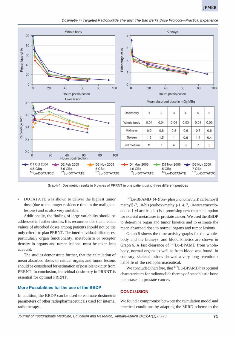
Dosimetry in Targeted Radionuclide Therapy: The Bad Berka Dose Protocol—Practical Experience
JPMER
Journal of Postgraduate Medicine, Education and Research, January-March 2013;47(1):65-73 71
• DOTATATE was shown to deliver the highest tumordose (due to the longer residence time in the malignantlesions) and is also very suitable.Additionally, the finding of large variability should be
addressed in further studies. It is recommended that medianvalues of absorbed doses among patients should not be theonly criteria to plan PRRNT. The interindividual differences,particularly organ functionality, metabolism or receptordensity in organs and tumor lesions, must be taken intoaccount.
The studies demonstrate further, that the calculation ofmean absorbed doses to critical organs and tumor lesionsshould be considered for estimation of possible toxicity fromPRRNT. In conclusion, individual dosimetry in PRRNT isessential for optimal PRRNT.
More Possibilities for the use of the BBDP
In addition, the BBDP can be used to estimate dosimetricparameters of other radiopharmaceuticals used for internalradiotherapy.
Graph 4: Dosimetric results in 6 cycles of PRRNT in one patient using three different peptides
177Lu-BPAMD ((4-{[bis-(phosphonomethyl)) carbamoyl]methyl}-7, 10-bis (carboxymethyl)-1, 4, 7, 10-tetraazacyclo-dodec-1-yl acetic acid) is a promising new treatment optionfor skeletal metastases in prostate cancer. We used the BBDPto determine organ and tumor kinetics and to estimate themean absorbed dose to normal organs and tumor lesions.
Graph 5 shows the time-activity graphs for the whole-body and the kidneys, and blood kinetics are shown inGraph 6. A fast clearance of 177Lu-BPAMD from whole-body, normal organs as well as from blood was found. Incontrary, skeletal lesions showed a very long retention /half-life of the radiopharmaceutical.
We concluded therefore, that 177Lu-BPAMD has optimalcharacteristics for radionuclide therapy of osteoblastic bonemetastases in prostate cancer.
CONCLUSION
We found a compromise between the calculation model andpractical conditions by adapting the MIRD scheme to the
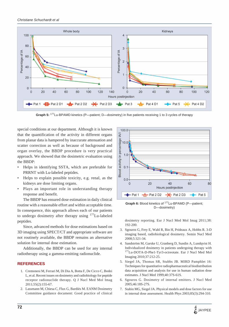
Christiane Schuchardt et al
72JAYPEE
special conditions at our department. Although it is knownthat the quantification of the activity in different organsfrom planar data is hampered by inaccurate attenuation andscatter correction as well as because of background andorgan overlay, the BBDP procedure is very practicalapproach. We showed that the dosimetric evaluation usingthe BBDP:• Helps in identifying SSTA, which are preferable for
PRRNT with Lu-labeled peptides.• Helps to explain possible toxicity, e.g. renal, as the
kidneys are dose limiting organs.• Plays an important role in understanding therapy
response and benefit.The BBDP has ensured dose estimation in daily clinical
routine with a reasonable effort and within acceptable time.In consequence, this approach allows each of our patientsto undergo dosimetry after therapy using 177Lu-labeledpeptides.
Since, advanced methods for dose estimations based on3D imaging using SPECT/CT and appropriate software arenot routinely available, the BBDP remains an alternativesolution for internal dose estimation.
Additionally, the BBDP can be used for any internalradiotherapy using a gamma-emitting radionuclide.
REFERENCES
1. Cremonesi M, Ferrari M, Di Dia A, Botta F, De Cicco C, BodeiL, et al. Recent issues on dosimetry and radiobiology for peptidereceptor radionuclide therapy. Q J Nucl Med Mol Imag2011;55(2):155-67.
2. Lassmann M, Chiesa C, Flux G, Bardiès M. EANM DosimetryCommittee guidance document: Good practice of clinical
Graph 5: 177Lu-BPAMD kinetics (P—patient; D—dosimetry) in five patients receiving 1 to 3 cycles of therapy
Graph 6: Blood kinetics of 177Lu-BPAMD (P—patient;D—dosimetry)
dosimetry reporting. Eur J Nucl Med Mol Imag 2011;38:192-200.
3. Sgouros G, Frey E, Wahl R, Bin H, Prideaux A, Hobbs R. 3-Dimaging based, radiobiological dosimetry. Semin Nucl Med2008;5:321-34.
4. Sandström M, Garske U, Granberg D, Sundin A, Lundqvist H.Individualized dosimetry in patients undergoing therapy with177Lu-DOTA-D-Phe1-Tyr3-octreotate. Eur J Nucl Med MolImaging 2010;37:212-25.
5. Siegel JA, Thomas SR, Stubbs JB. MIRD Pamphlet 16:Techniques for quantitative radiopharmaceutical biodistributiondata acquisition and analysis for use in human radiation doseestimates. J Nucl Med 1999;40:37S-61S.
6. Sgouros G. Dosimetry of internal emitters. J Nucl Med2005;46:18S-27S.
7. Stabin MG, Siegel JA. Physical models and dose factors for usein internal dose assessment. Health Phys 2003;85(3):294-310.

Dosimetry in Targeted Radionuclide Therapy: The Bad Berka Dose Protocol—Practical Experience
JPMER
Journal of Postgraduate Medicine, Education and Research, January-March 2013;47(1):65-73 73
8. Wehrmann C, Senftleben S, Zachert C, Mueller D, Baum RP.Results of individual patient dosimetry in peptide receptorradionuclide therapy with 177Lu DOTA-TATE and 177LuDOTANOC. Cancer Biother Radiopharm 2007;22(3):406-16.
9. Stabin MG, Sparks RP, Crowe E. OLINDA/EXM: The second-generation personal computer software for internal doseassessment in nuclear medicine. J Nucl Med 2005;46:1023-27.
10. Wessels BW, Bolch WE, Bouchet LG, et al. Bone marrowdosimetry using blood-based models for radiolabeled antibodytherapy: A multi-institutional comparison. J Nucl Med2004;45:1725-33.
11. Sgouros G. Bone marrow dosimetry for radioimmunotherapy:Theoretical considerations. J Nucl Med 1993;34:669.
12. Bardies M, Buvat I. Dosimetry in nuclear medicine therapy:What are the specifics in image quantification for dosimetry? QJ Nucl Med Mol Imag 2011;55(1):5-20.
13. Sandström M, Garske U, Granberg D, Sundin A, Lundqvist H.Individualized dosimetry in patients undergoing therapy with177Lu-DOTA-D-Phe1-Tyr3-octreotate. Eur J Nucl Med MolImag 2010;37:212-25.
14. Garkavij M, Nickel M, Sjögreen-Gleisner K, Ljungberg M, etal. 177Lu-[DOTA0,Tyr3] Octreotate therapy in patients withdisseminated neuroendocrine tumors: Analysis of dosimetry withimpact on future therapeutic strategy. Cancer 2010;116(4Suppl):1084-92.
ABOUT THE AUTHORS
Christiane Schuchardt
Medical Physicists, THERANOSTICS Center for MolecularRadiotherapy and Molecular Imaging, ENETS Center of Excellence,Zentralklinik Bad Berka, Germany
Harshad Kulkarni
Resident Physician, THERANOSTICS Center for MolecularRadiotherapy and Molecular Imaging, ENETS Center of ExcellenceZentralklinik Bad Berka, Germany
Carolin Zachert
Senior Physician, THERANOSTICS Center for MolecularRadiotherapy and Molecular Imaging, ENETS Center of ExcellenceZentralklinik Bad Berka, Germany
Richard P Baum (Corresponding Author)
Chairman and Clinical Director, THERANOSTICS Center for MolecularRadiotherapy and Molecular Imaging, ENETS Center of ExcellenceZentralklinik Bad Berka, 99437, Bad Berka, Germany, Phone: +49-364585-2200, Fax: +49-364-585-3515, e-mail: [email protected],www.zentralklinik-bad-berka.de

Jaya Shukla, BR Mittal
74JAYPEE
COMMENTARY
Ga-68: A Versatile PET Imaging RadionuclideJaya Shukla, BR Mittal
ABSTRACT
Gallium-68, a positron emitter, is available via 68Ge/68Gagenerators. The simple chemistry and easy availability hasincreased its application from the clinical diagnosis topersonalized therapy and has lot more potential in future.
Keywords: 68Ge/68Ga generator, Cyclotron, Bifunctionalchelators, Nanoparticles.
How to cite this article: Shukla J, Mittal BR. Ga-68: A VersatilePET Imaging Radionuclide. J Postgrad Med Edu Res2013;47(1):74-76.
Source of support: Nil
Conflict of interest: None declared
INTRODUCTION
The availability of 68Ga as a positron emission tomography(PET) radionuclide for imaging dates back to early 60’s ata time when neither PET nor fluorine-18 (F-18) wasestablished. With the emergence of cyclotron and automatedchemistry modules, F-18 radiopharmaceuticals establishedtheir empire in nuclear medicine. The resurgence of 68Ge/68Ga generator was due to the consistent efforts byradiochemists from Czechoslovakia and Russia in 20thcentury.1,2 The modern 68Ge/68Ga generators have provedto be a milestone for noninvasive state-of-art PET/CTimaging. After that there was no looking back for Ga-68imaging.
The advantages of 68Ga over other PET-basedradionuclides are its availability from an in-house generatorindependent of an onsite cyclotron. The half-life of Ga-68is 68 minutes. Eighty-nine percent of Ga-68 decays byemitting positron of 1.92 MeV and the rest 11% by electroncapture. The parent 68Ge is produced in accelerator by(p, 2n) reaction on Ga2O3 target. 68Ge decays with a half-life of 270.8 days by electron capture which enable longshelf life to generator (>6 months) and reduces the unit-dose cost. Due to short half life, Ga-68 can be eluted 2 to 3times a day (after 3-5 hours) as per the requirement/patientsnumber.
68Ge/68Ga GENERATOR SYSTEM
In 68Ge/68Ga generator system, 68Ge is strongly adsorbedon different solid supports such as, metal oxides (Al2O3,TiO2 or SnO2), organic (pyrogallol-formaldehyde resins)and inorganic supports (silica based).3-6 The Ga-68 fromcurrently available 68Ge/68Ga generators is eluted with dilute
10.5005/jp-journals-10028-1059
hydrochloric acid as a cationic 68Ga3+. Initially the longprocessing was required to remove metallic impurities ofsolid support and Ge-68 breakthrough from Ga-68 elute.With the development of nonmetallic silica-based column,the processing step for the elution of Ga-68 was eliminated.Silica has high binding affinity for Ge which reduces theGe-68 breakthrough to negligible level.
APPLICATION OF 68Ga3+
Gallium acts as an iron analog and binds to transferrin andlactoferrin. The complex diffuses through loose endothelialjunctions of capillaries at the sites of inflammation andenters the extracellular fluid. Leukocytes also migrate tosites of inflammation, degranulate and release largequantities of lactoferrin. Ga attaches to siderophores ofbacteria and therefore can be used in leukopenic patientswith bacterial infection and also in detecting sterileabscesses that provoke a leukocyte response.7 In earliertimes, Ga-67 citrate was very popular for infection imagingby exploiting above properties of Ga.8 Due to low energy,long imaging time (half life: 78 hours) and poor imagequality, the impact of Ga-67 imaging faded away. Theresurgence of Ga-68, a PET radionuclide has revived theimportance of 68Ga as natural in vivo infection/inflammationimaging agent.9,10 Now, the infection imaging is done using68Ga-citrate and 68GaCl3.
68Ga3+ CHEMISTRY
68Ga-complexes has simple aqueous coordinate chemistrybased on Me(III).11 Gallium, in aqueous solution, occurssolely in +3 oxidation state and is classified as a hard acidmetal. Gallium can bond to highly ionic hard base liganddonors, such as carboxylic acids, amino nitrogens,hydroxamates, thiols and phenolates. The Ga chemistry ishighly influenced by pH change. The optimum pH (3-5) isrequired for its aqueous chemistry. The pH below optimuminhibits the reaction and at pH above optimum range, i.e.>5, it tends to hydrolyze and leads to the precipitation asGa(OH)3.
Ga-68-LABELED MOLECULES
Several suitable bifunctional chelators have been developed,and coupled with biomolecules for Ga-68 labeling. DOTA,NOGADA and NOTA are commonly used bifunctionalchelators. Many peptides/biomolecules like receptor

Ga-68: A Versatile PET Imaging Radionuclide
JPMER
Journal of Postgraduate Medicine, Education and Research, January-March 2013;47(1):74-76 75
peptides and antibodies etc have now been successfullymodified by these chelating agents without compromisingtheir functional properties which further widened the roleof Ga-68 PET/CT imaging. These peptides/biomoleculesshow very fast target localization and fast blood clearancethus, making the short half-life ideal for clinical studies. Inthe last decade 68Ga-DOTA-octreotides replaced99mTc/111In-DTPA-octroescan used for neuroendocrinetumor (NET) imaging. 68Ga-DOTA-octreotides proved tobe a promising radiopharmaceutical for diagnosis, treatmentplanning, therapy response evaluation and diseaserecurrence of NET.12 Several peptides like somatostatin forimaging NET, integrin peptide for imaging neoangiogenesis,etc. are now available as cold kits.
Ga-68 labeling has also been explored with other peptidereceptors, like cholecystokinin/gastrin and GLP-1 analogsfor NETs, bombesin and neuropeptide-Y analogs forprostate or breast cancers.13-15 Arg-Gly-Asp (RGD) a cyclictripeptide is used to image neoangiogenic/angiogenicvessels and mediated cell adhesion molecule by targetingoverexpressed v3 integrin. Inflammatory bowel disease,inflamed synovial tissue of rheumatoid arthritis andinflammatory atherosclerotic plaques can also be visualizedby 68Ga-RGD peptide.16 Vascular adhesion protein-I(VAP 1) is an inflammation inducible endothelial cellmolecule. It also contributes to extravasation cascade andcontrols trafficking of leukocyte at the site of inflammation.VAP-1 is expressed on the endothelial surface of intestinalblood vessels in inflammatory diseases, in skin inflammation(psoriasis), synovial blood vessels of inflamed joints(rheumatic arthritis) and cardiovascular diseases. However,VAP-1 is absent from the endothelial surface of normaltissues. Ga-68-labeled peptide against VAP-1 have beenused for in vivo imaging of VAP-1 knockout.17
FUTURE
In the modern era of ‘personalized medicine’, Ga-68 has apromising role. The targets can be defined with the help ofdiagnostic Ga-68 PET/CT using appropriate ligands(peptides/biomolecules) for detection of disease,pretherapeutic measurement of organ and tumor doses. Thetherapeutic analog of imaging radionuclide (Lu-177/Y-90)can be selected for therapy using the same peptide.Nanomedicine in future has a great potential for earlydetection, accurate diagnosis and personalized treatment ofvarious diseases, particularly cancer. Nanomedicine canoffer unprecedented interactions with biomolecules, on thesurface as well as inside the cells which may revolutionizedisease diagnosis and treatment. Molecular imaging canmeasure the expression of molecular markers at different
stages of diseases and provide relevant and reliableinformation in an intact system. The information may speedup the drug development process and help in individualizedtreatment monitoring and dose optimization. Ga-68 is anideal radionuclide for labeling various nanoparticles likesingle-walled carbon nanotubes (SWNTs), quantum dots(QD), polymeric and metallic nanoparticles, etc. forevaluation of their biodistribution, pharmacokineticproperties and tumor targeting efficacy.18-20 The informationmay be utilized for early diagnosis, selecting better treatmentoptions and predicting the disease prognosis.
With each passing day, the reign of Ga-68 in researchand clinical application is increasingly being established. Ithas a lot in store for future. The easy availability and simplechemistry based on sophisticated chelating agents for Ga-68 will make it parallel to kit-based Tc-99m chemistry aspredicted by Deutsch.21
REFERENCES
1. Kopecký P, Mudrová B, Svoboda K. The study of conditionsfor the preparation and utilization of 68Ge-68Ga generator. Int JAppl Radiat Isot 1973;24:73-80.
2. Schuhmacher J, Maier-Borst W. 68Ge-68Ga generator for theproduction of 68Ga in an ionic form. Int J Appl Radiat Isot1974;25:263-68.
3. Mirzadeh S, Lambrecht R. Radiochemistry of germanium. JRadioanal Nucl Chem 1996;202:7-102.
4. Meyer GJ, Maecke H, Schuhmacher J, Knapp WH, HofmannM. 68Ga-labelled DOTA-derivatised peptide ligands. Eur J NuclMed Mol Imag 2004;31:1097-104.
5. Velikyan I, Beyer GJ, Langstrom B. Microwave-supportedpreparation of 68Ga bioconjugates with high specificradioactivity. Bioconjug Chem 2004;15:554-60.
6. Schuhmacher J, Maier-Borst W. A new 68Ge/68Ga radioisotopegenerator system for production of 68Ga in dilute HCl. Int JAppl Radiat Isot 1981;32:31-36.
7. El-Maghraby TA. Nuclear medicine methods for evaluation ofskeletal infection among other diagnostic modalities. Q J NuclMed Mol Imaging 2006;50:167-92.
8. Palestro CJ. The current role of gallium imaging in infection.Semin Nucl Med 1994;24:128-41.
9. Mäkinen TJ, Lankinen P, Pöyhönen T, Jalava J, Aro HT,Roivainen A. Comparison of 18F-FDG and 68Ga PET imagingin the assessment of experimental osteomyelitis due toStaphylococcus aureus. Eur J Nucl Med Mol Imaging2005;32:1259-68.
10. Silvola J, Laitinen I, Sipilä H, Laine VJO, Leppänen P, Ylä-Herttuala S, et al. Uptake of 68gallium in atherosclerotic plaquesin LDLR/ApoB100/100 mice. EJNMMI Res 2011;1:14.
11. Green MA, Welch MJ. Gallium radiopharmaceutical chemistry.Int J Rad Appl Instrum B 1989;16:435-48.
12. Naji M, AL-Nahhas A. 68Ga-labelled peptides in themanagement of neuroectodermal tumours. Eur J Nucl Med MolImaging 2012;39(Suppl 1):S61-67.
13. Von Guggen Berg E, Rangger C, Sosabowski J, Laverman P,Reubi JC, Virgolini IJ, et al. Preclinical evaluation ofradiolabeled DOTA-derivatized cyclic minigastrin analogs for

Jaya Shukla, BR Mittal
76JAYPEE
targeting cholecystokinin receptor expressing malignancies. MolImaging Biol 2012 Jun;14(3):366-75.
14. Brom M, Oyen WJG, Joosten L, Gotthardt M, Boerman OC.Ga-labelled exendin-3, a new agent for the detection ofinsulinomas with PET. Eur J Nucl Med Mol Imaging 2010;37:1345-55.
15. Schuhmacher J, Zhang H, Doll J, Mäcke HR, Matys R, HauserH, et al. GRP receptor-targeted PET of a rat pancreas carcinomaxenograft in nude mice with a 68Ga-labeled bombesin (6-14)analog. J Nucl Med 2005;46:691-99.
16. Knetsch PA, Petrik M, Griessinger CM, Rangger C, Fani M,Kesenheimer C, et al. [68Ga]NODAGA-RGD for imaging v3integrin expression. Eur J Nucl Med Mol Imaging 2011;38:1303-12.
17. Autio A, Ujula T, Luoto P, Salomäki S, Jalkanen S, RoivainenA. PET imaging of inflammation and adenocarcinomaxenografts using vascular adhesion protein 1 targeting peptide(68)Ga-DOTAVAP-P1: Comparison with (18)F-FDG. Eur JNucl Med Mol Imag 2010;37:1918-25.
18. Liu Z, Cai W, He L, Nakayama N, Chen K, Sun X, et al. In vivobiodistribution and highly efficient tumor targeting of carbonnanotubes in mice. Nat Nanotechnol 2007;2:47.
19. Cai W, Chen K, Li ZB, Gambhir SS, Chen X. Dual-functionprobe for PET and near-infrared fluorescence imaging of tumorvasculature. J Nucl Med 2007;48:1862-70.
20. Lee HY, Li Z, Chen K, Hsu AR, Xu C, Xie J, et al. PET/MRIdual-modality tumor imaging using arginine-glycine-aspartic(RGD)–conjugated radiolabeled iron oxide nanoparticles. J NuclMed 2008;49:1371-79.
21. Deutsch E. Clinical PET: Its time has come? J Nucl Med 1993;34:1132-33.
ABOUT THE AUTHORS
Jaya Shukla (Corresponding Author)
Assistant Professor, Department of Nuclear Medicine, PostgraduateInstitute of Medical Education and Research, Chandigarh, Indiae-mail: [email protected]
BR Mittal
Professor and Head, Department of Nuclear Medicine, PostgraduateInstitute of Medical Education and Research, Chandigarh, India