Embed Size (px)
Citation preview
Vol.64: e21200163, 2021 https://doi.org/10.1590/1678-4324-2021200163
ISSN 1678-4324 Online Edition
Brazilian Archives of Biology and Technology. Vol.64: e21200163, 2021 www.scielo.br/babt
Article – Biological and Applied Sciences
Isolation of Polyphenols from Soursop (Annona muricata L.) Leaves Using Green Chemistry Techniques and their Anticancer Effect
Valdez-Guerrero Daisy Yathzamiry1
https://orcid.org/0000-0002-3073-1635
Esparza-González Sandra Cecilia2 https://orcid.org/0000-0002-3336-8739
Morlett-Chávez Jesús Antonio1 https://orcid.org/0000-0001-7988-423X
Nery-Flores Sendar Daniel1 https://orcid.org/0000-0003-0739-2230
Flores-Gallegos Adriana Carolina1
https://orcid.org/0000-0001-5092-1404
Ascacio-Valdés Juan Alberto1
https://orcid.org/0000-0001-6595-863X
Rodríguez-Herrera Raúl1* https://orcid.org/0000-0002-6428-4925
1Universidad Autónoma de Coahuila, School of Chemistry, Food Research Department, Saltillo, Coahuila, México.; 2Universidad Autónoma de Coahuila. School of Dentistry,, Saltillo, Coahuila, México.
Editor-in-Chief: Paulo Vitor Farago Associate Editor:Jane Budel
Received: 2020.03.20; Accepted: 2020.12.18
*Correspondence: [email protected]; Tel.: +52(844)4169213 and 4161238. (R.H.R.).
Abstract: Cervical cancer is classified as the fourth most common malignancy in women. Natural compounds
are a therapeutic alternative in cancer therapy. The aim of the study is to isolate, fractionate, and characterize
extracts obtained from soursop leaves (Annona muricata L.) and determine their cytotoxic effect against HeLa
cervical cancer cells and non-carcinogenic fibroblast 3T3 cells. The phytochemicals of soursop leaves were
extracted through emerging green technologies such as the novel use of microwave-ultrasound hybridization
and the use of environmentally friendly solvents (water and ethanol), in addition to the purification of extracts
enriched in polyphenols by liquid chromatography with Amberlite XAD-16. Total aqueous and ethanolic
extract were purified, as well as the fraction one of each extract. The extracts recovered from soursop leaves
contained kaempferol and its isomers, procyanidins, catechin, and quercetin. The viability of the cells was
determined with the MTT (3-(4,5-dimethylthiazol-2-yl)-2,5-diphenyltetrazolium bromide) assay. HeLa and 3T3
cells were exposed to concentrations of 25, 50, 75, 100, 150, 200, and 250 ppm of a solution of soursop leaf
extract powder. The MTT assay showed that soursop leaf extracts were toxic to both cell lines in general,
however, the ethanolic extract at 25 and 50 ppm demonstrated inhibition in cell viability against the HeLa
HIGHLIGHTS
Isolate, fractionate and characterize extracts obtained from soursop leaves.
Use of emerging green technologies such as microwave-ultrasound hybridization.
The extracts contain kaempferol, procyanidins, catechin, and quercetin.
The total ethanolic extract demonstrates cytotoxic effect on HeLa cells.
2 Valdez-Guerrero, D.Y.; et al.
Brazilian Archives of Biology and Technology. Vol.64: e21200163, 2021 www.scielo.br/babt
cancer line and low cytotoxicity for 3T3 fibroblast cells. In conclusion, the novel microwave-ultrasound
hybridization technology allows the extraction of polyphenols that may have a potential cytotoxic effect on
cancer cells.
Keywords: green solvents; microwave-ultrasound; soursop; polyphenols; HeLa.
INTRODUCTION
Each year, more than a half-million women are diagnosed with cervical cancer and results in more than
300,000 deaths worldwide, also, this cancer is classified as the fourth most common malignancy in women
[1]. In 2012, 528,000 new cases were diagnosed, and 266,000 women died and almost 90 % of them were
registered in countries with low or medium income and it is predicted that without urgent care, deaths due to
cervical cancer could increase by almost 25 % over the next 10 years [2].
Cervical cancer is a malignant neoplasm that develops in the inner fibromuscular portion of the uterus,
which is projected within the vagina, and it is related to human Papillomavirus (HPV), in addition to other
factors of the local microenvironment, such as pH and the vaginal microbiota [3,4]. The treatment depends
on the stage of cancer, and more than one therapy can be administered, such as radiotherapy,
chemotherapy, or immunotherapy [5,6]. The most common drugs used to treat cervical cancer are cisplatin,
carboplatin, paclitaxel, topotecan, and gemcitabine. However, different side effects have been associated
with the use of these therapies [6]. In addition, the high cost of the treatments hinders their access to the
population [7]. Therefore, it is essential to search for new treatments that are cheaper and that do not cause
side effects. One of these alternatives is the use of bioactive compounds from natural sources and, therefore,
take advantage of the biological wealth of developing countries [8]. Moreover, the process of obtaining
bioactive compounds needs to incorporate “green extraction” methods, to achieve a faster extraction rate,
more effective energy use, reduction in the number in processing steps, use of safer solvents, and avoiding
waste production [9]. Currently, the use of "green" methodologies is being sought, which promotes the use
of cleaner extraction protocols, with the proposal of protecting the consumer and the environment [10].
Ultrasound and microwave techniques have proven to be a viable alternative for the extraction of natural
molecules. Likewise, the combination of analytical techniques is a strategy to save energy, time, and
resources [9].
It has been shown that some plants are a source of antitumor compounds of medical importance, so it
is important to conduct in vitro/in vivo research that supports their antineoplastic potential [11]. In species of
the Annonaceae family, the presence of bioactive compounds with antioxidant, antitumor,
immunosuppressive, anti-inflammatory, and antimicrobial activity among others has been reported [12].
Annona muricata L., widely known as soursop, is a tree 5-8 meters in height that has large, bright, and dark
green leaves [13]. Different parts of A. muricata have been widely used in the traditional medicine of different
countries for the treatment of various ailments and diseases [12]. In vitro studies have shown that plants
contain compounds that block some essential metabolites of cancer cells and this induces the death of
malignant cells [14-16]. Based on this background, the present study was established under the following
objectives: Obtain phytochemicals of soursop (Annona muricata) leaves through emerging green
technologies (hybridization of ultrasound and microwave) and environmentally friendly solvents, purify and
separate rich-phytochemical extract fractions, and finally evaluate the cytotoxic activity of the purified
fractions on HeLa and 3T3 cell lines.
MATERIAL AND METHODS
Plant material
Samples of dried soursop (Annona muricata) leaves were commercially acquired from the “Productos
Rickland” company, which specializes in soursop products in Mexico and is located in Las Varas, Nayarit
State, México. 1040 g of soursop leaves were purchased from the supplier. First, the leaves were selected,
eliminating those that had some physical or microorganism damage. The leaves were then allowed to dry for
three days at room temperature (22-26 °C) and 24 h in a Coldryer-NWT-5 brand dehydrator. Subsequently,
the leaves were cut into small pieces, which were ground in a blender, and then, the powder was recovered.

Soursop polyphenols and their cytotoxic effect 3
Brazilian Archives of Biology and Technology. Vol.64: e21200163, 2021 www.scielo.br/babt
Extract preparation
The extracts were obtained with two different solvents, water, and an ethanol/water mixture. To obtain
the aqueous extract, 62.5 g of the powder of A. muricata leaves were weighed and mixed with 1000 mL of
distilled water in a one litter reactor to obtain a 1:16 ratio mass: volume. For the ethanol extract, an
ethanol/water mixture was made (700 mL of ethanol 96% in 300 mL of distilled water), and then 62.5 g of the
crushed soursop leaves were mixed with the solution prepared in the one-liter reactor.
Phytochemical extraction
The mixtures of plant material and solvent previously prepared and contained in a 1 L reactor were
placed in the “Ultrasonic Microwave Comparative Workstation (ATPI0, Nanjing ATPIO Instruments
Manufacture Co., Ltd Company, China) equipment". The following conditions were used for Ultrasound:
Power Radio 20, Ultrasonic on Relay 10, Ultrasonic Off Relay 3, Amplitude Transformer 25, and Set 20; and
for Microwaves: Power Radio 800, Display Power 0, Set Time 70 °C and Holding Time 5. These conditions
were defined based on the previous work of the research group for the extraction of polyphenolic compounds
where an adequate extraction of the same and the conservation of biological properties were demonstrated
[17,18]. Finally, the aqueous and the ethanolic extracts were filtered through a Whatman No.41 filter paper
to remove the insoluble material and the liquid obtained was stored at 4 °C in an amber bottle.
Purification and fractionation of phytochemicals
Polyphenols extracts were obtained by using a liquid chromatography column with Amberlite XAD-16,
the column was washed with distilled water, then the soursop extract was added, first elution was carried out
with distilled water to discard undesirable compounds such as carbohydrates, lipids, and other impurities,
and then ethanol was used to recover the phenolic fraction for 6 h. This procedure was performed for both
extracts. The technique was repeated to obtain the fractions of the extracts. The fractions were collected at
intervals of 1 hour for 6 h; the six fractions of each extract were stored independently. The samples of the
complete chromatography and the fractions were placed in an oven at 45 °C for 12 h to remove the solvent
and thus recover polyphenol powders. To avoid the degradation of phenolic compounds in the drying process,
they were only heated up to 45 °C, since various articles have shown that the degradation process increases
from 60-70 °C [19-21]. Finally, the polyphenols powder was stored in amber bottles at 4 °C to avoid compound
degradation.
Identification of polyphenols by HPLC
For each sample, 1 mg of polyphenol powder obtained was mixed with 1 mL of ethanol (96%). The
compounds obtained after chromatographies were analyzed by HPLC (VarianProStar, USA) with a diode
array detector (280 nm). A mass spectrometer (MS) with a liquid chromatography ion trap (Varian 500-MS IT
Mass Spectrometer, USA) equipped with an electrospray ion source was also used. Samples (5 µL) were
injected into a Denali C18 column (3 µm, 150 mm × 2.1 mm, Grace, USA). The oven temperature was
maintained at 30 °C. The eluents were formic acid (0.2%, v/v; solvent A) and acetonitrile (solvent B). The
following gradient was applied: initial, 3% B; 0-5 min, 9% linear B; 5-15 min, 16% linear B; 15-45 min, 50%
linear B. The column was washed and reconditioned. The flow rate was maintained at 0.2 mL/min and the
elution was monitored at 245, 280, 320, and 550 nm. All eluent (0.2 mL/min) was injected into the mass
spectrometer, without dividing. All MS experiments were performed in negative mode [M-H]-1. The data was
collected and processed using the MS Workstation software (V 6.9). The samples were first analyzed in the
full scan mode acquired in the range 50-2000m/z. MS/MS analyses were performed on a series of selected
precursor ions. Use of standards was not necessary for this study since the identification of the compounds
is carried out by molecular weight (m/z), supported by the HPLC/MS analysis and, finally, the compounds
were compared using a database of bioactive compounds (WorkStation version 2.0 database, Varian, CA,
USA).
Cell culture
The HeLa cell line (ATCC® CCL-2™), which are epithelial cells of the human cervix adenocarcinoma,
and the 3T3 cell line (ATCC® CRL-1658™), fibroblasts from Swiss albino mouse embryonic tissues (3T3,
NYU, USA); were grown in DMEM medium (Dulbecco Modified Eagles Minimal Essential Medium)
supplemented with 100 U/mL penicillin, 100 mg/mL streptomycin, 0.1 mM non-essential amino acids, 10%
fetal bovine serum, and 2 mM L-glutamine in 5% CO2 at 37 °C.

4 Valdez-Guerrero, D.Y.; et al.
Brazilian Archives of Biology and Technology. Vol.64: e21200163, 2021 www.scielo.br/babt
Cytotoxicity by MTT assay
1 mg of each extract was placed in a microtube and re-suspended in 1 mL of DMEM cell culture medium
supplemented using vortex and sonication. Subsequently, dilutions of 25, 50, 75, 100, 150, 200, and 250
ppm were made. The cytotoxicity of the different concentrations of the purified molecules was determined by
the 3-(4,5-dimethylthiazol-2yl)-2,5-diphenyltetrazolium bromide (MTT) assay. 7x10³ cells per well were
transferred to a 96-well plate and grown under the conditions described above for 24 h. Then, 200 µL of the
culture medium with the extracts at different concentrations (25, 50, 75, 100, 150, 200, and 250 ppm) was
added to each well and allowed to incubate for 24 h. After the incubation, morphological changes were
examined with a Labomed inverted microscope at 100x. After this time, the supernatant was discarded and
100 µL of culture medium with 10 µL of the MTT solution (5 mg/mL) was added to each well, and the
microplates were incubated for 4 h at 37 °C. Finally, the culture medium was discarded and 150 µL of DMSO
was placed, and the absorbance was measured at 570 nm using a microplate reader (Multiskan, Thermo
Scientific, USA). The results are expressed as the percentage of cell viability. A reduction in viability greater
than 30% was considered a cytotoxic effect according to ISO 10993-5.
Statistical analysis
The data were expressed as mean ± standard error of the mean (SEM) of three replications. The results
were analyzed using a two-way ANOVA followed by Sidak’s test. p <0.05 were considered statistically
significant. GraphPad Prism 6.01 software (GraphPad Software Inc., La Jolla, CA, USA) was employed for
all analyses.
RESULTS
Obtainment of leaf polyphenols
Table 1 shows the yield in grams of the powder obtained compared with 100% amount of vegetable raw
material that was used to obtain the extracts. The percentage of yield was from 1.04-4.46%, this may be due
to the solvent or the type of chromatographic column used.
Table 1. Percentage yield of the extract obtained from soursop leave
Type of extract Vegetal raw
material (g)
Type of
chromatography
Amount (g) recovered
from extract
Yield (%)
Aqueous 62.5 Complete 1.2 1.92
Aqueous 62.5 Fractionated 0.65 1.04
Ethanolic 62.5 Complete 2.65 4.22
Ethanolic 62.5 Fractionated 2.79 4.46
Characterization of Polyphenols by HPLC
The HPLC scans, retention time, molecular mass, the compound family and the compounds that were
detected in the extracts are shown in Figures 1-4.
Soursop polyphenols and their cytotoxic effect 5
Brazilian Archives of Biology and Technology. Vol.64: e21200163, 2021 www.scielo.br/babt
Figure 1. (A) Chromatogram of the aqueous extract of the soursop leaf of fraction one of the chromatography. (B) Compounds detected in the aqueous extract of the soursop leaf of fraction one of the chromatography.

6 Valdez-Guerrero, D.Y.; et al.
Brazilian Archives of Biology and Technology. Vol.64: e21200163, 2021 www.scielo.br/babt
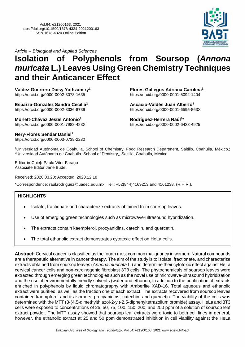
Figure 2. (A) Chromatogram of the aqueous extract of the soursop leaf of the complete chromatography. (B) Compounds detected in the aqueous extract of the soursop leaf of the complete chromatography.
Soursop polyphenols and their cytotoxic effect 7
Brazilian Archives of Biology and Technology. Vol.64: e21200163, 2021 www.scielo.br/babt
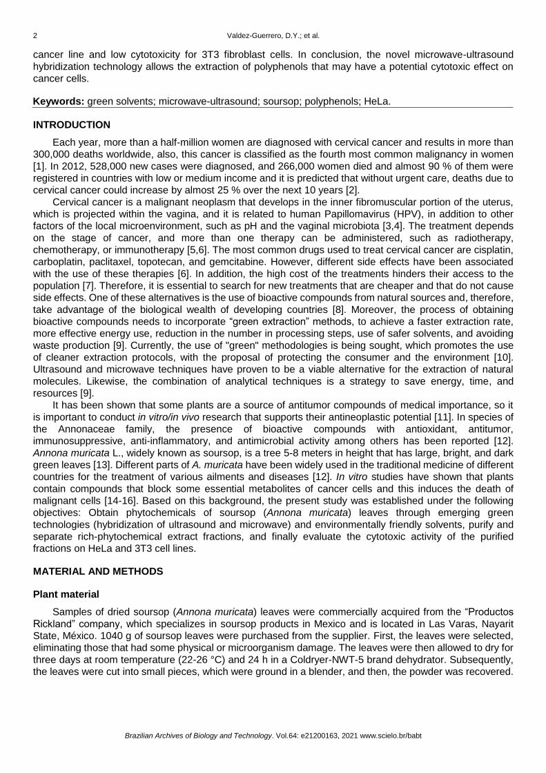
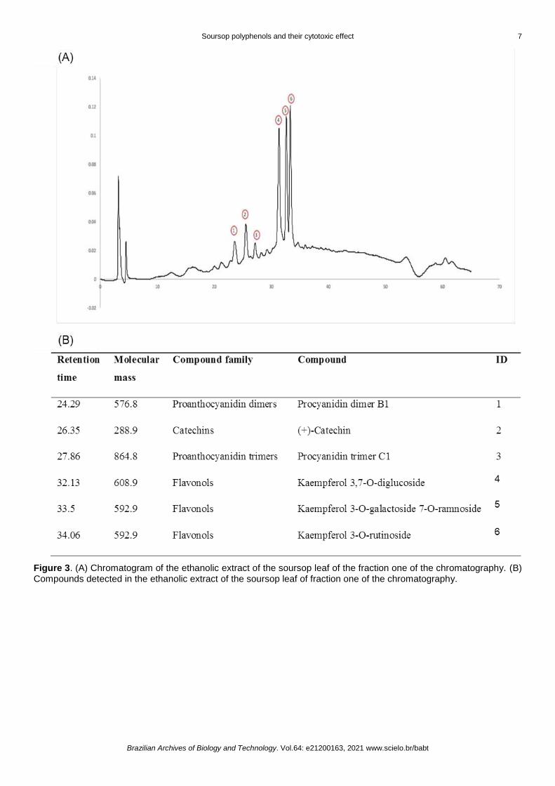
Figure 3. (A) Chromatogram of the ethanolic extract of the soursop leaf of the fraction one of the chromatography. (B) Compounds detected in the ethanolic extract of the soursop leaf of fraction one of the chromatography.
8 Valdez-Guerrero, D.Y.; et al.
Brazilian Archives of Biology and Technology. Vol.64: e21200163, 2021 www.scielo.br/babt
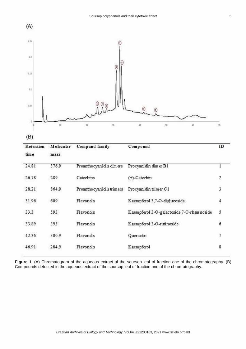
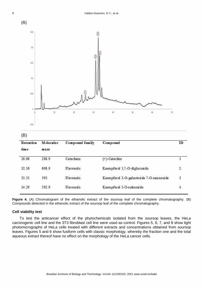
Figure 4. (A) Chromatogram of the ethanolic extract of the soursop leaf of the complete chromatography. (B) Compounds detected in the ethanolic extract of the soursop leaf of the complete chromatography.
Cell viability test
To test the anticancer effect of the phytochemicals isolated from the soursop leaves, the HeLa
carcinogenic cell line and the 3T3 fibroblast cell line were used as control. Figures 5, 6, 7, and 8 show light
photomicrographs of HeLa cells treated with different extracts and concentrations obtained from soursop
leaves. Figures 5 and 6 show fusiform cells with classic morphology, whereby the fraction one and the total
aqueous extract thereof have no effect on the morphology of the HeLa cancer cells.
Soursop polyphenols and their cytotoxic effect 9
Brazilian Archives of Biology and Technology. Vol.64: e21200163, 2021 www.scielo.br/babt
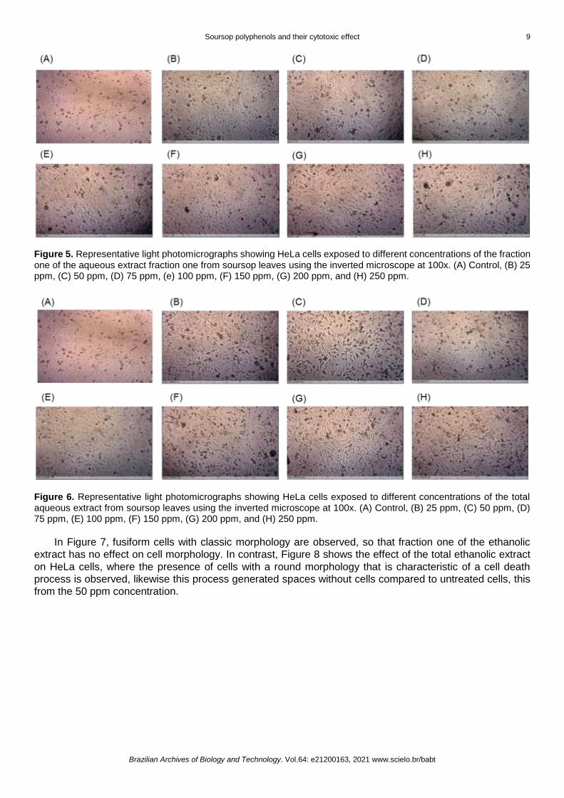
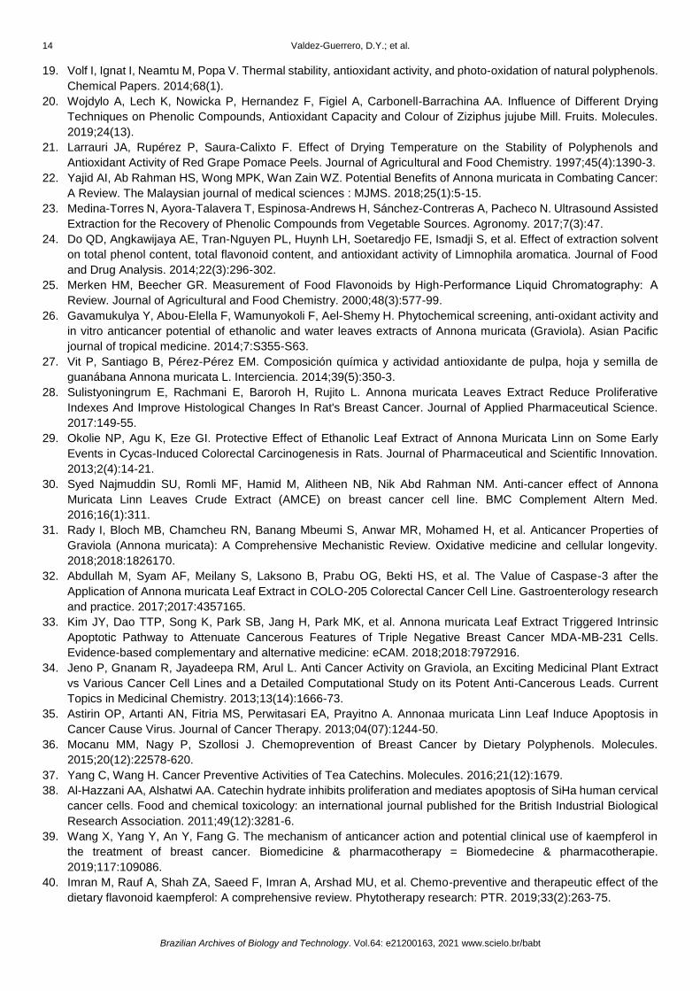
Figure 5. Representative light photomicrographs showing HeLa cells exposed to different concentrations of the fraction one of the aqueous extract fraction one from soursop leaves using the inverted microscope at 100x. (A) Control, (B) 25 ppm, (C) 50 ppm, (D) 75 ppm, (e) 100 ppm, (F) 150 ppm, (G) 200 ppm, and (H) 250 ppm.
Figure 6. Representative light photomicrographs showing HeLa cells exposed to different concentrations of the total aqueous extract from soursop leaves using the inverted microscope at 100x. (A) Control, (B) 25 ppm, (C) 50 ppm, (D) 75 ppm, (E) 100 ppm, (F) 150 ppm, (G) 200 ppm, and (H) 250 ppm.
In Figure 7, fusiform cells with classic morphology are observed, so that fraction one of the ethanolic
extract has no effect on cell morphology. In contrast, Figure 8 shows the effect of the total ethanolic extract
on HeLa cells, where the presence of cells with a round morphology that is characteristic of a cell death
process is observed, likewise this process generated spaces without cells compared to untreated cells, this
from the 50 ppm concentration.

10 Valdez-Guerrero, D.Y.; et al.
Brazilian Archives of Biology and Technology. Vol.64: e21200163, 2021 www.scielo.br/babt
Figure 7. Representative light photomicrographs show HeLa cells exposed to different concentrations of the fraction one of the ethanolic extract from soursop leaves using the inverted microscope at 100x. (A) Control, (B) 25 ppm, (C) 50 ppm, (D) 75 ppm, (E) 100 ppm, (F) 150 ppm, (G) 200 ppm, and (H) 250 ppm.
Figure 8. Representative light photomicrographs show HeLa cells exposed to different concentrations of the total ethanolic extract from soursop leaves using the inverted microscope at 100x. (A) Control, (B) 25 ppm, (C) 50 ppm, (D) 75 ppm, (E) 100 ppm, (F) 150 ppm, (G) 200 ppm, and (H) 250 ppm.
In general, aqueous and ethanol extracts presented higher toxicity for 3T3 cells. Fraction one of the
aqueous extracts had a cytotoxic effect on the 3T3 cell line at all concentrations used, and also did not show
a cytotoxic effect on HeLa cells. With respect to the total aqueous extract, only the concentration of 100 ppm
did not present a cytotoxic effect on 3T3 cells, whereas, for HeLa cells, only the concentration of 25 ppm did
not present a cytotoxic effect according to ISO 10993-5. In addition, doses starting at 100 ppm show a
significant decrease in the viability of HeLa cells with respect to the initial concentration of 25 ppm (*p < 0.05)
(Figure 9).

Soursop polyphenols and their cytotoxic effect 11
Brazilian Archives of Biology and Technology. Vol.64: e21200163, 2021 www.scielo.br/babt
Figure 9. Cytotoxic effect of different concentrations of fraction one and complete aqueous extract on HeLa and 3T3 cell lines. The viability percentage of each cell line is presented in the graph as mean ± SEM. *p < 0.05, **p < 0.01, ***p < 0.001.
Fraction one of the ethanolic extract does not affect the viability of HeLa cells at any concentration used,
however, concentrations of 150 and 200 showed a cytotoxic effect on 3T3 control cells according to ISO
10993-5 and only the concentration of 150 ppm showed a significant decrease in viability with respect to
HeLa cells (*p < 0.05). The total ethanolic extract had a non-selective cytotoxic effect on both cell lines,
except for concentrations of 25 and 50 ppm, which only affected the viability of HeLa cells without affecting
3T3 cells, this effect being more marked for the concentration of 50 ppm (***p < 0.001) (Figure 10).
Figure 10. Cytotoxic effect of different concentrations of fraction one and complete ethanolic extract on HeLa and 3T3 cell lines. The viability percentage of each cell line is presented in the graph as mean ± SEM. *p < 0.05, **p < 0.01, ***p < 0.001.
DISCUSSION
Medicinal plants can provide a useful or complementary alternative to conventional therapies used to
treat cancer [11]. A. muricata leaves have been used in traditional medicine for the treatment of various
inflammatory diseases as well as in the treatment of cancer [22]. This is due to the high content of flavonoids
present in A. muricata leaves, in comparison to its roots and stalks contents, in addition, leaves are the most
accessible source for obtaining polyphenols [16]. In the present work, the extraction of polyphenolic
compounds was carried out through the use of green methodologies such as combined extraction by
ultrasound-microwave. This extraction process consumes less energy compared to conventional methods,
in addition, the processing time and temperatures are minimized, which is useful for the extraction of
thermolabile compounds, such as polyphenols [9, 10, 23]. Likewise, the yield of the polyphenols obtained
was higher when using an ethanol extraction compared to the aqueous extraction. This may be due to the
fact that other compounds could be extracted in addition to the polyphenols, such as proteins and
carbohydrates, which were more soluble in water compared to ethanol or methanol [24].

12 Valdez-Guerrero, D.Y.; et al.
Brazilian Archives of Biology and Technology. Vol.64: e21200163, 2021 www.scielo.br/babt
The polyphenolic compounds of A. muricata leaf extracts were identified by the HPLC-ESI-MS technique.
These compounds were observed at a wavelength of 280 nm. The compounds detected were: quercetin,
procyanidins, kaempferol, and catechin. It has been reported that some flavonoids such as isoflavones,
catechins, flavones, and flavonol glycosides are absorbed between 260 and 290 nm [25]. In general, the
same compounds were observed in the different extracts, with the exception of the isomers of kaempferol,
procyanidin, and quercetin. This result may be due to the polarity of the solvents used, highlighting that the
water is highly polar, while the mixture of ethanol and water is medium or moderately polar. The difference
of the isomers or the compounds found could be due to the polyphenols present in the A. muricata leaf
solubilize less in ethanol solvents, than those found in aqueous extractions, since, the flavonoids present in
the soursop are more polar and more soluble in polar solvents and less soluble in moderately polar solvents.
This is consistent with other studies in which the total polyphenol content of the soursop leaves was shown
to be higher when polar solvents were used during the extraction process [26, 27]. When comparing the two
chromatographies, the complete and the fractionated one, there were fewer compounds in the first one than
in the fractionated one, regardless of the extraction conditions. The possible cause for more compounds in
the fraction one is because the compounds are very large in size and have more fluid desorption, therefore
these molecules are generally obtained first in purification because they are part of the group of extracted
molecules that do not enter the chromatographic packing by rapidly passing through the stationary phase of
the column, such as procyanidins and their isomers.
The aqueous and ethanolic extracts of the soursop leaves were tested against the HeLa and 3T3 cell
lines. Fraction one of the aqueous extracts and fraction one of ethanol extract did not have a cytotoxic effect
in HeLa cells at any concentration used, however, for 3T3 cells, fraction one of the aqueous extracts was
cytotoxic for this cell line. Regarding the total extracts, the aqueous extract had a non-selective cytotoxic
effect, decreasing the viability of both cell lines at the different concentrations used. With respect to the total
ethanolic extract, the results showed similar behavior in both cells types, without presenting a selective effect
of cell death, with the exception of concentrations of 25 and 50 ppm. These concentrations had a cytotoxic
effect according to ISO 10993-5 only for HeLa cells (69 and 68% cell inhibition) without affecting the viability
of 3T3 cells (28 and 3% cell inhibition). Several studies have demonstrated the anti-cancer effect of soursop
leaves both in vitro and in vivo [14, 16, 26, 28-30]. In general, aqueous and ethanolic extracts of A. muricata
leaves have shown a cytotoxic effect on different cancer cell lines [31]. The proposed mechanism of soursop
leaf extracts is to induced caspase-3 mediated apoptosis (mitochondrial route) and inhibition of cell growth
by promoting arrest in the G0/G1 phase of the cell cycle [12, 32, 33]. In the case of cervical cancer, Jeno and
coauthors [34] reported that HeLa cells exposed to 75 ppm of the crude extract of A. muricata showed 80%
cellular inhibition. Also, Astirin and coauthors [35] demonstrated that the soursop leaf extract, which uses
chloroform as a solvent, caused apoptosis on HeLa cells in a higher percentage compared to the aqueous
extract. In contrast, in the present study, only the polyphenols of the total ethanolic extract were those that
had an anticancer effect against the HeLa cell line without affecting the 3T3 control cells. The compounds
found in the total ethanol extract were (+) catechin and kaempferol. These flavonols can reduce the risk of
cancer, according to other studies [36, 37]. Catechin has been shown to induce apoptosis on SiHa cervical
cancer cells by increase the expression of pro-apoptotic genes such as caspase-3, -8, and -9 [38].
Kaempferol is one of the most common dietary flavonols and exerts anticancer activity through several
pathways, including induction of apoptosis, G2/M cell cycle arrest, and caspase 3-dependent apoptosis [39-
41]. Regarding cervical cancer, Tu, and col. [42] found that kaempferol inhibits the growth and proliferation
of SiHa cells in a time and dose-dependent manner, and induces apoptosis due to the disruption of
mitochondrial membrane potential. In another study, kaempferol has been shown to suppress the growth of
HeLa cells as compared with HFF cells (normal cells), also, reported that kaempferol effectively induced
apoptosis via the up-regulation of pro-apoptotic genes such as p53, p21, caspase-3, and -9 [43].
Therefore, the cytotoxic effect of the polyphenols obtained from the ethanolic extract described in this
work could be due to the synergistic activity of catechin and kampeferol. However, more studies are needed
to verify which molecules are responsible for the cytotoxic effect and to elucidate the mechanism of action of
the extracts from Annona muricata leaves.

Soursop polyphenols and their cytotoxic effect 13
Brazilian Archives of Biology and Technology. Vol.64: e21200163, 2021 www.scielo.br/babt
CONCLUSION
The extraction process of polyphenols through the combination of ultrasound and microwave is novel,
efficient, fast, and easy to carry out. In addition, the purification by chromatography with Amberlite XAD-16
proved to be a successful methodology for obtaining polyphenolic compounds from soursop leaves. The
polyphenols of the total ethanolic extract of the Annona muricata leaves at doses of 25 and 50 ppm were
cytotoxic against the HeLa cell line according to ISO 10993-5, without affecting the viability of the 3T3 line,
so it could be an alternative cancer therapy.
Funding: This study had financial support from The Secretary of Agriculture, Fishing and Livestock-Mexico, through the Project: FON.SEC. SAGARPA-CONACYT CV-2015-4-266936. Conflicts of Interest: The authors declare no conflict of interest.
REFERENCES
1. Cohen PA, Jhingran A, Oaknin A, Denny L. Cervical cancer. The Lancet. 2019;393(10167):169-82.
2. Zapata FV, Miranda de la Cruz A, Magaña-Olán L, Hernández JMG, Madrigal JDC. Factores Socioculturales Que
Interfieren En La Realización Del Papanicolaou En Mujeres Indígenas Mexicanas. European Scientific Journal,
ESJ. 2018;14(6):69.
3. Ibeanu OA. Molecular pathogenesis of cervical cancer. Cancer biology & therapy. 2011;11(3):295-306.
4. Mitra A, MacIntyre DA, Marchesi JR, Lee YS, Bennett PR, Kyrgiou M. The vaginal microbiota, human
papillomavirus infection and cervical intraepithelial neoplasia: what do we know and where are we going next?
Microbiome. 2016;4(1):58.
5. Vora C, Gupta S. Targeted therapy in cervical cancer. ESMO open. 2018;3(Suppl 1):e000462.
6. Gaffney DK, Hashibe M, Kepka D, Maurer KA, Werner TL. Too many women are dying from cervix cancer:
Problems and solutions. Gynecologic Oncology. 2018;151(3):547-54.
7. Kma L. Roles of plant extracts and constituents in cervical cancer therapy. Asian Pacific journal of cancer
prevention: APJCP. 2013;14(6):3429-36.
8. Greenwell M, Rahman PKSM. Medicinal Plants: Their Use in Anticancer Treatment. International journal of
pharmaceutical sciences and research. 2015;6(10):4103-12.
9. Soquetta MB, Terra LdM, Bastos CP. Green technologies for the extraction of bioactive compounds in fruits and
vegetables. CyTA - Journal of Food. 2018;16(1):400-12.
10. Chemat F, Vian MA, Cravotto G. Green extraction of natural products: concept and principles. International journal
of molecular sciences. 2012;13(7):8615-27.
11. Kooti W, Servatyari K, Behzadifar M, Asadi-Samani M, Sadeghi F, Nouri B, et al. Effective Medicinal Plant in Cancer
Treatment, Part 2: Review Study. Journal of evidence-based complementary & alternative medicine.
2017;22(4):982-95.
12. Coria-Téllez AV, Montalvo-Gónzalez E, Yahia EM, Obledo-Vázquez EN. Annona muricata: A comprehensive
review on its traditional medicinal uses, phytochemicals, pharmacological activities, mechanisms of action and
toxicity. Arabian Journal of Chemistry. 2018;11(5):662-91.
13. Abdul Wahab SM, Jantan I, Haque MA, Arshad L. Exploring the Leaves of Annona muricata L. as a Source of
Potential Anti-inflammatory and Anticancer Agents. Frontiers in pharmacology. 2018;9:661.
14. Liu N, Yang HL, Wang P, Lu YC, Yang YJ, Wang L, et al. Functional proteomic analysis revels that the ethanol
extract of Annona muricata L. induces liver cancer cell apoptosis through endoplasmic reticulum stress pathway.
Journal of Ethnopharmacology. 2016;189:210-7.
15. Arroyo A J, Prashad G M, Vásquez B Y, Li P E, Tomás C G. Actividad citotóxica in Vitro de la mezcla de Annona
muricata y Krameria Lappacea sobre células cancerosas de glándula mamaria, pulmón y sistema nervioso central.
Revista Peruana de Medicina Experimental y Salud Publica. 2005;22:247-53.
16. Pieme CA, Kumar SG, Dongmo MS, Moukette BM, Boyoum FF, Ngogang JY, et al. Antiproliferative activity and
induction of apoptosis by Annona muricata (Annonaceae) extract on human cancer cells. BMC Complement Altern
Med. 2014;14:516.
17. Mendez-Flores A, Hérnandez-Almanza A, Sáenz-Galindo A, Morlett-Chávez J, Aguilar CNA-V, Juan. Ultrasound-
assisted extraction of antioxidant polyphenolic compounds from Nephelium lappaceum L. (Mexican variety) husk.
Asian Pacific journal of tropical medicine. 2018;11(12):676.
18. Hernández-Hernández C, Aguilar CN, Flores-Gallegos AC, Sepúlveda L, Rodríguez-Herrera R, Morlett-Chávez J,
et al. Preliminary Testing of Ultrasound/Microwave-Assisted Extraction (U/M-AE) for the Isolation of Geraniin from
Nephelium lappaceum L. (Mexican Variety) Peel. Processes. 2020;8(5):572.
14 Valdez-Guerrero, D.Y.; et al.
Brazilian Archives of Biology and Technology. Vol.64: e21200163, 2021 www.scielo.br/babt
19. Volf I, Ignat I, Neamtu M, Popa V. Thermal stability, antioxidant activity, and photo-oxidation of natural polyphenols.
Chemical Papers. 2014;68(1).
20. Wojdylo A, Lech K, Nowicka P, Hernandez F, Figiel A, Carbonell-Barrachina AA. Influence of Different Drying
Techniques on Phenolic Compounds, Antioxidant Capacity and Colour of Ziziphus jujube Mill. Fruits. Molecules.
2019;24(13).
21. Larrauri JA, Rupérez P, Saura-Calixto F. Effect of Drying Temperature on the Stability of Polyphenols and
Antioxidant Activity of Red Grape Pomace Peels. Journal of Agricultural and Food Chemistry. 1997;45(4):1390-3.
22. Yajid AI, Ab Rahman HS, Wong MPK, Wan Zain WZ. Potential Benefits of Annona muricata in Combating Cancer:
A Review. The Malaysian journal of medical sciences : MJMS. 2018;25(1):5-15.
23. Medina-Torres N, Ayora-Talavera T, Espinosa-Andrews H, Sánchez-Contreras A, Pacheco N. Ultrasound Assisted
Extraction for the Recovery of Phenolic Compounds from Vegetable Sources. Agronomy. 2017;7(3):47.
24. Do QD, Angkawijaya AE, Tran-Nguyen PL, Huynh LH, Soetaredjo FE, Ismadji S, et al. Effect of extraction solvent
on total phenol content, total flavonoid content, and antioxidant activity of Limnophila aromatica. Journal of Food
and Drug Analysis. 2014;22(3):296-302.
25. Merken HM, Beecher GR. Measurement of Food Flavonoids by High-Performance Liquid Chromatography: A
Review. Journal of Agricultural and Food Chemistry. 2000;48(3):577-99.
26. Gavamukulya Y, Abou-Elella F, Wamunyokoli F, Ael-Shemy H. Phytochemical screening, anti-oxidant activity and
in vitro anticancer potential of ethanolic and water leaves extracts of Annona muricata (Graviola). Asian Pacific
journal of tropical medicine. 2014;7:S355-S63.
27. Vit P, Santiago B, Pérez-Pérez EM. Composición química y actividad antioxidante de pulpa, hoja y semilla de
guanábana Annona muricata L. Interciencia. 2014;39(5):350-3.
28. Sulistyoningrum E, Rachmani E, Baroroh H, Rujito L. Annona muricata Leaves Extract Reduce Proliferative
Indexes And Improve Histological Changes In Rat's Breast Cancer. Journal of Applied Pharmaceutical Science.
2017:149-55.
29. Okolie NP, Agu K, Eze GI. Protective Effect of Ethanolic Leaf Extract of Annona Muricata Linn on Some Early
Events in Cycas-Induced Colorectal Carcinogenesis in Rats. Journal of Pharmaceutical and Scientific Innovation.
2013;2(4):14-21.
30. Syed Najmuddin SU, Romli MF, Hamid M, Alitheen NB, Nik Abd Rahman NM. Anti-cancer effect of Annona
Muricata Linn Leaves Crude Extract (AMCE) on breast cancer cell line. BMC Complement Altern Med.
2016;16(1):311.
31. Rady I, Bloch MB, Chamcheu RN, Banang Mbeumi S, Anwar MR, Mohamed H, et al. Anticancer Properties of
Graviola (Annona muricata): A Comprehensive Mechanistic Review. Oxidative medicine and cellular longevity.
2018;2018:1826170.
32. Abdullah M, Syam AF, Meilany S, Laksono B, Prabu OG, Bekti HS, et al. The Value of Caspase-3 after the
Application of Annona muricata Leaf Extract in COLO-205 Colorectal Cancer Cell Line. Gastroenterology research
and practice. 2017;2017:4357165.
33. Kim JY, Dao TTP, Song K, Park SB, Jang H, Park MK, et al. Annona muricata Leaf Extract Triggered Intrinsic
Apoptotic Pathway to Attenuate Cancerous Features of Triple Negative Breast Cancer MDA-MB-231 Cells.
Evidence-based complementary and alternative medicine: eCAM. 2018;2018:7972916.
34. Jeno P, Gnanam R, Jayadeepa RM, Arul L. Anti Cancer Activity on Graviola, an Exciting Medicinal Plant Extract
vs Various Cancer Cell Lines and a Detailed Computational Study on its Potent Anti-Cancerous Leads. Current
Topics in Medicinal Chemistry. 2013;13(14):1666-73.
35. Astirin OP, Artanti AN, Fitria MS, Perwitasari EA, Prayitno A. Annonaa muricata Linn Leaf Induce Apoptosis in
Cancer Cause Virus. Journal of Cancer Therapy. 2013;04(07):1244-50.
36. Mocanu MM, Nagy P, Szollosi J. Chemoprevention of Breast Cancer by Dietary Polyphenols. Molecules.
2015;20(12):22578-620.
37. Yang C, Wang H. Cancer Preventive Activities of Tea Catechins. Molecules. 2016;21(12):1679.
38. Al-Hazzani AA, Alshatwi AA. Catechin hydrate inhibits proliferation and mediates apoptosis of SiHa human cervical
cancer cells. Food and chemical toxicology: an international journal published for the British Industrial Biological
Research Association. 2011;49(12):3281-6.
39. Wang X, Yang Y, An Y, Fang G. The mechanism of anticancer action and potential clinical use of kaempferol in
the treatment of breast cancer. Biomedicine & pharmacotherapy = Biomedecine & pharmacotherapie.
2019;117:109086.
40. Imran M, Rauf A, Shah ZA, Saeed F, Imran A, Arshad MU, et al. Chemo-preventive and therapeutic effect of the
dietary flavonoid kaempferol: A comprehensive review. Phytotherapy research: PTR. 2019;33(2):263-75.
Soursop polyphenols and their cytotoxic effect 15
Brazilian Archives of Biology and Technology. Vol.64: e21200163, 2021 www.scielo.br/babt
41. Imran M, Salehi B, Sharifi-Rad J, Aslam Gondal T, Saeed F, Imran A, et al. Kaempferol: A Key Emphasis to Its
Anticancer Potential. Molecules. 2019;24(12).
42. Tu LY, Bai HH, Cai JY, Deng SP. The mechanism of kaempferol induced apoptosis and inhibited proliferation in
human cervical cancer SiHa cell: From macro to nano. Scanning. 2016;38(6):644-53.
43. Kashafi E, Moradzadeh M, Mohamadkhani A, Erfanian S. Kaempferol increases apoptosis in human cervical
cancer HeLa cells via PI3K/AKT and telomerase pathways. Biomedicine & pharmacotherapy = Biomedecine &
pharmacotherapie. 2017;89:573-7.
© 2021 by the authors. Submitted for possible open access publication under the terms and
conditions of the Creative Commons Attribution (CC BY NC) license
(https://creativecommons.org/licenses/by-nc/4.0/).