Embed Size (px)
Citation preview
DISEASES OF AQUATIC ORGANISMSDis Aquat Org
Vol. 114: 117–125, 2015doi: 10.3354/dao02858
Published May 21
INTRODUCTION
Double-stranded, bisegmented RNA viruses of thefamily Birnaviridae are the most ubiquitous of patho-genic micro-organisms of aquatic animal species andare found in both marine and freshwater fish (Ro -driguez Saint-Jean et al. 2003, Roberts & Pearson2005). The genus Aquabirnavirus includes both viru-
lent and avirulent species (Reno 1999). The bestknown and most characterised aquatic birnavirus isinfectious pancreatic necrosis virus (IPNV), the typevirus of the genus Aquabirnavirus and a significantpathogen of salmonids in the northern hemisphere(Roberts & Pearson 2005). This virus is a cause of sig-nificant mortality in young salmonids, particularlypost-smolt rainbow trout Oncorhynchus mykiss Wal-
© Inter-Research 2015 · www.int-res.com*Corresponding author: christina.mccowan@ecodev. vic. gov. au
Isolation of a novel aquatic birnavirus fromrainbow trout Oncorhynchus mykiss in Australia
Christina McCowan1,*, Julian Motha1, Mark St. J. Crane2, Nicholas J. G. Moody2, Sandra Crameri2, Alex D. Hyatt2, Tracey Bradley3
1Victorian Department of Economic Development, Jobs, Transport and Resources, Agriculture Productivity Division, 5 Ring Road, Bundoora, Victoria 3083, Australia
2CSIRO Australian Animal Health Laboratory, 5 Portarlington Road, Geelong, Victoria 3220, Australia3Victorian Department of Economic Development, Jobs, Transport and Resources, Regulation and Compliance Group,
475 Mickleham Rd, Attwood, Victoria 3049, Australia
ABSTRACT: In November 2010, a rainbow trout (Oncorhynchus mykiss) hatchery in Victoriareported increased mortality rates in diploid and triploid female fingerlings. Live and moribundfish were submitted for laboratory investigation. All fish showed hyperpigmentation of the cranialhalf of the body. Histological lesions were seen in all areas of skin examined despite the localisednature of the gross lesions. There was irregular hyperplasia and spongiosis, alternating with areasof thinning and architectural disturbance. Occasionally, particularly in superficial layers of epithe-lium, cells showed large, eosinophilic inclusions that obscured other cellular detail. A small num-ber of fish had necrosis in dermis, subcutis and superficial muscles. Bacteriological culture of skinand gills was negative for all bacterial pathogens, including Flavibacterium columnare, the agentof columnaris disease. Attempts at virus isolation from the skin of affected fish resulted in thedevelopment of a cytopathic effect in RTG-2 cell cultures suggestive of the presence of a virus.Negative contrast electron microscopy of cell culture supernatant demonstrated the presence ofviral particles with the typical morphology of birnaviruses. Preliminary molecular characterisationidentified an aquabirnavirus that differed from both the Tasmanian aquabirnavirus (TABV) andother aquabirnaviruses exotic to Australia. Previous isolates of aquabirnaviruses in Australia andNew Zealand have been from healthy fish in a marine environment. This is the first report of anaquabirnavirus isolated from young salmonids at a freshwater hatchery in Australia. The role ofthe virus in the mortality event on the farm is uncertain as no further deaths attributable to thisvirus have occurred in the 4 yr since its initial discovery. The virus has been provisionally namedVictorian trout aquabirnavirus (VTAB).
KEY WORDS: Aquabirnavirus · Rainbow trout · Oncorhynchus mykiss · Australia
Resale or republication not permitted without written consent of the publisher
FREEREE ACCESSCCESS
Dis Aquat Org 114: 117–125, 2015
baum and brook trout Salvelinus fontinalis Mitchill(Reno 1999), but also in Atlantic salmon Salmo salarL. fry and fingerlings (Roberts & Pearson 2005). Clin-ical signs of IPNV infection are non-specific, includ-ing external darkening, exophthalmos, petechiationin fins and gill pallor (Roberts & Pearson 2005). Adultfish can be subclinical carriers; both horizontal trans-mission from infected water, infected fish, filter feed-ers and faeces of piscivorous birds, and vertical trans-mission through gonadal fluids but not gametes havebeen reported (Roberts & Pearson 2005).
Aquabirnaviruses have been isolated from sal mo -nids in both Australian and New Zealand waters, butin both instances the isolates have been found inmarine-phase fish only, never from freshwater hatch-eries, and they were not associated with disease (Tis-dall & Phipps 1987, Crane et al. 2000). These coun-tries are thus considered to be free of infectiouspancreatic necrosis.
In November 2010, a novel aquatic birnavirus wasisolated from rainbow trout fry during an episode ofmortality in a hatchery in Victoria (south-easternAustralia). The mortality episode resolved itself, hasnot recurred, and no further losses have been re -ported, leaving the role of this virus in the deathsuncertain.
MATERIALS AND METHODS
Affected fish and submission of samples. A rain-bow trout (Oncorhynchus mykiss) hatchery in Victo-ria reported increased mortality in 3-mo-old, diploidand triploid female fingerlings, averaging about 3 g.The population numbered about 380 000 fish, and thefarm was losing about 10 kg d−1 (or 3000 to 4000 fish).The farmer believed that the event coincided with aperiod of unusually high water temperatures. Mortal-ity rates subsided over an unspecified period of timeand had returned to normal by 3 wk following therestoration of lower temperatures by release of damwater to the river. Submission of fish for diagnosticinvestigation occurred in the later stages of the out-break, when water temperatures were low (12 to14°C).
Fish on the farm had suffered a mild to moderateoutbreak of Ichthyophthirius multifiliis (white spotdisease) approximately 2 wk previously, but trophontcounts from skin scrapes were performed on the farmand indicated that white spot was no longer an issue.The farmer noted a change in body colour, some-times in a saddle pattern around the dorsal fin, flar-ing of opercula and ‘tail rot’ in some of the fish, and
suspected columnaris disease as the cause. A pool of18 live and moribund fish from 3 affected populationswas submitted to the diagnostic laboratory; 10 fishwere autopsied, and samples were fixed in 10% neu-tral buffered formalin for microscopy. Of these 10,skin scrapings and gill clips were taken from 5 fishprior to fixation.
Histopathology. Using routine procedures, tissuesfrom the 10 autopsied fish were trimmed, processedand stained with haematoxylin and eosin. Skin, gillsand other organs including coelomic viscera, brainand eyes were examined. Gills were also stainedwith Gram stain and periodic acid-Schiff (PAS) stainto highlight bacteria or fungi.
Bacteriology. Samples of skin and gills were takenfrom 6 fish for bacteriological examination. Directsmears were examined, and culture was performedaccording to standard protocols using sheep bloodagar for both samples. For gills, freshwater Ordal’sagar and Sabouraud’s agar were used at 25°C for aminimum of 3 d.
Virus isolation. Routine virus isolation was per-formed on pooled tissue taken by deep scraping ofhyperpigmented skin from 3 fish, using cultures ofRTG-2 (rainbow trout gonad; ATCC Catalogue No.CCL55) and FHM (fat head minnow; ATCC Cata-logue No. CCL42) cell lines established in 25 cm2 cul-ture flasks and incubated at 20°C. In the absence ofhistological lesions, other organs were not examined.
A flask of RTG-2 cells showing cytopathic effect(CPE) typical of viral infection was submitted (Acces-sion Number 10-04677) to CSIRO’s Fish DiseasesLaboratory, Australian Animal Health Laboratory(AAHL) in Geelong for further characterisation andexclusion of infectious pancreatic necrosis virus. Cul-ture supernatant was further passaged twice in BF-2(blue gill fry; ATCC Catalogue No. CCL91) and RTG-2 cell lines. For electron microscopy, the virus wasalso passaged in CHSE-214 (Chinook salmon em -bryo; ATCC Catalogue No. CRL1681) cells.
Electron microscopy. Negative contrast electronmicroscopy was used to examine the supernatantfrom an infected cell culture. Supernatant was ad -sorbed onto parlodion-filmed 400 mesh copper gridscoated with carbon and stained (1 min) with nano-Wstain (Nanoprobes). Infected and control RTG-2 andCHSE-214 cells (5 d post-infection) were processedfor examination via ultra-thin sections as de scribed inWeir et al. (2012), with the exception that Sorensen’sphosphate buffer (300 mOsm kg−1, pH 7.2) was usedas the buffer. All samples were examined using aPhilips CM120 transmission electron microscope at100 kV.
118
McCowan et al.: Novel Australian aquatic birnavirus
Molecular analysis. Conventional reverse tran-scriptase polymerase chain reaction (RT-PCR) usinggeneric aquabirnavirus primers (GABF and GABR)described by Davies et al. (2010) to amplify a 775 bpsequence of Segment A of Tasmanian aquabirna -virus (TABV) and Serogroup A aquabirnavirus strainswas undertaken with the SuperScript III One StepRT-PCR with a Platinum Taq kit (Invitrogen, LifeTechnologies Australia Pty Ltd). The region targetedcorresponds to bases 555 to 1330 of IPNV StrainJasper D (NC_001915). Reaction mixtures, of 25 µlfinal volume containing 2 µl template, 12.5 µl 2×reaction buffer, 1 µl Superscript III/Platinum Taq Mix(Invitrogen) and 0.18 µM of each primer, were incu-bated at 50°C for 30 min, 95°C for 2 min, followed by35 cycles of 95°C for 45 s, 58°C for 45 s and 68°C for60 s. Final extension was at 68°C for 7 min. PCR prod-ucts were resolved by electrophoresis through 2%agarose gels in TAE, and the amplicons were visu-alised by staining with SYBR Safe DNA gel stain(Invitrogen) and blue-light transillumination. Ampli-cons of the expected size were cut from the gel usingsterile scalpel blades and purified from the agaroseusing a commercial spin column kit according to themanufacturer’s instructions (QIAamp PCR ProductPurification Kit, QIAGEN).
Davies et al. (2010) reported the presence of aunique ClaI restriction enzyme site within the TABVPCR amplicon, which enabled rapiddifferentiation from exotic Serogroup Aaquabirnavirus strains. Therefore, toconfirm/exclude TABV and IPNV whilesequencing of amplicons was beingundertaken, digestion with the re -striction enzyme ClaI was conduc ted.The reaction mix included 2 µl of 10×reaction buffer, 2 µl of bovine serumalbumin (BSA; 1 mg ml−1), 10 U of ClaI(Promega Australia), 10 µl of elutedamplicon and water to 20 µl were incu-bated at 37°C for 90 min. Products wereanalysed by agarose gel electrophore-sis as described above.
Sequences were generated by directproduct se quencing using BigDye Ter-minator v3.1 Cycle Se quen cing chem-istry and a 3130xl Genetic Analyzer(Applied Biosystems, Life TechnologiesAustralia) according to the ma nu fac -turer’s instructions. Each am pli con wassequenced using the forward and re -verse pri mers used in the initial RT-PCR. Chromatogram ana lysis and con-
sensus sequences and multiple se quen ce alignmentsusing ClustalW were produced using Ge neious Pro(Drummond et al. 2011). Phylogenetic analyses usingthe neighbour-joining method were conducted usingMEGA v5 (Tamura et al. 2011).
Immunocytochemistry. Immunocytochemistry wasperformed on infected RTG-2 cells using standardprocedures with a commercially available sheepanti-IPNV polyclonal antibody IPNV (Microtek Inter-national) and the Erwin strain of IPNV (Davies et al.2010) as the positive control.
RESULTS
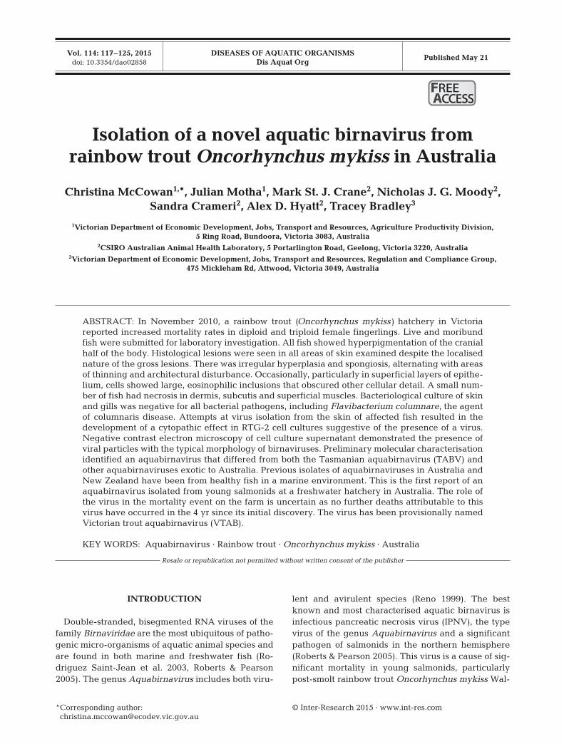
Water data for the section of the river supplying thefarm showed that the outbreak began as tempera-tures exceeded 16°C. This was higher than tempera-tures at the same time in pre vious years (Fig. 1).Water flow rates and oxygenation remained withinthe ranges experienced in previous months and years(Fig. 2).
Gross pathology. On arrival, ap proximately halfthe fish were dead and the remainder were chilledbut showed no obvious abnormalities of swimming orproprioception. All fish showed a sharp line of pig-mentary demarcation approximately half-way alongthe body, with the cranial skin having much darker
119
Fig. 1. Record of water temperatures. The period of increased mortality inrainbow trout Oncorhynchus mykiss fry is indicated by the shaded block.Rates increased in conjunction with an increase in water temperature>16°C and declined following restoration of temperatures. The tempera-tures in November 2010 were higher than those of the preceding months
(solid lines) or for the same time in the previous 2 yr (dashed lines)

Dis Aquat Org 114: 117–125, 2015
colouration than the caudal (Fig. 3). Some fish evi-denced fraying of the dorsal and/or tail fin. No othergross abnormalities were identified, and no signifi-cant changes were seen on wet mounts of skin scrap-ings from 5 fish.
Histology. Histological lesions were seen in the skinof all fish examined. All areas of skin examined, in-cluding both cranial and caudal skin, showed thesechanges, despite the localised nature of the gross le-sions. The epidermis showed irregular hyperplasiaand spongiosis, alternating with areas of thinning. Ep-
ithelial cells were disordered and irreg-ular in size and shape, often with large,pale, clea ved or reniform nuclei and dispersed chromatin. Mitoses oc curredoccasionally and were not confined tothe basal layer, and there were moderatenumbers of apoptotic cells, particularlyin superficial skin layers. Occasionalcells, particularly in superficial layers ofthe epithelium, showed large cyto -plasmic inclusions that ob scured othercellular details and had a slightly grainyap pearance. There was a multifocalmoderate in flammatory in filtrate withinthe epidermis that was mainly lym pho -his tiocytic, with some neutrophils. Somemacrophages contained cell debriswithin the cytoplasm (Fig. 4a).
Fish had mild, focal to locally exten-sive necrosis in dermis, subcutis andsuperficial muscles, with loss of tissuearchitecture and accumulation of smallnumbers of inflammatory cells, mainly
mononuclear (Fig. 4b). There were moderate num-bers of mononuclear cells clustered, probably inblood vessels, adjacent to large nerves. Some of thecells had central clearing of the nuclear chromatin,with refractile, eosinophilic granulation or linearinclusions. The lesions may have been more associ-ated with pale areas (seen as clumping of dermal pig-ment) than with dark areas, but this was equivocal.Gills did not show significant lesions in any fish, andspecial stains failed to highlight bacteria or fungi.
A diagnosis was made of severe, diffuse epidermaldysplasia, with multifocal regionally extensive der-mal hyperplasia, mild focal lymphocytic dermatitisand superficial myositis. The histological appearanceof the lesions was suggestive of a viral infection, and,although the mortality event had resolved itself withrestoration of water temperature, scrapings from skinof retained frozen fish were submitted for virus isolation.
Bacteriology. Cultures of skin and gills from 6 fishwere negative for all bacterial pathogens, includingFlavibacterium columnare, the agent of columnarisdisease.
Virus isolation. No CPEs were observed in theFHM cells, but CPE, typical of birnavirus infection,was observed in RTG-2 cells on Day 3 and rapidlyprogressed to about 80 to 90% by Day 6. The secondpassage of culture supernatant using BF-2 and RTG-2 cells at CSIRO resulted in the destruction of cul-tures within 24 h.
120
Fig. 2. Record of dissolved oxygen levels (DO2). The period of increasedmortality in rainbow trout Oncorhynchus mykiss fry is indicated by theshaded block. Dissolved oxygen in the water supply in November 2010 was
in the same range as in previous months and years
Fig. 3. Oncorhynchus mykiss. Clinical signs of disease. Sub-mitted fish were moribund or dead, and all showed marked
colour change cranially

McCowan et al.: Novel Australian aquatic birnavirus
Electron microscopy. In ultrathin sections virionswere observed within the cytoplasm — icosahedral inshape and non-enveloped, with an average diameterof 54 ± 4 nm (n = 12). Virions were only observed incells displaying extensive degranulation of the cyto-plasm (Fig. 5). No tubular forms of the virus wereidentified. Examination of negative-stained prepara-
tions revealed the presence of naked icosahedralparticles (68 ± 4 nm; n = 2) with hexagonal profilesdisplaying fine surface detail. These data (shape, sizeand cytopathology) indicate the viral structures aresimilar to birnaviruses as described by Delmas et al.(2012).
Molecular analysis. Amplicons of approximately775 bp size were produced for the 4 replicate extrac-tions tested (Fig. 6). An amplicon of the expected sizewas also produced for the TABV-positive control. Noamplicon was produced for template extracted fromuninfected cell culture supernatant, nor from anunrelated aquabirnavirus isolated from swordtails,Xiphophorus helleri (M. S. J. Crane & N. J. G. Moodyunpubl. results). Res triction enzyme digests of puri-fied amplicons using ClaI did not digest the ampli-cons produced from the test samples but did digestthe TABV-positive control, producing 2 fragments ofapproximately 652 and 123 bp (Fig. 7).
121
Fig. 4. Oncorhynchus mykiss. (a) Histological photomicrograph of skin and muscle showing small numbers of lymphocytes(arrows) in a thickened and disordered epithelium, with mild muscle necrosis and inflammation seen in some slides. (b) Withinsuperficial muscles focal myositis and myonecrosis, lymphohistiocytic infiltrates and loss of muscle fibres (asterisk) are evident
Fig. 5. Transmission electron micrograph of an ultrathin sec-tion of an infected CHSE-214 cell. Arrow indicates iscosahe-dral birnavirus particles within the cytoplasm. Insert: Nega-tive-stained particle with obvious icosahedral symmetry andfine surface detail from an infected RTG-2 cell culture super-
natant. Scale bars = 100 nm
Fig. 6. Amplicons produced by generic aquabirnavirus Sero -group A RT-PCR using the GABF/GABR0 primer set. Testsamples (Lanes 1 to 4), no template control (Lane 5), sword-tail aquabirnavirus (Lane 6), Tasmanian aquabirnavirus
(TABV) (Lane 7), M = 100 bp DNA ladder

Dis Aquat Org 114: 117–125, 2015
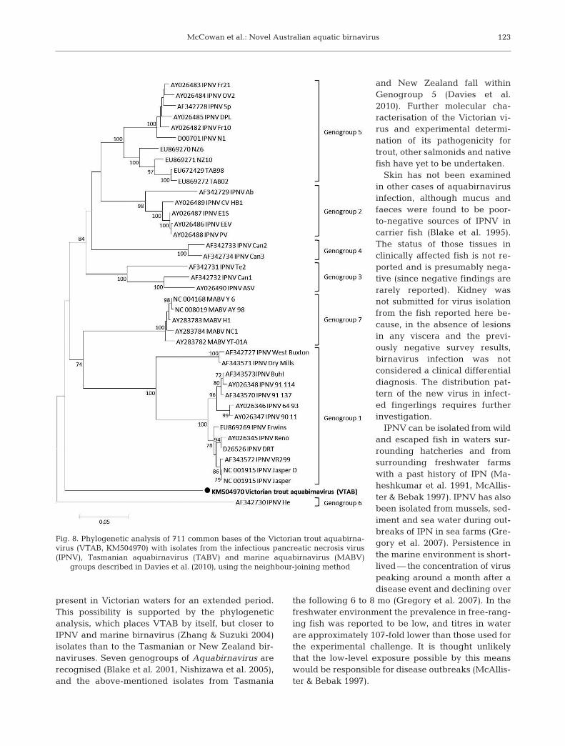
Sequencing of purified amplicons generated usingthe GABF/GABR primer set enabled analysis of a738 bp consensus sequence from 2 replicates of therainbow trout isolate and the TABV-positive control.The sequence obtained has the GenBank AccessionNo. KM504970. The sequences from the 2 ampliconsof the rainbow trout isolate were identical to eachother, and the TABV-positive control shared a 99.7%nucleotide similarity with TAB98 (EU672429). Therainbow trout isolate sequence shared only 78.9 and74.9% nucleotide similarities with TAB98 (EU -672429) and IPNV Erwin (EU869269), respectively.The novel nature of the rainbow trout virus was con-firmed by phylogenetic analysis (Fig. 8). The virushas been provisionally named Victorian trout aqua -birnavirus (VTAB).
Immunocytochemistry. Cell cultures infected withthe rainbow trout aquabirnavirus were test- negative,although positive controls (cultures in fected with theErwin strain of IPNV) showed a positive reaction, asexpected (results not shown). These results suggestthat the relationship of the virus to IPNV, to whichthe antibody was raised, was too distant to permitcross reactivity, and no staining of tissue sections wasattempted.
DISCUSSION
The family Birnaviridae consists of 4 genera and anumber of unclassified species (Delmas et al. 2012).
One genus, Entomobirnavirus (typified by the Dro so -phila X virus) contains species that are principallyinfective to insects. Infectious bursal disease virusis the sole member of the genus Avibirnavirus, andthe type virus for the genus Aquabirnavirus, whichcontains 3 species and several isolates awaiting classification, is represented by IPNV in salmonids.In 2008 the International Committee on Virus Taxo -nomy accepted a 4th genus, Blosnavirus, with a sin-gle mem ber species, the blotched snakehead virus,which shows a closer relationship to the avian virusesthan to the aquatic birnaviruses, despite its origin ina freshwater fish (Da Costa et al. 2003).
Aquabirnaviruses of low pathogenicity have beenisolated from farmed Atlantic salmon in sea cages inMacquarie Harbour, Tasmania (Crane et al. 2000,Davies et al. 2010), and a non-pathogenic strain hasbeen isolated from sea-adapted salmon from NewZealand (Tisdall & Phipps 1987). Viruses have notpreviously been isolated from freshwater-adaptedsalmonids in Australia or New Zealand, nor from fishraised in Australian mainland waters.
Following the identification of the aquabirnavirusin Tasmanian waters (Crane et al. 2000), surveillanceof salmonid hatcheries in Victoria, including the onedescribed in this report, was undertaken in 2000, butdid not turn up evidence of birnaviruses in milt orovarian fluids inoculated in cultures of CHSE-214 orBF-2 cell lines (Muller et al. 2000). Skin was notexamined, and fish were not sacrificed for examina-tion of viscera during this surveillance activity (Mullerat al. 2000).
However, although aquabirnavirus isolation fromreproductive tract fluids has been validated as a non-lethal screening method (Ahne 1983, McAllister et al.1987), higher viral titres are achieved when IPNVisolation is attempted from kidney tissues rather thanfrom reproductive secretions (Munro & Ellis 2008),reflecting the replication of IPNV within leukocytes(Swanson & Gillespie 1979, Ahne & Thomsen 1986,Johansson & Olesen 2009), particularly adherentleukocytes such as those of the renal haematopoietictissue (Johansson & Olesen 2009). In eels sufferingfrom aquabirnaviral gill lamellar pillar necrosis,lesions in viscera are often confined to gastric glandcell ne crosis, with minimal or no signs in the liver,kidney, or other organs (Lee et al. 1999), suggestingan epithelial tropism for this virus.
In view of this, previous Victorian testing may nothave been optimal, particularly for identification ofaquabirnaviruses other than IPNV. Moreover, nei-ther native fish nor birds have been screened, so itremains possible that this novel virus may have been
122
Fig. 7. Amplicons for generic Aquabirnavirus RT-PCR cutfrom the gel and purified for digestion with ClaI to distin-guish Aquabirnavirus (652 and 123 bp bands) from infec-tious pancreatic necrosis virus (IPNV) (775 bp band). Testsamples (Lanes 1 to 4), Tasmanian Aquabirnavirus (Lane 5),
M = 100 bp DNA ladder
McCowan et al.: Novel Australian aquatic birnavirus
present in Victorian waters for an extended period.This possibility is supported by the phylogeneticanalysis, which places VTAB by itself, but closer toIPNV and marine birnavirus (Zhang & Suzuki 2004)isolates than to the Tasmanian or New Zealand bir-naviruses. Seven ge no groups of Aquabirnavirus arerecognised (Blake et al. 2001, Nishizawa et al. 2005),and the above-mentioned isolates from Tasmania
and New Zealand fall withinGenogroup 5 (Davies et al.2010). Further molecular cha -racterisation of the Victorian vi -rus and experimental determi-nation of its pathogenicity fortrout, other salmonids and nativefish have yet to be undertaken.
Skin has not been examinedin other cases of aquabirnavirusin fection, although mucus andfaeces were found to be poor-to-negative sources of IPNV incarrier fish (Blake et al. 1995).The status of those tissues inclinically affected fish is not re -ported and is presumably nega-tive (since negative findings arerarely reported). Kidney wasnot submitted for virus isolationfrom the fish reported here be -cause, in the absence of lesionsin any viscera and the previ-ously negative survey results,birnavirus infection was notconsidered a clinical differentialdiagnosis. The distribution pat-tern of the new virus in infect -ed fingerlings requires furtherinvesti gation.
IPNV can be isolated from wildand escaped fish in waters sur-rounding hatcheries and fromsurrounding freshwater farmswith a past history of IPN (Ma -hesh kumar et al. 1991, McAllis-ter & Bebak 1997). IPNV has alsobeen isolated from mussels, sed-iment and sea water during out-breaks of IPN in sea farms (Gre-gory et al. 2007). Persistence inthe marine environment is short-lived — the concentration of viruspeaking around a month after adisease event and de clining over
the following 6 to 8 mo (Gregory et al. 2007). In thefreshwater environment the prevalence in free-rang-ing fish was reported to be low, and titres in waterare approximately 107-fold lower than those used forthe experimental challenge. It is thought unlikelythat the low-level exposure possible by this meanswould be responsible for disease outbreaks (McAllis-ter & Bebak 1997).
123
Fig. 8. Phylogenetic analysis of 711 common bases of the Victorian trout aquabirna -virus (VTAB, KM504970) with isolates from the infectious pancreatic necrosis virus(IPNV), Tasmanian aquabirnavirus (TABV) and marine aquabirnavirus (MABV)
groups described in Davies et al. (2010), using the neighbour-joining method
Dis Aquat Org 114: 117–125, 2015
Deaths from aquabirnavirus infection, both IPNVand others, are greatest in times of high water tem-peratures (Dorson & Torchy 1981, Jung et al. 1999,Reno 1999), and in young fish (Dorson & Torchy 1981,Rodger & Frerichs 1997, Jung et al. 1999, Roberts &Pearson 2005). The outbreak on the farm describedhere is consistent with this finding. Water records forthe year in question and the previous 2 yr showedthat the temperatures encountered were unusual. Itis likely that higher temperatures would be encoun-tered at the height of summer, but the increased ageof the fish at that time may decrease their susceptibil-ity to disease.
Other stress factors, including over-stocking/ crow -ding, handling or deterioration of water quality(Rodriguez Saint-Jean et al. 2003) and perhaps otheraquatic birnaviruses, may also contribute to out-breaks of IPN, but with the resolution of the whitespot infection, no adverse management issues of sig-nificance were associated with the outbreak de scri -bed here, and water oxygenation levels remainedwithin the ranges recorded for preceding months andfor the same time of year in preceding years.
In summary, we report here the isolation and iden-tification of a novel aquatic birnavirus from rainbowtrout, associated with an episode of increased mortal-ity during a period of high water temperature, andprovisionally name it Victorian trout aquabirnavirus(VTAB). This is the first report of an aquabirnavirusfrom mainland Australian waters, and the first reportof isolation from fish in freshwater in Australia orNew Zealand. No further mortality episodes havebeen reported, and the causal relationship betweenthe isolate and fish deaths remains unclear.
Acknowledgements. The authors acknowledge the contri-butions of Dr Mary Dep and Natalie Kvalheim, DEPI, andLynette Williams, John Hoad and Kelly Davies, CSIROAAHL, for expert scientific and technical assistance withbacteriology, histology, virus production, immunocytochem-istry and molecular studies.
LITERATURE CITED
Ahne W (1983) Presence of infectious pancreatic necrosisvirus in the seminal fluid of rainbow trout, Salmo gaird-neri Richardson. J Fish Dis 6: 377
Ahne W, Thomsen I (1986) Infectious pancreatic necrosis: detection of virus and antibodies in rainbow trout IPNV-carrier (Salmo gairdneri). J Vet Med B 33: 552−554
Blake SL, Schill WB, McAllister PE, Lee MK, Singer JT,Nicholson BL (1995) Detection and identification ofaquatic birnaviruses by PCR assay. J Clin Microbiol 33: 835−839
Blake S, Ma JY, Caporale DA, Jairath S, Nicholson BL (2001)Phylogenetic relationships of aquatic birnaviruses based
on deduced amino acid sequences of genome segment AcDNA. Dis Aquat Org 45: 89−102
Crane MSJ, Hardy-Smith P, Williams LM, Hyatt AD and others (2000) First isolation of an aquatic birnavirus fromfarmed and wild fish species in Australia. Dis Aquat Org43: 1−14
Da Costa B, Soignier S, Chevalier C, Henry C, Thory C,Huet JC, Delmas B (2003) Blotched snakehead virus is anew aquatic birnavirus that is slightly more related toavibirnavirus than to aquabirnavirus. J Virol 77: 719−725
Davies KR, McColl KA, Wang LF, Yu M, Williams LM, CraneMSJ (2010) Molecular characterisation of Australasianisolates of aquatic birnaviruses. Dis Aquat Org 93: 1−15
Delmas B, Mundt E, Vakharia VN, Wu JL (2012) Family Birnaviridae. In: King AMQ, Lefkowitz E, Adams MJ,Carstens EB (eds) Virus taxonomy, classification andnomenclature of viruses: 9th report of the InternationalCommittee on the Taxonomy of Viruses. Elsevier Acade -mic Press, San Diego, CA, p 499−507
Dorson M, Torchy C (1981) The influence of fish age andwater temperature on mortalities of rainbow trout, Salmogairdneri Richardson, caused by a European strain ofinfectious pancreatic necrosis virus. J Fish Dis 4: 213−221
Drummond AJ, Ashton B, Buxton S, Cheung M and others(2011) Geneious v5.4. Available at: www.geneious.com
Gregory A, Munro LA, Wallace IS, Bain N, Raynard RS(2007) Detection of infectious pancreatic necrosis virus(IPNV) from the environment in the vicinity of IPNV-infected Atlantic salmon farms in Scotland. J Fish Dis 30: 621−630
Johansson T, Olesen NJ (2009) Detection of infectious pan-creatic necrosis virus from rainbow trout, Oncorhynchusmykiss (Walbaum), using the macrophage lysis method.J Fish Dis 32: 563−566
Jung SJ, Kitamura S, Kawai K, Suzuki S (1999) Isolation ofdifferent types of birnavirus from ayu Plecoglossus alti -velis and amago salmon Oncorhynchus rhodurus cul-tured in the same geographic area. Dis Aquat Org 38: 87−91
Lee NS, Nomura Y, Miyazaki T (1999) Gill lamellar pillar cellnecrosis, a new birnavirus disease in Japanese eels. DisAquat Org 37: 13−21
Maheshkumar S, Goyal M, Economon PP (1991) Evaluationof a water concentration method for the detection ofinfectious pancreatic necrosis virus in a fish hatchery.J Appl Ichthyology 7: 115−119
McAllister PE, Bebak J (1997) Infectious pancreatic necrosisvirus in the environment: relationship to effluent fromaquaculture facilities. J Fish Dis 20: 201−207
McAllister PE, Owens WJ, Ruppenthal TM (1987) Detectionof infectious pancreatic necrosis virus in pelleted cell andparticulate components from ovarian fluid of brook troutSalvelinus fontinalis. Dis Aquat Org 2: 235−237
Muller JD, Wilks CR, Humphrey JD, Grant P (2000) Surveil-lance for aquabirnavirus in fish hatcheries in Victoria.Aust Vet J 78: 493−494
Munro ES, Ellis AE (2008) A comparison between non-destructive and destructive testing of Atlantic salmon,Salmo salar L., broodfish for IPNV — destructive testingis still the best at time of maturation. J Fish Dis 31: 187−195
Nishizawa T, Kinoshita S, Yoshimizu M (2005) An approachfor genogrouping of Japanese isolates of aquabirna -viruses in a new genogroup, VII, based on the VP2/NSjunction region. J Gen Virol 86: 1973−1978
124

McCowan et al.: Novel Australian aquatic birnavirus 125
Reno PW (1999) Infectious pancreatic necrosis and associ-ated aquatic birnaviruses. In: Woo PTK, Bruno WD (eds)Fish diseases and disorders, Vol 3. Viral, bacterial andfungal infections. CABI Publishing, Wallingford, p 1–55
Roberts RJ, Pearson MD (2005) Infectious pancreatic necro-sis in Atlantic salmon, Salmo salar L. J Fish Dis 28: 383−390
Rodger HD, Frerichs GN (1997) Clinical infectious pancre-atic necrosis virus infection in farmed halibut in theUnited Kingdom. Vet Rec 140: 401−402
Rodriguez Saint-Jean S, Borrego JJ, Perez-Prieto SI (2003)Infectious pancreatic necrosis virus: biology, pathogene-sis, and diagnostic methods. Adv Virus Res 62: 113−165
Swanson RN, Gillespie JH (1979) Pathogenesis of infectiouspancreatic necrosis in Atlantic salmon, Salmo salar.J Fish Res Board Can 36: 587−591
Tamura K, Peterson D, Peterson N, Stecher G, Nei M, KumarS (2011) MEGA5: molecular evolutionary genetics ana -lysis using maximum likelihood, evolutionary distance,and maximum parsimony methods. Mol Biol Evol 28: 2731−2739
Tisdall DJ, Phipps JC (1987) Isolation and characterisation ofa marine birnavirus from returning Quinnat salmon(Oncorhynchus tshawtscha) in the South Island of NewZealand. NZ Vet J 35: 217−218
Weir RP, Moody NJG, Hyatt AD, Crameri S, Voysey R, Pal-lister J, Jerrett IV (2012) Isolation and characterisation ofa novel Bohle-like virus from two frog species in the Darwin rural area, Australia. Dis Aquat Org 99: 169−177
Zhang CX, Suzuki S (2004) Aquabirnaviruses isolated frommarine organisms form a distinct genogroup from otheraquavirnaviruses. J Fish Dis 27: 633−643
Editorial responsibility: V. Gregory Chinchar, Jackson, Mississippi, USA
Submitted: November 20, 2014; Accepted: February 5, 2015Proofs received from author(s): May 13, 2015
➤
➤
➤
➤
➤
➤
➤
➤