Embed Size (px)
Citation preview
The Difco Manual 21
AgarBacto® Agar . Agar Flake . Agar, Granulated . Agar NobleAgar Bacteriological Technical
User Quality ControlIdentity Specifications
AGAR BACTO® AGAR AGAR FLAKE AGAR, GRANULATED AGAR NOBLE BACTERIOLOGICAL TECHNICAL
Dehydrated Very light beige, free Off-white to light Very light beige to White to off-white, free Very light to mediumAppearance: flowing, homogeneous, beige, free flowing, light tan, free flowing flowing,homogeneous, beige, free flowing,
granules. flakes. homogeneous, granules. fine granules. homogeneous.
Solution Solution is very light amber; Solution is very Solution is very light Solution is colorless, Solution is very light1.5% solution very slightly to slightly light to light amber, to medium amber, clear to very to medium amber,soluble in distilled opalescent. Clarity is less very slightly to very slightly opalescent slightly opalescent. opalescent.or deionized water than 10 Nephelometric slightly opalescent. to opalescent.upon boiling turbidity units.
Loss on Drying (LOD) 16-20% Less than or equal to 20% Less than or equal to 20% Less than or equal to 20% Less than or equal to 20%
Ash6 Less than or equal to 6.5% 2-5.2% Less than or equal to 6.5% Less than or equal to 2% Less than or equal to 6.5%
Calcium 300-3,000 ppm Less than or equal Less than or equal 100-2,600 ppm Less than or equalµg/g (ppm) to 3,400 ppm to 3,000 ppm to 3,000 ppm
Magnesium 50-1,000 ppm Less than or equal Less than or equal 0-750 ppm Less than or equalµg/g (ppm) to 1,850 ppm to 1,000 ppm to 1,300 ppm
Melting Point 83-89°C Greater than or 83-89°C Greater than or Greater than orequal to 85°C equal to 85°C equal to 85°C
Gelation Point 32-39°C 32-39°C 32-39°C 32-39°C 32-39°C
Cultural ResponsePrepare the agar formulation of Nutrient Broth (0003) or LB Broth, Miller (0446) by adding 1.5% agar.Sterilize and pour plates. Inoculate with 100-1,000 CFU of the indicated test organisms and incubateat 35 ± 2°C for 18-24 hours. Record recovery.
BACTO® AGAR AGAR, AGAR AGAR BACTERIOLOGICALAGAR FLAKE GRANULATED NOBLE TECHNICAL
Nutrient Broth with:Escherichia coli ATCC® 25922* Good Good Good GoodStaphylococcus aureus ATCC® 25923* Good Good Good Good
LB Broth, Miller with:Escherichia coli ATCC® 33694 (HB101) GoodSaccharomyces cerevisiae ATCC® 9763 Good
*These cultures are available as Bactrol™ Disks and should be used as directed in the Bactrol Disks Technical Information.
Can ofBacto Agar
Section II Agar
Intended UseBacto® Agar is a solidifying agent in which extraneous matter,pigmented portions and salts have been reduced to a minimum. Bacto®
Agar is used in preparing microbiological culture media.
Agar Flake is a solidifying agent used in preparing microbiologicalculture media.
Agar, Granulated is a solidifying agent used in preparing microbio-logical culture media.
Agar Noble is a solidifying agent that is essentially free of impurities.It is used in electrophoretic and nutritional procedures and in preparingmicrobiological culture media when increased purity is required.
Agar Bacteriological Technical is a solidifying agent used in pre-paring microbiological culture media. Although Agar BacteriologicalTechnical has wider quality control parameters than otherbacteriological agars, solubility, gelation temperature and solidity arecarefully monitored to permit its use.
Summary and ExplanationAgar is a phycocolloid extracted from a group of red-purple marinealgae (Class Rhodophyceae) including Gelidium, Pterocladia andGracilaria. Gelidium is the preferred source for Difco agars. Impurities,debris, minerals and pigment are reduced to specified levelsduring manufacture.
Agar was first suggested for microbiological purposes in 1881 byFannie Hesse.1, 2 By the early 1900s, agar became the gelling agent ofchoice over gelatin because agar remains firm at growth temperatures
22 The Difco Manual
for many pathogens. Agar is also generally resistant to a breakdownby bacterial enzymes. The use of agar in microbiological mediasignificantly contributed to the advance of microbiology, paving theway for pure culture isolation and study.
Agar is a gel at room temperature, remaining firm at temperaturesas high as 65°C.3 Agar melts at approximately 85°C, a differenttemperature from that at which it solidifies, 32-40°C. This property isknown as hysteresis. Agar is generally resistant to shear forces; however,different agars may have different gel strengths or degrees of stiffness.
Agar is typically used in a final concentration of 1-2% for solidifyingculture media. Smaller quantities (0.05-0.5%) are used in media formotility studies (0.5% w/v) and for growth of anaerobes (0.1%) andmicroaerophiles.3
Specifications for bacteriological grade agar include good clarity,controlled gelation temperature, controlled melting temperature, gooddiffusion characteristics, absence of toxic bacterial inhibitors, andrelative absence of metabolically useful minerals and compounds.
Product ApplicationsBacto® Agar is optimized for beneficial calcium and magnesiumcontent. Detrimental ions such as iron and copper are reduced. Bacto®
Agar is recommended for clinical applications, auxotrophic studies,bacterial and yeast transformation studies, and bacterial moleculargenetics applications.4, 5
Agar Flake is recommended for general bacteriological purposes. Thequality is similar to Bacto® Agar. However, the flakes are more easilywetted than the granules found in Bacto® Agar.
Agar, Granulated is qualified for culturing recombinant strains ofEscherichia coli (HB101) and Saccharomyces cerevisiae. Agar,Granulated may be used for general bacteriological purposes whereclarity is not a strict requirement.
Noble Agar is extensively washed and bleached. This agar should beused for applications where extreme clarity and high purity are required.Noble Agar is suitable for immunodiffusion, some electrophoreticapplications, and as a substrate for mammalian or plant tissue culture.
Agar Bacteriological Technical is suitable for many bacteriologicalapplications. This agar is not highly processed, has broader technicalspecifications than other Difco agars, and is not recommended forgrowth of fastidious organisms.
Typical AnalysisAGAR
BACTO® AGAR, AGAR BACTERIOLOGICALAGAR GRANULATED NOBLE TECHNICAL
Physical CharacteristicsAsh (%) 3.6 3.4 1.3 4.1Color lt. beige lt. beige off white lt beigeTexture granular granular fine granular
free-flowing free-flowing granular free-flowingfree flowing
Clarity, 1.5% Soln (NTU) 4.3 5.3 3.7 26.2Loss on Drying (%) 17.3 12.2 16.0 18.2pH, 1.5% Soln 6.5 6.6 5.7 6.9Gel Strength (g/cm2) 600 560 700 613Gelation Point(°C) 35°C 35°C 35°C 36°CMelting Point (°C) 88°C 88°C 87°C 88°C
AGARBACTO® AGAR, AGAR BACTERIOLOGICALAGAR GRANULATED NOBLE TECHNICAL
Biological Testing (CFU/g)Spore Count <1,000 <1,000 <1,000 4,300Standard Plate Count <1,000 <1,000 <1,000 2,725
Inorganics (%)Calcium 0.179 0.133 0.015 0.110Chloride 0.021 <0.005 <0.050 0.172Cobalt <0.001 <0.001 <0.001 <0.001Copper <0.001 <0.001 <0.001 <0.001Iron 0.002 0.003 <0.001 0.002Lead <0.001 <0.001 <0.001 <0.001Magnesium 0.068 0.041 0.002 0.093Manganese <0.001 <0.001 <0.001 <0.001Nitrate <0.005 <0.005 <0.050 <0.005Phosphate <0.005 0.010 <0.050 0.015Potassium 0.121 0.079 0.022 0.124Sodium 0.837 0.776 0.335 0.932Sulfate 1.778 1.710 0.663 0.367Sulfur 0.841 0.868 0.333 0.646Tin <0.001 <0.001 <0.001 <0.001Zinc <0.001 <0.001 <0.001 <0.001
Precautions1. For Laboratory and Manufacturing Use.2. Follow proper established laboratory procedures in handling and
disposing of infectious materials.
StorageStore dehydrated agar below 30°C. Dehydrated agar is very hygroscopic.Keep container tightly closed.
Expiration DateThe expiration date applies to the product in its intact container whenstored as directed. Do not use the product if it fails to meet specificationsfor identity and performance.
ProcedureMaterials ProvidedBacto® AgarAgar FlakeAgar, GranulatedAgar NobleAgar Bacteriological Technical
Materials Required But Not ProvidedMaterials vary depending on the application.
Method of PreparationMethod of preparation varies depending on the application.
Specimen Collection and PreparationObtain and process specimens according to the techniques andprocedures established by laboratory policy.
Test ProcedureSee appropriate references for specific procedures using Bacto® Agar,Agar Flake, Agar, Granulated, Agar Noble or Agar BacteriologicalTechnical.
Agar Section II
The Difco Manual 23
ResultsRefer to appropriate references and procedures for results.
References1. Hesse, W. 1894. Über die quantitative Bestimmung der in der Luft
enthaltenen Mikroorganismen. Mitt. a.d. Kaiserl. Gesh. Berlin2:182-207.
2. Hitchens, A. P., and M. C. Leikind. 1939. The introduction ofagar-agar into bacteriology. J. Bacteriology 37:485-493.
3. Selby, H. H., and T. A. Selby. 1959. Agar. In Whister (ed.),Industrial gums. Academic Press Inc., New York, NY.
4. Sambrook, J., E. F. Fritsch, and T. Maniatis. 1989. Molecularcloning, a laboratory manual, 2nd ed. Cold Spring HarborLaboratory Press, NY, NY.
5. Schiestl, R. H., and R. Daniel Geitz. 1989. High efficiencytransformation of intact yeast cells using single stranded nucleicacids as a carrier. Current Genetics 16:339-346.
Section II 2xYT
6. United States Pharmacopeial Convention. 1995. The UnitedStates pharmacopeia, 23rd ed. The United States PharmacopeialConvention. Rockville, MD.
PackagingBacto® Agar 100 g 0140-15
1 lb 0140-012 kg 0140-07
10 kg 0140-08
Agar Flake 500 g 0970-17
Agar, Granulated 100 g 0145-172 kg 0145-07
10 kg 0145-08
Agar Noble 100 g 0142-15500 g 0142-17
Agar Bacteriological Technical 500 g 0812-172 kg 0812-07
10 kg 0812-08
Bacto® 2xYTIntended UseBacto 2xYT is used for cultivating recombinant strains of Escherichia coli.
Summary and Explanation2xYT is a nutritionally rich growth medium designed for growth ofrecombinant strains of Escherichia coli. This medium is also used forpropagation of M13 bacteriophage for sequencing and phage display
research.1-3 The components of 2xYT provide nitrogen and growthfactors that allow bacteriophage to reproduce in large quantitieswithout exhausting the host. E. coli grows more rapidly in this richmedium because it provides amino acids, nucleotide precursors,vitamins and other metabolites that the cell would otherwise have tosynthesize.2
Principles of the ProcedureTryptone and Yeast Extract provide the necessary nutrients and cofactorsrequired for excellent growth of E. coli. Sodium Chloride is included toprovide a suitable osmotic environment.
Formula2xYT
Formula Per LiterBacto Tryptone . . . . . . . . . . . . . . . . . . . . . . . . . . . . . . . . . . 16 gBacto Yeast Extract . . . . . . . . . . . . . . . . . . . . . . . . . . . . . . . 10 gSodium Chloride . . . . . . . . . . . . . . . . . . . . . . . . . . . . . . . . . . 5 g
Final pH 7.0 ± 0.2 at 25°C
Precautions1. For Laboratory Use.2. Follow proper established laboratory procedure in handling and
disposing of infectious materials.
StorageStore the dehydrated medium below 30°C. The powder is very hy-groscopic. Keep container tightly closed.
Store the prepared medium at 2-8°C.
Expiration DateThe expiration date applies to the product in its intact container whenstored as directed. Do not use a product if it fails to meet specificationsfor identity and performance.
User Quality ControlIdentity Specifications
Dehydrated Appearance: Light beige, free-flowing, homogeneous.Solution: 3.1% solution, soluble in distilled or
deionized water. Solution is light tomedium amber, clear.
Prepared Medium: Light to medium amber, clear.Reaction of 3.1%Solution 25°C: pH 7.0 ± 0.2
Cultural ResponsePrepare 2xYT per label directions. Inoculate and incubate at35 ± 2°C for 18-24 hours.
INOCULUMORGANISM ATCC® CFU GROWTH
Escherichia coli (C600) 23724 100-300 GoodEscherichia coli (JM103) 39403 100-300 GoodEscherichia coli (JM107) 47014 100-300 GoodEscherichia coli (HB101) 33694 100-300 GoodEscherichia coli (DH-1) 33849 100-300 GoodEscherichia coli (DH-5) 53868 100-300 Good
The cultures listed are the minimum that should be used forperformance testing.
24 The Difco Manual
Bacto® A-1 Medium
A-1 Medium Section II
User Quality ControlIdentity Specifications
Dehydrated Appearance: Light beige, lumpy.Solution: 3.15% solution, soluble in distilled or deionized water
on boiling. Solution is light amber, opalescent immediatelyafter sterilization. Solution is light amber, clear, may haveflocculent precipitate upon cooling.
Prepared Medium: (When cooled to room temperature) - Light amber, clear,flocculent precipitate may be present.
Reaction of 3.15%Solution at 25°C: pH 6.9 ± 0.1
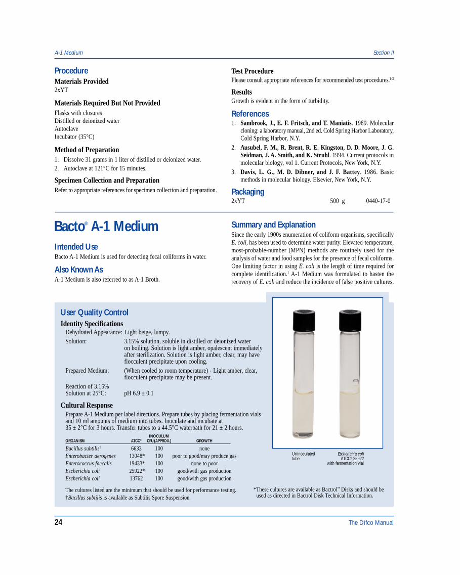
Cultural ResponsePrepare A-1 Medium per label directions. Prepare tubes by placing fermentation vialsand 10 ml amounts of medium into tubes. Inoculate and incubate at35 ± 2°C for 3 hours. Transfer tubes to a 44.5°C waterbath for 21 ± 2 hours.
INOCULUMORGANISM ATCC® CFU(APPROX.) GROWTH
Bacillus subtilis† 6633 100 noneEnterobacter aerogenes 13048* 100 poor to good/may produce gasEnterococcus faecalis 19433* 100 none to poorEscherichia coli 25922* 100 good/with gas productionEscherichia coli 13762 100 good/with gas production
The cultures listed are the minimum that should be used for performance testing.†Bacillus subtilis is available as Subtilis Spore Suspension.
*These cultures are available as Bactrol™ Disks and should be used as directed in Bactrol Disk Technical Information.
Escherichia coliATCC® 25922
with fermentation vial
Uninoculatedtube
Intended UseBacto A-1 Medium is used for detecting fecal coliforms in water.
Also Known AsA-1 Medium is also referred to as A-1 Broth.
Summary and ExplanationSince the early 1900s enumeration of coliform organisms, specificallyE. coli, has been used to determine water purity. Elevated-temperature,most-probable-number (MPN) methods are routinely used for theanalysis of water and food samples for the presence of fecal coliforms.One limiting factor in using E. coli is the length of time required forcomplete identification.1 A-1 Medium was formulated to hasten therecovery of E. coli and reduce the incidence of false positive cultures.
ProcedureMaterials Provided2xYT
Materials Required But Not ProvidedFlasks with closuresDistilled or deionized waterAutoclaveIncubator (35°C)
Method of Preparation1. Dissolve 31 grams in 1 liter of distilled or deionized water.2. Autoclave at 121°C for 15 minutes.
Specimen Collection and PreparationRefer to appropriate references for specimen collection and preparation.
Test ProcedurePlease consult appropriate references for recommended test procedures.1-3
ResultsGrowth is evident in the form of turbidity.
References1. Sambrook, J., E. F. Fritsch, and T. Maniatis. 1989. Molecular
cloning: a laboratory manual, 2nd ed. Cold Spring Harbor Laboratory,Cold Spring Harbor, N.Y.
2. Ausubel, F. M., R. Brent, R. E. Kingston, D. D. Moore, J. G.Seidman, J. A. Smith, and K. Struhl. 1994. Current protocols inmolecular biology, vol 1. Current Protocols, New York, N.Y.
3. Davis, L. G., M. D. Dibner, and J. F. Battey. 1986. Basicmethods in molecular biology. Elsevier, New York, N.Y.
Packaging2xYT 500 g 0440-17-0
The Difco Manual 25
Section II A-1 Medium
In 1972 Andrews and Presnell developed A-1 Medium. A-1 Mediumrecovers E. coli from estuarine water in 24 hours instead of 72 hours,and in greater numbers without the preenrichment step.2 Using a3-hour preincubation step for the enumeration of coliforms inchlorinated wastewater gave results that were statistically comparableto those obtained in the two-step MPN technique.3
A-1 Medium can be used in a single-step procedure for the detectionof fecal coliforms in source water, seawater, treated wastewater andfoods. Prior enrichment in a presumptive medium is not required.4
A-1 Medium conforms to standard methods for the isolation of fecalcoliforms in water and foods.4,5,6
Principles of the ProcedureTryptone provides the nitrogen, vitamins, minerals and amino acids inA-1 Medium. Lactose is the carbon source and, in combination withSalicin, provides energy for organism growth. Sodium Chloridemaintains the osmotic balance of the medium. Triton X-100 is a surfactant.
FormulaA-1 Medium
Formula Per LiterBacto Tryptone . . . . . . . . . . . . . . . . . . . . . . . . . . . . . . . . . . 20 gBacto Lactose . . . . . . . . . . . . . . . . . . . . . . . . . . . . . . . . . . . . 5 gSodium Chloride . . . . . . . . . . . . . . . . . . . . . . . . . . . . . . . . . . 5 gBacto Salicin . . . . . . . . . . . . . . . . . . . . . . . . . . . . . . . . . . . . 0.5 gTriton X-100 . . . . . . . . . . . . . . . . . . . . . . . . . . . . . . . . . . . . . 1 ml
Final pH 6.9 ± 0.1 at 25°C
Precautions1. For Laboratory Use.2. Follow proper established laboratory procedures in handling and
disposing of infectious materials.
StorageStore the dehydrated medium below 30°C. The dehydrated medium isvery hygroscopic. Keep container tightly closed.
Store prepared medium in the dark at room temperature for no longerthan 7 days.4
Expiration DateThe expiration date applies to the product in its intact container whenstored as directed. Do not use a product if it fails to meet the specificationsfor identity and performance.
ProcedureMaterials ProvidedA-1 Medium
Materials Required But Not ProvidedGlasswareFermentation vialsAutoclaveIncubator (35°C)Waterbath (44.5°C)Test tubesDistilled or deionized water
Method of Preparation1. Suspend 31.5 grams in 1 liter distilled or deionized water.2. Heat to boiling to dissolve completely.3. Dispense into tubes containing inverted fermentation vials.4. Autoclave at 121°C for 10 minutes.
NOTE: For 10 ml water samples, prepare double-strength medium toensure the ingredient concentrations are not reduced below those ofthe standard medium.4
Specimen Collection and PreparationObtain and process specimens according to the procedures establishedby laboratory policy or standard methods.4,5,6
Test Procedure1. Inoculate tubes of A-1 Medium as directed in standard methods.4,5,6
2. Incubate at 35 ± 0.5°C for 3 hours.3. Transfer tubes to a water bath at 44.5 ± 0.2°C and incubate for an
additional 21 ± 2 hours.4. Maintain water level in bath above level of liquid in inoculated tubes.
Results5
Gas production in the inverted vial, or dissolved gas that forms finebubbles when slightly agitated, is a positive reaction indicating thepresence of fecal coliforms. Calculate fecal coliform densities usingMPN tables from standard methods.
Limitations of the Procedure1. Since the nutritional requirements of organisms vary, some strains may
be encountered that fail to grow or grow poorly on this medium.2. Fecal coliform counts are usually greater than E. coli counts.5
3. Interpretation of test procedure using A-1 Medium requiresunderstanding of the microflora of the specimen.5
References1. Andrews, W. H., C. D. Diggs, and C. R. Wilson. 1975.
Evaluation of a medium for the rapid recovery of Escherichia colifrom shellfish. Appl. Microbiol. 29:130- 131.
2. Andrews, W. H., and M. W. Presnell. 1972. Rapid recovery ofEscherichia coli from estuarine water. Appl. Microbiol.23:521-523.
3. Standridge, and Delfino. 1981. Appl. Environ. Microbiol. 42:918.4. Eaton, A. D., L. S. Clesceri, and A. E. Greenberg (ed.). 1995.
Standard methods for the examination of water and wastewater,19th ed. American Public Health Association, Washington, D.C.
5. Vanderzant, C., and D. F. Splittstoesser (ed.). 1992.Compendium of methods for the microbiological examination offood, 3rd ed. American Public Health Association, Washington, D.C.
6. Association of Official Analytical Chemists. 1995. Bacteriologicalanalytical manual, 8th ed. AOAC International, Gaithersburg, MD.
PackagingA-1 Medium 500 g 1823-17
26 The Difco Manual
AC Broth & AC Broth w/o Dextrose Section II
Bacto® AC BrothBacto AC Broth w/o Dextrose
User Quality ControlIdentity Specifications
AC BrothDehydrated Appearance: Light tan, free-flowing, homogeneous.Solution: 3.4% solution, soluble in distilled or
deionized water. Solution is mediumto dark amber, clear to very slightlyopalescent.
Prepared Tubes: Light to medium amber, clear to veryslightly opalescent.
Reaction of 3.4%Solution at 25°C: pH 7.2 ± 0.2
AC Broth w/o DextroseDehydrated Appearance: Light tan, free-flowing, homogeneous.Solution: 2.92% solution, soluble in distilled or
deionized water. Solution is mediumto dark amber, clear to very slightlyopalescent.
Prepared Tubes: Medium to dark amber, clear to veryslightly opalescent.
Reaction of 2.92%Solution at 25°C: pH 7.2 ± 0.2
Cultural ResponsePrepare AC Broth or AC Broth w/o Dextrose per labeldirections. Inoculate and incubate at 35 ± 2°C for 18-48 hours.
INOCULUMORGANISM ATCC® CFU GROWTH
Corynebacterium diphtheriaeType mitis 8024 100-1,000 goodStreptococcus pneumoniae 6305 100-1,000 goodStreptococcus pyogenes 19615* 100-1,000 good
The cultures listed are the minimum that should be used forperformance testing.*This culture is available as Bactrol™ Disks and should be used as directed in Bactrol Disks Technical Information.
Intended UseBacto AC Broth is used for cultivating a wide variety of microorganismsand for the sterility testing of turbid or viscous solutions and othermaterials not containing mercurial preservatives.
Bacto AC Broth w/o Dextrose is used, with the addition of a carbohydrate,for cultivating a wide variety of microorganisms.
Summary and ExplanationAC Broth and AC Broth w/o Dextrose possess growth-promotingproperties for voluminous growth of a wide variety of microorganisms.Christensen1 and Malin and Finn2 reported that AC Medium does notexhibit the toxicity shown by media containing sodium thioglycollate.
Several early studies reported on the wide variety of organisms able togrow on AC Medium.3,4,5 AC Broth is suitable for use in the detectionof obligately aerobic contaminants in biologicals and other products.AC Broth and AC Broth w/o Dextrose are also useful in the isolationand cultivation of many common pathogenic and saprophytic aerobes.6
The media can be used to test the sterility of biologicals and solutionsthat do not contain mercurial preservatives. Fluid ThioglycollateMedium should be employed for the sterility testing of solutionscontaining mercurial preservatives.
AC Broth w/o Dextrose has the same formula as AC Broth except thatthe dextrose is omitted, allowing for the addition of other carbohydratesif desired.
Principles of the ProcedureProteose Peptone No. 3, Beef Extract, and Malt Extract providethe carbon and nitrogen sources required for good growth of a widevariety of organisms. Vitamins and cofactors required for growth aswell as additional sources of nitrogen and carbon are provided by YeastExtract. Dextrose is included in AC Broth as a carbon energy source.Ascorbic Acid is added to clarify the solution.
FormulaAC Broth
Formula Per LiterBacto Proteose Peptone No. 3 . . . . . . . . . . . . . . . . . . . . . . 20 gBacto Beef Extract . . . . . . . . . . . . . . . . . . . . . . . . . . . . . . . . 3 gBacto Yeast Extract . . . . . . . . . . . . . . . . . . . . . . . . . . . . . . . . 3 gBacto Malt Extract . . . . . . . . . . . . . . . . . . . . . . . . . . . . . . . . 3 gBacto Dextrose . . . . . . . . . . . . . . . . . . . . . . . . . . . . . . . . . . . 5 gAscorbic Acid . . . . . . . . . . . . . . . . . . . . . . . . . . . . . . . . . . . 0.2 g
Final pH 7.2 ± 0.2 at 25°C
AC Broth w/o DextroseFormula Per LiterBacto Proteose Peptone No. 3 . . . . . . . . . . . . . . . . . . . . . . 20 gBacto Beef Extract . . . . . . . . . . . . . . . . . . . . . . . . . . . . . . . . 3 gBacto Yeast Extract . . . . . . . . . . . . . . . . . . . . . . . . . . . . . . . . 3 gBacto Malt Extract . . . . . . . . . . . . . . . . . . . . . . . . . . . . . . . . 3 gAscorbic Acid . . . . . . . . . . . . . . . . . . . . . . . . . . . . . . . . . . . 0.2 g
Final pH 7.2 ± 0.2 at 25°C
Precautions1. For Laboratory Use.2. Follow proper established laboratory procedure in handling and
disposing of infectious materials.
StorageStore the dehydrated media below 30°C. The dehydrated media arevery hygroscopic. Keep container tightly closed.
AC BrothStore prepared medium at 15-30°C. After prolonged storage, reheat inflowing steam or a boiling water bath for a few minutes to drive offdissolved gases. Cool without agitation.
AC Broth w/o DextroseStore prepared medium at 15-30°C.
The Difco Manual 27
Section II APT Agar & APT Broth
Expiration DateThe expiration date applies to the products in their intact containerswhen stored as directed. Do not use a product if it fails to meetspecifications for identity and performance.
ProcedureMaterials ProvidedAC BrothAC Broth w/o Dextrose
Materials Required But Not ProvidedGlasswareAutoclaveIncubator (35°C)
Method of Preparation1. Suspend appropriate amount of medium in 1 liter distilled or
deionized water:AC Broth - 34 grams;AC Broth w/o Dextrose - 29.2 grams.
2. If necessary, warm slightly to dissolve completely.3. Dispense as desired. Autoclave at 121° C for 15 minutes.
If the medium is not used the same day it is sterilized, place inflowing steam or a boiling water bath for a few minutes to drive offdissolved gases. Allow to cool without agitation.
Test ProcedureSee appropriate references for specific procedures.
ResultsRefer to appropriate references and procedures for results.
Limitations of the Procedure1. When reheating prepared media to drive off dissolved gases do not
overheat because this may result in decreased growth.
References1. Paper read at New York Meeting. Am. Pub. Health Assoc., 1944.2. Malin, B., and R. K. Finn. 1951. The use of a synthetic resin in
anaerobic media. J. Bacteriol. 62:349-350.3. Reed, G. B., and J. H. Orr. 1943. Cultivation of anaerobes and
oxidative-reduction potentials. J. Bacteriol. 45:309-320.4. Schneiter, R., J. E. Dunn, and B. H. Caminita. 1945. Studies
in connection with the selection of a satisfactory culture mediumfor bacterial air sampling. Pub. Health Reports 60:789-806.
5. Kolb, R. W., and R. Schneiter. 1950. The germicidal andsporicidal efficacy of methyl bromide for Bacillus anthracis.J. Bacteriol. 59:401-412.
6. MacFaddin, J. D. 1985. Media for isolation-cultivation-identification-maintenance of medical bacteria, vol. 1, p. 13-14.Williams & Wilkins, Baltimore, MD.
PackagingAC Broth 500 g 0317-17
AC Broth w/o Dextrose 10 kg 0599-08
Bacto® APT AgarBacto APT BrothIntended UseBacto APT Agar is used for cultivating heterofermentative lactobacilliand other organisms requiring high thiamine content. It is also used formaintaining stock cultures of Lactobacillus viridescens ATCC® 12706used in the assay of thiamine.
Bacto APT Broth is used for culturing Lactobacillus viridescensATCC 12706 used in the assay of thiamine. It is also used for cultivatingheterofermentative lactobacilli and other organisms requiring highthiamine content.
Also Known AsAll Purpose Tween
Summary and ExplanationEvans and Niven1 investigated cultivating the heterofermentativelactobacilli that cause the faded or greenish discoloration of cured meatproducts, while Deibel, Evans and Niven2 investigated thiamine
requiring bacteria, specifically Lactobacillus viridescens. Theirformulations led to the development of APT Agar and APT Broth.
The lactic acid bacteria, a group of acid producing bacteria, includethe genera Streptococcus, Leuconostoc, Pediococcus and Lactobacillus.3
These organisms are widespread in nature and are associated withbacterial spoilage of foods such as dairy, meat and vegetableproducts.3 One use of APT Agar and APT Broth is for cultivatingthese heterofermentative lactic acid bacteria from food products.3
APT Agar and APT Broth are also used in the microbiological assay ofthiamine. In the assay, APT Agar is the maintenance medium thatpreserves the viability and sensitivity of Lactobacillus viridescensATCC 12706. APT Broth is used for growing Lactobacillus viridescensATCC 12706 and preparing the inoculum.
Principles of the ProcedureAPT Agar and APT Broth contain Tryptone as a source of carbon,nitrogen, vitamins and minerals. Yeast Extract supplies B-complexvitamins which stimulate bacterial growth. Dextrose is the carbohy-drate. The Manganese Chloride, Magnesium Sulfate and FerrousSulfate provide ions used in replication by lactobacilli. SorbitanMonooleate Complex is a source of fatty acids required by lactobacilli.Bacto Agar is the solidifying agent in APT Agar.
28 The Difco Manual
FormulaAPT Agar
Formula Per LiterBacto Yeast Extract . . . . . . . . . . . . . . . . . . . . . . . . . . . . . . . 7.5 gBacto Tryptone . . . . . . . . . . . . . . . . . . . . . . . . . . . . . . . . . 12.5 gBacto Dextrose . . . . . . . . . . . . . . . . . . . . . . . . . . . . . . . . . . 10 gSodium Citrate . . . . . . . . . . . . . . . . . . . . . . . . . . . . . . . . . . . 5 gThiamine Hydrochloride . . . . . . . . . . . . . . . . . . . . . . . . 0.001 gSodium Chloride . . . . . . . . . . . . . . . . . . . . . . . . . . . . . . . . . . 5 gDipotassium Phosphate . . . . . . . . . . . . . . . . . . . . . . . . . . . . 5 gManganese Chloride . . . . . . . . . . . . . . . . . . . . . . . . . . . . . 0.14 gMagnesium Sulfate . . . . . . . . . . . . . . . . . . . . . . . . . . . . . . . 0.8 gFerrous Sulfate . . . . . . . . . . . . . . . . . . . . . . . . . . . . . . . . . 0.04 gSorbitan Monooleate Complex . . . . . . . . . . . . . . . . . . . . . . 0.2 gBacto Agar . . . . . . . . . . . . . . . . . . . . . . . . . . . . . . . . . . . . . 15 g
Final pH 6.7 ± 0.2 at 25°C
APT BrothFormula Per LiterBacto Yeast Extract . . . . . . . . . . . . . . . . . . . . . . . . . . . . . . . 7.5 gBacto Tryptone . . . . . . . . . . . . . . . . . . . . . . . . . . . . . . . . . 12.5 gBacto Dextrose . . . . . . . . . . . . . . . . . . . . . . . . . . . . . . . . . . 10 gSodium Citrate . . . . . . . . . . . . . . . . . . . . . . . . . . . . . . . . . . . 5 gThiamine Hydrochloride . . . . . . . . . . . . . . . . . . . . . . . . 0.001 gSodium Chloride . . . . . . . . . . . . . . . . . . . . . . . . . . . . . . . . . . 5 gDipotassium Phosphate . . . . . . . . . . . . . . . . . . . . . . . . . . . . 5 gManganese Chloride . . . . . . . . . . . . . . . . . . . . . . . . . . . . . 0.14 gMagnesium Sulfate . . . . . . . . . . . . . . . . . . . . . . . . . . . . . . . 0.8 gFerrous Sulfate . . . . . . . . . . . . . . . . . . . . . . . . . . . . . . . . . 0.04 gSorbitan Monooleate Complex . . . . . . . . . . . . . . . . . . . . . . 0.2 g
Final pH 6.7 ± 0.2 at 25°C
Precautions1. For Laboratory Use.2. Follow proper established laboratory procedures in handling and
disposing of infectious materials.
StorageStore the dehydrated medium below 30°C. The dehydrated medium isvery hygroscopic. Keep container tightly closed.
Expiration DateThe expiration date applies to the product in its intact container whenstored as directed. Do not use a product if it fails to meet specificationsfor identity and performance.
ProcedureMaterials ProvidedAPT AgarAPT Broth
Materials Required but not ProvidedGlasswareDistilled or deionized waterAutoclaveIncubator (35°C)
Method of Preparation1. Suspend the medium in 1 liter distilled or deionized water:
APT Agar: 61.2 grams;APT Broth: 46.2 grams.
2. Heat to boiling to dissolve completely.3. Autoclave at 121°C for 15 minutes.4. Avoid overheating.
Specimen Collection and PreparationRefer to appropriate references for specimen collection and preparation.
Test ProcedureFor maintaining stock cultures of Lactobacillus viridescensATCC® 12706 prepare a stab inoculation. Prepare stock cultures intriplicate at monthly intervals. One of the transfers is saved for the
User Quality ControlIdentity Specifications
APT AgarDehydrated Appearance: Light beige, free-flowing,
homogeneous.Solution: 6.12%, soluble in distilled or
deionized water on boiling.Solution, upon cooling, is mediumamber, clear to slightly opalescent,may have a slight precipitate.
Prepared Medium: Medium amber, clear to slightlyopalescent, may have a slightprecipitate.
Reaction of 6.12%Solution at 25°C: pH 6.7 ± 0.2
APT BrothDehydrated Appearance: Light tan, free-flowing, homogeneous.Solution: 4.62%, soluble in distilled or
deionized water with slight heating.Solution, upon cooling, is light tomedium amber, clear to very slightlyopalescent, may have a slightprecipitate.
Prepared Medium: Light to medium amber, clear tovery slightly opalescent withoutsignificant precipitate.
Reaction of 4.62%Solution at 25°C: pH 6.7 ± 0.2
Cultural ResponsePrepare APT Agar and APT Broth per label directions.Inoculate and incubate at 35 ± 2°C for 24-48 hours.
INOCULUMORGANISM ATCC® CFU GROWTH
Lactobacillus fermentum 9338 100-1,000 goodLactobacillus viridescens 12706 100-1,000 good
The cultures listed are the minimum that should be used forperformance testing.
APT Agar & APT Broth Section II
The Difco Manual 29
preparation of stock cultures. The others are used to prepare inoculumin APT Broth for assay as needed. Following incubation at 35-37°C for24-48 hours, store stock cultures at 2-8°C.
ResultsRefer to appropriate references and procedures for results.
References1. Evans, J. B., and C. F. Niven, Jr. 1951. Nutrition of the
heterofermentative lactobacilli that cause greening of cured meatproducts. J. Bact. 62:599-603.
2. Deibel, R. H., J. B. Evans, and C. F. Niven, Jr. 1957.Microbiological assay for thiamine using Lactobacillusviridescens. J. Bact. 74:818-821.
Section II Acetate Differential Agar
3. Vedamuthu, E. R., M. Raccach, B. A. Glatz, E. W. Seitz, and M.S. Reddy. 1992. Acid-producing microorganisms, p. 225-238. InC. Vanderzant, and D. F. Splittstoesser (ed.), Compendium ofmethods for the microbiological examination of foods, 3rd ed.American Public Health Association, Washington, D.C.
PackagingAPT Agar 500 g 0654-17
2 kg 0654-0710 k 0654-08
APT Broth 500 g 0655-17
Bacto® Acetate Differential Agar
User Quality ControlIdentity Specifications
Dehydrated Appearance: Medium yellowish-tan to light green, free-flowing,homogeneous.
Solution: 2.92% solution, soluble in distilled or deionized wateron boiling. Solution is emerald green, slightly opalescent.
Prepared Medium: Emerald green to green, slightly opalescent.
Reaction of 2.92%Solution at 25°C: pH 6.7 ± 0.1
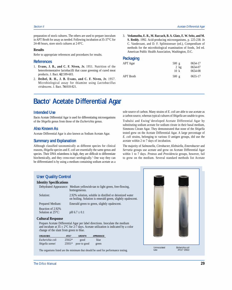
Cultural ResponsePrepare Acetate Differential Agar per label directions. Inoculate the mediumand incubate at 35 ± 2°C for 2-7 days. Acetate utilization is indicated by a colorchange of the slant from green to blue.
ORGANISMS ATCC® GROWTH APPEARANCE
Escherichia coli 25922* good blueShigella sonnei 25931* poor to good green
The organisms listed are the minimum that should be used for performance testing.Escherichia coli
ATCC® 25922Uninoculatedtube
Intended UseBacto Acetate Differential Agar is used for differentiating microorganismsof the Shigella genus from those of the Escherichia genus.
Also Known AsAcetate Differential Agar is also known as Sodium Acetate Agar.
Summary and ExplanationAlthough classified taxonomically as different species for clinicalreasons, Shigella species and E. coli are essentially the same genus andspecies. Their DNA relatedness is high, they are difficult to differentiatebiochemically, and they cross-react serologically.1 One way they canbe differentiated is by using a medium containing sodium acetate as a
sole source of carbon. Many strains of E. coli are able to use acetate asa carbon source, whereas typical cultures of Shigella are unable to grow.
Trabulsi and Ewing2 developed Acetate Differential Agar bysubstituting sodium acetate for sodium citrate in their basal medium,Simmons Citrate Agar. They demonstrated that none of the Shigellatested grew on the Acetate Differential Agar. A large percentage ofE. coli strains, belonging to various O antigen groups, did use theacetate within 2 to 7 days of incubation.
The majority of Salmonella, Citrobacter, Klebsiella, Enterobacter andSerratia groups use acetate and grow on Acetate Differential Agarwithin 1 to 7 days. Proteus and Providencia groups, however, failto grow on the medium. Several standard methods list Acetate
30 The Difco Manual
Differential Agar as a possible medium for the differentiation ofEnterobacteriaceae.2,3,4
Principles of the ProcedureAcetate Differential Agar consists of a mixture of salts and sodiumacetate as a sole source of carbon. Brom Thymol Blue is added todetect the alkaline products resulting from acetate utilization. MonoAmmonium Phosphate and Dipotassium Phosphate provide bufferingcapability. Bacto Agar is a solidifying agent.
FormulaAcetate Differential Agar
Formula Per LiterSodium Acetate . . . . . . . . . . . . . . . . . . . . . . . . . . . . . . . . . . . 2 gMagnesium Sulfate . . . . . . . . . . . . . . . . . . . . . . . . . . . . . . . 0.1 gSodium Chloride . . . . . . . . . . . . . . . . . . . . . . . . . . . . . . . . . . 5 gMono Ammonium Phosphate . . . . . . . . . . . . . . . . . . . . . . . . 1 gDipotassium Phosphate . . . . . . . . . . . . . . . . . . . . . . . . . . . . 1 gBacto Brom Thymol Blue . . . . . . . . . . . . . . . . . . . . . . . . 0.08 gBacto Agar . . . . . . . . . . . . . . . . . . . . . . . . . . . . . . . . . . . . . 20 g
Final pH 6.7 ± 0.1 at 25°C
Precautions1. For Laboratory Use.
2. IRRITANT. IRRITATING TO EYES, RESPIRATORY SYSTEMAND SKIN. Avoid contact with skin and eyes. Do not breathe dust.Wear suitable protective clothing. Keep container tightly closed.
FIRST AID: In case of contact with eyes, rinse immediately withplenty of water and seek medical advice. After contact with skin,wash immediately with plenty of water. If inhaled, remove to freshair. If not breathing, give artificial respiration. If breathing isdifficult, give oxygen. Seek medical advice. If swallowed seekmedical advice immediately and show this container or label.
3. Follow proper established laboratory procedure in handling anddisposing of infectious materials.
StorageStore the dehydrated medium below 30°C. The dehydrated medium isvery hygroscopic. Keep container tightly closed.
Expiration DateThe expiration date applies to the product in its intact container whenstored as directed. Do not use a product if it fails to meet specificationsfor identity and performance.
ProcedureMaterials ProvidedAcetate Differential Agar
Materials Required But Not ProvidedGlasswareAutoclave
Incubator (35°C)0.85% NaCl solution
Method of Preparation1. Suspend 29.2 grams in 1 liter distilled or deionized water.
2. Boil to dissolve completely.
3. Dispense into tubes to allow a 10 mm butt and a 30 mm slant.
4. Autoclave at 121°C for 15 minutes.
5. Allow tubes to cool in a slanted position to give the recommendedbutt and slant size.
Test Procedure1. Inoculate agar slant surfaces with 16-18 hour cultures emulsified
in 1 ml of 0.85% sodium chloride solution.
2. Incubate aerobically at 35 ± 2°C for at least 7 days; read daily,examining for a change in the color of the medium from greento blue.
ResultsPositive: BlueNegative: Green
Limitations of the Procedure1. Some strains of E. coli and nonmotile, anaerogenic E. coli
(Alkalescens-Dispar) grow slowly or not at all and, thus, may givea false-negative reaction.
2. Further biochemical, physiological and serological tests are requiredto differentiate species.
3. False-positive results may occur from a too heavy inoculum.4. MacFaddin5 suggests that correct results occur only when some
syneresis fluid is present in the bottom of the tube (junction of theslant and butt).
References1. Gray, L. D. 1995. Escherichia, Salmonella, Shigella, and Yersinia,
pp. 450-456. In P. R. Murray, E. J. Baron, M. A. Pfaller, F. C.Tenover, and R. H. Yolken (eds.). Manual of clinical microbiology,6th ed. American Society for Microbiology, Washington, D.C.
2. Vanderzant, C., and D. F. Splittstoesser (eds.). 1992. Compen-dium of methods for the microbiological examination of food,3rd ed. American Public Health Association, Washington, D.C.
3. Andrews, W. H., G. A. June, and P. S. Sherrod. 1995. Shigella,p. 6.01-6.06. In Bacteriological analytical manual, 8th ed. AOACInternational, Gaithersburg, MD.
4. MacFaddin, J. F. 1985. Media for isolation-cultivation-identification-maintenance of medical bacteria, vol. 1, p. 17-20.Williams & Wilkins, Baltimore, MD.
PackagingAcetate Differential Agar 500 g 0742-17
Acetate Differential Agar Section II
The Difco Manual 31
Section II Actinomycete Isolation Agar & Glycerol
Bacto® Actinomycete Isolation AgarBacto Glycerol
User Quality ControlIdentity Specifications
Actinomycete Isolation AgarDehydrated Appearance: Light beige, free-flowing, homogeneous.Solution: 2.2% solution, soluble in distilled or
deionized water on boiling. Solutionis light to medium amber, opalescentto opaque with precipitation.
Prepared Medium: Medium amber, opalescent.Reaction of 2.2%Solution with 0.5%Glycerol at 25°C: pH 8.1 ± 0.2
Cultural ResponsePrepare Actinomycete Isolation Agar per label directionswith the addition of 0.5% Glycerol. Inoculate and incubate at30 ± 2°C for up to 72 hours.
INOCULUMORGANISM ATCC® CFU GROWTH
Streptomyces achromogenes 12767 100-1,000 goodStreptomyces albus 3004 100-1,000 goodStreptomyces lavendulae 8664 100-1,000 good
The cultures listed are the minimum that should be used forperformance testing.
Intended UseBacto Actinomycete Isolation Agar is used with added glycerol forisolating and cultivating actinomycetes from soil and water.
Bacto Glycerol is used in preparing microbiological culture media.
Summary and ExplanationAlthough some genera are important to human medicine, most of theactinomycetes are part of the indigenous flora of soil, water, andvegetation. Actinomycetes may impart a musty odor to water or amuddy flavor to fish.2 Actinomycetes can cause massive growths whichwill form a thick foam in the activated sludge process, causing adisruption in wastewater treatment.3,4 Actinomycetes are grampositive, acid-fast cells, growing as filaments that may branch and mayform irregularly shaped rods and cocci.
Olsen1 formulated Actinomycete Isolation Agar for isolating andcultivating actinomycetes from soil and water. The formula,supplemented with Glycerol, is a highly purified fermentable alcoholused occasionally for differentiating certain bacteria and in media forisolating and culturing fastidious bacteria.
Principles of the ProcedureActinomycete Isolation Agar contains Sodium Caseinate which is asource of nitrogen. Asparagine is an amino acid and a source of
organic nitrogen. Sodium Propionate is a substrate used in anaerobicfermentation. Dipotassium Phosphate provides buffering capability tomaintain pH balance. Magnesium Sulfate and Ferrous Sulfate providesources of sulfates and metallic ions. Bacto Agar is the solidifyingagent. The added Glycerol is a source of carbon.
FormulaActinomycete Isolation Agar
Formula Per LiterSodium Caseinate . . . . . . . . . . . . . . . . . . . . . . . . . . . . . . . . . 2 gAsparagine . . . . . . . . . . . . . . . . . . . . . . . . . . . . . . . . . . . . . 0.1 gSodium Propionate . . . . . . . . . . . . . . . . . . . . . . . . . . . . . . . . 4 gDipotassium Phosphate . . . . . . . . . . . . . . . . . . . . . . . . . . . 0.5 gMagnesium Sulfate . . . . . . . . . . . . . . . . . . . . . . . . . . . . . . . 0.1 gFerrous Sulfate . . . . . . . . . . . . . . . . . . . . . . . . . . . . . . . . 0.001 gBacto Agar . . . . . . . . . . . . . . . . . . . . . . . . . . . . . . . . . . . . . 15 g
Final pH 8.1 ± 0.2 at 25°C
GlycerolNot applicable
Precautions1. For Laboratory Use.2. MAY BE IRRITATING TO EYES, RESPIRATORY SYSTEM
AND SKIN. (US) Avoid contact with skin and eyes. Do not breathedust. Wear suitable protective clothing. Keep container tightly closed.FIRST AID: In case of contact with eyes, rinse immediately withplenty of water and seek medical advice. After contact with skin,wash immediately with plenty of water. If inhaled, remove to freshair. If not breathing, give artificial respiration. If breathing isdifficult, give oxygen. Seek medical advice. If swallowed seekmedical advice immediately and show this container or label.
3. Follow proper established laboratory procedures in handling anddisposing of infectious materials.
StorageStore the dehydrated medium below 30°C. The dehydrated medium isvery hygroscopic. Keep container tightly closed.
Store Glycerol at 15-30°C.
Expiration DateThe expiration date applies to the product in its intact container whenstored as directed. Do not use a product if it fails to meet specificationsfor identity and performance.
ProcedureMaterials ProvidedActinomycete Isolation AgarGlycerol
32 The Difco Manual
Materials Required but not ProvidedGlasswarePetri dishesDistilled or deionized waterAutoclaveIncubator (30°C)
Method of Preparation1. Suspend 22 grams in 1 liter distilled or deionized water.2. Heat to boiling to dissolve completely.3. Add 5 grams Glycerol.4. Autoclave at 121°C for 15 minutes.
Specimen Collection and Preparation1. Collect specimens in sterile containers or with sterile swabs.
Transport immediately to the laboratory, in accordance withrecommended guidelines.
2. Process each specimen as appropriate for that specimen.
Test ProcedureInoculate medium and incubate at 30°C for up to 72 hours.
ResultsRefer to appropriate references and procedures for results.
References1. Olsen, E. H. 1960. Personal Communication.2. Eaton, A. D., L. S. Clesceri, and A. E. Greenberg. 1995.
Standard methods for the examination of water and wastewater,19th ed. American Public Health Association, Washington, D.C.
3. Lechevalier, H. A. 1975. Actinomycetes of sewage-treatmentplants. Environ. Protection Technol. Ser., EPA-600/2-75-031,U. S. Environmental Protection Agency, Cincinnati, OH.
4. Lechevalier, M. P., and H. A. Lechevalier. 1974. Nocardiaamarae, sp. nov., an actinomycete common in foaming activatedsludge. Int. J. Syst. Bacteriol. 24:278.
PackagingActinomycete Isolation Agar 100 g 0957-15
500 g 0957-17
Glycerol 100 g 0282-15500 g 0282-17
Agar Medium No. F Section II
Bacto® Agar Medium No. F
User Quality ControlIdentity Specifications
Dehydrated Appearance: Beige, free-flowing, homogeneous.Solution: 5.15% solution, soluble in distilled
or deionized water on boiling. Solutionis reddish-purple, slightly opalescent.
Prepared Medium: Reddish-purple, slightly opalescentwithout a precipitate.
Reaction of 5.15%Solution at 25°C: pH 7.4 ± 0.2
Cultural ResponsePrepare Agar Medium No. F per label directions. Inoculateand incubate at 35 ± 2°C for 18-24 hours.
INOCULUM COLONYORGANISM ATCC® CFU RECOVERY DESCRIPTION
Escherichia 11775 100-1,000 good reddish-purple, mayhave a slight precipitate
around the coloniesSalmonella 9184 100-1,000 good reddish-purple, maygallinarum have a slight precipitate
around the coloniesStaphylococcus 6538 1,000-2,000 inhibited –aureus
The cultures listed are the minimum that should be used forperformance testing.
Summary and ExplanationAgar Medium No. F is based on the formula for Agar Medium F (AgarMedium with Bile, Crystal Violet, Neutral Red and Glucose) describedin DAB, 10th Edition. Agar Medium No. F is recommended for use inthe detection of Enterobacteriaceae and other gram-negative bacteriain pharmaceuticals.1
Principles of the ProcedureAgar Medium No. F, based on Violet Red Bile Agar and Violet Red BileGlucose Agar, uses Sodium Cholate instead of the Bile SaltsNo. 3 used in Violet Red Bile Agar and Violet Red Bile Glucose Agar.Carbon and nitrogen sources required for growth of a variety oforganisms are provided by Bacto Peptone and Yeast Extract. Selectivityis due to the presence of Crystal Violet and Sodium Cholate whichmarkedly to completely inhibit growth of gram-positive microorganisms.Bacto Agar is the solidifying agent.
Differentiation is based on the fermentation of Dextrose and Lactose.Organisms growing in this medium that can ferment dextrose, such asmembers of the family Enterobacteriaceae, produce a localized pHdrop which, followed by absorption of the Neutral Red, imparts areddish-purple color to the colony. A zone of precipitated SodiumCholate may also be present due to this drop in pH. These reactions arefurther intensified in those organisms that can ferment both lactoseand dextrose.
FormulaAgar Medium No. F
Formula Per LiterBacto Peptone . . . . . . . . . . . . . . . . . . . . . . . . . . . . . . . . . . . . 7 gBacto Yeast Extract . . . . . . . . . . . . . . . . . . . . . . . . . . . . . . . . 3 gBacto Lactose . . . . . . . . . . . . . . . . . . . . . . . . . . . . . . . . . . . 10 gBacto Dextrose . . . . . . . . . . . . . . . . . . . . . . . . . . . . . . . . . . 10 g
Intended UseBacto Agar Medium No. F is a selective medium used for detectingEnterobacteriaceae and other gram-negative bacteria in pharmaceuticalproducts.
The Difco Manual 33
Section II Amino Acid Assay Media
Sodium Chloride . . . . . . . . . . . . . . . . . . . . . . . . . . . . . . . . . . 5 gSodium Cholate . . . . . . . . . . . . . . . . . . . . . . . . . . . . . . . . . 1.5 gNeutral Red . . . . . . . . . . . . . . . . . . . . . . . . . . . . . . . . . . . . 0.03 gCrystal Violet . . . . . . . . . . . . . . . . . . . . . . . . . . . . . . . . . 0.002 gBacto Agar . . . . . . . . . . . . . . . . . . . . . . . . . . . . . . . . . . . . . 15 g
Final pH 7.4 ± 0.2 at 25°C
Precautions1. For Laboratory Use.2. Follow proper, established laboratory procedures in handling and
disposing of infectious materials.
StorageStore the dehydrated medium below 30°C. The dehydrated medium isvery hygroscopic. Keep container tightly closed. Store the preparedmedium at 2-8°C.
Expiration DateThe expiration date applies to the product in its intact container whenstored as directed. Do not use a product if it fails to meet specificationsfor identity and performance.
ProcedureMaterials ProvidedAgar Medium No. F
Materials Required But Not ProvidedLactose BrothEnterobacteriaceae Enrichment Broth Mossel (EE Broth Mossel)Flasks with closuresDistilled or deionized waterIncubator (35°C)Polysorbate 20 or Polysorbate 80
Method of Preparation1. Suspend 51.5 grams in 1 liter distilled or deionized water.2. Heat to boiling to dissolve completely.3. Sterilize by steaming for 30 minutes. Do Not Autoclave.
Specimen Collection and Preparation1. Collect samples in sterile containers and transport immediately to
the laboratory following recommended guidelines.1,2
2. Process each sample using procedures appropriate for thatsample.1,2
Test Procedure1,2
1. Pre-enrich the sample in Lactose Broth. If the sample is insolublein water, add 0.1 ml of polysorbate 20 or polysorbate 80 to theLactose Broth.
2. Homogenize the mixture and incubate at 35 ± 2°C for 2-5 hours.3. Transfer 1 ml of enriched Lactose Broth to 100 ml of EE Broth
Mossel (Enterobacteriaceae Enrichment Broth-Mossel).4. Incubate at 35 ± 2°C for 24-48 hours.5. Subculture all enrichment broth cultures showing growth onto
Agar Medium No. F.6. Incubate at 35 ± 2°C for 18-24 hours.7. Examine plates for the presence of presumptive Enterobacteriaceae
colonies.
ResultsColonies of the family Enterobacteriaceae are reddish-purple in colorand are generally surrounded by a zone of precipitated bile salt. Growthof gram-positive organisms is markedly to completely suppressed.Further biochemical testing is necessary to confirm the presence andidentification of Enterobacteriaceae. Consult appropriate referencesfor further information on identification of Enterobacteriaceae.3,4
References1. DAB, 10th Edition. 1991. V.2 Biology, V.2.1.8 Proving Certain
Microorganisms, VIII.10 Media (Microbiological Pollution),Frankfurt/Main.
2. British Pharmacopoeia, Volume II, Appendix XVI. 1988.HMSO, London.
3. Farmer, J. J. 1995. Enterobacteriaceae: introduction andidentification. In P. R. Murray, E. J. Baron, M. A. Pfaller, F. C.Tenover, and R. H. Yolken (ed.). Manual of clinical microbiology,6th ed. American Society for Microbiology, Washington, D.C.
4. Baron, E. J., L. R. Peterson, and S. M. Finegold. 1994. Bailey &Scott’s diagnostic microbiology, 9th ed. Mosby-Year Book, Inc.,St. Louis, MO.
PackagingAgar Medium No. F 500 g 0666-17
Amino Acid Assay MediaBacto® Lysine Assay Medium . Bacto Methionine Assay MediumBacto Cystine Assay MediumIntended UseBacto Lysine Assay Medium is used for determining lysine concentrationby the microbiological assay technique.
Bacto Methionine Assay Medium is used for determining methionineconcentration by the microbiological assay technique.
Bacto Cystine Assay Medium is used for determining L-cystineconcentration by the microbiological assay technique.
Also Known AsLysine Assay Medium, Methionine Assay Medium and Cystine AssayMedium are also referred to as Amino Acid Assay Media.
34 The Difco Manual
Summary and ExplanationAmino Acid Assay Media are prepared for use in the microbiologicalassay of amino acids. Three types of media are used for this purpose:1. Maintenance Media: For carrying the stock culture to preserve the
viability and sensitivity of the test organism for its intended purpose.2. Inoculum Media: To condition the test culture for immediate use.3. Assay Media: To permit quantitation of the amino acid under test.
They contain all the factors necessary for optimal growth of the testorganism except the single essential amino acid to be determined.
Amino Acid Assay Media are prepared according to the formulationsof Steel et al.1 They are used in the microbiological assay of aminoacids using Pediococcus acidilactici ATCC® 8042 as the test organism.
Principles of the ProcedureLysine Assay Medium, Methionine Assay Medium and Cystine AssayMedium contain all the factors essential for the growth of Pediococcusacidilactici ATCC® 8042, except the amino acid under assay. Theaddition of the amino acid in specified increasing concentrations givesa growth response by the test organism.
FormulaLysine Assay Medium, Methionine Assay Medium, orCystine Assay Medium
All amino acid assay media contain the following formula. Omit theparticular amino acid to be assayed from the medium.
Formula Per LiterBacto Dextrose . . . . . . . . . . . . . . . . . . . . . . . . . . . . . . . . . . 50 gSodium Acetate . . . . . . . . . . . . . . . . . . . . . . . . . . . . . . . . . . 40 gAmmonium Chloride . . . . . . . . . . . . . . . . . . . . . . . . . . . . . . 6 gMonopotassium Phosphate . . . . . . . . . . . . . . . . . . . . . . . . . 1.2 gDipotassium Phosphate . . . . . . . . . . . . . . . . . . . . . . . . . . . 1.2 gMagnesium Sulfate . . . . . . . . . . . . . . . . . . . . . . . . . . . . . . . 0.4 gFerrous Sulfate . . . . . . . . . . . . . . . . . . . . . . . . . . . . . . . . . . 20 mgManganese Sulfate . . . . . . . . . . . . . . . . . . . . . . . . . . . . . . . 40 mgSodium Chloride . . . . . . . . . . . . . . . . . . . . . . . . . . . . . . . . . 20 mgAdenine Sulfate . . . . . . . . . . . . . . . . . . . . . . . . . . . . . . . . . 20 mgGuanine Hydrochloride . . . . . . . . . . . . . . . . . . . . . . . . . . . 20 mgUracil . . . . . . . . . . . . . . . . . . . . . . . . . . . . . . . . . . . . . . . . . . 20 mgXanthine . . . . . . . . . . . . . . . . . . . . . . . . . . . . . . . . . . . . . . . 20 mgThiamine Hydrochloride . . . . . . . . . . . . . . . . . . . . . . . . . . . 1 mgPyrodoxine Hydrochloride . . . . . . . . . . . . . . . . . . . . . . . . . . 2 mgPyridoxamine Hydrochloride . . . . . . . . . . . . . . . . . . . . . . 600 mgPyridoxal Hydrochloride . . . . . . . . . . . . . . . . . . . . . . . . . 600 mgCalcium Pantothenate . . . . . . . . . . . . . . . . . . . . . . . . . . . . . . 1 mgRiboflavin . . . . . . . . . . . . . . . . . . . . . . . . . . . . . . . . . . . . . . . 1 mgNicotinic Acid . . . . . . . . . . . . . . . . . . . . . . . . . . . . . . . . . . . . 2 mgp-Aminobenzoic Acid . . . . . . . . . . . . . . . . . . . . . . . . . . . . 200 µgBiotin . . . . . . . . . . . . . . . . . . . . . . . . . . . . . . . . . . . . . . . . . . . 2 µgFolic Acid . . . . . . . . . . . . . . . . . . . . . . . . . . . . . . . . . . . . . . 20 µgGlycine . . . . . . . . . . . . . . . . . . . . . . . . . . . . . . . . . . . . . . . . 0.2 gDL-Alanine . . . . . . . . . . . . . . . . . . . . . . . . . . . . . . . . . . . . . 0.4 gBacto Asparagine . . . . . . . . . . . . . . . . . . . . . . . . . . . . . . . . 0.8 gL-Aspartic Acid . . . . . . . . . . . . . . . . . . . . . . . . . . . . . . . . . 0.2 gL-Proline . . . . . . . . . . . . . . . . . . . . . . . . . . . . . . . . . . . . . . . 0.2 gDL-Serine . . . . . . . . . . . . . . . . . . . . . . . . . . . . . . . . . . . . . . 0.1 gDL-Tryptophane . . . . . . . . . . . . . . . . . . . . . . . . . . . . . . . . . 80 mgL-Cystine . . . . . . . . . . . . . . . . . . . . . . . . . . . . . . . . . . . . . . . 0.1 gL-Glutamic Acid . . . . . . . . . . . . . . . . . . . . . . . . . . . . . . . . . 0.6 gL-Histidine Hydrochloride . . . . . . . . . . . . . . . . . . . . . . . 0.124 gDL-Phenylalanine . . . . . . . . . . . . . . . . . . . . . . . . . . . . . . . . 0.2 gDL-Threonine . . . . . . . . . . . . . . . . . . . . . . . . . . . . . . . . . . . 0.4 gL-Tyrosine . . . . . . . . . . . . . . . . . . . . . . . . . . . . . . . . . . . . . . 0.2 gDL-Valine . . . . . . . . . . . . . . . . . . . . . . . . . . . . . . . . . . . . . . 0.5 gL-Lysine Hydrochloride . . . . . . . . . . . . . . . . . . . . . . . . . . . 0.5 gDL-Methionine . . . . . . . . . . . . . . . . . . . . . . . . . . . . . . . . . . 0.2 gDL-Isoleucine . . . . . . . . . . . . . . . . . . . . . . . . . . . . . . . . . . . 0.5 gDL-Leucine . . . . . . . . . . . . . . . . . . . . . . . . . . . . . . . . . . . . . 0.5 gL-Arginine Hydrochloride . . . . . . . . . . . . . . . . . . . . . . . 0.484 g
Final pH 6.7 ± 0.2 at 25ºC
Precautions1. For Laboratory Use.2. Great care to avoid contamination of media or glassware must be
taken in microbiological assay procedures. Extremely smallamounts of foreign material may be sufficient to give erroneousresults. Scrupulously clean glassware free from detergents andother chemicals must be used. Glassware is heated to 250°C for atleast 1 hour to burn off any organic residues that might be present.
3. Methionine Assay Medium and Cystine Assay MediumIRRITANT. IRRITATING TO EYES, RESPIRATORY SYSTEMAND SKIN. Avoid contact with skin and eyes. Do not breathe dust.
User Quality ControlIdentity Specifications
Lysine Assay Medium, Methionine Assay Medium,or Cystine Assay MediumDehydrated Appearance: White to off-white, homogeneous,
may have a tendency to clump.Solution: 5.25% (single strength) and 10.5%
(double strength) solution, solublein distilled or deionized water uponboiling. Solution (single strength) islight to medium amber, clear toslightly opalescent, may have aslight precipitate.
Prepared Medium: Single strength-light to mediumamber, clear.
Reaction of 5.25%Solution at 25°C: pH 6.7 ± 0.2
Cultural ResponsePrepare Lysine Assay Medium, Methionine Assay Medium andCystine Assay Medium per label directions. These media willsupport the growth of Pediococcus acidilactici ATCC® 8042when supplemented with the appropriate amino acid. TestLysine Assay Medium by creating a standard curve usingL-Lysine at 0 to 300 µg per 10 ml. Test Methionine AssayMedium by creating a standard curve using DL-Methionine at0 to 60 µg per 10 ml. Test Cystine Assay Medium by creatinga standard curve using L-Cystine at 0 to 50 µg per 10 ml.
The test organism listed is the minimum used for performance testing.
Amino Acid Assay Media Section II
The Difco Manual 35
Wear suitable protective clothing. Keep container tightly closed.TARGET ORGAN(S): Kidney, Bladder.FIRST AID: In case of contact with eyes, rinse immediately withplenty of water and seek medical advice. After contact with skin,wash immediately with plenty of water. If inhaled, remove to freshair. If not breathing, give artificial respiration. If breathing is diffi-cult, give oxygen. Seek medical advice. If swallowed seek medicaladvice immediately and show this container or label.
4. Take precautions to keep sterilizing and cooling conditions uniformthroughout the assay.
5. Follow proper established laboratory procedures in handling anddisposing of infectious materials.
StorageStore the dehydrated media at 2-8ºC. The dehydrated medium is veryhygroscopic and may be stored in a container with calcium chlorideor other desiccant. Keep container tightly closed.
Expiration DateThe expiration date applies to the product in its intact container whenstored as directed. Do not use a product if it fails to meet specificationsfor identity and performance.
ProcedureMaterials ProvidedLysine Assay Medium orMethionine Assay Medium orCystine Assay Medium
Materials Required But Not ProvidedGlasswareAutoclaveStock culture of Pediococcus acidilactici ATCC® 8042Sterile tubes, optically standardizedCentrifugeSpectrophotometer (660 nm)L-Lysine HClDL-MethionineL-CystineSterile 0.85% NaCl
Method of PreparationLysine Assay Medium, Methionine Assay Medium, and CystineAssay Medium1. Suspend 10.5 grams in 100 ml distilled or deionized water.2. Boil for 2-3 minutes to dissolve completely.
3. Dispense 5 ml amounts into tubes, evenly dispersing the precipitate.4. Add standard or test samples.5. Adjust tube volume to 10 ml with distilled or deionized water.6. Autoclave at 121°C for 10 minutes.
Specimen Collection and PreparationAssay samples are prepared according to references given in the specificassay procedure. The samples should be diluted to approximately thesame concentration as the standard solution.
Test ProcedureStock Culture and InoculumStock cultures of Pediococcus acidilactici ATCC® 8042 are prepared bystab inoculation into tubes of Lactobacilli Agar AOAC or Micro AssayCulture Agar. Incubate cultures at 35-37°C for 24 hours. Store stockcultures at 2-8°C. Make transfers at monthly intervals in triplicate.
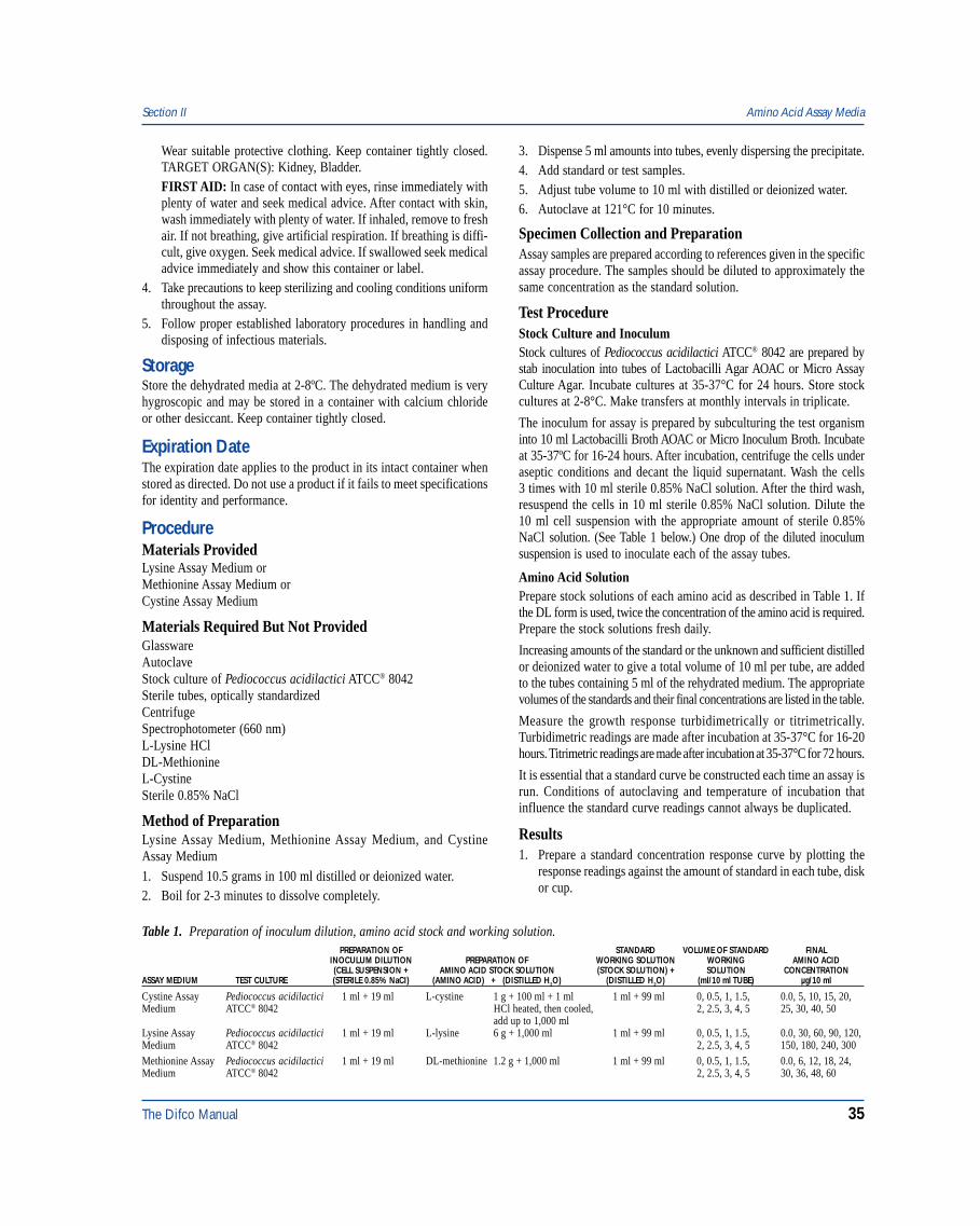
The inoculum for assay is prepared by subculturing the test organisminto 10 ml Lactobacilli Broth AOAC or Micro Inoculum Broth. Incubateat 35-37ºC for 16-24 hours. After incubation, centrifuge the cells underaseptic conditions and decant the liquid supernatant. Wash the cells3 times with 10 ml sterile 0.85% NaCl solution. After the third wash,resuspend the cells in 10 ml sterile 0.85% NaCl solution. Dilute the10 ml cell suspension with the appropriate amount of sterile 0.85%NaCl solution. (See Table 1 below.) One drop of the diluted inoculumsuspension is used to inoculate each of the assay tubes.
Amino Acid SolutionPrepare stock solutions of each amino acid as described in Table 1. Ifthe DL form is used, twice the concentration of the amino acid is required.Prepare the stock solutions fresh daily.
Increasing amounts of the standard or the unknown and sufficient distilledor deionized water to give a total volume of 10 ml per tube, are addedto the tubes containing 5 ml of the rehydrated medium. The appropriatevolumes of the standards and their final concentrations are listed in the table.
Measure the growth response turbidimetrically or titrimetrically.Turbidimetric readings are made after incubation at 35-37°C for 16-20hours. Titrimetric readings are made after incubation at 35-37°C for 72 hours.
It is essential that a standard curve be constructed each time an assay isrun. Conditions of autoclaving and temperature of incubation thatinfluence the standard curve readings cannot always be duplicated.
Results1. Prepare a standard concentration response curve by plotting the
response readings against the amount of standard in each tube, diskor cup.
Section II Amino Acid Assay Media
Table 1. Preparation of inoculum dilution, amino acid stock and working solution.PREPARATION OF STANDARD VOLUME OF STANDARD FINAL
INOCULUM DILUTION PREPARATION OF WORKING SOLUTION WORKING AMINO ACID(CELL SUSPENSION + AMINO ACID STOCK SOLUTION (STOCK SOLUTION) + SOLUTION CONCENTRATION
ASSAY MEDIUM TEST CULTURE (STERILE 0.85% NaCI) (AMINO ACID) + (DISTILLED H2O) (DISTILLED H2O) (ml/10 ml TUBE) µg/10 ml
Cystine Assay Pediococcus acidilactici 1 ml + 19 ml L-cystine 1 g + 100 ml + 1 ml 1 ml + 99 ml 0, 0.5, 1, 1.5, 0.0, 5, 10, 15, 20,Medium ATCC® 8042 HCl heated, then cooled, 2, 2.5, 3, 4, 5 25, 30, 40, 50
add up to 1,000 mlLysine Assay Pediococcus acidilactici 1 ml + 19 ml L-lysine 6 g + 1,000 ml 1 ml + 99 ml 0, 0.5, 1, 1.5, 0.0, 30, 60, 90, 120,Medium ATCC® 8042 2, 2.5, 3, 4, 5 150, 180, 240, 300Methionine Assay Pediococcus acidilactici 1 ml + 19 ml DL-methionine 1.2 g + 1,000 ml 1 ml + 99 ml 0, 0.5, 1, 1.5, 0.0, 6, 12, 18, 24,Medium ATCC® 8042 2, 2.5, 3, 4, 5 30, 36, 48, 60
36 The Difco Manual
2. Determine the amount of amino acid at each level of assay solutionby interpolation from the standard curve.
3. Calculate the concentration of amino acid in the sample from theaverage of these volumes. Use only those values that do not varymore than ±10% from the average. Use the results only if two thirdsof the values do not vary more than ±10%.
Limitations of the Procedure1. The test organism used for inoculating an assay medium must be
cultured and maintained on media recommended for this purpose.2. Aseptic technique should be used throughout the assay procedure.3. The use of altered or deficient media may cause mutants having
different nutritional requirements that will not give a satisfactoryresponse.
4. For successful results of these procedures, all conditions of theassay must be followed precisely.
References1. Steel, Sauberlich, Reynolds, and Baumann. 1949. J. Biol.
Chem. 177:533.
PackagingLysine Assay Medium 100 g 0422-15*
Methionine Assay Medium 100 g 0423-15*
Cystine Assay Medium 100 g 0467-15*
*Store at 2-8°C
Bacto® Anaerobic Agar
User Quality ControlIdentity Specifications
Dehydrated Appearance: Light beige, free-flowing, homogeneous.Solution: 5.8% solution, soluble in distilled or
deionized water on boiling. Lightamber, slightly opalescent. As themedium cools, it becomes greendue to aeration.
Prepared Medium: Light green, slightly opalescent.Reaction of 5.8%Solution at 25°C: pH 7.2 ± 0.2
Cultural ResponsePrepare Anaerobic Agar per label directions. Inoculate themedium and incubate at 35 ±2°C under anaerobic conditionsfor 18-48 hours.
INOCULUMORGANISM ATCC® CFU GROWTH
Bacteroides fragilis 25285* 100-1,000 goodClostridium perfringens 13124* 100-1,000 good
The cultures listed are the minimum that should be used forperformance testing.*These cultures are available as Bactrol™ Disks and should be used as directed in Bactrol Disks Technical Information.
Intended UseBacto Anaerobic Agar is used for cultivating anaerobic microorganisms.
Summary and ExplanationBrewer1 described a special Petri dish cover that allowed surface growthof anaerobes and microaerophiles without anaerobic equipment. Themicroorganisms were grown on an agar-based medium having a lowoxidation-reduction potential. Anaerobic Agar is a modification ofBrewer’s original formula. This medium is suitable for standardplating procedures used in cultivating anaerobic bacteria.2,3,4
Anaerobic bacteria cause a variety of infections in humans, includingotitis media, oral infections, endocarditis, meningitis, wound infectionsfollowing bowel surgery or trauma, and bacteremia.5,6 Anaerobicbacteria are the predominant flora colonizing the skin and mucousmembranes of the body.3 Anaerobes vary in their sensitivity to oxygenand nutritional requirements.2 Anaerobic bacteria lack cytochromes andthus are unable to use oxygen as a terminal electron acceptor.3
Principles of the ProcedureCasitone provides the nitrogen, vitamins and amino acids in AnaerobicAgar. Dextrose is a carbon source. Sodium Chloride maintains theosmotic equilibrium. Sodium Thioglycollate and Sodium FormaldehydeSulfoxylate are reducing agents. Methylene Blue serves as an indicatorof anaerobiosis with a blue color indicating the presence of oxygen.Bacto Agar is the solidifying agent.
FormulaAnaerobic Agar
Formula Per LiterBacto Casitone . . . . . . . . . . . . . . . . . . . . . . . . . . . . . . . . . . 20 gSodium Chloride . . . . . . . . . . . . . . . . . . . . . . . . . . . . . . . . . . 5 gBacto Dextrose . . . . . . . . . . . . . . . . . . . . . . . . . . . . . . . . . . 10 gBacto Agar . . . . . . . . . . . . . . . . . . . . . . . . . . . . . . . . . . . . . 20 gSodium Thioglycollate . . . . . . . . . . . . . . . . . . . . . . . . . . . . . 2 gSodium Formaldehyde Sulfoxylate . . . . . . . . . . . . . . . . . . . 1 gMethylene Blue . . . . . . . . . . . . . . . . . . . . . . . . . . . . . . . . 0.002 g
Final pH 7.2 ± 0.2 at 25°C
Precautions1. For Laboratory Use.2. Follow proper established laboratory procedures in handling and
disposing of infectious material.
StorageStore the dehydrated medium below 30°C. The dehydrated medium isvery hygroscopic. Keep container tightly closed.
Anaerobic Agar Section II
The Difco Manual 37
Expiration DateThe expiration date applies to the product in its intact container whenstored as directed. Do not use a product if it fails to meet specificationsfor identity and performance.
ProcedureMaterials ProvidedAnaerobic Agar
Materials Required But Not ProvidedGlasswareAutoclaveIncubator (35°C)Waterbath (45-50°C) (optional)Sterile Petri dishesBrewer Anaerobic Petri dish covers (optional)
Method of Preparation1. Suspend 58 grams in 1 liter distilled or deionized water.2. Heat to boiling to dissolve completely.3. Autoclave at 121°C for 15 minutes. Cool to 45-50°C.4. Dispense as desired.
Specimen Collection and PreparationAnaerobic bacteria are overlooked or missed unless the specimen isproperly collected and transported to the laboratory.2 Obtain andprocess specimens according to the techniques and proceduresestablished by institutional policy.
Test ProcedureStandard Petri Dishes:2
1. Inoculate a properly obtained specimen onto the medium and streakto obtain isolated colonies.
2. Immediately incubate anaerobically at 35°C.
3. Examine at 24 hours if incubating plates in an anaerobic chamber.Examine at 48 hours if incubating plates in an anaerobic jar oranaerobic pouch.
4. Extended incubation may be necessary to recover some anaerobes.
Brewer Anaerobic Agar Plates:1. Dispense 50-60 ml of Anaerobic Agar into a standard Petri dish.
For best results use porous tops to obtain a dry surface.
2. Inoculate the surface of the medium by streaking; avoid the edgesof the plates.
3. Replace the standard Petri dish lid with a sterile Brewer anaerobicPetri dish cover. The cover should not rest on the Petri dish bottom.The inner glass ridge should seal against the uninoculated periph-ery of the agar. It is essential that the sealing ring inside the coveris in contact with the medium. This seal must not be broken beforethe end of the incubation period. A small amount of air iscaught over the surface of the medium; however, the oxygen in thisspace reacts with reducing agents in the medium to form ananaerobic environment.
4. Incubate aerobically as desired.
For a complete discussion on anaerobic and microaerophilicbacteria from clinical specimens, refer to the appropriateprocedures outlined in the references.2,3,4 For the examination ofanaerobic bacteria in food, refer to Standard Methods.7,8,9
ResultsRefer to appropriate references and procedures for results.
Limitations of the Procedure1. Since the nutritional requirements of organisms vary, some
strains may be encountered that fail to grow or grow poorly onthis medium.
2. Clinical specimens must be obtained properly and transported tothe laboratory in a suitable anaerobic transport container.2
3. The microbiologist must be able to verify quality control of themedium and determine whether the environment is anaerobic.2
4. The microbiologist must perform aerotolerance testing on eachisolate recovered to ensure that the organism is an anaerobe.2
5. Methylene blue is toxic to some anaerobic bacteria.
References1. Brewer, J. H. 1942. A new Petri dish and technique for use in the
cultivation of anaerobes and microaerophiles. Science 95:587.
2. Isenberg, H. D. (ed.). 1992. Clinical microbiology procedureshandbook, American Society for Microbiology, Washington, D.C.
3. Baron, E. J., L. R. Peterson, and S. M. Finegold. 1994. Etiologicalagents recovered from clinical material, p. 474-503. Bailey& Scott’s diagnostic microbiology, 9th ed. Mosby-Year Book, Inc.St. Louis, MO.
4. Murray, P. R., E. J. Baron, M. A. Pfaller, F. C. Tenover, andR. H. Yolken (ed.). 1995. Manual of clinical microbiology, 6th ed.American Society for Microbiology, Washington, D.C.
5. Balows, A., W. J. Hausler, Jr., K. L. Herrmann, H. D. Isenberg,and H. J. Shadomy (ed.). 1991. Manual of clinical microbiology,5th ed. American Society for Microbiology, Washington, D.C.
6. Smith, L. D. S. 1975. The pathogenic anaerobic bacteria, 2nd ed.Charles C. Thomas, Springfield, Il.
7. Association of Official Analytical Chemists. 1995. Bacteriologicalanalytical manual, 8th ed. AOAC International, Gaithesburg, MD.
8. Vanderzant, C., and D. F. Splittstoesser (ed.). 1992. Compen-dium of methods for the microbiological examination of food,3rd ed. American Public Health Association, Washington, D.C.
9. Marshall, R. T. (ed.). 1992. Standard methods for the microbio-logical examination of dairy products, 16th ed. American PublicHealth Association, Washington, D.C.
PackagingAnaerobic Agar 500 g 0536-17
Section II Anaerobic Agar
38 The Difco Manual
Antibiotic Assay Media Section II
Bacto® Antibiotic Assay MediaBacto Antibiotic Medium 1 . Bacto Antibiotic Medium 2Bacto Antibiotic Medium 3 . Bacto Antibiotic Medium 4Bacto Antibiotic Medium 5 . Bacto Antibiotic Medium 8Bacto Antibiotic Medium 9 . Bacto Antibiotic Medium 10Bacto Antibiotic Medium 11 . Bacto Antibiotic Medium 12Bacto Antibiotic Medium 19
User Quality ControlIdentity Specifications
Antibiotic Medium 1Dehydrated Appearance: Beige, homogeneous, free-flowing.Solution: 3.05% solution, soluble in distilled
or deionized water upon boiling;light to medium amber, very slightlyto slightly opalescent.
Prepared Medium: Light to medium amber, slightlyopalescent.
Reaction of 3.05%Solution at 25°C: pH 6.55 ± 0.05
Antibiotic Medium 2Dehydrated Appearance: Light tan, homogeneous, free-flowing.Solution: 2.55% solution, soluble in distilled
or deionized water upon boiling;light- medium amber, very slightlyto slightly opalescent.
Prepared Medium: Light-medium amber, slightlyopalescent.
Reaction of 2.55%Solution at 25°C: pH 6.55 ± 0.05
Antibiotic Medium 3Dehydrated Appearance: Tan, free-flowing, homogeneous.Solution: 1.75% solution, soluble in distilled
or deionized water; light to mediumamber, clear.
Prepared Medium: Light to medium amber, clear.Reaction of 1.75%Solution at 25°C: pH 7.0 ± 0.05
continued on following page
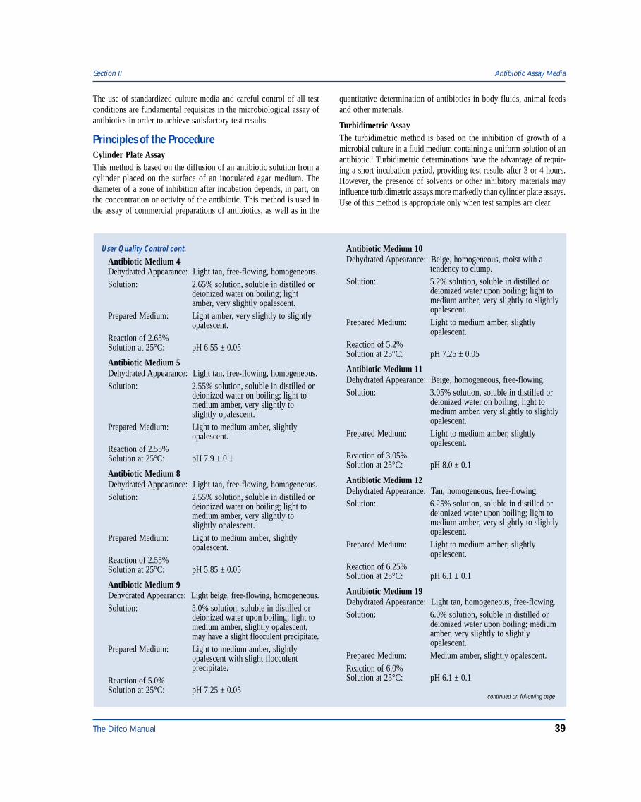
Intended UseBacto Antibiotic Assay Media are used for determining antibioticpotency by the microbiological assay technique.1,6,7
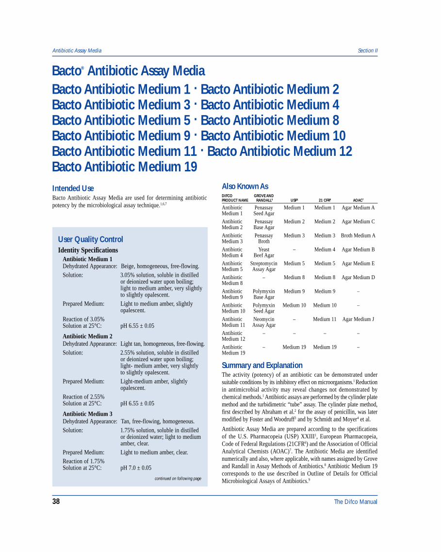
Also Known AsDIFCO GROVE ANDPRODUCT NAME RANDALL8 USP1 21 CFR6 AOAC7
Antibiotic Penassay Medium 1 Medium 1 Agar Medium AMedium 1 Seed Agar
Antibiotic Penassay Medium 2 Medium 2 Agar Medium CMedium 2 Base Agar
Antibiotic Penassay Medium 3 Medium 3 Broth Medium AMedium 3 Broth
Antibiotic Yeast – Medium 4 Agar Medium BMedium 4 Beef Agar
Antibiotic Streptomycin Medium 5 Medium 5 Agar Medium EMedium 5 Assay Agar
Antibiotic – Medium 8 Medium 8 Agar Medium DMedium 8
Antibiotic Polymyxin Medium 9 Medium 9 –Medium 9 Base Agar
Antibiotic Polymyxin Medium 10 Medium 10 –Medium 10 Seed Agar
Antibiotic Neomycin – Medium 11 Agar Medium JMedium 11 Assay Agar
Antibiotic – – – –Medium 12
Antibiotic – Medium 19 Medium 19 –Medium 19
Summary and ExplanationThe activity (potency) of an antibiotic can be demonstrated undersuitable conditions by its inhibitory effect on microorganisms.1 Reductionin antimicrobial activity may reveal changes not demonstrated bychemical methods.1 Antibiotic assays are performed by the cylinder platemethod and the turbidimetric “tube” assay. The cylinder plate method,first described by Abraham et al.2 for the assay of penicillin, was latermodified by Foster and Woodruff3 and by Schmidt and Moyer4 et al.
Antibiotic Assay Media are prepared according to the specificationsof the U.S. Pharmacopeia (USP) XXIII1, European Pharmacopeia,Code of Federal Regulations (21CFR6) and the Association of OfficialAnalytical Chemists (AOAC)7. The Antibiotic Media are identifiednumerically and also, where applicable, with names assigned by Groveand Randall in Assay Methods of Antibiotics.8 Antibiotic Medium 19corresponds to the use described in Outline of Details for OfficialMicrobiological Assays of Antibiotics.9
The Difco Manual 39
Section II Antibiotic Assay Media
The use of standardized culture media and careful control of all testconditions are fundamental requisites in the microbiological assay ofantibiotics in order to achieve satisfactory test results.
Principles of the ProcedureCylinder Plate AssayThis method is based on the diffusion of an antibiotic solution from acylinder placed on the surface of an inoculated agar medium. Thediameter of a zone of inhibition after incubation depends, in part, onthe concentration or activity of the antibiotic. This method is used inthe assay of commercial preparations of antibiotics, as well as in the
quantitative determination of antibiotics in body fluids, animal feedsand other materials.
Turbidimetric AssayThe turbidimetric method is based on the inhibition of growth of amicrobial culture in a fluid medium containing a uniform solution of anantibiotic.1 Turbidimetric determinations have the advantage of requir-ing a short incubation period, providing test results after 3 or 4 hours.However, the presence of solvents or other inhibitory materials mayinfluence turbidimetric assays more markedly than cylinder plate assays.Use of this method is appropriate only when test samples are clear.
Antibiotic Medium 10Dehydrated Appearance: Beige, homogeneous, moist with a
tendency to clump.Solution: 5.2% solution, soluble in distilled or
deionized water upon boiling; light tomedium amber, very slightly to slightlyopalescent.
Prepared Medium: Light to medium amber, slightlyopalescent.
Reaction of 5.2%Solution at 25°C: pH 7.25 ± 0.05
Antibiotic Medium 11Dehydrated Appearance: Beige, homogeneous, free-flowing.Solution: 3.05% solution, soluble in distilled or
deionized water on boiling; light tomedium amber, very slightly to slightlyopalescent.
Prepared Medium: Light to medium amber, slightlyopalescent.
Reaction of 3.05%Solution at 25°C: pH 8.0 ± 0.1
Antibiotic Medium 12Dehydrated Appearance: Tan, homogeneous, free-flowing.Solution: 6.25% solution, soluble in distilled or
deionized water upon boiling; light tomedium amber, very slightly to slightlyopalescent.
Prepared Medium: Light to medium amber, slightlyopalescent.
Reaction of 6.25%Solution at 25°C: pH 6.1 ± 0.1
Antibiotic Medium 19Dehydrated Appearance: Light tan, homogeneous, free-flowing.Solution: 6.0% solution, soluble in distilled or
deionized water upon boiling; mediumamber, very slightly to slightlyopalescent.
Prepared Medium: Medium amber, slightly opalescent.Reaction of 6.0%Solution at 25°C: pH 6.1 ± 0.1
User Quality Control cont.
Antibiotic Medium 4Dehydrated Appearance: Light tan, free-flowing, homogeneous.Solution: 2.65% solution, soluble in distilled or
deionized water on boiling; lightamber, very slightly opalescent.
Prepared Medium: Light amber, very slightly to slightlyopalescent.
Reaction of 2.65%Solution at 25°C: pH 6.55 ± 0.05
Antibiotic Medium 5Dehydrated Appearance: Light tan, free-flowing, homogeneous.Solution: 2.55% solution, soluble in distilled or
deionized water on boiling; light tomedium amber, very slightly toslightly opalescent.
Prepared Medium: Light to medium amber, slightlyopalescent.
Reaction of 2.55%Solution at 25°C: pH 7.9 ± 0.1
Antibiotic Medium 8Dehydrated Appearance: Light tan, free-flowing, homogeneous.Solution: 2.55% solution, soluble in distilled or
deionized water on boiling; light tomedium amber, very slightly toslightly opalescent.
Prepared Medium: Light to medium amber, slightlyopalescent.
Reaction of 2.55%Solution at 25°C: pH 5.85 ± 0.05
Antibiotic Medium 9Dehydrated Appearance: Light beige, free-flowing, homogeneous.Solution: 5.0% solution, soluble in distilled or
deionized water upon boiling; light tomedium amber, slightly opalescent,may have a slight flocculent precipitate.
Prepared Medium: Light to medium amber, slightlyopalescent with slight flocculentprecipitate.
Reaction of 5.0%Solution at 25°C: pH 7.25 ± 0.05
continued on following page
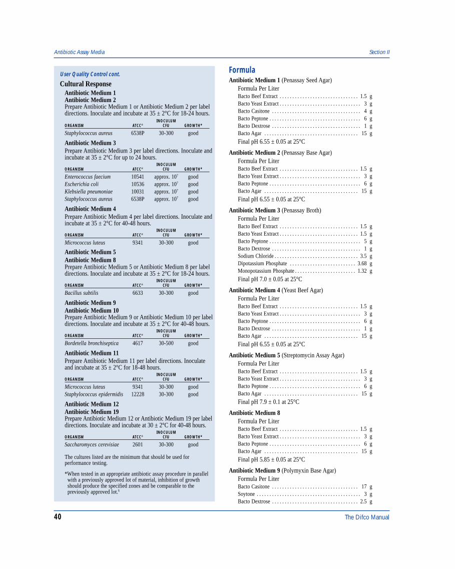
40 The Difco Manual
FormulaAntibiotic Medium 1 (Penassay Seed Agar)
Formula Per LiterBacto Beef Extract . . . . . . . . . . . . . . . . . . . . . . . . . . . . . . . 1.5 gBacto Yeast Extract . . . . . . . . . . . . . . . . . . . . . . . . . . . . . . . . 3 gBacto Casitone . . . . . . . . . . . . . . . . . . . . . . . . . . . . . . . . . . . 4 gBacto Peptone . . . . . . . . . . . . . . . . . . . . . . . . . . . . . . . . . . . . 6 gBacto Dextrose . . . . . . . . . . . . . . . . . . . . . . . . . . . . . . . . . . . 1 gBacto Agar . . . . . . . . . . . . . . . . . . . . . . . . . . . . . . . . . . . . . 15 g
Final pH 6.55 ± 0.05 at 25°C
Antibiotic Medium 2 (Penassay Base Agar)Formula Per LiterBacto Beef Extract . . . . . . . . . . . . . . . . . . . . . . . . . . . . . . . 1.5 gBacto Yeast Extract . . . . . . . . . . . . . . . . . . . . . . . . . . . . . . . . 3 gBacto Peptone . . . . . . . . . . . . . . . . . . . . . . . . . . . . . . . . . . . . 6 gBacto Agar . . . . . . . . . . . . . . . . . . . . . . . . . . . . . . . . . . . . . 15 g
Final pH 6.55 ± 0.05 at 25°C
Antibiotic Medium 3 (Penassay Broth)Formula Per LiterBacto Beef Extract . . . . . . . . . . . . . . . . . . . . . . . . . . . . . . . 1.5 gBacto Yeast Extract . . . . . . . . . . . . . . . . . . . . . . . . . . . . . . . 1.5 gBacto Peptone . . . . . . . . . . . . . . . . . . . . . . . . . . . . . . . . . . . . 5 gBacto Dextrose . . . . . . . . . . . . . . . . . . . . . . . . . . . . . . . . . . . 1 gSodium Chloride . . . . . . . . . . . . . . . . . . . . . . . . . . . . . . . . . 3.5 gDipotassium Phosphate . . . . . . . . . . . . . . . . . . . . . . . . . . 3.68 gMonopotassium Phosphate . . . . . . . . . . . . . . . . . . . . . . . . 1.32 g
Final pH 7.0 ± 0.05 at 25°C
Antibiotic Medium 4 (Yeast Beef Agar)Formula Per LiterBacto Beef Extract . . . . . . . . . . . . . . . . . . . . . . . . . . . . . . . 1.5 gBacto Yeast Extract . . . . . . . . . . . . . . . . . . . . . . . . . . . . . . . . 3 gBacto Peptone . . . . . . . . . . . . . . . . . . . . . . . . . . . . . . . . . . . . 6 gBacto Dextrose . . . . . . . . . . . . . . . . . . . . . . . . . . . . . . . . . . . 1 gBacto Agar . . . . . . . . . . . . . . . . . . . . . . . . . . . . . . . . . . . . . 15 g
Final pH 6.55 ± 0.05 at 25°C
Antibiotic Medium 5 (Streptomycin Assay Agar)Formula Per LiterBacto Beef Extract . . . . . . . . . . . . . . . . . . . . . . . . . . . . . . . 1.5 gBacto Yeast Extract . . . . . . . . . . . . . . . . . . . . . . . . . . . . . . . . 3 gBacto Peptone . . . . . . . . . . . . . . . . . . . . . . . . . . . . . . . . . . . . 6 gBacto Agar . . . . . . . . . . . . . . . . . . . . . . . . . . . . . . . . . . . . . 15 g
Final pH 7.9 ± 0.1 at 25°C
Antibiotic Medium 8Formula Per LiterBacto Beef Extract . . . . . . . . . . . . . . . . . . . . . . . . . . . . . . . 1.5 gBacto Yeast Extract . . . . . . . . . . . . . . . . . . . . . . . . . . . . . . . . 3 gBacto Peptone . . . . . . . . . . . . . . . . . . . . . . . . . . . . . . . . . . . . 6 gBacto Agar . . . . . . . . . . . . . . . . . . . . . . . . . . . . . . . . . . . . . 15 g
Final pH 5.85 ± 0.05 at 25°C
Antibiotic Medium 9 (Polymyxin Base Agar)Formula Per LiterBacto Casitone . . . . . . . . . . . . . . . . . . . . . . . . . . . . . . . . . . 17 gSoytone . . . . . . . . . . . . . . . . . . . . . . . . . . . . . . . . . . . . . . . . . 3 gBacto Dextrose . . . . . . . . . . . . . . . . . . . . . . . . . . . . . . . . . . 2.5 g
User Quality Control cont.
Cultural ResponseAntibiotic Medium 1Antibiotic Medium 2Prepare Antibiotic Medium 1 or Antibiotic Medium 2 per labeldirections. Inoculate and incubate at 35 ± 2°C for 18-24 hours.
INOCULUMORGANISM ATCC® CFU GROWTH*
Staphylococcus aureus 6538P 30-300 good
Antibiotic Medium 3Prepare Antibiotic Medium 3 per label directions. Inoculate andincubate at 35 ± 2°C for up to 24 hours.
INOCULUMORGANISM ATCC® CFU GROWTH*
Enterococcus faecium 10541 approx. 107 goodEscherichia coli 10536 approx. 107 goodKlebsiella pneumoniae 10031 approx. 107 goodStaphylococcus aureus 6538P approx. 107 good
Antibiotic Medium 4Prepare Antibiotic Medium 4 per label directions. Inoculate andincubate at 35 ± 2°C for 40-48 hours.
INOCULUMORGANISM ATCC® CFU GROWTH*
Micrococcus luteus 9341 30-300 good
Antibiotic Medium 5Antibiotic Medium 8Prepare Antibiotic Medium 5 or Antibiotic Medium 8 per labeldirections. Inoculate and incubate at 35 ± 2°C for 18-24 hours.
INOCULUMORGANISM ATCC® CFU GROWTH*
Bacillus subtilis 6633 30-300 good
Antibiotic Medium 9Antibiotic Medium 10Prepare Antibiotic Medium 9 or Antibiotic Medium 10 per labeldirections. Inoculate and incubate at 35 ± 2°C for 40-48 hours.
INOCULUMORGANISM ATCC® CFU GROWTH*
Bordetella bronchiseptica 4617 30-500 good
Antibiotic Medium 11Prepare Antibiotic Medium 11 per label directions. Inoculateand incubate at 35 ± 2°C for 18-48 hours.
INOCULUMORGANISM ATCC® CFU GROWTH*
Micrococcus luteus 9341 30-300 goodStaphylococcus epidermidis 12228 30-300 good
Antibiotic Medium 12Antibiotic Medium 19Prepare Antibiotic Medium 12 or Antibiotic Medium 19 per labeldirections. Inoculate and incubate at 30 ± 2°C for 40-48 hours.
INOCULUMORGANISM ATCC® CFU GROWTH*
Saccharomyces cerevisiae 2601 30-300 good
The cultures listed are the minimum that should be used forperformance testing.
*When tested in an appropriate antibiotic assay procedure in parallel with a previously approved lot of material, inhibition of growth should produce the specified zones and be comparable to the previously approved lot.6
Antibiotic Assay Media Section II
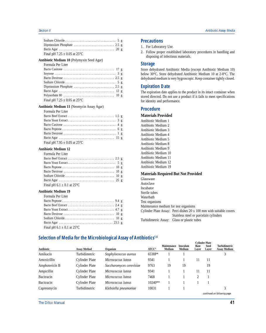
The Difco Manual 41
Sodium Chloride . . . . . . . . . . . . . . . . . . . . . . . . . . . . . . . . . . 5 gDipotassium Phosphate . . . . . . . . . . . . . . . . . . . . . . . . . . . 2.5 gBacto Agar . . . . . . . . . . . . . . . . . . . . . . . . . . . . . . . . . . . . . 20 gFinal pH 7.25 ± 0.05 at 25°C
Antibiotic Medium 10 (Polymyxin Seed Agar)Formula Per LiterBacto Casitone . . . . . . . . . . . . . . . . . . . . . . . . . . . . . . . . . . 17 gSoytone . . . . . . . . . . . . . . . . . . . . . . . . . . . . . . . . . . . . . . . . . 3 gBacto Dextrose . . . . . . . . . . . . . . . . . . . . . . . . . . . . . . . . . . 2.5 gSodium Chloride . . . . . . . . . . . . . . . . . . . . . . . . . . . . . . . . . . 5 gDipotassium Phosphate . . . . . . . . . . . . . . . . . . . . . . . . . . . 2.5 gBacto Agar . . . . . . . . . . . . . . . . . . . . . . . . . . . . . . . . . . . . . 12 gPolysorbate 80 . . . . . . . . . . . . . . . . . . . . . . . . . . . . . . . . . . 10 g
Final pH 7.25 ± 0.05 at 25°C
Antibiotic Medium 11 (Neomycin Assay Agar)Formula Per LiterBacto Beef Extract . . . . . . . . . . . . . . . . . . . . . . . . . . . . . . . 1.5 gBacto Yeast Extract . . . . . . . . . . . . . . . . . . . . . . . . . . . . . . . . 3 gBacto Casitone . . . . . . . . . . . . . . . . . . . . . . . . . . . . . . . . . . . 4 gBacto Peptone . . . . . . . . . . . . . . . . . . . . . . . . . . . . . . . . . . . . 6 gBacto Dextrose . . . . . . . . . . . . . . . . . . . . . . . . . . . . . . . . . . . 1 gBacto Agar . . . . . . . . . . . . . . . . . . . . . . . . . . . . . . . . . . . . . 15 g
Final pH 7.95 ± 0.05 at 25°C
Antibiotic Medium 12Formula Per LiterBacto Beef Extract . . . . . . . . . . . . . . . . . . . . . . . . . . . . . . . 2.5 gBacto Yeast Extract . . . . . . . . . . . . . . . . . . . . . . . . . . . . . . . . 5 gBacto Peptone . . . . . . . . . . . . . . . . . . . . . . . . . . . . . . . . . . . 10 gBacto Dextrose . . . . . . . . . . . . . . . . . . . . . . . . . . . . . . . . . . 10 gSodium Chloride . . . . . . . . . . . . . . . . . . . . . . . . . . . . . . . . . 10 gBacto Agar . . . . . . . . . . . . . . . . . . . . . . . . . . . . . . . . . . . . . 25 g
Final pH 6.1 ± 0.1 at 25°C
Antibiotic Medium 19Formula Per LiterBacto Peptone . . . . . . . . . . . . . . . . . . . . . . . . . . . . . . . . . . . 9.4 gBacto Beef Extract . . . . . . . . . . . . . . . . . . . . . . . . . . . . . . . 2.4 gBacto Yeast Extract . . . . . . . . . . . . . . . . . . . . . . . . . . . . . . . 4.7 gBacto Dextrose . . . . . . . . . . . . . . . . . . . . . . . . . . . . . . . . . . 10 gSodium Chloride . . . . . . . . . . . . . . . . . . . . . . . . . . . . . . . . . 10 gBacto Agar . . . . . . . . . . . . . . . . . . . . . . . . . . . . . . . . . . . . 23.5 g
Final pH 6.1 ± 0.1 at 25°C
Precautions1. For Laboratory Use.2. Follow proper established laboratory procedures in handling and
disposing of infectious materials.
StorageStore dehydrated Antibiotic Media (except Antibiotic Medium 10)below 30°C. Store dehydrated Antibiotic Medium 10 at 2-8°C. Thedehydrated medium is very hygroscopic. Keep container tightly closed.
Expiration DateThe expiration date applies to the product in its intact container whenstored directed. Do not use a product if it fails to meet specificationsfor identity and performance.
ProcedureMaterials ProvidedAntibiotic Medium 1Antibiotic Medium 2Antibiotic Medium 3Antibiotic Medium 4Antibiotic Medium 5Antibiotic Medium 8Antibiotic Medium 9Antibiotic Medium 10Antibiotic Medium 11Antibiotic Medium 12Antibiotic Medium 19
Materials Required But Not ProvidedGlasswareAutoclaveIncubatorSterile tubesWaterbathTest organismsMaintenance medium for test organismsCylinder Plate Assay: Petri dishes 20 x 100 mm with suitable covers
Stainless steel or porcelain cylindersTurbidimetric Assay: Glass or plastic tubes
Section II Antibiotic Assay Media
Selection of Media for the Microbiological Assay of Antibiotics1,6
Cylinder PlateMaintenance Inoculum Base Seed Turbidimetric
Antibiotic Assay Method Organism ATCC® Medium Medium Layer Layer Assay Medium
Amikacin Turbidimetric Staphylococcus aureus 6538P* 1 1 3
Amoxicillin Cylinder Plate Micrococcus luteus 9341 1 1 11 11
Amphotericin B Cylinder Plate Saccharomyces cerevisiae 9763 19 19 19
Ampicillin Cylinder Plate Micrococcus luteus 9341 1 1 11 11
Bacitracin Cylinder Plate Micrococcus luteus 7468 1 1 2 1
Bacitracin Cylinder Plate Micrococcus luteus 10240** 1 1 1 1
Capreomycin Turbidimetric Klebsiella pneumoniae 10031 1 1 3continued on following page
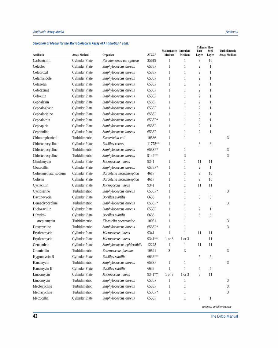
42 The Difco Manual
Selection of Media for the Microbiological Assay of Antibiotics1,6 cont. Cylinder Plate
Maintenance Inoculum Base Seed TurbidimetricAntibiotic Assay Method Organism ATCC® Medium Medium Layer Layer Assay Medium
Carbenicillin Cylinder Plate Pseudomonas aeruginosa 25619 1 1 9 10
Cefaclor Cylinder Plate Staphylococcus aureus 6538P 1 1 2 1
Cefadroxil Cylinder Plate Staphylococcus aureus 6538P 1 1 2 1
Cefamandole Cylinder Plate Staphylococcus aureus 6538P 1 1 2 1
Cefazolin Cylinder Plate Staphylococcus aureus 6538P 1 1 2 1
Cefotaxime Cylinder Plate Staphylococcus aureus 6538P 1 1 2 1
Cefoxitin Cylinder Plate Staphylococcus aureus 6538P 1 1 2 1
Cephalexin Cylinder Plate Staphylococcus aureus 6538P 1 1 2 1
Cephaloglycin Cylinder Plate Staphylococcus aureus 6538P 1 1 2 1
Cephaloridine Cylinder Plate Staphylococcus aureus 6538P 1 1 2 1
Cephalothin Cylinder Plate Staphylococcus aureus 6538P* 1 1 2 1
Cephapirin Cylinder Plate Staphylococcus aureus 6538P 1 1 2 1
Cephradine Cylinder Plate Staphylococcus aureus 6538P 1 1 2 1
Chloramphenicol Turbidimetric Escherichia coli 10536 1 1 3
Chlortetracycline Cylinder Plate Bacillus cereus 11778** 1 8 8
Chlortetracycline Turbidimetric Staphylococcus aureus 6538P* 1 1 3
Chlortetracycline Turbidimetric Staphylococcus aureus 9144** 3 3
Clindamycin Cylinder Plate Micrococcus luteus 9341 1 1 11 11
Cloxacillin Cylinder Plate Staphylococcus aureus 6538P* 1 1 2 1
Colistimethate, sodium Cylinder Plate Bordetella bronchiseptica 4617 1 1 9 10
Colistin Cylinder Plate Bordetella bronchiseptica 4617 1 1 9 10
Cyclacillin Cylinder Plate Micrococcus luteus 9341 1 1 11 11
Cycloserine Turbidimetric Staphylococcus aureus 6538P* 1 1 3
Dactinocycin Cylinder Plate Bacillus subtilis 6633 1 1 5 5
Demeclyocycline Turbidimetric Staphylococcus aureus 6538P* 1 1 3
Dicloxacillin Cylinder Plate Staphylococcus aureus 6538P 1 1 2 1
Dihydro- Cylinder Plate Bacillus subtilis 6633 1 1 5 5
streptomycin Turbidimetric Klebsiella pneumoniae 10031 1 1 3
Doxycycline Turbidimetric Staphylococcus aureus 6538P* 1 1 3
Erythromycin Cylinder Plate Micrococcus luteus 9341 1 1 11 11
Erythromycin Cylinder Plate Micrococcus luteus 9341** 1 or 3 1 or 3 11
Gentamicin Cylinder Plate Staphylococcus epidermidis 12228 1 1 11 11
Gramicidin Turbidimetric Enterococcus faecium 10541 3 3 3
Hygromycin B Cylinder Plate Bacillus subtilis 6633** 5 5
Kanamycin Turbidimetric Staphylococcus aureus 6538P 1 1 3
Kanamycin B Cylinder Plate Bacillus subtilis 6633 1 1 5 5
Lincomycin Cylinder Plate Micrococcus luteus 9341** 1 or 3 1 or 3 5 11
Lincomycin Turbidimetric Staphylococcus aureus 6538P 1 1 3
Meclocycline Turbidimetric Staphylococcus aureus 6538P 1 1 3
Methacycline Turbidimetric Staphylococcus aureus 6538P* 1 1 3
Methicillin Cylinder Plate Staphylococcus aureus 6538P 1 1 2 1
Antibiotic Assay Media Section II
continued on following page
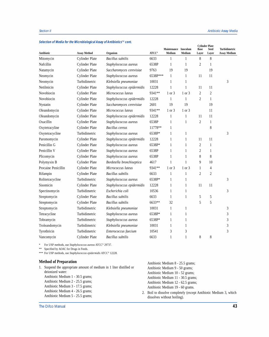
The Difco Manual 43
Selection of Media for the Microbiological Assay of Antibiotics1,6 cont. Cylinder Plate
Maintenance Inoculum Base Seed TurbidimetricAntibiotic Assay Method Organism ATCC® Medium Medium Layer Layer Assay Medium
Mitomycin Cylinder Plate Bacillus subtilis 6633 1 1 8 8
Nafcillin Cylinder Plate Staphylococcus aureus 6538P 1 1 2 1
Natamycin Cylinder Plate Saccharomyces cerevisiae 9763 19 19 19
Neomycin Cylinder Plate Staphylococcus aureus 6538P*** 1 1 11 11
Neomycin Turbidimetric Klebsiella pneumoniae 10031 1 1 3
Netilmicin Cylinder Plate Staphylococcus epidermidis 12228 1 1 11 11
Novobiocin Cylinder Plate Micrococcus luteus 9341** 1 or 3 1 or 3 2 2
Novobiocin Cylinder Plate Staphylococcus epidermidis 12228 1 1 2 1
Nystatin Cylinder Plate Saccharomyces cerevisiae 2601 19 19 19
Oleandomycin Cylinder Plate Micrococcus luteus 9341** 1 or 3 1 or 3 11
Oleandomycin Cylinder Plate Staphylococcus epidermidis 12228 1 1 11 11
Oxacillin Cylinder Plate Staphylococcus aureus 6538P 1 1 2 1
Oxytetracyline Cylinder Plate Bacillus cereus 11778** 1 8
Oxytetracycline Turbidimetric Staphylococcus aureus 6538P* 1 1 3
Paromomycin Cylinder Plate Staphylococcus epidermidis 12228 1 1 11 11
Penicillin G Cylinder Plate Staphylococcus aureus 6538P* 1 1 2 1
Penicillin V Cylinder Plate Staphylococcus aureus 6538P 1 1 2 1
Plicomycin Cylinder Plate Staphylococcus aureus 6538P 1 1 8 8
Polymyxin B Cylinder Plate Bordetella bronchiseptica 4617 1 1 9 10
Procaine Penicillin Cylinder Plate Micrococcus luteus 9341** 1 or 3 1 or 3 1 4
Rifampin Cylinder Plate Bacillus subtilis 6633 1 1 2 2
Rolitetracycline Turbidimetric Staphylococcus aureus 6538P* 1 1 3
Sisomicin Cylinder Plate Staphylococcus epidermidis 12228 1 1 11 11
Spectinomycin Turbidimetric Escherichia coli 10536 1 1 3
Streptomycin Cylinder Plate Bacillus subtilis 6633 1 1 5 5
Streptomycin Cylinder Plate Bacillus subtilis 6633** 32 5 5
Streptomycin Turbidimetric Klebsiella pneumoniae 10031 1 1 3
Tetracycline Turbidimetric Staphylococcus aureus 6538P* 1 1 3
Tobramycin Turbidimetric Staphylococcus aureus 6538P* 1 1 3
Troleandomycin Turbidimetric Klebsiella pneumoniae 10031 1 1 3
Tyrothricin Turbidimetric Enterococcus faecium 10541 3 3 3
Vancomycin Cylinder Plate Bacillus subtilis 6633 1 1 8 8
* For USP methods, use Staphylococcus aureus ATCC® 29737.** Specified by AOAC for Drugs in Feeds.*** For USP methods, use Staphylococcus epidermidis ATCC® 12228.
Section II Antibiotic Assay Media
Method of Preparation1. Suspend the appropriate amount of medium in 1 liter distilled or
deionized water:Antibiotic Medium 1 - 30.5 grams;Antibiotic Medium 2 - 25.5 grams;Antibiotic Medium 3 - 17.5 grams;Antibiotic Medium 4 - 26.5 grams;Antibiotic Medium 5 - 25.5 grams;
Antibiotic Medium 8 - 25.5 grams;Antibiotic Medium 9 - 50 grams;Antibiotic Medium 10 - 52 grams;Antibiotic Medium 11 - 30.5 grams;Antibiotic Medium 12 - 62.5 grams;Antibiotic Medium 19 - 60 grams.
2. Boil to dissolve completely (except Antibiotic Medium 3, whichdissolves without boiling).
44 The Difco Manual
3. Autoclave at 121°C for 15 minutes.4. Cool medium to 45-50°C.5. Antibiotic Medium 11, only: To alter the pH, add 1N HCl or 1N
NaOH to the medium at 45-50°C.6. Dispense as appropriate.
Specimen Collection and PreparationObtain and process specimens according to the techniques andprocedures established by laboratory policy.
Test Organism PreparationMaintain stock cultures on agar slants and make transfers at 1- or2-week intervals. Prepare the inoculum for assay by washing growthfrom a fresh 24-48 hour agar slant using sterile distilled water, salineor Antibiotic Medium 3 and further dilute the culture to obtain thedesired organism concentration. In some turbidimetric assays, a 18- to24-hour culture of the test organism in Antibiotic Medium 3, diluted toobtain the optimal number of organisms, is used.
When Bacillus subtilis is used as the test organism, inoculate it onAntibiotic Medium 1 and incubate at 37°C for 1 week, wash sporesfrom the agar surface, and heat the spores at 56°C for 30 minutes. Washthe spores 3 times in distilled water, heat again at 65°C for 30 minutes,and then dilute to the optimal concentration. This inoculum preparationshould produce a sharp zone in the assay.
Antibiotic Medium modified by the addition of 300 mg manganesesulfate (MnSO4
.H2O) per liter often aids the sporulation of B. subtilisand may be used in preparing the spore suspension. A standardizedspore suspension prepared from B. subtilis ATCC® 6633 is available asBacto Subtilis Spore Suspension.
When B. cereus var. mycoides is required, inoculate the organism onAntibiotic Medium 1 and incubate at 30°C for 1 week. Wash andprepare the spores as for B. subtilis, above. A standardized sporesuspension of B. cereus var. mycoides is available as Bacto CereusSpore Suspension.
Cylinder Plate AssayUse 20 x 100 mm Petri dishes with sufficient depth so that cylindersused in the assay will not be pushed into the medium by the cover.Porcelain covers glazed on the outside, only, are recommended.
Use stainless steel or porcelain assay cylinders having the followingdimensions (± 0.1 mm): 8 mm outside diameter, 6 mm inside diameterand 10 mm long.1 Carefully clean the cylinders to remove all residues,using an occasional acid bath, i.e., with approximately 2N nitric acidor with chromic acid.1 Four or six cylinders are generally used perplate, evenly spaced on a 2.8 cm radius.
To assure accurate assays, work on a level surface to obtain uniformlythick base and seed layers in the Petri dish. Allow the base layer tosolidify and then overlay the seed layer containing a proper concentra-tion of the test organism. The amount of medium in the layers variesfor different antibiotics, with most assays specifying a 21 ml base layerand a 4 ml seed layer. In any case, dishes with flat bottoms are requiredto assure complete coverage of the bottom of the dish when smallamounts of base medium are used. Tilt the plate to obtain even coverageof the base layer by the seed layer and allow it to solidify in a levelposition. Plates should be used the same day as prepared.
Turbidimetric AssayUse glass or plastic test tubes (i.e., 16 x 125 mm or 18 x 150 mm)that are relatively uniform in length, diameter and thickness andsubstantially free from surface blemishes.1 Tubes that will be placed inthe spectrophotometer should be matched and free of scratches orblemishes.1 Clean the tubes thoroughly to remove all antibioticresidues and traces of cleaning solution and, prior to subsequent use,sterilize tubes that have been previously used.1
Prepare working dilutions of the antibiotic reference standards inspecific concentrations. To a 1 ml quantity of each solution in asuitable tube, add 9 ml of inoculated broth, as required. Prepare similarsolutions of the assay materials containing approximately the sameamounts of antibiotic activity and place in tubes. Incubate the tubes for3-4 hours at the required temperature, generally in a water bath. Atthe end of the incubation period, stop growth by adding 0.5 ml of1:3 formalin. Determine the amount of growth by measuring lighttransmittance with a suitable spectrophotometer. Determine theconcentration of the antibiotic by comparing the growth obtained withthat given by reference standard solutions.
For a complete discussion of antibiotic assay methods, refer toappropriate procedures outlined in the references.1,5,6,7
ResultsRefer to appropriate procedures for results.1,5,6,7
Limitations of the Procedure1. Since the nutritional requirements of organisms vary, some strains
may be encountered that fail to grow or grow poorly on thismedium.
References1. United States Pharmacopeial Convention. 1995. The United
States pharmacopeia, 23rd ed. Biological Tests and Assays,p. 1690-1696. The United States Pharmacopeial Convention,Rockville, MD.
2. Abraham. 1941. Lancet. 2:177.3. Foster and Woodruff. 1943. J. Bacteriol. 46:187.4. Schmidt, W. H., and A. J. Moyer. 1944. Penicillin. I. Methods of
assay. J Bacteriol. 47:199.5. European Pharmacopoeia. 1994. Council of Europe, 2nd ed.
Maisonneuve S. A. Sainte-Ruffine, FR.6. Federal Register. 1992. Tests and methods of assay of
Antibiotics and Antibiotic-Containing Drugs. Fed. Regist.21:436.100-436.106.
7. Association of Official Analytical Chemists. 1995. Officialmethods of analysis of AOAC International, 16th ed. AOACInternational, Arlington, VA.
8. Grove, D. C., and W. A. Randall. 1955. Assay methods ofantibiotics. Medical Encyclopedia Inc., New York, NY.
9. Kirshbaum, A., and B. Arret. 1967. Outline of details forofficial microbiological assays of antibiotics. J. Pharm.Sci. 56:512.
Antibiotic Assay Media Section II
The Difco Manual 45
PackagingAntibiotic Medium 1 500 g 0263-17
2 kg 0263-0710 kg 0263-08
Antibiotic Medium 2 500 g 0270-1710 kg 0270-08
Antibiotic Medium 3 500 g 0243-172 kg 0243-07
Antibiotic Medium 4 500 g 0244-17
Section II Aseptic Commissioning Medium
Bacto® Aseptic Commissioning Medium
User Quality ControlIdentity Specifications
Dehydrated Appearance: Beige to pink, free-flowing,homogeneous.
Solution: 1.75% solution, soluble in distilledor deionized water on warming,orange-red, clear.
Prepared Medium: Orange-red, clear.Reaction of 1.75%Solution at 25°C: pH 7.2 ± 0.2
Cultural ResponsePrepare the medium per label directions. Inoculate testorganisms into tubes with fermentation vials and incubate at30 ± 2°C for 18-48 hours.
INOCULUMORGANISM ATCC® CFU GROWTH ACID GAS
Bacillus cereus 14579 100-1,000 good + –Enterobacter aerogenes 13048* 100-1,000 good + +Escherichia coli 25922* 100-1,000 good +** + or –Staphylococcus aureus 25923* 100-1,000 good + –
The cultures listed are the minimum that should be used forperformance testing.*These cultures are available as Bactrol™ Disks and should be used as directed in Bactrol Disks Technical Information.
**May revert to alkaline after prolonged incubation.
Intended UseBacto Aseptic Commissioning Medium is a fluid medium used invalidating aseptic packing lines.
Summary and ExplanationAseptic Commissioning Medium is a basic medium in which growthcan be demonstrated by either acid or gas production. It is ideally suitedfor validating and commissioning aseptic packing and filling lines.
Principles of ProcedurePeptone and Yeast Extract provide basic nutrients. Sucrose is a
carbohydrate source. Phenol Red is a pH indicator. Sodium Chloridemaintains the osmotic balance.
FormulaAseptic Commissioning Medium
Formula Per LiterPeptone . . . . . . . . . . . . . . . . . . . . . . . . . . . . . . . . . . . . . . . . . 5 gYeast Extract . . . . . . . . . . . . . . . . . . . . . . . . . . . . . . . . . . . . 2.5 gSucrose . . . . . . . . . . . . . . . . . . . . . . . . . . . . . . . . . . . . . . . . . 5 gPhenol Red . . . . . . . . . . . . . . . . . . . . . . . . . . . . . . . . . . . . . . 5 mgSodium Chloride . . . . . . . . . . . . . . . . . . . . . . . . . . . . . . . . . . 5 g
Final pH 7.2 ± 0.2 at 25°C
Precautions1. For Laboratory Use.2. Follow proper established laboratory procedures in handling and
disposing of infectious materials.
StorageStore the dehydrated medium below 30°C. The dehydrated medium isvery hygroscopic. Keep container tightly closed.
Expiration DateThe expiration date applies to the product in its intact container whenstored as directed. Do not use a product if it fails to meet specificationsfor identity and performance.
ProcedureMaterials ProvidedAseptic Commissioning Medium
Materials Required But Not ProvidedFlasks with closuresDistilled or deionized water
Method of Preparation1. Suspend 17.5 grams in 1 liter distilled or deionized water.2. Heat gently to dissolve completely.3. Autoclave at 121°C for 15 minutes.
Antibiotic Medium 5 500 g 0277-17
Antibiotic Medium 8 500 g 0667-17
Antibiotic Medium 9 500 g 0462-17
Antibiotic Medium 10 500 g 0463-17
Antibiotic Medium 11 500 g 0593-17
Antibiotic Medium 12 500 g 0669-17
Antibiotic Medium 19 500 g 0043-17
46 The Difco Manual
Test Procedure1. Dispense reconstituted medium into the packing line upstream of
the sterilization process.2. Incubate final packs at 30°C, as appropriate, for up to 7 days.
ResultsGas production is demonstrated by swelling of the pack and acid
Azide Blood Agar Base Section II
production by a color change of the medium to yellow. Growth isindicated by turbidity in the medium.
PackagingAseptic Commissioning Medium 500 g 1862-17
5 kg 1862-03
Bacto® Azide Blood Agar Base
User Quality ControlIdentity Specifications
Dehydrated Appearance: Tan, free-flowing, homogeneous.Solution: 3.3% solution, soluble in distilled or
deionized water upon boiling. Lightto medium amber, very slightly toslightly opalescent withoutsignificant precipitate.
Prepared Medium: Light to medium amber, slightlyopalescent without precipitate.With 5% blood, cherry red, opaque.
Reaction of 3.3%Solution at 25°C: pH 7.2 ± 0.2
Cultural ResponsePrepare Azide Blood Agar Base per label directions, enrichwith 5% sterile defibrinated blood. Inoculate prepared mediumand incubate at 35 ± 2°C. Read plates for growth, hemolysis,colony size at 18-24 and 40-48 hours.
INOCULUMORGANISM ATCC® CFU GROWTH HEMOLYSIS
Enterococcus 19433* 100-1,000 good alpha/faecalis gammaEscherichia coli 25922* 1,000-2,000 inhibited –Staphylococcus 25923* 100-1,000 good betaaureusStaphylococcus 12228* 100-1,000 good gammaepidermidisStreptococcus 6305 100-1,000 good alphapneumoniaeStreptococcus 19615* 100-1,000 good betapyogenes
The cultures listed are the minimum that should be used forperformance testing.*These cultures are available as Bactrol™ Disks and should be used as directed in Bactrol Disks Technical Information.
Summary and ExplanationIn 1933, Edwards1 used a liquid medium containing Crystal Violet andSodium Azide as a selective broth in the isolation of mastitisstreptococci. Snyder and Lichstein2,3 reported that 0.01% Sodium Azidein blood agar prevented the swarming of Proteus species,and permitted the isolation of streptococci from mixed bacterialpopulations. Packer4 modified Edwards’ medium and preparedInfusion Blood Agar containing 1:15,000 Sodium Azide and1:500,000 Crystal Violet for the study of bovine mastitis. Mallmann,Botwright and Churchill5 reported that Sodium Azide exerted abacteriostatic effect on gram negative bacteria. The Azide BloodAgar Base formulation was based on the work of these researchers.
Azide Blood Agar Base is used in the isolation of gram positiveorganisms from clinical and non-clinical specimens. Azide Blood AgarBase can be supplemented with 5-10% sheep, rabbit or horse bloodfor isolating, cultivating and determining hemolytic reactions offastidious pathogens.
Principles of the ProcedureTryptose and Beef Extract provide nitrogen, vitamins, carbon andamino acids. Sodium Chloride maintains osmotic balance. SodiumAzide is the selective agent, suppressing the growth of gram negativebacteria. Bacto Agar is the solidifying agent.
Supplementation with 5-10% blood provides additional growth factorsfor fastidious microorganisms, and is used to determine hemolyticpatterns of bacteria.
FormulaAzide Blood Agar Base
Formula Per LiterBacto Tryptose . . . . . . . . . . . . . . . . . . . . . . . . . . . . . . . . . . 10 gBacto Beef Extract . . . . . . . . . . . . . . . . . . . . . . . . . . . . . . . . 3 gSodium Chloride . . . . . . . . . . . . . . . . . . . . . . . . . . . . . . . . . . 5 gSodium Azide . . . . . . . . . . . . . . . . . . . . . . . . . . . . . . . . . . . 0.2 gBacto Agar . . . . . . . . . . . . . . . . . . . . . . . . . . . . . . . . . . . . . 15 g
Final pH 7.2 ± 0.2 at 25°C
Precautions1. For Laboratory Use.2. HARMFUL. HARMFUL BY INHALATION AND IF
SWALLOWED. IRRITATING TO EYES, RESPIRATORYSYSTEM AND SKIN. Avoid contact with skin and eyes. Donot breathe dust. Wear suitable protective clothing. Keep
Intended UseBacto Azide Blood Agar Base is used for isolating streptococci andstaphylococci; for use with blood in determining hemolytic reactions.
Also Known As“Blood Agar Base” may be abbreviated as BAB.
The Difco Manual 47
Section II Azide Blood Agar Base
container tightly closed. TARGET ORGAN(S): Cardiovascular,Lungs, Nerves.FIRST AID: In case of contact with eyes, rinse immediately withplenty of water and seek medical advice. After contact withskin, wash immediately with plenty of water. If inhaled, removeto fresh air. If not breathing, give artificial respiration. If breathingis difficult, give oxygen. Seek medical advice. If swallowedseek medical advice immediately and show this container or label.
3. Follow proper established laboratory procedures in handling anddisposing of infectious materials.
StorageStore the dehydrated medium below 30°C. The dehydrated medium isvery hygroscopic. Keep container tightly closed.
Expiration DateThe expiration date applies to the product in its intact container whenstored as directed. Do not use a product if it fails to meet specificationsfor identity and performance.
ProcedureMaterials ProvidedAzide Blood Agar Base
Materials Required But Not ProvidedGlasswareAutoclaveIncubator (35°C)Waterbath (45-50°C)Sterile defibrinated blood (optional)Sterile Petri dishes
Method of Preparation1. Suspend 33 grams in 1 liter distilled or deionized water.2. Heat to boiling to dissolve completely.3. Autoclave at 121°C for 15 minutes. Cool to 50°C.4. To prepare blood agar, aseptically add 5% sterile defibrinated blood
to the medium at 45-50°C. Mix well.5. Dispense into sterile Petri dishes.
Specimen Collection and PreparationCollect specimens in sterile containers or with sterile swabs. Transportimmediately to the laboratory in accordance with recommendedguidelines outlined in the references.
Test Procedure1. Process each specimen as appropriate, and inoculate directly
onto the surface of the medium. Streak for isolation with aninoculating loop, then stab the agar several times to depositbeta-hemolytic streptococci beneath the agar surface. Subsurfacegrowth will display the most reliable hemolytic reactionsdemonstrating both oxygen-stable and oxygen-labile streptolysins.6
2. Incubate plates aerobically, anaerobically or under conditionsof increased CO2 (5-10%) in accordance with establishedlaboratory procedures.
ResultsExamine plates for growth and hemolytic reactions after 18-24 and40-48 hours of incubation. Four different types of hemolysis on bloodagar media can be described:7
a. Alpha (α )-hemolysis is the reduction of hemoglobin tomethemoglobin in the medium surrounding the colony, causinga greenish discolorization of the medium.
b. Beta(β)-hemolysis is the lysis of red blood cells, resulting in aclear zone surrounding the colony.
c. Gamma(γ)-hemolysis indicates no hemolysis. No destruction ofred blood cells occurs, and there is no change in the medium.
d. Alpha-prime (α)-hemolysis is a small zone of completehemolysis that is surrounded by area of partial lysis.
Limitations of the Procedure1. Nutritional requirements of organisms vary. Strains may be
encountered that fail to grow or grow poorly on this medium.2. Azide Blood Agar Base is intended for selective use and should be
inoculated in parallel with nonselective media.3. Hemolytic patterns of streptococci grown on Azide Blood Agar
Base are somewhat different than those observed on ordinary bloodagar. Sodium azide enhances hemolysis. Alpha and beta zones maybe extended.4
4. Hemolytic patterns may vary with the source of animal blood orbase medium used.6
References1. Edwards, S. J. 1933. The diagnosis of Streptococcus mastitis by
cultural methods. J. Comp. Pathol. Ther. 46:211.2. Snyder, M. L., and H. C. Lichstein. 1940. Sodium azide as an
inhibition substance of gram-negative bacteria. J. Infect. Dis.67:113.
3. Lichstein, H. C., and M. L. Snyder. 1941. The inhibition of thespreading growth of Proteus and other bacteria to permit theisolation of associated streptococci. J. Bacteriol. 42:653.
4. Packer, R. A. 1943. The use of sodium azide (NaN3) as aninhibition substance of gram-negative bacteria. J. Infect. Dis.67:113.
5. Mallmann, Botwright, and Churchill. 1943. J. Bacteriol. 46:343.6. Ruoff, K. L. 1995. Streptococcus, p. 299-305. In P. R. Murray,
E. J. Baron, M. A. Pfaller, F. C. Tenover, and R. H. Yolken (ed.),Manual of clinical microbiology, 6th ed. American Society forMicrobiology, Washington, D.C.
7. Isenberg, H. D. (ed.). 1992. Clinical microbiology procedureshandbook, vol. 1. American Society for Microbiology,Washington, D.C.
PackagingAzide Blood Agar Base 500 g 0409-17
10 kg 0409-08
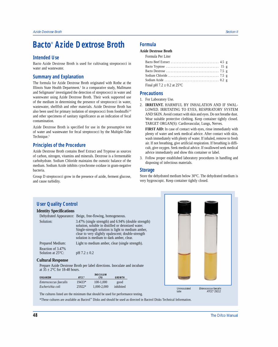
48 The Difco Manual
Enterococcus faecalisATCC® 29212
Uninoculatedtube
Azide Dextrose Broth Section II
Bacto® Azide Dextrose Broth
User Quality ControlIdentity Specifications
Dehydrated Appearance: Beige, free-flowing, homogeneous.Solution: 3.47% (single strength) and 6.94% (double strength)
solution, soluble in distilled or deionized water.Single-strength solution is light to medium amber,clear to very slightly opalescent; double-strengthsolution is medium to dark amber, clear.
Prepared Medium: Light to medium amber, clear (single strength).Reaction of 3.47%Solution at 25°C: pH 7.2 ± 0.2
Cultural ResponsePrepare Azide Dextrose Broth per label directions. Inoculate and incubateat 35 ± 2°C for 18-48 hours.
INOCULUMORGANISM ATCC® CFU GROWTH
Enterococcus faecalis 19433* 100-1,000 goodEscherichia coli 25922* 1,000-2,000 inhibited
The cultures listed are the minimum that should be used for performance testing.
Intended UseBacto Azide Dextrose Broth is used for cultivating streptococci inwater and wastewater.
Summary and ExplanationThe formula for Azide Dextrose Broth originated with Rothe at theIllinois State Health Department.1 In a comparative study, Mallmannand Seligmann2 investigated the detection of streptococci in water andwastewater using Azide Dextrose Broth. Their work supported useof the medium in determining the presence of streptococci in water,wastewater, shellfish and other materials. Azide Dextrose Broth hasalso been used for primary isolation of streptococci from foodstuffs3,4
and other specimens of sanitary significance as an indication of fecalcontamination.
Azide Dextrose Broth is specified for use in the presumptive testof water and wastewater for fecal streptococci by the Multiple-TubeTechnique.5
Principles of the ProcedureAzide Dextrose Broth contains Beef Extract and Tryptose as sourcesof carbon, nitrogen, vitamins and minerals. Dextrose is a fermentablecarbohydrate. Sodium Chloride maintains the osmotic balance of themedium. Sodium Azide inhibits cytochrome oxidase in gram-negativebacteria.
Group D streptococci grow in the presence of azide, ferment glucose,and cause turbidity.
FormulaAzide Dextrose Broth
Formula Per LiterBacto Beef Extract . . . . . . . . . . . . . . . . . . . . . . . . . . . . . . . 4.5 gBacto Tryptose . . . . . . . . . . . . . . . . . . . . . . . . . . . . . . . . . . 15 gBacto Dextrose . . . . . . . . . . . . . . . . . . . . . . . . . . . . . . . . . . 7.5 gSodium Chloride . . . . . . . . . . . . . . . . . . . . . . . . . . . . . . . . . 7.5 gSodium Azide . . . . . . . . . . . . . . . . . . . . . . . . . . . . . . . . . . . 0.2 g
Final pH 7.2 ± 0.2 at 25°C
Precautions1. For Laboratory Use.2. IRRITANT. HARMFUL BY INHALATION AND IF SWAL-
LOWED. IRRITATING TO EYES, RESPIRATORY SYSTEMAND SKIN. Avoid contact with skin and eyes. Do not breathe dust.Wear suitable protective clothing. Keep container tightly closed.TARGET ORGAN(S): Cardiovascular, Lungs, Nerves.FIRST AID: In case of contact with eyes, rinse immediately withplenty of water and seek medical advice. After contact with skin,wash immediately with plenty of water. If inhaled, remove to freshair. If not breathing, give artificial respiration. If breathing is diffi-cult, give oxygen. Seek medical advice. If swallowed seek medicaladvice immediately and show this container or label.
3. Follow proper established laboratory procedures in handling anddisposing of infectious materials.
StorageStore the dehydrated medium below 30°C. The dehydrated medium isvery hygroscopic. Keep container tightly closed.
*These cultures are available as Bactrol™ Disks and should be used as directed in Bactrol Disks Technical Information.
The Difco Manual 49
Section II B12 Assay Medium USP
Expiration DateThe expiration date applies to the product in its intact container whenstored as directed. Do not use a product if it fails to meet specificationsfor identity and performance.
ProcedureMaterials ProvidedAzide Dextrose Broth
Materials Required but not ProvidedGlasswareDistilled or deionized waterTubes with closuresAutoclaveIncubator (35°C)
Method of Preparation1. Suspend 34.7 grams in 1 liter distilled or deionized water.
Rehydrate with proportionally less water when liquid inocula willexceed 1 ml.
2. Autoclave at 121°C for 15 minutes.
Specimen Collection and PreparationRefer to appropriate references for specimen collection and preparation.
Presumptive Test Procedure5
1. Inoculate a series of Azide Dextrose Broth tubes with appropriatelygraduated quantities of sample. Use sample quantities of 10 ml orless. Use double-strength broth for 10 ml inocula. Consult anappropriate reference for suggested sample sizes.5
2. Incubate inoculated tubes at 35 ± 2°C for 20-48 hours.3. Examine each tube for turbidity at the end of 24 ± 2 hours. If
no turbidity is evident, reincubate and read again at the end of48 ± 3 hours.
ResultsA positive test is indicated by turbidity (cloudiness) in the broth. Anegative test remains clear.
All Azide Dextrose Broth tubes showing turbidity after 24- or 48-hoursincubation must be subjected to the Confirmed Test Procedure.Consult appropriate references for details of the Confirmed TestProcedure5 and further identification of Enterococcus.5,6
Limitations of the Procedure1. Azide Dextrose Broth is used to detect presumptive evidence of
fecal contamination. Further biochemical testing must be done forconfirmation.
2. For inoculum sizes of 10 ml or larger, use double strength mediumto prevent dilution of ingredients.5,6
References1. Rothe. 1948. Illinois State Health Department.2. Mallmann, W. L., and E. B. Seligmann. 1950. A comparative
study of media for the detection of streptococci in water andsewage. Am. J. Public Health 40:286.
3. Larkin, E. P., W. Litsky, and J. E. Fuller. 1955. Fecalstreptococci in frozen foods. I. A bacteriological survey of somecommercially frozen foods. Appl. Microbiol. 3:98.
4. Splittstoesser, D. F., R. Wright, and G. J. Hucker. 1961. Studieson media for enumerating enterococci in frozen vegetables. Appl.Microbiol. 9:303.
5. Eaton, A. D., L. S. Clesceri, and A. E. Greenberg (eds.). 1995.Standard methods for the examination of water and wastewater,19th ed. American Public Health Association, Washington, D.C.
6. MacFaddin, J. F. 1985. Media for isolation-cultivation-identification-maintenance of medical bacteria, vol. 1. Williams& Wilkins, Baltimore, MD.
PackagingAzide Dextrose Broth 500 g 0387-17
Bacto® B12 Assay Medium USPIntended UseBacto B12 Assay Medium USP is used for determining vitamin B12
concentration by the microbiological assay technique.
Also Known AsUSP is an abbreviation for United States Pharmacopeia.
Summary and ExplanationVitamin Assay Media are used in the microbiological assay of vitamins.Three types of media are used for this purpose:1. Maintenance Media: For carrying the stock culture to preserve the
viability and sensitivity of the test organism for its intended purpose;2. Inoculum Media: To condition the test culture for immediate use;3. Assay Media: To permit quantitation of the vitamin under test.
B12 Assay Medium USP is used in the microbiological assay of vitaminB12 according to the procedures of the Vitamin B12 Activity Assay inUSP1 and the Cobalamin (Vitamin B12 Activity) Assay in AOAC.2
Lactobacillus delbrueckii subsp. lactis ATCC® 7830 (Lactobacillusleichmannii) is the test organism used in this procedure.
Principles of the ProcedureB12 Assay Medium USP is a vitamin B12-free dehydrated mediumcontaining all other nutrients and vitamins essential for the cultivationof L. delbrueckii subsp. lactis ATCC® 7830. To obtain a standard curve,USP Cyanocobalamin Reference is added in specified increasingconcentrations giving a growth response that can be measuredtitrimetrically or turbidimetrically.
50 The Difco Manual
FormulaB12 Assay Medium USP
Formula Per LiterBacto Vitamin Assay Casamino Acids . . . . . . . . . . . . . . . . 15 gBacto Dextrose . . . . . . . . . . . . . . . . . . . . . . . . . . . . . . . . . . 40 gBacto Asparagine . . . . . . . . . . . . . . . . . . . . . . . . . . . . . . . . 0.2 gSodium Acetate . . . . . . . . . . . . . . . . . . . . . . . . . . . . . . . . . . 20 gAscorbic Acid . . . . . . . . . . . . . . . . . . . . . . . . . . . . . . . . . . . . 4 gL-Cystine . . . . . . . . . . . . . . . . . . . . . . . . . . . . . . . . . . . . . . . 0.4 gDL-Tryptophane . . . . . . . . . . . . . . . . . . . . . . . . . . . . . . . . . 0.4 gAdenine Sulfate . . . . . . . . . . . . . . . . . . . . . . . . . . . . . . . . . 20 mgGuanine Hydrochloride . . . . . . . . . . . . . . . . . . . . . . . . . . . 20 mgUracil . . . . . . . . . . . . . . . . . . . . . . . . . . . . . . . . . . . . . . . . . . 20 mgXanthine . . . . . . . . . . . . . . . . . . . . . . . . . . . . . . . . . . . . . . . 20 mgRiboflavin . . . . . . . . . . . . . . . . . . . . . . . . . . . . . . . . . . . . . . . 1 mgThiamine Hydrochloride . . . . . . . . . . . . . . . . . . . . . . . . . . . 1 mgBiotin . . . . . . . . . . . . . . . . . . . . . . . . . . . . . . . . . . . . . . . . . . 10 µgNiacin . . . . . . . . . . . . . . . . . . . . . . . . . . . . . . . . . . . . . . . . . . 2 mgp-Aminobenzoic Acid . . . . . . . . . . . . . . . . . . . . . . . . . . . . . . 2 mgCalcium Pantothenate . . . . . . . . . . . . . . . . . . . . . . . . . . . . . . 1 mgPyridoxine Hydrochloride . . . . . . . . . . . . . . . . . . . . . . . . . . 4 mgPyridoxal Hydrochloride . . . . . . . . . . . . . . . . . . . . . . . . . . . 4 mgPyridoxamine Hydrochloride . . . . . . . . . . . . . . . . . . . . . . 800 µgFolic Acid . . . . . . . . . . . . . . . . . . . . . . . . . . . . . . . . . . . . . 200 µgMonopotassium Phosphate . . . . . . . . . . . . . . . . . . . . . . . . . . 1 gDipotassium Phosphate . . . . . . . . . . . . . . . . . . . . . . . . . . . . 1 gMagnesium Sulfate . . . . . . . . . . . . . . . . . . . . . . . . . . . . . . . 0.4 gSodium Chloride . . . . . . . . . . . . . . . . . . . . . . . . . . . . . . . . . 20 mgFerrous Sulfate . . . . . . . . . . . . . . . . . . . . . . . . . . . . . . . . . . 20 mgManganese Sulfate . . . . . . . . . . . . . . . . . . . . . . . . . . . . . . . 20 mgSorbitan Monooleate Complex . . . . . . . . . . . . . . . . . . . . . . . 2 g
Final pH 6.0 ± 0.1 at 25°C
Precautions1. For Laboratory Use.2. Great care must be taken to avoid contamination of media or glass-
ware in microbiological assay procedures. Extremely smallamounts of foreign material may be sufficient to give erroneousresults. Scrupulously clean glassware free from detergents andother chemicals must be used. Glassware must be heated to 250°Cfor at least 1 hour to burn off any organic residues that might be present.
3. Take precautions to keep sterilization and cooling conditions uniformthroughout the assay.
4. Follow proper established laboratory procedures in handling anddisposing of infectious materials.
StorageStore the dehydrated medium at 2-8°C. The dehydrated medium is veryhygroscopic. Keep container tightly closed.
Expiration DateThe expiration date applies to the product in its intact container whenstored as directed. Do not use a product if it fails to meet specificationsfor identity and performance.
ProcedureMaterials ProvidedB12 Assay Medium USP
Materials Required But Not ProvidedGlasswareAutoclaveStock culture of Lactobacillus delbrueckii subsp. lactis ATCC® 7830Lactobacilli Agar AOAC or B12 Culture Agar USPLactobacilli Broth AOAC or B12 Inoculum Broth USPSterile 0.85% salineDistilled or deionized waterSpectophotometer or nephelometerB12 Culture Agar USPB12 Inoculum Broth USPCyanocobalamin USP (vitamin B12)
Method of Preparation1. Suspend 8.5 grams in 100 ml distilled or deionized water.2. Heat to boiling for 2-3 minutes to dissolve completely.3. Distribute 5 ml amounts into tubes, evenly dispersing the precipitate.4. Add standard or test samples.5. Adjust tube volume to 10 ml with distilled or deionized water.6. Autoclave at 121°C for 5 minutes.
Specimen Collection and PreparationAssay samples are prepared according to references given in thespecific assay procedures. For assay, the samples should be diluted toapproximately the same concentration as the standard solution.
Test ProcedureFollow assay procedures as outlined in USP1 or AOAC.2 Use levelsof B12 in the preparation of the standard curve according to these
B12 Assay Medium USP Section II
User Quality ControlIdentity Specifications
Dehydrated Appearance: Very light to light beige, homogeneous,with a tendency to clump.
Solution: 4.25% (single strength) or 8.5%(double strength) solution, solublein distilled or deionized water onboiling for 2-3 minutes. Light amber,clear, may have a slight precipitate(single strength).
Prepared Medium: (Single strength) very light to lightamber, clear, may have a slightprecipitate.
Reaction of 4.25%Solution at 25°C: pH 6.0 ± 0.1
Cultural ResponsePrepare B12 Assay Medium USP per label directions. Preparea standard curve using USP Cyanocobalamin ReferenceStandard at levels of 0.0 to 0.25 ng per 10 ml. The mediumsupports the growth of L. delbrueckii subsp. lactis ATCC® 7830when supplemented with cyanocobalamin (vitamin B12).
The Difco Manual 51
references. It is essential that a standard curve be constructed each timean assay is run. Autoclave and incubation conditions can influence thestandard curve reading and cannot always be duplicated.Generally satisfactory results are obtained with B12 at the followinglevels: 0.0, 0.025, 0.05, 0.075, 0.1, 0.125, 0.15, 0.2 and 0.25 ng perassay tube (10 ml).
Stock cultures of L. delbrueckii subsp. lactis ATCC® 7830 areprepared by stab inoculation into 10 ml of B12 Culture Agar USP orLactobacilli Agar AOAC. After 16-24 hours incubation at 35-37°C, thecultures are kept refrigerated. The inoculum for assay is prepared bysubculturing a stock culture of L. delbrueckii subsp. lactis into10 ml of B12 Inoculum Broth USP. For a complete discussion on B12
Culture Agar USP and B12 Inoculum Broth USP, refer to USP.1
Results1. Prepare a standard concentration response curve by plotting the
response readings against the amount of standard in each tube, diskor cup.
2. Determine the amount of vitamin at each level of assay solution byinterpolation from the standard curve.
3. Calculate the concentration of vitamin in the sample from theaverage of these volumes. Use only those values that do not vary
Section II B12 Culture Agar USP & B12 Inoculum Broth USP
more than ±10% from the average and use the results only if twothirds of the values do not vary more than ±10%.
Limitations of the Procedure1. The test organism used for inoculating an assay medium must be
cultured and maintained on media recommended for this purpose.2. For successful results to these procedures, all conditions of the
assay must be followed precisely.3. Aseptic technique should be used throughout the assay procedure.4. The use of altered or deficient media may cause mutants having
different nutritional requirements and will not give a satisfactoryresponse.
References1. The United States Pharmacopeial Convention. 1995. The United
States pharmacopeia, 23rd ed. The United States PharmacopeialConvention Inc., Rockville, MD.
2. Association of Official Analytical Chemists. 1995. Officialmethods of analysis of AOAC International, 16th ed. AOACInternational, Arlington, VA.
PackagingB12 Assay Medium USP 100 g 0457-15
Bacto® B12 Culture Agar USPBacto B12 Inoculum Broth USPIntended UseBacto B12 Inoculum Broth USP is used for preparing the inoculumof Lactobacillus delbrueckii subsp. lactis ATCC® 7830 used in theVitamin B12 Activity Assay.
Bacto B12 Culture Agar USP is used for cultivating L. delbrueckii subsp.lactis ATCC 7830 used in the Vitamin B12 Activity Assay.
Also Known AsUSP is an abbreviation for United States Pharmacopeia.
Summary and ExplanationVitamin Assay Media are prepared for use in the microbiological assayof vitamins. Three types of media are used for this purpose:1. Maintenance Media: For carrying the stock culture to preserve the
viability and sensitivity of the test organism for its intended purpose.2. Inoculum Media: To condition the test culture for immediate use.3. Assay Media: To permit quantitation of the vitamin under test.
They contain all the factors necessary for optimal growth of thetest organism except the single essential vitamin to be determined.
Lactobacillus species grow poorly on non-selective culture media andrequire special nutrients. Mickle and Breed2 reported the use oftomato juice in culture media for lactobacilli. Kulp,3 while investigatingthe use of tomato juice on bacterial development, found that growth ofLactobacillus acidophilus was enhanced.
B12 Culture Agar USP is recommended for maintaining stockcultures of L. delbrueckii subsp. lactis ATCC 7830 (Lactobacillusleichmannii) for use in the Vitamin B12 Activity Assay according toUS Pharmacopeia (USP).1
B12 Inoculum Broth USP is used for preparing the inoculum ofL. delbrueckii subsp. lactis ATCC 7830 in the microbiological assayof vitamin B12 according to USP.1
Principles of the ProcedureProteose Peptone No. 3 provides the nitrogen and amino acids in B12
Culture Agar USP and B12 Inoculum Broth USP. Yeast Extract is thevitamin source in the formulas. Tomato Juice is added to create theproper acidic environment. Dextrose is the carbon source, and SorbitanMonooleate Complex acts an emulsifier. Potassium Phosphate Dibasicacts as the buffering agent in B12 Inoculum Broth USP, andMonopotassium Phosphate is the buffering agent in B12 Culture AgarUSP. Bacto Agar is the solidifying agent in B12 Culture Agar USP.
FormulaB12 Culture Agar USP
Formula Per LiterTomato Juice . . . . . . . . . . . . . . . . . . . . . . . . . . . . . . . . . . . 100 mlBacto Proteose Peptone No. 3 . . . . . . . . . . . . . . . . . . . . . . 7.5 gBacto Yeast Extract . . . . . . . . . . . . . . . . . . . . . . . . . . . . . . . 7.5 gBacto Dextrose . . . . . . . . . . . . . . . . . . . . . . . . . . . . . . . . . . 10 gMonopotassium Phosphate . . . . . . . . . . . . . . . . . . . . . . . . . . 2 gSorbitan Monooleate Complex . . . . . . . . . . . . . . . . . . . . . . . 1 gBacto Agar . . . . . . . . . . . . . . . . . . . . . . . . . . . . . . . . . . . . . 15 g
Final pH 6.8 ± 0.1 at 25°C
52 The Difco Manual
B12 Inoculum Broth USPFormula Per LiterTomato Juice . . . . . . . . . . . . . . . . . . . . . . . . . . . . . . . . . . . 100 mlBacto Proteose Peptone No. 3 . . . . . . . . . . . . . . . . . . . . . . 7.5 gBacto Yeast Extract . . . . . . . . . . . . . . . . . . . . . . . . . . . . . . . 7.5 gBacto Dextrose . . . . . . . . . . . . . . . . . . . . . . . . . . . . . . . . . . 10 gSorbitan Monooleate Complex . . . . . . . . . . . . . . . . . . . . . . 0.1 gPotassium Phosphate Dibasic . . . . . . . . . . . . . . . . . . . . . . . . 2 g
Final pH 6.8 ± 0.1 at 25°C
Precautions1. For Laboratory Use.2. Follow proper established laboratory procedures in handling and
disposing of infectious materials.
3. Great care must be taken to avoid contamination of media or glass-ware in microbiological assay procedures. Extremely smallamounts of foreign material may be sufficient to give erroneousresults. Scrupulously clean glassware free from detergents andother chemicals must be used.
StorageStore the dehydrated media at 2-8°C. The dehydrated media are veryhygroscopic. Keep containers tightly closed.
Expiration DateThe expiration date applies to the product in its intact container whenstored as directed. Do not use a product if it fails to meet specificationsfor identity and performance.
ProcedureMaterials ProvidedB12 Culture Agar USPB12 Inoculum Broth USP
Materials Required But Not ProvidedGlasswareAutoclaveIncubatorDistilled or deionized waterInoculating needle
Method of Preparation1. Suspend the appropriate amount of medium in 1 liter distilled or
deionized water:B12 Culture Agar USP 47 gramsB12 Inoculum Broth USP 32 grams
2. Boil to dissolve completely. (B12 Culture Agar)3. Dispense 10 ml amounts into tubes.4. Autoclave at 121°C for 15 minutes.5. Allow tubes of B12 Culture Agar to cool in an upright position.
Stock Culture1. Prepare stock cultures in triplicate in sterile B12 Culture Agar USP.2. Inoculate the tubes using a straight wire inoculating needle.3. Incubate cultures for 16-24 hours at any temperature between
30-40°C, but held constant within ± 0.5°C.4. Store at 2-8°C.5. Before using a fresh culture for assay, make no fewer than 10
successive transfers of the culture in a 2 week period.6. Prepare stab cultures at least three times each week and do not use
a culture for preparing assay inoculum if over 4 days old.
InoculumPrepare inoculum as described in USP.1
Specimen Collection and PreparationAssay samples are prepared according to references given in thespecific assay procedures. For assay, the samples should be diluted toapproximately the same concentration as the standard solution.
B12 Culture Agar USP & B12 Inoculum Broth USP Section II
User Quality ControlIdentity Specifications
B12 Culture Agar USPDehydrated Appearance: Beige, free-flowing, homogeneous.Solution: 4.7% solution, soluble in distilled
or deionized water upon boiling.Solution is light to medium amber,opalescent when hot, slightlyopalescent with flocculent precipitatewhen cooled.
Prepared Medium: Light to medium amber, slightlyopalescent, may have a slightflocculent precipitate.
Reaction of 4.7%Solution at 25°C: pH 6.8 ± 0.1
B12 Inoculum Broth USPDehydrated Appearance: Tan, homogeneous, tendency to
clump.Solution: 3.2% solution, soluble in distilled or
deionized water on boiling. Solutionis medium to dark amber, opalescentwhen hot, clear when cooled to roomtemperature.
Prepared Medium: Medium amber, clear.Reaction of 3.2%Solution at 25°C: pH 6.8 ± 0.1
Cultural ResponseB12 Culture Agar USP or B12 Inoculum Broth USPPrepare B12 Culture Agar USP or B12 Inoculum Broth USP perlabel directions. Inoculate medium with test organism andincubate at 35 ± 2°C for 16-24 hours.
INOCULUMORGANISM ATCC® CFU GROWTH
Lactobacillus delbrueckii 7830 300-1,000 goodsubsp. lactis
The culture listed is the minimum that should be used forperformance testing.
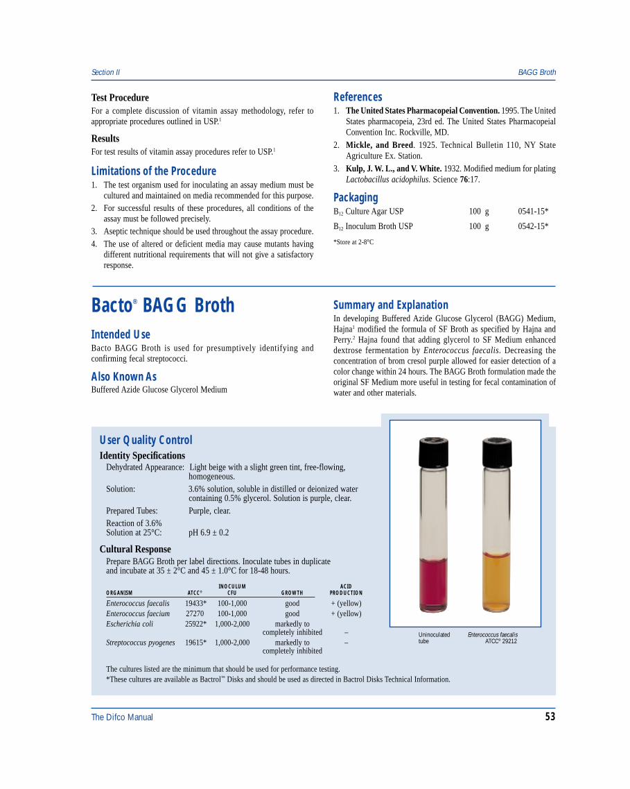
The Difco Manual 53
User Quality ControlIdentity Specifications
Dehydrated Appearance: Light beige with a slight green tint, free-flowing,homogeneous.
Solution: 3.6% solution, soluble in distilled or deionized watercontaining 0.5% glycerol. Solution is purple, clear.
Prepared Tubes: Purple, clear.Reaction of 3.6%Solution at 25°C: pH 6.9 ± 0.2
Cultural ResponsePrepare BAGG Broth per label directions. Inoculate tubes in duplicateand incubate at 35 ± 2°C and 45 ± 1.0°C for 18-48 hours.
INOCULUM ACIDORGANISM ATCC® CFU GROWTH PRODUCTION
Enterococcus faecalis 19433* 100-1,000 good + (yellow)Enterococcus faecium 27270 100-1,000 good + (yellow)Escherichia coli 25922* 1,000-2,000 markedly to
completely inhibited –Streptococcus pyogenes 19615* 1,000-2,000 markedly to –
completely inhibited
The cultures listed are the minimum that should be used for performance testing.*These cultures are available as Bactrol™ Disks and should be used as directed in Bactrol Disks Technical Information.
Enterococcus faecalisATCC® 29212
Uninoculatedtube
Section II BAGG Broth
Test ProcedureFor a complete discussion of vitamin assay methodology, refer toappropriate procedures outlined in USP.1
ResultsFor test results of vitamin assay procedures refer to USP.1
Limitations of the Procedure1. The test organism used for inoculating an assay medium must be
cultured and maintained on media recommended for this purpose.2. For successful results of these procedures, all conditions of the
assay must be followed precisely.3. Aseptic technique should be used throughout the assay procedure.4. The use of altered or deficient media may cause mutants having
different nutritional requirements that will not give a satisfactoryresponse.
Bacto® BAGG BrothIntended UseBacto BAGG Broth is used for presumptively identifying andconfirming fecal streptococci.
Also Known AsBuffered Azide Glucose Glycerol Medium
Summary and ExplanationIn developing Buffered Azide Glucose Glycerol (BAGG) Medium,Hajna1 modified the formula of SF Broth as specified by Hajna andPerry.2 Hajna found that adding glycerol to SF Medium enhanceddextrose fermentation by Enterococcus faecalis. Decreasing theconcentration of brom cresol purple allowed for easier detection of acolor change within 24 hours. The BAGG Broth formulation made theoriginal SF Medium more useful in testing for fecal contamination ofwater and other materials.
References1. The United States Pharmacopeial Convention. 1995. The United
States pharmacopeia, 23rd ed. The United States PharmacopeialConvention Inc. Rockville, MD.
2. Mickle, and Breed. 1925. Technical Bulletin 110, NY StateAgriculture Ex. Station.
3. Kulp, J. W. L., and V. White. 1932. Modified medium for platingLactobacillus acidophilus. Science 76:17.
PackagingB12 Culture Agar USP 100 g 0541-15*
B12 Inoculum Broth USP 100 g 0542-15*
*Store at 2-8°C

54 The Difco Manual
Principles of the ProcedureBAGG Broth contains Tryptose as a source for carbon, nitrogen,vitamins and minerals. Dextrose is a fermentable carbohydrate.Sodium Chloride maintains the osmotic balance of the medium.Sodium Azide inhibits gram-negative bacteria. Brom Cresol Purple isa pH indicator.
Enterococci grow in the presence of azide and ferment glucose,producing an acid pH that changes the color of the medium.
FormulaBAGG Broth
Formula Per LiterBacto Tryptose . . . . . . . . . . . . . . . . . . . . . . . . . . . . . . . . . . 20 gBacto Dextrose . . . . . . . . . . . . . . . . . . . . . . . . . . . . . . . . . . . 5 gDipotassium Phosphate . . . . . . . . . . . . . . . . . . . . . . . . . . . . 4 gMonopotassium Phosphate . . . . . . . . . . . . . . . . . . . . . . . . . 1.5 gSodium Chloride . . . . . . . . . . . . . . . . . . . . . . . . . . . . . . . . . . 5 gSodium Azide . . . . . . . . . . . . . . . . . . . . . . . . . . . . . . . . . . . 0.5 gBacto Brom Cresol Purple . . . . . . . . . . . . . . . . . . . . . . . 0.015 g
Final pH 6.9 ± 0.2 at 25°C
Precautions1. For Laboratory Use.2. HARMFUL. HARMFUL BY INHALATION AND IF SWAL-
LOWED. IRRITATING TO EYES, RESPIRATORY SYSTEMAND SKIN. Avoid contact with skin and eyes. Do not breathe dust.Wear suitable protective clothing. Keep container tightly closed.TARGET ORGAN(S): Cardiovascular, Lungs, Nerves.FIRST AID: In case of contact with eyes, rinse immediately withplenty of water and seek medical advice. After contact with skin,wash immediately with plenty of water. If inhaled, remove to freshair. If not breathing, give artificial respiration. If breathing is diffi-cult, give oxygen. Seek medical advice. If swallowed seek medicaladvice immediately and show this container or label.
3. Follow proper established laboratory procedures in handling anddisposing of infectious materials.
StorageStore the dehydrated medium below 30°C. The dehydrated medium isvery hygroscopic. Keep container tightly closed.
Expiration DateThe expiration date applies to the product in its intact container whenstored as directed. Do not use a product if it fails to meet specificationsfor identity and performance.
ProcedureMaterials ProvidedBAGG Broth
Materials Required but not ProvidedGlasswareDistilled or deionized water
GlycerolTubes with closuresAutoclaveIncubators (35 ± 2°C, 45 ± 1°C)
Method of Preparation1. Dissolve 36 grams in 1 liter distilled or deionized water containing
5 ml glycerol. Rehydrate with proportionally less water whenliquid inocula will exceed 1 ml.
2. Dispense into tubes with closures.3. Autoclave at 114 -118°C for 15 minutes.
Specimen Collection and PreparationRefer to appropriate references for specimen collection and preparation.
Test Procedure1-3
1. Inoculate duplicate tubes with sample. Use single-strength mediumfor inocula of 1 ml or less. Use double-strength medium forinocula of 10 ml.
2. Incubate one set of tubes at 35 ± 2°C for 18-48 hours. Incubate thesecond set at 45 ± 1°C for 18-48 hours.
3. Read tubes for growth and acid production.
Results1-3
1. A positive test is indicated by the production of a yellow color(acid) throughout the medium. This result is presumptive evidenceof the presence of fecal streptococci. Further testing must beperformed to confirm this result. Consult appropriate referencesfor further identification of Enterococcus.3
2. A negative result is indicated by no change in the medium (purplecolor).
Limitations of the Procedure1. The concentration of the medium must be adjusted to the inoculum
size. Refer to discussion in Test Procedure.
References1. Hajna, A. A. 1951. A buffered azide glucose-glycerol broth for
presumptive and confirmative tests for fecal streptococci. Pub.Health Lab. 9:80-81.
2. Hajna, A. A., and C. A. Perry. 1943. A comparative study ofpresumptive and confirmative media for bacteria of thecoliform group and for fecal streptococci. Am. J. Pub. Health33:550-556.
3. MacFaddin, J. F. 1985. Media for isolation-cultivation-identification-maintenance of medical bacteria, vol. 1. Williams& Wilkins, Baltimore, MD.
PackagingBAGG Broth 500 g 0442-17
BAGG Broth Section II
The Difco Manual 55
Section II BG Sulfa Agar, SBG Enrichment & SBG Sulfa Enrichment
Bacto® BG Sulfa Agar . Bacto SBG EnrichmentBacto SBG Sulfa EnrichmentIntended UseBacto BG Sulfa Agar is used for isolating Salmonella.
Bacto SBG Enrichment and Bacto SBG Sulfa Enrichment is used forenriching Salmonella prior to isolation procedures.
Also Known AsBG is an abbreviation for Brilliant Green and SBG is an abbreviationfor Selenite Brilliant Green.
Summary and ExplanationSalmonellosis continues to be an important public health problemworldwide, despite efforts to control the prevalence of Salmonella in
domesticated animals. Infection with non-typhi Salmonella often causesmild, self-limiting illness.1 The illness results from consumption ofraw, undercooked or improperly processed foods contaminated withSalmonella. Many of these cases of Salmonella-related gastroenteritisare due to improper handling of poultry products. Various poultryproducts are routinely monitored for Salmonella before their distri-bution for human consumption, but in many instances, contaminatedfood samples elude detection.
BG Sulfa Agar is a highly selective medium. Osborne and Stokes2
added 0.1% sodium sulfapyridine to Brilliant Green Agar to enhancethe selective properties of this medium for Salmonella. This formula isrecommended as a selective isolation medium for Salmonella followingenrichment. It is also recommended for direct inoculation with primaryspecimens for Salmonella isolation.
For food testing, BG Sulfa Agar has been used for detection of Salmonellain low and high moisture foods.3,4 It has also been used for detectingSalmonella in feeds and feed ingredients.5 This medium is recommendedwhen testing foods for Salmonella following USDA guidelines.6,7
SBG Enrichment and SBG Sulfa Enrichment are prepared accordingto the formulas described by Stokes and Osborne.8 The researchersfound that whole egg and egg yolk reduced the selective properties ofselenite brilliant green enrichment.2 They also found that the additionof sulfapyridine restored these selective properties.2
SBG Enrichment and SBG Sulfa Enrichment are selective enrichmentsfor the isolation of Salmonella species, especially from egg products.The shell and the contents of the egg at the time of oviposition aregenerally sterile or harbor very few microorganisms.9,10,11 Contami-nation of the shell occurs afterwards from nesting material, floorlitter, and avian fecal matter.12 Salmonellae are of most concern inegg products.12
Principles of the ProcedureIn BG Sulfa Agar, Proteose Peptone and Yeast Extract provide nitrogen,vitamins and minerals. Lactose and Sucrose are the sources ofcarbohydrates in the medium. Brilliant Green and Sodium Pyridineare complementary in inhibiting gram-positive bacteria and mostgram-negative bacilli other than Salmonella spp. Phenol Red is thepH indicator that turns the medium a yellow color with the formationof acid when lactose and/or sucrose is fermented. Bacto Agar is asolidifying agent.
Bacto Peptone provides the nitrogen, minerals and amino acids inSBG Enrichment and SBG Sulfa Enrichment. Yeast Extract is the vitaminsource. D-Mannitol is the carbon source to stimulate organism growth.The phosphates acts as buffers in the enrichments. Sodium Taurocholate,Sodium Selenite and Brilliant Green are the selective agents. Theselective agents are used to inhibit gram positive organisms andenteric bacteria other than Salmonella. Sodium Sulfapyridine is addedin SBG Sulfa Enrichment to increase selectivity.
User Quality ControlIdentity Specifications
BG Sulfa AgarDehydrated Appearance: Pink, free flowing, homogeneous.Solution: 5.9% solution, soluble in distilled or
deionized water on boiling. Solutionis very dark amber, very slightly toslightly opalescent.
Prepared Plates: Dark reddish-amber, slightlyopalescent.
Reaction of 5.9%Solution at 25°C: pH 6.9 ± 0.2
SBG EnrichmentDehydrated Appearance: Light beige, free-flowing,
homogeneous.Solution: 2.37% solution, soluble in distilled
or deionized water; green,opalescent with slight precipitate.
Prepared Medium: Green, opalescent withoutsignificant precipitation.
Reaction of 2.37%Solution at 25°C: pH 7.2 ± 0.2
SBG Sulfa EnrichmentDehydrated Appearance: Light beige, free-flowing,
homogeneous.Solution: 2.42% solution, soluble in distilled
or deionized water; green,opalescent without significantprecipitation.
Prepared Medium: Green, opalescent withoutsignificant precipitation.
Reaction of 2.42%Solution at 25°C: pH 7.2 ± 0.2
continued on following page
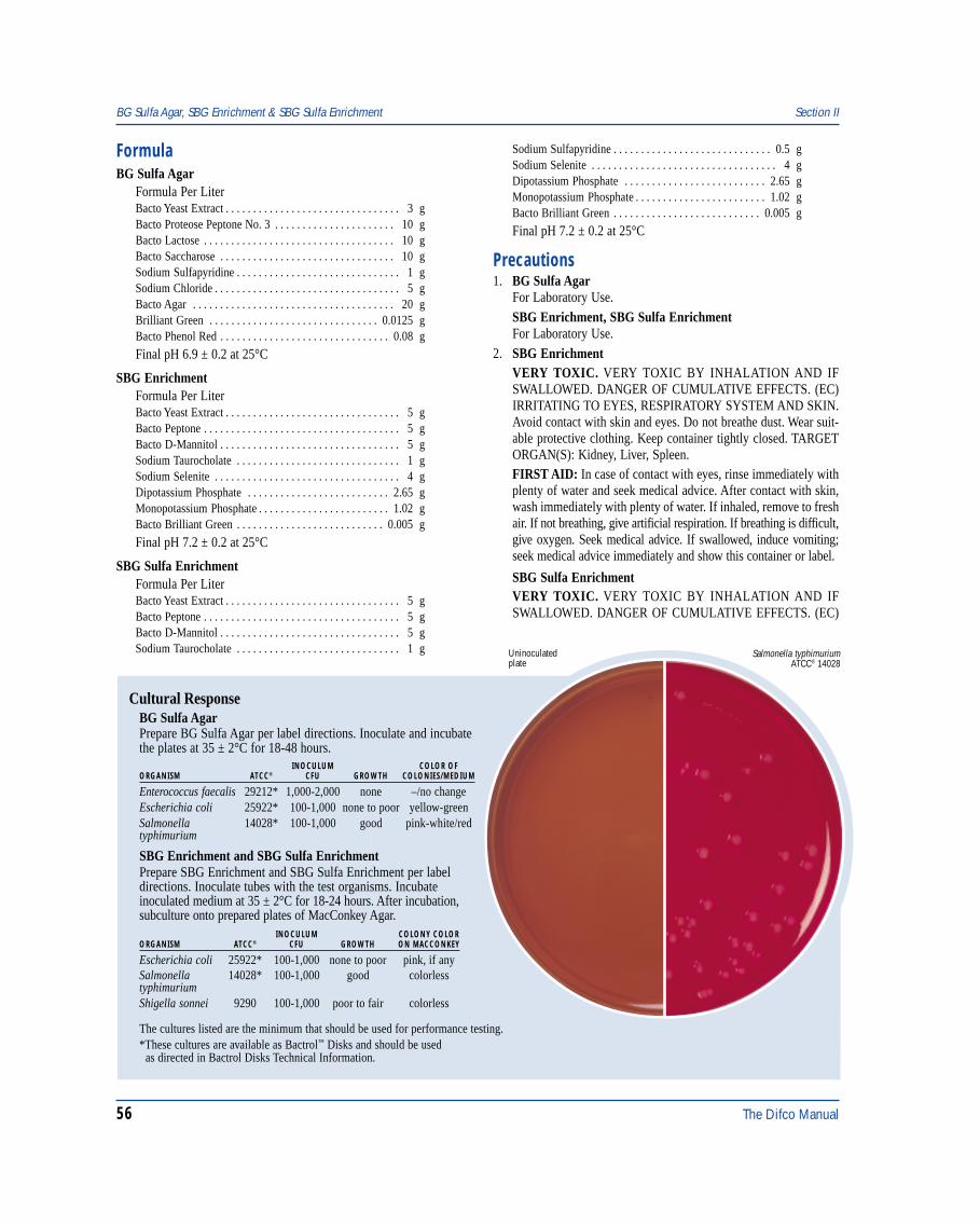
56 The Difco Manual
FormulaBG Sulfa Agar
Formula Per LiterBacto Yeast Extract . . . . . . . . . . . . . . . . . . . . . . . . . . . . . . . . 3 gBacto Proteose Peptone No. 3 . . . . . . . . . . . . . . . . . . . . . . 10 gBacto Lactose . . . . . . . . . . . . . . . . . . . . . . . . . . . . . . . . . . . 10 gBacto Saccharose . . . . . . . . . . . . . . . . . . . . . . . . . . . . . . . . 10 gSodium Sulfapyridine . . . . . . . . . . . . . . . . . . . . . . . . . . . . . . 1 gSodium Chloride . . . . . . . . . . . . . . . . . . . . . . . . . . . . . . . . . . 5 gBacto Agar . . . . . . . . . . . . . . . . . . . . . . . . . . . . . . . . . . . . . 20 gBrilliant Green . . . . . . . . . . . . . . . . . . . . . . . . . . . . . . . 0.0125 gBacto Phenol Red . . . . . . . . . . . . . . . . . . . . . . . . . . . . . . . 0.08 g
Final pH 6.9 ± 0.2 at 25°C
SBG EnrichmentFormula Per LiterBacto Yeast Extract . . . . . . . . . . . . . . . . . . . . . . . . . . . . . . . . 5 gBacto Peptone . . . . . . . . . . . . . . . . . . . . . . . . . . . . . . . . . . . . 5 gBacto D-Mannitol . . . . . . . . . . . . . . . . . . . . . . . . . . . . . . . . . 5 gSodium Taurocholate . . . . . . . . . . . . . . . . . . . . . . . . . . . . . . 1 gSodium Selenite . . . . . . . . . . . . . . . . . . . . . . . . . . . . . . . . . . 4 gDipotassium Phosphate . . . . . . . . . . . . . . . . . . . . . . . . . . 2.65 gMonopotassium Phosphate . . . . . . . . . . . . . . . . . . . . . . . . 1.02 gBacto Brilliant Green . . . . . . . . . . . . . . . . . . . . . . . . . . . 0.005 g
Final pH 7.2 ± 0.2 at 25°C
SBG Sulfa EnrichmentFormula Per LiterBacto Yeast Extract . . . . . . . . . . . . . . . . . . . . . . . . . . . . . . . . 5 gBacto Peptone . . . . . . . . . . . . . . . . . . . . . . . . . . . . . . . . . . . . 5 gBacto D-Mannitol . . . . . . . . . . . . . . . . . . . . . . . . . . . . . . . . . 5 gSodium Taurocholate . . . . . . . . . . . . . . . . . . . . . . . . . . . . . . 1 g
Sodium Sulfapyridine . . . . . . . . . . . . . . . . . . . . . . . . . . . . . 0.5 gSodium Selenite . . . . . . . . . . . . . . . . . . . . . . . . . . . . . . . . . . 4 gDipotassium Phosphate . . . . . . . . . . . . . . . . . . . . . . . . . . 2.65 gMonopotassium Phosphate . . . . . . . . . . . . . . . . . . . . . . . . 1.02 gBacto Brilliant Green . . . . . . . . . . . . . . . . . . . . . . . . . . . 0.005 g
Final pH 7.2 ± 0.2 at 25°C
Precautions1. BG Sulfa Agar
For Laboratory Use.SBG Enrichment, SBG Sulfa EnrichmentFor Laboratory Use.
2. SBG EnrichmentVERY TOXIC. VERY TOXIC BY INHALATION AND IFSWALLOWED. DANGER OF CUMULATIVE EFFECTS. (EC)IRRITATING TO EYES, RESPIRATORY SYSTEM AND SKIN.Avoid contact with skin and eyes. Do not breathe dust. Wear suit-able protective clothing. Keep container tightly closed. TARGETORGAN(S): Kidney, Liver, Spleen.FIRST AID: In case of contact with eyes, rinse immediately withplenty of water and seek medical advice. After contact with skin,wash immediately with plenty of water. If inhaled, remove to freshair. If not breathing, give artificial respiration. If breathing is difficult,give oxygen. Seek medical advice. If swallowed, induce vomiting;seek medical advice immediately and show this container or label.
SBG Sulfa EnrichmentVERY TOXIC. VERY TOXIC BY INHALATION AND IFSWALLOWED. DANGER OF CUMULATIVE EFFECTS. (EC)
BG Sulfa Agar, SBG Enrichment & SBG Sulfa Enrichment Section II
Cultural ResponseBG Sulfa AgarPrepare BG Sulfa Agar per label directions. Inoculate and incubatethe plates at 35 ± 2°C for 18-48 hours.
INOCULUM COLOR OFORGANISM ATCC® CFU GROWTH COLONIES/MEDIUM
Enterococcus faecalis 29212* 1,000-2,000 none –/no changeEscherichia coli 25922* 100-1,000 none to poor yellow-greenSalmonella 14028* 100-1,000 good pink-white/redtyphimurium
SBG Enrichment and SBG Sulfa EnrichmentPrepare SBG Enrichment and SBG Sulfa Enrichment per labeldirections. Inoculate tubes with the test organisms. Incubateinoculated medium at 35 ± 2°C for 18-24 hours. After incubation,subculture onto prepared plates of MacConkey Agar.
INOCULUM COLONY COLORORGANISM ATCC® CFU GROWTH ON MACCONKEY
Escherichia coli 25922* 100-1,000 none to poor pink, if anySalmonella 14028* 100-1,000 good colorlesstyphimuriumShigella sonnei 9290 100-1,000 poor to fair colorless
The cultures listed are the minimum that should be used for performance testing.*These cultures are available as Bactrol™ Disks and should be used as directed in Bactrol Disks Technical Information.
Uninoculatedplate
Salmonella typhimuriumATCC® 14028
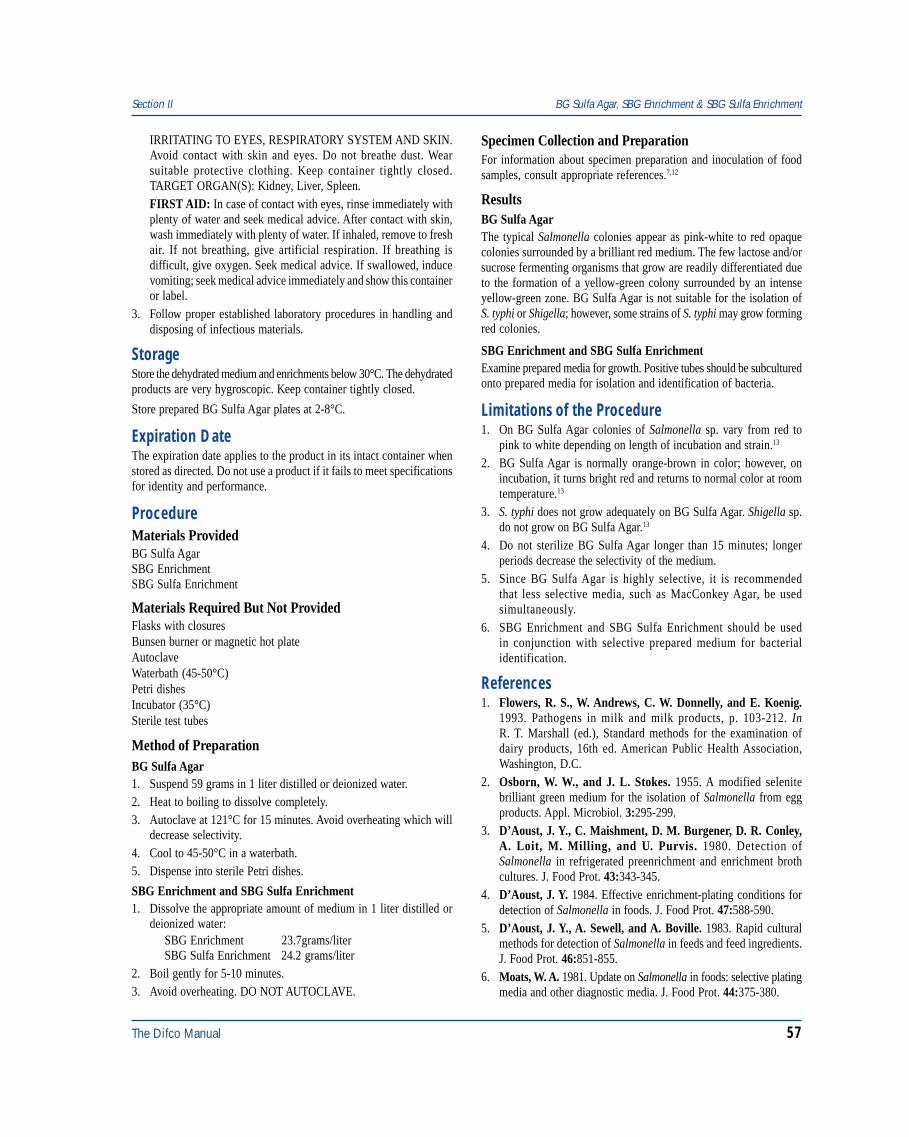
The Difco Manual 57
IRRITATING TO EYES, RESPIRATORY SYSTEM AND SKIN.Avoid contact with skin and eyes. Do not breathe dust. Wearsuitable protective clothing. Keep container tightly closed.TARGET ORGAN(S): Kidney, Liver, Spleen.FIRST AID: In case of contact with eyes, rinse immediately withplenty of water and seek medical advice. After contact with skin,wash immediately with plenty of water. If inhaled, remove to freshair. If not breathing, give artificial respiration. If breathing isdifficult, give oxygen. Seek medical advice. If swallowed, inducevomiting; seek medical advice immediately and show this containeror label.
3. Follow proper established laboratory procedures in handling anddisposing of infectious materials.
StorageStore the dehydrated medium and enrichments below 30°C. The dehydratedproducts are very hygroscopic. Keep container tightly closed.
Store prepared BG Sulfa Agar plates at 2-8°C.
Expiration DateThe expiration date applies to the product in its intact container whenstored as directed. Do not use a product if it fails to meet specificationsfor identity and performance.
ProcedureMaterials ProvidedBG Sulfa AgarSBG EnrichmentSBG Sulfa Enrichment
Materials Required But Not ProvidedFlasks with closuresBunsen burner or magnetic hot plateAutoclaveWaterbath (45-50°C)Petri dishesIncubator (35°C)Sterile test tubes
Method of PreparationBG Sulfa Agar1. Suspend 59 grams in 1 liter distilled or deionized water.2. Heat to boiling to dissolve completely.3. Autoclave at 121°C for 15 minutes. Avoid overheating which will
decrease selectivity.4. Cool to 45-50°C in a waterbath.5. Dispense into sterile Petri dishes.
SBG Enrichment and SBG Sulfa Enrichment1. Dissolve the appropriate amount of medium in 1 liter distilled or
deionized water:SBG Enrichment 23.7grams/literSBG Sulfa Enrichment 24.2 grams/liter
2. Boil gently for 5-10 minutes.3. Avoid overheating. DO NOT AUTOCLAVE.
Specimen Collection and PreparationFor information about specimen preparation and inoculation of foodsamples, consult appropriate references.7,12
ResultsBG Sulfa AgarThe typical Salmonella colonies appear as pink-white to red opaquecolonies surrounded by a brilliant red medium. The few lactose and/orsucrose fermenting organisms that grow are readily differentiated dueto the formation of a yellow-green colony surrounded by an intenseyellow-green zone. BG Sulfa Agar is not suitable for the isolation ofS. typhi or Shigella; however, some strains of S. typhi may grow formingred colonies.
SBG Enrichment and SBG Sulfa EnrichmentExamine prepared media for growth. Positive tubes should be subculturedonto prepared media for isolation and identification of bacteria.
Limitations of the Procedure1. On BG Sulfa Agar colonies of Salmonella sp. vary from red to
pink to white depending on length of incubation and strain.13
2. BG Sulfa Agar is normally orange-brown in color; however, onincubation, it turns bright red and returns to normal color at roomtemperature.13
3. S. typhi does not grow adequately on BG Sulfa Agar. Shigella sp.do not grow on BG Sulfa Agar.13
4. Do not sterilize BG Sulfa Agar longer than 15 minutes; longerperiods decrease the selectivity of the medium.
5. Since BG Sulfa Agar is highly selective, it is recommendedthat less selective media, such as MacConkey Agar, be usedsimultaneously.
6. SBG Enrichment and SBG Sulfa Enrichment should be usedin conjunction with selective prepared medium for bacterialidentification.
References1. Flowers, R. S., W. Andrews, C. W. Donnelly, and E. Koenig.
1993. Pathogens in milk and milk products, p. 103-212. InR. T. Marshall (ed.), Standard methods for the examination ofdairy products, 16th ed. American Public Health Association,Washington, D.C.
2. Osborn, W. W., and J. L. Stokes. 1955. A modified selenitebrilliant green medium for the isolation of Salmonella from eggproducts. Appl. Microbiol. 3:295-299.
3. D’Aoust, J. Y., C. Maishment, D. M. Burgener, D. R. Conley,A. Loit, M. Milling, and U. Purvis. 1980. Detection ofSalmonella in refrigerated preenrichment and enrichment brothcultures. J. Food Prot. 43:343-345.
4. D’Aoust, J. Y. 1984. Effective enrichment-plating conditions fordetection of Salmonella in foods. J. Food Prot. 47:588-590.
5. D’Aoust, J. Y., A. Sewell, and A. Boville. 1983. Rapid culturalmethods for detection of Salmonella in feeds and feed ingredients.J. Food Prot. 46:851-855.
6. Moats, W. A. 1981. Update on Salmonella in foods: selective platingmedia and other diagnostic media. J. Food Prot. 44:375-380.
Section II BG Sulfa Agar, SBG Enrichment & SBG Sulfa Enrichment
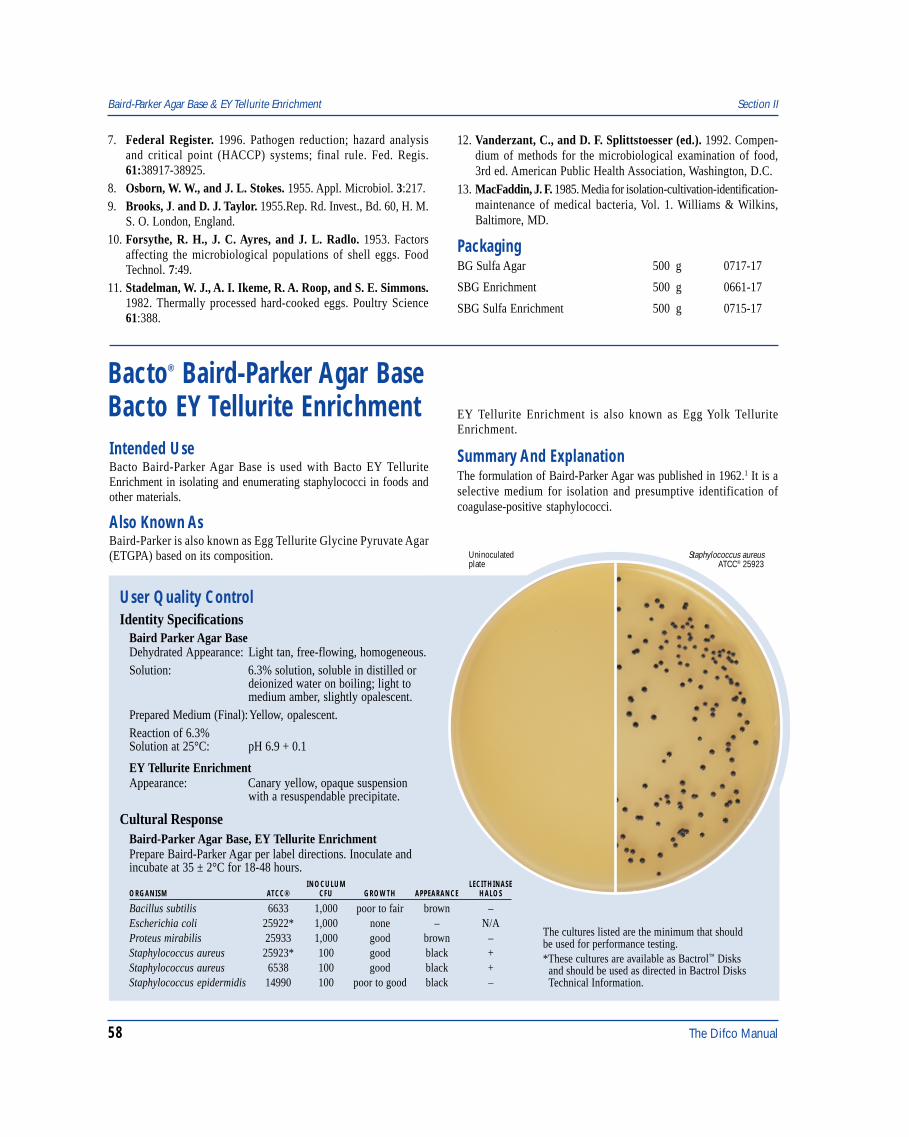
58 The Difco Manual
Baird-Parker Agar Base & EY Tellurite Enrichment Section II
Bacto® Baird-Parker Agar BaseBacto EY Tellurite Enrichment
User Quality ControlIdentity Specifications
Baird Parker Agar BaseDehydrated Appearance: Light tan, free-flowing, homogeneous.Solution: 6.3% solution, soluble in distilled or
deionized water on boiling; light tomedium amber, slightly opalescent.
Prepared Medium (Final):Yellow, opalescent.Reaction of 6.3%Solution at 25°C: pH 6.9 + 0.1
EY Tellurite EnrichmentAppearance: Canary yellow, opaque suspension
with a resuspendable precipitate.
Cultural ResponseBaird-Parker Agar Base, EY Tellurite EnrichmentPrepare Baird-Parker Agar per label directions. Inoculate andincubate at 35 ± 2°C for 18-48 hours.
INOCULUM LECITHINASEORGANISM ATCC® CFU GROWTH APPEARANCE HALOS
Bacillus subtilis 6633 1,000 poor to fair brown –Escherichia coli 25922* 1,000 none – N/AProteus mirabilis 25933 1,000 good brown –Staphylococcus aureus 25923* 100 good black +Staphylococcus aureus 6538 100 good black +Staphylococcus epidermidis 14990 100 poor to good black –
Staphylococcus aureusATCC® 25923
Uninoculatedplate
The cultures listed are the minimum that shouldbe used for performance testing.*These cultures are available as Bactrol™ Disks and should be used as directed in Bactrol Disks Technical Information.
7. Federal Register. 1996. Pathogen reduction; hazard analysisand critical point (HACCP) systems; final rule. Fed. Regis.61:38917-38925.
8. Osborn, W. W., and J. L. Stokes. 1955. Appl. Microbiol. 3:217.9. Brooks, J. and D. J. Taylor. 1955.Rep. Rd. Invest., Bd. 60, H. M.
S. O. London, England.10. Forsythe, R. H., J. C. Ayres, and J. L. Radlo. 1953. Factors
affecting the microbiological populations of shell eggs. FoodTechnol. 7:49.
11. Stadelman, W. J., A. I. Ikeme, R. A. Roop, and S. E. Simmons.1982. Thermally processed hard-cooked eggs. Poultry Science61:388.
12. Vanderzant, C., and D. F. Splittstoesser (ed.). 1992. Compen-dium of methods for the microbiological examination of food,3rd ed. American Public Health Association, Washington, D.C.
13. MacFaddin, J. F. 1985. Media for isolation-cultivation-identification-maintenance of medical bacteria, Vol. 1. Williams & Wilkins,Baltimore, MD.
PackagingBG Sulfa Agar 500 g 0717-17
SBG Enrichment 500 g 0661-17
SBG Sulfa Enrichment 500 g 0715-17
Intended UseBacto Baird-Parker Agar Base is used with Bacto EY TelluriteEnrichment in isolating and enumerating staphylococci in foods andother materials.
Also Known AsBaird-Parker is also known as Egg Tellurite Glycine Pyruvate Agar(ETGPA) based on its composition.
EY Tellurite Enrichment is also known as Egg Yolk TelluriteEnrichment.
Summary And ExplanationThe formulation of Baird-Parker Agar was published in 1962.1 It is aselective medium for isolation and presumptive identification ofcoagulase-positive staphylococci.
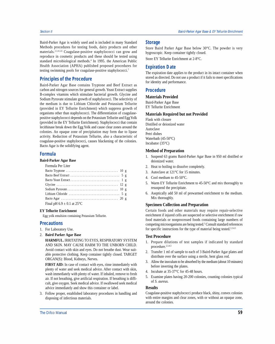
The Difco Manual 59
Section II Baird-Parker Agar Base & EY Tellurite Enrichment
Baird-Parker Agar is widely used and is included in many StandardMethods procedures for testing foods, dairy products and othermaterials.2,3,4,5,6 Coagulase-positive staphylococci can grow andreproduce in cosmetic products and these should be tested usingstandard microbiological methods.4 In 1995. the American PublicHealth Association (APHA) published proposed procedures fortesting swimming pools for coagulase-positive staphylococci.7
Principles of the ProcedureBaird-Parker Agar Base contains Tryptone and Beef Extract ascarbon and nitrogen sources for general growth. Yeast Extract suppliesB-complex vitamins which stimulate bacterial growth. Glycine andSodium Pyruvate stimulate growth of staphylococci. The selectivity ofthe medium is due to Lithium Chloride and Potassium Tellurite(provided in EY Tellurite Enrichment) which suppress growth oforganisms other than staphylococci. The differentiation of coagulase-positive staphylococci depends on the Potassium Tellurite and Egg Yolk(provided in the EY Tellurite Enrichment). Staphylococci that containlecithinase break down the Egg Yolk and cause clear zones around thecolonies. An opaque zone of precipitation may form due to lipaseactivity. Reduction of Potassium Tellurite, also a characteristic ofcoagulase-positive staphylococci, causes blackening of the colonies.Bacto Agar is the solidifying agent.
FormulaBaird-Parker Agar Base
Formula Per LiterBacto Tryptone . . . . . . . . . . . . . . . . . . . . . . . . . . . . . . . . . . 10 gBacto Beef Extract . . . . . . . . . . . . . . . . . . . . . . . . . . . . . . . . 5 gBacto Yeast Extract . . . . . . . . . . . . . . . . . . . . . . . . . . . . . . . . 1 gGlycine . . . . . . . . . . . . . . . . . . . . . . . . . . . . . . . . . . . . . . . . 12 gSodium Pyruvate . . . . . . . . . . . . . . . . . . . . . . . . . . . . . . . . . 10 gLithium Chloride . . . . . . . . . . . . . . . . . . . . . . . . . . . . . . . . . 5 gBacto Agar . . . . . . . . . . . . . . . . . . . . . . . . . . . . . . . . . . . . . 20 g
Final pH 6.9 ± 0.1 at 25°C
EY Tellurite EnrichmentEgg yolk emulsion containing Potassium Tellurite.
Precautions1. For Laboratory Use.2. Baird Parker Agar Base
HARMFUL. IRRITATING TO EYES, RESPIRATORY SYSTEMAND SKIN. MAY CAUSE HARM TO THE UNBORN CHILD.Avoid contact with skin and eyes. Do not breathe dust. Wear suit-able protective clothing. Keep container tightly closed. TARGETORGAN(S): Blood, Kidneys, Nerves.FIRST AID: In case of contact with eyes, rinse immediately withplenty of water and seek medical advice. After contact with skin,wash immediately with plenty of water. If inhaled, remove to freshair. If not breathing, give artificial respiration. If breathing is diffi-cult, give oxygen. Seek medical advice. If swallowed seek medicaladvice immediately and show this container or label.
3. Follow proper, established laboratory procedures in handling anddisposing of infectious materials.
StorageStore Baird Parker Agar Base below 30°C. The powder is veryhygroscopic. Keep container tightly closed.
Store EY Tellurite Enrichment at 2-8°C.
Expiration DateThe expiration date applies to the product in its intact container whenstored as directed. Do not use a product if it fails to meet specificationsfor identity and performance.
ProcedureMaterials ProvidedBaird-Parker Agar BaseEY Tellurite Enrichment
Materials Required but not ProvidedFlask with closureDistilled or deionized waterAutoclavePetri dishesWaterbath (45-50°C)Incubator (35°C)
Method of Preparation1. Suspend 63 grams Baird-Parker Agar Base in 950 ml distilled or
deionized water.2. Heat to boiling to dissolve completely.3. Autoclave at 121°C for 15 minutes.4. Cool medium to 45-50°C.5. Warm EY Tellurite Enrichment to 45-50°C and mix thoroughly to
resuspend the precipitate.6. Aseptically add 50 ml of prewarmed enrichment to the medium.
Mix thoroughly.
Specimen Collection and PreparationCertain foods and other materials may require repair-selectiveenrichment if injured cells are suspected or selective enrichment if rawfood materials or nonprocessed foods containing large numbers ofcompeting microorganisms are being tested.2 Consult standard referencesfor specific instructions for the type of material being tested.2,3,4,5
Test Procedure1. Prepare dilutions of test samples if indicated by standard
procedure.2,3,4,5
2. Transfer 1 ml of sample to each of 3 Baird-Parker Agar plates anddistribute over the surface using a sterile, bent glass rod.
3. Allow the inoculum to be absorbed by the medium (about 10 minutes)before inverting the plates.
4. Incubate at 35-37°C for 45-48 hours.5. Examine plates having 20-200 colonies, counting colonies typical
of S. aureus.
ResultsCoagulase-positive staphylococci produce black, shiny, convex colonieswith entire margins and clear zones, with or without an opaque zone,around the colonies.
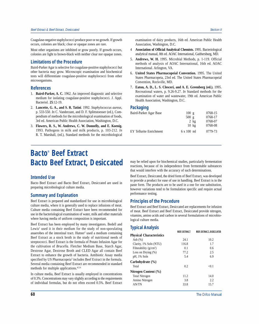
60 The Difco Manual
Coagulase-negative staphylococci produce poor or no growth. If growthoccurs, colonies are black; clear or opaque zones are rare.
Most other organisms are inhibited or grow poorly. If growth occurs,colonies are light to brown-black with neither clear nor opaque zones.
Limitations of the ProcedureBaird-Parker Agar is selective for coagulase-positive staphylococci butother bacteria may grow. Microscopic examination and biochemicaltests will differentiate coagulase-positive staphylococci from othermicroorganisms.
References1. Baird-Parker, A. C. 1962. An improved diagnostic and selective
medium for isolating coagulase-positive staphylococci. J. Appl.Bacteriol. 25:12-19.
2. Lancette, G. A., and S. R. Tatini. 1992. Staphylococcus aureus,p. 533-550. In C. Vanderzant, and D. F. Splittstoesser (ed.), Com-pendium of methods for the microbiological examination of foods,3rd ed. American Public Health Association, Washington, D.C.
3. Flowers, R. S., W. Andrews, C. W. Donnelly, and E. Koenig.1993. Pathogens in milk and milk products, p. 103-212. InR. T. Marshall, (ed.), Standard methods for the microbiological
examination of dairy products, 16th ed. American Public HealthAssociation, Washington, D.C.
4 Association of Official Analytical Chemists. 1995. Bacteriologicalanalytical manual, 8th ed. AOAC International, Gaithersburg, MD.
5. Andrews, W. H. 1995. Microbial Methods, p. 1-119. Officialmethods of analysis of AOAC International, 16th ed. AOACInternational. Arlington, VA.
6. United States Pharmacopeial Convention. 1995. The UnitedStates Pharmacopeia, 23rd ed. The United States PharmacopeialConvention, Rockville, MD.
7. Eaton, A. D., L. S. Clesceri, and A. E. Greenberg (ed.). 1995.Recreational waters, p. 9.26-9.27. In Standard methods for theexamination of water and wastewater, 19th ed. American PublicHealth Association, Washington, D.C.
PackagingBaird-Parker Agar Base 100 g 0768-15
500 g 0768-172 kg 0768-07
10 kg 0768-08
EY Tellurite Enrichment 6 x 100 ml 0779-73
Beef Extract & Beef Extract, Desiccated Section II
Bacto® Beef ExtractBacto Beef Extract, Desiccated
Intended UseBacto Beef Extract and Bacto Beef Extract, Desiccated are used inpreparing microbiological culture media.
Summary and ExplanationBeef Extract is prepared and standardized for use in microbiologicalculture media, where it is generally used to replace infusions of meat.Culture media containing Beef Extract have been recommended foruse in the bacteriological examination of water, milk and other materialswhere having media of uniform composition is important.
Beef Extract has been employed by many investigators. Bedell andLewis1 used it in their medium for the study of non-sporulatinganaerobes of the intestinal tract. Hutner2 used a medium containingBeef Extract as a stock broth in the study of nutritional needs ofstreptococci. Beef Extract is the formula of Potato Infusion Agar forthe cultivation of Brucella. Fletcher Medium Base, Starch Agar,Dextrose Agar, Dextrose Broth and CLED Agar all contain BeefExtract to enhance the growth of bacteria. Antibiotic Assay mediaspecified by US Pharmacopeia3 includes Beef Extract in the formula.Several media containing Beef Extract are recommended in standardmethods for multiple applications.4,5,6
In culture media, Beef Extract is usually employed in concentrationsof 0.3%. Concentrations may vary slightly according to the requirementsof individual formulas, but do not often exceed 0.5%. Beef Extract
may be relied upon for biochemical studies, particularly fermentationreactions, because of its independence from fermentable substancesthat would interfere with the accuracy of such determinations.
Beef Extract, Desiccated, the dried form of Beef Extract, was developedto provide a product for ease of use in handling. Beef Extract is in thepaste form. The products are to be used in a one for one substitution,however variations tend to be formulation specific and require actualperformance testing.
Principles of the ProcedureBeef Extract and Beef Extract, Desiccated are replacements for infusionof meat. Beef Extract and Beef Extract, Desiccated provide nitrogen,vitamins, amino acids and carbon in several formulations of microbio-logical culture media.
Typical AnalysisBEEF EXTRACT BEEF EXTRACT, DESICCATED
Physical CharacteristicsAsh (%) 24.1 10.2Clarity, 1% Soln (NTU) 116.8 1.7Filterability (g/cm2) 0.1 0.6Loss on Drying (%) 77.2 2.5pH, 1% Soln 5.4 6.9
Carbohydrate (%)Total 0.2 <0.1
Nitrogen Content (%)Total Nitrogen 11.2 14.0Amino Nitrogen 3.8 2.2AN/TN 33.8 15.7
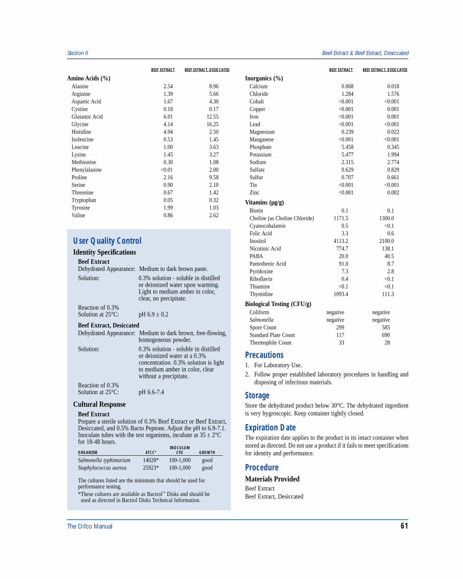
The Difco Manual 61
Section II Beef Extract & Beef Extract, Desiccated
User Quality ControlIdentity Specifications
Beef ExtractDehydrated Appearance: Medium to dark brown paste.Solution: 0.3% solution - soluble in distilled
or deionized water upon warming.Light to medium amber in color,clear, no precipitate.
Reaction of 0.3%Solution at 25°C: pH 6.9 ± 0.2
Beef Extract, DesiccatedDehydrated Appearance: Medium to dark brown, free-flowing,
homogeneous powder.Solution: 0.3% solution - soluble in distilled
or deionized water at a 0.3%concentration. 0.3% solution is lightto medium amber in color, clearwithout a precipitate.
Reaction of 0.3%Solution at 25°C: pH 6.6-7.4
Cultural ResponseBeef ExtractPrepare a sterile solution of 0.3% Beef Extract or Beef Extract,Desiccated, and 0.5% Bacto Peptone. Adjust the pH to 6.9-7.1.Inoculate tubes with the test organisms, incubate at 35 ± 2°Cfor 18-48 hours.
INOCULUMORGANISM ATCC® CFU GROWTH
Salmonella typhimurium 14028* 100-1,000 goodStaphylococcus aureus 25923* 100-1,000 good
The cultures listed are the minimum that should be used forperformance testing.*These cultures are available as Bactrol™ Disks and should be used as directed in Bactrol Disks Technical Information.
BEEF EXTRACT BEEF EXTRACT, DESICCATED
Amino Acids (%)Alanine 2.54 8.96Arginine 1.39 5.66Aspartic Acid 1.67 4.30Cystine 0.18 0.17Glutamic Acid 6.01 12.55Glycine 4.14 16.25Histidine 4.94 2.50Isoleucine 0.53 1.45Leucine 1.00 3.63Lysine 1.45 3.27Methionine 0.30 1.08Phenylalanine <0.01 2.00Proline 2.16 9.58Serine 0.90 2.10Threonine 0.67 1.42Tryptophan 0.05 0.32Tyrosine 1.99 1.03Valine 0.86 2.62
BEEF EXTRACT BEEF EXTRACT, DESICCATED
Inorganics (%)Calcium 0.068 0.018Chloride 1.284 1.576Cobalt <0.001 <0.001Copper <0.001 0.001Iron <0.001 0.001Lead <0.001 <0.001Magnesium 0.239 0.022Manganese <0.001 <0.001Phosphate 5.458 0.345Potassium 5.477 1.994Sodium 2.315 2.774Sulfate 0.629 0.829Sulfur 0.707 0.661Tin <0.001 <0.001Zinc <0.001 0.002
Vitamins (µg/g)Biotin 0.1 0.1Choline (as Choline Chloride) 1171.5 1300.0Cyanocobalamin 0.5 <0.1Folic Acid 3.3 0.6Inositol 4113.2 2100.0Nicotinic Acid 774.7 138.1PABA 20.0 40.5Pantothenic Acid 91.0 8.7Pyridoxine 7.3 2.8Riboflavin 0.4 <0.1Thiamine <0.1 <0.1Thymidine 1093.4 111.3
Biological Testing (CFU/g)Coliform negative negativeSalmonella negative negativeSpore Count 299 585Standard Plate Count 117 690Thermophile Count 33 28
Precautions1. For Laboratory Use.2. Follow proper established laboratory procedures in handling and
disposing of infectious materials.
StorageStore the dehydrated product below 30°C. The dehydrated ingredientis very hygroscopic. Keep container tightly closed.
Expiration DateThe expiration date applies to the product in its intact container whenstored as directed. Do not use a product if it fails to meet specificationsfor identity and performance.
ProcedureMaterials ProvidedBeef ExtractBeef Extract, Desiccated
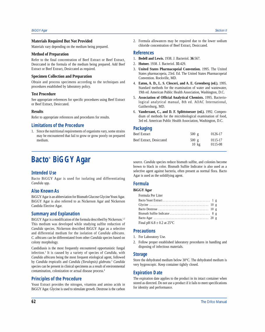
62 The Difco Manual
Materials Required But Not ProvidedMaterials vary depending on the medium being prepared.
Method of PreparationRefer to the final concentration of Beef Extract or Beef Extract,Desiccated in the formula of the medium being prepared. Add BeefExtract or Beef Extract, Desiccated as required.
Specimen Collection and PreparationObtain and process specimens according to the techniques andprocedures established by laboratory policy.
Test ProcedureSee appropriate references for specific procedures using Beef Extractor Beef Extract, Desiccated.
ResultsRefer to appropriate references and procedures for results.
Limitations of the Procedure1. Since the nutritional requirements of organisms vary, some strains
may be encountered that fail to grow or grow poorly on preparedmedium.
2. Formula allowances may be required due to the lower sodiumchloride concentration of Beef Extract, Desiccated.
References1. Bedell and Lewis. 1938. J. Bacteriol. 36:567.2. Hutner. 1938. J. Bacteriol. 35:429.3. United States Pharmacopeial Convention. 1995. The United
States pharmacopeia, 23rd. Ed. The United States PharmacopeialConvention. Rockville, MD.
4. Eaton, A. D., L. S. Clesceri, and A. E. Greenberg (ed.). 1995.Standard methods for the examination of water and wastewater,19th ed. American Public Health Association, Washington, D.C.
5. Association of Official Analytical Chemists. 1995. Bacterio-logical analytical manual, 8th ed. AOAC International,Gaithersburg, MD.
6. Vanderzant, C., and D. F. Splittstoesser (ed.). 1992. Compen-dium of methods for the microbiological examination of food,3rd ed. American Public Health Association, Washington, D.C.
PackagingBeef Extract 500 g 0126-17
Beef Extract, Desiccated 500 g 0115-1710 kg 0115-08
BiGGY Agar Section II
Bacto® BiGGY AgarIntended UseBacto BiGGY Agar is used for isolating and differentiatingCandida spp.
Also Known AsBiGGY Agar is an abbreviation for Bismuth Glucose Glycine Yeast Agar.BiGGY Agar is also referred to as Nickerson Agar and NickersonCandida Elective Agar.
Summary and ExplanationBiGGY Agar is a modification of the formula described by Nickerson.1,2
This medium was developed while studying sulfite reduction ofCandida species. Nickerson described BiGGY Agar as a selectiveand differential medium for the isolation of Candida albicans.C. albicans can be differentiated from other Candida species based oncolony morphology.
Candidiasis is the most frequently encountered opportunistic fungalinfection.3 It is caused by a variety of species of Candida, withCandida albicans being the most frequent etiological agent, followedby Candida tropicalis and Candida (Torulopsis) glabrata.3 Candidaspecies can be present in clinical specimens as a result of environmentalcontamination, colonization or actual disease process.4
Principles of the ProcedureYeast Extract provides the nitrogen, vitamins and amino acids inBiGGY Agar. Glycine is used to stimulate growth. Dextrose is the carbon
source. Candida species reduce bismuth sulfite, and colonies becomebrown to black in color. Bismuth Sulfite Indicator is also used as aselective agent against bacteria, often present as normal flora. BactoAgar is used as the solidifying agent.
FormulaBiGGY Agar
Formula Per LiterBacto Yeast Extract . . . . . . . . . . . . . . . . . . . . . . . . . . . . . . . . 1 gGlycine . . . . . . . . . . . . . . . . . . . . . . . . . . . . . . . . . . . . . . . . 10 gBacto Dextrose . . . . . . . . . . . . . . . . . . . . . . . . . . . . . . . . . . 10 gBismuth Sulfite Indicator . . . . . . . . . . . . . . . . . . . . . . . . . . . 8 gBacto Agar . . . . . . . . . . . . . . . . . . . . . . . . . . . . . . . . . . . . . 20 g
Final pH 6.8 ± 0.2 at 25°C
Precautions1. For Laboratory Use.2. Follow proper established laboratory procedures in handling and
disposing of infectious materials.
StorageStore the dehydrated medium below 30°C. The dehydrated medium isvery hygroscopic. Keep container tightly closed.
Expiration DateThe expiration date applies to the product in its intact container whenstored as directed. Do not use a product if it fails to meet specificationsfor identity and performance.
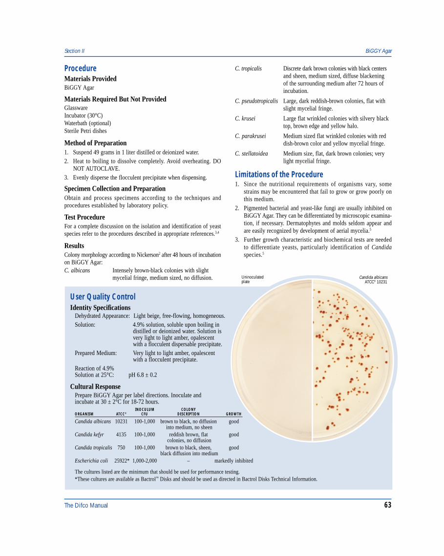
The Difco Manual 63
Section II BiGGY Agar
User Quality ControlIdentity Specifications
Dehydrated Appearance: Light beige, free-flowing, homogeneous.Solution: 4.9% solution, soluble upon boiling in
distilled or deionized water. Solution isvery light to light amber, opalescentwith a flocculent dispersable precipitate.
Prepared Medium: Very light to light amber, opalescentwith a flocculent precipitate.
Reaction of 4.9%Solution at 25°C: pH 6.8 ± 0.2
Cultural ResponsePrepare BiGGY Agar per label directions. Inoculate andincubate at 30 ± 2°C for 18-72 hours.
Candida albicansATCC® 10231
Uninoculatedplate
INOCULUM COLONYORGANISM ATCC® CFU DESCRIPTION GROWTH
Candida albicans 10231 100-1,000 brown to black, no diffusion good into medium, no sheen
Candida kefyr 4135 100-1,000 reddish brown, flat good colonies, no diffusion
Candida tropicalis 750 100-1,000 brown to black, sheen, good black diffusion into medium
Escherichia coli 25922* 1,000-2,000 – markedly inhibited
The cultures listed are the minimum that should be used for performance testing.*These cultures are available as Bactrol™ Disks and should be used as directed in Bactrol Disks Technical Information.
ProcedureMaterials ProvidedBiGGY Agar
Materials Required But Not ProvidedGlasswareIncubator (30°C)Waterbath (optional)Sterile Petri dishes
Method of Preparation1. Suspend 49 grams in 1 liter distilled or deionized water.2. Heat to boiling to dissolve completely. Avoid overheating. DO
NOT AUTOCLAVE.3. Evenly disperse the flocculent precipitate when dispensing.
Specimen Collection and PreparationObtain and process specimens according to the techniques andprocedures established by laboratory policy.
Test ProcedureFor a complete discussion on the isolation and identification of yeastspecies refer to the procedures described in appropriate references.3,4
ResultsColony morphology according to Nickerson2 after 48 hours of incubationon BiGGY Agar:C. albicans Intensely brown-black colonies with slight
mycelial fringe, medium sized, no diffusion.
C. tropicalis Discrete dark brown colonies with black centersand sheen, medium sized, diffuse blackeningof the surrounding medium after 72 hours ofincubation.
C. pseudotropicalis Large, dark reddish-brown colonies, flat withslight mycelial fringe.
C. krusei Large flat wrinkled colonies with silvery blacktop, brown edge and yellow halo.
C. parakrusei Medium sized flat wrinkled colonies with reddish-brown color and yellow mycelial fringe.
C. stellatoidea Medium size, flat, dark brown colonies; verylight mycelial fringe.
Limitations of the Procedure1. Since the nutritional requirements of organisms vary, some
strains may be encountered that fail to grow or grow poorly onthis medium.
2. Pigmented bacterial and yeast-like fungi are usually inhibited onBiGGY Agar. They can be differentiated by microscopic examina-tion, if necessary. Dermatophytes and molds seldom appear andare easily recognized by development of aerial mycelia.5
3. Further growth characteristic and biochemical tests are neededto differentiate yeasts, particularly identification of Candidaspecies.5
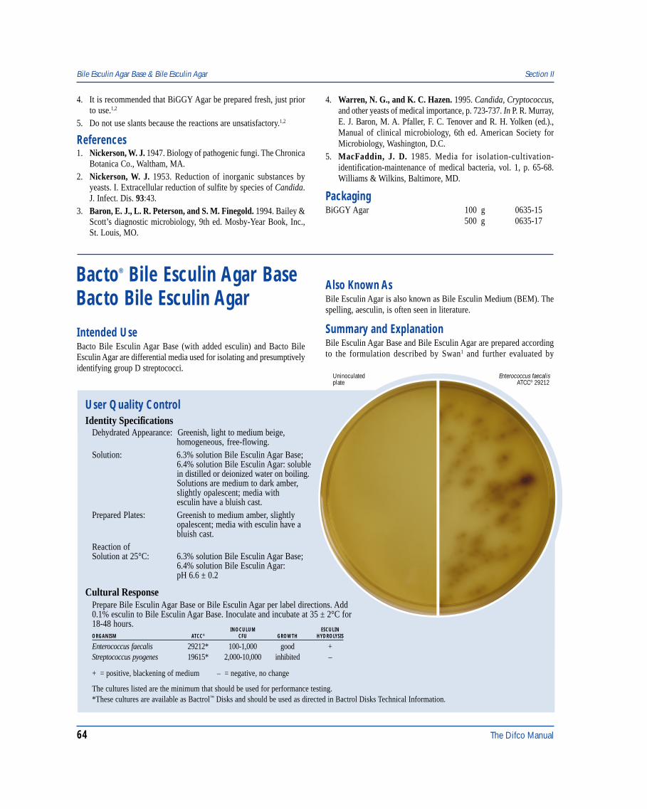
64 The Difco Manual
4. It is recommended that BiGGY Agar be prepared fresh, just priorto use.1,2
5. Do not use slants because the reactions are unsatisfactory.1,2
References1. Nickerson, W. J. 1947. Biology of pathogenic fungi. The Chronica
Botanica Co., Waltham, MA.2. Nickerson, W. J. 1953. Reduction of inorganic substances by
yeasts. I. Extracellular reduction of sulfite by species of Candida.J. Infect. Dis. 93:43.
3. Baron, E. J., L. R. Peterson, and S. M. Finegold. 1994. Bailey &Scott’s diagnostic microbiology, 9th ed. Mosby-Year Book, Inc.,St. Louis, MO.
Bile Esculin Agar Base & Bile Esculin Agar Section II
4. Warren, N. G., and K. C. Hazen. 1995. Candida, Cryptococcus,and other yeasts of medical importance, p. 723-737. In P. R. Murray,E. J. Baron, M. A. Pfaller, F. C. Tenover and R. H. Yolken (ed.).,Manual of clinical microbiology, 6th ed. American Society forMicrobiology, Washington, D.C.
5. MacFaddin, J. D. 1985. Media for isolation-cultivation-identification-maintenance of medical bacteria, vol. 1, p. 65-68.Williams & Wilkins, Baltimore, MD.
PackagingBiGGY Agar 100 g 0635-15
500 g 0635-17
Bacto® Bile Esculin Agar BaseBacto Bile Esculin Agar
User Quality ControlIdentity Specifications
Dehydrated Appearance: Greenish, light to medium beige,homogeneous, free-flowing.
Solution: 6.3% solution Bile Esculin Agar Base;6.4% solution Bile Esculin Agar: solublein distilled or deionized water on boiling.Solutions are medium to dark amber,slightly opalescent; media withesculin have a bluish cast.
Prepared Plates: Greenish to medium amber, slightlyopalescent; media with esculin have abluish cast.
Reaction ofSolution at 25°C: 6.3% solution Bile Esculin Agar Base;
6.4% solution Bile Esculin Agar:pH 6.6 ± 0.2
Cultural ResponsePrepare Bile Esculin Agar Base or Bile Esculin Agar per label directions. Add0.1% esculin to Bile Esculin Agar Base. Inoculate and incubate at 35 ± 2°C for18-48 hours.
Enterococcus faecalis ATCC® 29212
Uninoculatedplate
INOCULUM ESCULINORGANISM ATCC® CFU GROWTH HYDROLYSIS
Enterococcus faecalis 29212* 100-1,000 good +Streptococcus pyogenes 19615* 2,000-10,000 inhibited –
+ = positive, blackening of medium – = negative, no change
The cultures listed are the minimum that should be used for performance testing.*These cultures are available as Bactrol™ Disks and should be used as directed in Bactrol Disks Technical Information.
Intended UseBacto Bile Esculin Agar Base (with added esculin) and Bacto BileEsculin Agar are differential media used for isolating and presumptivelyidentifying group D streptococci.
Also Known AsBile Esculin Agar is also known as Bile Esculin Medium (BEM). Thespelling, aesculin, is often seen in literature.
Summary and ExplanationBile Esculin Agar Base and Bile Esculin Agar are prepared accordingto the formulation described by Swan1 and further evaluated by
The Difco Manual 65
Section II Bile Esculin Agar Base & Bile Esculin Agar
Precautions1. For Laboratory Use.2. Bile Esculin Agar Base:
IRRITANT. IRRITATING TO EYES, RESPIRATORY SYSTEMAND SKIN. Avoid contact with skin and eyes. Do not breathe dust.Wear suitable gloves and eye/face protection. Use only in wellventilated areas. Keep container tightly closed.FIRST AID: In case of contact with eyes, rinse immediately withplenty of water and seek medical advice. After contact with skin,wash immediately with plenty of water. If inhaled, remove to freshair. If not breathing, give artificial respiration. If breathing is diffi-cult, give oxygen. Seek medical advice. If swallowed seek medicaladvice immediately and show this container or label.Bacto Bile Esculin Agar:IRRITANT. IRRITATING TO EYES, RESPIRATORY SYSTEMAND SKIN. Avoid contact with skin and eyes. Do not breathe dust.Wear suitable protective clothing. Keep container tightly closed.TARGET ORGAN(S): Lungs.FIRST AID: In case of contact with eyes, rinse immediately withplenty of water and seek medical advice. After contact with skin,wash immediately with plenty of water. If inhaled, remove to freshair. If not breathing, give artificial respiration. If breathing is diffi-cult, give oxygen. Seek medical advice. If swallowed seek medicaladvice immediately and show this container or label.
3. Follow proper established laboratory procedure in handling anddisposing of infectious materials.
StorageStore the dehydrated medium below 30°C. The dehydrated medium isvery hygroscopic. Keep container tightly closed.
Expiration DateThe expiration date applies to the product in its intact container whenstored as directed. Do not use a product if it fails to meet specificationsfor identity and performance.
ProcedureMaterials ProvidedBile Esculin Agar BaseBile Esculin Agar
Materials Required But Not ProvidedGlasswareAutoclaveIncubator (35°C)Esculin (to be added to Bile Esculin Agar Base)Filter-sterilized horse serum (optional)Petri dishesTubes with closures
Method of Preparation1. Suspend the specidied amount of medium in 1 liter distilled or
deionized water:Bile Esculin Agar Base - 63 gramsBile Esculin Agar - 64 grams
Facklam and Moody.2 Rochaix3 first noted the value of esculin hydrolysisin the identification of enterococci. Meyer and Schönfeld4 added bileto the esculin medium and demonstrated that 61 of 62 enterococcistrains were able to grow and hydrolyze esculin, while the otherstreptococci could not.
Molecular taxonomic studies of the genus Streptococcus have placedenterococci, previously considered group D streptococci, in the distinctgenus Enterococcus.6 Streptococci with Lancefield group D antigeninclude the nonhemolytic species Streptococcus bovis.7 The ability tohydrolyze esculin in the presence of bile is a characteristic of enterococciand group D streptococci.
Swan1 compared the use of an esculin medium containing 40% bilesalts with the Lancefield serological method of grouping. He reportedthat a positive reaction on the bile esculin medium correlated with aserological group D precipitin reaction. Facklam and Moody,2 ina comparative study of tests used to presumptively identify group Dstreptococci, found that the bile esculin test provided a reliable meansof identifying group D streptococci and differentiating them fromnon-group D streptococci. Facklam5 further confirmed the usefulnessof Bile Esculin Agar in another study differentiating enterococci/groupD streptococci from non-group D streptococci.
Lindell and Quinn8 showed that the medium is also useful in thedifferentiation of the Klebsiella-Enterobacter-Serratia group from otherEnterobacteriaceae. Edberg et al.9 recommended the medium for routinetesting of the Enterobacteriaceae in order to differentiateKlebsiella-Enterobacter-Serratia spp. Bile Esculin Agar is listed in standardprocedures for the microbiological examination of food products.10-13
Principles of the ProcedureOrganisms positive for esculin hydrolysis hydrolyze the glycosideesculin to esculetin and dextrose. The esculetin reacts with the ferric citrateto form a dark brown or black complex. Oxgall (bile) is used to inhibitgram-positive bacteria other than enterococci. Beef Extract and BactoPeptone provide the carbon and nitrogen sources required for growth of awide variety of organisms. Bacto Agar is the solidifying agent.
FormulaBile Esculin Agar
Formula Per LiterBacto Beef Extract . . . . . . . . . . . . . . . . . . . . . . . . . . . . . . . . 3 gBacto Peptone . . . . . . . . . . . . . . . . . . . . . . . . . . . . . . . . . . . . 5 gEsculin . . . . . . . . . . . . . . . . . . . . . . . . . . . . . . . . . . . . . . . . . . 1 gBacto Oxgall . . . . . . . . . . . . . . . . . . . . . . . . . . . . . . . . . . . . 40 gFerric Citrate . . . . . . . . . . . . . . . . . . . . . . . . . . . . . . . . . . . . 0.5 gBacto Agar . . . . . . . . . . . . . . . . . . . . . . . . . . . . . . . . . . . . . 15 g
Final pH 6.6 ± 0.2 at 25°C
Bile Esculin Agar Base
Formula Per LiterBacto Beef Extract . . . . . . . . . . . . . . . . . . . . . . . . . . . . . . . . 3 gBacto Peptone . . . . . . . . . . . . . . . . . . . . . . . . . . . . . . . . . . . . 5 gBacto Oxgall . . . . . . . . . . . . . . . . . . . . . . . . . . . . . . . . . . . . 40 gFerric Citrate . . . . . . . . . . . . . . . . . . . . . . . . . . . . . . . . . . . . 0.5 gBacto Agar . . . . . . . . . . . . . . . . . . . . . . . . . . . . . . . . . . . . . 15 g
Final pH 6.6 ± 0.2 at 25°C
66 The Difco Manual
2. Heat to boiling to dissolve completely.3. Bile Esculin Agar Base, only: Add 1 gram (or another desired
amount) of Esculin and mix thoroughly.4. Autoclave at 121°C for 15 minutes. Overheating may cause
darkening of the media.5. Cool to 50-55°C.6. If desired, aseptically add 50 ml of filter-sterilized horse serum.
Mix thoroughly.7. Dispense as desired.
Specimen Collection and PreparationRefer to appropriate references for specimen collection and preparation.
Test ProcedureSee appropriate references for specific procedures.
ResultsRefer to appropriate references and procedures for results.
Limitations of the Procedure1. The bile esculin test was originally formulated to identify
enterococci. However, the properties of growth on 40% bile mediaand esculin hydrolysis are characteristics shared by most strains ofGroup D streptococci.14 The bile esculin test should be used incombination with other tests to make a positive identification.Facklam14 and Facklam et al.15 recommend a combination of the bileesculin test and salt tolerance (growth in 6.5% NaCl). Streptococcusbovis will give a positive reaction on Bile Esculin Agar, but unlikeEnterococcus spp., it cannot grow on 6.5% NaCl or at 10°C.16
2. Bile Esculin Agar should be considered a differential medium, butwith the addition of sodium azide (which inhibits gram-negativebacteria) the medium can be made more selective (see Bile EsculinAzide Agar).
3. Occasional viridans strains will be positive on Bile Esculin Agaror will display reactions that are difficult to interpret.17 Of theviridans group, 5 to 10% may be able to hydrolyze esculin in thepresence of bile.16
4. Use a light inoculum when testing Escherichia coli on Bile EsculinAgar. Wasilauskas18 suggests that the time required for an isolateto hydrolyze esculin is directly proportional to the size of theinoculum. For a tabulation of those Enterobacteriaceae that canhydrolyze esculin, refer to Farmer.19
References1. Swan, A. 1954. The use of bile-esculin medium and of Maxted’s
technique of Lancefield grouping in the identification of enterococci(group D streptococci). J. Clin. Pathol. 7:160.
2. Facklam, R. R., and M. D. Moody. 1970. Presumptive identifi-cation of group D streptococci: The bile-esculin test. Appl.Microbiol. 20:245.
3. Rochaix, A. 1924. Milieux a leculine pour le diagnostiddifferentieldes bacteries du groups strepto-entero-pneumocoque.Comt. Rend. Soc. Biol. 90: 771-772.
4. Meyer, K., and H. Schönfeld. 1926. Über die Untersheidung desEnterococcus vom Streptococcus viridans und die Beziehunger
beider zum Streptococcus lactis. Zentralbl. Bakteriol. Parasitenkd.Infektionskr. Hyg. Abt. I Orig. 99:402-416.
5. Facklam, R. R. 1973. Comparison of several laboratory media forpresumptive identification of enterococci and group D streptococci.Appl. Microbiol. 26:138.
6. Schleifer, K. H., and R. Kilpper-Balz. 1987. Molecular andchemotaxonomic approaches to the classification of streptococci,enterococci and lactococci: a review. Syst. Appl. Microbiol. 10:1-19.
7. Ruoff, K. L. 1995. Streptococcus. P. R. Murray, E. J. Baron, M. A.Pfaller, F. C. Tenover and R. H. Yolken (eds.), Manual of clinicalmicrobiology, 6th ed. American Society for Microbiology,Washington, D.C.
8. Lindell, S. S., and P. Quinn. 1975. Use of bile-esculin agar for rapiddifferentiation of Enterobacteriaceae. J. Clin. Microbiol. 1:440.
9. Edberg, S. C., S. Pittman, and J. M. Singer. 1977. Esculinhydrolysis by Enterobacteriaceae. J. Clin. Microbiol. 6:111.
10. Bacteriological Analytical Manual. 1995. 8th ed. AOACInternational, Gaithersburg, MD.
11. Vanderzant, C., and D. F. Splittstoesser (eds.). 1992. Compen-dium of methods for the microbiological examination of foods,3rd ed. American Public Health Association, Washington, D.C.
12. Marshall, R. T. (ed.) 1992. Standard methods for the examinationof dairy products, 16th ed. American Public Health Association,Washington, D.C.
13. Atlas, R. M. 1995. Handbook of microbiological media for theexamination of food. CRC Press, Boca Raton, FL.
14. Facklam, R. 1972. Recognition of group D streptococcal speciesof human origin by biochemical and physiological tests. Appl.Microbiol. 23:1131.
15. Facklam, R. R., J. F. Padula, L. G. Thacker, E. C. Wortham,and B. J. Sconyers. 1974. Presumptive identification of group A,B, and D streptococci. Appl. Microbiol. 27:107.
16. Baron, E. J., L. R. Peterson, and S. M. Finegold. 1994. Bailey& Scott’s diagnostic microbiology, 9th ed. Mosby-Year Book,Inc. St. Louis, MO.
17. Ruoff, K. L., S. I. Miller, C. V. Garner, M. J. Ferraro, and S. B.Calderwood. 1989. Bacteremia with Streptococcus bovis andStreptococcus salivarius: clinical correlates of more accurateidentification of isolates. J. Clin. Microbiol. 27:305-308.
18. Wasilauskas, B. L. 1971. Preliminary observations on the rapiddifferentiation of the Klebsiella-Enterobacter-Serratia group onbile-esculin agar. Appl. Microbiol. 21:162.
19. Farmer, J. J., III. 1995. Enterobacteriaceae. P. R. Murray, E. J.Baron, M. A. Pfaller, F. C. Tenover and R. H. Yolken (eds.), Manualof clinical microbiology, 6th ed. American Society for Microbiology,Washington, D.C.
PackagingBile Esculin Agar Base 500 g 0878-17
Bile Esculin Agar 100 g 0879-15500 g 0879-17
Esculin 10 g 0158-12
Bile Esculin Agar Base & Bile Esculin Agar Section II
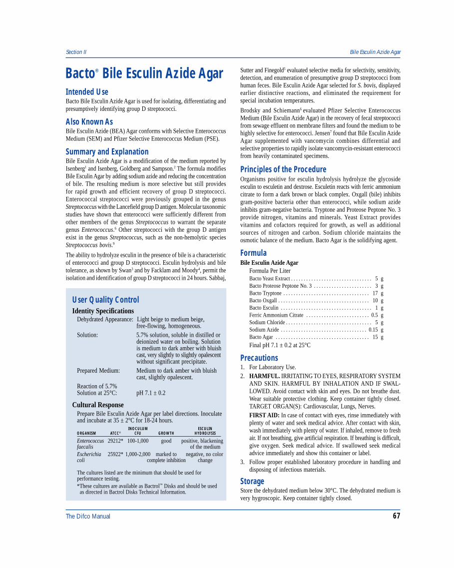
The Difco Manual 67
Section II Bile Esculin Azide Agar
Bacto® Bile Esculin Azide Agar
User Quality ControlIdentity Specifications
Dehydrated Appearance: Light beige to medium beige,free-flowing, homogeneous.
Solution: 5.7% solution, soluble in distilled ordeionized water on boiling. Solutionis medium to dark amber with bluishcast, very slightly to slightly opalescentwithout significant precipitate.
Prepared Medium: Medium to dark amber with bluishcast, slightly opalescent.
Reaction of 5.7%Solution at 25°C: pH 7.1 ± 0.2
Cultural ResponsePrepare Bile Esculin Azide Agar per label directions. Inoculateand incubate at 35 ± 2°C for 18-24 hours.
INOCULUM ESCULINORGANISM ATCC® CFU GROWTH HYDROLYSIS
Enterococcus 29212* 100-1,000 good positive, blackeningfaecalis of the mediumEscherichia 25922* 1,000-2,000 marked to negative, no colorcoli complete inhibition change
The cultures listed are the minimum that should be used forperformance testing.*These cultures are available as Bactrol™ Disks and should be used as directed in Bactrol Disks Technical Information.
Intended UseBacto Bile Esculin Azide Agar is used for isolating, differentiating andpresumptively identifying group D streptococci.
Also Known AsBile Esculin Azide (BEA) Agar conforms with Selective EnterococcusMedium (SEM) and Pfizer Selective Enterococcus Medium (PSE).
Summary and ExplanationBile Esculin Azide Agar is a modification of the medium reported byIsenberg1 and Isenberg, Goldberg and Sampson.2 The formula modifiesBile Esculin Agar by adding sodium azide and reducing the concentrationof bile. The resulting medium is more selective but still providesfor rapid growth and efficient recovery of group D streptococci.Enterococcal streptococci were previously grouped in the genusStreptococcus with the Lancefield group D antigen. Molecular taxonomicstudies have shown that enterococci were sufficiently different fromother members of the genus Streptococcus to warrant the separategenus Enterococcus.6 Other streptococci with the group D antigenexist in the genus Streptococcus, such as the non-hemolytic speciesStreptococcus bovis.9
The ability to hydrolyze esculin in the presence of bile is a characteristicof enterococci and group D streptococci. Esculin hydrolysis and biletolerance, as shown by Swan3 and by Facklam and Moody4, permit theisolation and identification of group D streptococci in 24 hours. Sabbaj,
Sutter and Finegold5 evaluated selective media for selectivity, sensitivity,detection, and enumeration of presumptive group D streptococci fromhuman feces. Bile Esculin Azide Agar selected for S. bovis, displayedearlier distinctive reactions, and eliminated the requirement forspecial incubation temperatures.
Brodsky and Schiemann6 evaluated Pfizer Selective EnterococcusMedium (Bile Esculin Azide Agar) in the recovery of fecal streptococcifrom sewage effluent on membrane filters and found the medium to behighly selective for enterococci. Jensen7 found that Bile Esculin AzideAgar supplemented with vancomycin combines differential andselective properties to rapidly isolate vancomycin-resistant enterococcifrom heavily contaminated specimens.
Principles of the ProcedureOrganisms positive for esculin hydrolysis hydrolyze the glycosideesculin to esculetin and dextrose. Esculetin reacts with ferric ammoniumcitrate to form a dark brown or black complex. Oxgall (bile) inhibitsgram-positive bacteria other than enterococci, while sodium azideinhibits gram-negative bacteria. Tryptone and Proteose Peptone No. 3provide nitrogen, vitamins and minerals. Yeast Extract providesvitamins and cofactors required for growth, as well as additionalsources of nitrogen and carbon. Sodium chloride maintains theosmotic balance of the medium. Bacto Agar is the solidifying agent.
FormulaBile Esculin Azide Agar
Formula Per LiterBacto Yeast Extract . . . . . . . . . . . . . . . . . . . . . . . . . . . . . . . . 5 gBacto Proteose Peptone No. 3 . . . . . . . . . . . . . . . . . . . . . . . 3 gBacto Tryptone . . . . . . . . . . . . . . . . . . . . . . . . . . . . . . . . . . 17 gBacto Oxgall . . . . . . . . . . . . . . . . . . . . . . . . . . . . . . . . . . . . 10 gBacto Esculin . . . . . . . . . . . . . . . . . . . . . . . . . . . . . . . . . . . . 1 gFerric Ammonium Citrate . . . . . . . . . . . . . . . . . . . . . . . . . 0.5 gSodium Chloride . . . . . . . . . . . . . . . . . . . . . . . . . . . . . . . . . . 5 gSodium Azide . . . . . . . . . . . . . . . . . . . . . . . . . . . . . . . . . . 0.15 gBacto Agar . . . . . . . . . . . . . . . . . . . . . . . . . . . . . . . . . . . . . 15 g
Final pH 7.1 ± 0.2 at 25°C
Precautions1. For Laboratory Use.2. HARMFUL. IRRITATING TO EYES, RESPIRATORY SYSTEM
AND SKIN. HARMFUL BY INHALATION AND IF SWAL-LOWED. Avoid contact with skin and eyes. Do not breathe dust.Wear suitable protective clothing. Keep container tightly closed.TARGET ORGAN(S): Cardiovascular, Lungs, Nerves.FIRST AID: In case of contact with eyes, rinse immediately withplenty of water and seek medical advice. After contact with skin,wash immediately with plenty of water. If inhaled, remove to freshair. If not breathing, give artificial respiration. If breathing is difficult,give oxygen. Seek medical advice. If swallowed seek medicaladvice immediately and show this container or label.
3. Follow proper established laboratory procedure in handling anddisposing of infectious materials.
StorageStore the dehydrated medium below 30°C. The dehydrated medium isvery hygroscopic. Keep container tightly closed.
68 The Difco Manual
Expiration DateThe expiration date applies to the product in its intact container whenstored as directed. Do not use a product if it fails to meet specificationsfor identity and performance.
ProcedureMaterials ProvidedBile Esculin Azide Agar
Materials Required But Not ProvidedGlasswareAutoclaveIncubator (35°C)Petri dishesHorse Serum, filter sterilized (optional)
Method of Preparation1. Suspend 57 grams in 1 liter distilled or deionized water.2. Boil to dissolve completely.3. Autoclave at 121°C for 15 minutes. Overheating may cause
darkening of the medium.4. If desired, aseptically add 50 ml of filter-sterilized horse serum.
Mix thoroughly.
Specimen Collection and PreparationRefer to appropriate references for specimen collection and preparation.
Test ProcedureFor isolation of group D streptococci, inoculate the sample onto a smallarea of one quadrant of a Bile Esculin Azide Agar plate and streak forisolation. This will permit development of discrete colonies. Incubateat 35°C for 18-24 hours. Examine for colonies having the characteristicmorphology of group D streptococci.
ResultsGroup D streptococci grow readily on this medium and hydrolyzeesculin, resulting in a dark brown color around the colonies after18-24 hours incubation.
Limitations of the Procedure1. Staphylococcus aureus and Staphylococcus epidermidis may
exhibit growth on the medium (less than 1 mm, white-graycolonies), but they will show no action on the esculin.2
2. Other than the enterococci, Listeria monocytogenes consistentlyblackens the medium around colonies. After 18-24 hours, there maybe a reddish to black-brown zone of hydrolysis surrounding pinpointListeria colonies. After 48 hours, white-gray pigmented colonies willbe seen. Listeria do not attain the same degree of esculin hydrolysisdisplayed by enterococci in this short incubation period.2
References1. Isenberg, H. D. 1970. Clin. Lab. Forum. July.2. Isenberg, H. D., D. Goldberg, and J. Sampson. 1970. Laboratory
studies with a selective enterococcus medium. Appl. Microbiol.20:433.
3. Swan, A. 1954. The use of bile-esculin medium and of Maxted’stechnique of Lancefield grouping in the identification ofenterococci (group D streptococci). J. Clin. Pathol. 7:160.
4. Facklam, R. R., and M. D. Moody. 1970. Presumptiveidentification of group D streptococci: The bile-esculin test.Appl. Microbiol. 20:245.
5. Sabbaj, J., V. L. Sutter, and S. M. Finegold. 1971. Comparisonof selective media for isolation of presumptive group Dstreptococci from human feces. Appl. Microbiol. 22:1008.
6. Brodsky, M. H., and D. A. Schiemann. 1976. Evaluation of Pfizerselective enterococcus and KF media for recovery of fecalstreptococci from water by membrane filtration. Appl. Environ.Microbiol. 31:695-699.
7. Jensen, B. J. 1996. Screening specimens for vancomycin-resistantEnterococcus. Laboratory Medicine 27:53-55.
8. Schleifer, K. H., and R. Kilpper-Balz. 1987. Molecular andchemotaxonomic approaches to the classification of streptococci,enterococci and lactococci: a review. Syst. Appl. Microbiol. 10:1-19.
9. Ruoff, K. L. 1995. Streptococcus. In P. R. Murray, E. J. Baron,M. A. Pfaller, F. C. Tenover, and R. H. Yolken (eds.), Manual ofclinical microbiology, 6th ed. American Society for Microbiology,Washington, D.C.
PackagingBile Esculin Azide Agar 100 g 0525-15
500 g 0525-172 kg 0525-07
Biotin Assay Medium Section II
Bacto® Biotin Assay MediumIntended UseBacto Biotin Assay Medium is used for determining biotin concentrationby the microbiological assay technique.
Summary and ExplanationVitamin Assay Media are used in the microbiological assay ofvitamins. Three types of media are used for this purpose:1. Maintenance Media: For carrying the stock culture to preserve the
viability and sensitivity of the test organism for its intended purpose;2. Inoculum Media: To condition the test culture for immediate use;
3. Assay Media: To permit quantitation of the vitamin under test.Assay media contain all the factors necessary for optimal growth ofthe test organism except the single essential vitamin to be determined.
Biotin Assay Medium is prepared for use in the microbiological assay ofbiotin using Lactobacillus plantarum ATCC® 8014 as the test organism.
Principles of the ProcedureBiotin Assay Medium is a biotin-free dehydrated medium containingall other nutrients and vitamins essential for the cultivation ofL. plantarum ATCC® 8014. The addition of biotin standard in specifiedincreasing concentrations gives a growth response by this organismthat can be measured titrimetrically or turbidimetrically.
The Difco Manual 69
Section II Biotin Assay Medium
User Quality ControlIdentity Specifications
Dehydrated Appearance: Light beige, homogeneous with atendency to clump.
Solution: 3.75% (single strength) solution,soluble in distilled or deionized wateron boiling 2-3 minutes. Light amber,clear, may have a slight precipitate.
Prepared Medium: (Single strength) light amber, clear,may have slight precipitate.
Reaction of 3.75%Solution at 25°C: pH 6.8 ± 0.2
Cultural ResponsePrepare Biotin Assay Medium per label directions. Prepare astandard curve using biotin at levels of 0.0, 0.1, 0.2, 0.3, 0.4,0.5, 0.6, 0.8 and 1 ng per 10 ml. The medium supports thegrowth of L. plantarum ATCC® 8014 when prepared in singlestrength and supplemented with biotin.
FormulaBiotin Assay Medium
Formula Per LiterBacto Vitamin Assay Casamino Acids . . . . . . . . . . . . . . . . 12 gBacto Dextrose . . . . . . . . . . . . . . . . . . . . . . . . . . . . . . . . . . 40 gSodium Acetate . . . . . . . . . . . . . . . . . . . . . . . . . . . . . . . . . . 20 gL-Cystine . . . . . . . . . . . . . . . . . . . . . . . . . . . . . . . . . . . . . . . 0.2 gDL-Tryptophane . . . . . . . . . . . . . . . . . . . . . . . . . . . . . . . . . 0.2 gAdenine Sulfate . . . . . . . . . . . . . . . . . . . . . . . . . . . . . . . . . 20 mgGuanine Hydrochloride . . . . . . . . . . . . . . . . . . . . . . . . . . . 20 mgUracil . . . . . . . . . . . . . . . . . . . . . . . . . . . . . . . . . . . . . . . . . . 20 mgThiamine Hydrochloride . . . . . . . . . . . . . . . . . . . . . . . . . . . 2 mgRiboflavin . . . . . . . . . . . . . . . . . . . . . . . . . . . . . . . . . . . . . . . 2 mgNiacin . . . . . . . . . . . . . . . . . . . . . . . . . . . . . . . . . . . . . . . . . . 2 mgCalcium Pantothenate . . . . . . . . . . . . . . . . . . . . . . . . . . . . . . 2 mgPyridoxine Hydrochloride . . . . . . . . . . . . . . . . . . . . . . . . . . 4 mgp-Aminobenzoic Acid . . . . . . . . . . . . . . . . . . . . . . . . . . . . 200 µgDipotassium Phosphate . . . . . . . . . . . . . . . . . . . . . . . . . . . . 1 gMonopotassium Phosphate . . . . . . . . . . . . . . . . . . . . . . . . . . 1 gMagnesium Sulfate . . . . . . . . . . . . . . . . . . . . . . . . . . . . . . . 0.4 gSodium Chloride . . . . . . . . . . . . . . . . . . . . . . . . . . . . . . . . . 20 mgFerrous Sulfate . . . . . . . . . . . . . . . . . . . . . . . . . . . . . . . . . . 20 mgManganese Sulfate . . . . . . . . . . . . . . . . . . . . . . . . . . . . . . . 20 mg
Final pH 6.8 ± 0.2 at 25°C
Precautions1. For Laboratory Use.2. Take great care to avoid contamination of media or glassware for
microbiological assay procedures. Extremely small amounts offoreign material may be sufficient to give erroneous results.Scrupulously clean glassware, free from detergents and otherchemicals, must be used. Glassware must be heated to 250°C for atleast 1 hour to burn off any organic residues that might be present.
3. Take precautions to keep sterilization and cooling conditionsuniform throughout the assay.
4. Follow proper established laboratory procedures in handling anddisposing of infectious materials.
StorageStore the dehydrated medium at 2-8°C. The dehydrated medium is veryhygroscopic. Keep container tightly closed.
Expiration DateThe expiration date applies to the product in its intact container whenstored as directed. Do not use a product if it fails to meet specificationsfor identity and performance.
ProcedureMaterials ProvidedBiotin Assay Medium
Materials Required But Not ProvidedLactobacilli Agar AOACCentrifugeSpectrophotometerBiotinGlasswareAutoclaveSterile tubesStock culture of Lactobacillus plantarum ATCC® 8014Sterile 0.85% salineDistilled or deionized water
Method of Preparation1. Suspend 7.5 grams in 100 ml distilled or deionized water.2. Boil 2-3 minutes to dissolve completely.3. Dispense 5 ml amounts into tubes, evenly dispersing the precipitate.4. Add standard or test samples.5. Adjust tube volume to 10 ml with distilled or deionized water.6. Autoclave at 121°C for 5 minutes.
Specimen Collection and PreparationAssay samples are prepared according to references given in thespecific assay procedures. For assay, the samples should be diluted toapproximately the same concentration as the standard solution.
Test ProcedureStock CulturesStock cultures of the test organism, L. plantarum ATCC® 8014, areprepared by stab inoculation of Lactobacilli Agar AOAC. After16-24 hours incubation at 35-37°C, the tubes are stored in therefrigerator. Transfers are made weekly.
InoculumInoculum for assay is prepared by subculturing from a stock cultureof L. plantarum ATCC® 8014 to 10 ml of single-strength BiotinAssay Medium supplemented with 0.5 ng biotin. After 16-24 hoursincubation at 35-37°C, the cells are centrifuged under aseptic conditionsand the supernatant liquid decanted. The cells are washed three timeswith 10 ml sterile 0.85% saline. After the third wash, the cells areresuspended in 10 ml sterile 0.85% saline and finally diluted 1:100with sterile 0.85% saline. One drop of this suspension is used to inoculateeach 10 ml assay tube.
70 The Difco Manual
Standard CurveIt is essential that a standard curve be constructed each time an assay isrun. Autoclave and incubation conditions can influence the standardcurve reading and cannot always be duplicated. The standard curve isobtained by using biotin at levels of 0.0, 0.1, 0.2, 0.3, 0.4, 0.5, 0.6, 0.8and 1 ng per assay tube (10 ml).
The concentration of biotin required for the preparation of thestandard curve may be prepared by dissolving 0.1 gram of d-Biotinor equivalent in 1,000 ml of 25% alcohol solution (100 µg per ml).Dilute the stock solution by adding 2 ml to 98 ml of distilled water.This solution is diluted by adding 1 ml to 999 ml distilled water, givinga solution of 2 ng of biotin per ml. This solution is further diluted byadding 10 ml to 90 ml distilled water, giving a final solution of 0.2 ngof biotin per ml. Use 0.0, 0.5, 1, 1.5, 2, 2.5, 3, 4 and 5 ml of this finalsolution. Prepare the stock solution fresh daily.
Biotin Assay Medium may be used for both turbidimetric and titrimetricanalysis. Before reading, the tubes are refrigerated for 15-30 minutesto stop growth. Turbidimetric readings should be made after 16-20hours at 35-37°C. Titrimetric determinations are made after 72 hoursincubation at 35-37°C. The most effective assay range, using BiotinAssay Medium, has been found to be between 0.1 ng and 1 ng biotin.
For a complete discussion of antibiotic assay methodology, refer toappropriate procedures outlined in the references.1,2
ResultsCalculations1. Prepare a standard concentration response curve by plotting the
response readings against the amount of standard in each tube,disk or cup.
2. Determine the amount of vitamin at each level of assay solution byinterpolation from the standard curve.
3. Calculate the concentration of vitamin in the sample from theaverage of these volumes. Use only those values that do not varymore than ±10% from the average. Use the results only if twothirds of the values do not vary by more than ±10%.
Limitations of the Procedure1. The test organism used for inoculating an assay medium must be
cultured and maintained on media recommended for thispurpose.
2. Aseptic technique should be used throughout the assay procedure.3. The use of altered or deficient media may cause mutants having
different nutritional requirements that will not give a satisfactoryresponse.
4. For successful results to these procedures, all conditions of the as-say must be followed precisely.
References1. Federal Register. 1992. Tests and methods of assay of antibiotics
and antibiotic-containing drugs. Fed. Regist. 21:436.100-436.106.2. United States Pharmacopeial Convention. 1995. The United
States pharmacopeia, 23rd ed. Biological tests and assay,p. 1690-1696. The United States Pharmacopeial Convention,Rockville, MD.
PackagingBiotin Assay Medium 100 g 0419-15
Bismuth Sulfite Agar Section II
Bacto® Bismuth Sulfite AgarIntended UseBacto Bismuth Sulfite Agar is used for isolating Salmonella spp,particularly Salmonella typhi, from food and clinical specimens.
Summary and ExplanationSalmonellosis continues to be an important public health problemworldwide, despite efforts to control the prevalence of Salmonella indomesticated animals. Infection with nontyphi Salmonella often causesmild, self-limiting illness.1 Typhoid fever, caused by S. typhi, ischaracterized by fever, headache, diarrhea, and abdominal pain, andcan produce fatal respiratory, hepatic, splenic, and/or neurologicaldamage. These illnesses result from consumption of raw, undercookedor improperly processed foods contaminated with Salmonella. Manycases of Salmonella-related gastroenteritis are due to improperhandling of poultry products. United States federal guidelines requirevarious poultry products to be routinely monitored before distributionfor human consumption but contaminated food samples oftenelude monitoring.
Bismuth Sulfite Agar is a modification of the Wilson and Blair2-4
formula. Wilson5,6 and Wilson and Blair2-4 clearly showed the superi-ority of Bismuth Sulfite medium for isolation of S. typhi. Cope andKasper7 increased their positive findings of typhoid from 1.2 to 16.8%
among food handlers and from 8.4 to 17.5% among contacts withBismuth Sulfite Agar. Employing this medium in the routine laboratoryexamination of fecal and urine specimens, these same authors8
obtained 40% more positive isolations of S. typhi than were obtainedon Endo medium. Gunther and Tuft,9 employing various media in acomparative way for the isolation of typhoid from stool and urinespecimens, found Bismuth Sulfite Agar most productive. On BismuthSulfite Agar, they obtained 38.4% more positives than on Endo Agar,33% more positives than on Eosin Methylene Blue Agar, and 80%more positives on Bismuth Sulfite Agar than on the Desoxycholatemedia. These workers found Bismuth Sulfite Agar to be superior toWilson’s original medium. Bismuth Sulfite Agar was stable, sensitiveand easier to prepare. Green and Beard,10 using Bismuth Sulfite Agar,claimed that this medium successfully inhibited sewage organisms.The value of Bismuth Sulfite Agar as a plating medium afterenrichment has been demonstrated by Hajna and Perry.11
Since these earlier references to the use of Bismuth Sulfite Agar, thismedium has been generally accepted as routine for the detection ofmost Salmonella. The value of the medium is demonstrated by the manyreferences to the use of Bismuth Sulfite Agar in scientific publications,laboratory manuals and texts. Bismuth Sulfite Agar is used in micro-bial limits testing as recommended by the United States Pharmacopeia.In this testing, pharmaceutical articles of all kinds, from raw materialsto the finished forms, are evaluated for freedom from Salmonella spp.12
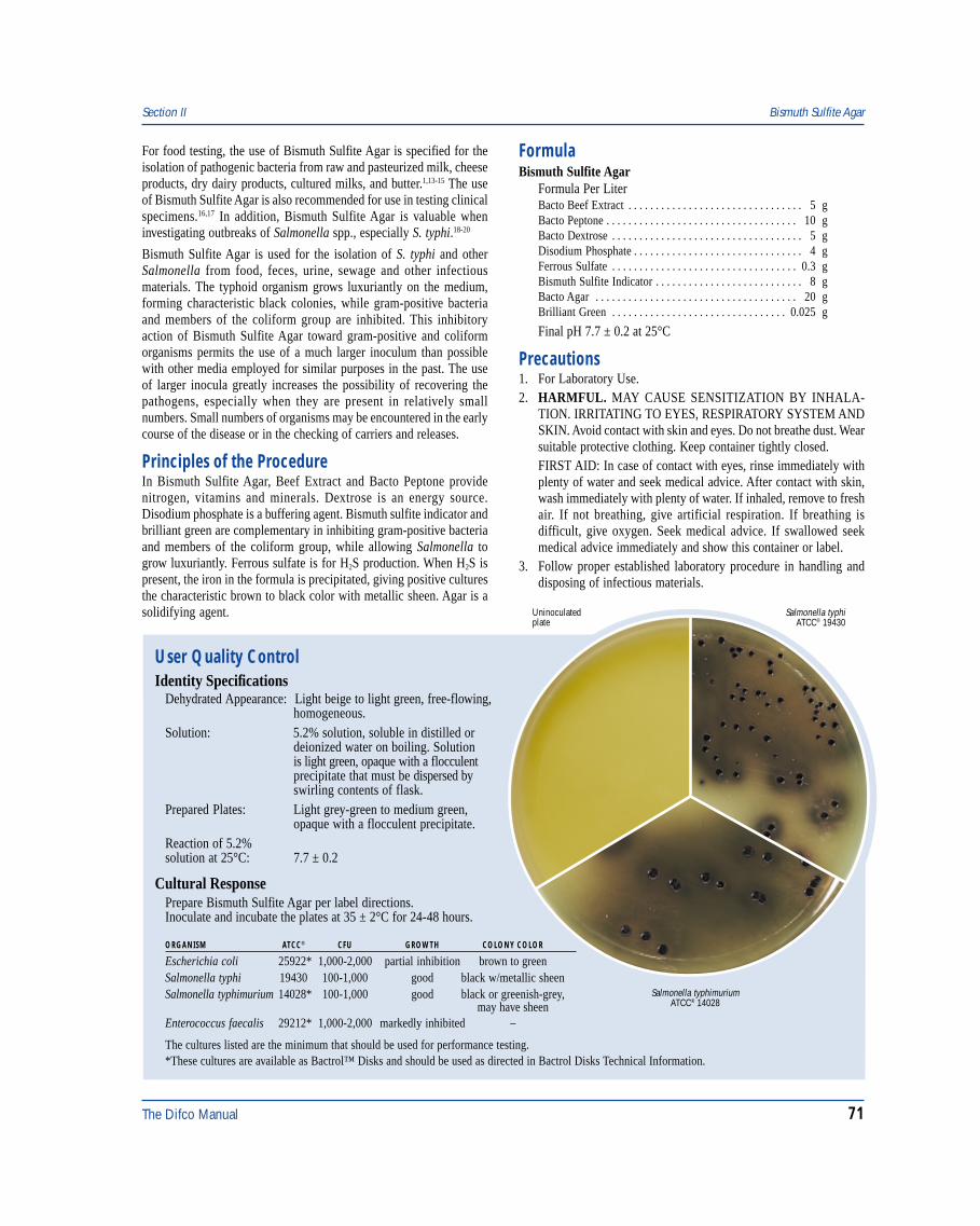
The Difco Manual 71
Section II Bismuth Sulfite Agar
User Quality ControlIdentity Specifications
Dehydrated Appearance: Light beige to light green, free-flowing,homogeneous.
Solution: 5.2% solution, soluble in distilled ordeionized water on boiling. Solutionis light green, opaque with a flocculentprecipitate that must be dispersed byswirling contents of flask.
Prepared Plates: Light grey-green to medium green,opaque with a flocculent precipitate.
Reaction of 5.2%solution at 25°C: 7.7 ± 0.2
Cultural ResponsePrepare Bismuth Sulfite Agar per label directions.Inoculate and incubate the plates at 35 ± 2°C for 24-48 hours.
ORGANISM ATCC® CFU GROWTH COLONY COLOR
Escherichia coli 25922* 1,000-2,000 partial inhibition brown to greenSalmonella typhi 19430 100-1,000 good black w/metallic sheenSalmonella typhimurium 14028* 100-1,000 good black or greenish-grey,
may have sheenEnterococcus faecalis 29212* 1,000-2,000 markedly inhibited –
Salmonella typhiATCC® 19430
Uninoculatedplate
Salmonella typhimuriumATCC® 14028
The cultures listed are the minimum that should be used for performance testing.*These cultures are available as Bactrol™ Disks and should be used as directed in Bactrol Disks Technical Information.
For food testing, the use of Bismuth Sulfite Agar is specified for theisolation of pathogenic bacteria from raw and pasteurized milk, cheeseproducts, dry dairy products, cultured milks, and butter.1,13-15 The useof Bismuth Sulfite Agar is also recommended for use in testing clinicalspecimens.16,17 In addition, Bismuth Sulfite Agar is valuable wheninvestigating outbreaks of Salmonella spp., especially S. typhi.18-20
Bismuth Sulfite Agar is used for the isolation of S. typhi and otherSalmonella from food, feces, urine, sewage and other infectiousmaterials. The typhoid organism grows luxuriantly on the medium,forming characteristic black colonies, while gram-positive bacteriaand members of the coliform group are inhibited. This inhibitoryaction of Bismuth Sulfite Agar toward gram-positive and coliformorganisms permits the use of a much larger inoculum than possiblewith other media employed for similar purposes in the past. The useof larger inocula greatly increases the possibility of recovering thepathogens, especially when they are present in relatively smallnumbers. Small numbers of organisms may be encountered in the earlycourse of the disease or in the checking of carriers and releases.
Principles of the ProcedureIn Bismuth Sulfite Agar, Beef Extract and Bacto Peptone providenitrogen, vitamins and minerals. Dextrose is an energy source.Disodium phosphate is a buffering agent. Bismuth sulfite indicator andbrilliant green are complementary in inhibiting gram-positive bacteriaand members of the coliform group, while allowing Salmonella togrow luxuriantly. Ferrous sulfate is for H2S production. When H2S ispresent, the iron in the formula is precipitated, giving positive culturesthe characteristic brown to black color with metallic sheen. Agar is asolidifying agent.
FormulaBismuth Sulfite Agar
Formula Per LiterBacto Beef Extract . . . . . . . . . . . . . . . . . . . . . . . . . . . . . . . . 5 gBacto Peptone . . . . . . . . . . . . . . . . . . . . . . . . . . . . . . . . . . . 10 gBacto Dextrose . . . . . . . . . . . . . . . . . . . . . . . . . . . . . . . . . . . 5 gDisodium Phosphate . . . . . . . . . . . . . . . . . . . . . . . . . . . . . . . 4 gFerrous Sulfate . . . . . . . . . . . . . . . . . . . . . . . . . . . . . . . . . . 0.3 gBismuth Sulfite Indicator . . . . . . . . . . . . . . . . . . . . . . . . . . . 8 gBacto Agar . . . . . . . . . . . . . . . . . . . . . . . . . . . . . . . . . . . . . 20 gBrilliant Green . . . . . . . . . . . . . . . . . . . . . . . . . . . . . . . . 0.025 g
Final pH 7.7 ± 0.2 at 25°C
Precautions1. For Laboratory Use.2. HARMFUL. MAY CAUSE SENSITIZATION BY INHALA-
TION. IRRITATING TO EYES, RESPIRATORY SYSTEM ANDSKIN. Avoid contact with skin and eyes. Do not breathe dust. Wearsuitable protective clothing. Keep container tightly closed.FIRST AID: In case of contact with eyes, rinse immediately withplenty of water and seek medical advice. After contact with skin,wash immediately with plenty of water. If inhaled, remove to freshair. If not breathing, give artificial respiration. If breathing isdifficult, give oxygen. Seek medical advice. If swallowed seekmedical advice immediately and show this container or label.
3. Follow proper established laboratory procedure in handling anddisposing of infectious materials.
72 The Difco Manual
StorageStore the dehydrated medium below 30°C. The dehydrated mediumis very hygroscopic. Keep container tightly closed. Store preparedplates at 2-8°C.
Expiration DateThe expiration date applies to the product in its intact container whenstored as directed. Do not use a product if it fails to meet specificationsfor identity and performance.
ProcedureMaterials ProvidedBismuth Sulfite Agar
Materials Required But Not ProvidedFlasks with closuresDistilled or deionized waterBunsen burner or magnetic hot plateWaterbath (45-50°C)Petri dishesIncubator (35°C)
Method of Preparation1. Suspend 52 grams in 1 liter distilled or deionized water.2. Heat to boiling no longer than 1-2 minutes to dissolve. Avoid
overheating. DO NOT AUTOCLAVE.3. Cool to 45-50°C in a waterbath.4. Gently swirl flask to evenly disperse the flocculent precipitate.
Dispense into sterile Petri dishes.NOTE: Best results are obtained when the medium is dissolvedand used immediately. The melted medium should not be allowedto solidify in flasks and remelted. Current references suggestthat the prepared plated medium should be aged for one daybefore use.13,21
Specimen Collection and Preparation1. Collect specimens or food samples in sterile containers or with
sterile swabs and transport immediately to the laboratory followingrecommended guidelines.1,13-20
2. Process each specimen, using procedures appropriate for thatspecimen or sample.1,13-20
Test ProcedureFor isolation of Salmonella spp. from food, samples are enriched andselectively enriched. Streak 10 µl of selective enrichment broth ontoBismuth Sulfite Agar. Incubate plates for 24-48 hours at 35°C.Examine plates for the presence of Salmonella spp. Refer to appropriatereferences for the complete procedure when testing food samples.1,13-15
For isolation of Salmonella spp. from clinical specimens, inoculatefecal specimens and rectal swabs onto a small area of one quadrant ofthe Bismuth Sulfite Agar plate and streak for isolation. This willpermit the development of discrete colonies. Incubate plates at 35°C.Examine at 24 hours and again at 48 hours for colonies resemblingSalmonella spp.
For additional information about specimen preparation and inoculationof clinical specimens, consult appropriate references.16-20
ResultsThe typical discrete S. typhi surface colony is black and surrounded bya black or brownish-black zone which may be several times the size ofthe colony. By reflected light, preferably daylight, this zone exhibits adistinctly characteristic metallic sheen. Plates heavily seeded withS. typhi may not show this reaction except near the margin of the massinoculation. In these heavy growth areas, this organism frequentlyappears as small light green colonies. This fact emphasizes theimportance of inoculating plates so that some areas are sparsely populatedwith discrete S. typhi colonies. Other strains of Salmonella produce blackto green colonies with little or no darkening of the surrounding medium.
Generally, Shigella spp. other than S. flexneri and S. sonnei areinhibited. Shigella flexneri and Shigella sonnei strains that do grow onthis medium produce brown to green, raised colonies with depressedcenters and exhibit a crater-like appearance.
E. coli is partially inhibited. Occasionally a strain will be encounteredthat will grow as small brown or greenish glistening colonies. Thiscolor is confined entirely to the colony itself and shows no metallicsheen. A few strains of Enterobacter aerogenes may develop on thismedium, forming raised, mucoid colonies. Enterobacter coloniesmay exhibit a silvery sheen, appreciably lighter in color than thatproduced by S. typhi. Some members of the coliform group thatproduce hydrogen sulfide may grow on the medium, giving coloniessimilar in appearance to S. typhi. These coliforms may be readilydifferentiated because they produce gas from lactose in differentialmedia, for example, Kligler Iron Agar or Triple Sugar Iron Agar. Thehydrolysis of urea, demonstrated in Urea Broth or on Urea Agar Base,may be used to identify Proteus sp.
To isolate S. typhi for agglutination or fermentation studies, pickcharacteristic black colonies from Bismuth Sulfite Agar and subculturethem on MacConkey Agar. The purified colonies from MacConkeyAgar may then be picked to differential tube media such asKligler Iron Agar, Triple Sugar Iron Agar or other satisfactorydifferential media for partial identification. All cultures that givereactions consistent with Salmonella spp. on these media should beconfirmed biochemically as Salmonella spp. before any serologicaltesting is performed. Agglutination tests may be performed from thefresh growth on the differential tube media or from the growth onnutrient agar slants inoculated from the differential media. The growthon the differential tube media may also be used for inoculatingcarbohydrate media for fermentation studies.
Limitations of the Procedure1. It is important to streak for well isolated colonies. In heavy growth
areas, S. typhi appears light green and may be misinterpreted asnegative growth for S. typhi.22
2. S. typhi and S. arizonae are the only enteric organisms to exhibittypical brown zones on the medium. Brown zones are not producedby other members of the Enterobacteriaceae. However, S. arizonaeis usually inhibited.22
3. Colonies on Bismuth Sulfite Agar may be contaminated withother viable organisms; therefore, isolated colonies should besubcultured to a less selective medium (e.g., MacConkey Agar).22
4. Typical S. typhi colonies usually develop within 24 hours;however, all plates should be incubated for a total of 48 hours toallow growth of all typhoid strains.22
Bismuth Sulfite Agar Section II
The Difco Manual 73
5. DO NOT AUTOCLAVE. Heating this medium for a period longerthan necessary to just dissolve the ingredients destroys its selectivity.
References1. Flowers, R. S., W. Andrews, C. W. Donnelly, and E. Koenig.
1993. Pathogens in milk and milk products, p. 103-212. InMarshall, R. T. (ed.), Standard methods for the examination ofdairy products, 16th ed. American Public Health Association,Washington, D.C.
2. Wilson, W. J., and E. M. Blair. 1926. A combination of bismuthand sodium sulphite affording an enrichment and selectivemedium for the typhoid-paratyphoid groups of bacteria. J. Pathol.Bacteriol. 29:310.
3. Wilson, W. J., and E. M. Blair. 1927. Use of a glucose bismuthsulphite iron medium for the isolation of B. typhosus andB. proteus. J. Hyg. 26:374-391.
4. Wilson, W. J., and E. M. Blair. 1931. Further experience of thebismuth sulphite media in the isolation of Bacillus typhosus andBacillus paratyphosus from faeces, sewage and water. J. Hyg.31:138-161.
5. Wilson, W. J. 1923. Reduction of sulphites by certain bacteria inmedia containing a fermentable carbohydrate and metallic salts.J. Hyg. 21:392.
6. Wilson, W. J. 1928. Isolation of B. typhosus from sewage andshellfish. Brit. Med. J. 1:1061.
7. Cope, E., and J. Kasper. 1937. A comparative study of methodsfor the isolation of typhoid bacilli from the stool of suspectedcarriers. Proceedings of local branches of the Society of AmericanBacteriologists. J. Bacteriol. 34:565.
8. Cope, E. J., and J. A. Kasper. 1938. Cultural methods for thedetection of typhoid carriers. Am. J. Public Health 28:1065-1068.
9. Gunther, M. S., and L. Tuft. 1939. A comparative study of mediaemployed in the isolation of typhoid bacilli from feces and urine.J. Lab. Clin. Med. 24:461-471.
10. Green, C. E., and P. J. Beard. 1938. Survival of E. typhi in sewagetreatment plant processes. Am. J. Public Health 28:762-770.
11. Hajna, A. A., and C. A. Perry. 1938. A comparative study ofselective media for the isolation of typhoid bacilli from stoolspecimens. J. Lab. Clin. Med. 23:1185-1193.
12. United States Pharmacopeial Convention. 1995. The UnitedStates pharmacopeia, 23rd ed. The United States PharmacopeialConvention, Rockville, MD.
13. Andrews, W. H., G. A. June, P. S. Sherrod, T. S. Hammack, andR. M. Amaguana. 1995. Salmonella, p. 5.01-5.20. In FDAbacteriological analytical manual, 8th ed. AOAC International,Gaithersburg, MD.
14. Flowers, R. S., J. D’Aoust, W. H. Andrews, and J. S. Bailey.1992. Salmonella, p. 371-422. In Vanderzant, C. and D. F.Splittstoesser (ed.), Compendium of methods for the microbiologicalexamination of foods, 3rd ed. American Public Health Association,Washington, D.C.
15. Andrews, W. H. (ed). 1995. Microbiological Methods, p. 17.1-17.119.In Cunniff, P. (ed.), Official methods of analysis of AOACInternational, 16th ed. AOAC International, Arlington, VA.
16. Washington, J. A. 1981. Initial processing for culture ofspecimens, p. 91-126. Laboratory procedures in clinical microbi-ology, p. 749. Springer-Verlag New York Inc. New York, NY.
17. Baron, E. J., L. R. Peterson, and S. M. Finegold. 1994.Microorganisms encountered in the gastrointestinal tract,p. 234-248. Bailey & Scott’s diagnostic microbiology, 9th ed.Mosby-Year Book, Inc. St. Louis, MO.
18. Gray, L. D. 1995. Escherichia, Salmonella, Shigella and Yersinia,p. 450-456. In Murray, P. R., E. J. Baron, M. A. Pfaller, F. C.Tenover, and R. H. Yolken (ed.), Manual of clinical microbiology,6th ed. American Society for Microbiology, Washington, D.C.
19. Citron, F. 1992. Initial processing, inoculation, and incubation ofaerobic bacteriology specimens, p. 1.4.1-1.4.19. In Isenberg, H. D.(ed.), Clinical microbiology procedures handbook, vol. 1. AmericanSociety for Microbiology, Washington, D.C.
20. Grasnick, A. 1992. Processing and interpretation of bacterialfecal cultures, p. 1.10.1-1.10.25. In Isenberg, H. D. (ed.), Clinicalmicrobiology procedures handbook, vol. 1. American Society forMicrobiology, Washington, D.C.
21. D’Aoust, J. Y. 1977. Effect of storage conditions on theperformance of bismuth sulfite agar. J. Clin. Microbiol. 5:122-124.
22. MacFaddin, J. F. 1985. Media for isolation-cultivation-identification-maintenance of medical bacteria, Vol. 1. Williams& Wilkins, Baltimore, MD.
PackagingBismuth Sulfite Agar 100 g 0073-15
500 g 0073-1710 kg 0073-08
Section II Blood Agar Base & Blood Agar Base No. 2
Bacto® Blood Agar BaseBacto Blood Agar Base No. 2Intended UseBacto Blood Agar Base is used for isolating and cultivating a widevariety of microorganisms and, with added blood, for cultivatingfastidious microorganisms.
Bacto Blood Agar Base No. 2 is used for isolating and cultivatingfastidious microorganisms with or without added blood.
Also Known AsBlood Agar Base is abbreviated as BAB, and may be referred to asInfusion Agar.
Summary and ExplanationBlood agar bases are typically supplemented with 5-10% sheep,rabbit or horse blood for use in isolating, cultivating and determininghemolytic reactions of fastidious pathogenic microorganisms. Withoutenrichment, blood agar bases can be used as general purpose media.
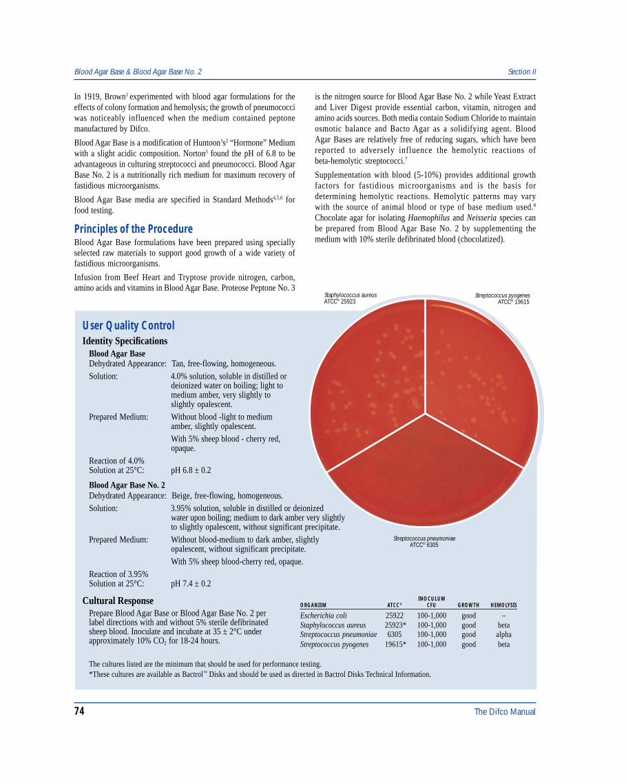
74 The Difco Manual
In 1919, Brown1 experimented with blood agar formulations for theeffects of colony formation and hemolysis; the growth of pneumococciwas noticeably influenced when the medium contained peptonemanufactured by Difco.
Blood Agar Base is a modification of Huntoon’s2 “Hormone” Mediumwith a slight acidic composition. Norton3 found the pH of 6.8 to beadvantageous in culturing streptococci and pneumococci. Blood AgarBase No. 2 is a nutritionally rich medium for maximum recovery offastidious microorganisms.
Blood Agar Base media are specified in Standard Methods4,5,6 forfood testing.
Principles of the ProcedureBlood Agar Base formulations have been prepared using speciallyselected raw materials to support good growth of a wide variety offastidious microorganisms.
Infusion from Beef Heart and Tryptose provide nitrogen, carbon,amino acids and vitamins in Blood Agar Base. Proteose Peptone No. 3
is the nitrogen source for Blood Agar Base No. 2 while Yeast Extractand Liver Digest provide essential carbon, vitamin, nitrogen andamino acids sources. Both media contain Sodium Chloride to maintainosmotic balance and Bacto Agar as a solidifying agent. BloodAgar Bases are relatively free of reducing sugars, which have beenreported to adversely influence the hemolytic reactions ofbeta-hemolytic streptococci.7
Supplementation with blood (5-10%) provides additional growthfactors for fastidious microorganisms and is the basis fordetermining hemolytic reactions. Hemolytic patterns may varywith the source of animal blood or type of base medium used.8
Chocolate agar for isolating Haemophilus and Neisseria species canbe prepared from Blood Agar Base No. 2 by supplementing themedium with 10% sterile defibrinated blood (chocolatized).
User Quality ControlIdentity Specifications
Blood Agar BaseDehydrated Appearance: Tan, free-flowing, homogeneous.Solution: 4.0% solution, soluble in distilled or
deionized water on boiling; light tomedium amber, very slightly toslightly opalescent.
Prepared Medium: Without blood -light to mediumamber, slightly opalescent.With 5% sheep blood - cherry red,opaque.
Reaction of 4.0%Solution at 25°C: pH 6.8 ± 0.2
Blood Agar Base No. 2Dehydrated Appearance: Beige, free-flowing, homogeneous.Solution: 3.95% solution, soluble in distilled or deionized
water upon boiling; medium to dark amber very slightlyto slightly opalescent, without significant precipitate.
Prepared Medium: Without blood-medium to dark amber, slightlyopalescent, without significant precipitate.With 5% sheep blood-cherry red, opaque.
Reaction of 3.95%Solution at 25°C: pH 7.4 ± 0.2
Cultural ResponsePrepare Blood Agar Base or Blood Agar Base No. 2 perlabel directions with and without 5% sterile defibrinatedsheep blood. Inoculate and incubate at 35 ± 2°C underapproximately 10% CO2 for 18-24 hours.
Streptococcus pyogenesATCC® 19615
Staphylococcus aureusATCC® 25923
INOCULUMORGANISM ATCC® CFU GROWTH HEMOLYSIS
Escherichia coli 25922 100-1,000 good –Staphylococcus aureus 25923* 100-1,000 good betaStreptococcus pneumoniae 6305 100-1,000 good alphaStreptococcus pyogenes 19615* 100-1,000 good beta
Streptococcus pneumoniaeATCC® 6305
Blood Agar Base & Blood Agar Base No. 2 Section II
The cultures listed are the minimum that should be used for performance testing.*These cultures are available as Bactrol™ Disks and should be used as directed in Bactrol Disks Technical Information.
The Difco Manual 75
FormulaBlood Agar Base
Formula Per LiterBeef Heart, Infusion from . . . . . . . . . . . . . . . . . . . . . . . . 500 gBacto Tryptose . . . . . . . . . . . . . . . . . . . . . . . . . . . . . . . . . . 10 gSodium Chloride . . . . . . . . . . . . . . . . . . . . . . . . . . . . . . . . . . 5 gBacto Agar . . . . . . . . . . . . . . . . . . . . . . . . . . . . . . . . . . . . . 15 g
Final pH 6.8 ± 0.2 at 25°C
Blood Agar Base No. 2Formula Per LiterBacto Proteose Peptone No. 3 . . . . . . . . . . . . . . . . . . . . . . 15 gLiver Digest . . . . . . . . . . . . . . . . . . . . . . . . . . . . . . . . . . . . . 2.5 gBacto Yeast Extract . . . . . . . . . . . . . . . . . . . . . . . . . . . . . . . . 5 gSodium Chloride . . . . . . . . . . . . . . . . . . . . . . . . . . . . . . . . . . 5 gBacto Agar . . . . . . . . . . . . . . . . . . . . . . . . . . . . . . . . . . . . . 12 g
Final pH 7.4 ± 0.2 at 25°C
Precautions1. For Laboratory Use.2. Follow proper established laboratory procedures in handling and
disposing of infectious materials.
StorageStore the dehydrated medium below 30°C. The dehydrated medium isvery hygroscopic. Keep container tightly closed.
Expiration DateThe expiration date applies to the product in its intact container whenstored as directed. Do not use a product if it fails to meet specificationsfor identity and performance.
ProcedureMaterials ProvidedBlood Agar BaseBlood Agar Base No. 2
Materials Required But Not ProvidedGlasswareAutoclaveIncubator (35°C)Waterbath (45-50°C) (optional)Sterile defibrinated bloodSterile Petri dishes
Method of Preparation1. Suspend the medium in 1 liter distilled or deionized water:
Blood Agar Base - 40 grams;Blood Agar Base No. 2 - 39.5 grams.
2. Heat to boiling to dissolve completely.3. Autoclave at 121°C for 15 minutes. Cool to 45-50°C.4. To prepare blood agar, aseptically add 5% sterile defibrinated
blood to the medium at 45-50°C. Mix well.5. To prepare chocolate agar, add 10% sterile defibrinated blood
to Blood Agar Base No. 2 at 80°C. Mix well.6. Dispense into sterile Petri dishes.
Specimen Collection and PreparationCollect specimens in sterile containers or with sterile swabs and transportimmediately to the laboratory in accordance with recommendedguidelines outlined in the references.
Test Procedure1. Process each specimen as appropriate, and inoculate directly
onto the surface of the medium. Streak for isolation with aninoculating loop, then stab the agar several times to depositbeta-hemolytic streptococci beneath the agar surface. Subsurfacegrowth will display the most reliable hemolytic reactions owing tothe activity of both oxygen-stable and oxygen-labile streptolysins.8
2. Incubate plates aerobically, anaerobically or under conditions ofincreased CO2 (5-10%) in accordance with established laboratoryprocedures.
ResultsExamine the medium for growth and hemolytic reactions after 18-24and 48 hours incubation. Four types of hemolysis on blood agar mediacan be described:9
a. Alpha hemolysis (α ) is the reduction of hemoglobin tomethemoglobin in the medium surrounding the colony. This causesa greenish discoloration of the medium.
b. Beta hemolysis (β) is the lysis of red blood cells, producing aclear zone surrounding the colony.
c. Gamma hemolysis (γ) indicates no hemolysis. No destruction ofred blood cells occurs and there is no change in the medium.
d. Alpha-prime hemolysis (α) is a small zone of complete hemolysisthat is surrounded by an area of partial lysis.
Limitations of the Procedure1. Blood Agar Base media are intended for use with blood
supplementation. Although certain diagnostic tests may beperformed directly on this medium, biochemical and, if indicated,immunological testing using pure cultures are recommendedfor complete identification. Consult appropriate references forfurther information.
2. Since the nutritional requirements of organisms vary, somestrains may be encountered that fail to grow or grow poorly onthis medium.
3. Hemolytic reactions of some strains of group D streptococcihave been shown to be affected by differences in animal blood.Such strains are beta-hemolytic on horse, human and rabbitblood agar and alpha-hemolytic on sheep blood agar.8
4. Colonies of Haemophilus haemolyticus are beta-hemolytic onhorse and rabbit blood agar and must be distinguished fromcolonies of beta-hemolytic streptococci using other criteria. Theuse of sheep blood has been suggested to obviate this problem sincesheep blood is deficient in pyridine nucleotides and does notsupport growth of H. haemolyticus.10
5. Atmosphere of incubation has been shown to influence hemolyticreactions of beta-hemolytic streptococci.8 For optimal performance,incubate blood agar base media under increased CO2 or anaerobicconditions.
Section II Blood Agar Base & Blood Agar Base No. 2
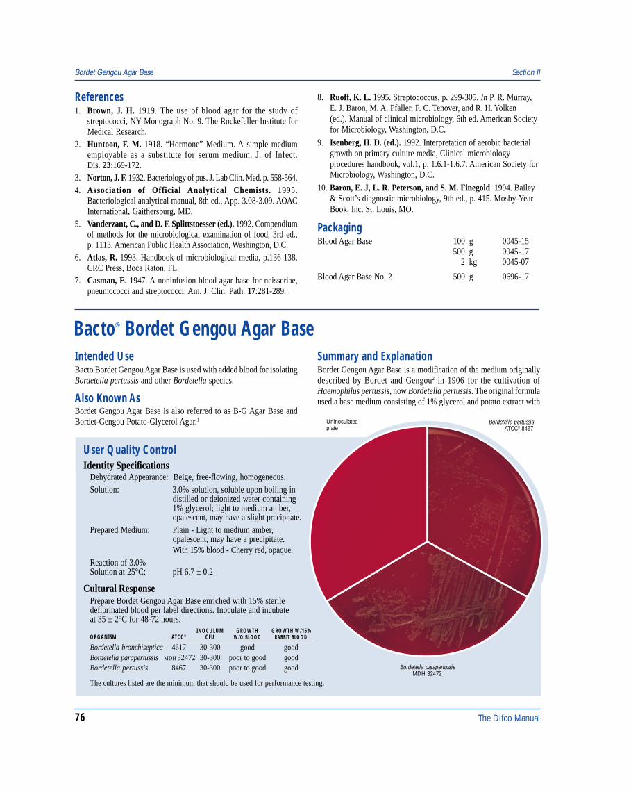
76 The Difco Manual
Bordetella pertussisATCC® 8467
Uninoculatedplate
Bordetella parapertussisMDH 32472
References1. Brown, J. H. 1919. The use of blood agar for the study of
streptococci, NY Monograph No. 9. The Rockefeller Institute forMedical Research.
2. Huntoon, F. M. 1918. “Hormone” Medium. A simple mediumemployable as a substitute for serum medium. J. of Infect.Dis. 23:169-172.
3. Norton, J. F. 1932. Bacteriology of pus. J. Lab Clin. Med. p. 558-564.4. Association of Official Analytical Chemists. 1995.
Bacteriological analytical manual, 8th ed., App. 3.08-3.09. AOACInternational, Gaithersburg, MD.
5. Vanderzant, C., and D. F. Splittstoesser (ed.). 1992. Compendiumof methods for the microbiological examination of food, 3rd ed.,p. 1113. American Public Health Association, Washington, D.C.
6. Atlas, R. 1993. Handbook of microbiological media, p.136-138.CRC Press, Boca Raton, FL.
7. Casman, E. 1947. A noninfusion blood agar base for neisseriae,pneumococci and streptococci. Am. J. Clin. Path. 17:281-289.
8. Ruoff, K. L. 1995. Streptococcus, p. 299-305. In P. R. Murray,E. J. Baron, M. A. Pfaller, F. C. Tenover, and R. H. Yolken(ed.). Manual of clinical microbiology, 6th ed. American Societyfor Microbiology, Washington, D.C.
9. Isenberg, H. D. (ed.). 1992. Interpretation of aerobic bacterialgrowth on primary culture media, Clinical microbiologyprocedures handbook, vol.1, p. 1.6.1-1.6.7. American Society forMicrobiology, Washington, D.C.
10. Baron, E. J, L. R. Peterson, and S. M. Finegold. 1994. Bailey& Scott’s diagnostic microbiology, 9th ed., p. 415. Mosby-YearBook, Inc. St. Louis, MO.
PackagingBlood Agar Base 100 g 0045-15
500 g 0045-172 kg 0045-07
Blood Agar Base No. 2 500 g 0696-17
Bordet Gengou Agar Base Section II
Bacto® Bordet Gengou Agar Base
User Quality ControlIdentity Specifications
Dehydrated Appearance: Beige, free-flowing, homogeneous.Solution: 3.0% solution, soluble upon boiling in
distilled or deionized water containing1% glycerol; light to medium amber,opalescent, may have a slight precipitate.
Prepared Medium: Plain - Light to medium amber,opalescent, may have a precipitate.With 15% blood - Cherry red, opaque.
Reaction of 3.0%Solution at 25°C: pH 6.7 ± 0.2
Cultural ResponsePrepare Bordet Gengou Agar Base enriched with 15% steriledefibrinated blood per label directions. Inoculate and incubateat 35 ± 2°C for 48-72 hours.
INOCULUM GROWTH GROWTH W/15%ORGANISM ATCC® CFU W/O BLOOD RABBIT BLOOD
Bordetella bronchiseptica 4617 30-300 good goodBordetella parapertussis MDH 32472 30-300 poor to good goodBordetella pertussis 8467 30-300 poor to good good
The cultures listed are the minimum that should be used for performance testing.
Intended UseBacto Bordet Gengou Agar Base is used with added blood for isolatingBordetella pertussis and other Bordetella species.
Also Known AsBordet Gengou Agar Base is also referred to as B-G Agar Base andBordet-Gengou Potato-Glycerol Agar.1
Summary and ExplanationBordet Gengou Agar Base is a modification of the medium originallydescribed by Bordet and Gengou2 in 1906 for the cultivation ofHaemophilus pertussis, now Bordetella pertussis. The original formulaused a base medium consisting of 1% glycerol and potato extract with
The Difco Manual 77
Section II Bordet Gengou Agar Base
an equal volume of human or rabbit blood. The modified medium isprepared according to the formula recommended by the AmericanPublic Health Association.3 Eldering and Kendrick4 reported that theaddition of 1% proteose peptone or neopeptone increased growth ofB. pertussis, thereby increasing the yield of vaccine.
The genus Bordetella consists of four species: Bordetella pertussis,B. parapertussis, B. bronchiseptica and B. avium.5 All Bordetella arerespiratory pathogens, residing on the mucous membranes of therespiratory tract. B. pertussis and B. parapertussis are uniquely humanpathogens. B. pertussis is the major cause of whooping cough orpertussis. B. parapertussis is associated with a milder form of thedisease.6 B. bronchiseptica is an opportunistic human pathogenassociated with both respiratory and non-respiratory infections, oftenoccurring in patients having close contact with animals.5
B. bronchiseptica has not been reported to cause pertussis. There havebeen no reports of recovery of B. avium from humans.5
The “cough plate” method for the diagnosis of whooping cough wasoriginally reported by Chievitz and Meyer.7 This technique is no longerrecommended. Nasopharyngeal washings or a nasopharyngeal swab(calcium alginate on a wire handle) should be collected within the firstweek of paroxysmal coughing.8
Principles of the ProcedureInfusion from Potato provides nitrogen, vitamins and amino acids.Glycerol is a carbon source. Sodium Chloride maintains the osmoticbalance of the medium. Bacto Agar is a solidifying agent. The additionof blood provides essential growth requirements for Bordetella species.
Many factors will inhibit growth of B. pertussis, including fatty acidspresent in nasal secretions or cotton from the collection swab. Starch,present from the Potato Infusion, absorbs fatty acids.
Modified Bordet Gengou medium, enriched with 15-20% blood, yieldstypical B. pertussis growth. The colonies appear small, white, opaqueand surrounded by a characteristic zone of hemolysis that is not sharplydefined but merges diffusely into the medium. The zone of hemolysisis usually absent if 30% or more blood is added to the medium andcannot be seen on charcoal-containing media.9 Sterile, defibrinatedsheep or rabbit blood can be used in preparing the medium.
FormulaBordet Gengou Agar Base
Formula Per LiterPotato, Infusion from . . . . . . . . . . . . . . . . . . . . . . . . . . . . 125 gSodium Chloride . . . . . . . . . . . . . . . . . . . . . . . . . . . . . . . . . 5.5 gBacto Agar . . . . . . . . . . . . . . . . . . . . . . . . . . . . . . . . . . . . . 20 g
Final pH 6.7 ± 0.2 at 25°C
Precautions1. For Laboratory Use.2. Follow proper established laboratory procedures in handling and
disposing of infectious materials.
StorageStore the dehydrated medium below 30°C. The dehydrated medium isvery hygroscopic. Keep container tightly closed.
Expiration DateThe expiration date applies to the product in its intact container whenstored as directed. Do not use a product if it fails to meet specificationsfor identity and performance.
ProcedureMaterials ProvidedBordet Gengou Agar Base
Materials Required But Not ProvidedGlasswareAutoclaveIncubator (35°C)Waterbath (45-50°C)Sterile defibrinated bloodSterile Petri dishes
Method of Preparation1. Suspend 30 grams in 1 liter distilled or deionized water containing
10 grams of glycerol.2. Heat to boiling to dissolve completely.3. Autoclave at 121°C for 15 minutes.4. Cool to 45-50°C. Aseptically add 15% sterile defibrinated sheep
or rabbit blood. Mix well.5. Dispense into sterile Petri dishes.
Specimen Collection and Preparation9
Specimens should be obtained during the early phases of the diseaseand prior to the convalescent stage and antimicrobial therapy. Thespecimen of choice is duplicate nasopharyngeal swabs. Direct platingof the specimen at bedside is recommended; when this is not possible,submerge both swabs into Regan-Lowe transport medium.
Test Procedure9
1. Roll one of the swabs over the primary inoculation area of theBordet Gengou plate and streak for isolation. Return the swab tothe transport medium. Incubate the transport medium for 48 hours.Plate the swabs onto a duplicate set of media.
2. Incubate the culture plates at 35°C for 5-7 days in a moist chamber.Increased CO2 is not recommended. Growth of B. pertussisappears in 3-5 days. Other bordetellae species can appear in 1-3 days.
3. Nasopharyngeal specimens may contain staphylococci that producea diffusible substance inhibitory to B. pertussis growth. For thesespecimens, use a plating medium with methicillin (2.5 µg/ml) orcephalexin (40 µg/ml) and a medium without antimicrobics.
4. Isolates suspected of being B. pertussis should be confirmed byusing a specific antiserum in either the slide agglutination orfluorescent antibody staining techniques.10
ResultsFor a complete discussion on the isolation and identification ofBordetella species refer to the appropriate procedures outlined inthe references.
Limitations of the Procedure1. Since the nutritional requirements of organisms vary, some strains may
be encountered that fail to grow or grow poorly on this medium.
78 The Difco Manual
2. Some Haemophilus species will grow on Bordetella isolationmedia and may cross-react with B. pertussis antisera. It may beprudent to rule out X and V factor dependence.
References1. MacFaddin, J. F. 1985. Media for isolation-cultivation-
identification- maintenance of medical bacteria, vol. 1, p. 86-92.Williams & Wilkins, Baltimore, MD.
2. Bordet, J., and D. Gengou. 1906. Le microbe de la coqueluche.Ann. Inst. Pasteur 20:731.
3. Kendrick, P. L., E. Eldering, and W. L. Bradford. 1970.Whooping cough, p.106-117. In H. L. Bodily, E. L. Updyke, andJ. O. Mason (ed.), Diagnostic procedures for bacterial, mycotic andparasitic infections, 5th ed. American Public Health Association,New York, NY.
4. Eldering, E., and P. L. Kendrick. 1936. Some practicalconsiderations in B. pertussis vaccine preparation. Am. J. PublicHealth 24:309.
5. Marcon, M . J. 1995. Bordetella, p. 566-573. In P. R. Murray, E. J.Baron, M. A. Pfaller, F. C. Tenover, and R. H. Yolken (ed.), Manualof clinical microbiology, 6th ed. American Society for Microbiology,Washington, D.C.
6. Linneman, C. C., and E. B. Pery. 1977. Bordetella parapertussis:recent experience and a review of the literature. Am. J. Dis. Child.131:560-563.
7. Chievitz, J., and A. H. Meyer. 1916. Recherches sur lacoqueluche. Ann. Inst. Pasteur 30:503.0
8. Baron, E. J., L. R. Peterson, and S. M. Finegold. 1994.Bailey & Scott’s diagnostic microbiology, 9th ed. Mosby-YearBook, Inc., St. Louis, MO.
9. Isenberg, H. D. (ed.). 1992. Clinical microbiology procedures hand-book, vol. 1. American Society for Microbiology, Washington, D.C.
10. Koneman, E. W. 1988. Color atlas and textbook of diagnosticmicrobiology, 3rd ed. J. B. Lippincott Company, Washington, D.C.
PackagingBordet Gengou Agar Base 100 g 0048-15
500 g 0048-17
Bovine Albumin 5% Section II
Bacto® Bovine Albumin 5%
User Quality ControlIdentity Specifications
Bovine Albumin 5%Appearance: Light amber, clear to very slightly
opalescent.Sterility Test: Negative.Reaction ofSolution at 25°C: pH 7.0 ± 0.2
Cultural ResponsePrepare Dubos Broth Base per label directions, substitutingBovine Albumin 5% for Dubos Medium Albumin. Inoculateand incubate at 35 ± 2°C under CO2 for up to three weeks.
INOCULUMORGANISM ATCC® CFU GROWTH
Mycobacterium 13950 100-1,000 goodintracellulareMycobacterium 25177 100-1,000 goodtuberculosis H37RaMycobacterium 27294 100-1,000 goodtuberculosis H37Ra
The cultures listed are the minimum that should be used forperformance testing.
Summary and ExplanationDavis and Dubos2 recommended the use of bovine albumin at a finalconcentration of 0.5% in liquid media for culturing Mycobacteriumtuberculosis. In this study, bovine albumin neutralized the toxicity offatty acids and permitted more luxuriant growth of M. tuberculosis.
Ellinghausen and McCullough3 used bovine albumin fraction V at a finalconcentration of 1% in liquid, semisolid and solid media for culturingleptospires. Morton et al.4 demonstrated that 1% bovine albuminstimulated growth of Mycoplasma (PPLO).
Bovine Albumin can be added to normally sterile specimens, tissuesand body fluids for direct inoculation onto culture media used forisolating mycobacteria. BSA is also used as an enrichment whencontaminated specimens are digested.
Bovine Albumin 5%, modified with added sodium chloride anddextrose, is available as Dubos Medium Albumin.
Principles of the ProcedureBovine Albumin 5% is a filter sterilized solution of Bovine AlbuminFraction V. BSA is suggested as a culture media enrichment because itsbuffering capacity and detoxifying effect on specimen sediment.1 BovineAlbumin 5% also increases adhesion of the specimen to solid media.1
Precautions1. For Laboratory Use.2. Follow proper established laboratory procedure in handling and
disposing of infectious materials.3. Mycobacterial organisms are BioSafety Level 2 pathogens. The
handling of clinical specimen material that is potentially infectedwith mycobacteria should be performed in a Class I or II biologicalsafety cabinet (BSC).1
StorageStore Bovine Albumin 5% at 2-8°C.
Intended UseBacto Bovine Albumin 5% is used to enrich media for cultivating alarge variety of microorganisms and tissue cells.
Also Known AsBovine Albumin can be abbreviated as BSA.1
The Difco Manual 79
Section II Brain Heart Infusion Media
Expiration DateThe expiration date applies to the product in its intact container whenstored as directed. Do not use a product if it fails to meet specificationsfor identity and performance.
ProcedureMaterials ProvidedBovine Albumin 5%
Materials Required But Not ProvidedMaterials vary depending on the specimen collected and the procedureperformed.
Method of PreparationRefer to the final concentration of Bovine Albumin in the procedurebeing used to inoculate specimens. A 0.2% solution of Bovine Albumin5% is recommended for the inoculation of sterile and contaminatedspecimens when isolating mycobacteria.1
Specimen Collection and PreparationMany different specimen types can be collected for mycobacterialcultures but the majority will be from the respiratory tract.1 Tissues,body fluids, urine, blood and gastric aspirates can also be tested for thepresence of mycobacteria. Refer to the procedures established bylaboratory policy or to appropriate references for specific guidelineson specimen collection and processing.
Test ProcedureSterile Specimens for the Isolation of Mycobacteria1
Normally sterile tissues may be ground in 0.2% BSA and inoculateddirectly in culture media. Concentrate body fluids before inoculationbecause they normally contain only a small number of mycobacteria.Centrifuge fluids at 3,000 x g and inoculate the sediment onto liquidor solid media. For a complete discussion of the inoculation of sterilespecimens, refer to appropriate references.
Contaminated Specimens for the Isolation of Mycobacteria1
A concentration of 0.2% Bovine Albumin fraction V can be added tospecimen sediment that has been digested and centrifuged by theNALC-NaOH digestion method. Using a separate sterile pipette foreach tube, add 1-2 ml of 0.2% BSA, then resuspend the sediment withthe pipette or by shaking the tube gently by hand.
Several digestion procedures exist. Consult appropriate references fora complete discussion on all digestion and decontamination methodsand other testing procedures.
ResultsAll media should be examined closely for evidence of growth. Refer tothe procedure established by laboratory policy or to appropriatereferences on typical growth patterns and confirmation tests.
Limitations of the Procedure1. Bovine Albumin 5% is not recommended for use with Bactec®
because BSA may delay detection times.1
References1. Nolte, F. S., and B. Metchock. 1995. Mycobacterium, p. 400-437.
In P. R. Murray, E. J. Baron, M. A. Pfaller, F. C. Tenover, andR. H. Yolken (ed.). 1995. Manual of clinical microbiology, 6th ed.American Society for Microbiology, Washington, D.C.
2. Davis and Dubos. 1945. J. Bacteriol. 55:11.3. Ellinghausen and McCullough. 1962. Bacteriol. Proc. 62:54.4. Morton, H. E., P. E. Smith, N. B. Williams, and C. F.
Eickenberg. 1951. Isolation of pleuropneumonia-like organismsfrom human saliva: A newly detected member of the oral flora.J. Dent. Res. 30:415-422.
PackagingBovine Albumin 5% 12 x 20 ml 0668-64
Brain Heart Infusion MediaBacto® Brain Heart Infusion . Bacto Brain Heart Infusion AgarBacto Clostridium Difficile Antimicrobic Supplement CCBacto Brain Heart CC Agar . Bacto Brain Heart Infusion w/PABand Agar . Bacto Brain Heart Infusion w/o DextroseIntended UseBacto Brain Heart Infusion is used for cultivating fastidious microor-ganisms, including streptococci, pneumococci and meningococci.Bacto Brain Heart Infusion Agar is used for cultivating fastidiousmicroorganisms, especially fungi and yeasts, and, with addedantibiotics, for isolating fungi.Bacto Clostridium Difficile Antimicrobic Supplement CC is used with
Brain Heart Infusion Agar in preparing Clostridium Difficile Agar.Bacto Brain Heart CC Agar is used for isolating and cultivatingfastidious fungi.Bacto Brain Heart Infusion w/PAB and Agar is used for cultivatingfastidious organisms, particularly from blood containing sulfonamides.Bacto Brain Heart Infusion w/o Dextrose is used for cultivatingfastidious organisms.
80 The Difco Manual
Clostridium Difficile Agar. The complete medium is based on theformula of Willey and Bartlett5 and recommended for use in the isolationof Clostridium difficile from fecal specimens. C. difficile is the majorcause of antibiotic-associated diarrhea and pseudomembranous colitis.6
Brain Heart CC Agar is prepared with chloramphenicol and cyclo-heximide (Actidione) according to the formulation of Ajello et al.7 andMcDonough et al.8 These selective agents restrict growth of bacteriaand saprophytic fungi. Brain Heart CC Agar is used in the isolation offungi that cause systemic disease, such as Histoplasma capsulatumand Blastomyces dermatiditis.
Brain Heart Infusion media are specified in several standard methodsreferences for food testing.9,10,11 Standard Methods for the Examinationof Water and Wastewater recommends Brain Heart Infusion media intests for the verification of fecal streptococci.12
Brain Heart Infusion is recommended by the National Committee forClinical Laboratory Standards (NCCLS) for the preparation of inoculaused in antimicrobial susceptibility tests.13
Brain Heart Infusion w/o Dextrose is a basal medium used with addedcarbohydrates for fermentation studies.
Modifications of BHI media include:14
• Brain Heart Infusion Agar with penicillin (20,000 U) andstreptomycin (40 mg) for the selective isolation of pathogenic
Brain Heart Infusion Media Section II
User Quality ControlIdentity Specifications
Brain Heart InfusionDehydrated Appearance: Light tan, free-flowing, homogeneous.Solution: 3.7% solution, soluble in distilled or
deionized water; light to medium amber,clear without significant precipitate.
Prepared Medium: Light to medium amber, clear withoutsignificant precipitate.
Reaction of 3.7%Solution at 25°C pH 7.4 ± 0.2
Brain Heart Infusion AgarDehydrated Appearance: Beige, free-flowing, homogeneous.Solution: 5.2% solution, soluble in distilled or
deionized water on boiling; light tomedium amber, slightly opalescent toopalescent with a flocculent precipitate.
Prepared Medium: Plain - light to medium amber, slightlyopalescent with a precipitate.With 5% sheep blood-cherryred, opaque.
Reaction of 5.2%Solution at 25°C: pH 7.4 ± 0.2
Brain Heart CC AgarDehydrated Appearance: Beige, free-flowing, homogeneous.Solution: 5.2% solution, soluble in distilled or
deionized water on boiling; mediumamber, slightly opalescent withoutsignificant precipitate.
Prepared Medium: Medium amber, slightly opalescentwithout a precipitate.
Reaction of 5.2%Solution at 25°C: pH 7.4 ± 0.2
Brain Heart Infusion w/PAB and AgarDehydrated Appearance: Light tan, free-flowing, homogeneous.Solution: 3.8% solution, soluble in distilled or
deionized water on boiling; light tomedium amber, slightly opalescent.
Prepared Medium: Light to medium amber, slightly opalescent.Reaction of 3.8%Solution at 25°C: pH 7.4 ± 0.2
Brain Heart Infusion w/o DextroseDehydrated Appearance: Light tan, free-flowing, homogeneous.Solution: 3.5% solution, soluble in distilled or
deionized water; light to mediumamber, clear.
Prepared Medium: Light to medium amber, clear.Reaction of 3.5%Solution at 25°C: pH 7.4 ± 0.2
Clostridium Difficile Antimicrobic Supplement CCLyophilized Appearance: White, homogeneous cake.Solution: Soluble in 5 ml sterile distilled or
deionized water. Colorless, clear.Microbial Limits Test: Satisfactory (negative).Reaction of RehydratedVial at 25°C: pH 5.9-6.3
Also Known AsBrain Heart Infusion is abbreviated as BHI.
Summary and ExplanationIn 1919, Rosenow1 devised an excellent medium for culturingstreptococci by supplementing dextrose broth with brain tissue.Hayden2 revised Rosenow’s procedure by adding crushed marble tothe medium and reported favorable growth of organisms from dentalpathogens. Brain Heart Infusion is a modification of the mediadescribed by Rosenow1 and Hayden2 in which infusion from calfbrains has replaced the brain tissue and disodium phosphate hasreplaced the calcium carbonate buffer.
Brain Heart Infusion Agar is used for cultivating a variety of fastidiousmicroorganisms, fungi and yeasts. This medium is used in combinationwith penicillin and streptomycin. Roseburg, Epps and Clark3 reportedthat the isolation and cultivation of Actinomyces israelii was enhancedon Brain Heart Infusion with 2% agar compared with 1% dextroseinfusion agar. Howell4 used Brain Heart Infusion with the additionof 2% Bacto Agar and 10% sterile defibrinated horse blood for thecultivation of Histoplasma capsulatum.
Brain Heart Infusion Agar can be used with Clostridium DifficileAntimicrobic Supplement CC, a selective supplement containinglyophilized cycloserine and cefoxitin, for the preparation of
continued on following page
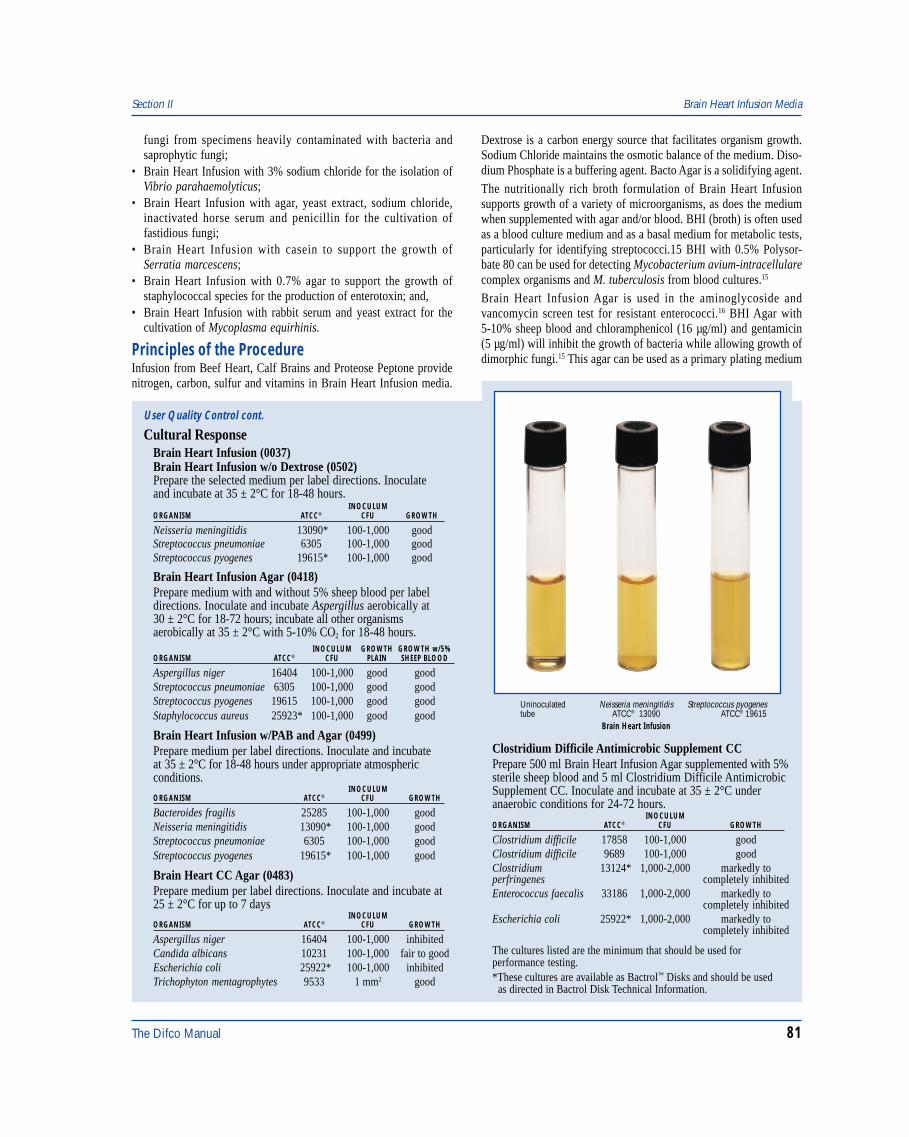
The Difco Manual 81
fungi from specimens heavily contaminated with bacteria andsaprophytic fungi;
• Brain Heart Infusion with 3% sodium chloride for the isolation ofVibrio parahaemolyticus;
• Brain Heart Infusion with agar, yeast extract, sodium chloride,inactivated horse serum and penicillin for the cultivation offastidious fungi;
• Brain Heart Infusion with casein to support the growth ofSerratia marcescens;
• Brain Heart Infusion with 0.7% agar to support the growth ofstaphylococcal species for the production of enterotoxin; and,
• Brain Heart Infusion with rabbit serum and yeast extract for thecultivation of Mycoplasma equirhinis.
Principles of the ProcedureInfusion from Beef Heart, Calf Brains and Proteose Peptone providenitrogen, carbon, sulfur and vitamins in Brain Heart Infusion media.
Dextrose is a carbon energy source that facilitates organism growth.Sodium Chloride maintains the osmotic balance of the medium. Diso-dium Phosphate is a buffering agent. Bacto Agar is a solidifying agent.The nutritionally rich broth formulation of Brain Heart Infusionsupports growth of a variety of microorganisms, as does the mediumwhen supplemented with agar and/or blood. BHI (broth) is often usedas a blood culture medium and as a basal medium for metabolic tests,particularly for identifying streptococci.15 BHI with 0.5% Polysor-bate 80 can be used for detecting Mycobacterium avium-intracellularecomplex organisms and M. tuberculosis from blood cultures.15
Brain Heart Infusion Agar is used in the aminoglycoside andvancomycin screen test for resistant enterococci.16 BHI Agar with5-10% sheep blood and chloramphenicol (16 µg/ml) and gentamicin(5 µg/ml) will inhibit the growth of bacteria while allowing growth ofdimorphic fungi.15 This agar can be used as a primary plating medium
Section II Brain Heart Infusion Media
User Quality Control cont.
Cultural ResponseBrain Heart Infusion (0037)Brain Heart Infusion w/o Dextrose (0502)Prepare the selected medium per label directions. Inoculateand incubate at 35 ± 2°C for 18-48 hours.
INOCULUMORGANISM ATCC® CFU GROWTH
Neisseria meningitidis 13090* 100-1,000 goodStreptococcus pneumoniae 6305 100-1,000 goodStreptococcus pyogenes 19615* 100-1,000 good
Brain Heart Infusion Agar (0418)Prepare medium with and without 5% sheep blood per labeldirections. Inoculate and incubate Aspergillus aerobically at30 ± 2°C for 18-72 hours; incubate all other organismsaerobically at 35 ± 2°C with 5-10% CO2 for 18-48 hours.
INOCULUM GROWTH GROWTH w/5%ORGANISM ATCC® CFU PLAIN SHEEP BLOOD
Aspergillus niger 16404 100-1,000 good goodStreptococcus pneumoniae 6305 100-1,000 good goodStreptococcus pyogenes 19615 100-1,000 good goodStaphylococcus aureus 25923* 100-1,000 good good
Brain Heart Infusion w/PAB and Agar (0499)Prepare medium per label directions. Inoculate and incubateat 35 ± 2°C for 18-48 hours under appropriate atmosphericconditions.
INOCULUMORGANISM ATCC® CFU GROWTH
Bacteroides fragilis 25285 100-1,000 goodNeisseria meningitidis 13090* 100-1,000 goodStreptococcus pneumoniae 6305 100-1,000 goodStreptococcus pyogenes 19615* 100-1,000 good
Brain Heart CC Agar (0483)Prepare medium per label directions. Inoculate and incubate at25 ± 2°C for up to 7 days
INOCULUMORGANISM ATCC® CFU GROWTH
Aspergillus niger 16404 100-1,000 inhibitedCandida albicans 10231 100-1,000 fair to goodEscherichia coli 25922* 100-1,000 inhibitedTrichophyton mentagrophytes 9533 1 mm2 good
Clostridium Difficile Antimicrobic Supplement CCPrepare 500 ml Brain Heart Infusion Agar supplemented with 5%sterile sheep blood and 5 ml Clostridium Difficile AntimicrobicSupplement CC. Inoculate and incubate at 35 ± 2°C underanaerobic conditions for 24-72 hours.
INOCULUMORGANISM ATCC® CFU GROWTH
Clostridium difficile 17858 100-1,000 goodClostridium difficile 9689 100-1,000 goodClostridium 13124* 1,000-2,000 markedly toperfringenes completely inhibitedEnterococcus faecalis 33186 1,000-2,000 markedly to
completely inhibitedEscherichia coli 25922* 1,000-2,000 markedly to
completely inhibited
The cultures listed are the minimum that should be used forperformance testing.*These cultures are available as Bactrol™ Disks and should be used as directed in Bactrol Disk Technical Information.
Streptococcus pyogenesATCC® 19615
Uninoculatedtube
Neisseria meningitidisATCC® 13090
Brain Heart Infusion
82 The Difco Manual
for the growth of fungi since it has been shown to yield better recoverythan the previously recommended Sabouraud Dextrose Agar.15 InBrain Heart CC Agar, chloramphenicol is used as a broad-spectrumantibiotic to inhibit a wide range of bacteria; cycloheximide inhibitssaprophytic fungi. Sheep blood provides essential growth factors forfastidious fungi.Clostridium Difficile Agar (Brain Heart Infusion Agar supplementedwith 5% sheep blood or 7% horse blood and Clostridium DifficileAntimicrobic Supplement CC) improves the growth and recovery ofC. difficile. Clostridium Difficile Agar markedly to completely inhibitsmost aerobic and anaerobic enteric organisms other than C. difficile.5
The final concentration of cycloserine and cefoxitin in ClostridiumDifficile Agar is 250 mcg/ml and 10 mcg/ml, respectively.Brain Heart CC Agar can be supplemented with sheep blood (5-10%)for enrichment and gentamicin (5 mg/l) for additional selectivity.14
McDonough et al17 demonstrated that the temperature of incubationaffects the sensitivity of some pathogenic fungi to antibiotics. Incubatethe medium containing antibiotics at room temperature. The specimensource and the type of fungus suspected will indicate the isolationmedium to be used. Both an antimicrobic-containing medium anda non-selective medium should be used on primary isolates withincubation at both 25°C and 37°C.Brain Heart Infusion w/PAB and Agar contains p-aminobenzoic acid(0.05 g/l) to neutralize sulfonamides in the blood of patients receivingthis therapy. This formulation will also inactivate streptomycin in theratio of 10 ml of medium to 100 units of streptomycin. The addition of0.1% agar to Brain Heart Infusion w/PAB and Agar provides optimumconditions for aerobic organisms, microaerophiles and obligate anaerobes.
FormulaBrain Heart Infusion
Formula Per LiterCalf Brains, Infusion from . . . . . . . . . . . . . . . . . . . . . . . . 200 gBeef Heart, Infusion from . . . . . . . . . . . . . . . . . . . . . . . . 250 gBacto Proteose Peptone . . . . . . . . . . . . . . . . . . . . . . . . . . . 10 gBacto Dextrose . . . . . . . . . . . . . . . . . . . . . . . . . . . . . . . . . . . 2 gSodium Chloride . . . . . . . . . . . . . . . . . . . . . . . . . . . . . . . . . . 5 gDisodium Phosphate . . . . . . . . . . . . . . . . . . . . . . . . . . . . . . 2.5 gFinal pH 7.4 ± 0.2 at 25°C
Brain Heart Infusion AgarFormula Per LiterCalf Brains, Infusion from . . . . . . . . . . . . . . . . . . . . . . . . 200 gBeef Heart, Infusion from . . . . . . . . . . . . . . . . . . . . . . . . 250 gBacto Proteose Peptone . . . . . . . . . . . . . . . . . . . . . . . . . . . 10 gBacto Dextrose . . . . . . . . . . . . . . . . . . . . . . . . . . . . . . . . . . . 2 gSodium Chloride . . . . . . . . . . . . . . . . . . . . . . . . . . . . . . . . . . 5 gDisodium Phosphate . . . . . . . . . . . . . . . . . . . . . . . . . . . . . . 2.5 gBacto Agar . . . . . . . . . . . . . . . . . . . . . . . . . . . . . . . . . . . . . 15 gFinal pH 7.4 ± 0.2 at 25°C
Brain Heart Infusion w/o DextroseFormula Per LiterCalf Brains, Infusion from . . . . . . . . . . . . . . . . . . . . . . . . 200 gBeef Heart, Infusion from . . . . . . . . . . . . . . . . . . . . . . . . 250 gBacto Proteose Peptone . . . . . . . . . . . . . . . . . . . . . . . . . . . 10 gSodium Chloride . . . . . . . . . . . . . . . . . . . . . . . . . . . . . . . . . . 5 gDisodium Phosphate . . . . . . . . . . . . . . . . . . . . . . . . . . . . . . 2.5 gFinal pH 7.4 ± 0.2 at 25°C
Brain Heart Infusion W/PAB and AgarFormula Per LiterCalf Brains, Infusion from . . . . . . . . . . . . . . . . . . . . . . . . 200 gBeef Heart, Infusion from . . . . . . . . . . . . . . . . . . . . . . . . 250 gBacto Proteose Peptone . . . . . . . . . . . . . . . . . . . . . . . . . . . 10 gBacto Dextrose . . . . . . . . . . . . . . . . . . . . . . . . . . . . . . . . . . . 2 gSodium Chloride . . . . . . . . . . . . . . . . . . . . . . . . . . . . . . . . . . 5 gDisodium Phosphate . . . . . . . . . . . . . . . . . . . . . . . . . . . . . . 2.5 gBacto Agar . . . . . . . . . . . . . . . . . . . . . . . . . . . . . . . . . . . . . . 1 gp-Aminobenzoic Acid . . . . . . . . . . . . . . . . . . . . . . . . . . . . 0.05 gFinal pH 7.4 ± 0.2 at 25°C
Brain Heart CC AgarFormula Per LiterCalf Brains, Infusion from . . . . . . . . . . . . . . . . . . . . . . . . 200 gBeef Heart, Infusion from . . . . . . . . . . . . . . . . . . . . . . . . 250 gBacto Proteose Peptone . . . . . . . . . . . . . . . . . . . . . . . . . . . 10 gBacto Dextrose . . . . . . . . . . . . . . . . . . . . . . . . . . . . . . . . . . . 2 gSodium Chloride . . . . . . . . . . . . . . . . . . . . . . . . . . . . . . . . . . 5 gDisodium Phosphate . . . . . . . . . . . . . . . . . . . . . . . . . . . . . . 2.5 gBacto Agar . . . . . . . . . . . . . . . . . . . . . . . . . . . . . . . . . . . . . 15 gChloramphenicol . . . . . . . . . . . . . . . . . . . . . . . . . . . . . . . . . 50 mgCycloheximide . . . . . . . . . . . . . . . . . . . . . . . . . . . . . . . . . 500 mgFinal pH 7.4 ± 0.2 at 25°C
Clostridium Difficile Antimicrobic Supplement CCFormula per 5 mlCycloserine . . . . . . . . . . . . . . . . . . . . . . . . . . . . . . . . . . . . 125 mgCefoxitin . . . . . . . . . . . . . . . . . . . . . . . . . . . . . . . . . . . . . . . . 5 mg
Precautions1. For Laboratory Use.2. Brain Heart CC Agar: HARMFUL. HARMFUL BY INHALATION
AND IF SWALLOWED. POSSIBLE RISK OF IRREVERSIBLEEFFECTS. POSSIBLE RISK OF HARM TO THE UNBORNCHILD. Do not breathe dust. In case of accident or if you feelunwell, seek medical advice immediately (show the label wherepossible). Wear suitable protective clothing. Keep container tightlyclosed. TARGET ORGAN(S): Eyes/Ears, Cardiovascular, Muscles,Blood, Lymph Glands, Nerves, Urogenital.FIRST AID: In case of contact with eyes, rinse immediately withplenty of water and seek medical advice. After contact with skin,wash immediately with plenty of water. If inhaled, remove to freshair. If not breathing, give artificial respiration. If breathing isdifficult, give oxygen. Seek medical advice. If swallowed, inducevomiting; seek medical advice immediately and show this containeror label.
3. Brain Heart CC Agar: Avoid overheating or holding the mediumin the melted state. Doing so tends to reduce the selective propertiesof the medium.
4. When testing human serum, treat all specimens as infectious agents.5. Follow proper established laboratory procedures in handling and
disposing of infectious materials.
StorageStore the dehydrated medium below 30°C. The dehydrated medium isvery hygroscopic. Keep container tightly closed.
Store Clostridium Difficile Antimicrobic Supplement CC at 2-8°C.
Brain Heart Infusion Media Section II
The Difco Manual 83
Expiration DateThe expiration date applies to the product in its intact container whenstored as directed. Do not use a product if it fails to meet specificationsfor identity and performance.
ProcedureMaterials ProvidedBrain Heart InfusionBrain Heart Infusion AgarClostridium Difficile Antimicrobic Supplement CCBrain Heart CC AgarBrain Heart Infusion w/PAB and AgarBrain Heart Infusion w/o Dextrose
Materials Required But Not ProvidedGlasswareAutoclaveIncubatorWaterbath (optional)Sterile defibrinated blood (optional)Sterile Petri dishesSterile tubesAnaerobic system for Clostridium Difficile Agar
Method of PreparationBrain Heart Infusion Media1. Suspend an appropriate amount of the selected medium in 1 liter
distilled or deionized water:Brain Heart Infusion - 37 grams;Brain Heart Infusion Agar - 52 grams;Brain Heart CC Agar - 52 grams;Brain Heart Infusion w/PAB and Agar - 38 grams;Brain Heart Infusion w/o Dextrose - 35 grams.
2. If the medium contains agar (Brain Heart Infusion Agar, BrainHeart CC Agar and Brain Heart Infusion w/PAB and Agar), heat itto boiling to dissolve completely. Avoid overheating.
3. Autoclave at 121°C for 15 minutes. Cool to room temperature.
Clostridium Difficile Agar1. Rehydrate and sterilize 500 ml of Brain Heart Infusion Agar per
label directions. Cool to 45-50°C.2. Aseptically rehydrate Clostridium Difficile Antimicrobic
Supplement CC with 5 ml sterile distilled or deionized water.Invert the vial gently several times to dissolve the contents. Useimmediately.
3. Aseptically add 5% sterile defibrinated sheep blood or 7%defibrinated horse blood and 5 ml of Clostridium DifficileAntimicrobic Supplement CC to the rehydrated medium.
4. Mix thoroughly, avoiding the formation of bubbles, and dispenseinto sterile Petri dishes.
Specimen Collection and PreparationObtain and process specimens according to the techniques andprocedures established by laboratory policy.
Test ProcedureSee appropriate references for specific procedures using Brain HeartInfusion Media.
Clostridium Difficile Agar1. Inoculate a representative portion of the specimen directly onto
the surface of a freshly prepared or previously reduced ClostridiumDifficile Agar plate and streak for isolation. The inoculum shouldinclude mucous, blood or membranous material, if present.
2. Incubate at 35°C under anaerobic conditions.3. Examine for growth after 24-48 hours incubation.
For a complete discussion on the isolation and identificationof Clostridium difficile refer to appropriate procedures in thereferences.15,18,20
ResultsClostridium Difficile AgarAfter 24 hours incubation, colonies of C. difficile appearnon-hemolytic, 1-3 mm in diameter, off-white to gray, flat and circularwith an undulated edge. Colonies become larger (3-5 mm) after48 hours incubation.
Limitations of the Procedure1. Since the nutritional requirements of organisms vary, some strains
may be encountered that fail to grow or grow poorly on this medium.2. Certain pathogenic fungi may be inhibited by the antibiotics in
Brain Heart CC Agar.19
3. Clostridium Difficile Antimicrobic Supplement CC is intended foruse in the preparation of Clostridium Difficile Agar. Althoughthis medium is selective for C. difficile, additional testing usingpure cultures is necessary for complete identification. Consultappropriate references for further information.15,18,20
4. Suspected colonies of C. difficile should be Gram stained andsubcultured anaerobically and aerobically on blood agar forcomplete identification.
5. Demonstration of the C. difficile toxin in feces in the presenceof clinically evident pseudomembranous colitis is required fordefinitive diagnosis.20
References1. Rosenow, E. C. 1919. Studies on elective localization. J. Dent.
Research 1:205- 249.2. Hayden, R. L. 1923. Elective localization in the eye of bacteria
from infected teeth. Arch. Int. Med. 32:828-849.3. Roseburg, T., L. J. Epps, and A. R. Clark. 1944. A study of the
isolation, cultivation and pathogenicity of Actinomyces israelirecovered from the human mouth and from actinomycosis in man.J. Infect. Dis. 29:390.
4. Howell, A. 1948. The efficiency of methods for the isolation ofHistoplasma capsulatum. Public Health Reports 63:173-178.
5. Willey, S. H., and J. G. Bartlett. 1979. Cultures for Clostridiumdifficile in stools containing a cytotoxin neutralized by Clostridiumsordellii antitoxin. J. Clin. Microbiol. 6:880-884.
6. Lyerly, D. M., D. E. Lockwood, S. H. Richardson, and T. D.Wilkins. 1982. Biological activities of toxins A and B ofClostridium difficile. Infect. Immum. 35:1147- 1150.
7. Ajello, L., L. K. Georg, W. Kaplan, and L. Kaufman. 1966.Laboratory manual for medical mycology (CDC), U.S. DHEW,Center for Disease Control, Atlanta, GA.
Section II Brain Heart Infusion Media
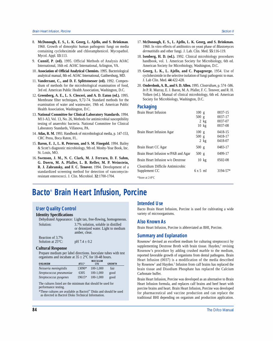
84 The Difco Manual
Brain Heart Infusion, Porcine Section II
Bacto® Brain Heart Infusion, Porcine
User Quality ControlIdentity Specifications
Dehydrated Appearance: Light tan, free-flowing, homogeneous.Solution: 3.7% solution, soluble in distilled
or deionized water. Light to mediumamber, clear.
Reaction of 3.7%Solution at 25°C: pH 7.4 ± 0.2
Cultural ResponsePrepare medium per label directions. Inoculate tubes with testorganisms and incubate at 35 ± 2°C for 18-48 hours.
INOCULUMORGANISM ATCC® CFU GROWTH
Neisseria meningitidis 13090* 100-1,000 fairStreptococcus pneumoniae 6305 100-1,000 goodStreptococcus pyogenes 19615* 100-1,000 good
The cultures listed are the minimum that should be used forperformance testing.*These cultures are available as Bactrol™ Disks and should be used as directed in Bactrol Disks Technical Information.
Intended UseBacto Brain Heart Infusion, Porcine is used for cultivating a widevariety of microorganisms.
Also Known AsBrain Heart Infusion, Porcine is abbreviated as BHI, Porcine.
Summary and ExplanationRosenow1 devised an excellent medium for culturing streptococci bysupplementing Dextrose Broth with brain tissue. Hayden,2 revisingRosenow’s procedure by adding crushed marble to the medium,reported favorable growth of organisms from dental pathogens. BrainHeart Infusion (0037) is a modification of the media describedby Rosenow1 and Hayden.2 Infusion from calf brains has replaced thebrain tissue and Disodium Phosphate has replaced the CalciumCarbonate buffer.
Brain Heart Infusion, Porcine was developed as an alternative to BrainHeart Infusion formula, and replaces calf brains and beef heart withporcine brains and heart. Brain Heart Infusion, Porcine was developedfor pharmaceutical and vaccine production and can replace thetraditional BHI depending on organism and production application.
8. McDonough, E. S., L. K. Georg, L. Ajello, and S. Brinkman.1960. Growth of dimorphic human pathogenic fungi on mediacontaining cycloheximide and chloramphenicol. Mycopathol.Mycol. Appl. 13:113.
9. Cunnif, P. (ed). 1995. Official Methods of Analysis AOACInternational, 16th ed. AOAC International, Arlington, VA.
10. Association of Official Analytical Chemists. 1995. Bacteriologicalanalytical manual, 8th ed. AOAC International, Gaithersburg, MD.
11. Vanderzant, C., and D. F. Splittstoesser (ed). 1992. Compen-dium of methods for the microbiological examination of food,3rd ed. American Public Health Association, Washington, D.C.
12. Greenberg, A. E., L. S. Clesceri, and A. D. Eaton (ed.). 1995.Membrane filter techniques, 9,72-74. Standard methods for theexamination of water and wastewater, 19th ed. American PublicHealth Association, Washington, D.C.
13. National Committee for Clinical Laboratory Standards. 1994.M11-A3, Vol. 13, No. 26, Methods for antimicrobial susceptibilitytesting of anaerobic bacteria. National Committee for ClinicalLaboratory Standards, Villanova, PA.
14. Atlas, R. M. 1993. Handbook of microbiological media, p. 147-153,CRC Press, Boca Raton, FL.
15. Baron, E. J., L. R. Peterson, and S. M. Finegold. 1994. Bailey& Scott’s diagnostic microbiology, 9th ed. Mosby-Year Book, Inc.St. Louis, MO.
16. Swenson, J. M., N. C. Clark, M. J. Ferraro, D. F. Sahm,G. Doern, M. A. Pfaller, L. B. Reller, M. P. Weinstein,R. J. Zabransky, and F. C. Tenover. 1994. Development of astandardized screening method for detection of vancomycin-resistant enterococci. J. Clin. Microbiol. 32:1700-1704.
17. McDonough, E. S., L. Ajello, L. K. Georg, and S. Brinkman.1960. In vitro effects of antibiotics on yeast phase of Blastomycesdermatitidis and other fungi. J. Lab. Clin. Med. 55:116-119.
18. Isenberg, H. D. (ed.). 1992. Clinical microbiology procedureshandbook, vol. 1. American Society for Microbiology, 6th ed.American Society for Microbiology, Washington, D.C.
19. Georg, L. K., L. Ajello, and C. Papageorge. 1954. Use ofcycloheximide in the selective isolation of fungi pathogenic to man.J. Lab Clin. Med. 44:422-428.
20. Onderdonk, A. B., and S. D. Allen. 1995. Clostridium, p. 574 -586.In P. R. Murray, E. J. Baron, M. A. Pfaller, F. C. Tenover, and R. H.Yolken (ed.). Manual of clinical microbiology, 6th ed. AmericanSociety for Microbiology, Washington, D.C.
PackagingBrain Heart Infusion 100 g 0037-15
500 g 0037-172 kg 0037-07
10 kg 0037-08
Brain Heart Infusion Agar 100 g 0418-15500 g 0418-17
2 kg 0418-07
Brain Heart CC Agar 500 g 0483-17
Brain Heart Infusion w/PAB and Agar 500 g 0499-17
Brain Heart Infusion w/o Dextrose 10 kg 0502-08
Clostridium Difficile AntimicrobicSupplement CC 6 x 5 ml 3194-57*
*Store at 2-8°C
The Difco Manual 85
BHI, Porcine was formulated with no bovine components to minimizeBovine Spongiform Encephalopathy (BSE) risk.The nutritionally rich formula of BHI is used to grow a variety ofmicroorganisms The original Brain Heart Infusion media are specifiedin standard methods for multiple applications.3,4,5,6
Principles of the ProcedureInfusion from pork brains, infusion from pork heart and Pork PeptoneNo. 2 provides nitrogen, carbon, sulfur and vitamins in Brain HeartInfusion, Porcine. Dextrose is the carbon energy source to facilitateorganism growth. Sodium Chloride maintains the osmotic balance ofthe medium. Disodium Phosphate is the buffering agent.
FormulaBrain Heart Infusion, Porcine
Formula Per LiterPork Brains, Infusion from . . . . . . . . . . . . . . . . . . . . . . . . 200 gPork Heart, Infusion from . . . . . . . . . . . . . . . . . . . . . . . . 250 gBacto Pork Peptone No. 2 . . . . . . . . . . . . . . . . . . . . . . . . . 10 gBacto Dextrose . . . . . . . . . . . . . . . . . . . . . . . . . . . . . . . . . . . 2 gSodium Chloride . . . . . . . . . . . . . . . . . . . . . . . . . . . . . . . . . . 5 gDisodium Phosphate . . . . . . . . . . . . . . . . . . . . . . . . . . . . . . 2.5 g
Final pH 7.4 ± 0.2 at 25°C
Precautions1. For Laboratory Use.2. Follow proper established laboratory procedures in handling and
disposing of infectious materials.
StorageStore the dehydrated medium below 30°C. The dehydrated medium isvery hygroscopic. Keep container tightly closed.
Expiration DateThe expiration date applies to the product in its intact container whenstored as directed. Do not use a product if it fails to meet specificationsfor identity and performance.
ProcedureMaterials ProvidedBrain Heart Infusion, Porcine
Materials Required But Not ProvidedGlasswareAutoclaveIncubatorWaterbath (optional)
Method of Preparation1. Dissolve 37 grams in 1 liter distilled or deionized water.2. Autoclave at 121°C for 15 minutes.3. Dispense as desired.
Specimen Collection and PreparationObtain and process specimens according to the techniques andprocedures established by laboratory policy.
Test ProcedureSee appropriate references for specific procedures using BrainHeart Infusion.
ResultsRefer to appropriate references and procedures for results.
References1. Rosenow, E. C. 1919. Studies on elective localization. J. Dent.
Res. 1:205-249.2. Hayden, R. L. 1923. Elective localization in the eye of bacteria
from infected teeth. Arch. Int. Med. 32:828-849.3. Vanderzant, C., and D. F. Splittstoesser (ed.). 1992. Compendium
of methods for the microbiological examination of food, 3rd ed.American Public Health Association, Washington, D.C.
4. Association of Official Analytical Chemists. 1995. Bacteriologicalanalytical manual, 8th ed. AOAC International, Gaithersburg, MD.
5. Eaton, A. D., L. S. Clesceri, and A. E. Greenberg (ed.). 1995.Standard methods for the examination of water and wastewater,19th ed. American Public Health Association, Washington, D.C.
6. Cunnif, P. (ed).1995. Official methods of analysis, AOACInternational, 16th ed. AOAC International, Arlington, VA.
PackagingBrain Heart Infusion, Porcine 500 g 0561-17
Section II Brewer Anaerobic Agar
Bacto® Brewer Anaerobic AgarIntended UseBacto Brewer Anaerobic Agar is used for cultivating anaerobic andmicroaerophilic bacteria.
Summary and ExplanationBrewer1 described a special Petri dish cover that allowed surface growthof anaerobes and microaerophiles without anaerobic equipment. Themicroorganisms were grown on agar with a low oxidation-reduction po-tential. Brewer Anaerobic Agar was originally formulated andmodified for the procedure described by Brewer.1 This medium is
suitable for standard plating procedures used in cultivatinganaerobic bacteria.2,3,4
Anaerobic bacteria cause a variety of infections in humans, includingotitis media, oral infections, endocarditis, meningitis, wound infectionsfollowing bowel surgery or trauma and bacteremia.5,6 Anaerobicbacteria are the predominant flora colonizing the skin and mucousmembranes of the body.3 Anaerobes vary in their sensitivity to oxygenand nutritional requirements.2 Anaerobic bacteria lack cytochromesand thus are unable to use oxygen as a terminal electron acceptor.3
Principles of the ProcedureTryptone, Proteose Peptone No. 3 and Yeast Extract provide the nitrogen,vitamins and amino acids in Brewer Anaerobic Agar. Dextrose
86 The Difco Manual
User Quality ControlIdentity Specifications
Dehydrated Appearance: Light beige, free-flowing, homogeneous.Solution: 5.8% solution, soluble in distilled or
deionized water on boiling. Lightamber, slightly opalescent while hot,turning red on aeration and cooling.
Prepared Medium: Light pink ring at outer edge, lightamber in center, slightly opalescent.
Reaction of 5.8%Solution at 25°C pH 7.2 ± 0.2
Cultural ResponsePrepare Brewer Anaerobic Agar per label directions. Inoculatethe plates using the streak method. Incubate plates at 35 ± 2°Canaerobically for 40-48 hours.
INOCULUMORGANISM ATCC® CFU GROWTH
Bacteroides fragilis 25285* 100-1,000 goodClostridium beijerinckii 17795 100-1,000 goodClostridium perfringens 12924 100-1,000 good
The cultures listed are the minimum that should be used forperformance testing.*This culture is available as Bactrol™ Disks and should be used as directed in Bactrol Disks Technical Information.
is the carbon source, and Sodium Chloride maintains osmoticequilibrium. Sodium Thioglycollate and Sodium FormaldehydeSulfoxylate are the reducing agents. Resazurin serves as anindicator of anaerobiosis with a pink color indicating the presence ofoxygen. Bacto Agar is the solidifying agent.
FormulaBrewer Anaerobic Agar
Formula Per LiterBacto Tryptone . . . . . . . . . . . . . . . . . . . . . . . . . . . . . . . . . . . 5 gBacto Proteose Peptone No. 3 . . . . . . . . . . . . . . . . . . . . . . 10 gBacto Yeast Extract . . . . . . . . . . . . . . . . . . . . . . . . . . . . . . . . 5 gBacto Dextrose . . . . . . . . . . . . . . . . . . . . . . . . . . . . . . . . . . 10 gSodium Chloride . . . . . . . . . . . . . . . . . . . . . . . . . . . . . . . . . . 5 gBacto Agar . . . . . . . . . . . . . . . . . . . . . . . . . . . . . . . . . . . . . 20 gSodium Thioglycollate . . . . . . . . . . . . . . . . . . . . . . . . . . . . . 2 gSodium Formaldehyde Sulfoxylate . . . . . . . . . . . . . . . . . . . 1 gResazurin, Certified . . . . . . . . . . . . . . . . . . . . . . . . . . . . 0.002 g
Final pH 7.2 ± 0.2 at 25°C
Precautions1. For Laboratory Use.2. Follow proper established laboratory procedures in handling and
disposing of infectious materials.
StorageStore the dehydrated medium below 30°C. The dehydrated medium isvery hygroscopic. Keep container tightly closed.
Expiration DateThe expiration date applies to the product in its intact container whenstored as directed. Do not use a product if it fails to meet specificationsfor identity and performance.
ProcedureMaterials ProvidedBrewer Anaerobic Agar
Materials Required But Not ProvidedGlasswareAutoclaveIncubator (35°C)Waterbath (45-50°C) (optional)Sterile Petri dishesBrewer Anaerobic Petri dish covers (optional)
Method of Preparation1. Suspend 58 grams in 1 liter distilled or deionized water.2. Heat to boiling to dissolve completely.3. Autoclave at 121°C for 15 minutes. Cool to 45-50°C.4. Dispense as desired.
Specimen Collection and PreparationAnaerobic bacteria are overlooked or missed unless the specimen isproperly collected and transported to the laboratory.2 Obtain andprocess specimens according to the techniques and proceduresestablished by institutional policy.
Test ProcedureStandard Petri Dishes:2
1. Inoculate a properly obtained specimen onto the medium, andstreak to obtain isolated colonies.
2. Immediately incubate anaerobically at 35 ± 2°C.3. Examine at 24 hours if incubating plates in an anaerobic chamber.
Examine at 48 hours if incubating plates in an anaerobic jar orpouch, or if using Brewer anaerobic dish cover.
4. Extended incubation may be necessary to recover some anaerobes.
Brewer Anaerobic Agar Plates:1. Dispense 50-60 ml of Brewer Anaerobic Agar into a standard
Petri dish. For best results use porous tops to obtain a dry surface.2. Inoculate the surface of the medium by streaking; avoid the edges
of the plates.3. Replace the standard Petri dish lid with a sterile Brewer anaerobic
dish cover. The cover should not rest on the Petri dish bottom.The inner glass ridge should seal against the uninoculatedperiphery of the agar. It is essential that the sealing ring inside thecover is in contact with the medium. This seal must not be brokenbefore the end of the incubation period. A small amount of air iscaught over the surface of the medium, and the oxygen in this spacereacts with the reducing agents to form an anaerobic environment.
4. Incubate aerobically as desired.For a complete discussion on anaerobic and microaerophilicbacteria from clinical specimens, refer to the appropriateprocedures outlined in the references.2,3,4 For the examination ofanaerobic bacteria in food refer to standard methods.7,8,9
Brewer Anaerobic Agar Section II
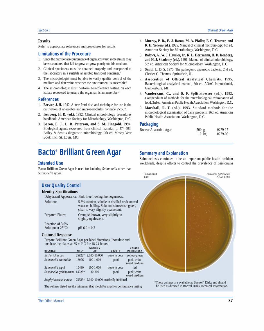
The Difco Manual 87
ResultsRefer to appropriate references and procedures for results.
Limitations of the Procedure1. Since the nutritional requirements of organisms vary, some strains may
be encountered that fail to grow or grow poorly on this medium.2. Clinical specimens must be obtained properly and transported to
the laboratory in a suitable anaerobic transport container.2
3. The microbiologist must be able to verify quality control of themedium and determine whether the environment is anaerobic.2
4. The microbiologist must perform aerotolerance testing on eachisolate recovered to ensure the organism is an anaerobe.2
References1. Brewer, J. H. 1942. A new Petri dish and technique for use in the
cultivation of anaerobes and microaerophiles. Science 95:587.2. Isenberg, H. D. (ed.). 1992. Clinical microbiology procedures
handbook, American Society for Microbiology, Washington, D.C.3. Baron, E. J., L. R. Peterson, and S. M. Finegold. 1994.
Etiological agents recovered from clinical material, p. 474-503.Bailey & Scott’s diagnostic microbiology, 9th ed. Mosby-YearBook, Inc., St. Louis, MO.
Section II Brilliant Green Agar
4. Murray, P. R., E. J. Baron, M. A. Pfaller, F. C. Tenover, andR. H. Yolken (ed.). 1995. Manual of clinical microbiology, 6th ed.American Society for Microbiology, Washington, D.C.
5. Balows, A., W. J. Hausler, Jr., K. L. Herrmann, H. D. Isenberg,and H. J. Shadomy (ed.). 1991. Manual of clinical microbiology,5th ed. American Society for Microbiology, Washington, D.C.
6. Smith, L. D. S. 1975. The pathogenic anaerobic bacteria, 2nd ed.Charles C. Thomas, Springfield, IL.
7. Association of Official Analytical Chemists. 1995.Bacteriological analytical manual, 8th ed. AOAC International,Gaithersburg, MD.
8. Vanderzant, C., and D. F. Splittstoesser (ed.). 1992.Compendium of methods for the microbiological examination offood, 3rd ed. American Public Health Association, Washington, D.C.
9. Marshall, R. T. (ed.). 1993. Standard methods for themicrobiological examination of dairy products, 16th ed. AmericanPublic Health Association, Washington, D.C.
PackagingBrewer Anaerobic Agar 500 g 0279-17
10 kg 0279-08
Bacto® Brilliant Green Agar
User Quality ControlIdentity Specifications
Dehydrated Appearance: Pink, free flowing, homogeneous.Solution: 5.8% solution, soluble in distilled or deionized
water on boiling. Solution is brownish-green,clear to very slightly opalescent.
Prepared Plates: Orangish-brown, very slightly toslightly opalescent.
Reaction of 3.6%Solution at 25°C: pH 6.9 ± 0.2
Cultural ResponsePrepare Brilliant Green Agar per label directions. Inoculate andincubate the plates at 35 ± 2°C for 18-24 hours.
INOCULUM COLONYORGANISM ATCC® CFU GROWTH MORPHOLOGY
Salmonella typhimuriumATCC® 14028
Uninoculatedplate
Escherichia coli 25922* 2,000-10,000 none to poor yellow-greenSalmonella enteritidis 13076 100-1,000 good pink-white
w/red mediumSalmonella typhi 19430 100-1,000 none to poor redSalmonella typhimurium 14028* 30-300 good pink-white
w/red mediumStaphylococcus aureus 25923* 2,000-10,000 markedly inhibited –
The cultures listed are the minimum that should be used for performance testing.*These cultures are available as Bactrol™ Disks and should be used as directed in Bactrol Disks Technical Information.
Intended UseBacto Brilliant Green Agar is used for isolating Salmonella other thanSalmonella typhi.
Summary and ExplanationSalmonellosis continues to be an important public health problemworldwide, despite efforts to control the prevalence of Salmonella
88 The Difco Manual
in domesticated animals. Infection with non-typhi Salmonellaoften causes mild, self-limiting illness.1 The illness results fromconsumption of raw, undercooked or improperly processed foodscontaminated with Salmonella. Many of these cases of Salmonella-related gastroenteritis are due to improper handling of poultryproducts. Various poultry products are routinely monitored forSalmonella before their distribution for human consumption, butin many instances, contaminated food samples elude monitoring.
The use of Brilliant Green Agar as a primary plating medium forthe isolation of Salmonella was first described by Kristensen, Lesterand Jurgens2 who reported it useful for the differentiation of“paratyphoid B” from other intestinal gram-negative bacilli. Later,Kauffmann3 modified their formula and used Brilliant Green Agarin addition to Tetrathionate Broth for the isolation of Salmonellafrom stool specimens. Gallon and Quan4 increased their positiveSalmonella findings by using Tetrathionate Broth and platingon Brilliant Green Agar. Broh-Kahn5 showed that the Kauffmannmodification of Brilliant Green Agar permitted the use of heavyinocula to obtain maximum recovery of Salmonella from fecalspecimens. Miller and Tate6 found that the addition of 20 mg per liter ofsodium novobiocin to Brilliant Green Agar reduced or completelyinhibited nuisance organisms commonly seen on agar media used forisolating salmonellae. Brilliant Green Agar with Novobiocin is alsorecommended for use when testing food for Salmonella.7
Brilliant Green Agar is recommended for use in testing clinicalspecimens.8,9 The outstanding selectivity of this medium permitsthe use of moderately heavy inocula, which should be evenlydistributed over the surface. Brilliant Green Agar is valuablewhen investigating outbreaks of Salmonella spp., other than S. typhiand S. paratyphi.8,9 In addition, Brilliant Green Agar is used in themicrobial limits test as recommended in the United StatesPharmacopeia. The microbial limits test is performed to ensurethat pharmaceutical articles are free of Salmonella spp.10
Principles of the ProcedureIn Brilliant Green Agar, Proteose Peptone No. 3 and Yeast Extractprovide nitrogen, vitamins and minerals. Lactose and Saccharoseare the carbohydrates in the medium. Phenol Red is the pH indicatorthat turns the medium a yellow color with the formation of acid whenlactose and/or sucrose is fermented. Sodium Chloride maintains theosmotic balance in the medium. Brilliant Green inhibits gram-positivebacteria and most gram-negative bacilli other than Salmonella spp.Lactose/sucrose fermenters are usually inhibited.11 Bacto Agar is thesolidifying agent.
FormulaBrilliant Green Agar
Formula Per LiterBacto Proteose Peptone No. 3 . . . . . . . . . . . . . . . . . . . . . . 10 gBacto Yeast Extract . . . . . . . . . . . . . . . . . . . . . . . . . . . . . . . . 3 gBacto Lactose . . . . . . . . . . . . . . . . . . . . . . . . . . . . . . . . . . . 10 gBacto Saccharose . . . . . . . . . . . . . . . . . . . . . . . . . . . . . . . . 10 gSodium Chloride . . . . . . . . . . . . . . . . . . . . . . . . . . . . . . . . . . 5 gBacto Agar . . . . . . . . . . . . . . . . . . . . . . . . . . . . . . . . . . . . . 20 gBrilliant Green . . . . . . . . . . . . . . . . . . . . . . . . . . . . . . . 0.0125 gBacto Phenol Red . . . . . . . . . . . . . . . . . . . . . . . . . . . . . . . 0.08 g
Final pH 6.9 ± 0.2 at 25°C
Precautions1. For Laboratory Use.
2. Follow proper established laboratory procedure in handling anddisposing of infectious materials.
StorageStore the dehydrated medium below 30°C. The dehydrated mediumis very hygroscopic. Keep container tightly closed. Store preparedplates at 2-8°C.
Expiration DateThe expiration date applies to the product in its intact containerwhen stored as directed. Do not use a product if it fails to meetspecifications for identity and performance.
ProcedureMaterials ProvidedBrilliant Green Agar
Materials Required But Not ProvidedFlasks with closuresDistilled or deionized waterBunsen burner or magnetic hot plateAutoclaveWaterbath (45-50°C)Petri dishesIncubator (35°C)
Method of Preparation1. Suspend 58 grams in 1 liter distilled or deionized water.2. Heat to boiling to dissolve completely.3. Autoclave at 121°C for 15 minutes. Avoid overheating.4. Cool to 45-50°C in a waterbath.5. Dispense into sterile Petri dishes.
Specimen Collection and Preparation1. Collect specimens in sterile containers or with sterile swabs and
transport immediately to the laboratory following recommendedguidelines.7.8.9
2. For specific information about specimen preparation andinoculation of clinical specimens, consult the appropriatereferences.7,8,9
Test ProcedureFor isolation of Salmonella from clinical specimens, inoculate fecalspecimens and rectal swabs onto a small area of one quadrant of theBrilliant Green Agar plate and streak for isolation. This will permitthe development of discrete colonies. Incubate plates at 35°C.Examine plates after 18-24 hours for colonies with characteristicmorphologies associated with Salmonella spp.
ResultsThe typical Salmonella colonies appear as pink-white opaque coloniessurrounded by a brilliant red medium. The few lactose and/or sucrosefermenting organisms that grow are readily differentiated due to theformation of a yellow-green colony surrounded by an intense
Brilliant Green Agar Section II
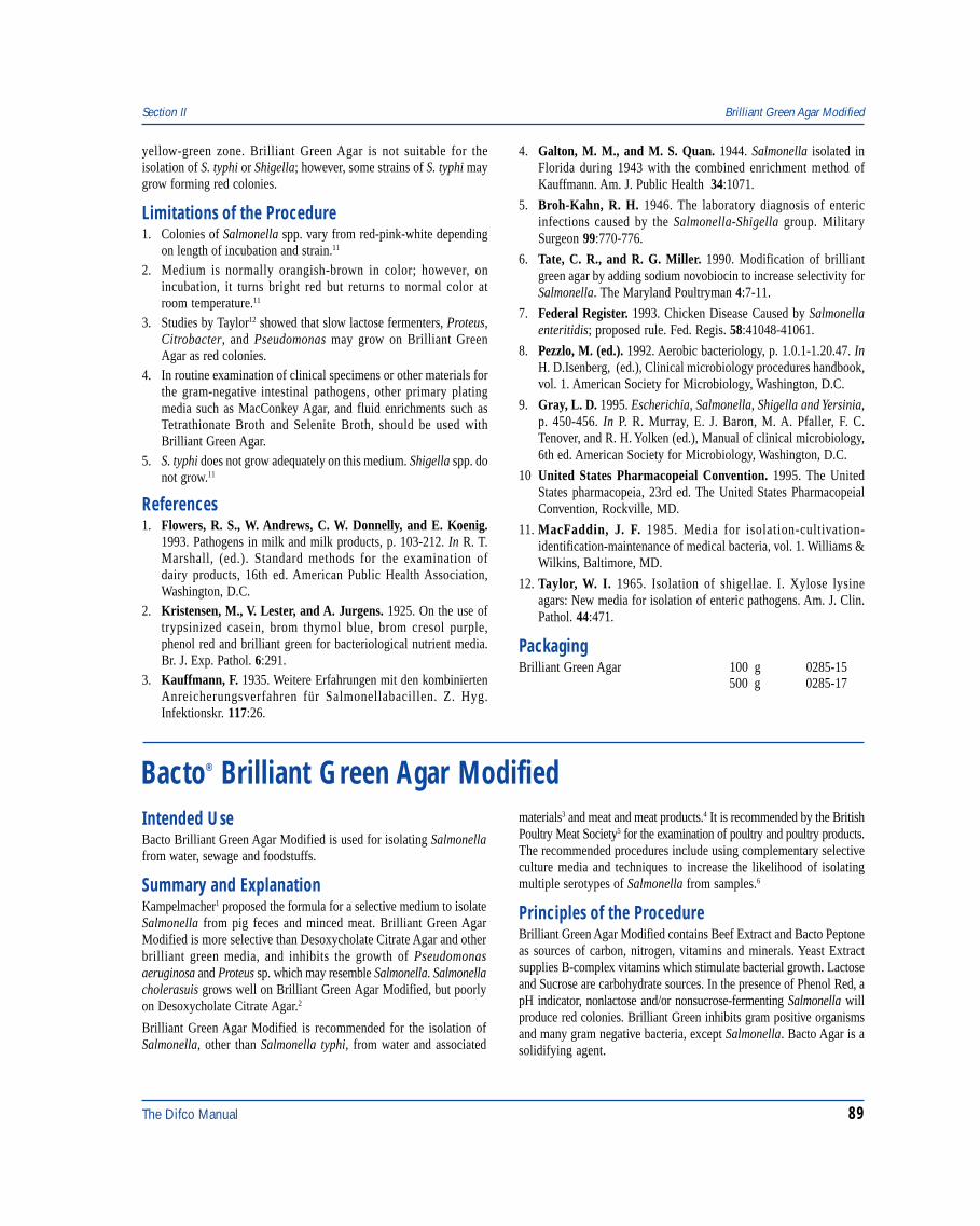
The Difco Manual 89
yellow-green zone. Brilliant Green Agar is not suitable for theisolation of S. typhi or Shigella; however, some strains of S. typhi maygrow forming red colonies.
Limitations of the Procedure1. Colonies of Salmonella spp. vary from red-pink-white depending
on length of incubation and strain.11
2. Medium is normally orangish-brown in color; however, onincubation, it turns bright red but returns to normal color atroom temperature.11
3. Studies by Taylor12 showed that slow lactose fermenters, Proteus,Citrobacter, and Pseudomonas may grow on Brilliant GreenAgar as red colonies.
4. In routine examination of clinical specimens or other materials forthe gram-negative intestinal pathogens, other primary platingmedia such as MacConkey Agar, and fluid enrichments such asTetrathionate Broth and Selenite Broth, should be used withBrilliant Green Agar.
5. S. typhi does not grow adequately on this medium. Shigella spp. donot grow.11
References1. Flowers, R. S., W. Andrews, C. W. Donnelly, and E. Koenig.
1993. Pathogens in milk and milk products, p. 103-212. In R. T.Marshall, (ed.). Standard methods for the examination ofdairy products, 16th ed. American Public Health Association,Washington, D.C.
2. Kristensen, M., V. Lester, and A. Jurgens. 1925. On the use oftrypsinized casein, brom thymol blue, brom cresol purple,phenol red and brilliant green for bacteriological nutrient media.Br. J. Exp. Pathol. 6:291.
3. Kauffmann, F. 1935. Weitere Erfahrungen mit den kombiniertenAnreicherungsverfahren für Salmonellabacillen. Z. Hyg.Infektionskr. 117:26.
4. Galton, M. M., and M. S. Quan. 1944. Salmonella isolated inFlorida during 1943 with the combined enrichment method ofKauffmann. Am. J. Public Health 34:1071.
5. Broh-Kahn, R. H. 1946. The laboratory diagnosis of entericinfections caused by the Salmonella-Shigella group. MilitarySurgeon 99:770-776.
6. Tate, C. R., and R. G. Miller. 1990. Modification of brilliantgreen agar by adding sodium novobiocin to increase selectivity forSalmonella. The Maryland Poultryman 4:7-11.
7. Federal Register. 1993. Chicken Disease Caused by Salmonellaenteritidis; proposed rule. Fed. Regis. 58:41048-41061.
8. Pezzlo, M. (ed.). 1992. Aerobic bacteriology, p. 1.0.1-1.20.47. InH. D.Isenberg, (ed.), Clinical microbiology procedures handbook,vol. 1. American Society for Microbiology, Washington, D.C.
9. Gray, L. D. 1995. Escherichia, Salmonella, Shigella and Yersinia,p. 450-456. In P. R. Murray, E. J. Baron, M. A. Pfaller, F. C.Tenover, and R. H. Yolken (ed.), Manual of clinical microbiology,6th ed. American Society for Microbiology, Washington, D.C.
10 United States Pharmacopeial Convention. 1995. The UnitedStates pharmacopeia, 23rd ed. The United States PharmacopeialConvention, Rockville, MD.
11. MacFaddin, J. F. 1985. Media for isolation-cultivation-identification-maintenance of medical bacteria, vol. 1. Williams &Wilkins, Baltimore, MD.
12. Taylor, W. I. 1965. Isolation of shigellae. I. Xylose lysineagars: New media for isolation of enteric pathogens. Am. J. Clin.Pathol. 44:471.
PackagingBrilliant Green Agar 100 g 0285-15
500 g 0285-17
Section II Brilliant Green Agar Modified
Bacto® Brilliant Green Agar ModifiedIntended UseBacto Brilliant Green Agar Modified is used for isolating Salmonellafrom water, sewage and foodstuffs.
Summary and ExplanationKampelmacher1 proposed the formula for a selective medium to isolateSalmonella from pig feces and minced meat. Brilliant Green AgarModified is more selective than Desoxycholate Citrate Agar and otherbrilliant green media, and inhibits the growth of Pseudomonasaeruginosa and Proteus sp. which may resemble Salmonella. Salmonellacholerasuis grows well on Brilliant Green Agar Modified, but poorlyon Desoxycholate Citrate Agar.2
Brilliant Green Agar Modified is recommended for the isolation ofSalmonella, other than Salmonella typhi, from water and associated
materials3 and meat and meat products.4 It is recommended by the BritishPoultry Meat Society5 for the examination of poultry and poultry products.The recommended procedures include using complementary selectiveculture media and techniques to increase the likelihood of isolatingmultiple serotypes of Salmonella from samples.6
Principles of the ProcedureBrilliant Green Agar Modified contains Beef Extract and Bacto Peptoneas sources of carbon, nitrogen, vitamins and minerals. Yeast Extractsupplies B-complex vitamins which stimulate bacterial growth. Lactoseand Sucrose are carbohydrate sources. In the presence of Phenol Red, apH indicator, nonlactose and/or nonsucrose-fermenting Salmonella willproduce red colonies. Brilliant Green inhibits gram positive organismsand many gram negative bacteria, except Salmonella. Bacto Agar is asolidifying agent.
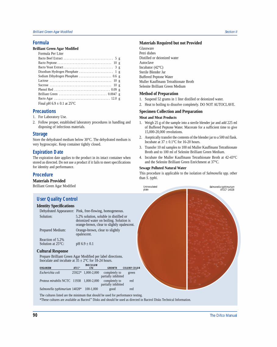
90 The Difco Manual
FormulaBrilliant Green Agar Modified
Formula Per LiterBacto Beef Extract . . . . . . . . . . . . . . . . . . . . . . . . . . . . . . . . 5 gBacto Peptone . . . . . . . . . . . . . . . . . . . . . . . . . . . . . . . . . . . 10 gBacto Yeast Extract . . . . . . . . . . . . . . . . . . . . . . . . . . . . . . . . 3 gDisodium Hydrogen Phosphate . . . . . . . . . . . . . . . . . . . . . . 1 gSodium Dihydrogen Phosphate . . . . . . . . . . . . . . . . . . . . . 0.6 gLactose . . . . . . . . . . . . . . . . . . . . . . . . . . . . . . . . . . . . . . . . 10 gSucrose . . . . . . . . . . . . . . . . . . . . . . . . . . . . . . . . . . . . . . . . 10 gPhenol Red . . . . . . . . . . . . . . . . . . . . . . . . . . . . . . . . . . . . 0.09 gBrilliant Green . . . . . . . . . . . . . . . . . . . . . . . . . . . . . . . 0.0047 gBacto Agar . . . . . . . . . . . . . . . . . . . . . . . . . . . . . . . . . . . . 12.0 g
Final pH 6.9 ± 0.1 at 25°C
Precautions1. For Laboratory Use.2. Follow proper, established laboratory procedures in handling and
disposing of infectious materials.
StorageStore the dehydrated medium below 30°C. The dehydrated medium isvery hygroscopic. Keep container tightly closed.
Expiration DateThe expiration date applies to the product in its intact container whenstored as directed. Do not use a product if it fails to meet specificationsfor identity and performance.
ProcedureMaterials ProvidedBrilliant Green Agar Modified
Materials Required but not ProvidedGlasswarePetri dishesDistilled or deionized waterAutoclaveIncubator (42°C)Sterile Blender JarBuffered Peptone WaterMuller Kauffmann Tetrathionate BrothSelenite Brilliant Green Medium
Method of Preparation1. Suspend 52 grams in 1 liter distilled or deionized water.2. Heat to boiling to dissolve completely. DO NOT AUTOCLAVE.
Specimen Collection and PreparationMeat and Meat Products1. Weigh 25 g of the sample into a sterile blender jar and add 225 ml
of Buffered Peptone Water. Macerate for a sufficient time to give15,000-20,000 revolutions.
2. Aseptically transfer the contents of the blender jar to a 500 ml flask.Incubate at 37 ± 0.1°C for 16-20 hours.
3. Transfer 10 ml samples to 100 ml Muller Kauffmann TetrathionateBroth and to 100 ml of Selenite Brilliant Green Medium.
4. Incubate the Muller Kauffmann Tetrathionate Broth at 42-43°Cand the Selenite Brilliant Green Enrichment at 37°C.
Sewage Polluted Natural WaterThis procedure is applicable to the isolation of Salmonella spp. otherthan S. typhi.
User Quality ControlIdentity Specifications
Dehydrated Appearance: Pink, free-flowing, homogeneous.Solution: 5.2% solution, soluble in distilled or
deionized water on boiling. Solution isorange-brown, clear to slightly opalescent.
Prepared Medium: Orange-brown, clear to slightlyopalescent.
Reaction of 5.2%Solution at 25°C: pH 6.9 ± 0.1
Cultural ResponsePrepare Brilliant Green Agar Modified per label directions.Inoculate and incubate at 35 ± 2°C for 18-24 hours.
INOCULUMORGANISM ATCC® CFU GROWTH COLONY COLOR
Escherichia coli 25922* 1,000-2,000 completely to greenpartially inhibited
Proteus mirabilis NCTC 11938 1,000-2,000 completely to redpartially inhibited
Salmonella typhimurium 14028* 100-1,000 good red
Salmonella typhimuriumATCC® 14028
Uninoculatedplate
The cultures listed are the minimum that should be used for performance testing.*These cultures are available as Bactrol™ Disks and should be used as directed in Bactrol Disks Technical Information.
Brilliant Green Agar Modified Section II
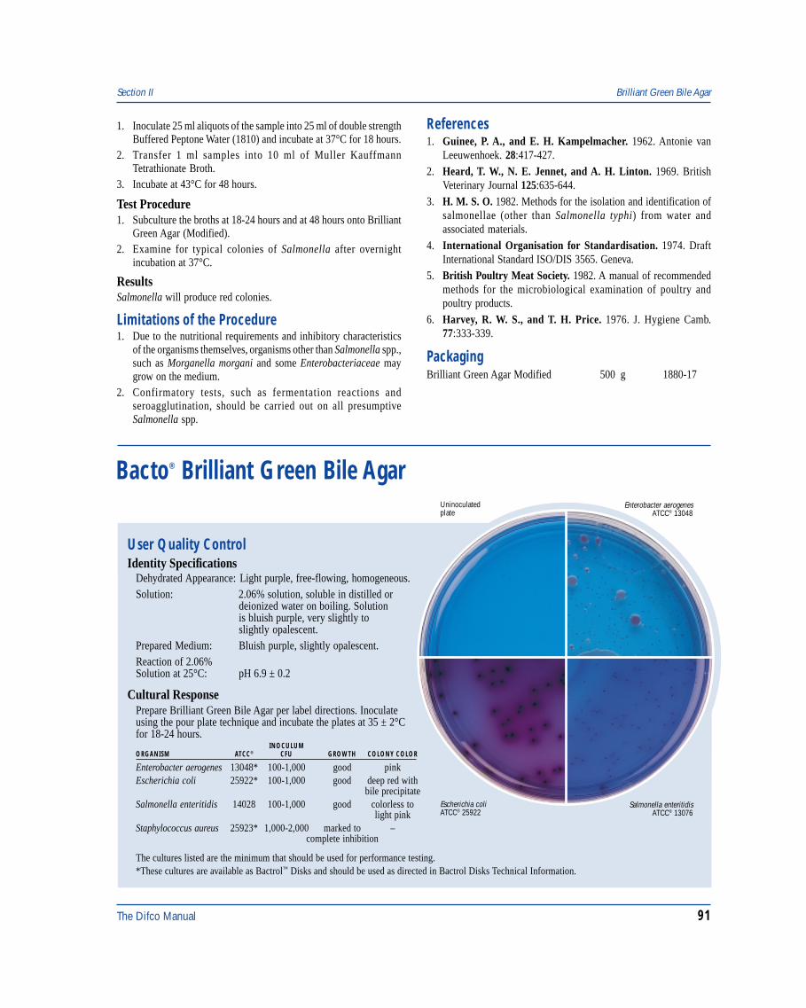
The Difco Manual 91
1. Inoculate 25 ml aliquots of the sample into 25 ml of double strengthBuffered Peptone Water (1810) and incubate at 37°C for 18 hours.
2. Transfer 1 ml samples into 10 ml of Muller KauffmannTetrathionate Broth.
3. Incubate at 43°C for 48 hours.
Test Procedure1. Subculture the broths at 18-24 hours and at 48 hours onto Brilliant
Green Agar (Modified).2. Examine for typical colonies of Salmonella after overnight
incubation at 37°C.
ResultsSalmonella will produce red colonies.
Limitations of the Procedure1. Due to the nutritional requirements and inhibitory characteristics
of the organisms themselves, organisms other than Salmonella spp.,such as Morganella morgani and some Enterobacteriaceae maygrow on the medium.
2. Confirmatory tests, such as fermentation reactions andseroagglutination, should be carried out on all presumptiveSalmonella spp.
References1. Guinee, P. A., and E. H. Kampelmacher. 1962. Antonie van
Leeuwenhoek. 28:417-427.2. Heard, T. W., N. E. Jennet, and A. H. Linton. 1969. British
Veterinary Journal 125:635-644.3. H. M. S. O. 1982. Methods for the isolation and identification of
salmonellae (other than Salmonella typhi) from water andassociated materials.
4. International Organisation for Standardisation. 1974. DraftInternational Standard ISO/DIS 3565. Geneva.
5. British Poultry Meat Society. 1982. A manual of recommendedmethods for the microbiological examination of poultry andpoultry products.
6. Harvey, R. W. S., and T. H. Price. 1976. J. Hygiene Camb.77:333-339.
PackagingBrilliant Green Agar Modified 500 g 1880-17
Section II Brilliant Green Bile Agar
Bacto® Brilliant Green Bile Agar
User Quality ControlIdentity Specifications
Dehydrated Appearance: Light purple, free-flowing, homogeneous.Solution: 2.06% solution, soluble in distilled or
deionized water on boiling. Solutionis bluish purple, very slightly toslightly opalescent.
Prepared Medium: Bluish purple, slightly opalescent.Reaction of 2.06%Solution at 25°C: pH 6.9 ± 0.2
Cultural ResponsePrepare Brilliant Green Bile Agar per label directions. Inoculateusing the pour plate technique and incubate the plates at 35 ± 2°Cfor 18-24 hours.
INOCULUMORGANISM ATCC® CFU GROWTH COLONY COLOR
Enterobacter aerogenes 13048* 100-1,000 good pinkEscherichia coli 25922* 100-1,000 good deep red with
bile precipitateSalmonella enteritidis 14028 100-1,000 good colorless to
light pinkStaphylococcus aureus 25923* 1,000-2,000 marked to –
complete inhibition
The cultures listed are the minimum that should be used for performance testing.*These cultures are available as Bactrol™ Disks and should be used as directed in Bactrol Disks Technical Information.
Enterobacter aerogenesATCC® 13048
Uninoculatedplate
Escherichia coliATCC® 25922
Salmonella enteritidisATCC® 13076
92 The Difco Manual
Brilliant Green Bile Agar Section II
Intended UseBacto Brilliant Green Bile Agar is used for isolating, differentiatingand enumerating coliform bacteria.
Also Known AsBrilliant Green Bile Agar (BGBA) is also known as Brilliant GreenAgar2 (BGA).
Summary and ExplanationNoble and Tonney1 described Brilliant Green Bile Agar for determiningthe relative density of coliform bacteria in water and sewage. Themedium is particularly useful in selectively isolating Salmonella spp.from other coliform bacteria. The American Public Health Association(APHA) specifies a qualitative procedure to isolate and identifySalmonella spp. from water and wastewater using concentration,enrichment and selective growth.2
Principles of the ProcedureBrilliant Green Bile Agar contains Bacto Peptone as a source of carbon,nitrogen, vitamins and minerals. Lactose is a fermentable carbohydrate.Oxgall (bile) and brilliant green inhibit gram-positive bacteria and mostgram-negative bacteria except coliforms. Basic Fuchsin is a pHindicator. Monopotassium phosphate is a buffering agent. Agar Noble isa solidifying agent.
Differentiation of the coliforms is based on fermentation of lactose.Bacteria that ferment lactose produce acid and, in the presence of basicfuchsin, form deep red colonies with a pink halo. Bacteria that donot ferment lactose form colorless to faint pink colonies. Coliformbacteria typically ferment lactose, producing deep red colonies, whileSalmonella spp., which do not ferment lactose, produce colorless tofaint pink colonies.
FormulaBrilliant Green Bile Agar
Formula Per LiterBacto Peptone . . . . . . . . . . . . . . . . . . . . . . . . . . . . . . . . . . 8.25 gBacto Lactose . . . . . . . . . . . . . . . . . . . . . . . . . . . . . . . . . . . 1.9 gBacto Oxgall . . . . . . . . . . . . . . . . . . . . . . . . . . . . . . . . 0.00295 gSodium Sulfite . . . . . . . . . . . . . . . . . . . . . . . . . . . . . . . . 0.205 gFerric Chloride . . . . . . . . . . . . . . . . . . . . . . . . . . . . . . . 0.0295 gMonopotassium Phosphate . . . . . . . . . . . . . . . . . . . . . . 0.0153 gAgar Noble . . . . . . . . . . . . . . . . . . . . . . . . . . . . . . . . . . . 10.15 gErioglaucine . . . . . . . . . . . . . . . . . . . . . . . . . . . . . . . . . 0.0649 gBacto Basic Fuchsin . . . . . . . . . . . . . . . . . . . . . . . . . . . 0.0776 gBrilliant Green . . . . . . . . . . . . . . . . . . . . . . . . . . . . 0.0000295 g
Final pH 6.9 ± 0.2 at 25°C
Precautions1. For Laboratory Use.
2. POSSIBLE RISK OF IRREVERSIBLE EFFECTS. (US) Avoidcontact with skin and eyes. Do not breathe dust. Wear suitableprotective clothing. Keep container tightly closed. TARGETORGAN(S): Liver, Thyroid.
FIRST AID: In case of contact with eyes, rinse immediately withplenty of water and seek medical advice. After contact with skin,wash immediately with plenty of water. If inhaled, remove to fresh
air. If not breathing, give artificial respiration. If breathing is difficult,give oxygen. Seek medical advice. If swallowed seek medicaladvice immediately and show this container or label.
3. Follow proper established laboratory procedures in handling anddisposing of infectious materials.
StorageStore the dehydrated medium below 30°C. The dehydrated medium isvery hygroscopic. Keep container tightly closed.
This medium is light sensitive. Protect from exposure to direct sunlight.
Expiration DateThe expiration date applies to the product in its intact container whenstored as directed. Do not use a product if it fails to meet specificationsfor identity and performance.
ProcedureMaterials ProvidedBrilliant Green Bile Agar
Materials Required but not ProvidedGlasswareDistilled or deionized waterAutoclaveIncubator (35°)
Method of Preparation1. Suspend 20.6 grams in 1 liter distilled or deionized water.2. Heat to boiling to dissolve completely.3. Autoclave at 121°C for 15 minutes. Cool to room temperature.
Specimen Collection and PreparationRefer to appropriate references for specimen collection and preparation.
Test ProcedureSee appropriate references for specific procedures.
ResultsRefer to appropriate references and procedures for results.
Limitations of the Procedure1. The medium is sensitive to light, particularly direct sunlight, which
produces a decrease in the productivity of the medium and a changein color from deep blue to purple or red. The medium should beprepared just prior to use and, when necessary to store themedium, it should be kept in the dark.
References1. Nobel and Tonney. 1935. J. Am. Water Works Assoc. 27:108.2. Eaton, A. D., L. S. Clesceri, and A. E. Greenberg (ed.). 1995.
Standard methods for the examination of water and wastewater,19th ed. American Public Health Association, Washington, D.C.
PackagingBrilliant Green Bile Agar 500 g 0014-17
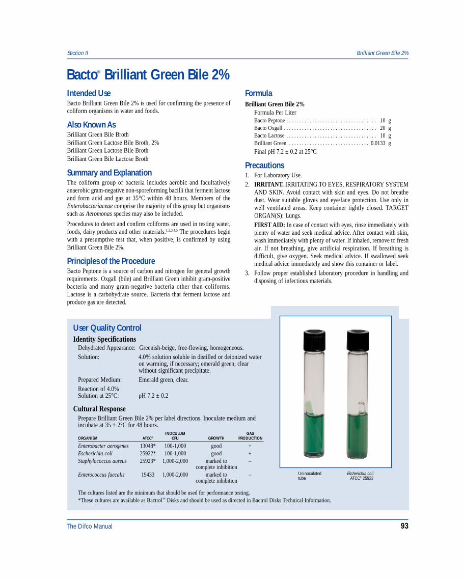
The Difco Manual 93
Section II Brilliant Green Bile 2%
Bacto® Brilliant Green Bile 2%
User Quality ControlIdentity Specifications
Dehydrated Appearance: Greenish-beige, free-flowing, homogeneous.Solution: 4.0% solution soluble in distilled or deionized water
on warming, if necessary; emerald green, clearwithout significant precipitate.
Prepared Medium: Emerald green, clear.Reaction of 4.0%Solution at 25°C: pH 7.2 ± 0.2
Cultural ResponsePrepare Brilliant Green Bile 2% per label directions. Inoculate medium andincubate at 35 ± 2°C for 48 hours.
INOCULUM GASORGANISM ATCC® CFU GROWTH PRODUCTION
Enterobacter aerogenes 13048* 100-1,000 good +Escherichia coli 25922* 100-1,000 good +Staphylococcus aureus 25923* 1,000-2,000 marked to –
complete inhibitionEnterococcus faecalis 19433 1,000-2,000 marked to –
complete inhibition
The cultures listed are the minimum that should be used for performance testing.*These cultures are available as Bactrol™ Disks and should be used as directed in Bactrol Disks Technical Information.
Escherichia coliATCC® 25922
Uninoculatedtube
Intended UseBacto Brilliant Green Bile 2% is used for confirming the presence ofcoliform organisms in water and foods.
Also Known AsBrilliant Green Bile BrothBrilliant Green Lactose Bile Broth, 2%Brilliant Green Lactose Bile BrothBrilliant Green Bile Lactose Broth
Summary and ExplanationThe coliform group of bacteria includes aerobic and facultativelyanaerobic gram-negative non-sporeforming bacilli that ferment lactoseand form acid and gas at 35°C within 48 hours. Members of theEnterobacteriaceae comprise the majority of this group but organismssuch as Aeromonas species may also be included.
Procedures to detect and confirm coliforms are used in testing water,foods, dairy products and other materials.1,2,3,4,5 The procedures beginwith a presumptive test that, when positive, is confirmed by usingBrilliant Green Bile 2%.
Principles of the ProcedureBacto Peptone is a source of carbon and nitrogen for general growthrequirements. Oxgall (bile) and Brilliant Green inhibit gram-positivebacteria and many gram-negative bacteria other than coliforms.Lactose is a carbohydrate source. Bacteria that ferment lactose andproduce gas are detected.
FormulaBrilliant Green Bile 2%
Formula Per LiterBacto Peptone . . . . . . . . . . . . . . . . . . . . . . . . . . . . . . . . . . . 10 gBacto Oxgall . . . . . . . . . . . . . . . . . . . . . . . . . . . . . . . . . . . . 20 gBacto Lactose . . . . . . . . . . . . . . . . . . . . . . . . . . . . . . . . . . . 10 gBrilliant Green . . . . . . . . . . . . . . . . . . . . . . . . . . . . . . . 0.0133 g
Final pH 7.2 ± 0.2 at 25°C
Precautions1. For Laboratory Use.2. IRRITANT. IRRITATING TO EYES, RESPIRATORY SYSTEM
AND SKIN. Avoid contact with skin and eyes. Do not breathedust. Wear suitable gloves and eye/face protection. Use only inwell ventilated areas. Keep container tightly closed. TARGETORGAN(S): Lungs.FIRST AID: In case of contact with eyes, rinse immediately withplenty of water and seek medical advice. After contact with skin,wash immediately with plenty of water. If inhaled, remove to freshair. If not breathing, give artificial respiration. If breathing isdifficult, give oxygen. Seek medical advice. If swallowed seekmedical advice immediately and show this container or label.
3. Follow proper established laboratory procedure in handling anddisposing of infectious materials.
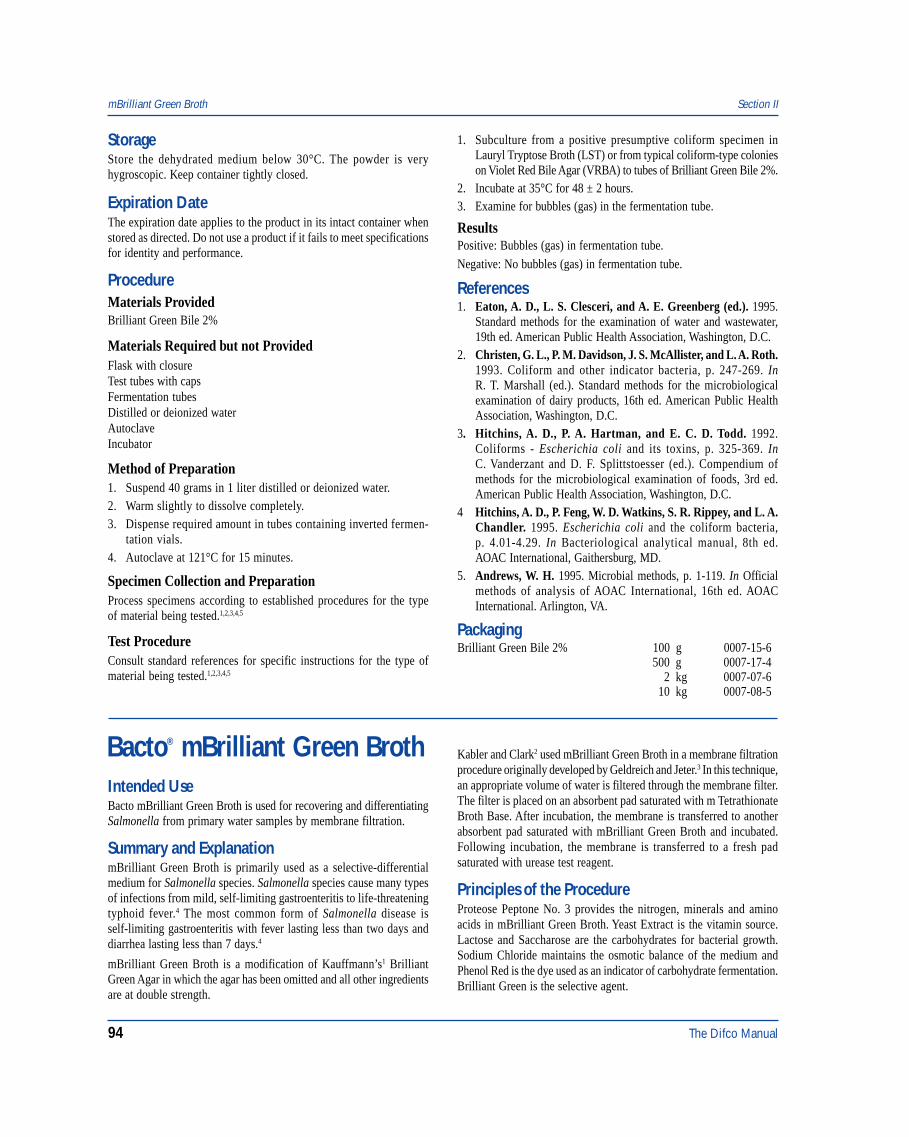
94 The Difco Manual
Bacto® mBrilliant Green BrothIntended UseBacto mBrilliant Green Broth is used for recovering and differentiatingSalmonella from primary water samples by membrane filtration.
Summary and ExplanationmBrilliant Green Broth is primarily used as a selective-differentialmedium for Salmonella species. Salmonella species cause many typesof infections from mild, self-limiting gastroenteritis to life-threateningtyphoid fever.4 The most common form of Salmonella disease isself-limiting gastroenteritis with fever lasting less than two days anddiarrhea lasting less than 7 days.4
mBrilliant Green Broth is a modification of Kauffmann’s1 BrilliantGreen Agar in which the agar has been omitted and all other ingredientsare at double strength.
Kabler and Clark2 used mBrilliant Green Broth in a membrane filtrationprocedure originally developed by Geldreich and Jeter.3 In this technique,an appropriate volume of water is filtered through the membrane filter.The filter is placed on an absorbent pad saturated with m TetrathionateBroth Base. After incubation, the membrane is transferred to anotherabsorbent pad saturated with mBrilliant Green Broth and incubated.Following incubation, the membrane is transferred to a fresh padsaturated with urease test reagent.
Principles of the ProcedureProteose Peptone No. 3 provides the nitrogen, minerals and aminoacids in mBrilliant Green Broth. Yeast Extract is the vitamin source.Lactose and Saccharose are the carbohydrates for bacterial growth.Sodium Chloride maintains the osmotic balance of the medium andPhenol Red is the dye used as an indicator of carbohydrate fermentation.Brilliant Green is the selective agent.
mBrilliant Green Broth Section II
StorageStore the dehydrated medium below 30°C. The powder is veryhygroscopic. Keep container tightly closed.
Expiration DateThe expiration date applies to the product in its intact container whenstored as directed. Do not use a product if it fails to meet specificationsfor identity and performance.
ProcedureMaterials ProvidedBrilliant Green Bile 2%
Materials Required but not ProvidedFlask with closureTest tubes with capsFermentation tubesDistilled or deionized waterAutoclaveIncubator
Method of Preparation1. Suspend 40 grams in 1 liter distilled or deionized water.2. Warm slightly to dissolve completely.3. Dispense required amount in tubes containing inverted fermen-
tation vials.4. Autoclave at 121°C for 15 minutes.
Specimen Collection and PreparationProcess specimens according to established procedures for the typeof material being tested.1,2,3,4,5
Test ProcedureConsult standard references for specific instructions for the type ofmaterial being tested.1,2,3,4,5
1. Subculture from a positive presumptive coliform specimen inLauryl Tryptose Broth (LST) or from typical coliform-type colonieson Violet Red Bile Agar (VRBA) to tubes of Brilliant Green Bile 2%.
2. Incubate at 35°C for 48 ± 2 hours.3. Examine for bubbles (gas) in the fermentation tube.
ResultsPositive: Bubbles (gas) in fermentation tube.
Negative: No bubbles (gas) in fermentation tube.
References1. Eaton, A. D., L. S. Clesceri, and A. E. Greenberg (ed.). 1995.
Standard methods for the examination of water and wastewater,19th ed. American Public Health Association, Washington, D.C.
2. Christen, G. L., P. M. Davidson, J. S. McAllister, and L. A. Roth.1993. Coliform and other indicator bacteria, p. 247-269. InR. T. Marshall (ed.). Standard methods for the microbiologicalexamination of dairy products, 16th ed. American Public HealthAssociation, Washington, D.C.
3. Hitchins, A. D., P. A. Hartman, and E. C. D. Todd. 1992.Coliforms - Escherichia coli and its toxins, p. 325-369. InC. Vanderzant and D. F. Splittstoesser (ed.). Compendium ofmethods for the microbiological examination of foods, 3rd ed.American Public Health Association, Washington, D.C.
4 Hitchins, A. D., P. Feng, W. D. Watkins, S. R. Rippey, and L. A.Chandler. 1995. Escherichia coli and the coliform bacteria,p. 4.01-4.29. In Bacteriological analytical manual, 8th ed.AOAC International, Gaithersburg, MD.
5. Andrews, W. H. 1995. Microbial methods, p. 1-119. In Officialmethods of analysis of AOAC International, 16th ed. AOACInternational. Arlington, VA.
PackagingBrilliant Green Bile 2% 100 g 0007-15-6
500 g 0007-17-42 kg 0007-07-6
10 kg 0007-08-5
The Difco Manual 95
User Quality ControlIdentity Specifications
Dehydrated Appearance: Pink, free-flowing, homogeneous.Solution: 7.6% solution, soluble in distilled or
deionized water; greenish-red, slightlyopalescent.
Prepared Medium Greenish-red, slightly opalescent.Reaction of 7.6%Solution at 25°C: pH 6.9 ± 0.2
Cultural ResponsePrepare mBrilliant Green Broth per label directions. Inoculate using themembrane filter technique and incubate at 35 ± 2°C in a humid atmospherefor 18-24 hours.
INOCULUMORGANISM ATCC® CFU GROWTH COLOR OF COLONY
Escherichia coli 25922* 20-80 good yellowSalmonella enteritidis 13076 20-80 good pink to redSalmonella typhimurium 14028* 20-80 good pink to red
The cultures listed are the minimum that should be used for performance testing.*These cultures are available as Bactrol™ Disks and should be used as directed in Bactrol Disks Technical Information.
Escherichia coliATCC® 25922
Salmonella typhimuriumATCC® 14028
FormulamBrilliant Green Broth
Formula Per LiterBacto Proteose Peptone No. 3 . . . . . . . . . . . . . . . . . . . . . . 20 gBacto Yeast Extract . . . . . . . . . . . . . . . . . . . . . . . . . . . . . . . . 6 gBacto Lactose . . . . . . . . . . . . . . . . . . . . . . . . . . . . . . . . . . . 20 gBacto Saccharose . . . . . . . . . . . . . . . . . . . . . . . . . . . . . . . . 20 gSodium Chloride . . . . . . . . . . . . . . . . . . . . . . . . . . . . . . . . . 10 gBacto Phenol Red . . . . . . . . . . . . . . . . . . . . . . . . . . . . . . . 0.16 gBrilliant Green . . . . . . . . . . . . . . . . . . . . . . . . . . . . . . . . 0.025 g
Final pH 6.9 ± 0.2 at 25°C
Precautions1. For Laboratory Use.2. Follow proper, established laboratory procedures in handling and
disposing of infectious materials.
StorageStore the dehydrated medium below 30°C. The dehydrated medium isvery hygroscopic. Keep container tightly closed.
Use the rehydrated medium within 24 hours.
Expiration DateThe expiration date applies to the product in its intact container whenstored as directed. Do not use a product if it fails to meet specificationsfor identity and performance.
ProcedureMaterials ProvidedmBrilliant Green Broth
Materials Required But Not ProvidedGlasswareSterile absorbent padMembrane filtration equipmentIncubator (35°C)Sterile Petri dishes, 50 x 9 mmDistilled or deionized water
Method of Preparation1. Suspend 7.6 grams in 100 ml of distilled or deionized water.2. Heat to boiling to dissolve completely. Do not autoclave.3. Cool to room temperature.4. Dispense 2 ml amounts onto sterile absorbent pads.5. Use the rehydrated medium within 24 hours.
Specimen Collection and PreparationObtain and process water samples according to the techniques andprocedures established by laboratory policy.
Test Procedure1. Inoculate a water sample using the membrane filtration procedure.2. Place the filter on a pad saturated with mBrilliant Green Broth.3. Incubate at 35 ± 2°C in a humid atmosphere for 18-24 hours.4. After incubation, examine for growth and the color of the colonies.
Section II mBrilliant Green Broth
96 The Difco Manual
ResultsSalmonella species form pink to red colonies.
Limitations of the Procedure1. Since the nutritional requirements of organisms vary, some strains may
be encountered that fail to grow or grow poorly on this medium.
References1. Kauffmann, F. 1935. Weitere Erfahrungen mit den kombinierten
Anreicherungsverfahren fur Salmonellabacillen. Z. Hyg.Infektionskr. 117:26.
2. Kabler and Clark. 1952. Am. J. Public Health 42:390.3. Geldreich and Jeter. 1952. Bacteriol. Proc. SAB., Boston, MA.4. Murray, P. R., E. J. Baron, M. A. Pfaller, F. C. Tenover, and
R. H. Yolken (ed.). 1995. Manual of clinical microbiology, 6th ed.American Society for Microbiology, Washington, D.C.
PackagingmBrilliant Green Broth 500 g 0494-17
Brucella Agar & Brucella Broth Section II
Bacto® Brucella AgarBacto Brucella Broth
User Quality ControlIdentity Specifications
Brucella AgarDehydrated Appearance: Beige, free-flowing, homogeneous.Solution: 4.3% solution, soluble in distilled
or deionized water with frequentagitation on boiling. Light amber,very slightly to slightly opalescent.
Prepared Medium: Light amber, slightly opalescent.Reaction of 4.3%Solution at 25°C: pH 7.0 ± 0.2
Brucella BrothDehydrated Appearance: Light beige, free-flowing, homogeneous.Solution: 2.8% solution, soluble in distilled
or deionized water. Light amber,clear to very slightly opalescent,no precipitate.
Prepared Medium: Light amber, clear to very slightlyopalescent, no precipitate.
Reaction of 2.8%Solution at 25°C: pH 7.0 ± 0.2
Cultural ResponsePrepare Brucella Agar or Brucella Broth per label directions.Inoculate and incubate at 35 ± 2°C under 5-10% CO2 for24-72 hours.
INOCULUMORGANISM ATCC® CFU GROWTH
Brucella abortus 4315 100-1,000 goodBrucella melitensis 4309 100-1,000 goodBrucella suis 4314 100-1,000 good
The cultures listed are the minimum that should be used forperformance testing.
Intended UseBacto Brucella Agar is used for isolating and cultivating Brucella.Bacto Brucella Broth is used for cultivating Brucella and otherfastidious microorganisms.
Summary and ExplanationBrucella Agar and Brucella Broth are prepared according to the APHAformula for Albimi Broth, which is used for the isolation of Brucellaspecies.1 Brucellosis is a zoonotic disease with a domestic-animalreservoir.2 Transmission by milk, milk products, meat and directcontact with infected animals is the usual route of exposure.2
Brucella Agar is used as a general medium for the cultivation offastidious microorganisms, e.g., Streptococcus pneumoniae,Streptococcus viridans and Neisseria meningitidis.3 With the additionof blood, Brucella Agar can be used to determine hemolytic reactionsof pathogenic bacteria.3 Brucella Agar can be used as a base for theisolation of Campylobacter species.3
Brucella Broth is recommended for the isolation of Brucella speciesfrom blood cultures.4,5 Brucella Broth is specified in the Compendiumof Methods for the Microbiological Examination of Food.6
Principles of the ProcedurePeptamin provides nitrogen and amino acids. Tryptone provides nitrogen.Yeast Extract adds essential vitamins. Dextrose is a carbon source;Sodium Bisulfite enhances growth. Sodium Chloride maintains theosmotic balance. Bacto Agar is the solidifying agent in Brucella Agar.Supplemental blood (5-10%) provides additional growth factors forfastidious microorganisms and is used to determine hemolytic reactions.
FormulaBrucella Agar
Formula Per LiterBacto Tryptone . . . . . . . . . . . . . . . . . . . . . . . . . . . . . . . . . . 10 gBacto Peptamin . . . . . . . . . . . . . . . . . . . . . . . . . . . . . . . . . . 10 gBacto Dextrose . . . . . . . . . . . . . . . . . . . . . . . . . . . . . . . . . . . 1 gBacto Yeast Extract . . . . . . . . . . . . . . . . . . . . . . . . . . . . . . . . 2 gSodium Chloride . . . . . . . . . . . . . . . . . . . . . . . . . . . . . . . . . . 5 gSodium Bisulfite . . . . . . . . . . . . . . . . . . . . . . . . . . . . . . . . . 0.1 gBacto Agar . . . . . . . . . . . . . . . . . . . . . . . . . . . . . . . . . . . . . 15 g
Final pH 7.0 ± 0.2 at 25°C
Brucella BrothFormula Per LiterBacto Tryptone . . . . . . . . . . . . . . . . . . . . . . . . . . . . . . . . . . 10 gBacto Peptamin . . . . . . . . . . . . . . . . . . . . . . . . . . . . . . . . . . 10 gBacto Dextrose . . . . . . . . . . . . . . . . . . . . . . . . . . . . . . . . . . . 1 gBacto Yeast Extract . . . . . . . . . . . . . . . . . . . . . . . . . . . . . . . . 2 gSodium Chloride . . . . . . . . . . . . . . . . . . . . . . . . . . . . . . . . . . 5 gSodium Bisulfite . . . . . . . . . . . . . . . . . . . . . . . . . . . . . . . . . 0.1 g
Final pH 7.0 ± 0.2 at 25°C
The Difco Manual 97
Section II Bryant and Burkey Medium
Precautions1. For Laboratory Use.2. Brucella species are classified as Biosafety Level 3 pathogens. All
manipulations with live cultures and antigens must be confined toa Class II biological safety cabinet (BSC).2
3. Follow proper established laboratory procedures in handling anddisposing of infectious materials.
StorageStore the dehydrated medium below 30°C. The dehydrated medium isvery hygroscopic. Keep container tightly closed.
Expiration DateThe expiration date applies to the product in its intact container whenstored as directed. Do not use a product if it fails to meet specificationsfor identity and performance.
ProcedureMaterials ProvidedBrucella AgarBrucella Broth
Materials Required But Not ProvidedGlasswareAutoclaveIncubator (35°C)Waterbath (45-50°C) (optional)Sterile Petri dishes or tubesSterile defibrinated blood (optional)
Method of Preparation1. Brucella Agar: Suspend 43 grams in 1 liter distilled or deionized
water and boil to dissolve completely.Brucella Broth: Dissolve 28 grams in 1 liter distilled or deionizedwater.
2. Autoclave at 121°C for 15 minutes. Cool to 45-55°C.3. OPTIONAL: To prepare Brucella Blood Agar, aseptically add
5-10% sterile defibrinated blood at 45-50°C. Mix well.
Specimen Collection and PreparationObtain and process specimens according to the techniques andprocedures established by institutional policy.
Test ProcedureFor a complete discussion of the isolation and identification ofBrucella, refer to appropriate procedures outlined in the references.2,4,5
ResultsRefer to apropriate references and procedures for results.
Limitations of the Procedure1. Since the nutritional requirements of organisms vary, some strains
may be encountered that fail to grow or grow poorly on this medium.2. Hemolytic reactions of many microorganisms are different on
horse blood from those on sheep blood agar; e.g., some Group Dstreptococci exhibit beta hemolysis on horse blood but not on sheepblood and are mistaken for Group A.3
References1. Hausler, W. J. (ed.). 1976. Standard methods for the examination
of dairy products, 14th ed. American Public Health Association,Washington, D.C.
2. Moyer, N. P., and L. A. Holcomb. 1995. Brucella, p. 549-555. InP. R. Murray, E. J. Baron, M. A. Pfaller, F. C. Tenover, and R. H.Yolken (ed.). Manual of clinical microbiology, 6th ed. AmericanSociety for Microbiology, Washington, D.C.
3. MacFaddin, J. D. 1985. Media for isolation-cultivation-identification-maintenance of medical bacteria, vol. 1, p.110-114.Williams & Wilkins, Baltimore, MD.
4. Isenberg, H. D. (ed.). 1992. Clinical microbiology procedureshandbook. American Society for Microbiology, Washington, D.C.
5. Baron, E. J., L. R. Peterson, and S. M. Finegold. 1994. Bailey &Scott’s diagnostic microbiology, 9th ed. Mosby-Year Book, Inc.,St. Louis, MO.
6. Vanderzant, C., and D. F. Splittstoesser (ed.). 1992. Compen-dium of methods for the microbiological examination of food,3rd ed. American Public Health Association, Washington, D.C.
PackagingBrucella Agar 500 g 0964-17
2 kg 0964-0710 kg 0964-08
Brucella Broth 500 g 0495-1710 kg 0495-08
Bacto® Bryant and Burkey MediumIntended UseBacto Bryant and Burkey Medium is used for detecting andenumerating spores of lactate-fermenting Clostridium in milk anddairy products.
Summary and ExplanationBryant and Burkey Medium is based on the lactate fermentation mediadescribed by Rosenberger1 and Bryant and Burkey2, as modified by
Bergère et al,3 who reported that their medium could be used for detectingand enumerating C. tyrobutyricum spores in milk and dairy products.3-5
Principles of the ProcedureTryptone, Yeast Extract, Beef Extract Desiccated and L-CysteineHydrochloride provide nutrients and cofactors required for goodgrowth of clostridia. Selectivity of this medium is achieved throughthe addition of Sodium Acetate, which is also the principal promoter of
98 The Difco Manual
germination by C. tyrobutyricum spores.6 Sodium Lactate isfermented under anaerobic conditions by C. tyrobutyricum and otherlactate-fermenting clostridia, producing hydrogen and carbon dioxide.Gas production is demonstrated by an upward movement of a paraffinplug which is overlaid on the medium. Resazurin is included in themedium to show anaerobiosis, turning from pink (aerobic) to colorlessunder anaerobic conditions.
During processing, the sample is heated at 75°C for 15 minutes to killvegetative cells and activate germination of spores.
FormulaBryant and Burkey Medium
Formula Per LiterBacto Tryptone . . . . . . . . . . . . . . . . . . . . . . . . . . . . . . . . . . 15 gBacto Yeast Extract . . . . . . . . . . . . . . . . . . . . . . . . . . . . . . . . 5 gBacto Beef Extract, Desiccated . . . . . . . . . . . . . . . . . . . . . 7.5 gSodium Acetate . . . . . . . . . . . . . . . . . . . . . . . . . . . . . . . . . . . 5 gL-Cysteine Hydrochloride . . . . . . . . . . . . . . . . . . . . . . . . . 0.5 gSodium Lactate . . . . . . . . . . . . . . . . . . . . . . . . . . . . . . . . . . . 5 gResazurin . . . . . . . . . . . . . . . . . . . . . . . . . . . . . . . . . . . . 0.0025 g
Final pH 5.9 ± 0.2 at 25°C
Precautions1. For Laboratory Use.2. Follow proper, established laboratory procedures in handling and
disposing of infectious materials.
Storage1. Store the dehydrated medium below 30°C. The powder is very
hygroscopic. Keep container tightly closed.2. Store prepared medium at 2-8°C.
Expiration DateThe expiration date applies to the product in its intact container whenstored as directed. Do not use a product if it fails to meet specificationsfor identity and performance.
ProcedureMaterials ProvidedBryant and Burkey Medium
Materials Required But Not ProvidedFlasks with closuresDistilled or deionized waterAutoclaveIncubator (35 ± 2°C)
Method of PreparationNOTE: This product contains sodium lactate; it is not necessary to addsodium lactate during preparation.
1. Dissolve 38 grams in 1 liter of distilled or deionized water.2. Dispense 10 ml amounts into tubes.3. Autoclave at 121°C for 15 minutes.
Specimen Collection and Preparation1. Collect food samples in sterile containers and transport immediately
to the laboratory following recommended guidelines.2. Process each food sample using procedures appropriate for that
sample.
Test ProcedureThree-tube Most Probable Number (MPN) Method1. Before use, heat tubes to boiling for 10 minutes to regenerate
anaerobic conditions. Note: This step is required only with tubesstored under aerobic conditions. Tubes stored under anaerobicconditions or freshly sterilized tubes do not need additional heating.
2. Prepare 10-fold dilutions of the sample and inoculate triplicatetubes of Bryant and Burkey Medium with 1 ml of each sampledilution.
3. Pour approximately 2 ml of melted paraffin (60-65°C), previouslyautoclaved at 121°C for 20 minutes, into each tube.
4. Heat the tubes at 75°C for 15 minutes to kill vegetative cells andactivate spores; allow to cool to room temperature.
5. Incubate tubes at 35°C for 6 days.6. Examine tubes for growth and gas production after 48 hours of
incubation and daily for up to 6 days.
ResultsTubes showing both growth and production of gas (indicated by upwardmovement of the paraffin more than 1 cm) are considered positive forthe presence of lactate-fermenting clostridial spores. Determine thespore count using the Most Probable Number (MPN) method.
Bryant and Burkey Medium Section II
User Quality ControlIdentity Specifications
Dehydrated Appearance: Tan, free-flowing, homogeneous.Solution: 3.8% solution, soluble in distilled or
deionized water. Solution is light tomedium amber, clear when hot,becoming red upon cooling.
Reaction of 3.8%Solution at 25°C: pH 5.9 ± 0.2
Cultural ResponsePrepare Bryant and Burkey Medium per label directions.Inoculate using Most Probable Number (MPN) method andincubate at 35 ± 2°C for 6 days.
ATCC® OR GASORGANISM STRAIN INOCULUM GROWTH PRODUCTION
Clostridium CNRZ 500 MPN method good >1 cm of gastyrobutyricum
Clostridium CNRZ 510 MPN method good >1 cm of gastyrobutyricum
Clostridium CNRZ 608 MPN method good >1 cm of gastyrobutyricum
Clostridium 25755 MPN method good >1 cm of gastyrobutyricum
The cultures listed are the minimum that should be used forperformance testing.
The Difco Manual 99
Section II Buffered Peptone Water & Modified Buffered Peptone Water
References1. Rosenberger, K. F. 1951. The development of methods for the
study of obligate anaerobes in silage. Proc. Soc. Appl. Bacteriol.14:161-164.
2. Bryant, M. P., and L. A. Burkey. 1956. The characteristics oflactate-fermenting sporeforming anaerobes from silage.J. Bacteriol. 71:43-46.
3. Bergère, J. L., P. Gouet, J. Hermier, and G. Mocquot. 1968. LesClostridium du groupe butyrique dans les produits laitiers. Ann.Inst. Pasteur Lille. 19:41-54.
4. Cerf, O., and J. L. Bergère. 1968. La numération des spores deClostridium et son application au lait et aux produits laitiers.
Numération des différents groupes de Clostridium. Le Lait48:501-519.
5. Bergère, J. L. 1979. Developpement de l’ensilage. Sesconséquences sur la qualité du lait et des produits laiters. Revuelaitère française.
6. Touraille, C., and J. L. Bergère. 1974. La germination de la sporede Clostridium tyrobutyricum. Biochimie. 56:404-422.
PackagingBryant and Burkey Medium 500 g 0645-17
2 kg 0645-07
Bacto® Buffered Peptone WaterBacto Modified Buffered Peptone Water
User Quality ControlIdentity Specifications
Buffered Peptone WaterDehydrated Appearance: Cream-white to light tan, free-flowing,
homogeneous.Solution: 2.0% solution, soluble in distilled or
deionized water. Solution is lightamber, clear.
Prepared Medium: Light amber, clear.Reaction of 2.0%Solution at 25°C: pH 7.2 ± 0.2
Modified Buffered Peptone WaterDehydrated Appearance: Light beige, free-flowing, homogenous.Solution: 2.5% solution, soluble in distilled or
deionized water. Solution is lightamber, clear.
Prepared Medium: Light amber, clear.Reaction of 2.5%Solution at 25°C: pH 7.2 ± 0.2
Cultural ResponsePrepare Buffered Peptone Water or Modified Buffered PeptoneWater per label directions. Inoculate and incubate at 35 ± 2°Cfor 18-24 hours.
INOCULUMORGANISM ATCC® CFU GROWTH
Salmonella enteritidis 13076 10-100 goodSalmonella typhi 19430 10-100 goodSalmonella typhimurium 14028* 10-100 good
The cultures listed are the minimum that should be used forperformance testing.*This culture is available as Bactrol™ Disks and should be used as directed in Bactrol Disks Technical Information.
Intended UseBacto Buffered Peptone Water is used for preenriching damagedSalmonella species from food specimens to increase recovery.
Bacto Modified Buffered Peptone Water is used for preenrichingSalmonella species from food specimens to increase recovery.
Summary and ExplanationEdel and Kampelmacher1 noted that food preservation techniquesinvolving heat, desiccation, preservatives, high osmotic pressure or pHchanges cause sublethal injury to salmonellae. Preenrichment in anonselective medium allows for repair of cell damage and facilitatesthe recovery of salmonellae. Lactose Broth is frequently used for thispurpose but it may be detrimental to recovering salmonellae.2 BufferedPeptone Water maintains a high pH over the preenrichment period andresults in repair of injured cells that may be sensitive to low pH.3 Thisis particularly important for vegetable specimens which have a lowbuffering capacity. These media can be used for testing dry poultryfeed.4 Buffered Peptone Water is a standard methods medium.5
Modified Buffered Peptone Water provides additional buffering capacitywhen organisms have been enriched in a pre-enrichment mediumcontaining a high carbohydrate concentration.
Principles of the ProcedureBuffered Peptone Water and Modified Buffered Peptone Water containPeptone as a source of carbon, nitrogen, vitamins and minerals.Sodium Chloride maintains the osmotic balance. Phosphates buffer themedium.
FormulaBuffered Peptone Water
Formula Per LiterPeptone . . . . . . . . . . . . . . . . . . . . . . . . . . . . . . . . . . . . . . . . 10 gSodium Chloride . . . . . . . . . . . . . . . . . . . . . . . . . . . . . . . . . . 5 gSodium Phosphate, Dibasic . . . . . . . . . . . . . . . . . . . . . . . . 3.5 gPotassium Phosphate, Monobasic . . . . . . . . . . . . . . . . . . . 1.5 g
Final pH 7.2 ± 0.2 at 25°C
100 The Difco Manual
Modified Buffered Peptone WaterFormula Per LiterPeptone . . . . . . . . . . . . . . . . . . . . . . . . . . . . . . . . . . . . . . . . 10 gSodium Chloride . . . . . . . . . . . . . . . . . . . . . . . . . . . . . . . . . . 5 gSodium Phosphate, Dibasic . . . . . . . . . . . . . . . . . . . . . . . . . 7 gPotassium Phosphate, Monobasic . . . . . . . . . . . . . . . . . . . . 3 g
Final pH 7.2 ± 0.2 at 25°C
Precautions1. For Laboratory Use.2. Buffered Peptone Water
MAY BE IRRITATING TO EYES, RESPIRATORY SYSTEMAND SKIN. (US) Avoid contact with skin and eyes. Do not breathedust. Wear suitable protective clothing. Keep container tightly closed.
Modified Buffered Peptone WaterIRRITANT. MAY BE IRRITATING TO EYES, RESPIRATORYSYSTEM AND SKIN. (US) Avoid contact with skin and eyes. Donot breathe dust. Wear suitable protective clothing. Keep containertightly closed.FIRST AID: In case of contact with eyes, rinse immediately withplenty of water and seek medical advice. After contact with skin,wash immediately with plenty of water. If inhaled, remove to freshair. If not breathing, give artificial respiration. If breathing is diffi-cult, give oxygen. Seek medical advice. If swallowed seek medicaladvice immediately and show this container or label.
3. Follow proper established laboratory procedures in handling anddisposing of infectious materials.
StorageStore the dehydrated medium below 30°C. The dehydrated medium isvery hygroscopic. Keep container tightly closed.
Expiration DateThe expiration date applies to the product in its intact container whenstored as directed. Do not use a product if it fails to meet specificationsfor identity and performance.
ProcedureMaterials ProvidedBuffered Peptone WaterModified Buffered Peptone Water
Materials Required but not ProvidedGlassware
Distilled or deionized waterAutoclaveIncubator (35°C)
Method of Preparation1. Dissolve the medium in 1 liter distilled or deionized water:
Buffered Peptone Water - 20 grams;Modified Buffered Peptone Water - 25 grams.
2. Autoclave at 121°C for 15 minutes.
Specimen Collection and PreparationCollect specimens according to recommended guidelines.
Test ProcedureTest specimens according to recommended guidelines.
ResultsGrowth is indicated by turbidity.
Limitations of the Procedure1. The types and numbers of competing flora in the test sample can
affect recovery and may overgrow salmonellae.
References1. Edel, W., and E. H. Kampelmacher. 1973. Bull. World Hlth.
Org. 48:167-174.2. Angelotti, R. 1963. Microbiological quality of foods. Academic
Press, New York.3. Sadovski, A. Y. 1977. J. Food Technol. 12:85-91.4. Juven, B. J., N. A. Cox, J. S. Bailey, J. E. Thomson, O. W.
Charles, and J. V. Schutze. 1984. Recovery of Salmonella fromartificially contaminated poultry feeds in non-selective andselective broth media. Jour. of Food Prot. 47:299-302.
5. Flowers, R. S., J-Y. D’Aoust, W. H. Andrews, and J. S. Bailey.1992. Salmonella, p. 371-422. In C. Vanderzant, and D. F.Splittstoesser (ed.). Compendium of methods for the microbiologicalexamination of foods, 3rd ed. American Public Health Association,Washington, D.C.
PackagingBuffered Peptone Water 500 g 1810-17
2 kg 1810-0710 kg 1810-08
Modified Buffered Peptone Water 500 g 1833-17
Bushnell-Haas Broth Section II
Bacto® Bushnell-Haas BrothIntended UseBacto Bushnell-Haas Broth is used for studying microbial utilizationof hydrocarbons.
Also Known AsBushnell-Haas Broth is also referred to as Bushnell-Haas marinesalts broth.
Summary and ExplanationBushnell-Haas Broth, prepared according to the formula described byBushnell and Haas1, is used to evaluate the ability of microorganismsto decompose hydrocarbons. It is formulated without a carbon sourcewhich allows for the addition of alternate hydrocarbons such as kerosene,light and heavy mineral oils, paraffin wax, and gasoline.
Bushnell-Haas was recommended for the microbiological examinationof fuels by the Society for Industrial Microbiology (SIM) Committeeon Microbiological Deterioration of Fuels.2 The medium was used to
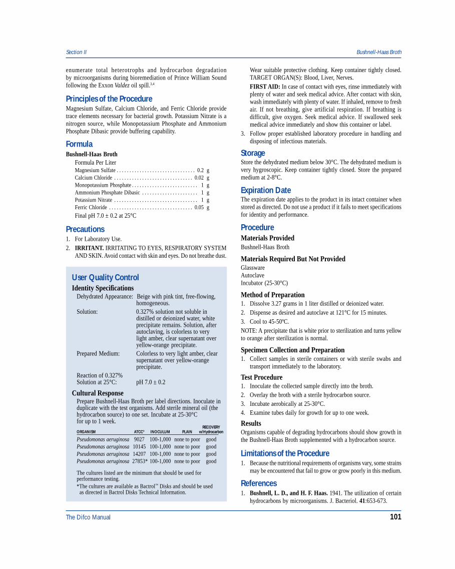
The Difco Manual 101
Section II Bushnell-Haas Broth
User Quality ControlIdentity Specifications
Dehydrated Appearance: Beige with pink tint, free-flowing,homogeneous.
Solution: 0.327% solution not soluble indistilled or deionized water, whiteprecipitate remains. Solution, afterautoclaving, is colorless to verylight amber, clear supernatant overyellow-orange precipitate.
Prepared Medium: Colorless to very light amber, clearsupernatant over yellow-orangeprecipitate.
Reaction of 0.327%Solution at 25°C: pH 7.0 ± 0.2
Cultural ResponsePrepare Bushnell-Haas Broth per label directions. Inoculate induplicate with the test organisms. Add sterile mineral oil (thehydrocarbon source) to one set. Incubate at 25-30°Cfor up to 1 week.
RECOVERYORGANISM ATCC® INOCULUM PLAIN w/Hydrocarbon
Pseudomonas aeruginosa 9027 100-1,000 none to poor goodPseudomonas aeruginosa 10145 100-1,000 none to poor goodPseudomonas aeruginosa 14207 100-1,000 none to poor goodPseudomonas aeruginosa 27853* 100-1,000 none to poor good
The cultures listed are the minimum that should be used forperformance testing.*The cultures are available as Bactrol™ Disks and should be used as directed in Bactrol Disks Technical Information.
enumerate total heterotrophs and hydrocarbon degradationby microorganisms during bioremediation of Prince William Soundfollowing the Exxon Valdez oil spill.3,4
Principles of the ProcedureMagnesium Sulfate, Calcium Chloride, and Ferric Chloride providetrace elements necessary for bacterial growth. Potassium Nitrate is anitrogen source, while Monopotassium Phosphate and AmmoniumPhosphate Dibasic provide buffering capability.
FormulaBushnell-Haas Broth
Formula Per LiterMagnesium Sulfate . . . . . . . . . . . . . . . . . . . . . . . . . . . . . . . 0.2 gCalcium Chloride . . . . . . . . . . . . . . . . . . . . . . . . . . . . . . . 0.02 gMonopotassium Phosphate . . . . . . . . . . . . . . . . . . . . . . . . . . 1 gAmmonium Phosphate Dibasic . . . . . . . . . . . . . . . . . . . . . . 1 gPotassium Nitrate . . . . . . . . . . . . . . . . . . . . . . . . . . . . . . . . . 1 gFerric Chloride . . . . . . . . . . . . . . . . . . . . . . . . . . . . . . . . . 0.05 g
Final pH 7.0 ± 0.2 at 25°C
Precautions1. For Laboratory Use.2. IRRITANT. IRRITATING TO EYES, RESPIRATORY SYSTEM
AND SKIN. Avoid contact with skin and eyes. Do not breathe dust.
Wear suitable protective clothing. Keep container tightly closed.TARGET ORGAN(S): Blood, Liver, Nerves.FIRST AID: In case of contact with eyes, rinse immediately withplenty of water and seek medical advice. After contact with skin,wash immediately with plenty of water. If inhaled, remove to freshair. If not breathing, give artificial respiration. If breathing isdifficult, give oxygen. Seek medical advice. If swallowed seekmedical advice immediately and show this container or label.
3. Follow proper established laboratory procedure in handling anddisposing of infectious materials.
StorageStore the dehydrated medium below 30°C. The dehydrated medium isvery hygroscopic. Keep container tightly closed. Store the preparedmedium at 2-8°C.
Expiration DateThe expiration date applies to the product in its intact container whenstored as directed. Do not use a product if it fails to meet specificationsfor identity and performance.
ProcedureMaterials ProvidedBushnell-Haas Broth
Materials Required But Not ProvidedGlasswareAutoclaveIncubator (25-30°C)
Method of Preparation1. Dissolve 3.27 grams in 1 liter distilled or deionized water.2. Dispense as desired and autoclave at 121°C for 15 minutes.3. Cool to 45-50ºC.NOTE: A precipitate that is white prior to sterilization and turns yellowto orange after sterilization is normal.
Specimen Collection and Preparation1. Collect samples in sterile containers or with sterile swabs and
transport immediately to the laboratory.
Test Procedure1. Inoculate the collected sample directly into the broth.2. Overlay the broth with a sterile hydrocarbon source.3. Incubate aerobically at 25-30°C.4. Examine tubes daily for growth for up to one week.
ResultsOrganisms capable of degrading hydrocarbons should show growth inthe Bushnell-Haas Broth supplemented with a hydrocarbon source.
Limitations of the Procedure1. Because the nutritional requirements of organisms vary, some strains
may be encountered that fail to grow or grow poorly in this medium.
References1. Bushnell, L. D., and H. F. Haas. 1941. The utilization of certain
hydrocarbons by microorganisms. J. Bacteriol. 41:653-673.
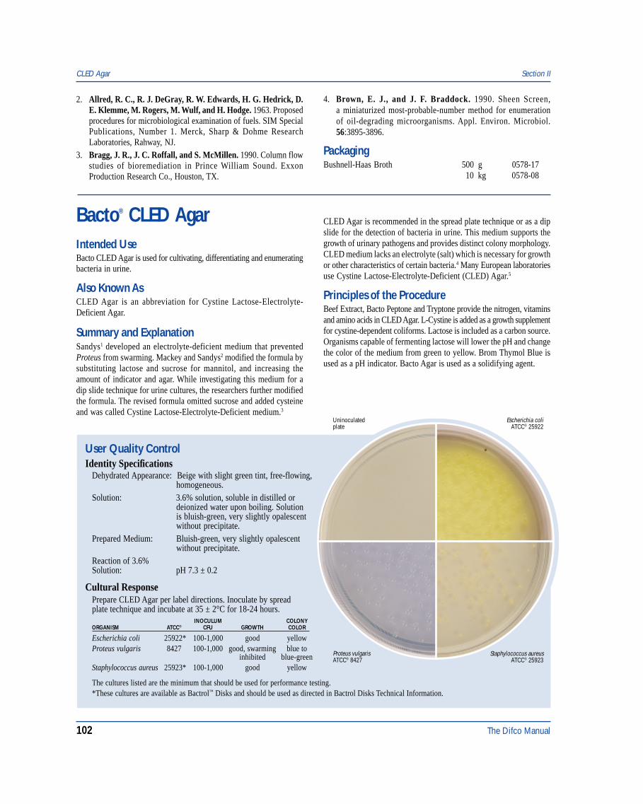
102 The Difco Manual
2. Allred, R. C., R. J. DeGray, R. W. Edwards, H. G. Hedrick, D.E. Klemme, M. Rogers, M. Wulf, and H. Hodge. 1963. Proposedprocedures for microbiological examination of fuels. SIM SpecialPublications, Number 1. Merck, Sharp & Dohme ResearchLaboratories, Rahway, NJ.
3. Bragg, J. R., J. C. Roffall, and S. McMillen. 1990. Column flowstudies of bioremediation in Prince William Sound. ExxonProduction Research Co., Houston, TX.
4. Brown, E. J., and J. F. Braddock. 1990. Sheen Screen,a miniaturized most-probable-number method for enumerationof oil-degrading microorganisms. Appl. Environ. Microbiol.56:3895-3896.
PackagingBushnell-Haas Broth 500 g 0578-17
10 kg 0578-08
Bacto® CLED Agar
User Quality ControlIdentity Specifications
Dehydrated Appearance: Beige with slight green tint, free-flowing,homogeneous.
Solution: 3.6% solution, soluble in distilled ordeionized water upon boiling. Solutionis bluish-green, very slightly opalescentwithout precipitate.
Prepared Medium: Bluish-green, very slightly opalescentwithout precipitate.
Reaction of 3.6%Solution: pH 7.3 ± 0.2
Cultural ResponsePrepare CLED Agar per label directions. Inoculate by spreadplate technique and incubate at 35 ± 2°C for 18-24 hours.
INOCULUM COLONYORGANISM ATCC® CFU GROWTH COLOR
Escherichia coli 25922* 100-1,000 good yellowProteus vulgaris 8427 100-1,000 good, swarming blue to
inhibited blue-greenStaphylococcus aureus 25923* 100-1,000 good yellow
Escherichia coliATCC® 25922
Uninoculatedplate
Proteus vulgarisATCC® 8427
Staphylococcus aureusATCC® 25923
Intended UseBacto CLED Agar is used for cultivating, differentiating and enumeratingbacteria in urine.
Also Known AsCLED Agar is an abbreviation for Cystine Lactose-Electrolyte-Deficient Agar.
Summary and ExplanationSandys1 developed an electrolyte-deficient medium that preventedProteus from swarming. Mackey and Sandys2 modified the formula bysubstituting lactose and sucrose for mannitol, and increasing theamount of indicator and agar. While investigating this medium for adip slide technique for urine cultures, the researchers further modifiedthe formula. The revised formula omitted sucrose and added cysteineand was called Cystine Lactose-Electrolyte-Deficient medium.3
CLED Agar is recommended in the spread plate technique or as a dipslide for the detection of bacteria in urine. This medium supports thegrowth of urinary pathogens and provides distinct colony morphology.CLED medium lacks an electrolyte (salt) which is necessary for growthor other characteristics of certain bacteria.4 Many European laboratoriesuse Cystine Lactose-Electrolyte-Deficient (CLED) Agar.5
Principles of the ProcedureBeef Extract, Bacto Peptone and Tryptone provide the nitrogen, vitaminsand amino acids in CLED Agar. L-Cystine is added as a growth supplementfor cystine-dependent coliforms. Lactose is included as a carbon source.Organisms capable of fermenting lactose will lower the pH and changethe color of the medium from green to yellow. Brom Thymol Blue isused as a pH indicator. Bacto Agar is used as a solidifying agent.
The cultures listed are the minimum that should be used for performance testing.*These cultures are available as Bactrol™ Disks and should be used as directed in Bactrol Disks Technical Information.
CLED Agar Section II
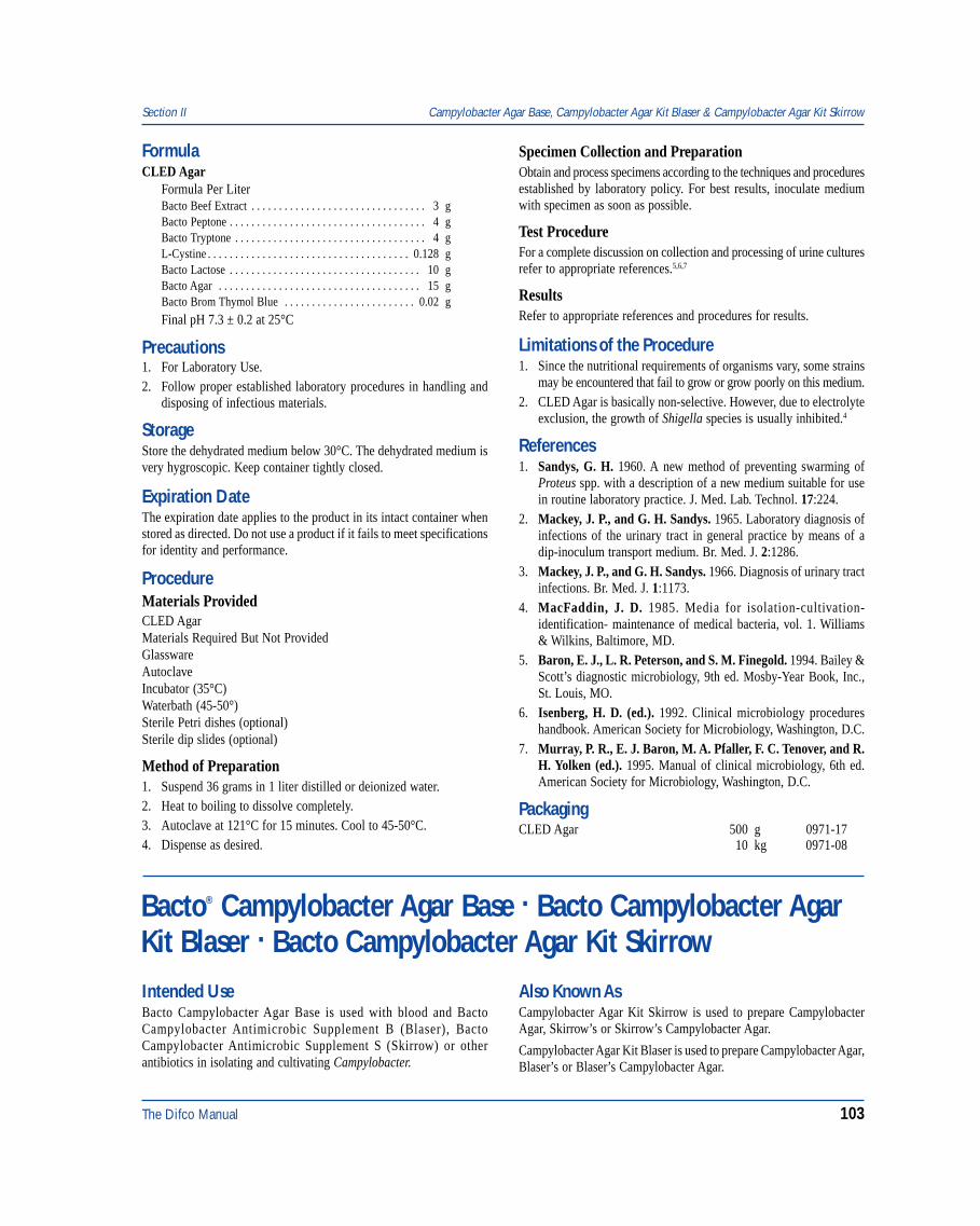
The Difco Manual 103
FormulaCLED Agar
Formula Per LiterBacto Beef Extract . . . . . . . . . . . . . . . . . . . . . . . . . . . . . . . . 3 gBacto Peptone . . . . . . . . . . . . . . . . . . . . . . . . . . . . . . . . . . . . 4 gBacto Tryptone . . . . . . . . . . . . . . . . . . . . . . . . . . . . . . . . . . . 4 gL-Cystine . . . . . . . . . . . . . . . . . . . . . . . . . . . . . . . . . . . . . 0.128 gBacto Lactose . . . . . . . . . . . . . . . . . . . . . . . . . . . . . . . . . . . 10 gBacto Agar . . . . . . . . . . . . . . . . . . . . . . . . . . . . . . . . . . . . . 15 gBacto Brom Thymol Blue . . . . . . . . . . . . . . . . . . . . . . . . 0.02 g
Final pH 7.3 ± 0.2 at 25°C
Precautions1. For Laboratory Use.2. Follow proper established laboratory procedures in handling and
disposing of infectious materials.
StorageStore the dehydrated medium below 30°C. The dehydrated medium isvery hygroscopic. Keep container tightly closed.
Expiration DateThe expiration date applies to the product in its intact container whenstored as directed. Do not use a product if it fails to meet specificationsfor identity and performance.
ProcedureMaterials ProvidedCLED AgarMaterials Required But Not ProvidedGlasswareAutoclaveIncubator (35°C)Waterbath (45-50°)Sterile Petri dishes (optional)Sterile dip slides (optional)
Method of Preparation1. Suspend 36 grams in 1 liter distilled or deionized water.2. Heat to boiling to dissolve completely.3. Autoclave at 121°C for 15 minutes. Cool to 45-50°C.4. Dispense as desired.
Specimen Collection and PreparationObtain and process specimens according to the techniques and proceduresestablished by laboratory policy. For best results, inoculate mediumwith specimen as soon as possible.
Test ProcedureFor a complete discussion on collection and processing of urine culturesrefer to appropriate references.5,6,7
ResultsRefer to appropriate references and procedures for results.
Limitations of the Procedure1. Since the nutritional requirements of organisms vary, some strains
may be encountered that fail to grow or grow poorly on this medium.2. CLED Agar is basically non-selective. However, due to electrolyte
exclusion, the growth of Shigella species is usually inhibited.4
References1. Sandys, G. H. 1960. A new method of preventing swarming of
Proteus spp. with a description of a new medium suitable for usein routine laboratory practice. J. Med. Lab. Technol. 17:224.
2. Mackey, J. P., and G. H. Sandys. 1965. Laboratory diagnosis ofinfections of the urinary tract in general practice by means of adip-inoculum transport medium. Br. Med. J. 2:1286.
3. Mackey, J. P., and G. H. Sandys. 1966. Diagnosis of urinary tractinfections. Br. Med. J. 1:1173.
4. MacFaddin, J. D. 1985. Media for isolation-cultivation-identification- maintenance of medical bacteria, vol. 1. Williams& Wilkins, Baltimore, MD.
5. Baron, E. J., L. R. Peterson, and S. M. Finegold. 1994. Bailey &Scott’s diagnostic microbiology, 9th ed. Mosby-Year Book, Inc.,St. Louis, MO.
6. Isenberg, H. D. (ed.). 1992. Clinical microbiology procedureshandbook. American Society for Microbiology, Washington, D.C.
7. Murray, P. R., E. J. Baron, M. A. Pfaller, F. C. Tenover, and R.H. Yolken (ed.). 1995. Manual of clinical microbiology, 6th ed.American Society for Microbiology, Washington, D.C.
PackagingCLED Agar 500 g 0971-17
10 kg 0971-08
Bacto® Campylobacter Agar Base . Bacto Campylobacter AgarKit Blaser . Bacto Campylobacter Agar Kit SkirrowIntended UseBacto Campylobacter Agar Base is used with blood and BactoCampylobacter Antimicrobic Supplement B (Blaser), BactoCampylobacter Antimicrobic Supplement S (Skirrow) or otherantibiotics in isolating and cultivating Campylobacter.
Also Known AsCampylobacter Agar Kit Skirrow is used to prepare CampylobacterAgar, Skirrow’s or Skirrow’s Campylobacter Agar.
Campylobacter Agar Kit Blaser is used to prepare Campylobacter Agar,Blaser’s or Blaser’s Campylobacter Agar.
Section II Campylobacter Agar Base, Campylobacter Agar Kit Blaser & Campylobacter Agar Kit Skirrow
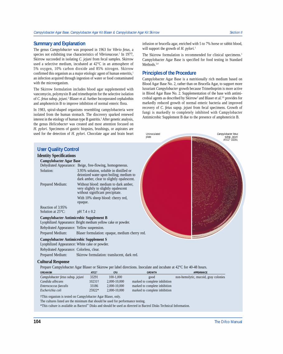
104 The Difco Manual
Campylobacter fetussubsp. jejuni
ATCC® 33291
Uninoculatedplate
User Quality ControlIdentity Specifications
Campylobacter Agar BaseDehydrated Appearance: Beige, free-flowing, homogeneous.Solution: 3.95% solution, soluble in distilled or
deionized water upon boiling; medium todark amber, clear to slightly opalescent.
Prepared Medium: Without blood: medium to dark amber,very slightly to slightly opalescentwithout significant precipitate.With 10% sheep blood: cherry red,opaque.
Reaction of 3.95%Solution at 25°C: pH 7.4 ± 0.2
Campylobacter Antimicrobic Supplement BLyophilized Appearance: Bright medium yellow cake or powder.Rehydrated Appearance: Yellow suspension.Prepared Medium: Blaser formulation: opaque, medium cherry red.
Campylobacter Antimicrobic Supplement SLyophilized Appearance: White cake or powder.Rehydrated Appearance: Colorless, clear.Prepared Medium: Skirrow formulation: translucent, dark red.
Summary and ExplanationThe genus Campylobacter was proposed in 1963 for Vibrio fetus, aspecies not exhibiting true characteristics of Vibrionaceae.1 In 1977,Skirrow succeeded in isolating C. jejuni from fecal samples. Skirrowused a selective medium, incubated at 42°C in an atmosphere of5% oxygen, 10% carbon dioxide and 85% nitrogen. Skirrowconfirmed this organism as a major etiologic agent of human enteritis,1
an infection acquired through ingestion of water or food contaminatedwith the microorganism.
The Skirrow formulation includes blood agar supplemented withvancomycin, polymyxin B and trimethoprim for the selective isolationof C. fetus subsp. jejuni.2 Blaser et al. further Incorporated cephalothinand amphotericin B to improve inhibition of normal enteric flora.
In 1983, spiral-shaped organisms resembling campylobacteria wereisolated from the human stomach. The discovery sparked renewedinterest in the etiology of human type B gastritis.1 After genetic analysis,the genus Helicobacter was created and most attention focused onH. pylori. Specimens of gastric biopsies, brushings, or aspirates areused for the detection of H. pylori. Chocolate agar and brain heart
infusion or brucella agar, enriched with 5 to 7% horse or rabbit blood,will support the growth of H. pylori.1
The Skirrow formulation is recommended for clinical specimens.1
Campylobacter Agar Base is specified for food testing in StandardMethods.3,4
Principles of the ProcedureCampylobacter Agar Base is a nutritionally rich medium based onBlood Agar Base No. 2, rather than on Brucella Agar, to support moreluxuriant Campylobacter growth because Trimethoprim is more activein Blood Agar Base No. 2. Supplementation of the base with antimi-crobial agents as described by Skirrow2 and Blaser et al.5,6 provides formarkedly reduced growth of normal enteric bacteria and improvedrecovery of C. fetus supsp. jejuni from fecal specimens. Growth offungi is markedly to completely inhibited with CampylobacterAntimicrobic Supplement B due to the presence of amphotericin B.
Campylobacter Agar Base, Campylobacter Agar Kit Blaser & Campylobacter Agar Kit Skirrow Section II
Cultural ResponsePrepare Campylobacter Agar Blaser or Skirrow per label directions. Inoculate and incubate at 42°C for 40-48 hours.ORGANISM ATCC® CFU GROWTH APPEARANCE
Campylobacter fetus subsp. jejuni 33291 100-1,000 good non-hemolytic, mucoid, gray coloniesCandida albicans 10231† 2,000-10,000 marked to complete inhibitionEnterococcus faecalis 33186 2,000-10,000 marked to complete inhibitionEscherichia coli 25922* 2,000-10,000 marked to complete inhibition
†This organism is tested on Campylobacter Agar Blaser, only.The cultures listed are the minimum that should be used for performance testing.*This culture is available as Bactrol™ Disks and should be used as directed in Bactrol Disks Technical Information.
The Difco Manual 105
FormulaCampylobacter Agar Base
Formula Per LiterBacto Proteose Peptone No. 3 . . . . . . . . . . . . . . . . . . . . . . 15 gLiver Digest . . . . . . . . . . . . . . . . . . . . . . . . . . . . . . . . . . . . 2.5 gBacto Yeast Extract . . . . . . . . . . . . . . . . . . . . . . . . . . . . . . . . 5 gSodium Chloride . . . . . . . . . . . . . . . . . . . . . . . . . . . . . . . . . . 5 gBacto Agar . . . . . . . . . . . . . . . . . . . . . . . . . . . . . . . . . . . . . 12 g
Campylobacter Antimicrobic Supplement BIngredients per vial 10 ml vial 5 ml vialVancomycin . . . . . . . . . . . . . . . . . . . . . . . 10 mg 5 mgPolymyxin B . . . . . . . . . . . . . . . . . . . . 2,500 units 1,250 unitsTrimethoprim . . . . . . . . . . . . . . . . . . . . . . 5 mg 2.5 mgCephalothin . . . . . . . . . . . . . . . . . . . . . . . 15 mg 7.5 mgAmphotericin B . . . . . . . . . . . . . . . . . . . . 3 mg 1 mg
Campylobacter Antimicrobic Supplement SIngredients per vial 10 ml vial 5 ml vialVancomycin . . . . . . . . . . . . . . . . . . . . . . . 10 mg 5 mgPolymyxin B . . . . . . . . . . . . . . . . . . . . 2,500 units 1,250 unitsTrimethoprim . . . . . . . . . . . . . . . . . . . . . . 5 mg 2.5 mg
Precautions1. For Laboratory Use.2. Campylobacter Antimicrobic Supplement B
HARMFUL. IRRITATING TO EYES, RESPIRATORY SYSTEMAND SKIN. MAY CAUSE SENSITIZATION BY INHALATIONAND SKIN CONTACT. POSSIBLE RISK OF IRREVERSIBLEEFFECTS. MAY CAUSE HARM TO THE UNBORN CHILD.Avoid contact with skin and eyes. Do not breathe dust. Wear suitableprotective clothing. Keep container tightly closed. Target Organs:Blood, Kidneys, Ears, Bone Marrow.
Campylobacter Antimicrobic Supplement SHARMFUL. IRRITATING TO EYES, RESPIRATORY SYSTEMAND SKIN. MAY CAUSE SENSITIZATION BY INHALATIONAND SKIN CONTACT. MAY CAUSE HARM TO THE UNBORNCHILD. Avoid contact with skin and eyes. Do not breathe dust.Wear suitable protective clothing. Keep container tightly closed.Target Organs: Kidneys, Ears, Bone Marrow.FIRST AID: In case of contact with eyes, rinse immediately withplenty of water and seek medical advice. After contact with skin,wash immediately with plenty of water. If inhaled, remove to freshair. If not breathing, give artificial respiration. If breathing is diffi-cult, give oxygen. Seek medical advice. If swallowed seek medicaladvice immediately and show this container or label.
3. Follow proper established laboratory procedures in handling anddisposing of infectious materials.
StorageStore the dehydrated medium below 30°C. The dehydrated medium isvery hygroscopic. Keep container tightly closed.
Store lyophilized and rehydrated Campylobacter Antimicrobic Supple-ments B and S at 2-8°C. Use the rehydrated supplement within 24 hours.
Expiration DateThe expiration date applies to the product in its intact container whenstored as directed.
Do not use a product if it fails to meet specifications for identity andperformance.
ProcedureMaterials ProvidedCampylobacter Agar BaseCampylobacter Antimicrobic Supplement BCampylobacter Antimicrobic Supplement S
Materials Required But Not ProvidedSpecimen collection containers or sterile rectal swabsMicroaerophilic environment systemBunsen burner or incineratorSterile defibrinated blood or sterile lysed horse bloodInoculating loopsIncubator (42°C)Sterile Petri dishes
Method of PreparationCampylobacter Agar Base:1. Suspend 39.5 grams of Campylobacter Agar Base in 1 liter of
distilled or deionized water.2. Heat to boiling to dissolve completely.3. Autoclave at 121°C for 15 minutes.4. Cool to 45-50°C. Aseptically add 5-7% sterile lysed horse blood
(final concentration) or 10% sterile defibrinated sheep blood(final concentration).
5. Aseptically add 1% rehydrated Campylobacter AntimicrobicSupplement B or Campylobacter Antimicrobic Supplement S(10 ml per liter or 5 ml per 500 ml of basal medium). Mix well.
6. Dispense 20 ml amounts into 90 mm Petri dishes.
Campylobacter Antimicrobic Supplement BCampylobacter Antimicrobic Supplement S1. Aseptically rehydrate the lyophilized supplement with 5 or 10 ml
of sterile distilled or deionized water, depending on label directions.2. Invert the vial gently several times to dissolve the powder. Use
within 24 hours of rehydration.
Specimen Collection and PreparationFecal specimens should be collected in sterile containers or with asterile rectal swab and transported immediately to the laboratory forprocessing. If the specimen cannot be inoculated onto appropriatemedia within four hours after collection, the specimen should bemaintained or transported in Cary-Blair Transport Medium.1
Test Procedure1. Inoculate the specimen directly onto the surface of the prepared
Campylobacter Agar plate and streak for isolation.2. Incubate at 42°C under a microaerophilic atmosphere containing
5-6% oxygen and 3-10% carbon dioxide. Consult appropriatereferences for specific information on establishing a microaerophilicenvironment.1,3,7
ResultsThe colonies of Campylobacter species appear as non-hemolytic, flatand gray with an irregular edge or raised and round with a mucoidappearance. Some strains may appear tan or slightly pink. Swarming
Section II Campylobacter Agar Base, Campylobacter Agar Kit Blaser & Campylobacter Agar Kit Skirrow
106 The Difco Manual
or spreading may be observed on moist surfaces. Growth of normalenteric bacteria is markedly to completely inhibited. Growth of fungiis markedly to completely inhibited on Campylobacter Agar Blaser.Colonies are selected for further biochemical characterization.
Identification is based on a positive oxidase reaction and characteristicdarting motility in a wet mount.1 For further differentiation intospecies and biotypes, test for catalase activity, urease, hydrogensulfide production, nitrate reduction, hippurate, indoxyl acetate, DNAhydrolysis and susceptibility to cephalothin and nalidixic acid.1
Limitations of the Procedure1. Campylobacter Agar prepared with either Campylobacter Antimi-
crobic Supplement S or Campylobacter Antimicrobic SupplementB is selective primarily for Campylobacter species. Biochemicaltesting using a pure culture is necessary for complete identification.Consult appropriate references for further information.1,3,7
2. Growth of Campylobacter fetus subsp. intestinalis may bedramatically inhibited on Campylobacter Agar Blaser due to thepresence of cephalothin. The use of Campylobacter Agar Skirrowand incubation at 35°C is suggested when isolating this organismfrom mixed populations.
3. Some strains of C. fetus subsp. jejuni may be encountered that failto grow or grow poorly on prepared Campylobacter Agar.
4. Some strains of normal enteric organisms may be encountered thatare not inhibited or only partially inhibited on Campylobacter Agar.
References1. Murray, P. R., E. J. Baron, M. A. Pfaller, F. C. Tenover, and
R. H. Yolken (ed.). Manual of clinical microbiology, 6th ed.American Society for Microbiology, Washington, D.C.
2. Skirrow, M. D. 1977. Campylobacter enteritis: A “new” disease.Br. Med. J. 2:9-11.
3. Vanderzant, C., and D. F. Splittstoesser (ed). 1992. Compen-dium of methods for the microbiological examination of food,3rd ed. American Public Health Association, Washington, D.C.
4. Association of Official Analytical Chemists. 1995. Bacteriologicalanalytical manual, 8th ed. AOAC International, Gaithersburg, MD.
5. Blaser, M. J., V. Berkowitz, F. M. LaForce, J. Cravens, L. B.Reller, and W. L. Wang. 1979. Campylobacter enteritis: clinicaland epidemiologic features. Ann. Intern. Med. 91:179-185.
6. Blaser, M. J., J. Cravens, B. W. Powers, and W. L. Wang. 1978.Campylobacter enteritis associated with canine infection. Lancet(ii):979-980.
7. Koneman E. W., S. D. Allen, W. M. Janda, P. C.Schreckenberger, W. C. Winn. 1983. Color atlas and textbookof diagnostic microbiology, 5th ed. J. B. Lippencott-RavenPublishers. Washington, D.C.
PackagingCampylobacter Agar Base 2 kg 1820-07Campylobacter Agar Kit Blaser 3279-32
To prepare: 6 x 1 literCampylobacter Agar Base 6 x 39.5 gramsCampylobacterAntimicrobic Supplement B 6 x 10 ml
Campylobacter Agar Kit Blaser 3279-40To prepare: 6 x 500 mlCampylobacter Agar Base 6 x 19.75 gramsCampylobacterAntimicrobic Supplement B 6 x 5 ml
Campylobacter Agar Kit Skirrow 3280-32To prepare: 6 x 1 literCampylobacter Agar Base 6 x 39.5 gramsCampylobacterAntimicrobic Supplement S 6 x 10 ml
Campylobacter Agar Kit Skirrow 3280-40To prepare: 6 x 500 mlCampylobacter Agar Base 6 x 19.75 gramsCampylobacterAntimicrobic Supplement S 6 x 5 ml
Bacto® Candida BCG Agar Base
Candida BCG Agar Base Section II
Intended UseBacto Candida BCG Agar Base is used with added neomycin in isolatingand differentiating Candida from primary specimens.
Also Known AsCandida BCG Agar Base is an abbreviation for Candida Brom CresolGreen Agar Base.
Summary and ExplanationCandida BCG Agar Base is prepared according to the formulation ofHarold and Snyder.1 Candida BCG Agar Base was developed after astudy demonstrated triphenyltetrazolium chloride (TTC) employed inPagano Levin medium retarded the growth of some Candida species.Harold and Snyder1 used brom cresol green as the indicator, which isnontoxic to Candida species. This medium is primarily used for
demonstrating morphological and biochemical reactions characterizingthe different Candida species for clinical diagnosis.
Candidiasis is the most frequently encountered opportunistic fungalinfection.2 It is caused by a variety of species of Candida, withCandida albicans being the most frequent etiological agent, followedby Candida tropicalis and Candida (Torulopsis) glabrata.2 Candidaspecies can be present in clinical specimens as a result of environmentalcontamination, colonization, or actual disease process.3
Principle of the ProcedureBacto Peptone provides the nitrogen and amino acids in Candida BCGAgar Base. Yeast Extract is the vitamin source. The high concentrationof Dextrose provides carbon as an energy source in this formula. BactoAgar is the solidifying agent. Brom cresol green is the pH indicator,and acid production changes the medium from blue-green to yellow.Due to pH changes, specific color patterns appear in the base andsurface of colonies for differentiation of Candida species.
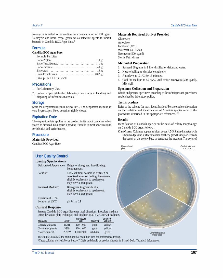
The Difco Manual 107
User Quality ControlIdentity Specifications
Dehydrated Appearance: Beige to blue-green, free-flowing,homogeneous.
Solution: 6.6% solution, soluble in distilled ordeionized water on boiling, blue-green,slightly opalescent to opalescent,may have a precipitate.
Prepared Medium: Blue-green to greenish blue,slightly opalescent to opalescent;may have a precipitate.
Reaction of 6.6%Solution at 25°C: pH 6.1 ± 0.1
Cultural ResponsePrepare Candida BCG Agar Base per label directions. Inoculate mediumusing the streak plate technique, and incubate at 30 ± 2°C for 24-48 hours.
INOCULUM COLOR OFORGANISM ATCC® CFU GROWTH MEDIUM
Candida albicans 10231 100-1,000 good yellowCandida tropicalis 3869 100-1,000 good yellowEscherichia coli 25922* 1,000-2,000 inhibited green
Candida albicansATCC® 10231
Uninoculatedplate
Candida tropicalisATCC® 3869
The cultures listed are the minimum that should be used for performance testing.*These cultures are available as Bactrol™ Disks and should be used as directed in Bactrol Disks Technical Information.
Neomycin is added to the medium in a concentration of 500 µg/ml.Neomycin and brom cresol green act as selective agents to inhibitbacteria in Candida BCG Agar Base.4
FormulaCandida BCG Agar Base
Formula Per LiterBacto Peptone . . . . . . . . . . . . . . . . . . . . . . . . . . . . . . . . . . . 10 gBacto Yeast Extract . . . . . . . . . . . . . . . . . . . . . . . . . . . . . . . . 1 gBacto Dextrose . . . . . . . . . . . . . . . . . . . . . . . . . . . . . . . . . . 40 gBacto Agar . . . . . . . . . . . . . . . . . . . . . . . . . . . . . . . . . . . . . 15 gBrom Cresol Green . . . . . . . . . . . . . . . . . . . . . . . . . . . . . . 0.02 gFinal pH 6.1 ± 0.1 at 25°C
Precautions1. For Laboratory Use.2. Follow proper established laboratory procedures in handling and
disposing of infectious materials.
StorageStore the dehydrated medium below 30°C. The dehydrated medium isvery hygroscopic. Keep container tightly closed.
Expiration DateThe expiration date applies to the product in its intact container whenstored as directed. Do not use a product if it fails to meet specificationsfor identity and performance.
ProcedureMaterials ProvidedCandida BCG Agar Base
Materials Required But Not ProvidedGlasswareAutoclaveIncubator (30°C)Waterbath (45-55°C)Neomycin (500 µg/ml)Sterile Petri dishes
Method of Preparation1. Suspend 66 grams in 1 liter distilled or deionized water.2. Heat to boiling to dissolve completely.3. Autoclave at 121°C for 15 minutes.4. Cool the medium to 50-55°C. Add sterile neomycin (500 µg/ml).
Mix well.
Specimen Collection and PreparationObtain and process specimens according to the techniques and proceduresestablished by laboratory policy.
Test ProcedureRefer to the scheme for yeast identification.3 For a complete discussionon the isolation and identification of Candida species refer to theprocedures described in the appropriate references.2,3,5
ResultsIdentification of Candida species on the basis of colony morphologyon Candida BCG Agar follows:C. albicans: Colonies appear as blunt cones 4.5-5.5 mm diameter with
smooth edges and surfaces; coarse feathery growths may arise fromthe center of the colony base to penetrate the medium. The color of
Section II Candida BCG Agar Base
108 The Difco Manual
the base and surface of the colonies is yellowish to bluish greenwith the intensity diminishing from a gray green center spot topaleness at the edge, although some strains may show a distinctgreen outer ring.
C. stellatoidea: Colonies appear convex 4.0-5.0 mm in diameter, withsmooth edges and smooth to irregular surfaces; there is a fine centralbasal feathery growth penetrating the medium. The color of bothbase and surface of colonies is yellow to green, the intensity of whichmay or may not diminish from center to border but is usually light.
C. tropicalis: Colonies appear convex or as low cones 4.5-5.0 mm indiameter with smooth to undulate edges, and smooth to granular orridged surfaces; deeply stained feathery growth arises from severalpoints in the base of the colony to form an effusive cloud. Thecolor of the submerged growth is normally an intense blue greencompared with that of the base which is much lighter; the surfaceis uniformly pale and may be yellowish green to green, reflecting alower pH than observed of the base.
C. pseudotropicalis: Colonies appear convex, 4.5-5.5 mm in diameterwith undulate to smooth edges, and smooth surfaces; occasionallythe surface is membranous but all colonies are shiny in appearance,and there is feathering growth emerging from several points in thebase of the colony. The color of a large central area in the base of thecolony is a medium green, which diminishes in intensity toward theedge; a similar distribution of color occurs on the surface, but thisgreen is bright in hue and is never grayed as it is with C. tropicalis.
C. krusei: Colonies appear as low cones 4.5-5.0 m in diameter withpseudohyphal edges, which may be weakly contractile or spreading,and have dull surfaces. There is abundant lightly colored growthpenetrating the medium from the base of the colony. The base ofthe colony is a medium blue green in the center diminishing inintensity to paleness at the edge; the surface is usually a light greento yellow green without much concentration in any part.
C. parapsilosis: Colonies appear as convex to low cones 3.5-4.5 mmin diameter with smooth or slightly spreading edges, but vary fromsmooth to granular or rough surfaces; there is no submergedgrowth. The color for both base and surface of the colony is blue
green over much of the colony, being more intense in the base thanthe surface which is modified by a thin grayish film of cells; theintensity in color fades abruptly leaving a broad pale edge.
C. guilliermondii: Colonies appear as low cones 4.0-5.0 mm indiameter with very smooth edges and highly glossy surfaces; theremaybe a weak, fine feathered submerged growth. Both base andsurface of the colony tend to have blue centers of medium intensityfading into a pale edge; however the surface may be blue greenwith the central third lightened with gray.
C. glabrata: Colonies are smooth and convex, 4.6-5.0 mm diameter;the surface color pattern is pale green in the center which becomesmedium green at the edge, and the base has the same color patternbut of less intensity.
Limitations of the Procedure1. Since the nutritional requirements of yeast vary, some strains may
be encountered that fail to grow or grow poorly on this medium.
References1. Harold, W., and M. Snyder. 1968. Personal Communication.2. Baron, E. J., L. R. Peterson, and S. M. Finegold. 1994. Bailey &
Scott’s diagnostic microbiology, 9th ed. Mosby-Year Book, Inc.,St. Louis, MO.
3. Warren, N. G., and K. C. Hazen. 1995. Candida, Cryptococcus,and other yeasts of medical importance, p. 723-737. In P. R.Murray, E. J. Baron, M. A. Pfaller, F. C. Tenover, and R. H. Yolken(ed.), Manual of clinical microbiology, 6th ed. American Societyfor Microbiology, Washington, D.C.
4. MacFaddin, J. D. 1985. Media for isolation-cultivation-identification-maintenance of medical bacteria, vol. 1, p. 136-137.Williams & Wilkins, Baltimore, MD.
5. Isenberg, H. D. (ed.). 1992. Clinical microbiology procedureshandbook. American Society for Microbiology, Washington, D.C.
PackagingCandida BCG Agar Base 500 g 0835-17
Candida Isolation Agar Section II
Bacto® Candida Isolation AgarIntended UseBacto Candida Isolation Agar is used for isolating and differentiatingCandida albicans.
Summary and ExplanationCandida Isolation Agar is a nutritionally rich medium that supportsgrowth of many yeasts and molds and is differential for Candidaalbicans. Candida Isolation Agar was developed using a modificationof YM Agar as described by Fung and Liang.1 Goldschmidt demonstratedthat YM Agar with Aniline Blue WS could be used to identifyC. albicans in clinical samples with high accuracy and predictability.2
Aniline Blue is metabolized by C. albicans to produce a fluorescentmoiety that can be detected under long wave UV light.2
Principles of the ProcedureYeast Extract provides nitrogen, carbon, vitamins and cofactors. MaltExtract provides carbon, protein and nutrients. Bacto Peptone provides
additional carbon and nitrogen. Dextrose is an energy source. AnilineBlue is a fluorescent indicator. Bacto Agar is a solidifying agent.
FormulaCandida Isolation Agar
Formula Per LiterBacto Yeast Extract . . . . . . . . . . . . . . . . . . . . . . . . . . . . . . . . 3 gBacto Malt Extract . . . . . . . . . . . . . . . . . . . . . . . . . . . . . . . . 3 gBacto Peptone . . . . . . . . . . . . . . . . . . . . . . . . . . . . . . . . . . . . 5 gBacto Dextrose . . . . . . . . . . . . . . . . . . . . . . . . . . . . . . . . . . 10 gBacto Agar . . . . . . . . . . . . . . . . . . . . . . . . . . . . . . . . . . . . . 20 gAniline Blue . . . . . . . . . . . . . . . . . . . . . . . . . . . . . . . . . . . . 0.1 g
Final pH 6.2 ± 0.2 at 25°C
Precautions1. For Laboratory Use.2. Follow proper established laboratory procedure in handling and
disposing of infectious materials.
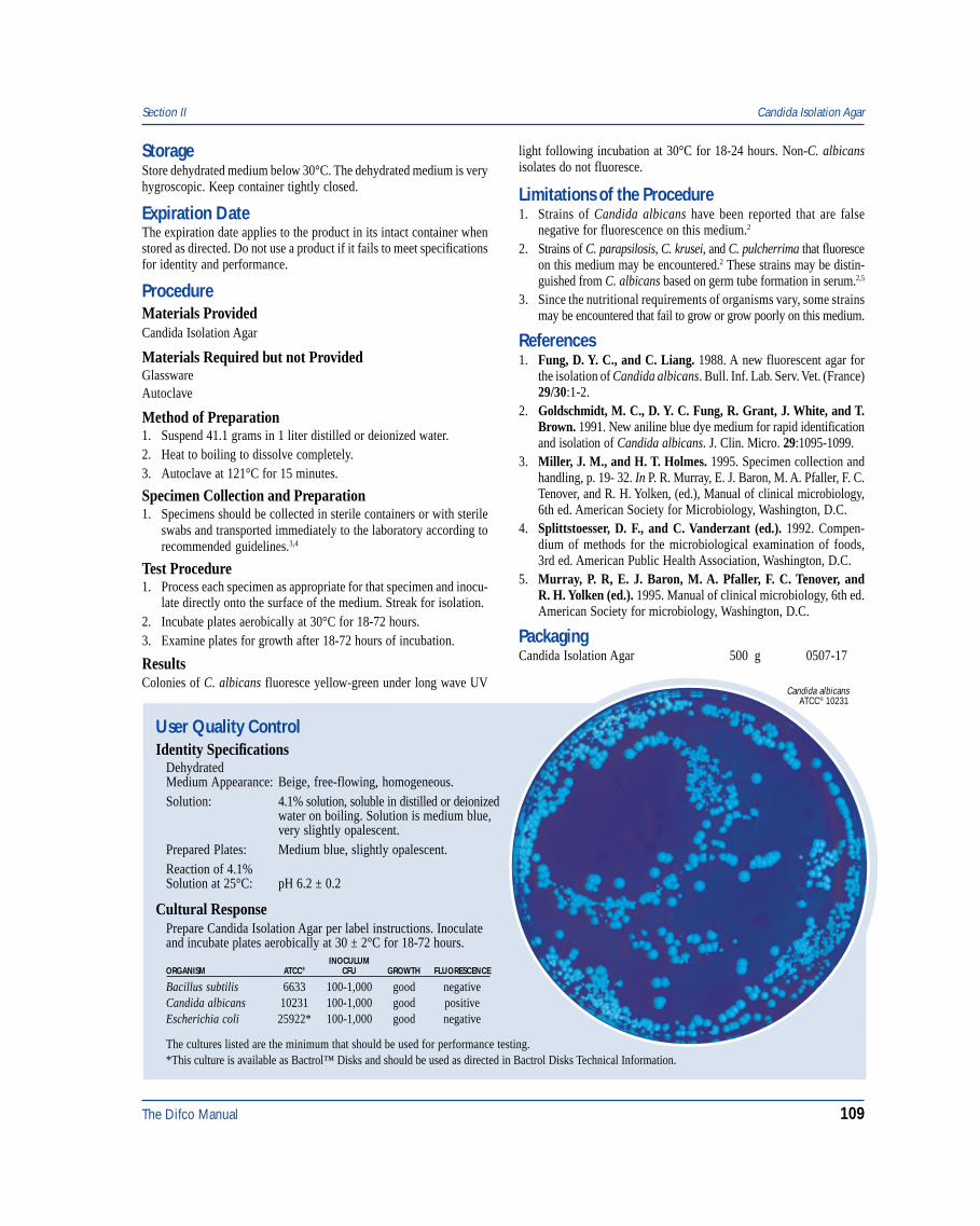
The Difco Manual 109
Section II Candida Isolation Agar
StorageStore dehydrated medium below 30°C. The dehydrated medium is veryhygroscopic. Keep container tightly closed.
Expiration DateThe expiration date applies to the product in its intact container whenstored as directed. Do not use a product if it fails to meet specificationsfor identity and performance.
ProcedureMaterials ProvidedCandida Isolation Agar
Materials Required but not ProvidedGlasswareAutoclave
Method of Preparation1. Suspend 41.1 grams in 1 liter distilled or deionized water.2. Heat to boiling to dissolve completely.3. Autoclave at 121°C for 15 minutes.
Specimen Collection and Preparation1. Specimens should be collected in sterile containers or with sterile
swabs and transported immediately to the laboratory according torecommended guidelines.3,4
Test Procedure1. Process each specimen as appropriate for that specimen and inocu-
late directly onto the surface of the medium. Streak for isolation.2. Incubate plates aerobically at 30°C for 18-72 hours.3. Examine plates for growth after 18-72 hours of incubation.
ResultsColonies of C. albicans fluoresce yellow-green under long wave UV
light following incubation at 30°C for 18-24 hours. Non-C. albicansisolates do not fluoresce.
Limitations of the Procedure1. Strains of Candida albicans have been reported that are false
negative for fluorescence on this medium.2
2. Strains of C. parapsilosis, C. krusei, and C. pulcherrima that fluoresceon this medium may be encountered.2 These strains may be distin-guished from C. albicans based on germ tube formation in serum.2,5
3. Since the nutritional requirements of organisms vary, some strainsmay be encountered that fail to grow or grow poorly on this medium.
References1. Fung, D. Y. C., and C. Liang. 1988. A new fluorescent agar for
the isolation of Candida albicans. Bull. Inf. Lab. Serv. Vet. (France)29/30:1-2.
2. Goldschmidt, M. C., D. Y. C. Fung, R. Grant, J. White, and T.Brown. 1991. New aniline blue dye medium for rapid identificationand isolation of Candida albicans. J. Clin. Micro. 29:1095-1099.
3. Miller, J. M., and H. T. Holmes. 1995. Specimen collection andhandling, p. 19- 32. In P. R. Murray, E. J. Baron, M. A. Pfaller, F. C.Tenover, and R. H. Yolken, (ed.), Manual of clinical microbiology,6th ed. American Society for Microbiology, Washington, D.C.
4. Splittstoesser, D. F., and C. Vanderzant (ed.). 1992. Compen-dium of methods for the microbiological examination of foods,3rd ed. American Public Health Association, Washington, D.C.
5. Murray, P. R, E. J. Baron, M. A. Pfaller, F. C. Tenover, andR. H. Yolken (ed.). 1995. Manual of clinical microbiology, 6th ed.American Society for microbiology, Washington, D.C.
PackagingCandida Isolation Agar 500 g 0507-17
User Quality ControlIdentity Specifications
DehydratedMedium Appearance: Beige, free-flowing, homogeneous.Solution: 4.1% solution, soluble in distilled or deionized
water on boiling. Solution is medium blue,very slightly opalescent.
Prepared Plates: Medium blue, slightly opalescent.Reaction of 4.1%Solution at 25°C: pH 6.2 ± 0.2
Cultural ResponsePrepare Candida Isolation Agar per label instructions. Inoculateand incubate plates aerobically at 30 ± 2°C for 18-72 hours.
INOCULUMORGANISM ATCC® CFU GROWTH FLUORESCENCE
Bacillus subtilis 6633 100-1,000 good negativeCandida albicans 10231 100-1,000 good positiveEscherichia coli 25922* 100-1,000 good negative
The cultures listed are the minimum that should be used for performance testing.*This culture is available as Bactrol™ Disks and should be used as directed in Bactrol Disks Technical Information.
Candida albicansATCC® 10231
110 The Difco Manual
Casamino Acids, Casamino Acids, Technical & Vitamin Assay Casamino Acids Section II
Bacto® Casamino Acids . Bacto Casamino Acids, TechnicalBacto Vitamin Assay Casamino Acids
User Quality ControlIdentity Specifications
Casamino AcidsDehydrated Appearance: Very light beige, free-flowing,
homogeneous.Solution: 1% solution-very light amber, clear
solution.2% solution-Light amber, clear,soluble in distilled or deionized waterupon slight heating.
Reaction of a 2%Solution at 25°C: pH 5.8-6.65
Casamino Acids, TechnicalDehydrated Appearance: Very light beige, free-flowing,
homogeneous.Solution: 1% solution, soluble in distilled or
deionized water. Solution is colorlessto very light amber and clear.
Reaction of 1%Solution at 25°C: pH 5.0-7.5
Vitamin Assay Casamino AcidsDehydrated Appearance: Light beige, free-flowing, homogeneous.Solution: 3% solution, soluble in distilled or
deionized water on boiling. Verylight to light amber, clear, may havea slight precipitate.
Reaction of 3%Solution at 25°C: pH 6.5-8.5
Cultural ResponseCasamino Acids and Casamino Acids, TechnicalPrepare a 1% solution and adjust the pH to 7.2 ± 0.2. Inoculatetubes with the test organisms, and incubate at 35 ± 2°C for18-48 hours.
INOCULUMORGANISM ATCC® CFU GROWTH
Escherichia coli 25922* 100-1,000 goodSalmonella typhi 19430 100-1,000 good
*This culture is available as Bactrol™ Disks and should be used as directed in Bactrol Disks Technical Information.
Vitamin Assay Casamino AcidsVitamin Assay Casamino Acids is prepared in various vitaminassay media to determine the vitamin content. It should notcontain a vitamin content higher than 20% above thefollowing values:
Vitamin B12 0.2 nanograms/gramBiotin 0.3 nanograms/gramFolic Acid 3.3 nanograms/gramNiacin 0.17 micrograms/gramPantothenate 0.04 micrograms/gramRiboflavin 0.1 micrograms/gramThiamine 0.1 micrograms/gram
The cultures listed are the minimum that should be used forperformance testing.
Intended UseBacto Casamino Acids is used in preparing microbiologicalculture media.
Bacto Casamino Acids, Technical is used in the preparation ofmicrobiological culture media.
Bacto Vitamin Assay Casamino Acids is used in vitamin assayprocedures.
Also Known AsCasamino Acids are also referred to as Casein Hydrolysate (Acid) orCasein Peptone, Acid Hydrolysate.
Summary and ExplanationCasamino Acids is acid hydrolyzed casein having low sodium chlorideand iron concentrations. Casamino Acids is recommended for use inmicrobiological culture media that require a completely hydrolyzedprotein as a nitrogen source. Casamino Acids is prepared according tothe method described by Mueller and Miller1 and Mueller and Johnson.2
Mueller3 prepared diphtheria toxin in a medium containing a caseinhydrolysate as the source of nitrogen. It was shown that the highsodium chloride content was the limiting factor in the amount of toxinthat could be produced in this medium. Mueller and Miller1 describeda method to reduce the sodium chloride and iron content of thehydrolyzed casein. This hydrolyzed casein, supplemented withinorganic salts, growth factors, cystine, maltose and an optimumamount of iron, was used to prepare diphtheria toxin.1,3 CasaminoAcids duplicates this specially treated hydrolyzed casein.
In Casamino Acids, hydrolysis is carried out until all the nitrogen inthe casein is converted to amino acids or other compounds of relativechemical simplicity. Casamino Acids is particularly well suited fornutritional studies, microbiological assays, and in the semi-syntheticmedium for testing disinfectants.4 Casamino Acids is also used in thepreparation of tetanus toxins, and pertussis vaccines, and for sulfonamideinhibitor studies.5
Casamino Acids, Technical is acid hydrolyzed casein. The hydrolysisis carried out as in the preparation of Casamino Acids, but the sodiumchloride and iron content of this product have not been decreased tothe same extent. Casamino Acids, Technical is recommended for usein culture media where amino acid mixtures are required for a nitrogensource, and the sodium chloride content is slightly increased. It isparticularly valuable in studying the growth requirements of bacteria.
Casamino Acids, Technical is prepared according to the methodsuggested by Mueller1 for use in the preparation of diphtheria toxin.Mueller and Hinton6 used Casamino Acids, Technical in a medium forprimary isolation of gonococcus and meningococcus. Casamino Acids,Technical was used in agar-free media for the isolation of Neisseria,and in a tellurite medium for the isolation of Corynebacterium,described by Levin.7 Wolf 8 used Casamino Acids, Technical in thepreparation of a medium for the testing of disinfectants.
Vitamin Assay Casamino Acids is an acid digest of casein speciallytreated to markedly reduce or eliminate certain vitamins. It is
The Difco Manual 111
Section II Casamino Acids, Casamino Acids, Technical & Vitamin Assay Casamino Acids
recommended for use in microbiological assay media and in studies ofthe growth requirements of microorganisms. Vitamin Assay CasaminoAcids is commonly used as the amino acid source in early phases ofnutrition work.9 Sarett10 used Vitamin Assay Casamino Acids as theacid hydrolyzed casein in his studies on p-aminobenzoic acid andp-teroylglutamic acid as growth factors for Lactobacillus species.
Several media containing Casamino Acids are specified in standardmethods for multiple applications.11,12,13
Principles of the ProcedureCasamino Acids, Casamino Acids, Technical and Vitamin AssayCasamino Acids are acid hydrolyzed casein. Casein is milk protein,and a rich source of amino acid nitrogen. Casamino Acids, CasaminoAcids, Technical and Vitamin Assay Casamino Acids provide nitrogen,vitamins, carbon and amino acids in microbiological culture media.Although Casamino Acids, Casamino Acids, Technical, and VitaminAssay Casamino Acids are added to media primarily because of theirorganic nitrogen and growth factor components, their inorganiccomponents also play a vital role.14
FormulaCasamino Acids is a dehydrated acid hydrolyzed casein in whichSodium Chloride and Iron are present in low concentrations permittingtoxin production.Casamino Acids, Technical is a dehydrated acid hydrolyzed casein.The Sodium Chloride and Iron content have not been reduced to sameextent as Casamino Acids.Vitamin Assay Casamino Acids is an acid hydrolyzed casein used toprepare media for microbiological assay of vitamins.
Typical AnalysisPhysical Characteristics
Ash (%) 24.4 Loss on Drying (%) 4.5Clarity, 1% Soln (NTU) 0.5 pH, 1% Soln 6.4Filterability (g/cm2) 2.9
Nitrogen Content (%)Total Nitrogen 10.5 AN/TN 83.8Amino Nitrogen 8.8
Amino Acids (%)Alanine 3.26 Lysine 5.71Arginine 2.20 Methionine 1.28Aspartic Acid 4.76 Phenylalanine 2.11Cystine 0.16 Proline 6.17Glutamic Acid 15.30 Serine 2.19Glycine 1.31 Threonine 2.41Histidine 1.66 Tryptophan <0.01Isoleucine 3.34 Tyrosine 0.47Leucine 5.47 Valine 4.30
Inorganics (%)Calcium <0.001 Phosphate 3.325Chloride 7.400 Potassium 0.410Cobalt <0.001 Sodium 8.710Copper <0.001 Sulfate 0.045Iron <0.001 Sulfur 0.420Lead <0.001 Tin <0.001Magnesium 0.002 Zinc <0.001Manganese <0.001
Vitamins (µg/g)Biotin <0.1 PABA <5.0Choline (as Choline Chloride) 160.0 Pantothenic Acid <0.1Cyanocobalamin <0.1 Pyridoxine <0.1Folic Acid <0.1 Riboflavin 1.8Inositol <100.0 Thiamine 1.2Nicotinic Acid <20.0 Thymidine <30.0
Biological Testing (CFU/g)Coliform negative Standard Plate Count 950Salmonella negative Thermophile Count 25Spore Count 390
Precautions1. For Laboratory Use.2. Follow proper established laboratory procedures in handling and
disposing of infectious materials.
StorageStore the dehydrated product below 30°C. The dehydrated product isvery hygroscopic. Keep container tightly closed.
Expiration DateThe expiration date applies to the product in its intact container whenstored as directed. Do not use a product if it fails to meet specificationsfor identity and performance.
ProcedureMaterials ProvidedCasamino AcidsCasamino Acids, TechnicalVitamin Assay Casamino Acids
Materials Required But Not ProvidedMaterials vary depending on the medium being prepared.
Method of PreparationRefer to the final concentration of Casamino Acids, Casamino Acids,Technical or Vitamin Assay Casamino Acids in the formula of themedium being prepared. Add Casamino Acids, Casamino Acids,Technical or Vitamin Assay Casamino Acids as required.
Specimen Collection and PreparationObtain and process specimens according to the techniques and proceduresestablished by laboratory policy.
Test ProcedureSee appropriate references for specific procedures using CasaminoAcids, Casamino Acids, Technical or Vitamin Assay Casamino Acids.
ResultsRefer to appropriate references and procedures for results.
References1. Mueller and Miller. 1941. J. Immunol. 40:21.2. Mueller and Johnson. 1941. J. Immunol. 40:33.3. Mueller. 1939. J. Immunol. 37:103.

112 The Difco Manual
Casein Digest Section II
Bacto® Casein Digest
User Quality ControlIdentity Specifications
Dehydrated Appearance: Tan, free-flowing, homogeneous.Solution: 1%, 2%, and 10% solutions, soluble
in distilled or deionized water:1%-Light amber, clear;2%-Medium amber, clear;10%-Dark amber, clear, no significantprecipitate.
Reaction of 1%Solution at 25°C: pH 7.2 ± 0.2
Cultural ResponsePrepare NZM Broth per formula. Inoculate and incubate at35 ± 2°C for 18-24 hours.
INOCULUMORGANISM ATCC® CFU GROWTH
Bacillus subtilis† 6633 100-1,000 goodEscherichia coli (HB101) 33694 100-1,000 goodEscherichia coli (JM107) 47014 100-1,000 goodEscherichia coli (DH5) 53868 100-1,000 goodSaccharomyces cerevisiae 9763 100-1,000 goodStreptomyces avermitilis 31267 100-1,000 good
The cultures listed are the minimum that should be used forperformance testing.
†Bacillus subtilis is available as Subtilis Spore Suspension.
Intended UseBacto Casein Digest is used in preparing microbiological culture media.
Also Known AsCasein Digest is similar to N-Z-Amine A.
Summary and ExplanationCasein Digest, an enzymatic digest of casein, was developed for use inmolecular genetics media. This product is digested under conditionsdifferent from other enzymatic digests of casein, including Tryptoneand Casitone.
Casein Digest is contained in the formulas of NZ media (NZCYMBroth, NZYM Broth and NZM Broth), which are used for cultivatingrecombinant strains of Escherichia coli. E. coli grows rapidly in theserich media because they provide amino acids, nucleotide precursors,vitamins and other metabolites that the cells would otherwise have tosynthesize.1 Consult appropriate references for recommended testprocedures using NZ media.1,2
Principles of the ProcedureCasein Digest is a nitrogen and amino acid source for microbiologicalculture media. Casein is raw milk protein, a rich source of amino acidnitrogen.
Precautions1. For Laboratory Use.2. Follow proper established laboratory procedures in handling and
disposing of infectious materials.
StorageStore Casein Digest below 30°C. The product is very hygroscopic.Keep container tightly closed.
Expiration DateThe expiration date applies to the product in its intact container whenstored as directed. Do not use a product if it fails to meet specificationsfor identity and performance.
ProcedureMaterials ProvidedCasein Digest
Materials Required But Not ProvidedMaterials vary depending on the medium being prepared.
4. Klarman and Wright. 1945. Soap and San. Chem. 21:113.
5. Straus, Dingle and Finland. 1941. J. Immunol. 42:331.
6. Mueller and Hinton. 1941. Proc. Soc. Exp. Biol. Med. 48:330.
7. Levin. 1943. J. Bacteriol. 46:233.
8. Wolf. 1945. J. Bacteriol. 49:463.
9. Nolan, R. A. 1971. Amino acids and growth factors in vitamin-freecasamino acids. Mycol. 63:1231-1234.
10. Sarett. 1947. J. Biol. Chem. 171:265.
11. Vanderzant, C., and D. F. Splittstoesser (ed.). 1992. Compendiumof methods for the microbiological examination of food, 3rd. ed.American Public Health Association, Washington, D.C.
12. Association of Official Analytical Chemists. 1995. Bacteriologicalanalytical manual, 8th ed. AOAC International, Gaithersburg, MD.
13. Eaton, A. D., L. S. Clesceri, and A. E. Greenberg (ed.). 1995.Standard methods for the examination of water and wastewater,19th ed. American Public Health Association, Washington, D.C.
14. Nolan, R. A., and W. G. Nolan. 1972. Elemental analysis ofvitamin-free casamino acids. Appl. Microbiol. 24:290-291.
PackagingCasamino Acids 100 g 0230-15
500 g 0230-172 kg 0230-07
10 kg 0230-08
Casamino Acids, Technical 500 g 0231-1710 kg 0231-08
Vitamin Assay Casamino Acids 100 g 0288-15500 g 0288-17
The Difco Manual 113
Section II Casitone
Method of PreparationRefer to the final concentration of Casein Digest in the formula of themedium being prepared. Add Casein Digest as required.
Specimen Collection and PreparationObtain and process specimens according to the techniques and proce-dures established by laboratory policy.
Test ProcedureSee appropriate references for specific procedures using Casein Digest.
ResultsRefer to appropriate references and procedures for results.
References1. Ausubel, F. M., R. Brent, R. E. Kingston, D. D. Moore, J. G.
Seidman, J. A. Smith, and K. Struhl (ed.). 1994. Currentprotocols in molecular biology, vol.1. Current Protocols,New York, NY.
2. Sambrook, J., E. F. Fritsch, and T. Maniatis. 1989. Molecularcloning: a laboratory manual, 2nd ed. Cold Spring HarborLaboratory, Cold Spring Harbor, NY.
PackagingCasein Digest 500 g 0116-17
Bacto® Casitone
User Quality ControlIdentity Specifications
Dehydrated Appearance: Tan, free-flowing granules.Solution: 1%, 2% and 10% solutions are
soluble in distilled or deionized water.1%-Light amber, clear, no precipitate.2%-Light to medium amber, clear,may have a slight precipitate.10%-Medium to dark amber, clearto very slightly opalescent, mayhave a precipitate.
Reaction of 1%Solution at 25°C: pH 6.8 - 7.4
Cultural ResponseAll solutions are prepared with the pH adjusted to 7.2 - 7.4.TEST SOLUTION ORGANISM ATCC® RESULT INOCULUM
Fermentable 2% Escherichia 25922* negative –Carbohydrates coliIndole 0.1% Escherichia 25922* positive –Production coliAcetylmethylcarbinol 0.1% Enterobacter 13048* positive –Production aerogenesHydrogen Sulfide 1% Salmonella 6539 positive –Production typhiToxicity 2%w/0.5% Escherichia 25922* good 100-1,000
NaCl & coli growth1.5% Agar
Toxicity 2%w/0.5% Staphylococcus 25923* good 100-1,000NaCl & aureus growth
1.5% Agar
The cultures listed are the minimum that should be used forperformance testing.*These cultures are available as Bactrol™ Disks and should be used as directed in Bactrol Disks Technical Information.
Intended UseBacto Casitone is used in preparing microbiological culture media.
Summary and ExplanationCasitone, is recommended for preparing media where an enzymatichydrolyzed casein is desired. Casitone is used to support the growth offastidious microorganisms. The high tryptophan content of Casitonemakes it valuable for use in detecting indole production.
Dubos Broth and Dubos Oleic Agar media that support the growth ofMycobacterium tuberculosis contain Casitone. Media used for the enumeration of coliforms in water, m Endo Agar and m Endo BrothMF®, use Casitone as a nitrogen source. Several Thioglycollate mediaused for detecting microorganisms in normally sterile materials,include Casitone as a nitrogen and amino acid source.
Casitone is recommended for preparing media for sterility testingaccording to US Pharmacopeia XXIII (USP).1 Several media containingCasitone are specified in standard methods2,3,4,5 for multiple applications.
Principles of the ProcedureCasitone is a pancreatic digest of casein. Casein is the main protein ofmilk, and a rich source of amino acid nitrogen.
Precautions1. For Laboratory Use.2. Follow proper established laboratory procedures in handling and
disposing of infectious materials.
StorageStore the dehydrated product below 30°C. The dehydrated product isvery hygroscopic. Keep container tightly closed.
Expiration DateThe expiration date applies to the product in its intact container whenstored as directed. Do not use a product if it fails to meet specificationsfor identity and performance.
Typical AnalysisPhysical Characteristics
Ash (%) 7.0 Loss on Drying (%) 3.7Clarity, 1% Soln (NTU) 0.6 pH, 1% Soln 7.2Filterability (g/cm2) 1.7
Carbohydrate (%)Total 0.2
114 The Difco Manual
Nitrogen Content (%)Total Nitrogen 13.3 AN/TN 35.3Amino Nitrogen 4.7
Amino Acids (%)Alanine 3.01 Lysine 13.62Arginine 3.76 Methionine 1.71Aspartic Acid 6.61 Phenylalanine 4.02Cystine 0.02 Proline 8.57Glutamic Acid 20.03 Serine 4.82Glycine 1.97 Threonine 3.74Histidine 2.17 Tryptophan 0.14Isoleucine 4.16 Tyrosine 2.09Leucine 8.74 Valine 4.06
Inorganics (%)Calcium 0.010 Phosphate 2.604Chloride 0.110 Potassium 0.162Cobalt <0.001 Sodium 3.073Copper <0.001 Sulfate 0.339Iron 0.003 Sulfur 0.676Lead <0.001 Tin <0.001Magnesium 0.019 Zinc 0.004Manganese <0.001
Vitamins (µg/g)Biotin 0.2 PABA 15.9Choline (as Choline Chloride) 550.0 Pantothenic Acid 7.7Cyanocobalamin <0.1 Pyridoxine 1.3Folic Acid 0.8 Riboflavin 0.4Inositol 980.0 Thiamine <0.1Nicotinic Acid 20.3 Thymidine 342.9
Biological Testing (CFU/g)Coliform negative Standard Plate Count 1850Salmonella negative Thermophile Count 100Spore Count 300
ProcedureMaterials ProvidedCasitone
Materials Required But Not ProvidedMaterials vary depending on the medium being prepared.
Method of PreparationRefer to the final concentration of Casitone in the formula of themedium being prepared. Add Casitone as required.
Specimen Collection and PreparationObtain and process specimens according to the techniques andprocedures established by laboratory policy.
Test ProcedureSee appropriate references for specific procedures using Casitone.
ResultsRefer to appropriate references and procedures for results.
References1. The United States Pharmacopeial Convention. 1995. The United
States Pharmacopeia, 23rd ed. Sterility test, p. 1686-1690. TheUnited States Pharmacopeial Convention Inc., Rockville, MD.
2. Vanderzant, C., and D. F. Splittstoesser (ed.). 1992.Compendium of methods for the microbiological examination offood, 3rd ed. American Public Health Association, Washington, D.C.
3. Association of Official Analytical Chemists. 1995. Bacteriologicalanalytical manual, 8th ed. AOAC International, Gaithersburg, MD.
4. Eaton, A. D., L. S. Clesceri, and A. E. Greenberg (ed.). 1995.Standard methods for the examination of water and wastewater,19th ed. American Public Health Association, Washington, D.C.
5. Marshall, R. T. (ed.). 1993. Standard methods for the examinationof dairy products, 16th ed., American Public Health Association,Washington, D.C.
PackagingCasitone 100 g 0259-15
500 g 0259-1710 kg 0259-08
Casman Medium Base Section II
Bacto® Casman Medium BaseIntended UseBacto Casman Medium Base is used with blood in isolating fastidiousmicroorganisms under reduced oxygen tension.
Summary and ExplanationIn 1947, Casman1,2,3 described an infusion-free medium enriched with5% blood for fastidious microorganisms incubated anaerobically. Thismedium replaced labor intensive formulas containing fresh meatinfusion and unheated and heated blood.1 Casman adjusted themedium after experiments revealed that nicotinamide disrupted theaction of a blood enzyme that inactivates V factor (NAD).2 Usingunheated human blood in the formula, Haemophilus influenzae grewwell and Neisseria was inhibited. The concentration of nicotinamidewas lowered to support growth of Neisseria species.2,3
Casman Agar Base with rabbit blood can be used for the cultivationand maintenance of Gardnerella vaginalis.4
Principles of the ProcedureProteose Peptone No.3, Tryptose and Beef Extract provide nitrogen,vitamins and amino acids. Nicotinamide enhances growth ofN. gonorrhoeae and H. influenzae by impeding the removal ofcoenzyme (V factor) by nucleotidase from the enriched blood. Thesmall amount of Dextrose is added to enhance growth of pathogeniccocci. Sodium chloride maintains the osmotic balance of the medium.Para-aminobenzoic acid is a preservative. Corn starch is added toensure that any toxic metabolites produced are absorbed, to neutralizeglucose inhibition of beta-hemolysis4 and to enhance growth ofNeisseria species. Agar Noble is a solidifying agent.
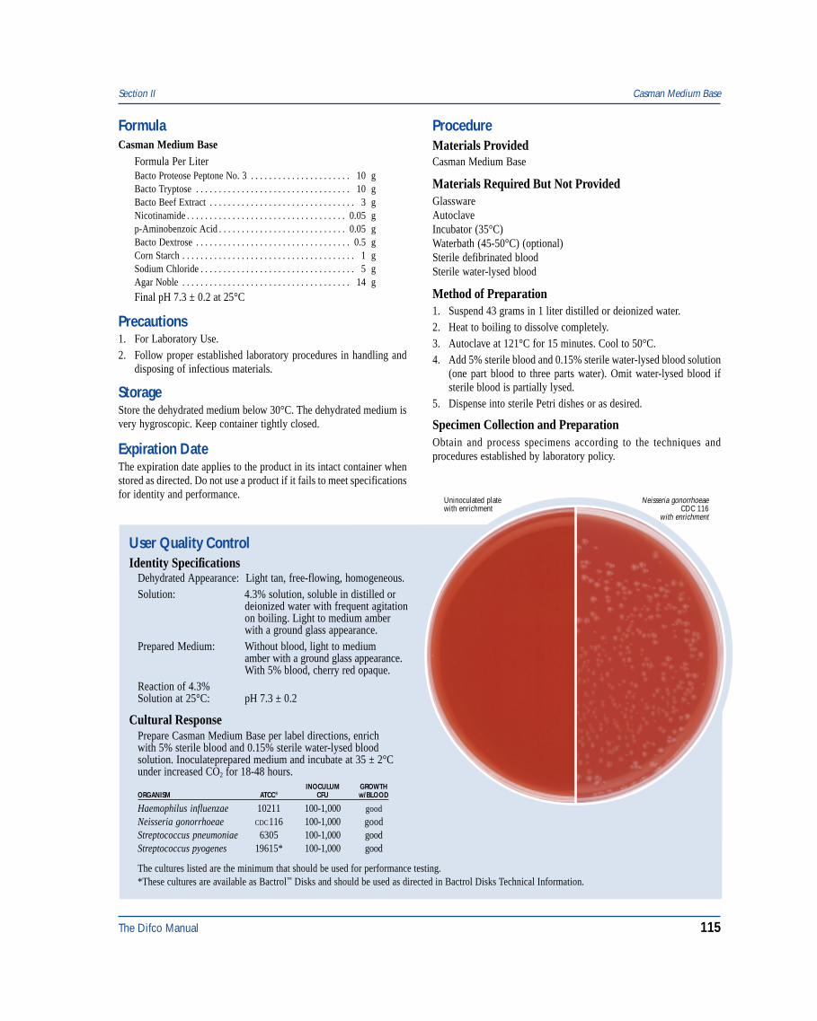
The Difco Manual 115
Section II Casman Medium Base
User Quality ControlIdentity Specifications
Dehydrated Appearance: Light tan, free-flowing, homogeneous.Solution: 4.3% solution, soluble in distilled or
deionized water with frequent agitationon boiling. Light to medium amberwith a ground glass appearance.
Prepared Medium: Without blood, light to mediumamber with a ground glass appearance.With 5% blood, cherry red opaque.
Reaction of 4.3%Solution at 25°C: pH 7.3 ± 0.2
Cultural ResponsePrepare Casman Medium Base per label directions, enrichwith 5% sterile blood and 0.15% sterile water-lysed bloodsolution. Inoculateprepared medium and incubate at 35 ± 2°Cunder increased CO2 for 18-48 hours.
INOCULUM GROWTHORGANISM ATCC® CFU w/BLOOD
Haemophilus influenzae 10211 100-1,000 good
Neisseria gonorrhoeae CDC116 100-1,000 goodStreptococcus pneumoniae 6305 100-1,000 goodStreptococcus pyogenes 19615* 100-1,000 good
Neisseria gonorrhoeaeCDC 116
with enrichment
Uninoculated platewith enrichment
The cultures listed are the minimum that should be used for performance testing.*These cultures are available as Bactrol™ Disks and should be used as directed in Bactrol Disks Technical Information.
FormulaCasman Medium Base
Formula Per LiterBacto Proteose Peptone No. 3 . . . . . . . . . . . . . . . . . . . . . . 10 gBacto Tryptose . . . . . . . . . . . . . . . . . . . . . . . . . . . . . . . . . . 10 gBacto Beef Extract . . . . . . . . . . . . . . . . . . . . . . . . . . . . . . . . 3 gNicotinamide . . . . . . . . . . . . . . . . . . . . . . . . . . . . . . . . . . . 0.05 gp-Aminobenzoic Acid . . . . . . . . . . . . . . . . . . . . . . . . . . . . 0.05 gBacto Dextrose . . . . . . . . . . . . . . . . . . . . . . . . . . . . . . . . . . 0.5 gCorn Starch . . . . . . . . . . . . . . . . . . . . . . . . . . . . . . . . . . . . . . 1 gSodium Chloride . . . . . . . . . . . . . . . . . . . . . . . . . . . . . . . . . . 5 gAgar Noble . . . . . . . . . . . . . . . . . . . . . . . . . . . . . . . . . . . . . 14 g
Final pH 7.3 ± 0.2 at 25°C
Precautions1. For Laboratory Use.2. Follow proper established laboratory procedures in handling and
disposing of infectious materials.
StorageStore the dehydrated medium below 30°C. The dehydrated medium isvery hygroscopic. Keep container tightly closed.
Expiration DateThe expiration date applies to the product in its intact container whenstored as directed. Do not use a product if it fails to meet specificationsfor identity and performance.
ProcedureMaterials ProvidedCasman Medium Base
Materials Required But Not ProvidedGlasswareAutoclaveIncubator (35°C)Waterbath (45-50°C) (optional)Sterile defibrinated bloodSterile water-lysed blood
Method of Preparation1. Suspend 43 grams in 1 liter distilled or deionized water.2. Heat to boiling to dissolve completely.3. Autoclave at 121°C for 15 minutes. Cool to 50°C.4. Add 5% sterile blood and 0.15% sterile water-lysed blood solution
(one part blood to three parts water). Omit water-lysed blood ifsterile blood is partially lysed.
5. Dispense into sterile Petri dishes or as desired.
Specimen Collection and PreparationObtain and process specimens according to the techniques andprocedures established by laboratory policy.
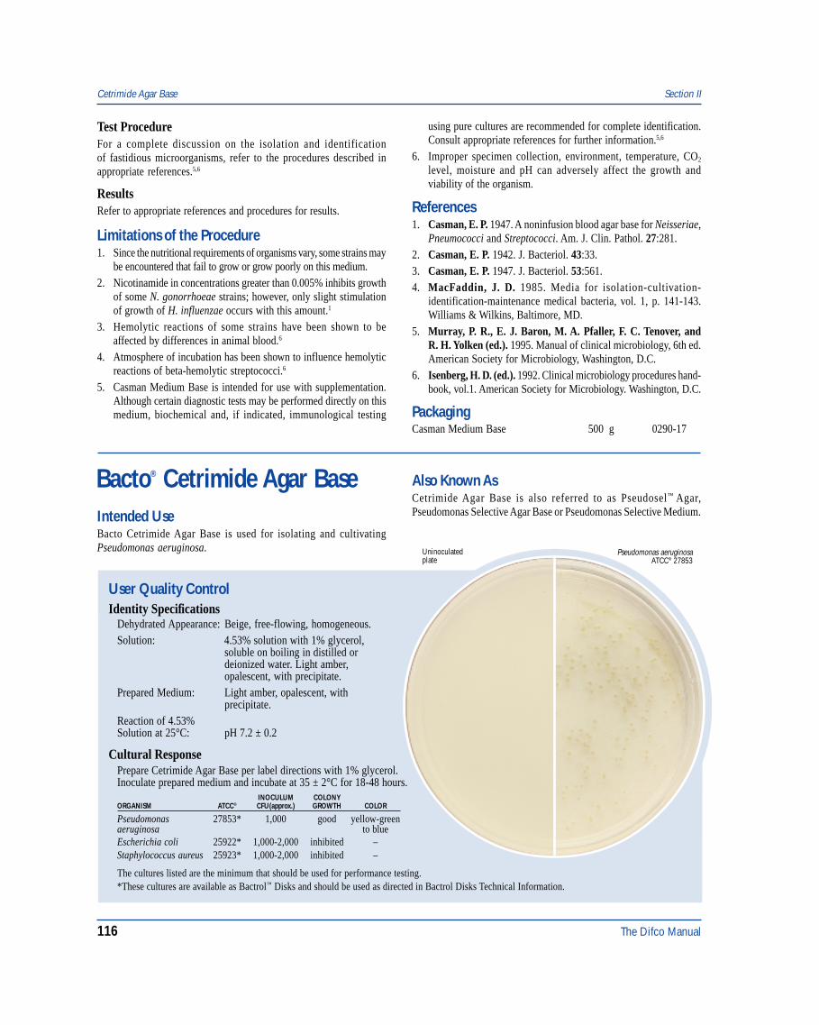
116 The Difco Manual
Test ProcedureFor a complete discussion on the isolation and identificationof fastidious microorganisms, refer to the procedures described inappropriate references.5,6
ResultsRefer to appropriate references and procedures for results.
Limitations of the Procedure1. Since the nutritional requirements of organisms vary, some strains may
be encountered that fail to grow or grow poorly on this medium.2. Nicotinamide in concentrations greater than 0.005% inhibits growth
of some N. gonorrhoeae strains; however, only slight stimulationof growth of H. influenzae occurs with this amount.1
3. Hemolytic reactions of some strains have been shown to beaffected by differences in animal blood.6
4. Atmosphere of incubation has been shown to influence hemolyticreactions of beta-hemolytic streptococci.6
5. Casman Medium Base is intended for use with supplementation.Although certain diagnostic tests may be performed directly on thismedium, biochemical and, if indicated, immunological testing
using pure cultures are recommended for complete identification.Consult appropriate references for further information.5,6
6. Improper specimen collection, environment, temperature, CO2
level, moisture and pH can adversely affect the growth andviability of the organism.
References1. Casman, E. P. 1947. A noninfusion blood agar base for Neisseriae,
Pneumococci and Streptococci. Am. J. Clin. Pathol. 27:281.2. Casman, E. P. 1942. J. Bacteriol. 43:33.3. Casman, E. P. 1947. J. Bacteriol. 53:561.4. MacFaddin, J. D. 1985. Media for isolation-cultivation-
identification-maintenance medical bacteria, vol. 1, p. 141-143.Williams & Wilkins, Baltimore, MD.
5. Murray, P. R., E. J. Baron, M. A. Pfaller, F. C. Tenover, andR. H. Yolken (ed.). 1995. Manual of clinical microbiology, 6th ed.American Society for Microbiology, Washington, D.C.
6. Isenberg, H. D. (ed.). 1992. Clinical microbiology procedures hand-book, vol.1. American Society for Microbiology. Washington, D.C.
PackagingCasman Medium Base 500 g 0290-17
Bacto® Cetrimide Agar Base
User Quality ControlIdentity Specifications
Dehydrated Appearance: Beige, free-flowing, homogeneous.Solution: 4.53% solution with 1% glycerol,
soluble on boiling in distilled ordeionized water. Light amber,opalescent, with precipitate.
Prepared Medium: Light amber, opalescent, withprecipitate.
Reaction of 4.53%Solution at 25°C: pH 7.2 ± 0.2
Cultural ResponsePrepare Cetrimide Agar Base per label directions with 1% glycerol.Inoculate prepared medium and incubate at 35 ± 2°C for 18-48 hours.
INOCULUM COLONYORGANISM ATCC® CFU(approx.) GROWTH COLOR
Pseudomonas 27853* 1,000 good yellow-greenaeruginosa to blueEscherichia coli 25922* 1,000-2,000 inhibited –Staphylococcus aureus 25923* 1,000-2,000 inhibited –
Pseudomonas aeruginosaATCC® 27853
Uninoculatedplate
The cultures listed are the minimum that should be used for performance testing.*These cultures are available as Bactrol™ Disks and should be used as directed in Bactrol Disks Technical Information.
Intended UseBacto Cetrimide Agar Base is used for isolating and cultivatingPseudomonas aeruginosa.
Also Known AsCetrimide Agar Base is also referred to as Pseudosel™ Agar,Pseudomonas Selective Agar Base or Pseudomonas Selective Medium.
Cetrimide Agar Base Section II
The Difco Manual 117
Summary and ExplanationPseudomonas aeruginosa has one of the broadest ranges of infectivityamong pathogens, and is the most frequently isolated nonfermentativebacillus in clinical specimens.3 It is a significant cause of burn andnosocomial infections.4 The ability of P. aeruginosa strains to destroytissue may be related to the production of various extracellularenzymes.3 In addition, virulent strains produce an exotoxin A, whichinhibits protein synthesis.3
Pseudomonas aeruginosa produces a number of water-solublepigments, including the yellow-green or yellow-brown fluorescentpigment pyoverdin.4 When pyoverdin combines with the bluewater-soluble pigment pyocyanin, the bright green color characteristicof P. aeruginosa is created.4 Fluorescent pigment-producing strainsfluoresce under short-wave ultraviolet light, and are observedat 254 nm using a standard Wood’s lamp.3 Agar containing cetrimidehas been used successfully to isolate P. aeruginosa fromcontaminated specimens.5
King, Ward and Raney1 developed Medium A (Tech Agar) to enhancethe production of pyocyanin in Pseudomonas species. Cetrimide AgarBase is prepared according to this formula with the additionof cetrimide.1 Brown and Lowbury2 used cetrimide in the Medium Bformulation of King, Ward and Raney1 to demonstrate the productionof fluorescein in P. aeruginosa.
Cetrimide Agar Base is recommended in the examination of food6
and in United States Pharmacopeia (USP XXIII) for use in MicrobialLimit Tests.7
Principles of the ProcedureBacto Peptone provides the nitrogen, vitamins and amino acids inCetrimide Agar Base. Magnesium Chloride and Potassium Sulfateenhance the production of pyocyanin and fluorescein.8 Cetrimide(cetyltrimethylammonium bromide) is the selective agent. Cetrimideacts as a quaternary ammonium cationic detergent causing nitrogenand phosphorous to be released from bacterial cells other thanP. aeruginosa. Bacto Agar is the solidifying agent. Cetrimide AgarBase is supplemented with 1% Glycerol as a source of carbon.
FormulaCetrimide Agar Base
Formula Per LiterBacto Peptone . . . . . . . . . . . . . . . . . . . . . . . . . . . . . . . . . . . 20 gMagnesium Chloride . . . . . . . . . . . . . . . . . . . . . . . . . . . . . 1.4 gPotassium Sulfate . . . . . . . . . . . . . . . . . . . . . . . . . . . . . . . . 10 gCetrimide (Cetyltrimethylammonium Bromide) . . . . . . . . 0.3 gBacto Agar . . . . . . . . . . . . . . . . . . . . . . . . . . . . . . . . . . . . 13.6 g
Final pH 7.2 ± 0.2 at 25°C
Precautions1. For Laboratory Use.2. Follow proper established laboratory procedures in handling and
disposing of infectious materials.
StorageStore the dehydrated medium below 30°C. The dehydrated medium isvery hygroscopic. Keep container tightly closed.
ProcedureMaterials ProvidedCetrimide Agar Base
Materials Required But Not ProvidedGlasswareAutoclaveIncubator (35°C)Waterbath (45-50°C)GlycerolSterile Petri dishes
Method of Preparation1. Suspend 45.3 grams in 1 liter distilled or deionized water
containing 10 ml of glycerol.2. Heat to boiling to dissolve completely.3. Autoclave at 121°C for 15 minutes. Cool to room temperature.4. Dispense into sterile Petri dishes, or as desired.
Specimen Collection and PreparationObtain and process specimens according to the techniques andprocedures established by laboratory policy.
Test ProcedureFor the isolation of P. aeruginosa plates of Cetrimide Agar Base maybe inoculated by the streak method from nonselective medium ordirectly from the specimen. When plating directly from the specimenthe inoculum level should be sufficiently high.
ResultsExamine plates or tubes for the presence of characteristic blue,blue-green, yellow-green pigment. Pseudomonas aeruginosa typicallyproduce both pyocyanin and fluorescein.
Limitations of the Procedure1. Since the nutritional requirements of organisms vary, some strains
may be encountered that fail to grow or grow poorly on this medium.2. The type of peptone used in base may affect pigment production.1,9
3. No single medium can be depended upon to exhibit all pigmentproducing P. aeruginosa strains.
4. Occasionally some enterics will exhibit a slight yellowing of themedium; however, this coloration is easily distinguished fromfluorescein production since this yellowing does not fluoresce.1
5. Some nonfermenters and some aerobic spore formers may exhibita water-soluble tan to brown pigmentation on this medium.Serratia strains may exhibit a pink pigmentation.1
6. Studies of Lowbury and Collins10 showed Ps. aeruginosa maylose its fluorescence under UV if the cultures are left at roomtemperature for a short time. Fluorescence reappears when platesare reincubated.
7. Further tests are necessary for definitive identification ofP. aeruginosa.
References1. King, E. O., M. K. Ward, and E. E. Raney. 1954. Two simple
media for the demonstration of pyocyanin and fluorescein. J. Lab.Clin. Med. 44:301.
Section II Cetrimide Agar Base
118 The Difco Manual
2. Brown, V.I., and E. J. L. Lowbury. 1965. Use of an improvedCetrimide Agar Medium and of culture methods for Pseudomonasaeruginosa. J. Clin. Pathol. 18:752.
3. Baron, E. J., L. R. Peterson, and S. M. Finegold. 1994.Nonfermentative gram-negative bacilli and coccobacilli,p. 386-405. Bailey & Scott’s diagnostic microbiology, 9th ed.Mosby-Year Book, Inc., St. Louis, MO.
4. Gilligan, P. H. 1995. Pseudomonas and Burkholderia, p. 509-519.In P. R. Murray, E. J. Baron, M. A. Pfaller, F. C. Tenover, andR. H. Yolken (ed.)., Manual of clinical microbiology, 6th ed.American Society for Microbiology, Washington, D.C.
5. Robin, T., and J. M. Janda. 1984. Enhanced recovery ofPseudomonas aeruginosa from diverse clinical specimens on anew selective agar. Diag. Microbiol. Infect. Dis. 2:207.
6. Association of Official Analytical Chemists. 1995. Bacteriologicalanalytical manual, 8th ed. AOAC International, Gaithersburg, MD.
Chapman Stone Medium Section II
7. The United States Pharmacopeia. 1995. Microbiological LimitsTests, The United States pharmacopeia, 23rd ed. United StatesPharmacopeial Convention, Rockville, MD.
8. MacFaddin, J. D. 1985. Media for isolation-cultivation-identification-maintenance of medical bacteria, vol. 1, p. 146-149.Williams & Wilkins, Baltimore, MD.
9. Goto, S., and S. Enomoto. 1970. Nalidixic acid cetrimide agar:A new selective plating medium for the selective isolation ofPseudomonas aeruginosa. Jpn. J. Microbiol. 14:65.
10. Lowbury, E. J. L., and A. G. Collins. 1955. The use of a newcetrimide product in a selective medium for Pseudomonasaeruginosa. J. Clin. Pathol. 8:47.
PackagingCetrimide Agar Base 100 g 0854-15
500 g 0854-17
Glycerol 100 g 0282-15500 g 0282-17
User Quality ControlIdentity Specifications
Dehydrated Appearance: Light beige, free-flowing, homogeneouswith a tendency to cake.
Solution: 20.2% solution, soluble in distilled ordeionized water on boiling. Solution islight amber, opalescent with precipitation.
Prepared medium: Light to medium amber, opalescentwith a precipitate.
Reaction of 20.2%Solution at 25°C: pH 7.0 ± 0.2
Cultural ResponsePrepare Chapman Stone Medium per label directions.Inoculate and incubate at 30 ± 2°C for 18-48 hours. Add BromCresol Purple indicator to determine mannitol fermentation(yellow = positive).
INOCULUM HALO MANNITOLORGANISM ATCC® CFU GROWTH (Gelatinase) FERMENTATION
Escherichia 25922* 100-1,000 inhibited – –coliStaphylococcus 25923* 100-1,000 good + +aureusStaphylococcus 12228* 100-1,000 good + –epidermidis
The cultures listed are the minimum that should be used forperformance testing.*These cultures are available as Bactrol™ Disks and should be used as directed in Bactrol Disks Technical Information.
Also Known AsChapman Stone Medium conforms with Chapman Stone Agar.
Summary and ExplanationChapman Stone Medium is prepared according to the formuladescribed by Chapman.1 It is similar to Staphylococcus Medium 110,previously described by Chapman,2 except that the sodium chlorideconcentration is reduced to 5.5% and ammonium sulfate is included inthe formulation. The inclusion of ammonium sulfate in the mediumnegates the need to add a reagent after growth has been obtained inorder to detect gelatinase activity by Stone’s method. Chapman StoneMedium is especially recommended for suspected food poisoningstudies involving Staphylococcus.3 It is selective, due to the relativelyhigh salt content, and is differential due to pigmentation, mannitolfermentation, and the presence or absence of gelatin liquefaction.
Principles of the ProcedureYeast Extract and Tryptone provide nitrogen, carbon, sulfur, vitamins,and trace nutrients essential for growth. Gelatin serves as a substratefor gelatinase activity. Ammonium Sulfate allows detection of gelatinhydrolysis. D-Mannitol is the fermentable carbohydrate. SodiumChloride acts as a selective agent because most bacterial species areinhibited by the high salt content. Dipotassium Phosphate providesbuffering capability. Bacto Agar is the solidifying agent.
FormulaChapman Stone Medium
Formula Per LiterBacto Yeast Extract . . . . . . . . . . . . . . . . . . . . . . . . . . . . . . . 2.5 gBacto Tryptone . . . . . . . . . . . . . . . . . . . . . . . . . . . . . . . . . . 10 gBacto Gelatin . . . . . . . . . . . . . . . . . . . . . . . . . . . . . . . . . . . 30 gBacto D-Mannitol . . . . . . . . . . . . . . . . . . . . . . . . . . . . . . . . 10 gSodium Chloride . . . . . . . . . . . . . . . . . . . . . . . . . . . . . . . . . 55 gAmmonium Sulfate . . . . . . . . . . . . . . . . . . . . . . . . . . . . . . . 75 gDipotassium Phosphate . . . . . . . . . . . . . . . . . . . . . . . . . . . . 5 gBacto Agar . . . . . . . . . . . . . . . . . . . . . . . . . . . . . . . . . . . . . 15 gFinal pH 7.0 ± 0.2 at 25°C
Bacto® Chapman Stone MediumIntended UseBacto Chapman Stone Medium is used for isolating and differentiatingstaphylococci based on mannitol fermentation and gelatinase activity.

The Difco Manual 119
Section II Charcoal Agar
Precautions1. For Laboratory Use.2. HARMFUL. HARMFUL IF SWALLOWED. (EC) IRRITATING
TO EYES, RESPIRATORY SYSTEM AND SKIN. Avoid contactwith skin and eyes. Do not breathe dust. Wear suitable protectiveclothing. Keep container tightly closed. TARGET ORGAN(S):Lungs, Intestines.FIRST AID: In case of contact with eyes, rinse immediately withplenty of water and seek medical advice. After contact with skin,wash immediately with plenty of water. If inhaled, remove to freshair. If not breathing, give artificial respiration. If breathing is diffi-cult, give oxygen. Seek medical advice. If swallowed seek medicaladvice immediately and show this container or label.
3. Follow proper established laboratory procedure in handling anddisposing of infectious materials.
StorageStore the dehydrated medium below 30°C. The dehydrated medium isvery hygroscopic. Keep container tightly closed. Store prepared mediumat 2-8°C.
Expiration DateThe expiration date applies to the product in its intact container whenstored as directed. Do not use a product if it fails to meet specificationsfor identity and performance.
ProcedureMaterials ProvidedChapman Stone Medium
Materials Required But Not ProvidedGlasswareAutoclaveIncubator (30°C)Sterile Petri dishesBrom Cresol Purple
Method of Preparation1. Suspend 20.2 grams in 100 ml distilled or deionized water.2. Boil to dissolve completely.3. Autoclave at 121°C for 10 minutes. Omit sterilization if prepared
medium is to be used within 12 hours.4. Dispense as desired.
Specimen Collection and PreparationRefer to appropriate references for specimen collection and preparation.
Test Procedure1. Streak a sample of the specimen onto the surface of the agar. Make
several stabs into the medium along the streak.2. Incubate, aerobically, at 30 ± 2°C for up to 48 hours.3. Examine for growth and the presence or absence of clear zones
around colonies.4. To determine mannitol fermentation, add a few drops of Brom
Cresol Purple to areas on the medium from which colonies havebeen removed. Any change in color of the indicator, compared withthat of the uninoculated medium, indicates fermentation of mannitol.
ResultsMannitol fermentation: Positive = change in color of the indicator toyellow.
Gelatinase activity: Positive Stone reaction = formation of clear zonesaround the colonies.
Any mannitol-positive, yellow or orange colonies surrounded by a clearzone are presumptively identified as Staphylococcus aureus. White ornonpigmented colonies, with or without a clear zone, are probablyS. epidermidis.
Limitations of the Procedure1. Confirm the presumptive identification of pathogenic staphylococci
with additional tests, such as coagulase activity.2. Enterococci and/or Group D streptococci may exhibit growth on
the medium and show slight mannitol fermentation. The colonies,however, are tiny and can easily be differentiated from staphylococciby Gram stain and the catalase test.3
References1. Chapman, G. H. 1948. An improved Stone medium for the isolation
and testing of food-poisoning staphylococci. Food Res. 13:100-105.2. Chapman, G. H. 1946. A single culture medium for selective
isolation of plasma-coagulating staphylococci and for improvedtesting of chromogenesis, plasma coagulation, mannitol fermentation,and the Stone reaction. J. Bacteriol. 51:409-410.
3. MacFaddin, J. F. 1985. Media for isolation-cultivation-identification-maintenance of medical bacteria. Williams &Wilkins, Baltimore, MD.
PackagingChapman Stone Medium 500 g 0313-17
10 kg 0313-08
Bacto® Charcoal AgarIntended UseBacto Charcoal Agar is used for cultivating fastidious organisms,especially Bordetella pertussis, for vaccine production and stockculture maintenance.
Summary and ExplanationCharcoal Agar is prepared according to the method of Mishulow,Sharpe and Cohen.1 The authors found this medium to be anefficient substitute for Bordet-Gengou Agar in the production ofB. pertussis vaccines.
The genus Bordetella consists of four species: Bordetella pertussis, B.parapertussis, B. bronchiseptica and B. avium.2 All Bordetella are

120 The Difco Manual
respiratory pathogens, residing on the mucous membranes of therespiratory tract. B. pertussis is the major cause of whooping coughor pertussis. B. parapertussis is associated with a milder form ofthe disease.3 B. bronchiseptica is an opportunistic human pathogenassociated with both respiratory and non-respiratory infections,often occurring in patients having close contact with animals.2
B. bronchiseptica has not been reported to cause pertussis. Therehave been no reports of recovery of B. avium from humans.2
Charcoal Agar supplemented with Horse Blood is used for thecultivation and isolation of Haemophilus influenzae.4
Principles of the ProcedureInfusion from Beef Heart and Bacto Peptone provide the nitrogen,carbon and amino acids in Charcoal Agar. Yeast Extract is a vitaminsource. Sodium Chloride maintains osmotic balance. Bacto Agar is asolidifying agent. Soluble Starch absorbs toxic metabolites. Norit SG,charcoal, provides growth requirements and selective properties.
FormulaCharcoal Agar
Formula Per LiterBeef Heart, Infusion from . . . . . . . . . . . . . . . . . . . . . . . . 500 gBacto Peptone . . . . . . . . . . . . . . . . . . . . . . . . . . . . . . . . . . . 10 gSodium Chloride . . . . . . . . . . . . . . . . . . . . . . . . . . . . . . . . . . 5 gBacto Soluble Starch . . . . . . . . . . . . . . . . . . . . . . . . . . . . . 10 gBacto Yeast Extract . . . . . . . . . . . . . . . . . . . . . . . . . . . . . . . 3.5 gNorit SG . . . . . . . . . . . . . . . . . . . . . . . . . . . . . . . . . . . . . . . . 4 gBacto Agar . . . . . . . . . . . . . . . . . . . . . . . . . . . . . . . . . . . . . 18 g
Final pH 7.3 ± 0.2 at 25°C
Precautions1. For Laboratory Use.
2. Follow proper established laboratory procedures in handling anddisposing of infectious materials.
StorageStore the dehydrated medium below 30°C. The dehydrated medium isvery hygroscopic. Keep container tightly closed.
Expiration DateThe expiration date applies to the product in its intact container whenstored as directed. Do not use a product if it fails to meet specificationsfor identity and performance.
ProcedureMaterials ProvidedCharcoal Agar
Materials Required But Not ProvidedGlasswareAutoclaveIncubator (35°C)Waterbath (45-50°C)Sterile Petri dishes
Method of Preparation1. Suspend 62.5 grams in 1 liter distilled or deionized water.2. Heat to boiling to dissolve completely.3. Autoclave at 121°C for 15 minutes.4. Mix thoroughly during dispensing to uniformly distribute
the charcoal.
User Quality ControlIdentity Specifications
Dehydrated Appearance: Gray, free-flowing, homogeneous.Solution: 6.25% solution, soluble in distilled or
deionized water on boiling; black,opaque with a precipitate.
Prepared Medium: Black, opaque.Reaction of 6.25%Solution at 25°C pH 7.3 ± 0.2
Cultural ResponsePrepare Charcoal Agar per label directions. Inoculate andincubate at 35 ± 2°C for 18-72 hours.
INOCULUMORGANISM ATCC® CFU GROWTH
Bordetella bronchiseptica 4617 100-1,000 goodBordetella parapertussis 15237 100-1,000 goodBordetella pertussis 8467 100-1,000 good
The cultures listed are the minimum that should be used forperformance testing.
Bordetella bronchisepticaATCC® 4617
Uninoculatedplate
Charcoal Agar Section II
The Difco Manual 121
Specimen Collection and PreparationObtain and process specimens according to the techniques andprocedures established by institutional policy.
Test ProcedureFor a complete discussion on the isolation and maintenance offastidious microorganisms refer to the procedures described inappropriate references.2,4,5
ResultsRefer to appropriate references and procedures for results.
Limitations of the Procedure1. Since the nutritional requirements of organisms vary, some strains
may be encountered that fail to grow or grow poorly on this medium.2. Charcoal has a tendency to settle out of the medium. Swirl the flask
gently when dispensing to obtain a uniform charcoal suspension.4
References1. Mishulow, L., L. S. Sharpe, and L. L. Cohen. 1953. Beef-heart
charcoal agar for the preparation of pertussis vaccines. Am. J.Public Health, 43:1466.
2. Marcon, M. J. 1995. Bordetella, p. 566-573. In P. R. Murray, E. J.Baron, M. A. Pfaller, F. C. Tenover, and R. H. Yolken (ed.).,Manual of clinical microbiology, 6th ed. American Society forMicrobiology, Washington, D.C.
3. Linneman, C. C., and E. B. Pery. 1977. Bordetella parapertussis;recent experience and a review of the literature. Am. J. Dis. Child.131:560-563. In P. R. Murray, E. J. Baron, M. A. Pfaller, F. C.Tenover, and R. H. Yolken (ed.)., Manual of clinical microbiology,6th ed. American Society for Microbiology, Washington, D.C.
4. MacFaddin, J. F. 1985. Media for isolation-cultivation-identification-maintenance of medical bacteria, vol 1, p. 154-159.Williams & Wilkins, Baltimore, MD.
5. Isenberg, H. D. (ed.). 1992. Clinical microbiology procedureshandbook, vol. 1. American Society for Microbiology,Washington, D.C.
PackagingCharcoal Agar 500 g 0894-17
Section II Choline Assay Medium
Bacto® Choline Assay Medium
User Quality ControlIdentity Specifications
Dehydrated Appearance: White, free-flowing, homogeneous.Solution: 2.85% (single strength) and 5.7%
(double strength) solution, soluble indistilled or deionized water uponboiling. Solution is colorless, clear,may have a slight precipitate.
Prepared Medium: Colorless, clear, may have a slightprecipitate.
Reaction of 2.85%Solution at 25°C: pH 5.5 ± 0.2
Cultural ResponsePrepare Choline Assay Medium per label directions. Preparea standard curve using choline at levels from 0 to 25 µg per10 ml. The medium supports the growth of Neurospora crassaATCC® 9277 when supplemented with choline chloride.
Intended UseBacto Choline Assay Medium is used for determining choline concen-tration by the microbiological assay technique.
Summary and ExplanationVitamin Assay Media are used in the microbiological assay of vitamins.Three types of media are used for this purpose:1. Maintenance Media: For carrying the stock culture to preserve the
viability and sensitivity of the test organism for its intended purpose;
2. Inoculum Media: To condition the test culture for immediate use;3. Assay Media: To permit quantitation of the vitamin under test.Choline Assay Medium is a slight modification of the mediumdescribed by Horowitz and Beadle.1 Neurospora crassa ATCC® 9277is the test organism used in this microbiological assay.
Principles of the ProcedureCholine Assay Medium is a choline-free dehydrated medium containingall other nutrients and vitamins essential for the cultivation of N. crassaATCC® 9277. The addition of choline standard in specified increasingconcentrations gives a growth response by this organism that can bemeasured gravimetrically.
FormulaCholine Assay Medium
Formula Per LiterBacto Sucrose . . . . . . . . . . . . . . . . . . . . . . . . . . . . . . . . . . . 40 gAmmonium Nitrate . . . . . . . . . . . . . . . . . . . . . . . . . . . . . . . . 2 gBiotin . . . . . . . . . . . . . . . . . . . . . . . . . . . . . . . . . . . . . . . . . . 10 µgPotassium Sodium Tartrate . . . . . . . . . . . . . . . . . . . . . . . . 11.4 gMonopotassium Phosphate . . . . . . . . . . . . . . . . . . . . . . . . . . 2 gMagnesium Sulfate . . . . . . . . . . . . . . . . . . . . . . . . . . . . . . . . 1 gSodium Chloride . . . . . . . . . . . . . . . . . . . . . . . . . . . . . . . . . 0.2 gCalcium Chloride . . . . . . . . . . . . . . . . . . . . . . . . . . . . . . . . 0.2 gSodium Borate . . . . . . . . . . . . . . . . . . . . . . . . . . . . . . . . . 700 µgAmmonium Molybdate . . . . . . . . . . . . . . . . . . . . . . . . . . . 500 µgFerrous Sulfate . . . . . . . . . . . . . . . . . . . . . . . . . . . . . . . . . . 1.1 mgCuprous Chloride . . . . . . . . . . . . . . . . . . . . . . . . . . . . . . . 300 µgManganese Sulfate . . . . . . . . . . . . . . . . . . . . . . . . . . . . . . 110 µgZinc Sulfate . . . . . . . . . . . . . . . . . . . . . . . . . . . . . . . . . . . . 17.6 mg
Final pH 5.5 ± 0.2 at 25°C
122 The Difco Manual
Precautions1. For Laboratory Use.2. Follow proper established laboratory procedures in handling and
disposing of infectious materials.3. Great care must be taken to avoid contamination of media or
glassware in microbiological assay procedures. Extremely smallamounts of foreign material may be sufficient to give erroneousresults. Scrupulously clean glassware free from detergents andother chemicals must be used. Glassware must be heated to 250°Cfor at least 1 hour to burn off any organic residues that might bepresent.
4. Take precautions to keep sterilization and cooling conditionsuniform throughout the assay.
StorageStore the dehydrated medium at 2-8°C. The dehydrated medium is veryhygroscopic. Keep container tightly closed.
Expiration DateThe expiration date applies to the product in its intact container whenstored as directed. Do not use a product if it fails to meet specificationsfor identity and performance.
ProcedureMaterials ProvidedCholine Assay Medium
Materials Required But Not ProvidedGlasswareAutoclaveStock culture of Neurospora crassa ATCC® 9277Sterile 0.85% salineDistilled or deionized waterInoculating loopNeurospora Culture AgarWire needle or glass rodPaper towelsVacuum ovenPorcelain spot plateScale
Method of Preparation1. Suspend 5.7 grams in 100 ml of distilled or deionized water.2. Boil 2-3 minutes to dissolve completely.3. Dispense 5 ml amounts into flasks, evenly dispersing the precipitate.4. Add standard or test samples.5. Adjust flask volume to 10 ml with distilled or deionized water.6. Autoclave at 121°C for 10 minutes.
Specimen Collection and PreparationAssay samples are prepared according to references given in thespecific assay procedures. For assay, the samples should be diluted toapproximately the same concentration as the standard solution.
Test ProcedureRemove 1 loop of spores from a 48-hour culture of N. crassaATCC® 9277 grown on Neurospora Culture Agar and suspend it in100 ml sterile saline. Add 1 drop of this spore suspension to eachflask of medium. Incubate at 25-30°C for 3 days. At the end of theincubation period, steam the flask at 100°C for 5 minutes. Removeall the mycelium from the flask using a stiff wire needle or glass rod,press dry between paper towels, and roll into a small pellet. Dry thepellet at 100°C in a vacuum oven for 2 hours. (A glazed porcelainspot plate is convenient for handling the mycelium during drying andweighing.) Weigh to the nearest 0.5 mg. A standard curve is thenconstructed from the weights obtained, and the unknown determinedby interpolation. In the assay for choline, 50 ml Erlenmeyer flaskscontaining a total volume of 10 ml each are used.
It is essential that a standard curve be constructed each time an assay isrun. Autoclave and incubation conditions can influence the standardcurve reading and cannot always be duplicated. The standard curve isobtained by using choline at levels of 0.0, 2.5, 5, 10, 15, 20 and 25 µgper assay flask (10 ml). The most effective assay range using CholineAssay Medium is between 2.5 and 30 µg choline.
The concentration of choline required for the preparation of the standardcurve may be prepared by dissolving 0.5 grams choline chloride in1,000 ml distilled water. This is the stock solution (500 µg per ml).Dilute the stock solution by adding 1 ml to 99 ml distilled water. Use0.0, 0.5, 1, 2, 3, 4 and 5 ml of this diluted solution per flask. Preparethe stock solution fresh daily.
Results1. Prepare a standard concentration response curve by plotting the
response readings against the amount of standard in each tube, diskor cup.
2. Determine the amount of vitamin at each level of assay solution byinterpolation from the standard curve.
3. Calculate the concentration of vitamin in the sample from theaverage of these volumes. Use only those values that do not varymore than ±10% from the average. Use the results only if twothirds of the values do not vary more than ±10%.
Limitations of the Procedure1. The test organism used for inoculating an assay medium must be
cultured and maintained on media recommended for this purpose.2. Aseptic technique should be used throughout the assay procedure.3. The use of altered or deficient media may cause mutants having
different nutritional requirements that will not give a satisfactoryresponse.
4. For successful results to these procedures, all conditions of theassay must be followed precisely.
References1. Horowitz and Beadle. 1943. J. Biol. Chem. 150:325.
PackagingCholine Assay Medium 100 g 0460-15
Choline Assay Medium Section II
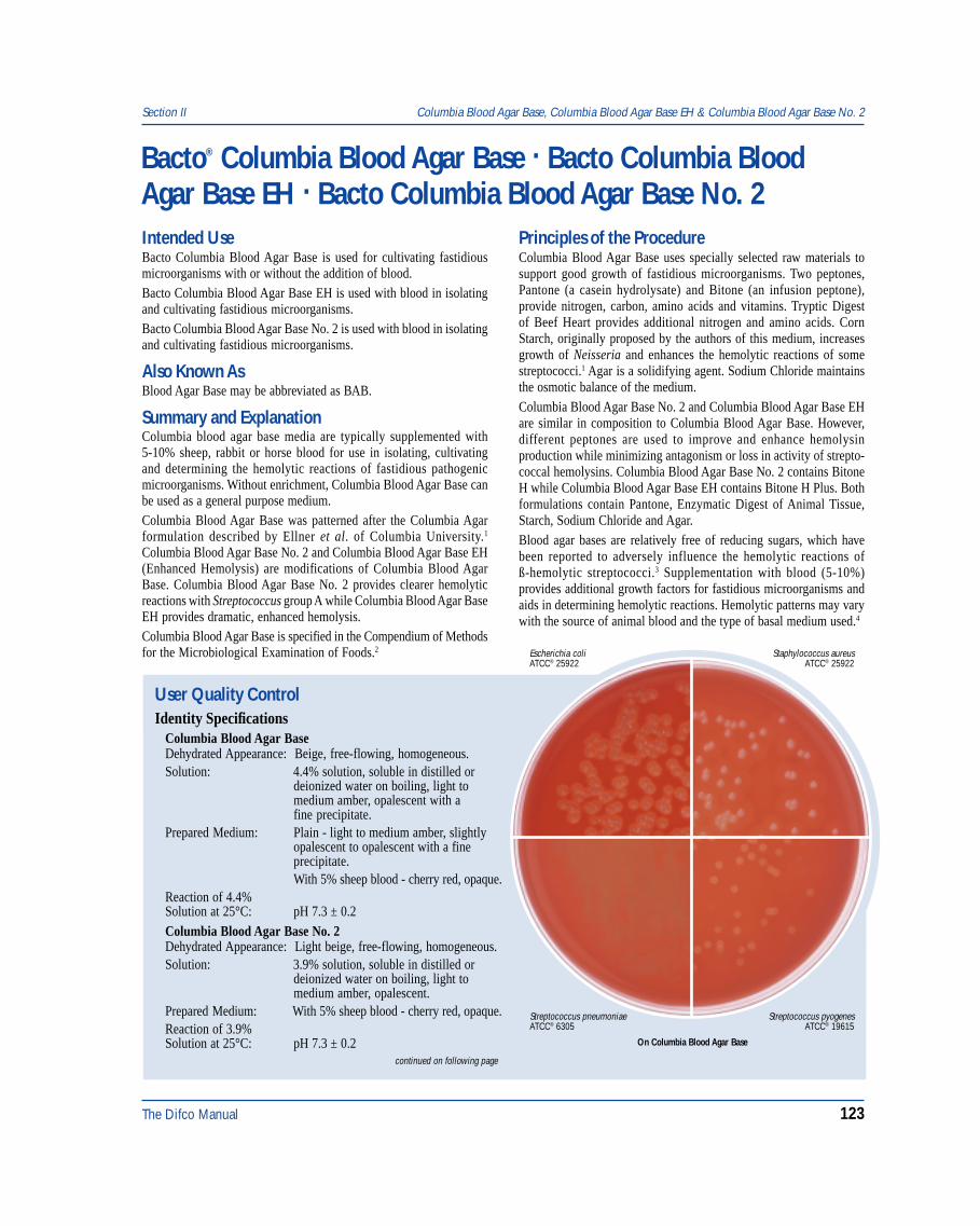
The Difco Manual 123
Section II Columbia Blood Agar Base, Columbia Blood Agar Base EH & Columbia Blood Agar Base No. 2
Bacto® Columbia Blood Agar Base . Bacto Columbia BloodAgar Base EH . Bacto Columbia Blood Agar Base No. 2
User Quality ControlIdentity Specifications
Columbia Blood Agar BaseDehydrated Appearance: Beige, free-flowing, homogeneous.Solution: 4.4% solution, soluble in distilled or
deionized water on boiling, light tomedium amber, opalescent with afine precipitate.
Prepared Medium: Plain - light to medium amber, slightlyopalescent to opalescent with a fineprecipitate.With 5% sheep blood - cherry red, opaque.
Reaction of 4.4%Solution at 25°C: pH 7.3 ± 0.2Columbia Blood Agar Base No. 2Dehydrated Appearance: Light beige, free-flowing, homogeneous.Solution: 3.9% solution, soluble in distilled or
deionized water on boiling, light tomedium amber, opalescent.
Prepared Medium: With 5% sheep blood - cherry red, opaque.Reaction of 3.9%Solution at 25°C: pH 7.3 ± 0.2
Staphylococcus aureusATCC® 25922
Escherichia coliATCC® 25922
Streptococcus pneumoniaeATCC® 6305
Streptococcus pyogenesATCC® 19615
continued on following page
On Columbia Blood Agar Base
Intended UseBacto Columbia Blood Agar Base is used for cultivating fastidiousmicroorganisms with or without the addition of blood.Bacto Columbia Blood Agar Base EH is used with blood in isolatingand cultivating fastidious microorganisms.Bacto Columbia Blood Agar Base No. 2 is used with blood in isolatingand cultivating fastidious microorganisms.
Also Known AsBlood Agar Base may be abbreviated as BAB.
Summary and ExplanationColumbia blood agar base media are typically supplemented with5-10% sheep, rabbit or horse blood for use in isolating, cultivatingand determining the hemolytic reactions of fastidious pathogenicmicroorganisms. Without enrichment, Columbia Blood Agar Base canbe used as a general purpose medium.Columbia Blood Agar Base was patterned after the Columbia Agarformulation described by Ellner et al. of Columbia University.1
Columbia Blood Agar Base No. 2 and Columbia Blood Agar Base EH(Enhanced Hemolysis) are modifications of Columbia Blood AgarBase. Columbia Blood Agar Base No. 2 provides clearer hemolyticreactions with Streptococcus group A while Columbia Blood Agar BaseEH provides dramatic, enhanced hemolysis.Columbia Blood Agar Base is specified in the Compendium of Methodsfor the Microbiological Examination of Foods.2
Principles of the ProcedureColumbia Blood Agar Base uses specially selected raw materials tosupport good growth of fastidious microorganisms. Two peptones,Pantone (a casein hydrolysate) and Bitone (an infusion peptone),provide nitrogen, carbon, amino acids and vitamins. Tryptic Digestof Beef Heart provides additional nitrogen and amino acids. CornStarch, originally proposed by the authors of this medium, increasesgrowth of Neisseria and enhances the hemolytic reactions of somestreptococci.1 Agar is a solidifying agent. Sodium Chloride maintainsthe osmotic balance of the medium.Columbia Blood Agar Base No. 2 and Columbia Blood Agar Base EHare similar in composition to Columbia Blood Agar Base. However,different peptones are used to improve and enhance hemolysinproduction while minimizing antagonism or loss in activity of strepto-coccal hemolysins. Columbia Blood Agar Base No. 2 contains BitoneH while Columbia Blood Agar Base EH contains Bitone H Plus. Bothformulations contain Pantone, Enzymatic Digest of Animal Tissue,Starch, Sodium Chloride and Agar.Blood agar bases are relatively free of reducing sugars, which havebeen reported to adversely influence the hemolytic reactions ofß-hemolytic streptococci.3 Supplementation with blood (5-10%)provides additional growth factors for fastidious microorganisms andaids in determining hemolytic reactions. Hemolytic patterns may varywith the source of animal blood and the type of basal medium used.4
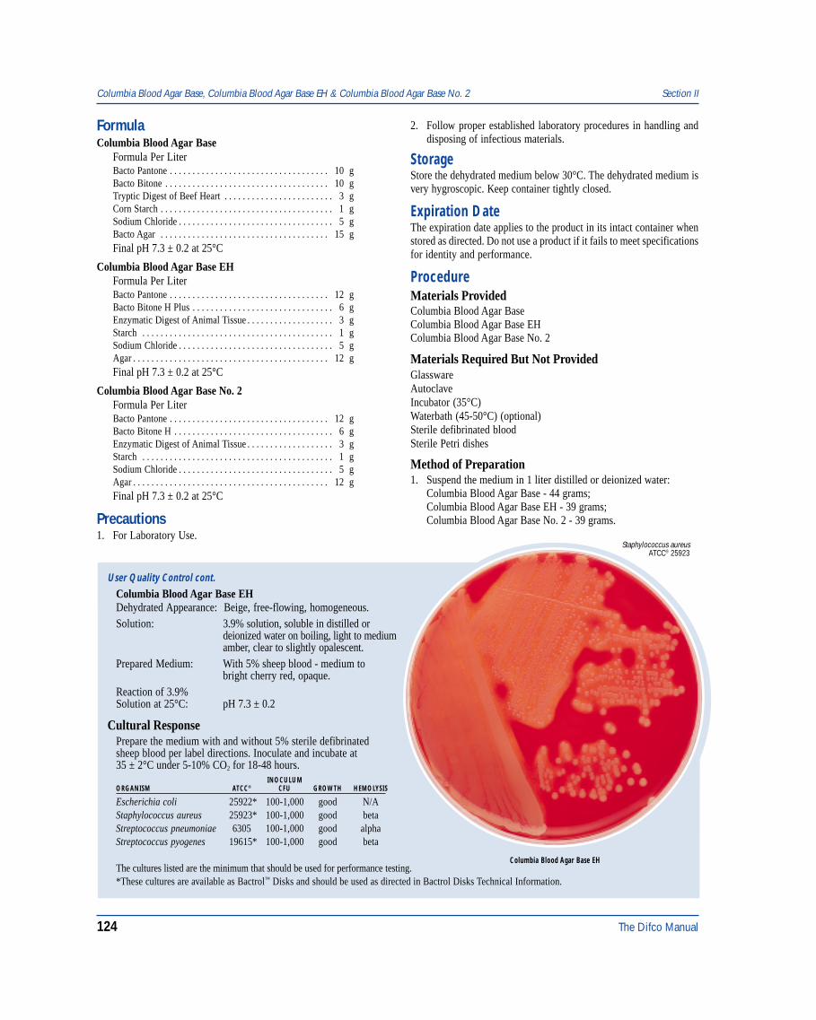
124 The Difco Manual
FormulaColumbia Blood Agar Base
Formula Per LiterBacto Pantone . . . . . . . . . . . . . . . . . . . . . . . . . . . . . . . . . . . 10 gBacto Bitone . . . . . . . . . . . . . . . . . . . . . . . . . . . . . . . . . . . . 10 gTryptic Digest of Beef Heart . . . . . . . . . . . . . . . . . . . . . . . . 3 gCorn Starch . . . . . . . . . . . . . . . . . . . . . . . . . . . . . . . . . . . . . . 1 gSodium Chloride . . . . . . . . . . . . . . . . . . . . . . . . . . . . . . . . . . 5 gBacto Agar . . . . . . . . . . . . . . . . . . . . . . . . . . . . . . . . . . . . . 15 gFinal pH 7.3 ± 0.2 at 25°C
Columbia Blood Agar Base EHFormula Per LiterBacto Pantone . . . . . . . . . . . . . . . . . . . . . . . . . . . . . . . . . . . 12 gBacto Bitone H Plus . . . . . . . . . . . . . . . . . . . . . . . . . . . . . . . 6 gEnzymatic Digest of Animal Tissue . . . . . . . . . . . . . . . . . . . 3 gStarch . . . . . . . . . . . . . . . . . . . . . . . . . . . . . . . . . . . . . . . . . . 1 gSodium Chloride . . . . . . . . . . . . . . . . . . . . . . . . . . . . . . . . . . 5 gAgar . . . . . . . . . . . . . . . . . . . . . . . . . . . . . . . . . . . . . . . . . . . 12 gFinal pH 7.3 ± 0.2 at 25°C
Columbia Blood Agar Base No. 2Formula Per LiterBacto Pantone . . . . . . . . . . . . . . . . . . . . . . . . . . . . . . . . . . . 12 gBacto Bitone H . . . . . . . . . . . . . . . . . . . . . . . . . . . . . . . . . . . 6 gEnzymatic Digest of Animal Tissue . . . . . . . . . . . . . . . . . . . 3 gStarch . . . . . . . . . . . . . . . . . . . . . . . . . . . . . . . . . . . . . . . . . . 1 gSodium Chloride . . . . . . . . . . . . . . . . . . . . . . . . . . . . . . . . . . 5 gAgar . . . . . . . . . . . . . . . . . . . . . . . . . . . . . . . . . . . . . . . . . . . 12 gFinal pH 7.3 ± 0.2 at 25°C
Precautions1. For Laboratory Use.
Columbia Blood Agar Base, Columbia Blood Agar Base EH & Columbia Blood Agar Base No. 2 Section II
User Quality Control cont.
Columbia Blood Agar Base EHDehydrated Appearance: Beige, free-flowing, homogeneous.Solution: 3.9% solution, soluble in distilled or
deionized water on boiling, light to mediumamber, clear to slightly opalescent.
Prepared Medium: With 5% sheep blood - medium tobright cherry red, opaque.
Reaction of 3.9%Solution at 25°C: pH 7.3 ± 0.2
Cultural ResponsePrepare the medium with and without 5% sterile defibrinatedsheep blood per label directions. Inoculate and incubate at35 ± 2°C under 5-10% CO2 for 18-48 hours.
INOCULUMORGANISM ATCC® CFU GROWTH HEMOLYSIS
Escherichia coli 25922* 100-1,000 good N/AStaphylococcus aureus 25923* 100-1,000 good betaStreptococcus pneumoniae 6305 100-1,000 good alphaStreptococcus pyogenes 19615* 100-1,000 good beta
2. Follow proper established laboratory procedures in handling anddisposing of infectious materials.
StorageStore the dehydrated medium below 30°C. The dehydrated medium isvery hygroscopic. Keep container tightly closed.
Expiration DateThe expiration date applies to the product in its intact container whenstored as directed. Do not use a product if it fails to meet specificationsfor identity and performance.
ProcedureMaterials ProvidedColumbia Blood Agar BaseColumbia Blood Agar Base EHColumbia Blood Agar Base No. 2
Materials Required But Not ProvidedGlasswareAutoclaveIncubator (35°C)Waterbath (45-50°C) (optional)Sterile defibrinated bloodSterile Petri dishes
Method of Preparation1. Suspend the medium in 1 liter distilled or deionized water:
Columbia Blood Agar Base - 44 grams;Columbia Blood Agar Base EH - 39 grams;Columbia Blood Agar Base No. 2 - 39 grams.
Staphylococcus aureusATCC® 25923
Columbia Blood Agar Base EHThe cultures listed are the minimum that should be used for performance testing.*These cultures are available as Bactrol™ Disks and should be used as directed in Bactrol Disks Technical Information.
The Difco Manual 125
2. Heat to boiling to dissolve completely.3. Autoclave at 121°C for 15 minutes. Cool to 45-50°C.4. To prepare blood agar, aseptically add 5% sterile defibrinated blood
to the medium at 45-50°C. Mix well.5. Dispense into sterile Petri dishes.
Specimen Collection and PreparationCollect specimens in sterile containers or with sterile swabs andtransport immediately to the laboratory in accordance withrecommended guidelines outlined in the references.
Test Procedure1. Process each specimen as appropriate and inoculate directly onto
the surface of the medium. Streak for isolation with an inoculatingloop, then stab the agar several times to deposit ß-hemolyticstreptococci beneath the agar surface. Subsurface growth willdemonstrate the most reliable hemolytic reactions due to theactivity of both oxygen-stable and oxygen-labile streptolysins.4
2. Incubate plates aerobically, anaerobically or under conditionsof increased CO2 (5-10%) in accordance with establishedlaboratory procedures.
Results1. Examine plates for growth and hemolytic reactions after 18-24 and
48 hours of incubation. Four types of hemolysis on blood agarmedia can be described:5
a. Alpha-hemolysis (α ) is the reduction of hemoglobin tomethemoglobin in the medium surrounding the colony, causinga greenish discolorization of the medium.
b. Beta-hemolysis (ß) is the lysis of red blood cells, producing aclear zone surrounding the colony.
c. Gamma-hemolysis (γ) indicates no hemolysis. No destructionof red blood cells occurs and there is no change in the medium.
d. Alpha-prime-hemolysis (α) is a small zone of complete hemolysisthat is surrounded by an area of partial lysis.
Limitations of the Procedure1. Blood agar base media are intended for use with blood
supplementation. Although certain diagnostic tests maybe performed directly on these media, biochemical and, ifindicated, immunological testing using pure cultures isrecommended for complete identification. Consult appropriatereferences for further information.
2. Since the nutritional requirements of organisms vary, some strainsmay be encountered that fail to grow or grow poorly on this medium.
Section II Columbia Broth
3. Hemolytic reactions of some strains of group D streptococci havebeen shown to be affected by differences in animal blood. Suchstrains are ß-hemolytic on horse, human and rabbit blood agar andα-hemolytic on sheep blood agar.4
4. Colonies of Haemophilus haemolyticus are ß-hemolytic on horseand rabbit blood agar and must be distinguished from coloniesof ß-hemolytic streptococci using other criteria. The use of sheepblood has been suggested to obviate this problem since sheepblood is deficient in pyridine nucleotides and does not supportgrowth of H. haemolyticus.6
5. Atmosphere of incubation has been shown to influence hemolyticreactions of ß-hemolytic streptococci.4 For optimal performance,incubate blood agar media under increased CO2 or anaerobic conditions.
References1. Ellner, P. D., C. J. Stoessel, E. Drakeford, and F. Vasi. 1966.
A new culture medium for medical bacteriology. Am. J. Clin.Pathol. 45:502-504.
2. Vanderzant, C. and D. F. Splittstoesser (ed.). 1992. Compen-dium of methods for the microbiological examination of foods,3rd ed. American Public Health Association, Washington, D.C.
3. Casman, E. P. 1947. A noninfusion blood agar base for neisseriae,pneumococci and streptococci. Am. J. Clin Pathol. 17:281-289.
4. Ruoff, K. L. 1995. Streptococcus, p. 299-305. In P. R. Murray,E. J. Baron, M. A. Pfaller, F. C. Tenover, and R. H. Yolken (ed.).Manual of clinical microbiology, 6th ed. American Society forMicrobiology, Washington, D.C.
5. Isenberg, H. D. (ed). 1992. Clinical microbiology procedures hand-book, vol.1. American Society for Microbiology, Washington, D.C.
6. Baron, E. J., L. R. Peterson, and S. M. Finegold. 1994. Bailey &Scott’s diagnostic microbiology, 9th ed. Mosby-Year Book, Inc.St. Louis, MO.
PackagingColumbia Blood Agar Base 500 g 0792-17
2 kg 0792-0710 kg 0792-08
Columbia Blood Agar Base EH 500 g 0790-172 kg 0790-07
10 kg 0790-08
Columbia Blood Agar Base No. 2 500 g 0793-172 kg 0793-07
10 kg 0793-08
Bacto® Columbia BrothIntended UseBacto Columbia Broth is used for cultivating fastidious microorganisms.
Summary and ExplanationColumbia Broth is prepared according to the formulation described byMorello and Ellner.1 In their study Columbia Broth, a mediumdeveloped for blood cultures, was superior to a commonly usedgeneral purpose broth for faster growth of Staphylococcus aureus,E. coli and streptococci (viridans and enterococcus groups). Columbia
Broth, in the presence of CO2 and supplemented with SPS, is anexcellent blood culture medium.2 In the study by Morello and Ellner,1
the addition of sodium polyanetholsulfonate (SPS) in Columbia Brothwas emphasized. SPS is an anticoagulant that inhibits serumbactericidal activity against many bacteria, inhibits phagocytosis,inactivates complement, and neutralizes lysozymes and theaminoglycoside class of antibiotics.2
Principles of the ProcedureColumbia Broth was formulated from Pantone and Bitone. Dextrose isadded to the formula as a carbon energy source. The medium is
126 The Difco Manual
buffered with Tris. Corn Starch is omitted to reduce opalescence.1
Cysteine is the reducing agent. Magnesium and Iron are added tofacilitate organism growth.
FormulaColumbia Broth
Formula Per LiterBacto Pantone . . . . . . . . . . . . . . . . . . . . . . . . . . . . . . . . . . . 10 gBacto Bitone . . . . . . . . . . . . . . . . . . . . . . . . . . . . . . . . . . . . 10 gTryptic Digest of Beef Heart . . . . . . . . . . . . . . . . . . . . . . . . 3 gL-Cysteine Hydrochloride . . . . . . . . . . . . . . . . . . . . . . . . 0.1 gBacto Dextrose . . . . . . . . . . . . . . . . . . . . . . . . . . . . . . . . . 2.5 gSodium Chloride . . . . . . . . . . . . . . . . . . . . . . . . . . . . . . . . . . 5 gMagnesium Sulfate Anhydrous . . . . . . . . . . . . . . . . . . . . 0.1 gFerrous Sulfate . . . . . . . . . . . . . . . . . . . . . . . . . . . . . . . . 0.02 gSodium Carbonate . . . . . . . . . . . . . . . . . . . . . . . . . . . . . . . 0.6 gTris (Hydroxymethyl) Aminomethane . . . . . . . . . . . . . . 0.83 gTris (Hydroxymethyl) Aminomethane HCl . . . . . . . . . . . 2.86 gFinal pH 7.5 ± 0.2 at 25oC
Precautions1. For Laboratory Use.2. Follow proper established laboratory procedures in handling and
disposing of infectious materials.
StorageStore the dehydrated medium below 30°C. The dehydrated medium isvery hygroscopic. Keep container tightly closed.
Expiration DateThe expiration date applies to the product in its intact container whenstored as directed. Do not use a product if it fails to meet specificationsfor identity and performance.
ProcedureMaterials ProvidedColumbia Broth
Materials Required But Not ProvidedGlasswareAutoclaveIncubator (35°C)Waterbath (45-50°C)Sterile tubesSodium polyanetholesulfonate (SPS)
Method of Preparation1. Dissolve 35 grams in 1 liter distilled or deionized water.2. Warm slightly if necessary to dissolve completely.3. OPTIONAL: Sodium polyanetholesulfonate (SPS) may be added
at this time with agitation to ensure a uniform solution. The culturemedium should contain 0.025 to 0.05% SPS.
4. Distribute in suitable containers. Autoclave at 121°C for 15 minutes.5. Allow to cool to room temperature before using.
Specimen Collection and PreparationObtain and process specimens according to the techniques and proceduresestablished by laboratory policy.
Test ProcedureProcess clinical specimens from different body sites as described inClinical Microbiology Procedures Handbook,2 Manual of ClinicalMicrobiology3 or according to laboratory procedures.
ResultsRefer to appropriate references and procedures for results.
Limitations of the Procedure1. Since the nutritional requirements of organisms vary, some strains
may be encountered that fail to grow or grow poorly on this medium.2. Neisseria spp. may be inhibited by SPS in Columbia Broth. The
addition of 1.2% gelatin may counteract the inhibitory effect, butSPS may also inhibit other organisms.2
3. Opalescence in Columbia Broth cannot always be relied upon asevidence of bacterial growth in the bottle.
4. It is possible for significant numbers of viable bacteria to be presentin an inoculated and incubated blood culture bottle without theusual signs of bacterial growth.
References1. Morello, J. A., and P. D. Ellner. 1969. New medium for blood
cultures. Appl. Microbiol. 17:68-70.2. Isenberg, H. D. (ed). 1992. Clinical microbiology procedures hand-
book, vol.1. American Society for Microbiology, Washington, D.C.
User Quality ControlIdentity Specifications
Dehydrated Appearance: Light beige, free-flowing homogeneous.Solution: 3.5% solution, soluble in distilled
or deionized water on warming.Solution is light amber, clear tovery slightly opalescent, may havea slight amount of fine precipitate.
Prepared Medium: Light amber, clear to very slightlyopalescent, may have a slightamount of fine precipitate.
Reaction of 3.5%Solution at 25°C: pH 7.5 ± 0.2
Cultural ResponsePrepare Columbia Broth per label directions. Inoculate andincubate at 35 ± 2°C under appropriate conditions for18-48 hours. Incubate Bacteroides fragilis anaerobically.
INOCULUMORGANISM ATCC® CFU GROWTH
Neisseria meningitidis 13090 100-1,000 goodStaphylococcus aureus 25923* 100-1,000 goodStreptococcus pyogenes 19615* 100-1,000 goodBacteroides fragilis 25285 100-1,000 goodPseudomonas aeruginosa 27853 100-1,000 good
The cultures listed are the minimum that should be used forperformance testing.*These cultures are available as Bactrol™ Disks and should be used as directed in Bactrol Disks Technical Information.
Columbia Broth Section II
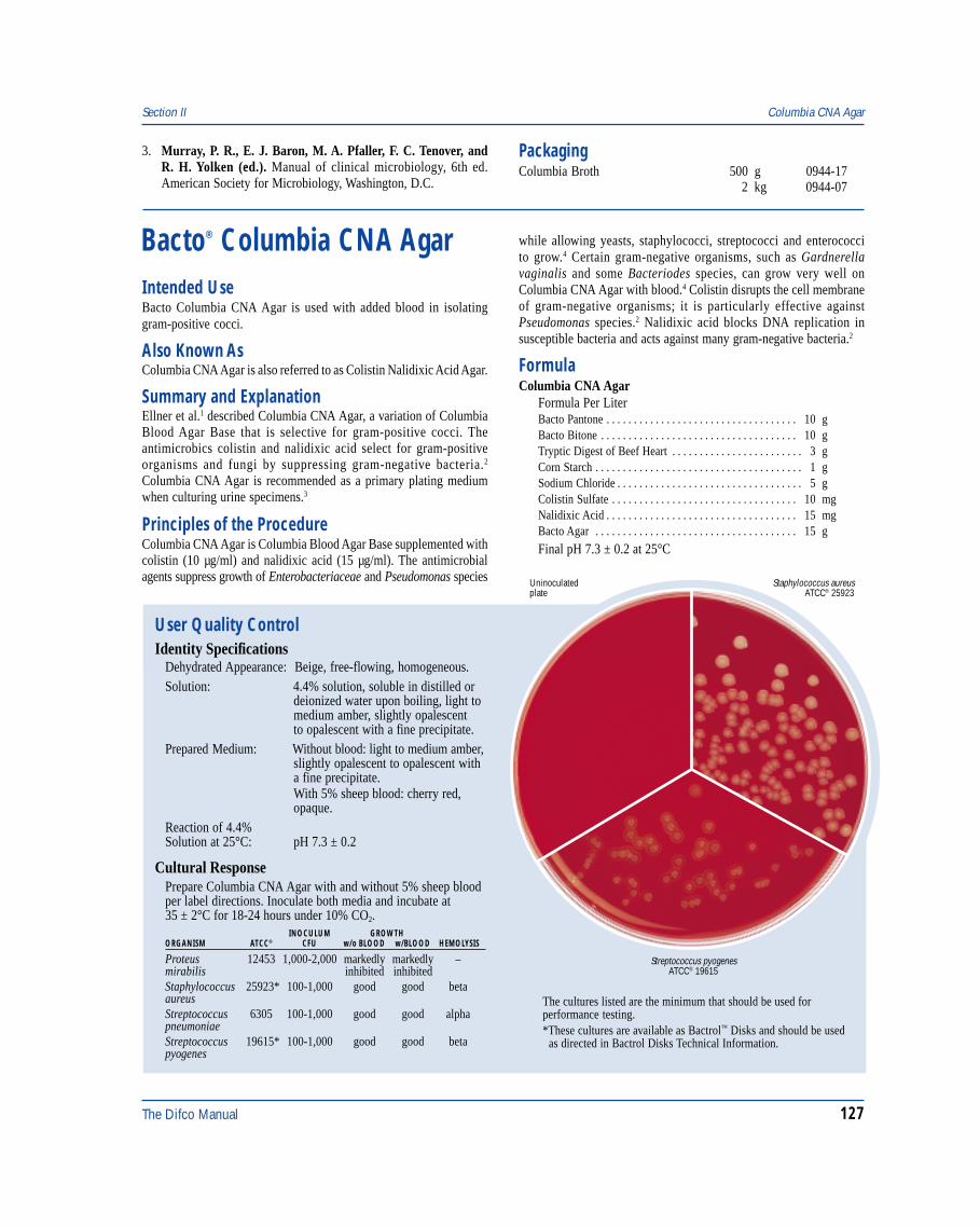
The Difco Manual 127
Section II Columbia CNA Agar
3. Murray, P. R., E. J. Baron, M. A. Pfaller, F. C. Tenover, andR. H. Yolken (ed.). Manual of clinical microbiology, 6th ed.American Society for Microbiology, Washington, D.C.
Bacto® Columbia CNA AgarIntended UseBacto Columbia CNA Agar is used with added blood in isolatinggram-positive cocci.
Also Known AsColumbia CNA Agar is also referred to as Colistin Nalidixic Acid Agar.
Summary and ExplanationEllner et al.1 described Columbia CNA Agar, a variation of ColumbiaBlood Agar Base that is selective for gram-positive cocci. Theantimicrobics colistin and nalidixic acid select for gram-positiveorganisms and fungi by suppressing gram-negative bacteria.2
Columbia CNA Agar is recommended as a primary plating mediumwhen culturing urine specimens.3
Principles of the ProcedureColumbia CNA Agar is Columbia Blood Agar Base supplemented withcolistin (10 µg/ml) and nalidixic acid (15 µg/ml). The antimicrobialagents suppress growth of Enterobacteriaceae and Pseudomonas species
while allowing yeasts, staphylococci, streptococci and enterococcito grow.4 Certain gram-negative organisms, such as Gardnerellavaginalis and some Bacteriodes species, can grow very well onColumbia CNA Agar with blood.4 Colistin disrupts the cell membraneof gram-negative organisms; it is particularly effective againstPseudomonas species.2 Nalidixic acid blocks DNA replication insusceptible bacteria and acts against many gram-negative bacteria.2
FormulaColumbia CNA Agar
Formula Per LiterBacto Pantone . . . . . . . . . . . . . . . . . . . . . . . . . . . . . . . . . . . 10 gBacto Bitone . . . . . . . . . . . . . . . . . . . . . . . . . . . . . . . . . . . . 10 gTryptic Digest of Beef Heart . . . . . . . . . . . . . . . . . . . . . . . . 3 gCorn Starch . . . . . . . . . . . . . . . . . . . . . . . . . . . . . . . . . . . . . . 1 gSodium Chloride . . . . . . . . . . . . . . . . . . . . . . . . . . . . . . . . . . 5 gColistin Sulfate . . . . . . . . . . . . . . . . . . . . . . . . . . . . . . . . . . 10 mgNalidixic Acid . . . . . . . . . . . . . . . . . . . . . . . . . . . . . . . . . . . 15 mgBacto Agar . . . . . . . . . . . . . . . . . . . . . . . . . . . . . . . . . . . . . 15 g
Final pH 7.3 ± 0.2 at 25°C
Staphylococcus aureusATCC® 25923
Uninoculatedplate
Streptococcus pyogenesATCC® 19615
User Quality ControlIdentity Specifications
Dehydrated Appearance: Beige, free-flowing, homogeneous.Solution: 4.4% solution, soluble in distilled or
deionized water upon boiling, light tomedium amber, slightly opalescentto opalescent with a fine precipitate.
Prepared Medium: Without blood: light to medium amber,slightly opalescent to opalescent witha fine precipitate.With 5% sheep blood: cherry red,opaque.
Reaction of 4.4%Solution at 25°C: pH 7.3 ± 0.2
Cultural ResponsePrepare Columbia CNA Agar with and without 5% sheep bloodper label directions. Inoculate both media and incubate at35 ± 2°C for 18-24 hours under 10% CO2.
INOCULUM GROWTHORGANISM ATCC® CFU w/o BLOOD w/BLOOD HEMOLYSIS
Proteus 12453 1,000-2,000 markedly markedly –mirabilis inhibited inhibitedStaphylococcus 25923* 100-1,000 good good betaaureusStreptococcus 6305 100-1,000 good good alphapneumoniaeStreptococcus 19615* 100-1,000 good good betapyogenes
The cultures listed are the minimum that should be used forperformance testing.*These cultures are available as Bactrol™ Disks and should be used as directed in Bactrol Disks Technical Information.
PackagingColumbia Broth 500 g 0944-17
2 kg 0944-07
128 The Difco Manual
Precautions1. For Laboratory Use.2. Follow proper established laboratory procedures in handling and
disposing of infectious materials.
StorageStore the dehydrated medium below 30°C. The dehydrated medium isvery hygroscopic. Keep container tightly closed.
Expiration DateThe expiration date applies to the product in its intact containerwhenstored as directed. Do not use a product if it fails to meetspecificationfor identity and performance.
ProcedureMaterials ProvidedColumbia CNA Agar
Materials Required But Not ProvidedGlasswareAutoclaveIncubator (35°C)WaterbathSterile Petri dishes
Method of Preparation1. Suspend 44 grams in 1 liter distilled or deionized water.2. Heat to boiling to dissolve completely.3. Autoclave at 121° C for 15 minutes. Avoid overheating.4. Cool to 45-50°C.5. Aseptically add 5% sterile defibrinated blood to the medium at
45-50°C. Mix well.6. Dispense into sterile Petri dishes or tubes as desired.
Specimen Collection and PreparationObtain and process specimens according to the techniques andprocedures established by laboratory policy.
Test Procedure1. Inoculate specimens directly onto the surface of the medium. Streak
for isolation with an inoculating loop, then stab the agar severaltimes to deposit beta-hemolytic streptococci beneath the agarsurface. Subsurface growth will display the most reliable hemolyticreactions due to the activity of both oxygen-stable andoxygen-labile streptolysins.5
2. Incubate plates aerobically, anaerobically or under conditionsof increased CO2 (5-10%) in accordance with establishedlaboratory procedures.
ResultsExamine plates for growth and hemolytic reactions after 18-24 and48 hours incubation. Four different types of hemolysis on blood agarmedia can be described:3
a. Alpha (α)-hemolysis is the reduction of hemoglobin tomethemoglobin in the medium surrounding the colony. Thiscauses a greenish discolorization of the medium.
b. Beta (β)-hemolysis is the lysis of red blood cells, resulting in aclear zone surrounding the colony.
c. Gamma (γ)-hemolysis indicates no hemolysis. No destruction ofred blood cells occurs and there is no change in the medium.
d. Alpha-prime (α)-hemolysis is a small zone of complete hemolysisthat is surrounded by an area of partial lysis.
Limitations of the Procedure1. Because the nutritional requirements of organisms vary, some
strains may be encountered that fail to grow or grow poorly onthis medium.
2. Hemolytic reactions of some strains of group D streptococci havebeen shown to be affected by differences in animal blood. Suchstrains are beta-hemolytic on horse, human and rabbit blood agarand alpha-hemolytic on sheep blood agar.5
3. Colonies of Haemophilus haemolyticus are beta-hemolytic on horseand rabbit blood agar and must be distinguished from colonieson beta-hemolytic streptococci using other criteria. The use ofsheep blood has been suggested to obviate this problem since sheepblood is deficient in pyridine nucleotides and does not supportgrowth of H. haemolyticus.4
4. Atmosphere of incubation has been shown to influencehemolytic reactions of beta-hemolytic streptococci.5 For optimalperformance, incubate blood agar base media under increased CO2
or anaerobic conditions.5. Proteus species occasionally grow on CNA Agar and may initially
be confused with streptococci because of the small size of thecolonies.2
References1. Ellner, P. D., C. J. Stoessel, E. Drakeford, and F. Vasi. 1966.
A new culture medium for medical bacteriology. Am. J. Clin.Pathol. 45:502-504.
2. Estevez, E. G. 1984. Bacteriologic plate media: review ofmechanisms of action. Lab Med. 15:258-262.
3. Isenberg, H. D. (ed.). 1992. Clinical microbiology procedures hand-book, vol.1. American Society for Microbiology, Washington, D.C.
4. Baron, E. J., L. R. Peterson, and S.M. Finegold. 1994. Bailey &Scott’s diagnostic microbiology, 9th ed. Mosby-Year Book, Inc.St. Louis, MO.
5. Ruoff, K. L. 1995. Streptococcus, p. 299-305. In P. R. Murray,E. J. Baron, M. A. Pfaller, F. C. Tenover, and R. H. Yolken (ed.).Manual of clinical microbiology, 6th ed. American Society forMicrobiology, Washington, D.C.
PackagingColumbia CNA Agar 500 g 0867-17
2 kg 0867-07
Columbia CNA Agar Section II
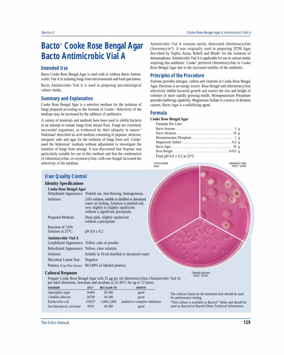
The Difco Manual 129
Section II Cooke Rose Bengal Agar & Antimicrobic Vial A
Bacto® Cooke Rose Bengal AgarBacto Antimicrobic Vial AIntended UseBacto Cooke Rose Bengal Agar is used with or without Bacto Antimi-crobic Vial A in isolating fungi from environmental and food specimens.
Bacto Antimicrobic Vial A is used in preparing microbiologicalculture media.
Summary and ExplanationCooke Rose Bengal Agar is a selective medium for the isolation offungi prepared according to the formula of Cooke.1 Selectivity of themedium may be increased by the addition of antibiotics.
A variety of materials and methods have been used to inhibit bacteriain an attempt to isolate fungi from mixed flora. Fungi are extremelysuccessful organisms, as evidenced by their ubiquity in nature.2
Waksman3 described an acid medium consisting of peptone, dextrose,inorganic salts and agar for the isolation of fungi from soil. Cooke1
used the Waksman3 medium without adjustment to investigate theisolation of fungi from sewage. It was discovered that Soytone wasparticularly suitable for use in this medium and that the combinationof chlortetracycline, or oxytetracycline, with rose bengal increased theselectivity of the medium.
Antimicrobic Vial A contains sterile, desiccated chlortetracycline(Aureomycin®). It was originally used in preparing DTM Agardescribed by Taplin, Azias, Rebell and Blank4 for the isolation ofdermatophytes. Antimicrobic Vial A is applicable for use in various mediarequiring this antibiotic. Cooke1 preferred chlortetracycline in CookeRose Bengal Agar due to the increased stability of the antibiotic.
Principles of the ProcedureSoytone provides nitrogen, carbon and vitamins in Cooke Rose BengalAgar. Dextrose is an energy source. Rose Bengal and chlortetracyclineselectively inhibit bacterial growth and restrict the size and height ofcolonies of more rapidly growing molds. Monopotassium Phosphateprovides buffering capability. Magnesium Sulfate is a source of divalentcations. Bacto Agar is a solidifying agent.
FormulaCooke Rose Bengal Agar
Formula Per LiterBacto Soytone . . . . . . . . . . . . . . . . . . . . . . . . . . . . . . . . . . . . 5 gBacto Dextrose . . . . . . . . . . . . . . . . . . . . . . . . . . . . . . . . . . 10 gMonopotassium Phosphate . . . . . . . . . . . . . . . . . . . . . . . . . . 1 gMagnesium Sulfate . . . . . . . . . . . . . . . . . . . . . . . . . . . . . . . 0.5 gBacto Agar . . . . . . . . . . . . . . . . . . . . . . . . . . . . . . . . . . . . . 20 gRose Bengal . . . . . . . . . . . . . . . . . . . . . . . . . . . . . . . . . . 0.035 gFinal pH 6.0 ± 0.2 at 25°C
User Quality ControlIdentity Specifications
Cooke Rose Bengal AgarDehydrated Appearance: Pinkish tan, free-flowing, homogeneous.Solution: 3.6% solution, soluble in distilled or deionized
water on boiling. Solution is pinkish red,very slightly to slightly opalescentwithout a significant precipitate.
Prepared Medium: Deep pink, slightly opalescentwithout a precipitate.
Reaction of 3.6%Solution at 25°C: pH 6.0 ± 0.2
Antimicrobic Vial ALyophilized Appearance: Yellow cake or powder.Rehydrated Appearance: Yellow, clear solution.Solution: Soluble in 10 ml distilled or deionized water.Microbial Limits Test: Negative.Potency (Cup-Plate Assay): 90-140% of labeled potency.
Cultural ResponsePrepare Cooke Rose Bengal Agar with 35 µg per ml chlortetracycline (Antimicrobic Vial A)per label directions. Inoculate and incubate at 25-30°C for up to 72 hours.ORGANISM ATCC® INOCULUM CFU GROWTH
Aspergillus niger 16404 30-300 goodCandida albicans 26790 30-300 goodEscherichia coli 25922* 1,000-2,000 marked to complete inhibitionSaccharomyces cerevisae 9763 30-300 good
Aspergillus nigerATCC® 16404
Uninoculatedplate
Candida albicansATCC® 26790
The cultures listed are the minimum that should be usedfor performance testing.*This culture is available as Bactrol™ Disks and should beused as directed in Bactrol Disks Technical Information.
130 The Difco Manual
Antimicrobic Vial AAntimicrobic Vial A contains 25 mg sterile desiccated chlortetracycline(Aureomycin®) per 10 ml vial.
Precautions1. For Laboratory Use.2. Antimicrobic Vial A
HARMFUL. MAY CAUSE ALLERGIC EYE, RESPIRATORYSYSTEM AND SKIN REACTION. (US) POSSIBLE RISK OFHARM TO THE UNBORN CHILD. Avoid contact with skin andeyes. Do not breathe dust. Wear suitable protective clothing. Keepcontainer tightly closed. Target Organs: Teeth, Bones.FIRST AID: In case of contact with eyes, rinse immediately withplenty of water and seek medical advice. After contact with skin,wash immediately with plenty of water. If inhaled, remove to freshair. If not breathing, give artificial respiration. If breathing is diffi-cult, give oxygen. Seek medical advice. If swallowed seek medicaladvice immediately and show this container or label.
3. Follow proper established laboratory procedure in handling anddisposing of infectious materials.
StorageStore dehydrated medium below 30°C. The dehydrated medium is veryhygroscopic. Keep container tightly closed.
Store Antimicrobic Vial A at 2-8°C.
Expiration DateThe expiration date applies to the product in its intact container whenstored as directed. Do not use a product if it fails to meet specificationsfor identity and performance.
ProcedureMaterials ProvidedCooke Rose Bengal AgarAntimicrobic Vial A
Materials Required but not ProvidedGlasswareAutoclaveSterile Petri dishesIncubatorWaterbath (optional)
Method of PreparationAntimicrobic Vial A
Cooked Meat Medium Section II
1. Aseptically add 10 ml sterile distilled or deionized water toAntimicrobic Vial A.
2. Agitate gently to dissolve completely.3. The resulting concentration of the rehydrated solution is 2.5 mg
chlortetrycycline per ml.
Cooke Rose Bengal Agar1. Suspend 36 grams in 1 liter distilled or deionized water.2. Heat to boiling to dissolve completely.3. Autoclave at 121°C for 15 minutes.4. Cool to 45°C.5. OPTIONAL: To increase selectivity, aseptically add 14 ml of
rehydrated Antimicrobic Vial A to achieve a final concentration of35 µg of chlortetracycline per ml of medium or an appropriateamount of another antibiotic.
Specimen Collection and PreparationObtain and process specimens according to the techniques andprocedures established by laboratory policy.
Test ProcedureRefer to appropriate references for specific procedures on the isolationand cultivation of fungi.
ResultsRefer to appropriate references and procedures for results.
Limitations of the Procedure1. Since the nutritional requirements of organisms vary, some strains
may be encountered that fail to grow or grow poorly on this medium.
References1. Cooke. 1922. J. Bact. 7:339.2. Dixon, D. M., and R. A. Fromtling. 1995. Morphology, taxonomy,
and classification of the fungi, p. 699-708. In P. R. Murray, E. J.Baron, M. A. Pfaller, F. C. Tenover, and R. H. Yolken (ed.). Manualof clinical microbiology, 6th ed. American Society for Microbiology,Washington, D.C.
3. Waksman. 1954. Antibiotics and Chemotherapy 4:657.4. Taplin, Azias, Rebell, and Blank. 1969. Arch. Dermatol. 99:203.
PackagingCooke Rose Bengal Agar 500 g 0703-17
Antimicrobic Vial A 6 x 10 ml 3333-60
Bacto® Cooked Meat MediumIntended UseBacto Cooked Meat Medium is used for cultivating anaerobicmicroorganisms and for maintaining stock cultures.
Also Known AsCooked Meat Medium (CMM) is also called Chopped MeatMedium.
Summary and ExplanationIn 1890, Theobald Smith1 made use of fresh unheated animal tissue forcultivating anaerobic organisms. Tarozzi2 confirmed Smith’s1
findings and discovered the meat-broth could be heated to104-105°C for 15 minutes without destroying medium nutrients.A steam sterilized emulsion of brain tissue in water was employedby von Hibler3.4 for cultivating anaerobic microorganisms.Von Hibler3,4 found organisms in cooked brain broth were lesssusceptible to harmful effects of toxic metabolic products than in
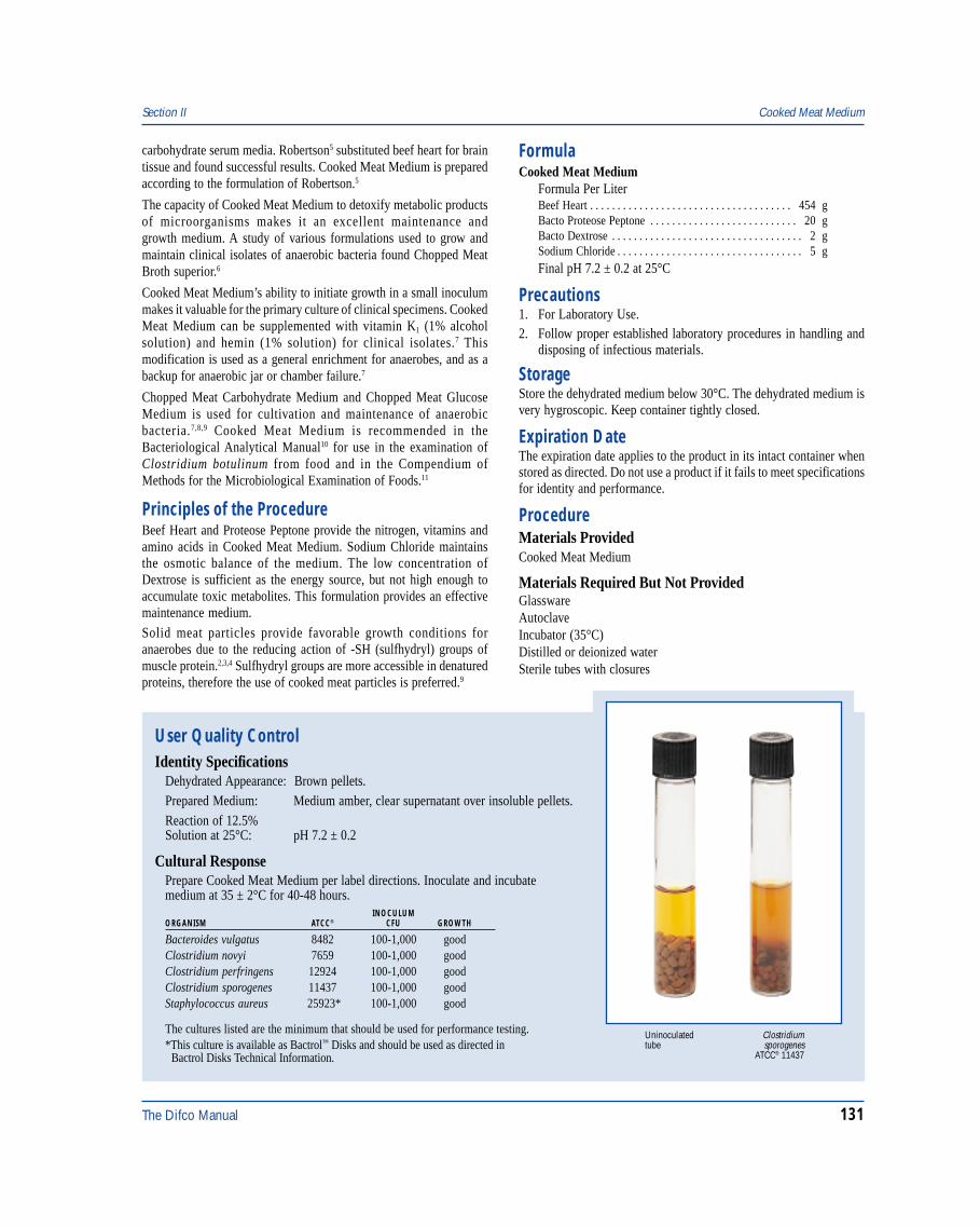
The Difco Manual 131
Section II Cooked Meat Medium
carbohydrate serum media. Robertson5 substituted beef heart for braintissue and found successful results. Cooked Meat Medium is preparedaccording to the formulation of Robertson.5
The capacity of Cooked Meat Medium to detoxify metabolic productsof microorganisms makes it an excellent maintenance andgrowth medium. A study of various formulations used to grow andmaintain clinical isolates of anaerobic bacteria found Chopped MeatBroth superior.6
Cooked Meat Medium’s ability to initiate growth in a small inoculummakes it valuable for the primary culture of clinical specimens. CookedMeat Medium can be supplemented with vitamin K1 (1% alcoholsolution) and hemin (1% solution) for clinical isolates.7 Thismodification is used as a general enrichment for anaerobes, and as abackup for anaerobic jar or chamber failure.7
Chopped Meat Carbohydrate Medium and Chopped Meat GlucoseMedium is used for cultivation and maintenance of anaerobicbacteria.7,8,9 Cooked Meat Medium is recommended in theBacteriological Analytical Manual10 for use in the examination ofClostridium botulinum from food and in the Compendium ofMethods for the Microbiological Examination of Foods.11
Principles of the ProcedureBeef Heart and Proteose Peptone provide the nitrogen, vitamins andamino acids in Cooked Meat Medium. Sodium Chloride maintainsthe osmotic balance of the medium. The low concentration ofDextrose is sufficient as the energy source, but not high enough toaccumulate toxic metabolites. This formulation provides an effectivemaintenance medium.
Solid meat particles provide favorable growth conditions foranaerobes due to the reducing action of -SH (sulfhydryl) groups ofmuscle protein.2,3,4 Sulfhydryl groups are more accessible in denaturedproteins, therefore the use of cooked meat particles is preferred.9
FormulaCooked Meat Medium
Formula Per LiterBeef Heart . . . . . . . . . . . . . . . . . . . . . . . . . . . . . . . . . . . . . 454 gBacto Proteose Peptone . . . . . . . . . . . . . . . . . . . . . . . . . . . 20 gBacto Dextrose . . . . . . . . . . . . . . . . . . . . . . . . . . . . . . . . . . . 2 gSodium Chloride . . . . . . . . . . . . . . . . . . . . . . . . . . . . . . . . . . 5 gFinal pH 7.2 ± 0.2 at 25°C
Precautions1. For Laboratory Use.2. Follow proper established laboratory procedures in handling and
disposing of infectious materials.
StorageStore the dehydrated medium below 30°C. The dehydrated medium isvery hygroscopic. Keep container tightly closed.
Expiration DateThe expiration date applies to the product in its intact container whenstored as directed. Do not use a product if it fails to meet specificationsfor identity and performance.
ProcedureMaterials ProvidedCooked Meat Medium
Materials Required But Not ProvidedGlasswareAutoclaveIncubator (35°C)Distilled or deionized waterSterile tubes with closures
Clostridiumsporogenes
ATCC® 11437
Uninoculatedtube
User Quality ControlIdentity Specifications
Dehydrated Appearance: Brown pellets.Prepared Medium: Medium amber, clear supernatant over insoluble pellets.Reaction of 12.5%Solution at 25°C: pH 7.2 ± 0.2
Cultural ResponsePrepare Cooked Meat Medium per label directions. Inoculate and incubatemedium at 35 ± 2°C for 40-48 hours.
INOCULUMORGANISM ATCC® CFU GROWTH
Bacteroides vulgatus 8482 100-1,000 goodClostridium novyi 7659 100-1,000 goodClostridium perfringens 12924 100-1,000 goodClostridium sporogenes 11437 100-1,000 goodStaphylococcus aureus 25923* 100-1,000 good
The cultures listed are the minimum that should be used for performance testing.*This culture is available as Bactrol™ Disks and should be used as directed in Bactrol Disks Technical Information.
132 The Difco Manual
Method of Preparation1. Suspend 12.5 grams in 100 ml distilled or deionized water (1.25 g
per 10 ml).2. Let stand until all particles are thoroughly wetted and form an
even suspension.3. Autoclave at 121°C for 15 minutes. Reduce pressure slowly.4. Cool without agitation.5. If not used within 24 hours, reheat (100°C) prior to use to drive off
dissolved oxygen.
Specimen Collection and PreparationObtain and process specimens according to the techniques andprocedures established by institutional policy.
Test Procedure1. Inoculate specimen well into the meat particles (bottom of the
tube). Tissue specimens should be ground prior to inoculation.2. Growth is indicated by turbidity and/or the presence of gas bubbles.3. For a complete discussion on the isolation and identification
of aerobic and anaerobic bacteria, refer to appropriate proceduresoutlined in the references.
ResultsRefer to appropriate references and procedures for results.
Limitations1. Since the nutritional requirements of organisms vary, some
strains may be encountered that fail to grow or grow poorly onthis medium.
References1. Smith, T. 1890. Centr. Bakteriol. 7:509.
2. Tarozzi, G. 1905. Uber ein leicht in aerober Weise ausfuhrbaresKulturmittel von einigen bis jetzt fuu strenge Anaerobengehlatenen Keimen. Zentralb. Bakteriol. 38:619.
3. von Hibler, E. 1899. Beitrage zur Kenntnis der durch anaerobeSpaltpilze erzeugen Infektions-krankheiten der Tiere und desMenschen etc. Centr. Bakteriol. 25: 513,594,631.
4. von Hibler, E. 1908. Untersuchungen uber die pathogenenAnaerobier, Jena: Verlag Fischer.
5. Robertson, M. 1916. Notes upon certain anaerobes isolated fromwounds. J. Pathol. Bacteriol. 20:327.
6. Claros, M. C., D. M. Citron, and E. J. C. Goldstein. 1995.Survival of anaerobic bacteria in various thioglycollate and choppedmeat broth formulations. J. Clin. Microbiol. 33:2505-2507.
7. Isenberg, H. E. (ed.). 1992. Clinical microbiology procedureshandbook. American Society for Microbiology, Washington, D.C.
8. Atlas, R. M. 1993. Handbook of microbiological media,p. 224-226. CRC Press, Boca Raton, FL.
9. MacFaddin, J. D. 1985. Media for isolation-cultivation-identification- maintenance of medical bacteria, vol. 1, p. 240-246.Williams & Wilkins, Baltimore, MD.
10. Association of Official Analytical Chemists. 1995.Bacteriological analytical manual, 8th ed. AOAC International,Gaithersburg, MD.
11. Vanderzant, C., and D. F. Splittstoesser (ed.). 1992.Compendium of methods for the microbiological examination of food,3rd ed. American Public Health Association, Washington, D.C.
PackagingCooked Meat Medium 100 g 0267-15
500 g 0267-1710 kg 0267-15r
Corn Meal Agar Section II
Bacto® Corn Meal AgarIntended UseBacto Corn Meal Agar is used for stimulating the production ofchlamydospores by most strains of Candida albicans and forcultivating phytopathological fungi.
Summary and ExplanationNumerous culture media formulations have been described for thedetection, isolation, and identification of Candida albicans, theetiological agent in candidiasis. The various media were designedto bring out morphological or physiological characteristics in thisorganism which would differentiate it from other members of thegenus as well as from other genera.
One of the most important differential characteristics of C. albicansin its ability to form chlamydospores on certain media. This propertyis perhaps the best criterion for identification. Corn Meal Agar isvaluable for morphologic differentiation of many yeast-like organisms.It suppresses vegetative growth of many fungi while stimulatingsporulation.1
Corn Meal Agar has been used with varying degrees of success forshowing chlamydospore formation in C. albicans. Chlamydosporeproduction is the best diagnostic criterion for identification of thepathogenic yeast C. albicans.2 Kelly and Funigeillo3 reported that theaddition of 1% Tween 80 enhanced chlamydospore formation byC. albicans. With this improvement, Corn Meal Agar may be the mostaccurate routine tool available for identification of C. albicans.4
Principles of the ProcedureInfusion from corn meal is a source of carbon, protein and nutrients.Bacto Agar is a solidifying agent.
FormulaCorn Meal Agar
Formula Per LiterCorn Meal, Infusion from . . . . . . . . . . . . . . . . . . . . . . . . . . 50 gBacto Agar . . . . . . . . . . . . . . . . . . . . . . . . . . . . . . . . . . . . . 15 g
Final pH 6.0 ± 0.2 at 25°C
Precautions1. For Laboratory Use.
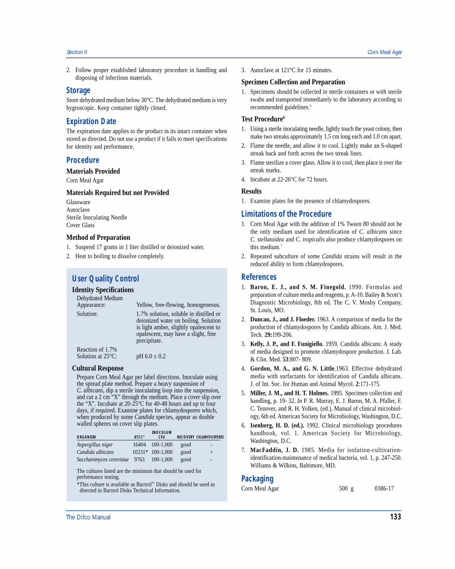
The Difco Manual 133
Section II Corn Meal Agar
2. Follow proper established laboratory procedure in handling anddisposing of infectious materials.
StorageStore dehydrated medium below 30°C. The dehydrated medium is veryhygroscopic. Keep container tightly closed.
Expiration DateThe expiration date applies to the product in its intact container whenstored as directed. Do not use a product if it fails to meet specificationsfor identity and performance.
ProcedureMaterials ProvidedCorn Meal Agar
Materials Required but not ProvidedGlasswareAutoclaveSterile Inoculating NeedleCover Glass
Method of Preparation1. Suspend 17 grams in 1 liter distilled or deionized water.2. Heat to boiling to dissolve completely.
3. Autoclave at 121°C for 15 minutes.
Specimen Collection and Preparation1. Specimens should be collected in sterile containers or with sterile
swabs and transported immediately to the laboratory according torecommended guidelines.5
Test Procedure6
1. Using a sterile inoculating needle, lightly touch the yeast colony, thenmake two streaks approximately 1.5 cm long each and 1.0 cm apart.
2. Flame the needle, and allow it to cool. Lightly make an S-shapedstreak back and forth across the two streak lines.
3. Flame sterilize a cover glass. Allow it to cool, then place it over thestreak marks.
4. Incubate at 22-26°C for 72 hours.
Results1. Examine plates for the presence of chlamydospores.
Limitations of the Procedure1. Corn Meal Agar with the addition of 1% Tween 80 should not be
the only medium used for identification of C. albicans sinceC. stellatoidea and C. tropicalis also produce chlamydospores onthis medium.7
2. Repeated subculture of some Candida strains will result in thereduced ability to form chlamydospores.
References1. Baron, E. J., and S. M. Finegold. 1990. Formulas and
preparation of culture media and reagents, p. A-10. Bailey & Scott’sDiagnostic Microbiology, 8th ed. The C. V. Mosby Company,St. Louis, MO.
2. Duncan, J., and J. Floeder. 1963. A comparison of media for theproduction of chlamydospores by Candida albicans. Am. J. Med.Tech. 29:199-206.
3. Kelly, J. P., and F. Funigiello. 1959. Candida albicans: A studyof media designed to promote chlamydospore production. J. Lab.& Clin. Med. 53:807- 809.
4. Gordon, M. A., and G. N. Little.1963. Effective dehydratedmedia with surfactants for identification of Candida albicans.J. of Int. Soc. for Human and Animal Mycol. 2:171-175.
5. Miller, J. M., and H. T. Holmes. 1995. Specimen collection andhandling, p. 19- 32. In P. R. Murray, E. J. Baron, M. A. Pfaller, F.C. Tenover, and R. H. Yolken, (ed.), Manual of clinical microbiol-ogy, 6th ed. American Society for Microbiology, Washington, D.C.
6. Isenberg, H. D. (ed.). 1992. Clinical microbiology procedureshandbook, vol. 1. American Society for Microbiology,Washington, D.C.
7. MacFaddin, J. D. 1985. Media for isolation-cultivation-identification-maintenance of medical bacteria, vol. 1, p. 247-250.Williams & Wilkins, Baltimore, MD.
PackagingCorn Meal Agar 500 g 0386-17
User Quality ControlIdentity Specifications
Dehydrated MediumAppearance: Yellow, free-flowing, homogeneous.Solution: 1.7% solution, soluble in distilled or
deionized water on boiling. Solutionis light amber, slightly opalescent toopalescent, may have a slight, fineprecipitate.
Reaction of 1.7%Solution at 25°C: pH 6.0 ± 0.2
Cultural ResponsePrepare Corn Meal Agar per label directions. Inoculate usingthe spread plate method. Prepare a heavy suspension ofC. albicans, dip a sterile inoculating loop into the suspension,and cut a 2 cm “X” through the medium. Place a cover slip overthe “X”. Incubate at 20-25°C for 40-48 hours and up to fourdays, if required. Examine plates for chlamydospores which,when produced by some Candida species, appear as doublewalled spheres on cover slip plates.
INOCULUMORGANISM ATCC® CFU RECOVERY CHLAMYDOSPORES
Aspergillus niger 16404 100-1,000 good –Candida albicans 10231* 100-1,000 good +Saccharomyces cerevisiae 9763 100-1,000 good –
The cultures listed are the minimum that should be used forperformance testing.*This culture is available as Bactrol™ Disks and should be used as directed in Bactrol Disks Technical Information.
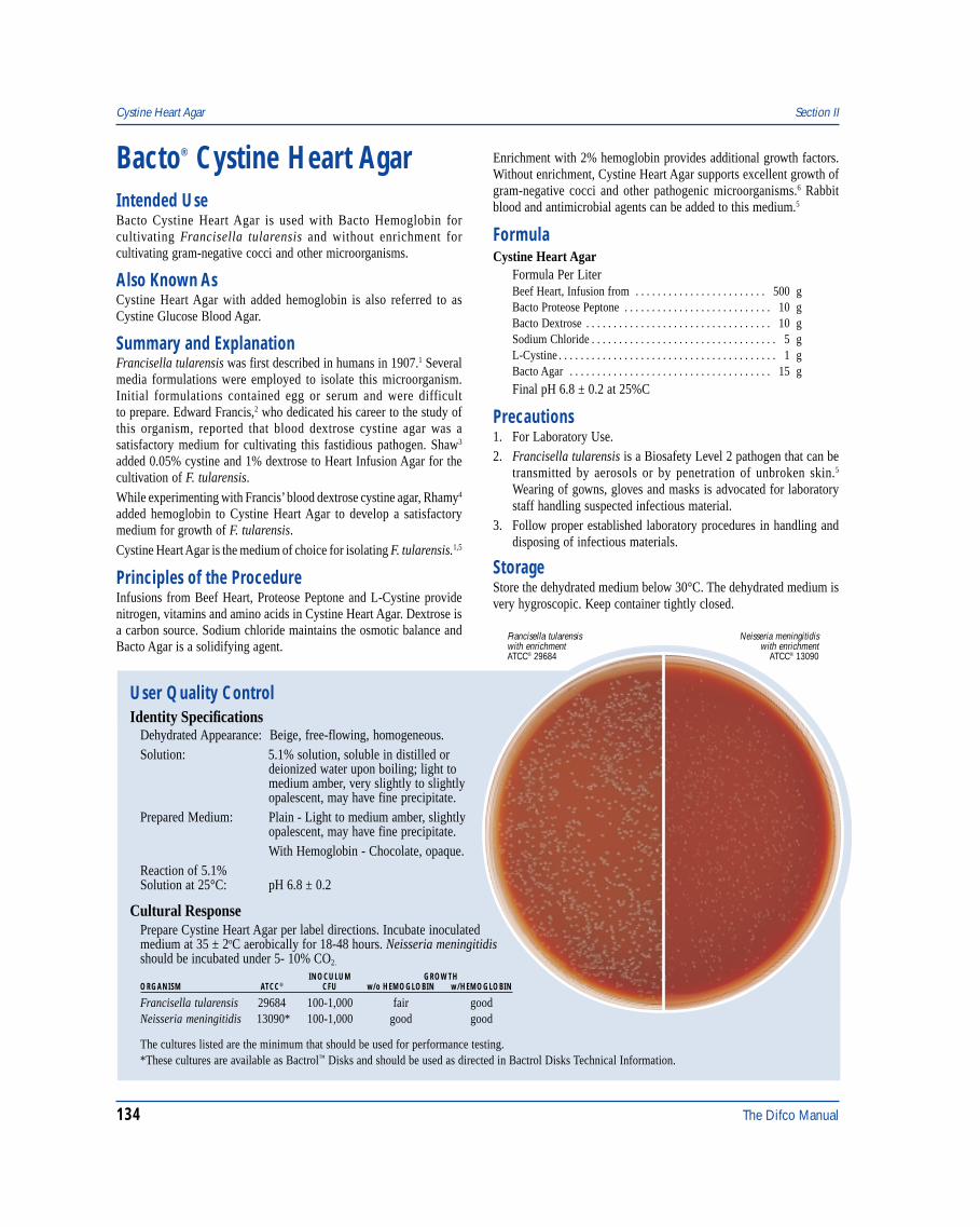
134 The Difco Manual
Bacto® Cystine Heart Agar
User Quality ControlIdentity Specifications
Dehydrated Appearance: Beige, free-flowing, homogeneous.Solution: 5.1% solution, soluble in distilled or
deionized water upon boiling; light tomedium amber, very slightly to slightlyopalescent, may have fine precipitate.
Prepared Medium: Plain - Light to medium amber, slightlyopalescent, may have fine precipitate.With Hemoglobin - Chocolate, opaque.
Reaction of 5.1%Solution at 25°C: pH 6.8 ± 0.2
Cultural ResponsePrepare Cystine Heart Agar per label directions. Incubate inoculatedmedium at 35 ± 2oC aerobically for 18-48 hours. Neisseria meningitidisshould be incubated under 5- 10% CO2.
INOCULUM GROWTHORGANISM ATCC® CFU w/o HEMOGLOBIN w/HEMOGLOBIN
Francisella tularensis 29684 100-1,000 fair goodNeisseria meningitidis 13090* 100-1,000 good good
The cultures listed are the minimum that should be used for performance testing.*These cultures are available as Bactrol™ Disks and should be used as directed in Bactrol Disks Technical Information.
Neisseria meningitidiswith enrichment
ATCC® 13090
Francisella tularensiswith enrichmentATCC® 29684
Intended UseBacto Cystine Heart Agar is used with Bacto Hemoglobin forcultivating Francisella tularensis and without enrichment forcultivating gram-negative cocci and other microorganisms.
Also Known AsCystine Heart Agar with added hemoglobin is also referred to asCystine Glucose Blood Agar.
Summary and ExplanationFrancisella tularensis was first described in humans in 1907.1 Severalmedia formulations were employed to isolate this microorganism.Initial formulations contained egg or serum and were difficultto prepare. Edward Francis,2 who dedicated his career to the study ofthis organism, reported that blood dextrose cystine agar was asatisfactory medium for cultivating this fastidious pathogen. Shaw3
added 0.05% cystine and 1% dextrose to Heart Infusion Agar for thecultivation of F. tularensis.While experimenting with Francis’ blood dextrose cystine agar, Rhamy4
added hemoglobin to Cystine Heart Agar to develop a satisfactorymedium for growth of F. tularensis.Cystine Heart Agar is the medium of choice for isolating F. tularensis.1,5
Principles of the ProcedureInfusions from Beef Heart, Proteose Peptone and L-Cystine providenitrogen, vitamins and amino acids in Cystine Heart Agar. Dextrose isa carbon source. Sodium chloride maintains the osmotic balance andBacto Agar is a solidifying agent.
Enrichment with 2% hemoglobin provides additional growth factors.Without enrichment, Cystine Heart Agar supports excellent growth ofgram-negative cocci and other pathogenic microorganisms.6 Rabbitblood and antimicrobial agents can be added to this medium.5
FormulaCystine Heart Agar
Formula Per LiterBeef Heart, Infusion from . . . . . . . . . . . . . . . . . . . . . . . . 500 gBacto Proteose Peptone . . . . . . . . . . . . . . . . . . . . . . . . . . . 10 gBacto Dextrose . . . . . . . . . . . . . . . . . . . . . . . . . . . . . . . . . . 10 gSodium Chloride . . . . . . . . . . . . . . . . . . . . . . . . . . . . . . . . . . 5 gL-Cystine . . . . . . . . . . . . . . . . . . . . . . . . . . . . . . . . . . . . . . . . 1 gBacto Agar . . . . . . . . . . . . . . . . . . . . . . . . . . . . . . . . . . . . . 15 g
Final pH 6.8 ± 0.2 at 25%C
Precautions1. For Laboratory Use.2. Francisella tularensis is a Biosafety Level 2 pathogen that can be
transmitted by aerosols or by penetration of unbroken skin.5
Wearing of gowns, gloves and masks is advocated for laboratorystaff handling suspected infectious material.
3. Follow proper established laboratory procedures in handling anddisposing of infectious materials.
StorageStore the dehydrated medium below 30°C. The dehydrated medium isvery hygroscopic. Keep container tightly closed.
Cystine Heart Agar Section II
The Difco Manual 135
Expiration DateThe expiration date applies to the product in its intact container whenstored as directed. Do not use a product if it fails to meet specificationsfor identity and performance.
ProcedureMaterials ProvidedCystine Heart Agar
Materials Required But Not ProvidedGlasswareAutoclaveIncubator (35°C)Waterbath (45-50°C) (optional)Hemoglobin Solution 2% or Hemoglobin (optional)Sterile Petri dishes or tubes
Method of PreparationEnriched Medium:1. Suspend 10.2 grams in 100 ml distilled or deionized water.2. Heat to boiling to dissolve completely.3. Autoclave at 121°C for 15 minutes. Cool to 50-60°C.4. Add 100 ml sterile 2% hemoglobin solution and mix well. Use:
• Hemoglobin Solution 2%; or,• Prepare a 2% hemoglobin solution as follows: Place 2 g r a m s
of Hemoglobin in a dry flask. Add 100 ml of colddistilled or deionized water while agitating vigorously.Continue intermittent agitation for 10-15 minutes untilsolution is complete. Autoclave at 121°C for 15 minutes.Cool to 50-60°C
5. Dispense into sterile Petri dishes or tubes.
Unenriched Medium:1. Suspend 51 grams in 1 liter distilled or deionized water.2. Heat to boiling to dissolve completely.3. Autoclave at 121°C for 15 minutes. Cool to 45-50°C.4. Dispense into sterile Petri dishes.
Specimen Collection and PreparationCollect specimens in sterile containers or with sterile swabs. Transportimmediately to the laboratory in accordance with recommendedguidelines outlined in the references.
Test Procedure1. Inoculate and streak specimens as soon as possible. For a complete
discussion on the inoculation and identification of Francisella,consult appropriate references.
2. Overgrowth by contaminating organisms can be reduced byincorporating 100- 500 units penicillin per ml into the medium.6
ResultsRefer to appropriate references and procedures for results.
Limitations of the Procedure1. Since the nutritional requirements of organisms vary, some
strains may be encountered that fail to grow or grow poorlyon this medium.
References1. Stewart, S. J. 1995. In P. R. Murray., E. J. Baron, M. A.
Pfaller, F. C. Tenover, and R. H. Yolken (ed.), Manual of clinicalmicrobiology, 6th ed. American Society for Microbiology,Washington, D.C.
2. Francis, E. 1928. Symptoms, diagnosis and pathology of tularemia.J. Am. Med. Assoc. 91:1155-1160.
3. Shaw, F. W. 1930. Culture medium for Bacterium tularense.Zentr. Bakt. I. Abt. Orig. 118:216-217.
4. Rhamy, B. W. 1933. A new and simplified medium for Pasteurellatularensis and other delicate organisms. Am. J. Clin. Pathol.3:121-124.
5. Isenberg, H. D. (ed.). 1992. Clinical microbiology procedures hand-book, vol.1. American Society for Microbiology, Washington, D.C.
6. Stewart, S. J. 1995. Francisella, p. 545-548. In P. R. Murray,E. J. Baron, M. A. Pfaller, F. C. Tenover and R. H. Yolken (ed.),Manual of clinical microbiology, 6th ed. American Society forMicrobiology, Washington, D.C.
PackagingCystine Heart Agar 500 g 0047-17
Hemoglobin 100 g 0136-15500 g 0136-17
2 kg 0136-0710 kg 0136-08
Hemoglobin Solution 2% 6 x 100 ml 3248-73
Section II Cystine Tryptic Agar
Bacto® Cystine Tryptic AgarIntended UseBacto Cystine Tryptic Agar is used with added carbohydrates in differ-entiating microorganisms based on fermentation reactions and motility.
Also Known AsCystine Tryptic Agar is abbreviated as CTA, and referred to asCT Medium.
Summary and ExplanationCystine Tryptic Agar is a semi-solid basal medium prepared accordingto the formula of Vera.1 Many tests used to differentiate among membersof the Enterobacteriaceae determine the organism’s ability to utilize acarbohydrate with the production of acid metabolic end products.2 CTAis free of fermentable carbohydrates, and the carbohydrate content canbe adjusted for specific reactions. The carbohydrate concentration usedmost frequently in fermentation reactions is 0.5 or 1%.
Some researchers prefer 1% to insure against reversion of the reactiondue to depletion of the carbohydrate by the microorganism.
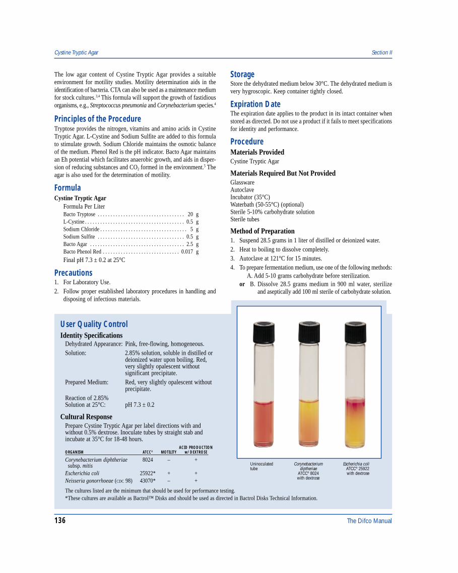
136 The Difco Manual
The low agar content of Cystine Tryptic Agar provides a suitableenvironment for motility studies. Motility determination aids in theidentification of bacteria. CTA can also be used as a maintenance mediumfor stock cultures.3,4 This formula will support the growth of fastidiousorganisms, e.g., Streptococcus pneumonia and Corynebacterium species.4
Principles of the ProcedureTryptose provides the nitrogen, vitamins and amino acids in CystineTryptic Agar. L-Cystine and Sodium Sulfite are added to this formulato stimulate growth. Sodium Chloride maintains the osmotic balanceof the medium. Phenol Red is the pH indicator. Bacto Agar maintainsan Eh potential which facilitates anaerobic growth, and aids in disper-sion of reducing substances and CO2 formed in the environment.5 Theagar is also used for the determination of motility.
FormulaCystine Tryptic Agar
Formula Per LiterBacto Tryptose . . . . . . . . . . . . . . . . . . . . . . . . . . . . . . . . . . 20 gL-Cystine . . . . . . . . . . . . . . . . . . . . . . . . . . . . . . . . . . . . . . . 0.5 gSodium Chloride . . . . . . . . . . . . . . . . . . . . . . . . . . . . . . . . . . 5 gSodium Sulfite . . . . . . . . . . . . . . . . . . . . . . . . . . . . . . . . . . 0.5 gBacto Agar . . . . . . . . . . . . . . . . . . . . . . . . . . . . . . . . . . . . . 2.5 gBacto Phenol Red . . . . . . . . . . . . . . . . . . . . . . . . . . . . . . 0.017 g
Final pH 7.3 ± 0.2 at 25°C
Precautions1. For Laboratory Use.2. Follow proper established laboratory procedures in handling and
disposing of infectious materials.
StorageStore the dehydrated medium below 30°C. The dehydrated medium isvery hygroscopic. Keep container tightly closed.
Expiration DateThe expiration date applies to the product in its intact container whenstored as directed. Do not use a product if it fails to meet specificationsfor identity and performance.
ProcedureMaterials ProvidedCystine Tryptic Agar
Materials Required But Not ProvidedGlasswareAutoclaveIncubator (35°C)Waterbath (50-55°C) (optional)Sterile 5-10% carbohydrate solutionSterile tubes
Method of Preparation1. Suspend 28.5 grams in 1 liter of distilled or deionized water.2. Heat to boiling to dissolve completely.3. Autoclave at 121°C for 15 minutes.4. To prepare fermentation medium, use one of the following methods:
A. Add 5-10 grams carbohydrate before sterilization.or B. Dissolve 28.5 grams medium in 900 ml water, sterilize and aseptically add 100 ml sterile of carbohydrate solution.
User Quality ControlIdentity Specifications
Dehydrated Appearance: Pink, free-flowing, homogeneous.Solution: 2.85% solution, soluble in distilled or
deionized water upon boiling. Red,very slightly opalescent withoutsignificant precipitate.
Prepared Medium: Red, very slightly opalescent withoutprecipitate.
Reaction of 2.85%Solution at 25°C: pH 7.3 ± 0.2
Cultural ResponsePrepare Cystine Tryptic Agar per label directions with andwithout 0.5% dextrose. Inoculate tubes by straight stab andincubate at 35°C for 18-48 hours.
ACID PRODUCTIONORGANISM ATCC® MOTILITY w/ DEXTROSE
Corynebacterium diphtheriae 8024 – + subsp. mitisEscherichia coli 25922* + +Neisseria gonorrhoeae (CDC 98) 43070* – +
The cultures listed are the minimum that should be used for performance testing.*These cultures are available as Bactrol™ Disks and should be used as directed in Bactrol Disks Technical Information.
Cystine Tryptic Agar Section II
Escherichia coliATCC® 25922with dextrose
Uninoculatedtube
Corynebacteriumdiptheriae
ATCC® 8024with dextrose
The Difco Manual 137
Specimen Collection and PreparationObtain and process specimens according to the techniques andprocedures established by laboratory policy.
Test ProcedureFor a complete discussion on motility and carbohydrate fermentationstudies refer to procedures described in appropriate references.2,6,7
Results1. Fermentation of the test carbohydrate is observed when acid is
formed and the medium turns from red to yellow.2. Motility of an organism is evident as a haze of growth extending
into the agar from the stab line.2
Limitations of the Procedure1. Since the nutritional requirements of organisms vary, some strains
may be encountered that fail to grow or grow poorly on this medium.2. CTA requires a heavy inoculum.5
3. Prolonged incubation may lead to changes in pH indicator orabnormal lactose/sucrose reactions with Neisseria pathogens.8,9
4. Neisseria species usually produce acid only in the area of stabs(upper third). If there is a strong acid (yellow color) throughout themedium, a contaminating organism may be present. If in doubtabout a tube containing a Neisseria species, a Gram stain andoxidase test should be performed on the growth.5
References1. Vera, H. D. 1948. A simple medium for identification and mainte-
nance of the gonococcus and other bacteria. J. Bacteriol. 55:531.
2. Baron, E. J., L. R. Peterson, and S. M. Finegold. 1994. Bailey &Scott’s Diagnostic Microbiology, 9th ed. Mosby-Year Book, Inc.,St. Louis, MO.
3. Myers, R. M., and G. Koshy. 1961. Beta-hemolytic streptococciin survey throat cultures in an Indian population. Am. J. PublicHealth 51:1872.
4. Alford, J. A., G. E. Wiese, and J. J. Gunter. 1955. Heatresistance in Corynebacterium and the relationship of the genus toMicrobacterium. J. Bacteriol. 69:516.
5. MacFaddin, J. D. 1985. Media for isolation-cultivation-identification-maintenance of medical bacteria, vol. 1, p. 254-259,802-804. Williams & Wilkins, Baltimore, MD.
6. Isenberg, H. D. (ed.). 1995. Clinical microbiology procedureshandbook, American Society for Microbiology, Washington, D.C.
7. Murray, P. R., E. J. Baron, M. A. Pfaller, F. C. Tenover, andR. H. Yolken (ed.). 1995. Manual of clinical microbiology, 6th ed.American Society for Microbiology, Washington, D.C.
8. Faur, Y. C., M. H. Weisburd, and M. E. Wilson. 1975.Carbohydrate fermentation plate medium for confirmation ofNeisseria species. J. Clin. Microbiol. 1:294.
9. Applebaum, P. C., and R. B. Lawrence. 1979. Comparison ofthree methods for identification of pathogenic Neisseria species.J. Clin. Microbiol. 9:598.
PackagingCystine Tryptic Agar 500 g 0523-17
Section II Czapek-Dox Broth & Czapek Solution Agar
Bacto® Czapek-Dox BrothBacto Czapek Solution AgarIntended UseBacto Czapek-Dox Broth and Czapek Solution Agar are used forcultivating fungi and bacteria capable of using inorganic nitrogen.
Summary and ExplanationCzapek-Dox Broth and Czapek Solution Agar are a modification of theCzapek1 and Dox2 formula prepared according to Thom and Raper.3
The media are prepared with only inorganic sources of nitrogen andchemically defined compounds sources of carbon. Czapek-Dox mediaare useful in a variety of microbiological procedures, including soilmicrobiology and fungi and mildew resistance tests. Thom and Raper3
reported Czapek-Dox Broth and Czapek Solution Agar will producemoderately vigorous growth of most saprophytic aspergilli and yieldcharacteristic mycelia and conidia.
Czapek Solution Agar is recommended in Standard Methods for theExamination of Water and Wastewater5 for the isolation of Aspergillus,Penicillium and related fungi.
Principles of the ProcedureSaccharose is the sole carbon source, and Sodium Nitrate is the solenitrogen source in Czapek-Dox Broth and Czapek Solution Agar.
Dipotassium Phosphate is the buffering agent, and Potassium Chloridecontains essential ions. Magnesium Sulfate and Ferrous Sulfate sourcesof cations. Bacto Agar is the solidifying agent in Czapek Solution Agar.
FormulaCzapek-Dox Broth
Formula Per LiterBacto Saccharose . . . . . . . . . . . . . . . . . . . . . . . . . . . . . . . . 30 gSodium Nitrate . . . . . . . . . . . . . . . . . . . . . . . . . . . . . . . . . . . 3 gDipotassium Phosphate . . . . . . . . . . . . . . . . . . . . . . . . . . . . 1 gMagnesium Sulfate . . . . . . . . . . . . . . . . . . . . . . . . . . . . . . . 0.5 gPotassium Chloride . . . . . . . . . . . . . . . . . . . . . . . . . . . . . . . 0.5 gFerrous Sulfate . . . . . . . . . . . . . . . . . . . . . . . . . . . . . . . . . 0.01 g
Final pH 7.3 ± 0.2 at 25°C
Czapek Solution AgarFormula Per LiterBacto Saccharose . . . . . . . . . . . . . . . . . . . . . . . . . . . . . . . . 30 gSodium Nitrate . . . . . . . . . . . . . . . . . . . . . . . . . . . . . . . . . . . 2 gDipotassium Phosphate . . . . . . . . . . . . . . . . . . . . . . . . . . . . 1 gMagnesium Sulfate . . . . . . . . . . . . . . . . . . . . . . . . . . . . . . . 0.5 gPotassium Chloride . . . . . . . . . . . . . . . . . . . . . . . . . . . . . . . 0.5 gFerrous Sulfate . . . . . . . . . . . . . . . . . . . . . . . . . . . . . . . . . 0.01 gBacto Agar . . . . . . . . . . . . . . . . . . . . . . . . . . . . . . . . . . . . . 15 g
Final pH 7.3 ± 0.2 at 25°C
138 The Difco Manual
PrecautionsCzapek-Dox Broth1. For Laboratory Use.
Czapek Solution Agar1. For Laboratory Use.2. Follow proper established laboratory procedures in handling and
disposing of infectious materials.
StorageStore dehydrated medium below 30°C. The dehydrated medium is veryhygroscopic. Keep container tightly closed.
Expiration DateThe expiration date applies to the product in its intact container when
stored as directed. Do not use a product if it fails to meet specificationsfor identity and performance.
ProcedureMaterials ProvidedCzapek-Dox BrothCzapek Solution Agar
Materials Required but not ProvidedGlasswareAutoclaveIncubatorWaterbath (optional)
Method of PreparationCzapek-Dox Broth1. Dissolve 35 grams in 1 liter distilled or deionized water.2. Autoclave at 121°C for 15 minutes.3. Dispense as desired.
Czapek Solution Agar1. Suspend 49 grams in 1 liter distilled or deionized water.2. Boil to dissolve completely.3. Autoclave at 121°C for 15 minutes.4. Dispense as desired.
Specimen Collection and PreparationObtain and process specimens according to the techniques andprocedures established by laboratory policy.
Test ProcedureRefer to appropriate references for specific procedures for the cultivationof fungi and bacteria capable of utilizing inorganic nitrogen.
ResultsRefer to appropriate references and procedures for results.
References1. Czapek, F. 1902-1903. Untersuchungen uber die
stickstoffgewinnung und EiweiBbildung der Pflanze. Beitr.Chem. Physiol. Pathol. 1:540.
2. Dox, A. W. 1910. The intracellular enzymes of Penicillium andAspergillus with special references to those of P. camenberti. U.S.Dept. Agr. Bur. Anim. Ind. Bull. 120:70.
3. Thom, C., and K. B. Raper. 1945. Manual of the aspergilli, vol. 39.4. Thom, C., and M. B. Church. 1926. The aspergilli. Williams and
Wilkins Co., Baltimore, MD.5. Eaton, A. D., L. S. Cleseri, and A. E. Greenberg (ed.). 1995.
Standard methods for the examination of water and wastewater,19th ed. American Public Health Association, Washington, D.C.
PackagingCzapek-Dox Broth 500 g 0338-17
Czapek Solution Agar 500 g 0339-17
User Quality ControlIdentity Specifications
Czapek-Dox BrothDehydrated Appearance: White, free-flowing, homogeneous.Solution: 3.5% solution, soluble in distilled or
deionized water. Solution is colorless,clear to very slightly opalescent andmay have a slight precipitate.
Prepared Medium: Colorless, clear to very slightlyopalescent, may have slight precipitate.
Reaction of 3.5%Solution at 25°C: pH 7.3 ± 0.2
Czapek Solution AgarDehydrated Appearance: Very light beige, free-flowing,
homogeneous.Solution: 4.9% solution, soluble in distilled or
deionized water on boiling; light amber,opalescent with a uniform flocculentprecipitate.
Prepared Medium: Light amber, slightly opalescent;may have slight precipitate.
Reaction of 4.9%Solution at 25°C: pH 7.3 ± 0.2
Cultural ResponseCzapek-Dox BrothPrepare Czapek-Dox Broth per label directions. Inoculate tubeswith the test organisms. Incubate inoculated medium at 30 ± 2°Cfor 48-72 hours.
Czapek Solution AgarPrepare Czapek Solution Agar per label directions. Inoculateprepared medium with the test organisms. Incubate at 30 ± 2°Cfor 18-48 hours, or up to 72 hours if necessary.
INOCULUMORGANISM ATCC® CFU GROWTH
Aspergillus niger 9642 100-1,000 goodCandida albicans 10231 100-1,000 good
The cultures listed are the minimum that should be used forperformance testing.
Czapek-Dox Broth & Czapek Solution Agar Section II